Embed Size (px)
Citation preview
*For correspondence:U.B.
Present address: †Department
of Molecular and Cellular
Physiology, Stanford University,
Stanford, United states
Competing interests: The
authors declare that no
competing interests exist.
Funding: See page 20
Received: 20 March 2015
Accepted: 09 December 2015
Published: 09 December 2015
Reviewing editor: Richard
Aldrich, The University of Texas
at Austin, United States
Copyright Fechner et al. This
article is distributed under the
terms of the Creative Commons
Attribution License, which
permits unrestricted use and
redistribution provided that the
original author and source are
credited.
A K+-selective CNG channel orchestratesCa2+ signalling in zebrafish spermSylvia Fechner1†, Luis Alvarez1, Wolfgang Bonigk1, Astrid Muller1,Thomas K Berger1, Rene Pascal1, Christian Trotschel2, Ansgar Poetsch2,Gabriel Stolting3, Kellee R Siegfried4, Elisabeth Kremmer5, Reinhard Seifert1,U Benjamin Kaupp1*
1Abteilung Molekulare Neurosensorik, Center of Advanced European Studies andResearch, Bonn, Germany; 2Lehrstuhl Biochemie der Pflanzen, Ruhr-UniversitatBochum, Bochum, Germany; 3Institute of Complex Systems 4, ForschungszentrumJulich, Julich, Germany; 4Biology Department, University of Massachusetts Boston,Boston, United States; 5Institut fur Molekulare Immunologie, Helmholtz-ZentrumMunchen, Munchen, Germany
Abstract Calcium in the flagellum controls sperm navigation. In sperm of marine invertebrates
and mammals, Ca2+ signalling has been intensely studied, whereas for fish little is known. In sea
urchin sperm, a cyclic nucleotide-gated K+ channel (CNGK) mediates a cGMP-induced
hyperpolarization that evokes Ca2+ influx. Here, we identify in sperm of the freshwater fish Danio
rerio a novel CNGK family member featuring non-canonical properties. It is located in the sperm
head rather than the flagellum and is controlled by intracellular pH, but not cyclic nucleotides.
Alkalization hyperpolarizes sperm and produces Ca2+ entry. Ca2+ induces spinning-like swimming,
different from swimming of sperm from other species. The “spinning” mode probably guides
sperm into the micropyle, a narrow entrance on the surface of fish eggs. A picture is emerging of
sperm channel orthologues that employ different activation mechanisms and serve different
functions. The channel inventories probably reflect adaptations to species-specific challenges
during fertilization.
DOI: 10.7554/eLife.07624.001
IntroductionFertilization is a complex task that, for different species, happens in entirely different spatial com-
partments or ionic milieus. In aquatic habitats, gametes are released into the water where sperm
acquire motility and navigate to the egg. By contrast, mammalian fertilization happens in confined
compartments of the female oviduct. From invertebrates to mammals, sperm use various sensing
mechanisms, including chemotaxis, rheotaxis, and thermotaxis, to gather physical or chemical cues
to spot the egg. These sensory cues activate various cellular signalling pathways that ultimately con-
trol the intracellular Ca2+ concentration ([Ca2+]i) and, thereby, the flagellar beat and swimming
behaviours (Alvarez et al., 2012; Darszon et al., 2008; Eisenbach and Giojalas, 2006;
Florman et al., 2008; Guerrero et al., 2010; Ho and Suarez, 2001; Kaupp et al., 2008;
Publicover et al., 2008). In species as phylogenetically distant as sea urchin and mammals, these
pathways target a sperm-specific, voltage-dependent Ca2+ channel, called CatSper. Signalling
events open CatSper by shifting its voltage-dependence to permissive, more negative Vm values.
This shift is achieved by different means. In sea urchin sperm, opening of a K+-selective cyclic nucleo-
tide-gated channel (CNGK) causes a transient hyperpolarization (Bonigk et al., 2009;
Strunker et al., 2006); the hyperpolarization activates a sperm-specific Na+/H+ exchanger (sNHE)
(Lee, 1984, 1985; Lee and Garbers, 1986) resulting in a long-lasting alkalization that shifts the
Fechner et al. eLife 2015;4:e07624. DOI: 10.7554/eLife.07624 1 of 25
RESEARCH ARTICLE
voltage dependence of CatSper and leads to a Ca2+ influx (Kaupp et al., 2003; Seifert et al.,
2015). By contrast, in human sperm, the shift is achieved by direct stimulation of CatSper with pros-
taglandins and progesterone in the seminal fluid or the oviduct (Brenker et al., 2012; Lishko et al.,
2011; Smith et al., 2013; Strunker et al., 2011).
Navigation of fish sperm and the underlying signalling pathways must be arguably different. First,
teleost fish are lacking CatSper channels (Cai and Clapham, 2008), although activation of sperm
motility requires Ca2+ influx (Alavi and Cosson, 2006; Billard, 1986; Cosson et al., 2008; Mori-
sawa, 2008; Takai and Morisawa, 1995) stimulated by hyper- or hypoosmotic shock after spawning
into seawater or freshwater, respectively (Alavi and Cosson, 2006; Cherr et al., 2008;
Krasznai et al., 2000; Morisawa, 2008; Vines et al., 2002). Therefore, Ca2+ signalling in fish sperm
must involve molecules different from those in marine invertebrates and mammals.
Second, the ionic milieu seriously constrains ion channel function. Sperm of freshwater fish,
marine invertebrates, and mammals are facing entirely different ionic milieus. K+ and Na+ concentra-
tions in freshwater are extremely low (70 mM and 200 mM, respectively) compared to the orders-of-
magnitude higher concentrations in seawater or the oviduct (Alavi and Cosson, 2006;
Hugentobler et al., 2007). Furthermore, [Ca2+] in seawater is high (10 mM), whereas in freshwater it
is low (< 1 mM). The low salt concentrations in freshwater probably require distinctively different ion
channels. In fact, none of the ion channels controlling electrical excitation and Ca2+ signalling of fish
sperm are known.
Finally, fish sperm are not actively attracted to the whole egg from afar by chemical or physical
cues, i.e. chemotaxis, thermotaxis, or rheotaxis (Cosson et al., 2008; Morisawa, 2008;
Yanagimachi et al., 2013). Instead, many fishes deposit sperm directly onto the eggs. For fertiliza-
tion, sperm must search for the narrow entrance to a cone-shaped funnel in the egg coat – the
micropyle – that provides access to the egg membrane. Sperm reach the micropyle probably by
haptic interactions with tethered molecules that line the egg surface and the opening or interior of
the micropyle (Iwamatsu et al., 1997; Ohta and Iwamatsu, 1983; Yanagimachi et al., 2013). At
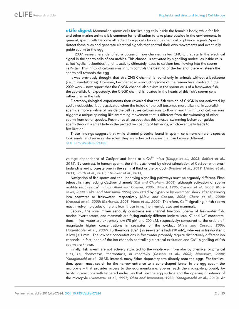
eLife digest Mammalian sperm cells fertilize egg cells inside the female’s body; while for fish
and other marine animals it is common for fertilization to take place outside in the environment. In
general, sperm cells become attracted to egg cells by various chemical or physical signals. Sperm
detect these cues and generate electrical signals that control their own movements and eventually
guide sperm to the egg.
In 2009, researchers identified a potassium ion channel, called CNGK, that starts the electrical
signal in the sperm cells of sea urchins. This channel is activated by signalling molecules inside cells,
called ‘cyclic nucleotides’, and its activity ultimately leads to calcium ions flowing into the sperm
cell’s tail. This influx of calcium ions in turn controls the beating of the tail and, thereby, steers the
sperm cell towards the egg.
It was previously thought that this CNGK channel is found only in animals without a backbone
(i.e. in invertebrates). However, Fechner et al. – including some of the researchers involved in the
2009 work – now report that the CNGK channel also exists in the sperm cells of a freshwater fish,
the zebrafish. Unexpectedly, the CNGK channel is located in the heads of this fish’s sperm cells
rather than in the tails.
Electrophysiological experiments then revealed that the fish version of CNGK is not activated by
cyclic nucleotides, but is activated when the inside of the cell becomes more alkaline. In zebrafish
sperm, a more alkaline pH inside the cell causes calcium ions to flow in and this influx of calcium ions
triggers a unique spinning-like swimming movement that is different from the swimming of other
sperm from other species. Fechner et al. suspect that this unusual swimming behaviour guides
sperm through a small hole in the protective coating of fish eggs, which eventually leads to
fertilization.
These findings suggest that while channel proteins found in sperm cells from different species
look similar and serve similar roles, they are activated in ways that can be very different.
DOI: 10.7554/eLife.07624.002
Fechner et al. eLife 2015;4:e07624. DOI: 10.7554/eLife.07624 2 of 25
Research article Biophysics and structural biology Cell biology
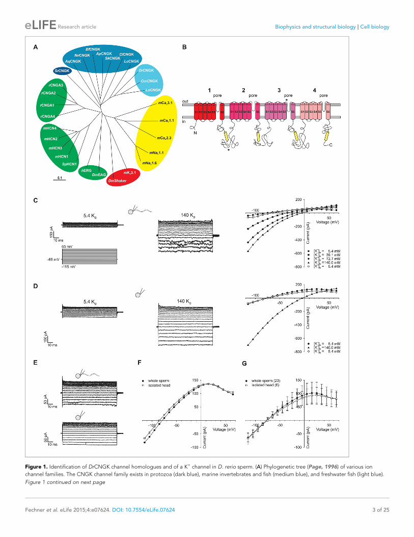
Figure 1. Identification of DrCNGK channel homologues and of a K+ channel in D. rerio sperm. (A) Phylogenetic tree (Page, 1996) of various ion
channel families. The CNGK channel family exists in protozoa (dark blue), marine invertebrates and fish (medium blue), and freshwater fish (light blue).
Figure 1 continued on next page
Fechner et al. eLife 2015;4:e07624. DOI: 10.7554/eLife.07624 3 of 25
Research article Biophysics and structural biology Cell biology

surfaces, sperm swim with their flagellum slightly inclined, which pushes the head against the wall
and stabilizes sperm at the surface (Denissenko et al., 2012; Elgeti et al., 2010). Thus, fish sperm
motility might be governed by specific hydrodynamic and haptic interactions with the egg surface
and the micropyle.
Although the principal targets of a CNGK-mediated hyperpolarization – the Na+/H+ exchanger
and CatSper – are absent in fish, vertebrate orthologues of the sperm CNGK channel are present in
various fish genomes (Figure 1A). Here, we study the function of the CNGK channel in sperm of the
freshwater fish Danio rerio (DrCNGK). The DrCNGK channel constitutes the principal K+ channel in
D. rerio sperm. Unexpectedly, cyclic nucleotides neither regulate CNGK channel activity nor sperm
motility; instead, intracellular alkalization, a key mechanism to control sperm function in many spe-
cies, strongly activates CNGK and, thereby, triggers a Ca2+ signal and a motility response. Although
its mechanism of activation is entirely different compared to sea urchin sperm, the principal CNGK
function, namely to provide a hyperpolarization that triggers a Ca2+ signal, is conserved. Our results
show that sperm signalling among aquatic species shows unique variations that probably represent
adaptations to vastly different ionic milieus and fertilization habits.
ResultsIn several genomes, we identified genes encoding putative cyclic nucleotide-gated K+ channels
(CNGK) (Figure 1A). CNGK channels are primarily present in marine invertebrates, yet absent in the
genomes of vertebrates such as birds, amphibians, and mammals, except freshwater fish and coela-
canths. The CNGKs of freshwater fish appear to form a phylogenetic sub-group on their own
(Figure 1A). Moreover, the CNGK channel exists in the unicellular choanoflagellate (Salpingocea
rosetta), the closest living relative of animals (Levin and King, 2013; Umen and Heitman, 2013).
CNGK channels feature a chimeric structure. Their overall four-repeat pseudotetrameric architec-
ture is reminiscent of voltage-dependent Nav and Cav channels, whereas the pore carries the canoni-
cal GYG or GFG motif of K+-selective channels (Figure 1B and Figure 1—figure supplement 1).
Furthermore, CNGK channels are phylogenetic cousins of cyclic nucleotide-gated (CNG) channels
and of hyperpolarization-activated and cyclic nucleotide-gated (HCN) channels (Figure 1A); each of
the four repeats harbours a cyclic nucleotide-binding domain (CNBD) (Figure 1B). In fact, the CNGK
channel of sea urchin sperm is activated at nanomolar cGMP concentrations (Bonigk et al., 2009).
Figure 1 continued
The HCN, CNG, and KCNH channel families are highlighted in green; voltage-gated Nav and Cav channels are highlighted in yellow; and voltage-gated
Kv channels are highlighted in red. The following ion channel sequences were used: CNGK channels from zebrafish (DrCNGK), rainbow trout
(OmCNGK), spotted gar (LoCNGK), West Indian Ocean coelacanth (LcCNGK), sea urchin (ApCNGK), acorn worm (SkCNGK), amphioxus (BfCNGK),
starlet sea anemone (NvCNGK), vasa tunicate (CiCNGK), sponge (AqCNGK), choanoflagellate (SrCNGK); murine HCN channel subunits 1 (mHCN1), 2
(mHCN2), 3 (mHCN3), 4 (mHCN4), and the HCN channel from sea urchin (SpHCN1); rat CNGA subunits A1 (rCNGA1), A2 (rCNGA2), A3 (rCNGA3), and
A4 (rCNGA4); the KCNH channels from fruit fly (DmEAG) and human (hERG); murine voltage-gated Nav (mNav 1.1 and mNav 1.6) and Cav channels
(mCav1.1, mCav2.3 and mCav3.1) and voltage-gated Kv channels from fruit fly (DmShaker) and mouse (mKv3.1). Full-length Latin names and accession
numbers are given in experimental procedures. Scale bar represents 0.1 substitutions per site. (B) Pseudo-tetrameric structure of CNGK channels.
Numbers 1 to 4, homologous repeats; S1 to S6, transmembrane segments; yellow cylinders, cyclic nucleotide-binding domain CNBD; asterisks,
epitopes recognized by antibodies anti-repeat1 of DrCNGK (polyclonal) and anti-repeat3 of DrCNGK (YENT1E2, monoclonal). (C) Whole-cell recordings
from zebrafish sperm at low (left upper panel) and high (middle panel) extracellular K+ concentrations. Left lower panel: Voltage step protocol. Right
panel: corresponding IV relations. (D) Whole-cell recordings from an isolated sperm head. Description see part C. (E) Whole-cell recording from
zebrafish sperm (upper panel) and an isolated head (lower panel). (F) IV relation of recordings from part E. (G) Pooled IV relations ( ± sd) of currents
from zebrafish sperm (filled circle, n = 23) and sperm heads (open squares, n = 6).
DOI: 10.7554/eLife.07624.003
The following figure supplements are available for figure 1:
Figure supplement 1. Amino-acid sequence of the DrCNGK channel.
DOI: 10.7554/eLife.07624.004
Figure supplement 2. Separation of heads and flagella from whole sperm.
DOI: 10.7554/eLife.07624.005
Figure supplement 3. Electrophysiological characterization of currents recorded from zebrafish sperm.
DOI: 10.7554/eLife.07624.006
Fechner et al. eLife 2015;4:e07624. DOI: 10.7554/eLife.07624 4 of 25
Research article Biophysics and structural biology Cell biology
Identification of a K+ current in sperm of D. rerioWe recorded currents from whole D. rerio sperm (Figure 1—figure supplement 2, left panel) in the
whole-cell patch-clamp configuration. Voltage steps from a holding potential of -65 mV evoked
slightly inwardly rectifying currents (Figure 1C). Two pieces of evidence established that currents are
carried by K+ channels and not by Cl- channels. The reversal potential (Vrev) shifted from -77 ± 3 mV
(n = 18) at 5.4 mM extracellular [K+]o to -7 ± 1 mV (n = 7) at 140 mM [K+]o (4Vrev = 51 ± 3 mV/log
[K+], n = 7) (Figure 1C, Figure 1—figure supplement 3C). Changing the intracellular [Cl-] did not
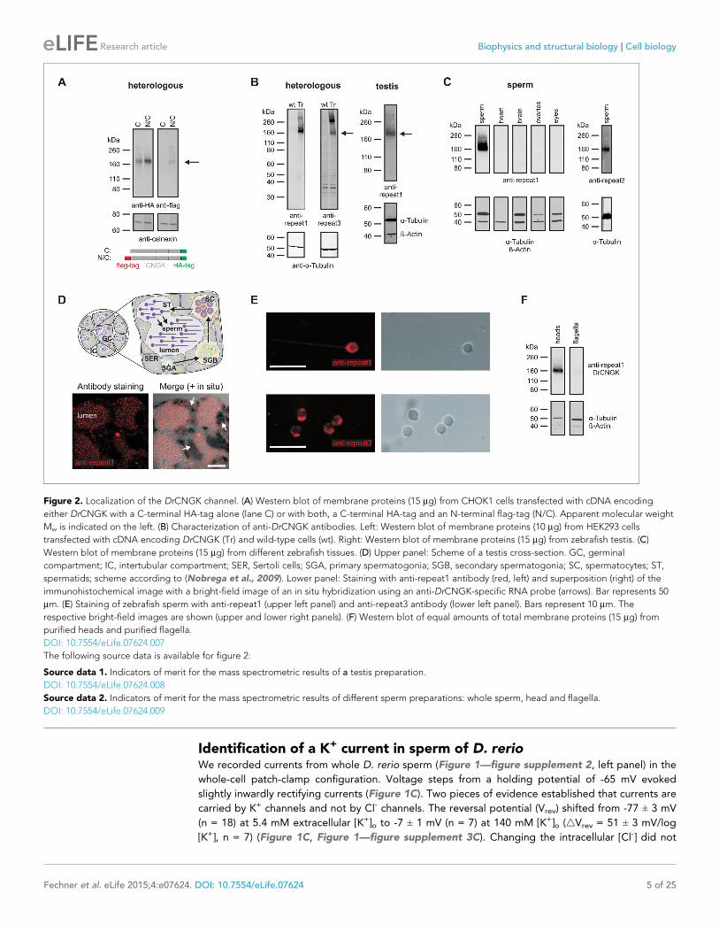
Figure 2. Localization of the DrCNGK channel. (A) Western blot of membrane proteins (15 mg) from CHOK1 cells transfected with cDNA encoding
either DrCNGK with a C-terminal HA-tag alone (lane C) or with both, a C-terminal HA-tag and an N-terminal flag-tag (N/C). Apparent molecular weight
Mw is indicated on the left. (B) Characterization of anti-DrCNGK antibodies. Left: Western blot of membrane proteins (10 mg) from HEK293 cells
transfected with cDNA encoding DrCNGK (Tr) and wild-type cells (wt). Right: Western blot of membrane proteins (15 mg) from zebrafish testis. (C)
Western blot of membrane proteins (15 mg) from different zebrafish tissues. (D) Upper panel: Scheme of a testis cross-section. GC, germinal
compartment; IC, intertubular compartment; SER, Sertoli cells; SGA, primary spermatogonia; SGB, secondary spermatogonia; SC, spermatocytes; ST,
spermatids; scheme according to (Nobrega et al., 2009). Lower panel: Staining with anti-repeat1 antibody (red, left) and superposition (right) of the
immunohistochemical image with a bright-field image of an in situ hybridization using an anti-DrCNGK-specific RNA probe (arrows). Bar represents 50
mm. (E) Staining of zebrafish sperm with anti-repeat1 (upper left panel) and anti-repeat3 antibody (lower left panel). Bars represent 10 mm. The
respective bright-field images are shown (upper and lower right panels). (F) Western blot of equal amounts of total membrane proteins (15 mg) from
purified heads and purified flagella.
DOI: 10.7554/eLife.07624.007
The following source data is available for figure 2:
Source data 1. Indicators of merit for the mass spectrometric results of a testis preparation.
DOI: 10.7554/eLife.07624.008
Source data 2. Indicators of merit for the mass spectrometric results of different sperm preparations: whole sperm, head and flagella.
DOI: 10.7554/eLife.07624.009
Fechner et al. eLife 2015;4:e07624. DOI: 10.7554/eLife.07624 5 of 25
Research article Biophysics and structural biology Cell biology
affect Vrev (Figure 1—figure supplement 3A,B). These results demonstrate that the current is pre-
dominantly carried by K+ ions. To localize the underlying K+ channel, we recorded currents from iso-
lated sperm heads (Figure 1D-G, Figure 1—figure supplement 2, middle panel). Head and whole-
sperm currents displayed a similar K+ dependence (4Vrev = 52 ± 2 mV/log [K+], n = 6) (Figure 1D),
rectification (Figure 1F,G), and amplitude (Figure 1F,G), suggesting that the underlying K+ channel
is primarily located in the head.
This result is unexpected, as ion channels involved in sperm signalling are usually localized to the
flagellum. To test whether the DrCNGK channel is also localized to the head, we used Western blot
analysis and immunocytochemistry. To this end, the DrCNGK protein was first characterized by het-
erologous expression in mammalian cell lines. DrCNGK constructs with a C-terminal HA-tag or with
two tags, a C-terminal HA-tag and an N-terminal flag-tag, were expressed in CHOK1 cells
(Figure 2A). In Western blots, the anti-HA-tag and the anti-flag-tag antibody labelled proteins of the
same apparent molecular mass (Mw) (174 ± 4 kDa (n = 13) and 175 ± 4 kDa (n = 3), respectively)
(Figure 2A). The Mw is smaller than the predicted Mw of 244.4 kDa. Because flag-tag and HA-tag
antibodies recognized the N- and C-terminal end of the CNGK protein, respectively, we conclude
that the 175-kDa band represents the full-length protein that, however, displays an abnormal elec-
trophoretic mobility similar to other CNG channels (Korschen et al., 1995; 1999).
We raised two antibodies against epitopes in repeat 1 and 3 of the DrCNGK protein (Figure 1B,
asterisks). Both antibodies labelled membrane proteins of about 170 kDa in Western blots of
DrCNGK-expressing CHOK1 cells, D. rerio testis, and sperm, but not of heart, brain, ovaries, and
eyes (Figure 2B,C). To scrutinize the antibody specificity, we analyzed by mass spectrometry the
~170-kDa protein band from testis, mature whole sperm, isolated heads, and isolated flagella; 7, 23,
18, and 15 proteotypic DrCNGK peptides were identified, respectively (Figure 1—figure supple-
ment 1, Figure 2—source data 1,2). Peptides covered almost the entire polypeptide sequence (Fig-
ure 1—figure supplement 1). The presence of DrCNGK in testis was confirmed by
immunohistochemistry and in situ hybridization of D. rerio testis slices. The anti-repeat1 antibody
labelled structures, most likely sperm, in the lumen of testicular compartments (Figure 2D, bottom
left). An antisense RNA probe stained sperm precursor cells, in particular spermatocytes (Figure 2D,
bottom right), but almost no primary or secondary spermatogonia.
Finally, the anti-repeat1 and anti-repeat3 antibodies intensely labelled the head and, to a lesser
extent, the flagellum of single sperm cells (Figure 2E). In Western blots of isolated heads and fla-
gella, the DrCNGK was readily identified in head preparations, yet was barely detectable in flagella
preparations (Figure 2F, n = 4). In summary, the CNGK channel is located primarily in the head of
mature D. rerio sperm.
The DrCNGK channel is not sensitive to cyclic nucleotidesThe sea urchin ApCNGK channel is opened by cyclic nucleotides and mediates the chemoattractant-
induced hyperpolarization (Bonigk et al., 2009; Strunker et al., 2006). Unexpectedly, patch-clamp
recordings of K+ currents from D. rerio sperm required no cyclic nucleotides in the pipette
(Figure 1C-G). Therefore, we scrutinized the action of cyclic nucleotides on sperm K+ currents. Mean
current amplitudes were similar in controls and in the presence of either cAMP or cGMP (100 mM) in
the pipette solution (Figure 3A). We used also caged cyclic nucleotides to study the K+ current in
the absence and presence of cyclic nucleotides in the same sperm cell (Kaupp et al., 2003). Photo-
release of cAMP or cGMP from caged precursors did not affect K+ currents, suggesting that cyclic
nucleotides do not modulate DrCNGK (Figure 3B). In contrast, the photo-release of cAMP or cGMP
induced a rapid current increase in heterologously expressed cyclic nucleotide-gated channels
ApCNGK from sea urchin (Figure 3—figure supplement 3A). We also heterologously expressed the
DrCNGK channel in X. laevis oocytes. The current-voltage (IV) relation (Figure 3C–F), K+ depen-
dence (Figure 3—figure supplement 1A-D), and block by external TEA (Figure 3—figure supple-
ment 1E,F) was similar to that of the K+ current recorded from D. rerio sperm. Moreover, currents in
oocytes were also insensitive to the membrane-permeable analogs 8Br-cAMP and 8Br-cGMP
(Figure 3C–F), whereas perfusion with 8Br-cGMP increased currents in oocytes that express the
ApCNGK channel from A. punctulata sperm (Figure 3—figure supplement 3B).
Membrane-permeant caged cyclic nucleotides have successfully been used to study sperm motil-
ity in sea urchin (Bohmer et al., 2005; Kashikar et al., 2012; Wood et al., 2005) and humans
(Gakamsky et al., 2009). We studied D. rerio sperm motility before and after photo-release of
Fechner et al. eLife 2015;4:e07624. DOI: 10.7554/eLife.07624 6 of 25
Research article Biophysics and structural biology Cell biology
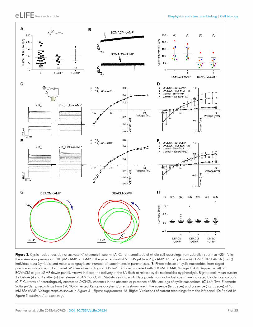
Figure 3. Cyclic nucleotides do not activate K+ channels in sperm. (A) Current amplitude of whole-cell recordings from zebrafish sperm at +25 mV in
the absence or presence of 100 mM cAMP or cGMP in the pipette (control: 91 ± 49 pA (n = 23); cAMP: 73 ± 25 pA (n = 6); cGMP: 109 ± 44 pA (n = 5)).
Individual data (symbols) and mean ± sd (gray bars), number of experiments in parentheses. (B) Photo-release of cyclic nucleotides from caged
precursors inside sperm. Left panel: Whole-cell recordings at +15 mV from sperm loaded with 100 mM BCMACM-caged cAMP (upper panel) or
BCMACM-caged cGMP (lower panel). Arrows indicate the delivery of the UV flash to release cyclic nucleotides by photolysis. Right panel: Mean current
3 s before (-) and 3 s after (+) the release of cAMP or cGMP. Statistics as in part A. Data points from individual sperm are indicated by identical colours.
(C-F) Currents of heterologously expressed DrCNGK channels in the absence or presence of 8Br- analogs of cyclic nucleotides. (C) Left: Two-Electrode
Voltage-Clamp recordings from DrCNGK-injected Xenopus oocytes. Currents shown are in the absence (left traces) and presence (right traces) of 10
mM 8Br-cAMP. Voltage steps as shown in Figure 3—figure supplement 1A. Right: IV relations of current recordings from the left panel. (D) Pooled IV
Figure 3 continued on next page
Fechner et al. eLife 2015;4:e07624. DOI: 10.7554/eLife.07624 7 of 25
Research article Biophysics and structural biology Cell biology
cAMP (Figure 3G, left) or cGMP (Figure 3G, right) from caged precursors. The photo-release was
followed by the increase of fluorescence of the free coumaryl cage (Figure 3—figure supplement 5)
(Bonigk et al., 2009). Swimming behaviour, i.e. path curvature (Figure 3H) and swimming speed
(Figure 3—figure supplement 4A) were not altered by photo-release, showing that neither cAMP
nor cGMP play a major role in the control of sperm motility. In conclusion, we observe no action of
cyclic nucleotides on the DrCNGK channel and on the swimming behaviour of D. rerio sperm.
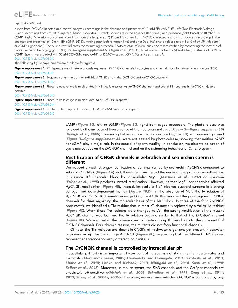
Rectification of CNGK channels in zebrafish and sea urchin sperm isdifferentWe noticed a much stronger rectification of currents carried by sea urchin ApCNGK compared to
zebrafish DrCNGK (Figure 4A) and, therefore, investigated the origin of this pronounced difference.
In classical K+ channels, block by intracellular Mg2+ (Matsuda et al., 1987) or spermine
(Fakler et al., 1995) produces inward rectification. However, neither Mg2+ nor spermine affected
ApCNGK rectification (Figure 4B). Instead, intracellular Na+ blocked outward currents in a strong
voltage- and dose-dependent fashion (Figure 4B,D). In the absence of Na+, the IV relation of
ApCNGK and DrCNGK channels converged (Figure 4A,B). We searched the pore regions of CNGK
channels for clues regarding the molecular basis of the Na+ block. In three of the four ApCNGK
pore motifs, we identified a Thr residue that in most K+ channels is replaced by a Val or Ile residue
(Figure 4C). When these Thr residues were changed to Val, the strong rectification of the mutant
ApCNGK channel was lost and the IV relation became similar to that of the DrCNGK channel
(Figure 4E). We also tested the reverse construct, introducing Thr residues into the pore motif of
DrCNGK channels. For unknown reasons, the mutants did not form functional channels.
Of note, the Thr residues are absent in CNGKs of freshwater organisms yet present in seawater
organisms except for the sponge AqCNGK (Figure 4C), suggesting that the different CNGK pores
represent adaptations to vastly different ionic milieus.
The DrCNGK channel is controlled by intracellular pHIntracellular pH (pHi) is an important factor controlling sperm motility in marine invertebrates and
mammals (Alavi and Cosson, 2005; Dziewulska and Domagala, 2013; Hirohashi et al., 2013;
Lishko et al., 2010; Lishko and Kirichok, 2010; Nishigaki et al., 2014; Santi et al., 1998;
Seifert et al., 2015). Moreover, in mouse sperm, the Slo3 channels and the CatSper channels are
exquisitely pH-sensitive (Kirichok et al., 2006; Schreiber et al., 1998; Zeng et al., 2011;
2013; Zhang et al., 2006a; 2006b). Therefore, we examined whether DrCNGK is controlled by pHi.
Figure 3 continued
curves from DrCNGK injected and control oocytes; recordings in the absence and presence of 10 mM 8Br-cAMP. (E) Left: Two-Electrode Voltage-
Clamp recordings from DrCNGK injected Xenopus oocytes. Currents shown are in the absence (left traces) and presence (right traces) of 10 mM 8Br-
cGMP. Right: IV relations of current recordings from the left panel. (F) Pooled IV curves from DrCNGK-injected and control oocytes; recordings in the
absence and presence of 10 mM 8Br-cGMP. (G) Swimming path before (green line) and after (red line) photo-release (black flash) of cAMP (left panel)
or cGMP (right panel). The blue arrow indicates the swimming direction. Photo-release of cyclic nucleotides was verified by monitoring the increase of
fluorescence of the caging group (Figure 3—figure supplement 5) (Hagen et al., 2003). (H) Path curvature before (-) and after (+) release of cAMP or
cGMP. Sperm were loaded with 30 mM DEACM-caged cAMP or DEACM-caged cGMP. Statistics as in part A.
DOI: 10.7554/eLife.07624.010
The following figure supplements are available for figure 3:
Figure supplement 1. K+ dependence of heterologously expressed DrCNGK channels in oocytes and channel block by tetraethylammonium (TEA).
DOI: 10.7554/eLife.07624.011
Figure supplement 2. Sequence alignment of the individual CNBDs from the DrCNGK and ApCNGK channels.
DOI: 10.7554/eLife.07624.012
Figure supplement 3. Photo-release of cyclic nucleotides in HEK cells expressing ApCNGK channels and use of 8Br-analogs in ApCNGK-injected
oocytes.
DOI: 10.7554/eLife.07624.013
Figure supplement 4. Photo-release of cyclic nucleotides (A) or Ca2+ (B) in sperm.
DOI: 10.7554/eLife.07624.014
Figure supplement 5. Control of loading and release of DEACM-cAMP in zebrafish sperm.
DOI: 10.7554/eLife.07624.015
Fechner et al. eLife 2015;4:e07624. DOI: 10.7554/eLife.07624 8 of 25
Research article Biophysics and structural biology Cell biology
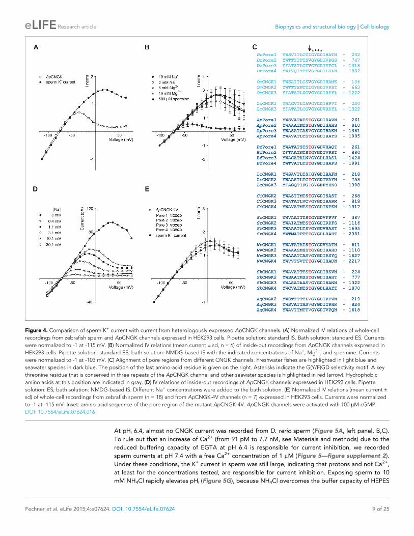
At pHi 6.4, almost no CNGK current was recorded from D. rerio sperm (Figure 5A, left panel, B,C).
To rule out that an increase of Ca2+ (from 91 pM to 7.7 nM, see Materials and methods) due to the
reduced buffering capacity of EGTA at pH 6.4 is responsible for current inhibition, we recorded
sperm currents at pH 7.4 with a free Ca2+ concentration of 1 mM (Figure 5—figure supplement 2).
Under these conditions, the K+ current in sperm was still large, indicating that protons and not Ca2+,
at least for the concentrations tested, are responsible for current inhibition. Exposing sperm to 10
mM NH4Cl rapidly elevates pHi (Figure 5G), because NH4Cl overcomes the buffer capacity of HEPES
Figure 4. Comparison of sperm K+ current with current from heterologously expressed ApCNGK channels. (A) Normalized IV relations of whole-cell
recordings from zebrafish sperm and ApCNGK channels expressed in HEK293 cells. Pipette solution: standard IS. Bath solution: standard ES. Currents
were normalized to -1 at -115 mV. (B) Normalized IV relations (mean current ± sd, n = 6) of inside-out recordings from ApCNGK channels expressed in
HEK293 cells. Pipette solution: standard ES, bath solution: NMDG-based IS with the indicated concentrations of Na+, Mg2+, and spermine. Currents
were normalized to -1 at -103 mV. (C) Alignment of pore regions from different CNGK channels. Freshwater fishes are highlighted in light blue and
seawater species in dark blue. The position of the last amino-acid residue is given on the right. Asterisks indicate the G(Y/F)GD selectivity motif. A key
threonine residue that is conserved in three repeats of the ApCNGK channel and other seawater species is highlighted in red (arrow). Hydrophobic
amino acids at this position are indicated in gray. (D) IV relations of inside-out recordings of ApCNGK channels expressed in HEK293 cells. Pipette
solution: ES; bath solution: NMDG-based IS. Different Na+ concentrations were added to the bath solution. (E) Normalized IV relations (mean current ±
sd) of whole-cell recordings from zebrafish sperm (n = 18) and from ApCNGK-4V channels (n = 7) expressed in HEK293 cells. Currents were normalized
to -1 at -115 mV. Inset: amino-acid sequence of the pore region of the mutant ApCNGK-4V. ApCNGK channels were activated with 100 mM cGMP.
DOI: 10.7554/eLife.07624.016
Fechner et al. eLife 2015;4:e07624. DOI: 10.7554/eLife.07624 9 of 25
Research article Biophysics and structural biology Cell biology
Figure 5. pH regulation of the DrCNGK channel. (A) Whole-cell recordings from zebrafish sperm after perfusion with NH4Cl or propionic acid. Voltage
steps as shown in Figure 1C. Recordings at extracellular pH 7.4 and pipette pH 6.4 (left). NH4Cl (10 mM, middle) or propionic acid (10 mM, right) was
added to the bath. (B) Pooled IV curves for recordings from zebrafish sperm at a pipette pHi of 6.4 and in the presence of 10 mM NH4Cl or 10 mM
propionic acid (PA). (C) Pooled IV curves from recordings of zebrafish sperm at different intracellular pHi. (D) Dependence of mean current ( ± sd) on
intracellular pHi (circles, bottom axis) or in the presence of either 10 mM propionic acid (PA) or different NH4Cl concentrations (triangles, top axis). (E)
Recording of the voltage signal of zebrafish sperm in the current-clamp configuration. Pipette solution with an intracellular pHi of 6.4; recording in the
presence of 10 mM NH4Cl or 10 mM propionic acid (PA). Left panel: single recording. Right panel: individual data (symbols) and mean ± sd (gray bars),
n = 10. (F) Pooled IV curves of Two-Electrode Voltage-Clamp recordings from heterologously expressed DrCNGK channels and uninjected wild-type
oocytes in 96 mM K+ bicarbonate solution (black and red symbols) or 96 mM K+ gluconate, including 1 mM NH4Cl (white symbols, see Figure 5—
figure supplement 1 for recordings). (G) Changes in fluorescence of a zebrafish sperm population incubated with the pH indicator BCECF, recorded as
the ratio of fluorescence at 549/15 nm and 494/20 nm (excited at 452/28 nm), before (black) and after the addition of 10 mM (red) or 30 mM (green)
NH4Cl. (H) Stimulation of sperm with NH4Cl as in panel G using the Ca2+ indicator Cal-520. Fluorescence was excited at 494/20 nm and recorded at
536/40 nm. Fluorescence F was normalized to the control value F0 before stimulation.
DOI: 10.7554/eLife.07624.017
The following figure supplements are available for figure 5:
Figure supplement 1. pH dependence of heterologously expressed DrCNGK channels in oocytes.
DOI: 10.7554/eLife.07624.018
Figure supplement 2. High intracellular Ca2+ does not suppress DrCNGK currents.
DOI: 10.7554/eLife.07624.019
Figure supplement 3. Hypoosmotic conditions do not stimulate or diminish DrCNGK currents in Xenopus oocytes.
DOI: 10.7554/eLife.07624.020
Fechner et al. eLife 2015;4:e07624. DOI: 10.7554/eLife.07624 10 of 25
Research article Biophysics and structural biology Cell biology
at pH 6.4 (Boron and De Weer, 1976; Seifert et al., 2015; Strunker et al., 2011). Alkaline pHi
strongly enhanced CNGK currents (Figure 5A, middle panel, B,C). Subsequent superfusion with 10
mM propionic acid, which lowers pHi, completely reversed the NH4Cl-induced CNGK currents
(Figure 5A, right panel, B and C). The NH4Cl action was very pronounced: 1 mM activated
Figure 6. Sperm swimming behaviour upon Ca2+ release. (A), (B), and (C) representative swimming paths of three different DMSO loaded sperm before
and after application of UV light. (D), (E), and (F) representative averaged swimming paths of three different sperm before (green) and after Ca2+
release by one (red) or two (cyan) consecutive UV flashes (black arrows). Curved blue arrows indicate the swimming direction of sperm. (G) Same
swimming path shown in (F) including a temporal axis to facilitate the visualization of the changes in swimming path after consecutive flashes. Upon
release (black arrows), the curvature of the swimming path progressively increases and the cell finally spins around the same position. (H)
Representative flagellar shapes before (-), after Ca2+ release by one (+) or two consecutive flashes (++), and during cell spinning against the wall
(bottom right). Consecutive frames every 100 ms are shown in different colours. Sequence order: red, green, blue, and yellow. (I) Mean curvature before
(-) and after one (+) or two (++) UV flashes. Individual data (symbols) and mean ± sd (gray bars), number of experiments in parentheses.
DOI: 10.7554/eLife.07624.021
Fechner et al. eLife 2015;4:e07624. DOI: 10.7554/eLife.07624 11 of 25
Research article Biophysics and structural biology Cell biology
approximately 40% of the CNGK current; at 10
mM, the current was maximal (Figure 5D, trian-
gles). To quantitatively determine the pH depen-
dence, we recorded sperm K+ currents at
different intracellular pHi values (Figure 5C,D
circles). The current is half-maximally activated
at pH 7.08. The pH dependence allows to cali-
brate the NH4Cl action by superposing the data
of the two different experimental conditions
(Figure 5D): For example, at an NH4Cl concen-
tration of 1.5 mM, pHi in sperm will increase
from 6.4 to 7.08 (Figure 5D). Under current-
clamp conditions, alkalization of D. rerio sperm
with 10 mM NH4Cl evoked a rapid and revers-
ible hyperpolarization from -49 ± 7 mV to -71 ±
4 mV (Figure 5E, n = 10). We also studied the
pH regulation of the DrCNGK channel expressed
in Xenopus oocytes. Oocytes were first perfused
with a K+ bicarbonate solution, followed by a K+
gluconate-based solution including 1 mM NH4Cl
(Figure 5—figure supplement 1). 1 mM NH4Cl
reversibly increased DrCNGK currents by
approximately 71%, demonstrating regulation
by pHi (Figure 5F). Higher concentrations of
NH4Cl further increased the DrCNGK current;
however, these conditions also elicited signifi-
cant currents in control oocytes, thus precluding
quantitative analysis. Furthermore, we tested,
whether DrCNGK channels in oocytes are activated by hypoosmotic conditions. Reducing the osmo-
larity by ~50% does not significantly change DrCNGK currents in oocytes (Figure 5—figure supple-
ment 3, n = 4). In sea urchin sperm, CNGK-mediated hyperpolarization leads to an increase of
intracellular Ca2+. Therefore, we tested, whether the NH4Cl-induced hyperpolarization (Figure 5E)
and alkalization (Figure 5G) evokes a Ca2+ response. In fact, mixing of D. rerio sperm with 10 or 30
mM NH4Cl gave rise to a rapid Ca2+ signal (Figure 5H, n = 4). The time course of the pHi- and Ca2+
signal was similar, suggesting that the CNGK-mediated hyperpolarization triggers a Ca2+ influx. We
conclude that DrCNGK represents a pH-sensitive channel that is strongly activated at alkaline pHi;
the ensuing hyperpolarization, like in sea urchin sperm, produces a Ca2+ signal.
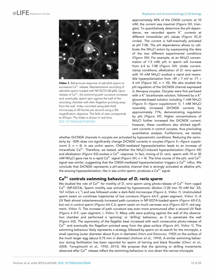
Ca2+ controls swimming behaviour of D. rerio spermWe studied the role of Ca2+ for motility of D. rerio sperm using photo-release of Ca2+ from caged
Ca2+ (NP-EGTA). Sperm motility was activated by hypoosmotic dilution (1:20 into 70 mM Na+ ES,
167 mOsm x L-1) and was followed under a dark-field microscope (Figure 6, Video 1). Unstimulated
sperm swam on curvilinear trajectories of low curvature (Figure 6A-F, green segment, Video 1). A
UV flash almost instantaneously increased path curvature in NP-EGTA-loaded sperm (Figure 6D-F,I),
but not in control sperm (Figure 6A-C,I); sperm swam on much narrower arcs (Figure 6D-F, red seg-
ment, Video 1). The increase of path curvature was even more pronounced after a second UV flash
(Figure 6 D-F, cyan segment, I, Video 1). Many cells were pushing against the wall of the observa-
tion chamber and performed a ‘spinning’ or ‘drilling’ behaviour, as if to penetrate the wall
(Figure 6G). The asymmetry of the flagellar beat increased with each consecutive photo-release of
Ca2+ and eventually the flagellum pointed away from the glass surface (Figure 6H, Video 1). This
swimming behaviour likely represents a strategy followed by sperm on its search for the micropyle, a
small opening (outer diameter about 8 mm in diameter) (Hart and Danovan, 1983) on the surface of
the much larger egg (about 0.75 mm in diameter) (Selman et al., 1993). A similar swimming behav-
iour during fertilization has been reported for sperm of herring and black flounder (Cherr et al.,
2008; Yanagimachi et al., 1992; 2013). We propose that the spinning or drilling movements
observed after Ca2+ release reflect the swimming behaviour in vivo down the narrow micropyle.
Video 1. Behavioural response of zebrafish sperm to
successive Ca2+ release. Representative recording of
zebrafish sperm loaded with NP-EGTA (40 mM). Upon
release of Ca2+, the swimming path curvature increases
and, eventually, sperm spin against the wall of the
recording chamber with their flagellum pointing away
from the wall. Video recorded using dark-field
microscopy at 30 frames per second using a 20x
magnification objective. The field of view corresponds
to 410 mm. The Video is shown in real time.
DOI: 10.7554/eLife.07624.022
Fechner et al. eLife 2015;4:e07624. DOI: 10.7554/eLife.07624 12 of 25
Research article Biophysics and structural biology Cell biology
DiscussionA growing body of evidence reveals unexpected commonalities, but also notable differences among
sperm from different species (for review (Darszon et al., 2006; Kaupp et al., 2008; Yoshida and
Yoshida, 2011). Organisms, as phylogenetically distant as sea urchins and humans, share the CatS-
per channel as a common site of Ca2+ entry into the sperm flagellum. By the same token, Slo3 K+
channel orthologues in mouse and human sperm evolved different selectivity for intracellular ligands
and might serve different functions (Brenker et al., 2014; Chavez et al., 2013; Lishko et al., 2012;
Santi et al., 2009; Zeng et al., 2013). Here, we characterize a novel variant of CNGK channels in
zebrafish sperm, whose key features depart from those of CNGK channels of marine invertebrates
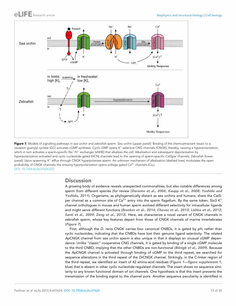
(Figure 7).
First, although the D. rerio CNGK carries four canonical CNBDs, it is gated by pHi rather than
cyclic nucleotides, indicating that the CNBDs have lost their genuine ligand selectivity. The related
ApCNGK channel from sea urchin sperm is also unique in that it displays an unusual cGMP depen-
dence: Unlike “classic” cooperative CNG channels, it is gated by binding of a single cGMP molecule
to the third CNBD, implying that the other CNBDs are non-functional (Bonigk et al., 2009). Because
the ApCNGK channel is activated through binding of cGMP to the third repeat, we searched for
sequence alterations in the third repeat of the DrCNGK channel. Strikingly, in the C-linker region of
the third repeat, we identified an insert of 42 amino-acid residues (Figure 1—figure supplement 1,
blue) that is absent in other cyclic nucleotide-regulated channels. The insert shows no sequence simi-
larity to any known functional domain of ion channels. One hypothesis is that this insert prevents the
transmission of the binding signal to the channel pore. Another sequence peculiarity is identified in
Figure 7. Models of signalling pathways in sea urchin and zebrafish sperm. Sea urchin (upper panel): Binding of the chemoattractant resact to a
receptor guanylyl cyclase (GC) activates cGMP synthesis. Cyclic GMP opens K+-selective CNG channels (CNGK), thereby, causing a hyperpolarization,
which in turn activates a sperm-specific Na+/H+ exchanger (sNHE) that alkalizes the cell. Alkalization and subsequent depolarization by
hyperpolarization-activated and cyclic nucleotide-gated (HCN) channels lead to the opening of sperm-specific CatSper channels. Zebrafish (lower
panel): Upon spawning, K+ efflux through CNGK hyperpolarizes sperm. An unknown mechanism of alkalization (dashed lines) modulates the open
probability of CNGK channels; the ensuing hyperpolarization opens voltage-gated Ca2+ channels (Cav).
DOI: 10.7554/eLife.07624.023
Fechner et al. eLife 2015;4:e07624. DOI: 10.7554/eLife.07624 13 of 25
Research article Biophysics and structural biology Cell biology

the second repeat (Figure 3—figure supplement 2). At amino-acid position 934, an Ala residue
replaces a highly conserved Arg residue that is crucial for cyclic-nucleotide binding (Kaupp and Sei-
fert, 2002). The CNBDs of repeat 1 and 4 do not show obvious sequence abnormalities and could
represent bona fide CNBDs (Figure 3—figure supplement 2). In recent years, many structures of
CNBDs have been solved (Clayton et al., 2004; Kesters et al., 2015; Kim et al., 2007;
Rehmann et al., 2003; Schunke et al., 2011; Schunke et al., 2009; Zagotta et al., 2003). Can we
learn from these structures something about the DrCNGK channel? Most of the CNBD structures
feature a similar fold and are able to bind both cAMP and cGMP. However, for some CNG channels,
ligands are full agonists, like cAMP and cGMP in the CNGA2 channel (Dhallan et al., 1990), only
partial agonists, like cAMP in the CNGA1 channel (Altenhofen et al., 1991), or competitive antago-
nists, like cGMP in the bacterial SthK channel (Brams et al., 2014). Finally, in HCN channels, CNBDs
interact and form a so-called gating ring (Zagotta et al., 2003), whereas in MloK1 channel, CNBDs
do not interact at all (Cukkemane et al., 2007; Schunke et al., 2009; 2011). In conclusion, at pres-
ent, no sequence features can be identified that unequivocally explain the lack of cyclic-nucleotide
regulation of the DrCNGK channel.
The insensitivity of the DrCNGK channel to cyclic nucleotides is, however, reminiscent of EAG
and hERG channels that carry classic CNBDs, yet are not gated by cyclic nucleotides (Brelidze et al.,
2009; 2010; 2012). Instead, small molecules such as flavonoids have been suggested as ligands that
bind to the CNBD and modulate channel activity (Brelidze et al., 2010; Carlson et al., 2013). More-
over, in the C-terminus of these CNBDs, a conserved segment of residues was identified that occu-
pies the CNBD and serves as an intrinsic “ligand” (Brelidze et al., 2012; Carlson et al., 2013). We
can only speculate, that, in addition to protons, as yet unidentified ligands might bind to and regu-
late the DrCNGK channel. The apparent pKa value for channel activation by pH was approximately
7, suggesting that a His residue controls channel opening. There are a number of His residues in the
C-linker of the four repeats that might serve as candidate sites. Future work is necessary to identify
the site of pH regulation of the DrCNGK channel.
To take on a new ligand selectivity or activation mechanism is also reminiscent of orthologues of
the sperm-specific K+ channel Slo3. Whereas the mouse Slo3 channel is exclusively controlled by pHi
(Brenker et al., 2014; Schreiber et al., 1998; Yang et al., 2011; Zeng et al., 2011; Zhang et al.,
2006a), the human Slo3 is primarily regulated by Ca2+ (Brenker et al., 2014). In conclusion, the
zebrafish CNGK is a striking example for a channel featuring a CNBD that is not gated by cyclic
nucleotides. In general, CNBDs might represent sensor domains that can relay information on
ligands other than cyclic nucleotides.
Second, signalling pathways that control sperm motility are located to the flagellum: The GC
receptor for chemoattractant binding in sea urchin (Bonigk et al., 2009; Pichlo et al., 2014), the
CatSper channel in humans, mice, and sea urchin (Chung et al., 2014; Kirichok et al., 2006;
Seifert et al., 2015), the Slo3 K+ channel in mice and humans (Brenker et al., 2014; Navarro et al.,
2007), and the HCN and the CNGK channel in sea urchin (Bonigk et al., 2009; Gauss et al., 1998).
In contrast, the DrCNGK channel is located in the head rather than the flagellum. What might be the
functional significance of such a peculiar location? The CNGK channel probably serves two related
functions.
In seminal fluid, sperm of freshwater fish are immotile due to a high [K+] and high osmolarity.
Upon release into hypoosmotic freshwater, sperm become motile for a few minutes
(Morisawa et al., 1983; Takai and Morisawa, 1995; Wilson-Leedy et al., 2009). The osmolarity-
induced activation hyperpolarizes sperm and induces a Ca2+ signal (Krasznai et al., 2000). We pro-
pose that the CNGK triggers Ca2+ signalling events upon spawning: In the high-K+ seminal fluid,
partially open CNGK channels keep sperm depolarized. When exposed to low-K+ hypoosmolar con-
ditions, sperm hyperpolarize and, ultimately, Ca2+ is entering the cell and activates general motility
(Figure 7).
Moreover, during the search for the micropyle on the egg surface, the sense of direction might
be provided by haptic interaction with tethered molecules that line the opening or the funnel of the
micropyle (Iwamatsu et al., 1997; Ohta and Iwamatsu, 1983; Yanagimachi et al., 2013). The hap-
tic interactions could directly control CNGK activity in the head. For example, near or inside the
micropyle, the CNGK might become further activated by alkaline pH and initiate the Ca2+-depen-
dent ‘drilling’ behaviour.
Fechner et al. eLife 2015;4:e07624. DOI: 10.7554/eLife.07624 14 of 25
Research article Biophysics and structural biology Cell biology

On a final note, the study of zebrafish sperm provides insight into adaptive mechanisms of sperm
evolution. Boundary conditions might constrain sperm to develop different signalling strategies for
similar functions. One obvious constraint is the ionic milieu, which strongly affects ion channel func-
tion. In freshwater, ion concentrations are low and opening of Na+-, K+-, and non-selective cation
channels would hyperpolarize rather than depolarize cells. We speculate that, in freshwater fish, a
depolarization-activated Ca2+ channel like CatSper may not work and has been replaced by another
Cav channel.
Furthermore, the Thr/Val difference in ApCNGK versus DrCNGK, which determines Na+ block-
age, probably represents an adaptation to the respective ionic milieu. Na+ blockage of sea urchin
CNGK resists hyperpolarization in seawater and, thereby, facilitates the opening of depolarization-
activated CatSper channels. The observation that CNGK channels from seawater organisms carry
this Thr residue indicates a specific evolutionary pressure on this pore residue. Why is this Thr resi-
due lost in CNGK channels of freshwater organisms? We speculate that Na+ blockage disappeared
along with the loss of CatSper genes and that Ca2+ ions enter fish sperm through a Ca2+ channel
that is activated by hyperpolarization rather than depolarization. Future work needs to identify this
Ca2+ channel, its mechanism of activation, and its role for fertilization of teleost fish.
In summary, we identify a zebrafish CNGK channel that is activated at alkaline pH, and is set apart
from its cousins of sea urchins that are activated by cGMP. Orthologues of CNGK also exist in the
choanoflagellate S. rosetta, suggesting that this channel sub-family is phylogenetically ancient. Inter-
estingly, this protozoon has a sexual life cycle: during anisogamous mating, small flagellated cells
fuse with large cells (Levin and King, 2013). This mating behaviour represents the ancestor of sexual
reproduction in animals (Levin and King, 2013; Umen and Heitman, 2013). The role of the S.
rosetta CNGK channel for sexual reproduction without sperm will be interesting to study.
Materials and methods
Materials and reagentsChemicals were purchased from AppliChem (Darmstadt, Germany), Biozym (Hessisch Oldendorf,
Germany), Carl Roth (Karlsruhe, Germany), GE Healthcare Life Sciences (Munich, Germany), Life
Technologies (Carlsbad, CA), Merck KgAa (Darmstadt, Germany), Merck Millipore (Billerica,
MA), PolyScience (Warrington, PA), Qiagen (Hilden, Germany), Serva (Heidelberg, Germany), Sigma-
Aldrich (Steinheim, Germany), and Thermo Scientifica (Waltham, MA). Enzymes and corresponding
buffer solutions were ordered from Ambion (Austin, TX), MBI Fermentas (Vilnius, Lithuania), New
England Biolabs (Frankfurt on the Main, Germany), and Roche (Basel, Switzerland). Primers were syn-
thesized from Eurofins Genomics (Ebersberg, Germany). Chemicals for mammalian cell culture were
ordered from Carl Roth, Life Technologies, and Biochrom (Berlin, Germany). CHOK1 and HEK293
cells were obtained from the American Type Culture Collection (ATCC, Manassas, VA).
Cloning of the DrCNGK geneWe identified two putative annotated sequences in the database: Eb934551 and XM_0013354.
Eb934551 contained the putative N-terminal region and XM_0013354 contained the putative repeat
3, repeat 4, and parts of repeat 2. Using four sets of primer pairs, we did nested PCR reactions on
testis cDNA to obtain the full-length sequence: the primer pairs #4811/#4812 and #4813/#4814
were used for XM_0013354 and the primer pairs C0274/C0275 and C0276/C0277 for Eb934551.
The primer sequences were: TATTTCAAGTAGCTGTTACCG (#4811), ACATTCCCTTATAATAAT-
GTCC (#4812), AAAAAAGCTAAGCTTTTCAGAAACACAG (#4813), AAAATCTGACAGGTACCCTG-
CAGAATGC (#4814), CATACAGGATGCATGACCCC (C0274), CCAGGAATGTATGTGTAGGTC
(C0275), GAGGAATTCATGCATGACCCCAGAGAAATGAAG (C0276) and CTCGGATCCGTATGTGT-
AGGTCTTTAATTTCAGGG (C0277). Due to failure of expression, we used a codon-optimized ver-
sion (human codon usage) of the DrCNGK gene separated into three modules (Eurofins Genomics).
Each module was flanked by restriction sites. The first module contained bases 1 to 2,108 and was
flanked on the 5’ end with BamHI and on the 3’ end with XbaI; the second module contained bases
2,109 to 4,655 and was flanked on the 5’ end with XbaI and on the 3’ end with EcoRI; the third mod-
ule contained bases 4,656 to 6,360 and was flanked on the 5’ end with EcoRI and on the 3’ end with
NotI. At the 3’ end, the coding sequence for the hemagglutinin tag (HA-tag) was added. The
Fechner et al. eLife 2015;4:e07624. DOI: 10.7554/eLife.07624 15 of 25
Research article Biophysics and structural biology Cell biology
construct was cloned into the pcDNA3.1 vector (Life technologies) (DrCNGK). To enhance expres-
sion levels, we added a QBI SP163 sequence (Stein et al., 1998) in front of the start codon (QBI-
DrCNGK).
Moreover, we added the coding sequence for a flag-tag at the 5’ end of the DrCNGK gene. We
performed two PCR reactions with primer pairs C0991/C0962 and C0417/C0990 and a recombinant
PCR reaction on the resulting PCR products with primer pair C0417/C0962. Primer sequences were:
CCCGGACGGCCTCCGAAACCATGGACTACAAGGACGACGACGACAAGC (C0991), TTCAGAC-
CGGCATTCCAAGCCC (C0962), CGCGGATCCAGCGCAGAGGCTTGGGGCAGC (C0417), GTCGT-
CGTCGTCCTTGTAGTCCATGGTTTCGGAGGCCGTCCGGG (C0990).
ApCNGK pore mutants: for the pore mutant ApCNGK-4V, the following amino-acid substitutions
in the ApCNGK wild-type channel (Bonigk et al., 2009) were produced: T252V, T801V and T1986V.
Three PCR reactions were required for each mutation: two with the primers containing the point
mutation and one recombinant PCR reaction. For the amino-acid exchange T252V, the primer pairs
#4433/C0531 and C0530/#4409 were used and for the recombinant PCR, the primer pair #4433/
#4409. The PCR product was cloned into the ApCNGK gene with restriction enzymes BamHI and
XhoI. For the amino-acid exchange T802V, primer pairs #4436/C0533 and C0532/#4439 were used
and for the recombinant PCR the primer pair #4436/#4439. The PCR product was cloned into the
ApCNGK gene with restriction enzymes XhoI and XbaI. For the amino-acid exchange T1986V, the
primer pairs #4412/C0535 and C0534/#4447 were used and for the recombinant PCR, the primer
pair #4412/#4447. The PCR product was cloned into the ApCNGK gene with restriction enzymes
BamHI and XbaI. The primer sequences were: GGTTCTGCTCGAGATTCTGTAGG (#4409), CAACAC-
CGGATCCGGTGAGAGCAGTG (#4412), AAAGTTGGGATCCAATACAGCG (#4433), TACAGAATCT-
CGAGCAGAACC (#4436), AAGTCTAGACGGTAGACTGATCGCCTGG (#4439),
AAATCTAGATTAGGCATAATCGGGCACATCATAGGGATACACCACCGTTTGTCTCAGCG (#4447),
GCCACCTCTGTAGGCTACGGAGAC (C0530), GTCTCCGTAGCCTACAGAGGTGGC (C0531), ATG-
ACATCCGTGGGCTACGGAGAC (C0532), GTCTCCGTAGCCCACGGATGTCAT (C0533), CTGACCT-
CCGTTGGCTACGGTGACATC (C0534), GTCACCGTAGCCAACGGAGGTCAGAG (C0535).
For expression in X. laevis oocytes, the DrCNGK and QBI-DrCNGK constructs were cloned into a
modified version of the expression vector pGEMHEnew (Liman et al., 1992); because cloning of
DrCNGK was only possible using BamHI and NotI, a NotI restriction site present in pGEMHEnew
was removed and a new one was introduced into the multiple-cloning site. This new vector has been
named pGEMHEnew-NotI. In vitro transcription to generate cRNA was performed using the mMES-
SAGEmMACHINEKit (Ambion); the plasmid was linearized with SpeI.
For in situ hybridization, a short fragment of the DrCNGK gene coding for amino acids 1,085-
1,219 was cloned into the pBluescript vector using PstI and HindIII. Nested PCR reactions were per-
formed using for the 1st reaction the primer pair #4791/#4792 and for the 2nd reaction the primer
pair #4793/#4794. Primer sequences were: ATTTTGCCGTGGAGTCCATGG (#4791), AAGTCAATAT-
TAAACGTTGCATCC (#4792), GGAAGCTTTCCGAAGCATTACAGCCG (#4793), GTTGGATCCAAG-
TGTGTCACCCATGAC (#4794). For the antisense probe, the plasmid was linearized with HindIII and
transcribed by T7 RNA polymerase. RNA was labeled with Digoxigenin (DIG RNA Labelling Mix,
Roche).
Preparation of testis, sperm, heads and flagellaAnimals were sacrificed according to the “Guidelines for housing and care, transport, and euthanasia
of laboratory fishes”, (“Empfehlung fur die Haltung, den Transport und das tierschutzgerechte Toten
von Versuchsfischen”, published by the Tierarztliche Vereinigung fur Tierschutz e.V. January 2010).
To obtain intact sperm, zebrafish male were anesthetized with MS-222 (0.5 mM, 3 min). After a brief
wash with fresh water, the head was quickly separated from the body. The body of the fish was ven-
trally opened and two testis strands were removed and transferred into ES buffer (see Electrophysi-
ology) or phosphate-buffered saline (PBS) containing (in mM: NaCl 137, KCl 2.7, Na2HPO4 6.5,
KH2PO4 1.5, pH 7.4), additionally containing 1.3 mM EDTA, mPIC protease inhibitory cocktail
(Sigma-Aldrich), and 1 mM DTT. Sperm were collected with a pipette tip. After 15 min, 80% of the
supernatant was transferred into a fresh reaction tube. To separate heads from flagella, the sperm
suspension was sheared 30-40 times on ice with a 24 gauge needle (Braun, Bethlehem, PA). The
sheared suspension was centrifuged for 10 min (800xg, 4˚C) to sediment intact sperm and sperm
heads. This procedure was repeated twice. The purity of flagella preparations was assessed using
Fechner et al. eLife 2015;4:e07624. DOI: 10.7554/eLife.07624 16 of 25
Research article Biophysics and structural biology Cell biology
dark-field microscopy (Figure 1—figure supplement 2). For testis sections, males were ventrally
sliced and kept overnight in 4% paraformaldehyde. After 24 hr, testis strands were removed and
embedded in paraffin. Sections (8 mm) were made using a microtome (Leica, Wetzlar, Germany).
Primary antibodiesA rabbit polyclonal antibody produced by Peptide Specialty Laboratories (PSL, Heidelberg, Ger-
many) was directed against the cytosolic loop C-terminal of the CNBD of the first repeat (anti-
repeat1, amino acids 483 - 497). Antibodies were purified with a peptide affinity column provided
by PSL. Rat monoclonal antibody YENT1E2 (anti-repeat3) was directed against the extracellular loop
between S5 and the pore region of the third repeat (amino acids 1,254 - 1,269). Anti-a-tubulin
(mouse, B-5-1-2, Sigma-Aldrich), anti-b-actin (mouse, abcam, Cambridge, United Kingdom), anti-HA
(rat, 3F10, Roche), anti-calnexin (rabbit, abcam), and anti-flag-tag (mouse, M2, Sigma-Aldrich) anti-
bodies were used as controls.
Immunocytochemistry, in situ hybridization, and Western blot analysisSperm were immobilized on SuperFrost Plus microscope slides (Thermo Fisher Scientific, Waltham,
MA) and fixed for 5 min with 4% paraformaldehyde. After preincubation with 0.5% Triton X-100 and
5% chemiblocker (Merck Millipore) in PBS, sperm were incubated for 1 hr with antibodies YENT1E2
(1:10) or anti-repeat1 (1:500) diluted in 5% chemiblocker (Merck Millipore) and 0.5% TritonX-100 in
PBS (pH 7.4). Sperm were visualized with Cy3-conjugated secondary antibody (Jackson ImmunoRe-
search Laboratories, West Grove, PA).
For in situ hybridization, tissue was permeabilized with protein kinase K (1 mg/ml in 0.1 M Tris/
HCl, pH 8.0) and hybridized using the DrCNGK-3 antisense probe. After washing, the antibody stain-
ing was performed using an anti-Digoxigenin antibody (1:500, Roche) conjugated with alkaline phos-
phatase. RNA was visualized with a mixture of nitro-blue tetrazolium chloride (500 mg) and 5-bromo-
4-chloro-3’-indolyphosphate p-toluidine salt (188 mg, Roche). Cross sections were covered with a
glass slip. For antibody staining of the in situ hybridization sections, cover slips were removed keep-
ing the slides 5 min in xylene and briefly in PBS. This step was repeated; the fixative was removed
from the sections. Afterwards, sections were stained as described for sperm immunocytochemistry.
Proteins were probed with antibodies: anti-repeat1 (1:500) or anti-repeat3 (1:10) and visualized with
Cy3-conjugated secondary antibody (Jackson ImmunoResearch Laboratories).
For Western blotting, zebrafish tissue or cells heterologously expressing the DrCNGK channel
were resuspended in PBS buffer containing 1.3 mM EDTA, mPIC protease inhibitor cocktail (Sigma-
Aldrich), and 1 mM DTT. For lysis, cells were triturated 20x with a cannula (24G, Braun) and soni-
cated three times for 15 s. After a clearing spin (25,000xg, 30 min, 4˚C), the pellet was resuspended
and sonicated two times for 10 s in 200 mM NaCl, 50 mM Hepes (pH 7.5), mPIC, and 1 mM DTT. Tri-
ton X-100 was added to a final concentration of 1%. Proteins were solubilized for 1–2 hr at 4˚C. Afinal clearing spin (10,000xg, 20 min, 4˚C) was performed. For Western blot analysis, proteins were
separated using 4-12% NuPAGE gradient gels (Life Technologies) and transferred overnight (4˚C,12–15 V) onto PVDF membranes (Immobilion FL, Merck Millipore), using a Xcell SureLock minigel
chamber (Life Technologies). Membranes were incubated with Odyssey blocking buffer (LI-COR Bio-
sciences, Lincoln, NE). Proteins were probed with the following antibodies: anti-repeat1 (1:1,000),
anti-repeat3 (1:10), anti-a-tubulin (1:2,000), anti-b-actin (1:1,000), anti-HA (1:1,000), anti-calnexin
(1:5,000), and anti-flag-tag (1:200). Proteins were visualized using IRDye800CW-conjugated second-
ary antibodies (1:10,000, LI-COR Biosciences), IRDye680-conjugated secondary antibodies (1:10,000,
LI-COR Biosciences), or horseradish peroxidase-conjugated secondary antibodies (1:5,000, Jackson
ImmunoResearch Laboratories). Visualization took place either with a chemiluminescence detection
system (LAS-3000 Luminescent Image Analyzer, FUJIFILM, Life Science, Stamford, CT) or with fluo-
rescent secondary antibodies (Odyssey infrared imaging system, Li-Cor Bioscience). The Novex
Sharp pre-stained protein standard (Life Technologies) was used as molecular mass standard.
ElectrophysiologyWe electrically recorded from intact zebrafish sperm and from isolated sperm heads using the
patch-clamp technique in the whole-cell configuration. Recordings were accomplished within 4 hr
after preparation. Seals between pipette and sperm were formed at the neck region in standard
Fechner et al. eLife 2015;4:e07624. DOI: 10.7554/eLife.07624 17 of 25
Research article Biophysics and structural biology Cell biology
extracellular solution (ES). The following pipette solutions were used: standard intracellular solution
(IS) (in mM): NaCl 10, K+ aspartate 130, MgCl2 2, EGTA 1, Na2ATP 2, and Hepes 10 at pH 8.4, 7.9,
7.4, 6.9, or 6.4 adjusted with KOH; Cl--based IS (in mM): NaCl 10, KCl 130, MgCl2 2, EGTA 1,
Na2ATP 2, and Hepes 10 at pH 7.4 adjusted with KOH. The following bath solutions were used: stan-
dard ES (in mM): NaCl 140, KCl 5.4, MgCl2 1, CaCl2 1.8, glucose 10, and Hepes 5 at pH 7.4 adjusted
with NaOH; for K+-based ES solutions, the equivalent amount of Na+ was replaced by K+ (concentra-
tions are indicated in the Figure legends). Calculations of the free Ca2+ concentrations were carried
out using the Maxchelator program (http://maxchelator.stanford.edu/webmaxc/webmaxcE.htm)
assuming a residual Ca2+ concentration in water of 1 mM. At pH 6.4, [Ca2+]i was 7.7 nM and at pH
7.4, it was 91 pM. To obtain an intracellular solution with 1 mM free Ca2+, 1 mM CaCl2 was added to
the IS solution at pH 7.4.
Caged compounds (100 mM BCMACM-caged cAMP or 100 mM BCMACM-caged cGMP) were
added to the IS. The final concentration of DMSO was 0.1%. The compounds were photolyzed by a
~1 ms flash of ultraviolet light from a Xenon flash lamp (JML-C2; Rapp OptoElectronic, Wedel, Ger-
many). The flash was passed through a BP295-395 nm filter (Rapp OptoElectronic) and delivered to
the patch-clamp chamber in the microscope by a liquid light guide. Pipette resistance in IS/ES was
between 11.5 and 15.0 MW. Voltages were corrected for liquid junction potentials.
For functional studies in X. laevis oocytes, 50 nl DrCNGK RNA (0.3, 0.4, and 0.6 mg/ml) per oocyte
were injected. Oocytes were purchased from EcoCyte Bioscience (Castrop-Rauxel, Germany) or pre-
pared from dissected animals. Briefly, frogs were anesthetized with MS-222 (0.5%, 10-20 min), fol-
licles were removed, opened with forceps and washed several times with ND96 solution. For
defolliculation, oocytes were transferred for 1–2 hr (RT) into Ca2+-free OR-2 solution containing 3
mg/ml collagenase type IV (Worthingthon Biochemical Corp., Lakewood, NJ). Defolliculated oocytes
were stored in ND96 solution containing (in mM): NaCl 96, KCl 2, MgCl2 1, CaCl2 1.8, Hepes 10 at
pH 7.6, pyruvate 2.5, and gentamycin 1. The OR-2 solution contained (in mM): NaCl 82.5, KCl 2.5,
MgCl2 1, Hepes 5 at pH 7.6. We recorded in the Two-Electrode Voltage-Clamp configuration. Most
data were recorded with a Dagan Clampator One (CA-1B, Dagan, Minneapolis, MN) amplifier and
digitized with Digidata 1320A (Axon Instruments, Molecular Devices, Sunnyvale, CA). Analogue sig-
nals were sampled at 2 kHz. The holding potential was -80 mV. Pipette solution: 3 M KCl. Bath solu-
tions were ND96-7K (in mM): NaCl 96, KCl 7, MgCl2 1, CaCl2 1.8, Hepes 10 at pH 7.4 adjusted with
NaOH; K+- based solution K96-7Na (in mM): NaCl 7, KCl 96, MgCl2 1, CaCl2 1.8, Hepes 10 at pH 7.4
adjusted with KOH. Recordings with reduced osmolarity were carried out in ND48-7K solution (in
mM): NaCl 48, KCl 7, CaCl2 1.8, MgCl2 1, HEPES 10 at pH 7.4 adjusted with NaOH. Pipette resis-
tance of voltage electrodes ranged between 1.5 and 3.0 MW and of current electrodes between 0.5
and 1.5 MW. Different analogues of cyclic nucleotides were added to the bath solution as indicated.
Oocytes recordings with bicarbonate-based solutions were performed at Stanford University. Data
were recorded with an OC-725C amplifier (Warner Instruments, Hamden, CT) using Patchmaster
(HEKA Elektronik, Lambrecht, Germany) as acquisition software. Analogue signals were sampled at 1
kHz. The holding potential was -60 mV. Pipette solutions and pipette resistance as described above.
Bath solutions: K+ bicarbonate-based solution (in mM): NaCl 7, K-bicarbonate 96, MgCl2 1, CaCl21.8, Hepes 5 at pH 7.65. Solution was made fresh on each day of recording; K+ gluconate-based
solution (in mM): NaCl 7, K-gluconate 96, MgCl2 1, CaCl2 1.8, Hepes 5 at pH 7.65 adjusted with
KOH. NH4Cl was dissolved in K+gluconate-based solution.
We recorded ApCNGK and mutant ApCNGK currents from transfected (Lipofectamine 2000, Life
technologies) HEK293 cells with the patch-clamp technique in the whole-cell configuration. A
HEK293 cell line stably expressing the ApCNGK channel was used for inside-out recordings. The
pipette solution for whole-cell recordings was standard IS. Channels were activated with 100 mM
cGMP. The pipette solution for inside-out recordings was standard ES. The following bath solutions
were used for inside-out recordings: IS-30 NMDG-0Na+ solution (in mM): NaCl 0, NMDG 30, KCl
110, EGTA 0.1, Hepes 10 at pH 7.4 adjusted with KOH; IS-NMDG-30Na+ solution (in mM): NaCl 30,
KCl 110, EGTA 0.1, Hepes 10 at pH 7.4 adjusted with KOH. 30 NMDG-0Na+ and 0 NMDG-30Na+
solutions were mixed to obtain the desired Na+ concentrations. 100 mM Na+-cGMP was added to
the bath solution. For the solution with 0 mM Na+, we used 100 mM Na+-free cGMP. Pipette resis-
tance in IS/ES was between 4.0 and 7.0 MW.
Fechner et al. eLife 2015;4:e07624. DOI: 10.7554/eLife.07624 18 of 25
Research article Biophysics and structural biology Cell biology
Measurement of changes in intracellular Ca2+ concentration and pHWe measured changes in [Ca2+]i, and pHi in a rapid-mixing device (SFM-4000; BioLogic, Claix,
France) in the stopped-flow mode using the Ca2+ indicator Cal-520-AM (AAT Bioquest, Sunnyvale,
CA) or the pH indicator BCECF-AM (Life Technologies). All sperm from a zebrafish male were diluted
into 100 ml of ES solution and incubated with either 10 mM Cal-520-AM and 0.5% Pluronic for 120–
180 min or 10 mM BCECF-AM for 10 min. Sperm were washed once, diluted 1:20 into ES solution,
and loaded into the stopped-flow device. The sperm suspension was rapidly mixed 1:1 (vol/vol) with
control ES solution or with ES solution containing NH4Cl to obtain final concentrations of 10 mM
and 30 mM after mixing. Fluorescence was excited by a SpectraX Light Engine (Lumencor, Beaver-
ton, OR). Cal-520 was excited with a 494/20 nm (Semrock, Rochester, NY), BCECF with a 452/45 nm
(Semrock) excitation filter. Emission was recorded by photomultiplier modules (H9656-20; Hama-
matsu Photonics). Fluorescence of Cal-520 was recorded using a 536/40 nm (Semrock) emission filter
and normalized (without background subtraction) to the value before stimulation. BCECF fluores-
cence was recorded in the dual emission mode using a 494/20 nm (Semrock) and a 549/15 nm (Sem-
rock) emission filter. The pHi signals represent the ratio of F494/549 and were normalized (without
background subtraction) to the value before stimulation. All stopped-flow traces represent the aver-
age of 3–6 recordings. The signals were normalized to the first 5-10 data points before the onset of
the signal to yield 4F/F and 4R/R, respectively.
Mass spectrometric identification of the DrCNGK channelProteins of whole sperm, isolated heads, or flagella were resuspended in an SDS sample buffer and
loaded on a SDS gel; after proteins had migrated approximately 1 cm into the separation gel, the
gel was stained with Coomassie. The single gel band was excised for every sample, and proteins
were in-gel digested with trypsin (Promega, Sunnyvale, CA); peptides were separated by RP-LC (180
min gradient 2–85% acetonitrile, (Thermo Fisher Scientific)) using a nanoAcquity LC System (Waters,
Milford, MA) equipped with a HSS T3 analytical column (1.8 mm particle, 75 mm x 150 mm) (Waters)
and analyzed twice by ESI-LC-MS/MS, using an LTQ Orbitrap Elite mass spectrometer (Thermo
Fisher Scientific) with a 300-2,000 m/z survey scan at 240,000 resolution, and parallel CID of the 20
most intense precursors from most to least intense (top20) and from least to most intense (bot-
tom20) with 60 s dynamic exclusion. All database searches were performed using SEQUEST and MS
Amanda algorithm (Dorfer et al., 2014), embedded in Proteome Discoverer (Rev. 1.4, Thermo Elec-
tron 2008-2011, Thermo Fisher Scientific), with both a NCBI (26,623 entries, accessed December
20th, 2010) and a Uniprot (40,895 entries, accessed April 24th, 2014) zebrafish sequence protein
database, both supplemented with the DrCNGK protein sequence (Figure 1—figure supplement
2). Only peptides originating from protein cleavage after lysine and arginine with up to two missed
cleavages were accepted. Oxidation of methionine was permitted as variable modification. The mass
tolerance for precursor ions was set to 8 ppm; the mass tolerance for fragment ions was set to 0.6
amu. For filtering of search results and identification of DrCNGK, a peptide FDR threshold of 0.01
(q-value) according to Percolator (Kall et al., 2007) two unique peptides per protein and peptides
with search result rank 1 were required.
Sequence analysisAlignments for the calculation of the phylogenetic tree were done with ClustalOmega. Tree was
depicted with Tree view (Page, 1996). The following ion channel sequences were used for the phylo-
genetic tree: CNGK channels from zebrafish (Danio rerio, DrCNGK, XP_001335499.5); rainbow trout
(Oncorhynchus mykiss, OmCNGK, CDQ79437.1); spotted gar (Lepisosteus oculatus, LoCNGK,
W5MTF2); West Indian ocean coelacanth (Latimeria chalumnae, LcCNGK, H3BE11); sea urchin (Arba-
cia punctulata, ApCNGK); acorn worm (Saccoglossus kowalevskii, SkCNGK, XP_002731383.1);
amphioxus (Branchiostoma floridae, BfCNGK, XP_002592428.1); starlet sea anemone (Nematostella
vectensis, NvCNGK, XP_001627832); vasa tunicate (Ciona intestinalis, CiCNGK, XP_002123955);
sponge (Amphimedon queenslandica, AqCNGK, I1G982); choanoflagellate (Salpingoeca rosetta,
SrCNGK, XP_004992545.1); murine HCN channel 1 (Mus musculus, mHCN1, NP_034538), 2
(mHCN2, NP_032252), 3 (mHCN3, NP_032253.1), and 4 (mHCN4, NP_001074661), and the HCN
channel from sea urchin (Strongylocentrotus purpuratus, SpHCN1, NP_999729); rat cyclic nucleo-
tide-gated channels CNGA1 (Rattus rattus, rCNGA1, NP_445949), A2 (rCNGA2, NP_037060), A3
Fechner et al. eLife 2015;4:e07624. DOI: 10.7554/eLife.07624 19 of 25
Research article Biophysics and structural biology Cell biology
(rCNGA3, NP_445947.1), and A4 (rCNGA4, Q64359); the KCNH channels from fruit fly (Drosophila
melanogaster, DmEAG, AAA28495) and human (Homo sapiens, hERG, BAA37096.1); murine volt-
age-gated Nav channels (Mus musculus, mNav 1.1, NP_061203 and mNav 1.6, NM_001077499.2);
murine voltage-gated Cav channels (Mus musculus, mCav1.1, NP_055008, mCav2.3, NP_033912.2,
and mCav3.1, NP_033913.2); and voltage-gated Kv channels from fruit fly (Drosophila melanogaster,
DmShaker, CAA29917.1) and mouse (Mus musculus, mKv3.1, NM_001112739.1).
Sperm motilitySperm were loaded for 45 min at room temperature with either 30 mM DEACM-caged cAMP, 30 mM
DEACM-caged cGMP, or 40 mM caged Ca2+ (NP-EGTA, Life Technologies). A UV light-emitting
diode (365-nm LED; M365L2-C, Thorlabs, Newton, NJ) was used for photolysis of caged com-
pounds. Experiments using caged Ca2+ and DEACM-caged nucleotides were carried out using a UV
power of 25 and 22 mW, respectively. Flash duration was 300 ms. Pluronic (0.5%) was added to the
incubation solution. Sperm were kept quiescent during incubation in ES solution (292 mOsm x L-1).
Swimming was initiated by a hypoosmotic shock diluting sperm 1:20 in an activation solution con-
taining (in mM): NaCl 70, KCl 5.4, MgCl2 1, CaCl2 1.8, glucose 10, and Hepes 5 at pH 7.4 adjusted
with NaOH (167 mOsm x L-1). Swimming behaviour was observed with a dark-field condenser in an
inverse microscope (IX71, Olympus, Tokio, Japan) with 20x magnification (UPLSAPO, NA 0.75). Mov-
ies were recorded at 30 Hz using a back-illuminated electron-multiplying charge-coupled device
camera (DU-897D; Andor Technology, Belfast, United Kingdom). Sperm trajectories were tracked
using custom-made software written in MATLAB (Mathworks). The software can be made available
upon request. The average swimming path (ASP) was calculated by filtering the tracked coordinates
with a second degree Savitzky-Golay filter with a 200 ms span. The curvature (k) of the swimming
path was calculated using the formula:bbb
k ¼_x y � _y x_x2þ _y2
� �3=2, where x and y are the coordinates of the ASP.
To assess cAMP loading and release, we recorded the fluorescence increase due to DEACM-OH
release after photolysis of DEACM-caged cAMP (Bonigk et al. 2009). Release and fluorescence exci-
tation was achieved simultaneously using the same UV LED (power 1.75 mW). Light was coupled to
the microscope with a dichroic mirror (455DRLP, XF2034, Omega Optical, Brattleboro, VT) and fluo-
rescence was long-pass filtered (460ALP; XF309; Omega Optical). Single cells were recorded at 50
Hz.
Data analysis: Statistical analysis and fitting of data were performed, unless otherwise stated,
using Sigma Plot 11.0, GraphPadPrism 5, or Clampfit 10.2 (Molecular Devices). All data are given as
mean ± standard deviation (number of experiments).
Note: All cell lines used in this study will be sent for STR profiling. Mycoplasma testing was per-
formed using the Promokine Mycoplasma Test Kit 1/C (PromoCell GmbH, Heidelberg, Germany).
Results of this test can be supplied upon request.
AcknowledgementsWe thank Drs. K Benndorf, and J Kusch (University Jena) and Drs. E Miranda-Laferte and P Hidalgo
(Research Center Julich), and Dr. M Goodman (Stanford University) for support with oocyte expres-
sion and H Krause for preparing the manuscript. SF was a fellow of the Boehringer Ingelheim Fonds.
The authors declare no competing financial interests.
Additional information
Funding
Funder Grant reference number Author
Boehringer Ingelheim Fonds Ph.D. Student Fellowship Sylvia Fechner
The funders had no role in study design, data collection and interpretation, or the decision tosubmit the work for publication.
Fechner et al. eLife 2015;4:e07624. DOI: 10.7554/eLife.07624 20 of 25
Research article Biophysics and structural biology Cell biology
Author contributions
SF, Designed the project and experiments, Performed electrophysiology in mammalian cells, sperm
and oocytes, biochemistry, cell biology, motility and stopped-flow experiments, Analysis and inter-
pretation of data, Wrote, read and corrected the manuscript; LA, Performed motility experiments
and analysis, Wrote, read and corrected the manuscript; WB, AM, Cloned the CNGK gene and
mutants; TKB, Performed electrophysiology in sperm, Analysis and interpretation of data; RP, Per-
formed motility experiments, Analysis and interpretation of data; CT, Performed the MS analysis;
AP, Performed MS analysis; GS, Guided electrophysiology in oocytes; KRS, Performed in situ hybrid-
ization; EK, Produced the monoclonal antibodies; RS, Designed the project and experiments, Per-
formed stopped-flow experiments, Performed electrophysiology in sperm, Analysis and
interpretation of data, Wrote, read and corrected the manuscript; UBK, Designed the project and
experiments, Wrote and corrected the manuscript, Analysis and interpretation of data, Drafting or
revising the article
Author ORCIDs
Gabriel Stolting, http://orcid.org/0000-0002-2339-0545
ReferencesAlavi SM, Cosson J. 2005. Sperm motility in fishes. I. Effects of temperature and pH: a review. Cell BiologyInternational 29:101–110. doi: 10.1016/j.cellbi.2004.11.021
Alavi SM, Cosson J. 2006. Sperm motility in fishes. II. Effects of ions and osmolality: a review. Cell BiologyInternational 30:1–14. doi: 10.1016/j.cellbi.2005.06.004
Altenhofen W, Ludwig J, Eismann E, Kraus W, Bonigk W, Kaupp UB. 1991. Control of ligand specificity in cyclicnucleotide-gated channels from rod photoreceptors and olfactory epithelium. Proceedings of the NationalAcademy of Sciences of the United States of America 88:9868–9872. doi: 10.1073/pnas.88.21.9868
Alvarez L, Dai L, Friedrich BM, Kashikar ND, Gregor I, Pascal R, Kaupp UB. 2012. The rate of change in Ca2+concentration controls sperm chemotaxis. The Journal of Cell Biology 196:653–663. doi: 10.1083/jcb.201106096
Billard R. 1986. Spermatogenesis and spermatology of some teleost fish species. Reproduction NutritionDeveloppement 26:877–920. doi: 10.1051/rnd:19860601
Bonigk W, Loogen A, Seifert R, Kashikar N, Klemm C, Krause E, Hagen V, Kremmer E, Strunker T, Kaupp UB.2009. An atypical CNG channel activated by a single cGMP molecule controls sperm chemotaxis. ScienceSignaling 2:ra68. doi: 10.1126/scisignal.2000516
Boron WF, De Weer P. 1976. Intracellular pH transients in squid giant axons caused by CO2, NH3, and metabolicinhibitors. The Journal of General Physiology 67:91–112. doi: 10.1085/jgp.67.1.91
Brams M, Kusch J, Spurny R, Benndorf K, Ulens C. 2014. Family of prokaryote cyclic nucleotide-modulated ionchannels. Proceedings of the National Academy of Sciences of the United States of America 111:7855–7860.doi: 10.1073/pnas.1401917111
Brelidze TI, Carlson AE, Davies DR, Stewart LJ, Zagotta WN. 2010. Identifying regulators for EAG1 channels witha novel electrophysiology and tryptophan fluorescence based screen. PLoS One 5:e12523. doi: 10.1371/journal.pone.0012523
Brelidze TI, Carlson AE, Sankaran B, Zagotta WN. 2012. Structure of the carboxy-terminal region of a KCNHchannel. Nature 481:530–533. doi: 10.1038/nature10735
Brelidze TI, Carlson AE, Zagotta WN. 2009. Absence of direct cyclic nucleotide modulation of mEAG1 andhERG1 channels revealed with fluorescence and electrophysiological methods. Journal of Biological Chemistry284:27989–27997. doi: 10.1074/jbc.M109.016337
Brenker C, Goodwin N, Weyand I, Kashikar ND, Naruse M, Krahling M, Muller A, Kaupp UB, Strunker T. 2012.The CatSper channel: a polymodal chemosensor in human sperm. The EMBO Journal 31:1654–1665. doi: 10.1038/emboj.2012.30
Brenker C, Zhou Y, Muller A, Echeverry FA, Trotschel C, Poetsch A, Xia X-M, Bonigk W, Lingle CJ, Kaupp UB,Strunker T. 2014. The Ca2+-activated K+ current of human sperm is mediated by Slo3. eLife 3:e01438. doi: 10.7554/eLife.01438
Bohmer M, Van Q, Weyand I, Hagen V, Beyermann M, Matsumoto M, Hoshi M, Hildebrand E, Kaupp UB. 2005.Ca2+ spikes in the flagellum control chemotactic behavior of sperm. The EMBO Journal 24:2741–2752. doi: 10.1038/sj.emboj.7600744
Cai X, Clapham DE, . 2008. Evolutionary genomics reveals lineage-specific gene loss and rapid evolution of asperm-specific ion channel complex: CatSpers and CatSperb. PLoS ONE 3:e3569. doi: 10.1371/journal.pone.0003569
Carlson AE, Brelidze TI, Zagotta WN. 2013. Flavonoid regulation of EAG1 channels. The Journal of GeneralPhysiology 141:347–358. doi: 10.1085/jgp.201210900
Fechner et al. eLife 2015;4:e07624. DOI: 10.7554/eLife.07624 21 of 25
Research article Biophysics and structural biology Cell biology
Cherr GN, Morisawa M, Vines CA, Yoshida K, Smith EH, Matsubara T, Pillai MC, Griffin FJ, Yanagimachi R. 2008.Two egg-derived molecules in sperm motility initiation and fertilization in the pacific herring (clupea pallasi).The International Journal of Developmental Biology 52:743–752. doi: 10.1387/ijdb.072566gc
Chung J-J, Shim S-H, Everley RA, Gygi SP, Zhuang X, Clapham DE. 2014. Structurally distinct Ca2+ signalingdomains of sperm flagella orchestrate tyrosine phosphorylation and motility. Cell 157:808–822. doi: 10.1016/j.cell.2014.02.056
Chavez JC, de la Vega-Beltran JL, Escoffier J, Visconti PE, Trevino CL, Darszon A, Salkoff L, Santi CM. 2013. Ionpermeabilities in mouse sperm reveal an external trigger for SLO3-dependent hyperpolarization. PLoS One 8:e60578. doi: 10.1371/journal.pone.0060578
Clayton GM, Silverman WR, Heginbotham L, Morais-Cabral JH. 2004. Structural basis of ligand activation in acyclic nucleotide regulated potassium channel. Cell 119:615–627. doi: 10.1016/j.cell.2004.10.030
Cosson J, Groison A-L, Suquet M, Fauvel C, Dreanno C, Billard R. 2008. Marine fish spermatozoa: racingephemeral swimmers. Reproduction 136:277–294. doi: 10.1530/REP-07-0522
Cukkemane A, Gruter B, Novak K, Gensch T, Bonigk W, Gerharz T, Kaupp UB, Seifert R. 2007. Subunits actindependently in a cyclic nucleotide-activated K+ channel. EMBO Reports 8:749–755. doi: 10.1038/sj.embor.7401025
Darszon A, Acevedo JJ, Galindo BE, Hernandez-Gonzalez EO, Nishigaki T, Trevino CL, Wood C, Beltran C. 2006.Sperm channel diversity and functional multiplicity. Reproduction 131:977–988. doi: 10.1530/rep.1.00612
Darszon A, Guerrero A, Galindo BE, Nishigaki T, Wood CD. 2008. Sperm-activating peptides in the regulation ofion fluxes, signal transduction and motility. The International Journal of Developmental Biology 52:595–606.doi: 10.1387/ijdb.072550ad
Denissenko P, Kantsler V, Smith DJ, Kirkman-Brown J. 2012. Human spermatozoa migration in microchannelsreveals boundary-following navigation. Proceedings of the National Academy of Sciences of the United Statesof America 109:8007–8010. doi: 10.1073/pnas.1202934109
Dhallan RS, Yau K-W, Schrader KA, Reed RR. 1990. Primary structure and functional expression of a cyclicnucleotide-activated channel from olfactory neurons. Nature 347:184–187. doi: 10.1038/347184a0
Dorfer V, Pichler P, Stranzl T, Stadlmann J, Taus T, Winkler S, Mechtler K. 2014. MS amanda, a universalidentification algorithm optimized for high accuracy tandem mass spectra. Journal of Proteome Research 13:3679–3684. doi: 10.1021/pr500202e
Dziewulska K, Domagała J. 2013. Effect of pH and cation concentrations on spermatozoan motility of sea trout(salmo trutta m. trutta l.). Theriogenology 79:48–58. doi: 10.1016/j.theriogenology.2012.09.008
Eisenbach M, Giojalas LC. 2006. Sperm guidance in mammals — an unpaved road to the egg. Nature ReviewsMolecular Cell Biology 7:276–285. doi: 10.1038/nrm1893
Elgeti J, Kaupp UB, Gompper G. 2010. Hydrodynamics of sperm cells near surfaces. Biophysical Journal 99:1018–1026. doi: 10.1016/j.bpj.2010.05.015
Fakler B, Brandle U, Glowatzki E, Weidemann S, Zenner H-P, Ruppersberg JP. 1995. Strong voltage-dependentinward rectification of inward rectifier K+ channels is caused by intracellular spermine. Cell 80:149–154. doi: 10.1016/0092-8674(95)90459-X
Florman HM, Jungnickel MK, Sutton KA. 2008. Regulating the acrosome reaction. The International Journal ofDevelopmental Biology 52:503–510. doi: 10.1387/ijdb.082696hf
Gakamsky A, Armon L, Eisenbach M. 2009. Behavioral response of human spermatozoa to a concentration jumpof chemoattractants or intracellular cyclic nucleotides. Human Reproduction 24:1152–1163. doi: 10.1093/humrep/den409
Gauss R, Seifert R, Kaupp UB. 1998. Molecular identification of a hyperpolarization-activated channel in seaurchin sperm. Nature 393:583–587. doi: 10.1038/31248
Guerrero A, Wood CD, Nishigaki T, Carneiro J, Darszon A. 2010. Tuning sperm chemotaxis. Biochemical SocietyTransactions 38:1270–1274. doi: 10.1042/BST0381270
Hagen V, Frings S, Wiesner B, Helm S, Kaupp UB, Bendig J. 2003. [7-(dialkylamino)coumarin-4-yl]methyl-cagedcompounds as ultrafast and effective long-wavelength phototriggers of 8Bromo-substituted cyclic nucleotides.ChemBioChem 4:434–442. doi: 10.1002/cbic.200300561
Hart NH, Donovan M. 1983. Fine structure of the chorion and site of sperm entry in the egg of Brachydanio.Journal of Experimental Zoology 227:277–296. doi: 10.1002/jez.1402270212
Hirohashi N, Alvarez L, Shiba K, Fujiwara E, Iwata Y, Mohri T, Inaba K, Chiba K, Ochi H, Supuran CT, Kotzur N,Kakiuchi Y, Kaupp UB, Baba SA. 2013. Sperm from sneaker male squids exhibit chemotactic swarming to CO2.Current Biology 23:775–781. doi: 10.1016/j.cub.2013.03.040
Ho HC, Suarez SS. 2001. Hyperactivation of mammalian spermatozoa: function and regulation. Reproduction122:519–526. doi: 10.1530/rep.0.1220519
Hugentobler SA, Morris DG, Sreenan JM, Diskin MG. 2007. Ion concentrations in oviduct and uterine fluid andblood serum during the estrous cycle in the bovine. Theriogenology 68:538–548. doi: 10.1016/j.theriogenology.2007.04.049
Iwamatsu T, Yoshizaki N, Shibata Y. 1997. Changes in the chorion and sperm entry into the micropyle duringfertilization in the teleostean fish, oryzias latipes. Development, Growth and Differentiation 39:33–41. doi: 10.1046/j.1440-169X.1997.00005.x
Kashikar ND, Alvarez L, Seifert R, Gregor I, Jackle O, Beyermann M, Krause E, Kaupp UB. 2012. Temporalsampling, resetting, and adaptation orchestrate gradient sensing in sperm. The Journal of Cell Biology 198:1075–1091. doi: 10.1083/jcb.201204024
Fechner et al. eLife 2015;4:e07624. DOI: 10.7554/eLife.07624 22 of 25
Research article Biophysics and structural biology Cell biology
Kaupp UB, Kashikar ND, Weyand I. 2008. Mechanisms of sperm chemotaxis. Annual Review of Physiology 70:93–117. doi: 10.1146/annurev.physiol.70.113006.100654
Kaupp UB, Seifert R. 2002. Cyclic nucleotide-gated ion channels. Physiological Reviews 82:769–824. doi: 10.1152/physrev.00008.2002
Kaupp UB, Solzin J, Hildebrand E, Brown JE, Helbig A, Hagen V, Beyermann M, Pampaloni F, Weyand I. 2003.The signal flow and motor response controling chemotaxis of sea urchin sperm. Nature Cell Biology 5:109–117.doi: 10.1038/ncb915
Kesters D, Brams M, Nys M, Wijckmans E, Spurny R, Voets T, Tytgat J, Kusch J, Ulens C. 2015. Structure of theSthK carboxy-terminal region reveals a gating mechanism for cyclic nucleotide-modulated ion channels. PLOSOne 10:e0116369. doi: 10.1371/journal.pone.0116369
Kim C, Cheng CY, Saldanha SA, Taylor SS. 2007. PKA-I holoenzyme structure reveals a mechanism for cAMP-dependent activation. Cell 130:1032–1043. doi: 10.1016/j.cell.2007.07.018
Kirichok Y, Navarro B, Clapham DE. 2006. Whole-cell patch-clamp measurements of spermatozoa reveal analkaline-activated Ca2+ channel. Nature 439:737–740. doi: 10.1038/nature04417
Korschen HG, Illing M, Seifert R, Sesti F, Williams A, Gotzes S, Colville C, Muller F, Dose´ A, Godde M, Molday L,Kaupp UB, Molday RS. 1995. A 240 kDa protein represents the complete b subunit of the cyclic nucleotide-gated channel from rod photoreceptor. Neuron 15:627–636. doi: 10.1016/0896-6273(95)90151-5
Krasznai Z, Marian T, Izumi H, Damjanovich S, Balkay L, Tron L, Morisawa M. 2000. Membrane hyperpolarizationremoves inactivation of Ca2+ channels, leading to Ca2+ influx and subsequent initiation of sperm motility inthe common carp. Proceedings of the National Academy of Sciences of the United States of America 97:2052–2057. doi: 10.1073/pnas.040558097
Kall L, Canterbury JD, Weston J, Noble WS, MacCoss MJ. 2007. Semi-supervised learning for peptideidentification from shotgun proteomics datasets. Nature Methods 4:923–925. doi: 10.1038/nmeth1113
Korschen HG, Beyermann M, Muller F, Heck M, Vantler M, Koch KW, Kellner R, Wolfrum U, Bode C, HofmannKP, Kaupp UB. 1999. Interaction of glutamic-acid-rich proteins with the cGMP signalling pathway in rodphotoreceptors. Nature 400:761–766. doi: 10.1038/23468
Lee HC, Garbers DL. 1986. Modulation of the voltage-sensitive Na+/H+ exchange in sea urchin spermatozoathrough membrane potential changes induced by the egg peptide speract. The Journal of Biological Chemistry261:16026–16032.
Lee HC. 1984. Sodium and proton transport in flagella isolated from sea urchin spermatozoa. The Journal ofBiological Chemistry 259:4957–4963.
Lee HC. 1985. The voltage-sensitive Na+/H+ exchange in sea urchin spermatozoa flagellar membrane vesiclesstudied with an entrapped pH probe. The Journal of Biological Chemistry 260:10794–10799.
Levin TC, King N. 2013. Evidence for sex and recombination in the choanoflagellate Salpingoeca rosetta. CurrentBiology 23:2176–2180. doi: 10.1016/j.cub.2013.08.061
Liman ER, Tytgat J, Hess P. 1992. Subunit stoichiometry of a mammalian K+ channel determined by constructionof multimeric cDNAs. Neuron 9:861–871. doi: 10.1016/0896-6273(92)90239-A
Lishko PV, Botchkina IL, Fedorenko A, Kirichok Y. 2010. Acid extrusion from human spermatozoa is mediated byflagellar voltage-gated proton channel. Cell 140:327–337. doi: 10.1016/j.cell.2009.12.053
Lishko PV, Botchkina IL, Kirichok Y. 2011. Progesterone activates the principal Ca2+ channel of human sperm.Nature 471:387–391. doi: 10.1038/nature09767
Lishko PV, Kirichok Y, Ren D, Navarro B, Chung J-J, Clapham DE. 2012. The control of male fertility byspermatozoan ion channels. Annual Review of Physiology 74:453–475. doi: 10.1146/annurev-physiol-020911-153258
Lishko PV, Kirichok Y. 2010. The role of Hv1 and CatSper channels in sperm activation. The Journal of Physiology588:4667–4672. doi: 10.1113/jphysiol.2010.194142
Matsuda H, Saigusa A, Irisawa H. 1987. Ohmic conductance through the inwardly rectifying K channel andblocking by internal Mg2+. Nature 325:156–159. doi: 10.1038/325156a0
Morisawa M, Suzuki K, Shimizu H, Morisawa S, Yasuda K. 1983. Effects of osmolality and potassium on motilityof spermatozoa from freshwater cyprinid fishes. The Journal of Experimental Biology 107:95–103.
Morisawa M. 2008. Adaptation and strategy for fertilization in the sperm of teleost fish. Journal of AppliedIchthyology 24:362–370. doi: 10.1111/j.1439-0426.2008.01126.x
Navarro B, Kirichok Y, Clapham DE. 2007. KSper, a pH-sensitive K+ current that controls sperm membranepotential. Proceedings of the National Academy of Sciences of the United States of America 104:7688–7692.doi: 10.1073/pnas.0702018104
Nishigaki T, Jose O, Gonzalez-Cota AL, Romero F, Trevino CL, Darszon A. 2014. Intracellular pH in spermphysiology. Biochemical and Biophysical Research Communications 450:1149–1158. doi: 10.1016/j.bbrc.2014.05.100
Nobrega RH, Batlouni SR, Franca LR. 2009. An overview of functional and stereological evaluation ofspermatogenesis and germ cell transplantation in fish. Fish Physiology and Biochemistry 35:197–206. doi: 10.1007/s10695-008-9252-z
Ohta T, Iwamatsu T. 1983. Electron microscopic observations on sperm entry into eggs of the rose bitterling,Rhodeus ocellatus. Journal of Experimental Zoology 227:109–119. doi: 10.1002/jez.1402270115
Page RD. 1996. TreeView: an application to display phylogenetic trees on personal computers. ComputerApplications in the Biosciences 12:357–358.
Pichlo M, Bungert-Plumke S, Weyand I, Seifert R, Bonigk W, Strunker T, Kashikar ND, Goodwin N, Muller A,Korschen HG, Collienne U, Pelzer P, Van Q, Enderlein J, Klemm C, Krause E, Trotschel C, Poetsch A, Kremmer
Fechner et al. eLife 2015;4:e07624. DOI: 10.7554/eLife.07624 23 of 25
Research article Biophysics and structural biology Cell biology
E, Kaupp UB. 2014. High density and ligand affinity confer ultrasensitive signal detection by a guanylyl cyclasechemoreceptor. The Journal of Cell Biology 206:541–557. doi: 10.1083/jcb.201402027
Publicover SJ, Giojalas LC, Teves ME, de Oliveira GS, Garcia AA, Barratt CL, Harper CV. 2008. Ca2+ signalling inthe control of motility and guidance in mammalian sperm. Frontiers in Bioscience 13:5623–5637. doi: 10.2741/3105
Rehmann H, Prakash B, Wolf E, Rueppel A, de Rooij J, Bos JL, Wittinghofer A. 2003. Structure and regulation ofthe cAMP-binding domains of Epac2. Nature Structural Biology 10:26–32. doi: 10.1038/nsb878
Santi CM, Butler A, Kuhn J, Wei A, Salkoff L. 2009. Bovine and mouse SLO3 K+ channels: evolutionarydivergence points to an RCK1 region of critical function. Journal of Biological Chemistry 284:21589–21598. doi:10.1074/jbc.M109.015040
Santi CM, Santos T, Hernandez-Cruz A, Darszon A. 1998. Properties of a novel pH-dependent Ca2+ permeationpathway present in male germ cells with possible roles in spermatogenesis and mature sperm function. TheJournal of General Physiology 112:33–53. doi: 10.1085/jgp.112.1.33
Schreiber M, Wei A, Yuan A, Gaut J, Saito M, Salkoff L. 1998. Slo3, a novel pH-sensitive K+ channel frommammalian spermatocytes. Journal of Biological Chemistry 273:3509–3516. doi: 10.1074/jbc.273.6.3509
Schunke S, Stoldt M, Novak K, Kaupp UB, Willbold D. 2009. Solution structure of the Mesorhizobium loti K1channel cyclic nucleotide-binding domain in complex with cAMP. EMBO Reports 10:729–735. doi: 10.1038/embor.2009.68
Schunke S, Stoldt M, Lecher J, Kaupp UB, Willbold D. 2011. Structural insights into conformational changes of acyclic nucleotide-binding domain in solution from Mesorhizobium loti K1 channel. Proceedings of the NationalAcademy of Sciences of the United States of America 108:6121–6126. doi: 10.1073/pnas.1015890108
Seifert R, Flick M, Bonigk W, Alvarez L, Trotschel C, Poetsch A, Muller A, Goodwin N, Pelzer P, Kashikar ND,Kremmer E, Jikeli J, Timmermann B, Kuhl H, Fridman D, Windler F, Kaupp UB, Strunker T. 2015. The CatSperchannel controls chemosensation in sea urchin sperm. The EMBO Journal 34:379–392. doi: 10.15252/embj.201489376
Selman K, Wallace RA, Sarka A, Qi X. 1993. Stages of oocyte development in the zebrafish,brachydanio rerio.Journal of Morphology 218:203–224. doi: 10.1002/jmor.1052180209
Smith JF, Syritsyna O, Fellous M, Serres C, Mannowetz N, Kirichok Y, Lishko PV. 2013. Disruption of theprincipal, progesterone-activated sperm Ca2+ channel in a CatSper2-deficient infertile patient. Proceedings ofthe National Academy of Sciences of the United States of America 110:6823–6828. doi: 10.1073/pnas.1216588110
Stein I, Itin A, Einat P, Skaliter R, Grossman Z, Keshet E. 1998. Translation of vascular endothelial growth factormRNA by internal ribosome entry: implications for translation under hypoxia. Molecular and Cellular Biology18:3112–3119. doi: 10.1128/MCB.18.6.3112
Strunker T, Goodwin N, Brenker C, Kashikar ND, Weyand I, Seifert R, Kaupp UB. 2011. The CatSper channelmediates progesterone-induced Ca2+ influx in human sperm. Nature 471:382–386. doi: 10.1038/nature09769
Strunker T, Weyand I, Bonigk W, Van Q, Loogen A, Brown JE, Kashikar N, Hagen V, Krause E, Kaupp UB. 2006.A K+-selective cGMP-gated ion channel controls chemosensation of sperm. Nature Cell Biology 8:1149–1154.doi: 10.1038/ncb1473
Takai H, Morisawa M. 1995. Change in intracellular K+ concentration caused by external osmolality changeregulates sperm motility of marine and freshwater teleosts. Journal of Cell Science 108 (Pt 3):1175–1181.
Umen J, Heitman J. 2013. Evolution of sex: mating rituals of a pre-metazoan. Current Biology 23:R1006–R1008.doi: 10.1016/j.cub.2013.10.009
Vines CA, Yoshida K, Griffin FJ, Pillai MC, Morisawa M, Yanagimachi R, Cherr GN. 2002. Motility initiation inherring sperm is regulated by reverse sodium-calcium exchange. Proceedings of the National Academy ofSciences of the United States of America 99:2026–2031. doi: 10.1073/pnas.042700899
Wilson-Leedy JG, Kanuga MK, Ingermann RL. 2009. Influence of osmolality and ions on the activation andcharacteristics of zebrafish sperm motility. Theriogenology 71:1054–1062. doi: 10.1016/j.theriogenology.2008.11.006
Wood CD, Nishigaki T, Furuta T, Baba SA, Darszon A. 2005. Real-time analysis of the role of Ca2+ in flagellarmovement and motility in single sea urchin sperm. The Journal of Cell Biology 169:725–731. doi: 10.1083/jcb.200411001
Yanagimachi R, Cherr G, Matsubara T, Andoh T, Harumi T, Vines C, Pillai M, Griffin F, Matsubara H, WeatherbyT, Kaneshiro K. 2013. Sperm attractant in the micropyle region of fish and insect eggs. Biology of Reproduction88:47. doi: 10.1095/biolreprod.112.105072
Yanagimachi R, Cherr GN, Pillai MC, Baldwin JD. 1992. Factors controlling sperm entry into the micropyles ofsalmonid and herring eggs. (fish/sperm/egg/micropyle/fertilization). Development, Growth and Differentiation34:447–461. doi: 10.1111/j.1440-169X.1992.00447.x
Yang C, Zeng X-H, Zhou Y, Xia X-M, Lingle CJ. 2011. LRRC52 (leucine-rich-repeat-containing protein 52), a testis-specific auxiliary subunit of the alkalization-activated Slo3 channel. Proceedings of the National Academy ofSciences of the United States of America 108:19419–19424. doi: 10.1073/pnas.1111104108
Yoshida M, Yoshida K. 2011. Sperm chemotaxis and regulation of flagellar movement by Ca2+. MolecularHuman Reproduction 17:457–465. doi: 10.1093/molehr/gar041
Zagotta WN, Olivier NB, Black KD, Young EC, Olson R, Gouaux E. 2003. Structural basis for modulation andagonist specificity of HCN pacemaker channels. Nature 425:200–205. doi: 10.1038/nature01922
Fechner et al. eLife 2015;4:e07624. DOI: 10.7554/eLife.07624 24 of 25
Research article Biophysics and structural biology Cell biology
Zeng X-H, Navarro B, Xia X-M, Clapham DE, Lingle CJ. 2013. Simultaneous knockout of Slo3 and CatSper1abolishes all alkalization- and voltage-activated current in mouse spermatozoa. The Journal of GeneralPhysiology 142:305–313. doi: 10.1085/jgp.201311011
Zeng X-H, Yang C, Kim ST, Lingle CJ, Xia X-M. 2011. Deletion of the Slo3 gene abolishes alkalization-activated K+ current in mouse spermatozoa. Proceedings of the National Academy of Sciences of the United States ofAmerica 108:5879–5884. doi: 10.1073/pnas.1100240108
Zhang X, Zeng X, Lingle CJ. 2006a. Slo3 K+ channels: voltage and pH dependence of macroscopic currents. TheJournal of General Physiology 128:317–336. doi: 10.1085/jgp.200609552
Zhang X, Zeng X, Xia X-M, Lingle CJ. 2006b. pH-regulated Slo3 K+ channels: properties of unitary currents. TheJournal of General Physiology 128:301–315. doi: 10.1085/jgp.200609551
Fechner et al. eLife 2015;4:e07624. DOI: 10.7554/eLife.07624 25 of 25
Research article Biophysics and structural biology Cell biology


![Molekulare Biotechnologie, BSc · Studiengang Molekulare Biotechnologie Universität Heidelberg, Fakultät für Biowissenschaften [1] MODULHANDBUCH Molekulare Biotechnologie, BSc](https://img.pdfslide.net/doc/110x75/5e1dc9b208ba112f8f2362ad/molekulare-biotechnologie-studiengang-molekulare-biotechnologie-universitt-heidelberg.jpg)