Embed Size (px)
Citation preview
C L I N I C A L A R T I C L E
A literature review and hypothesis for the etiologies of cervicaland root caries
JohnO. Grippo1 | Thomas A. Coleman2 | Antonello Maria Messina3 | Daniel S. Oh4
1Department of Biomedical Engineering,
Western New England University, 1215
Wilbraham Road, Springfield, Massachusetts
2Retired Clinical Practitioner, Colchester,
Vermont
3Via Salaria 394/b, Rome, 00199, Italy
4Division of Oral and Maxillofacial Surgery,
College of Dental Medicine, Columbia
University, New York
Correspondence
John O. Grippo, Department of Biomedical
Engineering, Western New England
University, 1215Wilbraham Road, Springfield,
MA 01119, USA.
Email: [email protected]
Abstract
The presence of endogenous acids from bacteria acting on a suitable substrate combined with
sources of exogenous biocorrosives such as exogenous acids and proteolytic enzymes in areas of
stress concentration are hypothesized to lead to the development and progression of cervical and
root caries (RC). Quantifying the effects of each of the mechanisms (stress and biocorrosion) is a
daunting task to investigate since so many factors are involved at various times in the etiology of
noncarious cervical lesions (NCCLs), cervical caries (CC), and RC. Frictional action of the tongue
has a cleansing effect and lingual serous saliva, which has a high flow rate buffering capacity from
bicarbonates seem to account for the paucity of lingual NCCLs, cervical, and RC in these areas of
teeth. Future studies are indicated to determine the effects of stress and biocorrosion and their
factors in the etiology of CC and RC.
Clinical significance
This manuscript presents hypothetical and literary information that the combined effects of stress
concentration and biocorrosion contribute to the formation as well as progression of cervical and
root caries.
K E YWORD S
biocorrosion, cervical caries, root caries, stress, stress concentration
1 | HISTORICAL DEVELOPMENT OF CARIESFROM BACTERIA
Following the dawn of microbiology pioneered by Louis Pasteur the
first extensive research of bacteria affecting teeth was conducted by
W. D. Miller, who studied with Robert Koch a preeminent bacteriol-
ogist in Berlin. Pasteur had discovered that bacteria can ferment
sugars into lactic acid1 and another Frenchman Emil Magitot,
showed that fermentations of sugars could dissolve teeth in the lab-
oratory.2 Bacteria had been observed inside carious dentin by
Underwood and Miles in 1881, and these researchers also proposed
that bacterial acids were necessary for removing the mineral of
teeth.3 With this background, Miller then developed his oral micro-
biological research. While working in Koch’s laboratory he began
numerous research projects that introduced biological principles into
dentistry. In 1890, he postulated a “chemico-parasitic” origin of
caries, which has become the foundation of all modern research in
the microbiology of dentistry. His theory contended that caries is
caused by acids produced by oral bacteria following fermentation of
sugars. In 1891, he published his landmark book “The Microorganisms
of the Human Mouth,” which set forth a new theory regarding the
cause of dental caries.4 His principles of the chemo-parasitic theory
have been accepted since that time. Miller believed that no single
species of bacteria could cause caries.
Though various bacteria are involved in caries, no specific
microorganism has been shown by any researcher to be responsible
for RC. More recent examination of the microbiology of carious
lesions using 16S rRNA and high-throughput DNA sequencing indi-
cates that colonies of diverse organisms may be more important
than individual species such as Streptococcus mutans as the primary
pathogen, which has been stated.5 Bowden found in 19906 that
Streptococcus sobrinus in addition to S. mutans was also a primary
pathogen for RC. S. sobrinus produces more acid than other species
of mutans streptococci.7 Newbrun reported that organisms involved
in RC are different from those in other smooth surface lesions
because the initial lesion is in cementum and dentin, not enamel.8
J Esthet Restor Dent. 2018;30:187–192. wileyonlinelibrary.com/journal/jerd VC 2018Wiley Periodicals, Inc. | 187
Received: 6 December 2017 | Accepted: 14 December 2017
DOI: 10.1111/jerd.12365
He found from bacteriological investigation that samplings of pla-
que, now termed biofilm, covering caries of the root surface
yielded predominantly Actinomyces viscosus and that microbial
samplings of softened human dentin from RC also revealed
the presence of other species of the genus Actinomyces (viz.,
A. naeslundii, A. odontolyticus, A. eriksonii) as well as Rothia
dentocariosa, Nocardia, and S. mutans.
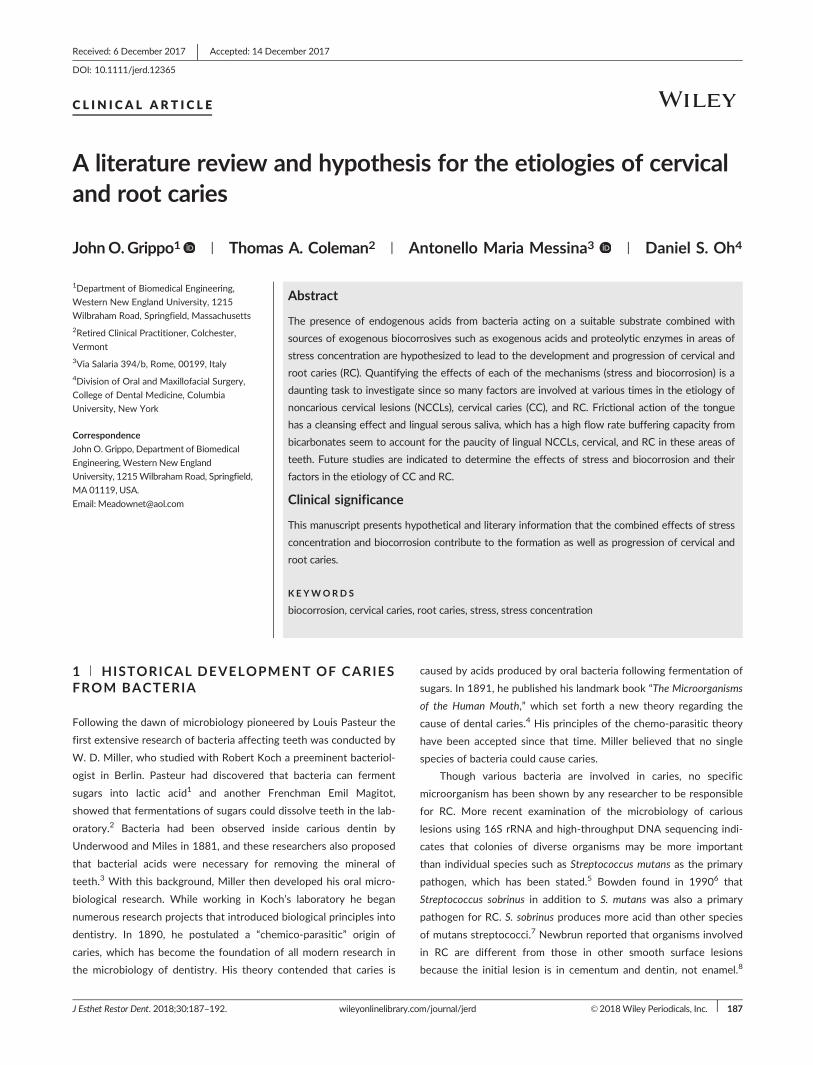
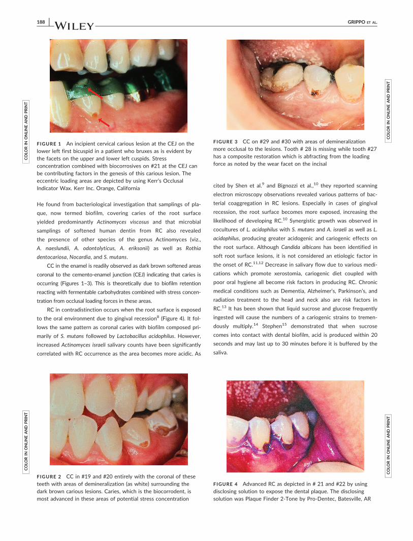
CC in the enamel is readily observed as dark brown softened areas
coronal to the cemento-enamel junction (CEJ) indicating that caries is
occurring (Figures 1–3). This is theoretically due to biofilm retention
reacting with fermentable carbohydrates combined with stress concen-
tration from occlusal loading forces in these areas.
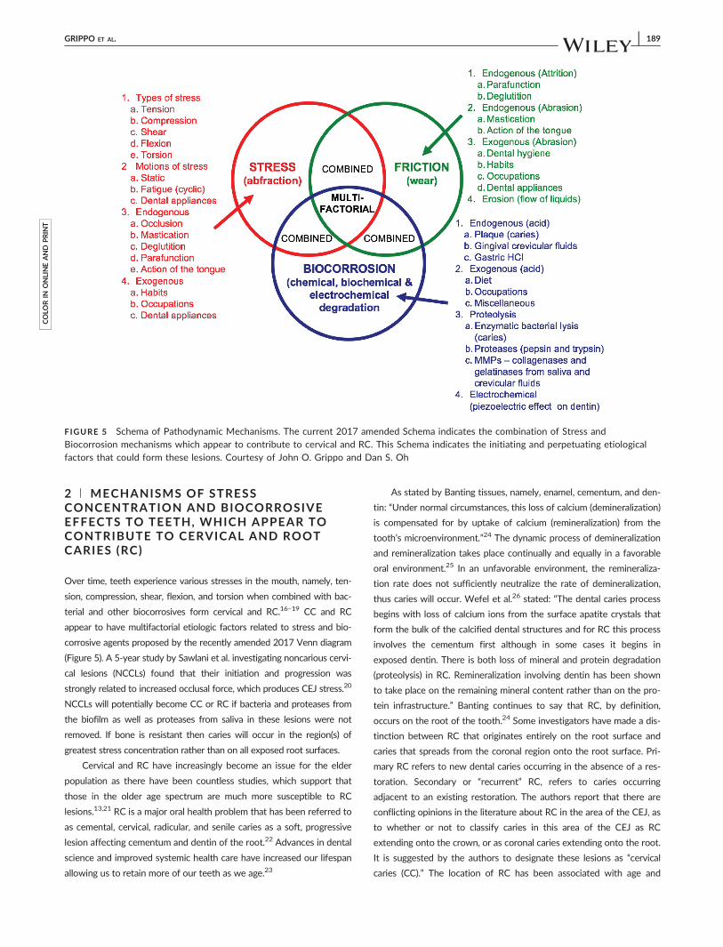
RC in contradistinction occurs when the root surface is exposed
to the oral environment due to gingival recession8 (Figure 4). It fol-
lows the same pattern as coronal caries with biofilm composed pri-
marily of S. mutans followed by Lactobacillus acidophilus. However,
increased Actinomyces israeli salivary counts have been significantly
correlated with RC occurrence as the area becomes more acidic. As
cited by Shen et al.9 and Bignozzi et al.,10 they reported scanning
electron microscopy observations revealed various patterns of bac-
terial coaggregation in RC lesions. Especially in cases of gingival
recession, the root surface becomes more exposed, increasing the
likelihood of developing RC.10 Synergistic growth was observed in
cocultures of L. acidophilus with S. mutans and A. israeli as well as L.
acidophilus, producing greater acidogenic and cariogenic effects on
the root surface. Although Candida albicans has been identified in
soft root surface lesions, it is not considered an etiologic factor in
the onset of RC.11,12 Decrease in salivary flow due to various medi-
cations which promote xerostomia, cariogenic diet coupled with
poor oral hygiene all become risk factors in producing RC. Chronic
medical conditions such as Dementia, Alzheimer’s, Parkinson’s, and
radiation treatment to the head and neck also are risk factors in
RC.13 It has been shown that liquid sucrose and glucose frequently
ingested will cause the numbers of a cariogenic strains to tremen-
dously multiply.14 Stephen15 demonstrated that when sucrose
comes into contact with dental biofilm, acid is produced within 20
seconds and may last up to 30 minutes before it is buffered by the
saliva.
COLO
RIN
ONLINEAND
PRIN
T
F IGURE 1 An incipient cervical carious lesion at the CEJ on thelower left first bicuspid in a patient who bruxes as is evident bythe facets on the upper and lower left cuspids. Stressconcentration combined with biocorrosives on #21 at the CEJ canbe contributing factors in the genesis of this carious lesion. Theeccentric loading areas are depicted by using Kerr’s OcclusalIndicator Wax. Kerr Inc. Orange, California
COLO
RIN
ONLINEAND
PRIN
T
F IGURE 2 CC in #19 and #20 entirely with the coronal of theseteeth with areas of demineralization (as white) surrounding thedark brown carious lesions. Caries, which is the biocorrodent, ismost advanced in these areas of potential stress concentration
COLO
RIN
ONLINEAND
PRIN
T
F IGURE 3 CC on #29 and #30 with areas of demineralizationmore occlusal to the lesions. Tooth # 28 is missing while tooth #27has a composite restoration which is abfracting from the loadingforce as noted by the wear facet on the incisal
COLO
RIN
ONLINEAND
PRIN
T
F IGURE 4 Advanced RC as depicted in # 21 and #22 by usingdisclosing solution to expose the dental plaque. The disclosingsolution was Plaque Finder 2-Tone by Pro-Dentec, Batesville, AR
188 | GRIPPO ET AL.
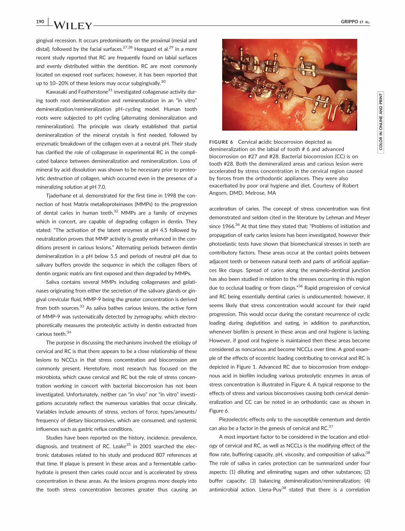
2 | MECHANISMS OF STRESSCONCENTRATION AND BIOCORROSIVEEFFECTS TO TEETH, WHICH APPEAR TOCONTRIBUTE TO CERVICAL AND ROOTCARIES (RC)
Over time, teeth experience various stresses in the mouth, namely, ten-
sion, compression, shear, flexion, and torsion when combined with bac-
terial and other biocorrosives form cervical and RC.16–19 CC and RC
appear to have multifactorial etiologic factors related to stress and bio-
corrosive agents proposed by the recently amended 2017 Venn diagram
(Figure 5). A 5-year study by Sawlani et al. investigating noncarious cervi-
cal lesions (NCCLs) found that their initiation and progression was
strongly related to increased occlusal force, which produces CEJ stress.20
NCCLs will potentially become CC or RC if bacteria and proteases from
the biofilm as well as proteases from saliva in these lesions were not
removed. If bone is resistant then caries will occur in the region(s) of
greatest stress concentration rather than on all exposed root surfaces.
Cervical and RC have increasingly become an issue for the elder
population as there have been countless studies, which support that
those in the older age spectrum are much more susceptible to RC
lesions.13,21 RC is a major oral health problem that has been referred to
as cemental, cervical, radicular, and senile caries as a soft, progressive
lesion affecting cementum and dentin of the root.22 Advances in dental
science and improved systemic health care have increased our lifespan
allowing us to retain more of our teeth as we age.23
As stated by Banting tissues, namely, enamel, cementum, and den-
tin: “Under normal circumstances, this loss of calcium (demineralization)
is compensated for by uptake of calcium (remineralization) from the
tooth’s microenvironment.”24 The dynamic process of demineralization
and remineralization takes place continually and equally in a favorable
oral environment.25 In an unfavorable environment, the remineraliza-
tion rate does not sufficiently neutralize the rate of demineralization,
thus caries will occur. Wefel et al.26 stated: “The dental caries process
begins with loss of calcium ions from the surface apatite crystals that
form the bulk of the calcified dental structures and for RC this process
involves the cementum first although in some cases it begins in
exposed dentin. There is both loss of mineral and protein degradation
(proteolysis) in RC. Remineralization involving dentin has been shown
to take place on the remaining mineral content rather than on the pro-
tein infrastructure.” Banting continues to say that RC, by definition,
occurs on the root of the tooth.24 Some investigators have made a dis-
tinction between RC that originates entirely on the root surface and
caries that spreads from the coronal region onto the root surface. Pri-
mary RC refers to new dental caries occurring in the absence of a res-
toration. Secondary or “recurrent” RC, refers to caries occurring
adjacent to an existing restoration. The authors report that there are
conflicting opinions in the literature about RC in the area of the CEJ, as
to whether or not to classify caries in this area of the CEJ as RC
extending onto the crown, or as coronal caries extending onto the root.
It is suggested by the authors to designate these lesions as “cervical
caries (CC).” The location of RC has been associated with age and
COLO
RIN
ONLINEAND
PRIN
T
F IGURE 5 Schema of Pathodynamic Mechanisms. The current 2017 amended Schema indicates the combination of Stress andBiocorrosion mechanisms which appear to contribute to cervical and RC. This Schema indicates the initiating and perpetuating etiologicalfactors that could form these lesions. Courtesy of John O. Grippo and Dan S. Oh
GRIPPO ET AL. | 189
gingival recession. It occurs predominantly on the proximal (mesial and
distal), followed by the facial surfaces.27,28 Heegaard et al.29 in a more
recent study reported that RC are frequently found on labial surfaces
and evenly distributed within the dentition. RC are most commonly
located on exposed root surfaces; however, it has been reported that
up to 10–20% of these lesions may occur subgingivally.30
Kawasaki and Featherstone31 investigated collagenase activity dur-
ing tooth root demineralization and remineralization in an “in vitro”
demineralization/remineralization pH–cycling model. Human tooth
roots were subjected to pH cycling (alternating demineralization and
remineralization). The principle was clearly established that partial
demineralization of the mineral crystals is first needed, followed by
enzymatic breakdown of the collagen even at a neutral pH. Their study
has clarified the role of collagenase in experimental RC in the compli-
cated balance between demineralization and remineralization. Loss of
mineral by acid dissolution was shown to be necessary prior to proteo-
lytic destruction of collagen, which occurred even in the presence of a
mineralizing solution at pH 7.0.
Tjaderhane et al. demonstrated for the first time in 1998 the con-
nection of host Matrix metalloproteinases (MMPs) to the progression
of dental caries in human teeth.32 MMPs are a family of enzymes
which in concert, are capable of degrading collagen in dentin. They
stated: “The activation of the latent enzymes at pH 4.5 followed by
neutralization proves that MMP activity is greatly enhanced in the con-
ditions present in carious lesions.” Alternating periods between dentin
demineralization in a pH below 5.5 and periods of neutral pH due to
salivary buffers provide the sequence in which the collagen fibers of
dentin organic matrix are first exposed and then degraded by MMPs.
Saliva contains several MMPs including collagenases and gelati-
nases originating from either the secretion of the salivary glands or gin-
gival crevicular fluid, MMP-9 being the greater concentration is derived
from both sources.33 As saliva bathes carious lesions, the active form
of MMP-9 was systematically detected by zymography, which electro-
phoretically measures the proteolytic activity in dentin extracted from
carious teeth.34
The purpose in discussing the mechanisms involved the etiology of
cervical and RC is that there appears to be a close relationship of these
lesions to NCCLs in that stress concentration and biocorrosion are
commonly present. Heretofore, most research has focused on the
microbiota, which cause cervical and RC but the role of stress concen-
tration working in concert with bacterial biocorrosion has not been
investigated. Unfortunately, neither can “in vivo” nor “in vitro” investi-
gations accurately reflect the numerous variables that occur clinically.
Variables include amounts of stress, vectors of force, types/amounts/
frequency of dietary biocorrosives, which are consumed, and systemic
influences such as gastric reflux conditions.
Studies have been reported on the history, incidence, prevalence,
diagnosis, and treatment of RC. Leake35 in 2001 searched the elec-
tronic databases related to his study and produced 807 references at
that time. If plaque is present in these areas and a fermentable carbo-
hydrate is present then caries could occur and is accelerated by stress
concentration in these areas. As the lesions progress more deeply into
the tooth stress concentration becomes greater thus causing an
acceleration of caries. The concept of stress concentration was first
demonstrated and seldom cited in the literature by Lehman and Meyer
since 1966.36 At that time they stated that: “Problems of initiation and
propagation of early caries lesions has been investigated, however their
photoelastic tests have shown that biomechanical stresses in teeth are
contributory factors. These areas occur at the contact points between
adjacent teeth or between natural teeth and parts of artificial applian-
ces like clasps. Spread of caries along the enamelo-dentinal junction
has also been studied in relation to the stresses occurring in this region
due to occlusal loading or from clasps.”36 Rapid progression of cervical
and RC being essentially dentinal caries is undocumented; however, it
seems likely that stress concentration would account for their rapid
progression. This would occur during the constant recurrence of cyclic
loading during deglutition and eating, in addition to parafunction,
whenever biofilm is present in these areas and oral hygiene is lacking.
However, if good oral hygiene is maintained then these areas become
considered as noncarious and become NCCLs over time. A good exam-
ple of the effects of eccentric loading contributing to cervical and RC is
depicted in Figure 1. Advanced RC due to biocorrosion from endoge-
nous acid in biofilm including various proteolytic enzymes in areas of
stress concentration is illustrated in Figure 4. A typical response to the
effects of stress and various biocorrosives causing both cervical demin-
eralization and CC can be noted in an orthodontic case as shown in
Figure 6.
Piezoelectric effects only to the susceptible cementum and dentin
can also be a factor in the genesis of cervical and RC.37
A most important factor to be considered in the location and etiol-
ogy of cervical and RC, as well as NCCLs is the modifying effect of the
flow rate, buffering capacity, pH, viscosity, and composition of saliva.38
The role of saliva in caries protection can be summarized under four
aspects: (1) diluting and eliminating sugars and other substances; (2)
buffer capacity; (3) balancing demineralization/remineralization; (4)
antimicrobial action. Llena-Puy38 stated that there is a correlation
COLO
RIN
ONLINEAND
PRIN
T
F IGURE 6 Cervical acidic biocorrosion depicted asdemineralization on the labial of tooth # 6 and advancedbiocorrosion on #27 and #28. Bacterial biocorrosion (CC) is ontooth #28. Both the demineralized areas and carious lesion wereaccelerated by stress concentration in the cervical region causedby forces from the orthodontic appliances. They were alsoexacerbated by poor oral hygiene and diet. Courtesy of RobertAngorn, DMD, Melrose, MA
190 | GRIPPO ET AL.
between pH changes in the plaque and sugar clearance from saliva.
Furthermore, pH recovery is not the same on all tooth surfaces.
Although saliva plays an important role in balancing the pH, it also
contains specific buffering components such as phosphates, bicarbon-
ates with proteins emanating from the salivary glands.39 Phosphate
buffer plays an essential role when salivary flow is low. Buffering mech-
anisms do not act equally on all tooth surfaces. Their effect is less on
interproximal surfaces and greater on the free surfaces which are cov-
ered by a thin layer of bacteria.40,41
Israel Kleinberg, a preeminent emeritus scholar of saliva, (SUNY
Stony Brook of NY, personal communication with JOG, 2006) avows
that there is five times more saliva on the lingual surfaces than in the
vestibule. That observation was first reported by Jenkins, et al.42 These
reputable sources of information support the contention that saliva,
particularly lingual serous saliva, which has a high flow rate and buffer-
ing capacity from bicarbonates accounts for the paucity of lingual
NCCLs, cervical, and RC on these areas of teeth. Furthermore, the fric-
tional action of the tongue also assists in cleansing the lingual surfaces.
3 | SUMMATION
In summation, little absolute direct evidence currently exists to fully
explain the interaction of the various co-variables that exist with
regards to the formation of root and CC. However, in light of the many
co-related studies that appear in the literature regarding the multiple
potential contributing factors of stress and biocorrosion for the etiol-
ogy of NCCLs, it appears that there is a strong likelihood that the
“missing link” to the initiation and rapid progression of cervical and RC
could well involve these same two mechanisms. Enzymatic breakdown
of CC and RC by proteolysis and stress concentration working in con-
cert with the biocorrosive effects of bacterial endogenous as well as
exogenous acidic agents suggests that these factors may all contribute
to these types of caries. Future research is needed to fully confirm or
refute the proposed mechanisms of stress and biocorrosion with their
various related factors in the etiology of CC and RC.
DISCLOSURE
The authors do not have any financial interest in the company whose
product is mentioned in this article.
ORCID
John O. Grippo http://orcid.org/0000-0002-8213-7849
Antonello Maria Messina http://orcid.org/0000-0003-4920-8032
REFERENCES
[1] Pasteur L. The Physiological Theory of Fermentation. University of
Adelaide, 1870.
[2] Magitot E. Treatise on Dental Caries. Houghton, MA, USA: Osgood
and Company Boston; 1878.
[3] Underwood, Miles. The British Journal of Dental Science. Vol. 43.
London: Oxford House. January–December 1905:3.
[4] Miller WD. The human mouth as a focus of infection. Dent Cosmos.
1891;33:689–913.
[5] Gross EM, Beall CJ, Kutsch SR, et al. Beyond Streptococcus Mutans:
dental caries onset linked to multiple species by 16 S.rRNA commu-
nity analysis. PLoS One, 2012;7(10):e47722. https://doi.org/10.137/
journal.pone.0047722
[6] Bowden GH. Microbiology of root surface caries in humans. J Dent
Res. 1990;69:205–210.
[7] De Soet JJ, Toor FA, de Graff J. Acidogenesis by oral streptococci
at different pH values. Caries Res. 1989;23:14–17.
[8] Newbrun E. Cariology. 3rd ed. Chicago, IL: Quintessence Interna-
tional Publishing Inc.; 1989.
[9] Shen S, Samaranayake LP, Yip HK. In vitro growth, acidogenicity
and cariogenicity of predominant human root caries flora. J Dent.
2004;32:667–668.
[10] Bignozzi I, Crea A, Capri D, et al. Root caries: a periodontal perspec-
tive. J Periodont Res. 2014;49:143–163.
[11] Powell LV, Leroux BG, Persson RE, Kiyak HA. Factors associated
with caries incidence in an elderly population. Commun Dent Oral
Epidemiol. 1998;26:170–176.
[12] Fure S. Ten-year cross-sectional and incidence study of coronal and
root caries and some related factors in elderly Swedish individuals.
Gerodontology, 2004;21:130–140.
[13] Bansal V, Sohi RK, Verresha KL, et al. Root caries: a problem of
growing age. J Indian Acad Dent Spec Res. 2011;2(2):43–45.
[14] Carlsson J, Sundstrom B. Variation in composition of early dental plaque
following the ingestion of sucrose and glucose.Odont Rev. 1968;12:1297.
[15] Stephen RM. Changes in hydrogen ion concentration on tooth
surfaces and in carious lesions. J Am Dent Assoc. 1940;27:718.
[16] Grippo JO, Simring M, Coleman TA. Abfraction, abrasion, biocorro-
sion and the enigma of noncarious cervical lesions: A 20–year per-
spective. J Esthet Restor Dent. 2012;24:10–13.
[17] Lee WC, Eakle WS. Possible role of tensile stress in the etiology of
cervical erosive lesions of teeth. J Prosthet Dent. 1984;52(3):374–380.
[18] Braem M, Lambrechts P, Vanherle G. Stress-induced cervical
lesions. J Prosthet Dent. 1992;67(5):718–722.
[19] Lee HE, Lin CL, Wang CH, et al. Stresses at the cervical lesion of
maxillary premolar–a finite element investigation. J Dent. 2002;30
(7–8):283–290.
[20] Sawlani K, Lawson NC, Burgess JO, et al. Factors influencing the
progression of noncarious cervical lesions: a 5-year prospective clin-
ical evaluation. J Prosthet Dent. 2016;115:571–577.
[21] Hazen SP, Chilton NW, Mumma RD. The problem of root caries:
review and clinical description. J Am Dent Assoc. 1973;86:137–144.
[22] Newman E, Armitage G, Daniels TE, et al. Root Caries. Calif Dent J.
1984;12:68–73.
[23] Griffin SO, Griffin PM, Swann JL, Zlobin N. Estimating rates of new
root caries in older adults. J Dent Res. 2004;83(8):634–638.
[24] Banting DW. The Diagnosis of Root Caries. J Dent Educ. 2001;65
(10):991–996.
[25] Koulourides T. Dynamics of biologic mineralization applied to dental
caries. In: L. Menker, ed. The biologic basis of dental caries. New
York: Harper Rowe Publishers; 1980.
[26] Wefel JS, Clarkson BH, Heilman JR. Natural root caries: a histology
and microradiographic evaluation. J Oral Pathol. 1985;14(8):615–623.
[27] Shacken MJ, Keljtjens HM, van der Hoeven JS. Effects of fluoride
and chlorhexidine on the microflora of dental root surfaces and pro-
gression of root- surface caries. J Dent Res. 1991;70(2):150–153.
GRIPPO ET AL. | 191
[28] Fure S. Five–year incidence of coronal and root caries in 60-, 70-,
and 80- year–old Swedish individuals. Caries Res. 1997;31:249–258.
[29] Heegaard KM, Holm-Pedersen P, Bardow A, et al. The Copenhagen
Oral Health Senior Cohort: design, population and dental health.
Gerodontology, 2011;28:165–176.
[30] Stamm JW, Banting DW, Imrey PB. Adult root caries survey of two
similar communities with contrasting natural water fluoride levels.
J Am Dent Assoc. 1990;120:143–149.
[31] Kawasaki K, Featherstone JD. Effects of Collagnease on root
demineralization. J Dent Res. 1997;76(1):588–595.
[32] Tjaderhane L, Larjava H, Sorsa T, et al. The activation and function
of host matrix metalloproteinases in dentin matrix breakdown in
caries lesions. J Dent Res. 1998;77(8):1622–1629.
[33] Ingman T, Sorsa T, Lindy O, et al. Multiple forms of gelatinases/
type 1V collagenases in saliva and gingival crevicular fluid of perio-
dontitis patients. J Clin Periodontol. 1994;21:26–31.
[34] Sorsa T, Ding YL, Ingman T, et al. Cellular source, activation and
inhibition of dental plaque collagenase. J Clin Periodontol. 1995;22:
709–717.
[35] Leake JL. Clinical decision making for caries management in root
surfaces. J Dent Educ. 2001;10:1147–1153.
[36] Lehman ML, Meyer ML. Relationship of dental caries and stress:
concentrations in teeth as revealed by photoelastic test. J Dent Res.
1966;45:1706–1714.
[37] Marino AA, Gross BD. Piezoelectricity in cementum, dentine and
bone. Arch Biol. 1989;4(7):7–9.
[38] Llena-Puy C. The role of saliva in maintaining oral health and as an
aid to diagnosis. Med Oral Patol Oral Cir Bucal. 2006;11(5):E449–455. PMID: 16878065
[39] Nauntofte B, Tenevuo JO, Lagerl€of F. Secretion and composition of
saliva. In: Fejerskov O, Kidd E, eds. Dental caries. The disease and
its clinical management. Oxford. Blackwell Munksgard; 2003:7–29.
[40] Axelson P. Internal modifying factors in dental caries. In: Axelson P,
ed. Diagnosis and caries risk prediction of dental caries. Vol. 2. Chi-
cago: Quintessence Publishing; 2000:91–150.
[41] Tenovuo J. Salivary parameters of relevance for assessing caries
activity in individuals and populations. Commun Dent Oral Epidemiol.
1997;25(1):82–6. Review. PMID: 9088696.
[42] Jenkins GN. Salivary effects on plaque pH. In: Kleinberg I, Ellison
SA, Mandel ID, eds. Saliva and dental caries. New York, NY: Infor-
mation Retrieval, Inc.; 1979 (Microbiology Abstracts):307–322.
How to cite this article: Grippo JO, Coleman TA, Messina AM,
Oh DS. A literature review and hypothesis for the etiologies of
cervical and root caries. J Esthet Restor Dent. 2018;30:187–192.
https://doi.org/10.1111/jerd.12365
192 | GRIPPO ET AL.