Embed Size (px)
Citation preview

JOHN J. WOZNIAK
A METHOD FOR THE SUTURELESS ANASTOMOSIS OF BLOOD VESSELS
Penetrating-type wounds that are inflicted during combat or accidents often result in trauma to blood vessels that can lead to morbidity or mortality if anastomosis (rejoining) is not performed rapidly. APL, in collaboration with the Johns Hopkins Medical Institutions and the University of Maryland, has developed a unique vascular-everting instrument and a biocompatible, heat-shrinkable sleeve that are used in a procedure to accomplish end-to-end anastomoses without the need for sutures.
INTRODUCTION
Anastomosis of blood vessels was successfully performed by A. Carrel in the early 1900s; 1 however, it was not until after World War 11, 2 in the Korean and Vietnam wars,3 that the techniques of vascular surgery and the training of its practitioners were developed to the point where vascular surgery became standard practice for major trauma. In the case of wounds of the extremities, this resulted in a tremendous increase of personnel returning to duty who, in earlier years, would have undergone amputation. Two key problems in vascular surgery that must be faced when dealing with a large number of combat casualties are the availability of surgeons skilled in needed techniques and the amount of time required to perform each surgical procedure.
Vascular anastomosis with sutures is accomplished with a curved needle. The sutures must be placed precisely, piercing the adventitia (fibrous sheath) from the outside and the intima (innermost tissue layer) from the inside at several locations for proper vessel approximation (tissue aligned and in good contact). Either individual or continuous-running sutures are used. Typically, for an artery with a 3-mm lumen, approximately 20 stitches are taken around the circumference using 6-0 (O. l-mm diameter) thread. Usually a single end-to-end anastomosis on vessels of this size cannot be performed in less than 20 min.
The Naval Medical R&D Command was approached with the concept of a sutureless anastomosis, and a collaboration was formed among APL, the Johns Hopkins Medical Institutions, and the University of Maryland for its implementation. Figure 1 shows the steps involved in the sutureless anastomosis technique. Figure 1a shows the severed vessel, which characteristically is constricted and retracted by the smooth muscle (media) within the vessel wall. In the first step, vascular clamps are applied, and the vessel is rinsed with saline to dislodge any clots or tissue from the lumen. A visual estimate is made of the size, and ferrules are selected that approximately match the outside diameter of the vessel.
Each vessel end is then everted over a ferrule (Fig. 1b). Everting fully opens the vessel and ensures continuous in-. timal contact, which is vital to prevent thrombosis formation after the anastomosis is completed.
Johns Hopkins APL Technical Digest, Volume 9, Number 1 (1988)
(a)
(b)
(d)
Figure 1-Sutureless vascular anastomosis.
In the next step, a sleeve, fabricated from a low-temperature, heat-shrinkable biocompatible polymer, is placed on one of the vessel ends. The two ends of the vessel are brought together, and the sleeve is centered over the junction (Fig. 1c). Warm saline is used to contract the sleeve and bind the everted vessel sections together (Fig. 1d).
35
Wozniak - A Method for the Sutureless Anastomosis of Blood Vessels
Finally, the proximal clip is removed (Fig. Id), and a hypodermic needle is inserted into the vessel lumen to remove any entrapped air. When a steady flow of blood through the hypodermic needle is achieved, the needle and distal clip are removed.
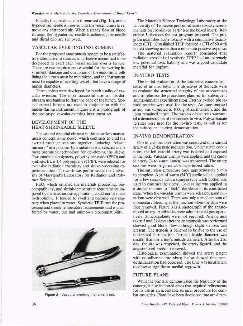
VASCULAR-EVERTING INSTRUMENT
For the proposed anastomosis system to be a satisfactory alternative to sutures, an effective means had to be developed to evert each vessel section over a ferrule. There are two requirements essential for the everting instrument: damage and disruption of the endothelial cells lining the lumen must be minimized, and the instrument must be capable of everting vessels that have a range of lumen diameters.
Three devices were developed for bench studies of vascular eversion. The most successful uses an iris-diaphragm mechanism to flare the edge of the lumen. Special curved forceps are used in conjunction with the lumen-flaring instrument. Figure 2 is a photograph of the prototype vascular -everting instrument set.
DEVELOPMENT OF THE HEAT -SHRINKABLE SLEEVE
The second essential element in the sutureless anastomosis concept is the sleeve, which contracts to bind the everted v.ascular sections together. Inducing "elastic memory" in a polymer by irradiation was selected as the most promising technology for developing the sleeve. Two candidate polymers, polyethylene oxide (pEa) and synthetic trans-l,4 polyisoprene (TPIP), were selected for extensive radiation chemistry and sleeve-processing experimentation. The work was performed at the University of Maryland's Laboratory for Radiation and Polymer Science. 4
PEa, which satisfied the materials processing, biocompatibility, and shrink-temperature requirements imposed by the anastomosis application, could not be made hydrophobic. It tended to swell and become very slippery when placed in water. Synthetic TPIP met the processing and shrink-temperature requirements and is unaffected by water, but had unknown biocompatibility.
Figure 2-Vascular-everting instrument set.
36
The Materials Science Toxicology Laboratory at the University of Tennessee performed acute toxicity screening tests on crosslinked TPIP (see the boxed insert). Reference 5 discusses the test program protocol. The program quantifies acute toxicity with a cumulative toxicity index (CTI). Crosslinked TPIP received a CTI of 50 with no test showing more than a minimum positive response.
The material evaluation report 6 concluded that radiation-crosslinked synthetic TPIP had an extremely low potential toxic liability and was a good candidate material for implant.
IN-VITRO TESTS The initial evaluation of the sutureless concept con
sisted of in-vitro tests. The objectives of the tests were to evaluate the structural integrity of the anastomosis and to rehearse the procedures planned for subsequent animal-implant experimentation. Freshly excised pig carotid arteries were used for the tests. An anastomosed artery was subjected to 1 ()()() g of axial tension, and the joint remained intact. The success of the tests warranted a demonstration of the concept in vivo. Polycarbonate ferrules were used for the in-vitro tests, as well as for the subsequent in-vivo demonstration.
IN-VIVO DEMONSTRATION
One in-vivo demonstration was conducted on a carotid artery of a 25-kg male mongrel dog. Under sterile conditions, the left carotid artery was isolated and exposed in the neck. Vascular clamps were applied, and the carotid artery (3- to 4-mm lumen) was transected. The artery sections were irrigated with heparinized saline.
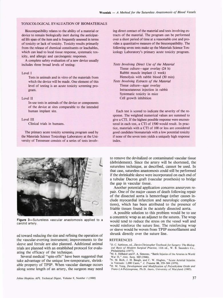
The sutureless procedure took approximately 5 min to complete. A jet of warm (54°C) sterile saline, applied for a few seconds with a squeeze-type wash bottle, was used to contract the sleeve. Cold saline was applied in a similar manner to "lock" the sleeve in its contracted state. When the vascular clamps were released, good pulsations were observed. There was only a small amount of momentary bleeding at the junction when the clips were first removed. Figure 3 is a photograph of the anastomosed artery. Antibiotics were administered postoperatively; anticoagulants were not required. Angiograms taken 5 and 21 days after the anastomosis was performed showed good blood flow although slight stenosis was present. The stenosis is believed to be due to the use of undersized ferrules (the ferrule's inside diameter was smaller than the artery's outside diameter). After the 21st day, the site was reopened, the artery ligated, and the anastomosed section removed.
Histological examination showed the artery patent with no adherent thrombus; it also showed that reendothelialization had occurred. The time was insufficient to observe significant medial regrowth.
FUTURE PLANS While the one trial demonstrated the feasibility of the
concept, it also indicated areas that required refinements for its use as an acceptable surgical procedure for combat casualties. Plans have been developed that are direct-
fohn s Hopkins APL Technical Digest, Volume 9, Number I (1988)
Wozniak - A Method jor the Sutureless Anastomosis oj Blood Vessels
TOXICOLOGICAL EVALUATION OF BIOMA TERIALS
Biocompatibility relates to the ability of a material or device to remain biologically inert during the anticipated life span of the item and is frequently assessed in terms of toxicity or lack of toxicity. Toxicity results primarily from the release of chemical constituents or leachables, which can lead to local tissue response, systematic toxicity, and allergic and carcinogenic responses.
A complete safety evaluation of a new device usually includes three broad levels of testing:
Level I Tests in animals and in vitro of the materials from which the device will be made. One element of this level of testing is an acute toxicity screening program.
Level II In-use tests in animals of the device or components of the device at sites comparable to the intended human implant site.
Level III Clinical trials in humans.
The primary acute toxicity screening program used by the Materials Science Toxicology Laboratory at the University of Tennessee consists of a series of tests involv-
Figure 3-Sutureless vascular anastomosis applied to a carotid artery .
ed toward reducing the size and refining the operation of the vascular-everting instrument; improvements to the sleeve and ferrule are also planned. Additional animal trials are planned with an established protocol for evaluating the efficacy of the technique.
Several medical "spin-offs" have been suggested that take advantage of the unique low-temperature, shrinkable property of TPIP. When vascular damage occurs along some length of an artery, the surgeon may need
Johns Hopkins APL Technical Digest, Volume 9, Number 1 (1988)
ing direct contact of the material and tests involving extracts of the material. The program can be performed over a short period of time at a reasonable cost and provides a quantitative measure of the biocompatibility. The following seven tests make up the Materials Science Toxicology Laboratory's primary acute toxicity program.
Tests Involving Direct Use of the Material Tissue culture-agar overlay (24 h) Rabbit muscle implant (l week) Hemolysis with rabbit blood (30 min)
Tests Involving Extracts of the Material Tissue culture-agar overlay Intracutaneous injection in rabbit Systematic toxicity in mice Cell growth inhibition
Each test is scored to indicate the severity of the response. The weighted numerical values are summed to give a CTI. If the highest possible response were encountered in each test, a CTI of 1500 would result. In practice, materials with a CTI of 100 or less are considered good candidate biomaterials with a low potential toxicity if none of the seven tests yields a uniquely high response index.
to remove the devitalized or contaminated vascular tissue (debridement). Since the artery will be shortened, the sutureless technique, as described, cannot be used. In that case, sutureless anastomosis could still be performed if the shrinkable sleeve were incorporated on each end of a tubular Dacron graft (vascular prosthesis) to bridge the gap in vascular tissue.
Another potential application concerns aneurysm repair. One of the major causes of death following repair of the dissected aorta is hemorrhage (other causes include myocardial infarction and neurologic complications), which has been attributed to the presence of friable tissues found in the acutely dissected aorta.
A possible solution to this problem would be to use a concentric wrap as an adjunct to the sutures. The wrap would tend to reduce the stress on the vessel wall and would reinforce the suture line. The reinforcing wrap or sleeve would be woven from TPIP monofIlament and shrunk directly over the suture line.
REFERENCES 1 D. C. Sabiston, ed., Davis-Christopher Textbookjor Surgery: The Biological Basis oj Modern Surgical Practice, 11th ed ., W. B. Saunders Co ., Philadelphia (1977).
2M. E. DeBaker and F. A. Simeone, "Battle Injuries of the Arteries in World War II," Ann. Surg. 123 (1946).
3N. M. Rich , 1. H. Baugh, and C. W . Hughes, "Acute Arterial Injuries in Vietnam: 1,000 Cases," J. Trauma 10, 359- 369 (1970).
4H . M. Yang, Development and Modification oj Polyethylene Oxide and Trans-l,4-Polyisoprene, Ph.D . thesis, University of Maryland (1985).
37
Wozniak - A Method jor the Sutureless Anastomosis oj Blood Vessels
5 J . Autian, "Toxicological Evaluation of Biomaterials: Primary Acute Toxicity Screening Program," Artif Organs 1, 53-60 (1977) .
6 J. Autian, private correspondence, " Report on Toxicity of Synthetic Trans-l,4 Polyisoprene under Project Number PT 0.1921, " Materials Science Toxicology Laboratory, University of Tennessee (1984).
THE AUTHOR
ACKNOWLEDGMENTS-This work was supponed by the Naval Medical R&D Command and APL's Independent Research and Development funds . The author wishes to acknowledge the contributions of J . Silverman and M. Yank at the University of Maryland, M. Epstein at the Johns Hopkins Medical Institutions, and R. Flower and D. Walters (retired) at APL.
JOHN J. WOZNIAK received a B.S. degree in mechanical engineering from Lowell Technological Institute in 1966 and the M .S. degree in mechanical engineering from the University of New Hampshire in 1971. He joined APL in 1973 and during 1973-79 worked as a specialist in submarine dynamics and missile underwater flight in the Strategic Systems Department. In 1979, he joined the Aeronautics Department to work on a variety of problems involving hydrodynamics and aerodynamics. In 1982, Mr. Wozniak became associated with the Biomedical Program when he initiated the
collaborative effort to investigate the sutureless anastomosis technique. He holds a number of patents that have medical and biomaterial applkations. Mr. Wozniak is now on the Principal Professional Staff at APL and is section supervisor of the Applied Fluid Dynamics Section.
38 Johns Hopkins APL Technical Digest, Volume 9, Number 1 (198.8)

![Experimental Model for Sutureless Proximal Anastomosis by ...This technique was initially described by Alexis Carrel, in 1902[1], and despite several technical improvements it has](https://img.pdfslide.net/doc/110x75/5f0d578e7e708231d439dfa2/experimental-model-for-sutureless-proximal-anastomosis-by-this-technique-was.jpg)