Embed Size (px)
Citation preview
Page 1 Lovato et al.
A Molecular Mechanism of Temperature Sensitivity for Mutations Affecting the Drosophila Muscle Regulator
Myocyte Enhancer Factor-2
TyAnna L. Lovato, Melanie M. Adams, Phillip W. Baker and Richard M. Cripps1
Dept. of Biology, University of New Mexico, Albuquerque, New Mexico 87131, USA
Running title: Temperature sensitive Mef2 mutants in Drosophila
1 Corresponding author: +1 (505) 277-2822
E-mail: [email protected]
Genetics: Published Articles Ahead of Print, published on June 29, 2009 as 10.1534/genetics.109.105056
Page 2 Lovato et al.
Abstract
Temperature sensitive (TS) mutations are a useful tool for elucidating gene function where a
gene of interest is essential at multiple stages of development. However, the molecular
mechanisms behind TS alleles vary. TS mutations of the myogenic regulator Myocyte
enhancer factor-2 (MEF2) in Drosophila arise in the heteroallelic combination Mef230-5/Mef244-5.
We show that the 30-5 mutation affects the N-terminal MADS domain, causing impaired DNA
binding ability and failure of homozygous mutants to survive to adulthood. The 44-5 mutation
deletes a downstream splice acceptor site, and results in a truncated protein that is unable to
activate MEF2 targets. 44-5 homozygotes consequently show severely impaired myogenesis
and die as embryos. We propose that in heteroallelic mutants at the permissive temperature,
30-5/44-5 heterodimers form and have a sufficiently stable interaction with DNA to activate
myogenic gene expression; at the restrictive temperature, 44-5 homodimers displace 30-5/44-
5 heterodimers from target genes, thus acting in a dominant-negative manner. To test this, we
show that 30-5/44-5 heterodimers can form, and we study additional Mef2 alleles for
complementation with the 30-5 allele. An allele affecting the DNA binding domain fails to
complement 30-5, whereas two alleles affecting downstream residues show temperature-
dependent complementation. Thus, by combining one MEF2 isoform having weakened DNA
binding ability with a second truncated MEF2 mutant that has lost its activation ability, a TS
form of intragenic complementation can be generated. These findings will provide new insight
and guidance into the functions of dimeric proteins, and how they might be engineered to
generate TS combinations.
Page 3 Lovato et al.
Introduction
Temperature sensitive (TS) mutants have a noted history in defining gene function. Using TS
alleles, geneticists are able to attenuate gene function at a specific time and then observe the
phenotypic effects of this genetic manipulation. TS mutants have been used for such diverse
studies as identifying yeast genes critical in the various stages of the cell cycle (HARTWELL et
al. 1970), and in metazoans to dissect the requirements for genes which function at multiple
developmental stages (see for example COX and BAYLIES 2005). This powerful tool has
proven especially useful in identifying and categorizing genes which function at multiple stages
to support organism viability.
Defining the molecular mechanisms behind temperature-sensitivity enables us to more
thoroughly understand the synthesis, folding, collaboration and function of a protein of interest.
SADLER and NOVICK (1964) categorized bacteriophage TS mutants by the mechanism of
their dysfunction (SADLER AND NOVICK 1964). The TL (thermolabile) class of mutants
demonstrated a mutant phenotype when grown at a restrictive temperature or when shifted to
a restrictive temperature during a later stage of development. The mutant phenotype in this
instance was usually due to the destabilization of the encoded protein, and the subsequent
loss of protein function as a result of increased temperature. This could be due to decreased
melting temperature from the loss of an hydrophobic amino acid, or due to decreased ability to
interact with DNA or other proteins as dimers or multimers.
Another category of TS mutants, TSF (temperature sensitive folding) only showed a
phenotype if incubated at the restrictive temperature during synthesis of the encoded protein. If
Page 4 Lovato et al.
the mutants were shifted to the restrictive temperature later, the protein did not lose
conformation and no phenotype was apparent. These mutations were recessive and did not
demonstrate intragenic complementation as did the thermolabile types. It was postulated that
many mutants in this class affect the formation, folding or initial integration of the encoded
protein into larger complexes; once the complex is formed, the mutant protein is stabilized by
its interactions with other subunits (GORDON and KING 1993; EDGAR and LIELAUSIS 1963).
Clearly, the mechanism of TS for a particular protein is strongly dependent upon its function as
part of a multimeric or macromolecular complex, and upon its half-life in the cell.
TS mutants have generally been isolated by screening through large numbers of
organisms that have undergone random mutagenesis. More recently, researchers have looked
for ways to engineer TS proteins. One approach was to introduce charged amino acids into the
internal region of the CcdB toxin encoded by the F’ plasmid (CHAKSHUSMATHI et al. 2004).
CcdB is a good candidate for TS screening because when transformed into Escherichia coli, it
is lethal when functional. Several of the introduced mutations successfully destabilized the
protein at the restrictive temperature; however this approach was not always fully effective.
Clearly, the engineering of new TS mutants will benefit from further characterization of the
molecular basis of existing TS alleles.
We have recently described the isolation of a temperature-sensitive combination of
alleles for the muscle transcription factor Myocyte enhancer factor 2 (MEF2; BAKER et al.
2005). Vertebrates possess four MEF2 genes, all of which contain a highly conserved, N-
terminal MADS domain [MCM1, Agamous, Deficiens, serum response factor (SRF)] spanning
amino acids 1 through 57, and a 29 amino acid MEF2 domain immediately adjacent (reviewed
in BLACK and OLSON 1998; BLACK and CRIPPS 2009). Both mutagenesis studies and
Page 5 Lovato et al.
biophysical determination of the solution structure of MEF2A, have clearly shown that the
MADS domain is required for DNA binding; also, a portion of the MADS domain (residues 35-
50) along with the adjacent MEF2 domain, is required for proper dimerization (MOLKENTIN et
al. 1996a; SANTELLI and RICHMOND, 2000; HUANG et al. 2000).
Upon dimerization and DNA binding, MEF2 is a potent activator of target structural
genes, both directly and in complex with other factors (MOLKENTIN et al. 1995; HAMAMORI
et al. 1997; KELLY TANAKA et al. 2008). Much of this transcriptional activation ability of MEF2
proteins arises from the function of residues located C-terminal to the MEF2 domain, which are
both required and sufficient for MEF2 transcriptional activity (MARTIN et al., 1994; WONG et
al., 1995; MOLKENTIN et al., 1995). Clearly, MEF2 is an highly modular protein with distinct
functional domains.
Drosophila contains a single Mef2 gene which is expressed in muscles at all stages of
development (NGUYEN et al. 1994; LILLY et al, 1994; TAYLOR et al, 1995; BAKER et al.
2005). Mef2 null mutants do not survive beyond embryogenesis and display a profound lack of
differentiated muscle (BOUR et al., 1995; LILLY et al., 1995), making this system useful for the
study of MEF2 protein function. In this paper, we define a molecular mechanism for the
temperature sensitivity of a subset of Drosophila Mef2 mutant alleles. We show that a mutation
affecting the MADS domain (allele 30-5) can complement a mutation affecting C-terminal
residues (allele 44-5), but that this complementation is thermolabile. We provide evidence that
the thermolability arises from competition between the attenuated DNA binding ability of the
30-5 isoform, and the strong DNA binding ability of the 44-5 isoform which acts as a dominant-
negative factor at the restrictive temperature. Our analyses of several additional Mef2 mutants
confirm this model. These studies provide important new insight into critical regions of the
Page 6 Lovato et al.
MEF2 factor, and more broadly contribute to our understanding of the molecular basis of TS
mutants.
Materials and Methods
Drosophila stocks and crosses: Drosophila stocks were grown on Carpenter’s medium
(CARPENTER 1950) at the indicated temperatures. Mef2 mutants were obtained from Elliot
Goldstein (Arizona State University; GOLDSTEIN et al. 2001) and balanced over a CyO, Cy
Kr-GFP balancer chromosome (CASSO et al. 1999). Viability studies were achieved by
crossing CyO, Cy Kr-GFP / Mef2x with CyO, Cy Kr-GFP / Mef2y. We counted the number of
heteroallelic escaper adults and the total number of progeny. Viability counts reflect the
percentage of heteroallelic escapers detected, as a proportion of the number of such escapers
expected if their viability were the same as their heterozygous siblings.
Immunohistochemistry: Embryos were collected and stained as described by PATEL (1994).
Primary antibodies were rabbit anti-Myosin heavy-chain (1:250; (KIEHART and FEGHALI
1986), mouse anti –GFP (1:250; INVITROGEN CORP.), and rat anti-Tropomyosin (1:250;
PECKHAM et al., 1992, ABCAM IMMUNOCHEMICALS). Secondary detection was using the
Vectastain Elite kit (VECTOR LABORATORIES) and diaminobenzidine (DAB) stain. Samples
were cleared in glycerol and mounted for photography with an Olympus BX51
photomicroscope using DIC optics. Images were collated using Adobe Photoshop.
DNA and RNA methods: To identify the lesions associated with each Mef2 allele, RNA was
purified from larvae of the genotype CyO, Cy Kr-GFP / Mef2x. The RNA was purified using the
Qiagen RNeasy Mini kit and RNase-free DNase set, and then subjected to reverse
Page 7 Lovato et al.
transcription followed by PCR, using Invitrogen Superscript III One-Step RT-PCR. Primers for
the PCR were (5’-ATGGGCCGCAAAAAAATTCAAATATC-3’) and (5’-
CTATGTGCCCCATCCGCC-3’), which were designed to amplify the entire Mef2 coding
region. PCR products were cloned into the pGEM-T Easy vector (Promega Corp.), and twelve
positive clones for each genotype were sequenced in their entirety, to ensure that a cDNA
arising from the mutant Mef2 allele was sequenced several times. In all cases, we found
several wild-type cDNA clones and several clones which consistently showed a departure from
wild-type.
To confirm the sequence alterations observed in the cDNA as arising from the Mef2
mutant allele, we isolated DNA from heterozygotes and amplified the appropriate genomic
region by PCR. Primers used to characterize 44-5 splice variants were (5’-
GCTGGAGATGTCGAACG-3’ and 5’-CTGCATATCCCACATCATCC-3’). For all mutants
characterized, the lesion at the DNA level matched or accounted for the changes observed in
the cDNAs.
Electrophoretic mobility shift assays: Wild-type MEF2 protein was generated from pSK-
DMef2 (LILLY et al., 1995) using T3 RNA polymerase and the TnT Coupled
Transcription/Translation system (PROMEGA). For mutant MEF2 proteins, the appropriate
sequence change was introduced into pSK-DMef2 using the Invitrogen Gene-Tailor Site
Directed Mutagenesis Kit, and confirmed by sequencing. Protein was then generated as
described for wild-type. In order to use equivalent amounts of protein for wild-type and mutant
shifts, parallel reactions were set up so that wild-type and mutant proteins were generated in
the presence of either radioactive 35S-methionine, or non-radioactive methionine. Next,
radioactive reactions were analyzed by SDS-PAGE and the radioactive bands corresponding
Page 8 Lovato et al.
to full-length MEF2 protein were quantified using the Cyclone Packard Phosphorimager. Based
upon the efficiency of MEF2 protein generation as determined in the radioactive reactions, the
volume of non-radioactive MEF2 lysate was correspondingly adjusted in the gel shift reactions
to ensure that equal amounts of protein were used. Differences in the volumes used of
programmed lysate were corrected by addition of unprogrammed lysate.
Electrophoretic mobility shift assays were set up according to standard methods
(SAMBROOK et al.,1998; KELLY TANAKA et al., 2008). For analysis of the individual DNA
binding ability of MEF2 isoforms, reactions were incubated in water baths at either 18ºC or
29ºC, and the gels were run at a constant temperature of 18ºC or 29ºC by circulating water of
the appropriate temperature through the electrophoresis rig. The dimerization assays were
carried out at room temperature. After running, gels were dried and radioactive bands were
quantified using the Packard Cyclone Phosphorimager. Data shown represent an average of at
least two reactions for each mutant compared to wild-type at each temperature.
Cell culture and transfections: For a wild-type expression plasmid we used pPac-Pl-Mef2
(KELLY TANAKA et al., 2008). Mutant expression plasmids were generated by sub-cloning the
mutant cDNAs into pPac-Pl. The reporter plasmid used was the -593/+2 Actin 57B-lacZ
construct described by KELLY et al. (2002). Transfections and analyses were as described in
KELLY TANAKA et al. (2008). All experiments were carried out in triplicate, and the average
fold activation for each experiment is reported.
Page 9 Lovato et al.
Results
Temperature dependent phenotypes of 30-5 and 44-5 alleles: In order to define the basis
of the 30-5/44-5 temperature-sensitivity, we first studied the extent of embryonic muscle
differentiation in homozygotes for each mutant Mef2 allele. These studies were performed at
18ºC and 29ºC, and were compared to wild-type and to the heteroallelic combination that we
had previously described (BAKER et al., 2005).
At stage 16, wild-type embryos stained for the accumulation of Tropomyosin showed
extensive skeletal muscle differentiation at both temperatures (Figure 1 A, B); and the
heteroallelic combination showed relatively normal patterning of skeletal muscles at the
permissive temperature, but a strong hypomorphic phenotype at the restrictive temperature
(Figure 1C, D).
For 30-5 homozygotes, muscle differentiation appeared relatively normal at both
temperatures (Figure 1 E, F), suggesting that this allele on its own had significant wild-type
function, and did not show a strong temperature dependence in its activity. Given the relatively
normal pattern of muscles in 30-5 animals, we determined if they showed survival to adulthood
at either 18ºC or 29ºC. 30-5 homozygotes never showed viability to the adult stage, although
this could arise from second-site mutations that might have accumulated on the 30-5
chromosome. We therefore tested the viability of mutants heteroallelic for 30-5 and the Mef2
null allele P544 (LILLY et al., 1995). Since P544 was isolated in a different genetic screen to
the GOLDSTEIN et al. (2001) alleles, the P544 chromosome is unlikely to share any second-
site lethal mutations with 30-5. We found in this instance that 13% of 30-5/P544 mutants
survived to adulthood at the permissive temperature (12 observed out of 89 expected) but
none survived at the restrictive temperature (0 observed, 238 expected). We conclude that the
Page 10 Lovato et al.
30-5 allele is a mild hypomorphic mutation, which shows a slight attenuation in its function with
increasing temperature. This mild temperature-dependent effect does not account for the
profound TS observed in 30-5/44-5 mutants.
For the 44-5 allele, homozygotes showed severe defects in muscle development at both
temperatures (Figure 1 G, H). Again, there was not a strong effect of temperature upon the
severity of the embryonic phenotype.
We conclude that these two alleles, while individually not strongly temperature-
dependent, somehow interacted to generate a temperature-dependent effect in heteroallelic
combinations. We note that at the permissive temperature the 30-5/44-5 combination appears
phenotypically similar to the 30-5 homozygotes, whereas at the restrictive temperature the
heteroallelic combination appears similar to the 44-5 mutants. Thus we reasoned that the 44-5
allele has an antimorphic effect, but only at the restrictive temperature, and only in combination
with 30-5 mutants.
Sequence analysis of 30-5: Our next goal was to understand the molecular mechanism
underlying this temperature sensitive effect. We isolated RNA from 30-5/+ heterozygotes and
carried out an RT-PCR, amplifying Mef2 cDNA using primers flanking the coding region. We
cloned and sequenced the resulting products, and discovered a point mutation that was
consistently observed in approximately half of the sequenced cDNAs. We found that the 30-5
cDNA contained a G to A point mutation in the MADS box (Figure 2A) resulting in an alanine to
threonine conversion at amino acid 32 (Figure 2C). The same mutation was observed in
clones of genomic DNA isolated from 30-5/+ heterozygotes. Given that 30-5 homozygotes
develop a fairly normal pattern of skeletal muscles, we hypothesized that 30-5 mutants were
Page 11 Lovato et al.
capable of forming MEF2 dimers and activating downstream target genes, albeit with reduced
efficiency compared to wild-type.
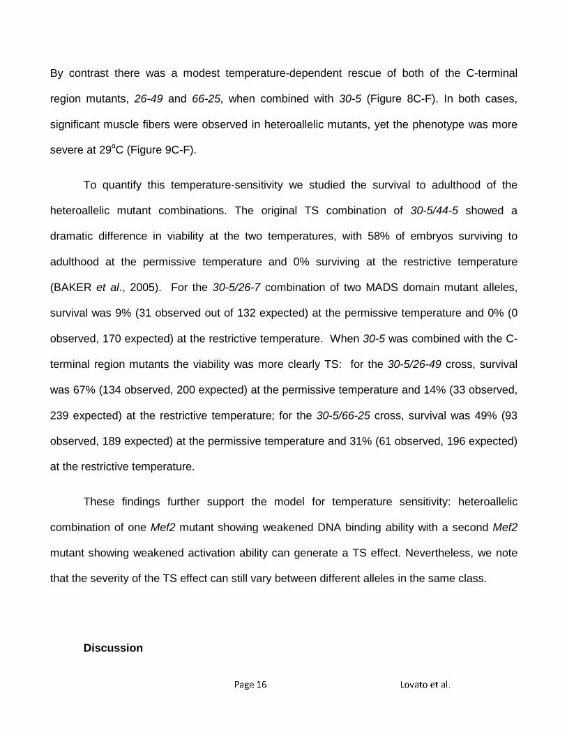
The 30-5 protein demonstrates decreased DNA binding ability: To test this hypothesis, we
performed an electrophoretic mobility shift assay (EMSA) using a radioactively labeled Actin
57B MEF2 site as a probe. The reaction was carried out at both 18ºC and 29ºC, and equal
amounts of wild-type and 30-5 protein were used for binding reactions (see Materials and
Methods for details). The results demonstrated that the 30-5 isoform consistently showed a
reduced ability to bind to the probe relative to wild-type MEF2. However, this reduction in DNA
binding was not affected by temperature (Figure 3A, B). In cotransfection assays 30-5 was
able to activate Act57B in cell culture at both temperatures equally well (data not shown).
These experiments, along with the phenotypic characterization of terminal differentiation in 30-
5 mutants, support the idea that the 30-5 isoform in isolation is capable of activating the
muscle differentiation pathway. However, due to its decreased DNA binding ability, the
pathway is not activated robustly enough in vivo for the animal to survive to adulthood.
Sequence analysis and functional characterization of 44-5 isoforms: We next isolated
RNA from 44-5/+ heterozygotes and cloned and sequenced Mef2 cDNAs as performed
previously for 30-5. We found two distinct mutant transcripts, 44-5 (7) and 44-5 (11) (Figure 2
B, C), both of which lacked the normal downstream splice acceptor site of exon 8. The 44-5 (7)
transcript was missing the first 44 bases of exon 8 and contained extra nucleotides that did not
match the Mef2 coding sequence, but did match sequence from intron 7. The 44-5 (11)
transcript lacked the first 51 bases of exon 8. We then analyzed Mef2 genomic clones and
discovered that in the 44-5 allele there was a 44 bp deletion which removed the exon 8 splice
Page 12 Lovato et al.
acceptor site and a portion of exon 8 coding sequence; all but the last 3bp of intron 7 were
retained.
The effects of these altered transcripts upon the encoded MEF2 protein are potentially
profound (Figure 2C). The 44-5 (11) variant predicts a 22 amino acid internal deletion; and the
44-5 (7) splice variant encodes a polypeptide with a large C-terminal deletion. Both of these
isoforms contain mutations in a region that has not been fully characterized, but which may
encode the activation domain of MEF2. We predicted that 44-5 protein isoforms would be able
to bind to DNA, but we were uncertain if the binding ability of the truncated 44-5 (7) isoform
would be reduced at the restrictive temperature. We found that each of the 44-5 (7) and 44-5
(11) isoforms bound to the Act57B MEF2 site at 18ºC and 29ºC extremely well (Figure 4A, B).
In fact, the 44-5 (11) isoform consistently bound to DNA even more effectively that wild-type
MEF2.
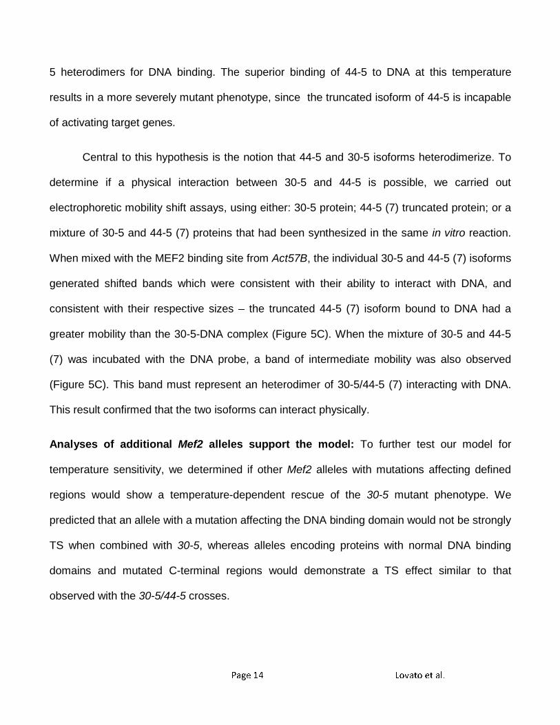
Since DNA binding ability of the 44-5 isoforms was not reduced, but terminal
differentiation of 44-5 homozygotes was severely affected at both temperatures, we concluded
that the 44-5 isoforms must not be able to activate downstream target genes. We tested this
notion using co-transfection assays (Figure 4C), and found that the ability of the 44-5 (7)
isoform to activate downstream targets was severely reduced at both temperatures. However,
the 44-5 (11) isoform was capable of activating transcription as effectively as wild-type MEF2
at both temperatures.
That one of the 44-5 mutant isoforms was capable of activating a canonical MEF2 target
gene was inconsistent with our demonstration of a severe muscle phenotype in 44-5
homozygotes. We therefore postulated that there was differential expression of the two mutant

Page 13 Lovato et al.
isoforms. To test this, we performed another RT-PCR on 44-5/+ heterozygotes, using a primer
pair which would generate characteristic products for each of the mutant and wild-type
isoforms (described in Figure 5A). This RT-PCR was carried out using mRNA isolated from
animals raised at either 18ºC or 29ºC. We found that, at both temperatures, the 44-5 (7)
isoform was the predominant mutant transcript (Figure 5B). While we have not confirmed at
the protein level the prevalence of the truncated isoform in vivo, earlier studies have shown
that truncated MEF2 isoforms are readily observed (MARTIN et al., 1995), although we note
that the similar allele Mef2113 shows reduced stability of the protein (RANGANAYAKULU et al.,
1995). In addition, the presence of longer isoforms might render a truncated MEF2 more
stable. These results support the severity of the phenotypic data in 44-5 homozygotes at both
temperatures, since the predominant isoform detected by RT-PCR can bind to target genes
very well, but is incapable of transcriptional activation.
A model to explain temperature dependency of 30-5/44-5 mutants: We have observed that
the TS effect of 30-5/44-5 shows a phenotype that is closer to the homozygous 30-5 mutant at
the permissive temperature, and closer to the 44-5 mutant at the restrictive temperature.
Having established that the 30-5 isoform has a defect in DNA binding, and that the
predominant 44-5 isoform can bind DNA but fails to activate transcription, we proposed the
following model to explain the temperature sensitivity of this combination:
In 30-5/44-5 trans-heterozygotes, a 30-5/44-5 heterodimer can bind to DNA and
activate downstream genes, due to the interaction of the 44-5 DNA binding ability and the 30-5
activation ability. This interaction occurs successfully at the permissive temperature, and
rescues the lethality of 30-5 homozygotes. At the restrictive temperature molecular interactions
are more restrictive to low-affinity binding, thus the 44-5 homodimers out-compete the 30-5/44-
Page 14 Lovato et al.
5 heterodimers for DNA binding. The superior binding of 44-5 to DNA at this temperature
results in a more severely mutant phenotype, since the truncated isoform of 44-5 is incapable
of activating target genes.
Central to this hypothesis is the notion that 44-5 and 30-5 isoforms heterodimerize. To
determine if a physical interaction between 30-5 and 44-5 is possible, we carried out
electrophoretic mobility shift assays, using either: 30-5 protein; 44-5 (7) truncated protein; or a
mixture of 30-5 and 44-5 (7) proteins that had been synthesized in the same in vitro reaction.
When mixed with the MEF2 binding site from Act57B, the individual 30-5 and 44-5 (7) isoforms
generated shifted bands which were consistent with their ability to interact with DNA, and
consistent with their respective sizes – the truncated 44-5 (7) isoform bound to DNA had a
greater mobility than the 30-5-DNA complex (Figure 5C). When the mixture of 30-5 and 44-5
(7) was incubated with the DNA probe, a band of intermediate mobility was also observed
(Figure 5C). This band must represent an heterodimer of 30-5/44-5 (7) interacting with DNA.
This result confirmed that the two isoforms can interact physically.
Analyses of additional Mef2 alleles support the model: To further test our model for
temperature sensitivity, we determined if other Mef2 alleles with mutations affecting defined
regions would show a temperature-dependent rescue of the 30-5 mutant phenotype. We
predicted that an allele with a mutation affecting the DNA binding domain would not be strongly
TS when combined with 30-5, whereas alleles encoding proteins with normal DNA binding
domains and mutated C-terminal regions would demonstrate a TS effect similar to that
observed with the 30-5/44-5 crosses.
Page 15 Lovato et al.
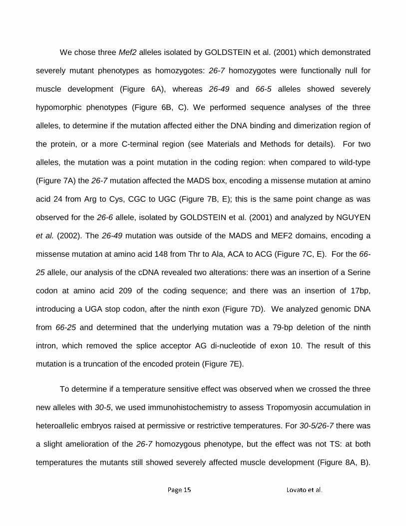
We chose three Mef2 alleles isolated by GOLDSTEIN et al. (2001) which demonstrated
severely mutant phenotypes as homozygotes: 26-7 homozygotes were functionally null for
muscle development (Figure 6A), whereas 26-49 and 66-5 alleles showed severely
hypomorphic phenotypes (Figure 6B, C). We performed sequence analyses of the three
alleles, to determine if the mutation affected either the DNA binding and dimerization region of
the protein, or a more C-terminal region (see Materials and Methods for details). For two
alleles, the mutation was a point mutation in the coding region: when compared to wild-type
(Figure 7A) the 26-7 mutation affected the MADS box, encoding a missense mutation at amino
acid 24 from Arg to Cys, CGC to UGC (Figure 7B, E); this is the same point change as was
observed for the 26-6 allele, isolated by GOLDSTEIN et al. (2001) and analyzed by NGUYEN
et al. (2002). The 26-49 mutation was outside of the MADS and MEF2 domains, encoding a
missense mutation at amino acid 148 from Thr to Ala, ACA to ACG (Figure 7C, E). For the 66-
25 allele, our analysis of the cDNA revealed two alterations: there was an insertion of a Serine
codon at amino acid 209 of the coding sequence; and there was an insertion of 17bp,
introducing a UGA stop codon, after the ninth exon (Figure 7D). We analyzed genomic DNA
from 66-25 and determined that the underlying mutation was a 79-bp deletion of the ninth
intron, which removed the splice acceptor AG di-nucleotide of exon 10. The result of this
mutation is a truncation of the encoded protein (Figure 7E).
To determine if a temperature sensitive effect was observed when we crossed the three
new alleles with 30-5, we used immunohistochemistry to assess Tropomyosin accumulation in
heteroallelic embryos raised at permissive or restrictive temperatures. For 30-5/26-7 there was
a slight amelioration of the 26-7 homozygous phenotype, but the effect was not TS: at both
temperatures the mutants still showed severely affected muscle development (Figure 8A, B).
Page 16 Lovato et al.
By contrast there was a modest temperature-dependent rescue of both of the C-terminal
region mutants, 26-49 and 66-25, when combined with 30-5 (Figure 8C-F). In both cases,
significant muscle fibers were observed in heteroallelic mutants, yet the phenotype was more
severe at 29οC (Figure 9C-F).
To quantify this temperature-sensitivity we studied the survival to adulthood of the
heteroallelic mutant combinations. The original TS combination of 30-5/44-5 showed a
dramatic difference in viability at the two temperatures, with 58% of embryos surviving to
adulthood at the permissive temperature and 0% surviving at the restrictive temperature
(BAKER et al., 2005). For the 30-5/26-7 combination of two MADS domain mutant alleles,
survival was 9% (31 observed out of 132 expected) at the permissive temperature and 0% (0
observed, 170 expected) at the restrictive temperature. When 30-5 was combined with the C-
terminal region mutants the viability was more clearly TS: for the 30-5/26-49 cross, survival
was 67% (134 observed, 200 expected) at the permissive temperature and 14% (33 observed,
239 expected) at the restrictive temperature; for the 30-5/66-25 cross, survival was 49% (93
observed, 189 expected) at the permissive temperature and 31% (61 observed, 196 expected)
at the restrictive temperature.
These findings further support the model for temperature sensitivity: heteroallelic
combination of one Mef2 mutant showing weakened DNA binding ability with a second Mef2
mutant showing weakened activation ability can generate a TS effect. Nevertheless, we note
that the severity of the TS effect can still vary between different alleles in the same class.
Discussion
Page 17 Lovato et al.
MEF2 is a widely expressed, multi-functional dimeric protein that works in collaboration with
other factors to influence a variety of developmental processes at multiple stages of
development (reviewed in BLACK and CRIPPS, 2009; POTTHOFF and OLSON 2007). We
have previously exploited the temperature-sensitivity of the 30-5/44-5 combination to elucidate
a role for MEF2 in adult myogenesis, where we showed that a strong reduction in MEF2
function could nevertheless support several aspects of adult muscle development (BAKER et
al. 2005). In this paper we have characterized the molecular mechanism behind this TS
combination, in an effort to acquire more information about the dynamics of temperature
sensitivity and the mechanisms of MEF2 function.
Current models of temperature sensitivity take into account mutations that affect the
proper formation or folding of a protein when synthesized at a restrictive temperature, and
mutations that destabilize a protein when shifted to a restrictive temperature (SADLER AND
NOVICK, 1964). Mutations that are capable of destabilizing a protein can be found in DNA
binding domains, protein-protein interaction domains or internal hydrophobic regions
(SUNDBERG and DAVIS 1997, SMITH et al.1980, GORDON and KING 1993, EDGAR and
LIELAUSIS, 1963). In addition, the temperature-sensitivity of homodimeric proteins has
received attention. In several instances, intragenic complementation has been observed for
dimeric or multimeric proteins, and the classical literature has noted that in several cases such
complementation is TS (FINCHAM, 1966). This relationship was underlined by SUNDBERG
and DAVIS (1997), who showed that mutations affecting different functional regions of the S.
cerevisiae spindle protein Spc110p could show effective TS intragenic complementation.
The 30-5/44-5 TS findings that we describe fit the latter model effectively. Combination
of the 30-5 DNA-binding mutant with 44-5 or with either of two additional C-terminal mutants
Page 18 Lovato et al.
demonstrated a TS effect, whereas combination of 30-5 with an allele affecting the DNA
binding domain did not show effective rescue and temperature-sensitivity. We note that the
severity of the various TS effects that we have observed is still the most striking with 30-5/44-
5. This might be attributed to a less severe disruption of the activation domain in the C-
terminal mutants 26-49 or 66-25. Indeed, the 26-49 isoform contains only a single amino acid
substitution in the C-terminus, but still produces a full-length protein. The 66-25 mutant protein
isoform is truncated like that of 44-5 (7), however it encodes approximately 71 more amino
acids in the C-terminal region. As homozygotes, these alleles are severe, lethal hypomorphs,
but in combination with 30-5 they complement well and demonstrate a TS effect.
Our findings for the temperature-sensitivity of Mef2 allelic combinations could be applied
to the analysis of other transcription factors. A combination of alleles for the Drosophila twist
gene also shows TS in the heteroallelic arrangement (THISSE et al. 1997). Neither of these
mutations affect the DNA binding domain (BAYLIES and BATE, 1997), however they might
nevertheless impact regions of the protein with distinct functions.
Regarding MEF2 functional domains, an important regulatory domain of MEF2 appears
to lie between amino acids 219-241. This region has a potentially inhibitory function based
upon the increased transcriptional activation activity of the 44-5 (11) isoform that lacks these
amino acids. Previous work using murine MEF2s has identified a repression domain located in
exon 9 (GULICK and ZHU 2004). Alternative splice variants lacking this domain have an
increased ability to activate downstream MEF2 targets in cell culture as was seen in the cell
culture assay for 44-5 (11). While the region identified by GULICK and ZHU (2004) probably
does not correspond to amino acids 219-241, these finding nevertheless confirm that there is
much still to be learned regarding the function of the MEF2 C-terminal region.
Page 19 Lovato et al.
Acknowledgements
We are very grateful to the individuals who supplied reagents essential to this work: Elliot
Goldstein, Daniel Kiehart and Bruce Paterson. This work was supported by a grant from the
NIH (GM61738) to RMC. MMA was supported by a MARC fellowship from the NIH
(5T34GM008751). We acknowledge technical support from the Department of Biology’s
Molecular Biology Facility, supported by NIH grant number 1P20RR18754 from the Institute
Development Award (IDeA) Program of the National Center for Research Resources.
References
BAKER, P., KELLY TANAKA, K.K., KLITGORD, N. and CRIPPS, R.M. (2005). Adult myogenesis in Drosophila melanogaster can proceed independently of myocyte enhancer factor-2. Genetics 170: 1747-1759.
BAYLIES, M. K., and M. BATE, 1996 twist: A myogenic switch in Drosophila. Science 272:
1481-1484. BLACK B.L. and OLSON, E.N. (1998) Transcriptional control of muscle development by
myocyte enhancer factor-2 (MEF2) proteins. Annu Rev Cell Dev Biol. 14:167-96. BLACK, B.L., CRIPPS, R.M., ED. (2009). Myocyte enhancer factor-2 transcription factors in
heart development and disease. Heart Development and Disease, Academic Press. BOUR, B.A., O'BRIEN, M.A., LOCKWOOD, W.L., GOLDSTEIN, E.S., and BODMER, R.
(1995) Drosophila MEF2, a transcription factor that is essential for myogenesis. Genes and Development. 9: 730-741.
CARPENTER, J. M. (1950). A new semisynthetic food medium for Drosophila. Drosophila
Inform. Serv. 24: 96-97. CASSO, D., F.-A. RAMIREZ-WEBER and T. B. KORNBERG, 1999 GFP-tagged balancer
chromosomes for Drosophila melanogaster. Mechanisms of Development 88: 229-232.
CHAKSHUSMATHI, G., MONDAL, K., LAKSHMI, G.S., SINGH, G., ROY, A., CH, R.B., MADHUSUDHANAN, S. and VARADARAJAN, R. (2004). Design of temperature-sensitive mutants solely from amino acid sequence. Proceedings of the National Academy of Sciences 101(21): 7925-7930.
Page 20 Lovato et al.
COX, D. M., DU, M., MARBACK, M., YANG, E. C., CHAN, J., SIU, K. W. and MCDERMOTT, J. C.(2003). Phosphorylation motifs regulating the stability and function of myocyte enhancer factor 2A. J Biol Chem 278: 15297-303.
COX, VT and BAYLIES MK. (2005). Specification of individual Slouch muscle progenitors in
Drosophila requires sequential Wingless signaling. Development 132:713-724. EDGAR, R. S. and LIELAUSIS, I. (1963). "Temperature-Sensitive Mutants of Bacteriophage
T4D: Their Isolation and Genetic Characterization." Genetics 49: 649-662. FINCHAM J.R.S. (1966). Genetic Complementation. New York: W.A. Benjamin, Inc. GOLDSTEIN E.S., TREADWAY S.L. STEPHENSON A.E. GRAMSTAD G.D., KEILTY A. et al.
(2001). A genetic analysis of the cytological region 46C-F containing the Drosophila melanogaster homolog of the jun proto-oncogene. Mol. Genet. Genomics 266: 695-700.
GORDON, C. L. and KING, J. (1993). Genetic Properties of Temperature-Sensitive Folding
Mutants of the Coat Protein of Phage P22. Genetics 136: 427-438. GULICK T. and ZHU B. (2004). Phosphorylation and alternative pre-mRNA splicing converge
to regulate myocyte enhancer factor 2C activity. Mol Cell Biol. 24(18): 8264-75. HARTWELL, L. H., CULOTTI, J. and REID, B. (1970). Genetic Control of the Cell-Division
Cycle in Yeast, I. Detection of Mutants. Proceedings of the National Academy of Sciences 66(No.2): 352-259.
HAMAMORI, Y., HUNG-YI WU, SARTORELLI, V. and KEDES, L. (1997). The basic Domain of
myogenic basic helix-loop-helix (bHLH) proteins is the novel target for direct inhibition by another bHLH protein, Twist. Molecular and Cellular Biology. 17(No.11): 6563-6573.
HUANG K., LOUIS J.M., DONALDSON L., LIM F.L. SHARROCKS A.D. and CLORE G.M.
(2000). Solution structure of the MEF2A-DNA complex: structural basis for the modulation of DNA bending and specificity by MADS-box transcription factors. EMBO J 19: 2615-28.
KELLY, K.K., MEADOWS, S.M., and CRIPPS, R.M. (2002). Drosophila MEF2 is a direct
regulator of Actin 57B transcription in cardiac, skeletal, and visceral muscle lineages. Mech Dev. 110(1-2): 39-50.
KELLY TANAKA, K.K., BRYANTSEV, A.L., and CRIPPS, R.M. (2008). Myocyte enhancer
factor 2 and chorion factor 2 collaborate activation of the myogenic program in Drosophila. Molecular and Cellular Biology. 28(No.5):1616-1629.
KIEHART, D. P. and FEGHALI, R. (1986). Cytoplasmic myosin from Drosophila melanogaster.
Journal of Cell Biology 103: 1517-1525.
Page 21 Lovato et al.
LILLY, B., S. GALEWSKY, A. B. FIRULLI, R. A. SCHULZ and E. N. OLSON, 1994 D-MEF2: A
MADS box transcription factor expressed in differentiating mesoderm and muscle cell lineages during Drosophila embryogenesis. Proceedings of the National Academy of Sciences USA 91: 5662-5666.
LILLY, B., ZHAO, B., RANGANAYAKULU, G. PATERSON, B.M., SCHULZ, R.A. and OLSON, E.N. (1995). Requirement of MADS domain transcription factor D-MEF2 for muscle formation in Drosophila. Science 267: 688-693.
MARTIN J.F., MIANO J.M. HUSTAD C.M., COPELAND N.G. JENKINS N.A. and OLSON E.N.
(1994). A Mef2 gene that generates a muscle-specific isoform via alternative mRNA splicing. Mol Cell biol. 14: 1647-56.
MOLKENTIN J.D. BLACK B.L., MARTIN J.F. and OLSON E.N. (1995). Cooperative activation
of muscle gene expression by MEF2 and myogenic bHLH proteins. Cell 83: 1125-36. MOLKENTIN, J.D., BLACK, B.L., MARTIN, J.F., and OLSON, E.N. (1996)a. Mutational
analysis of the DNA binding, dimerization, and transcriptional activation domains of MEF2C. Molecular and Cellular Biology. 16(No.6): 2627-2636.
MOLKENTIN, J.D., LI, L. and OLSON, E.N. (1996)b. Phosphorylation of the MADS-Box
transcription factor MEF2C enhances its DNA binding activity. J Biol Chem 271: 17199-204.
NGUYEN H.T., BODMER R., ABMAYR S.M. MCDERMOTT J.C. and SPOEREL N.A. (1994).
D-mef2: a Drosophila mesoderm-specific MADS box-containing gene with a biphasic expression profile during embryogenesis. Proc. Natl. Acad. Sci. USA 91: 7520-7524.
NGUYEN, T., J. B. WANG and R. A. SCHULZ, 2002 Mutations within the conserved MADS
box of the D-MEF2 muscle differentiation factor result in a loss of DNA binding ability and lethality in Drosophila. Differentiation 70: 438-446.
PATEL, N., 1994 Imaging neuronal subsets and other cell types in whole-mount Drosophila embryos and larvae using antibody probes. In Methods in Cell Biology, edited by E. A. FYRBERG and L. S. B. GOLDSTEIN. Academic Press Inc., Boston, MA.
POTTHOFF, M.J. and OLSON, E.N. (2007). MEF2: a central regulator of diverse developmental programs. Development 134(23): 4131-40.
PECKHAM, M, CRIPPS, RM, WHITE, DCS and BULLARD, B (1992) Mechanics and protein content of insect flight muscles. J. Exp. Biol. 168: 57-76.
SADLER, J. R., NOVICK, A. (1964). "The Properties of Repressor and the Kinetics of its Action." Journal of Molecular Biology 12: 305-327.
SANTELLI E. and RICHMOND T.J. (2000). Crystal Structure of MEF2A Core Bound to DNA at 1.5 Å Resolution. J. Mol. Biol. 297: 437-449.
Page 22 Lovato et al.
SMITH D.H., BERGET P.B. and KING J. (1980). Temperature-sensitive mutants blocked in the
folding or subunit assembly of the bacteriophage P22 tail-spike protein. I. Fine-structure mapping. Genetics. 2: 331-52.
SUNDBERG H.A. and DAVIS T.N. (1997) A mutational analysis identifies three functional
regions of the spindle pole component Spc110p in Saccharomyces cerevisiae. Mol Biol Cell. 12: 2575-90.
TAYLOR M.V., BEATTY K.E., HUNTER H.K. and BAYLIES M.K. (1995). Drosophila MEF2 is
regulated by twist and is expressed in both the primordia and differentiated cells of the embryonic somatic, visceral and heart musculature. Mech Dev 50:29-41.
THISSE, B., M. E. MESSAL and F. PERRIN-SCHMITT., (1987). The twist gene: Isolation of a
Drosophila zygotic gene necessary for the establishment of dorsoventral pattern. Nucleic Acids Res. 15: 3439–3453.
Page 23 Lovato et al.
Figure Legends
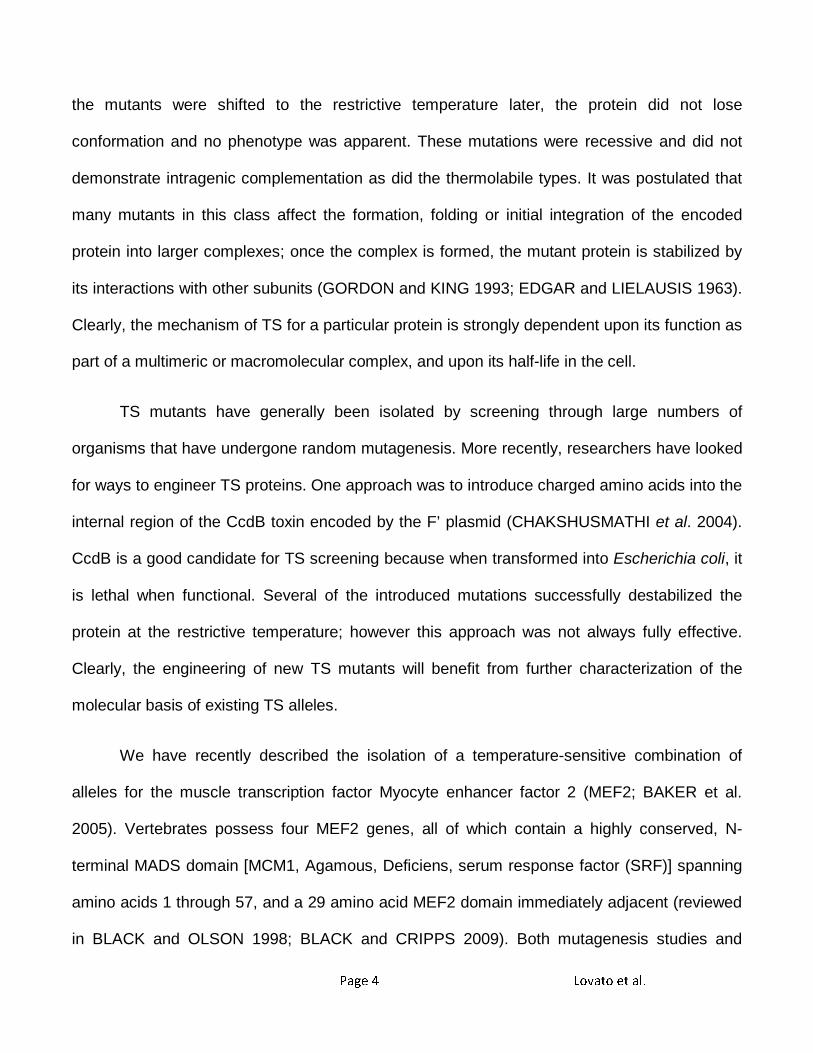
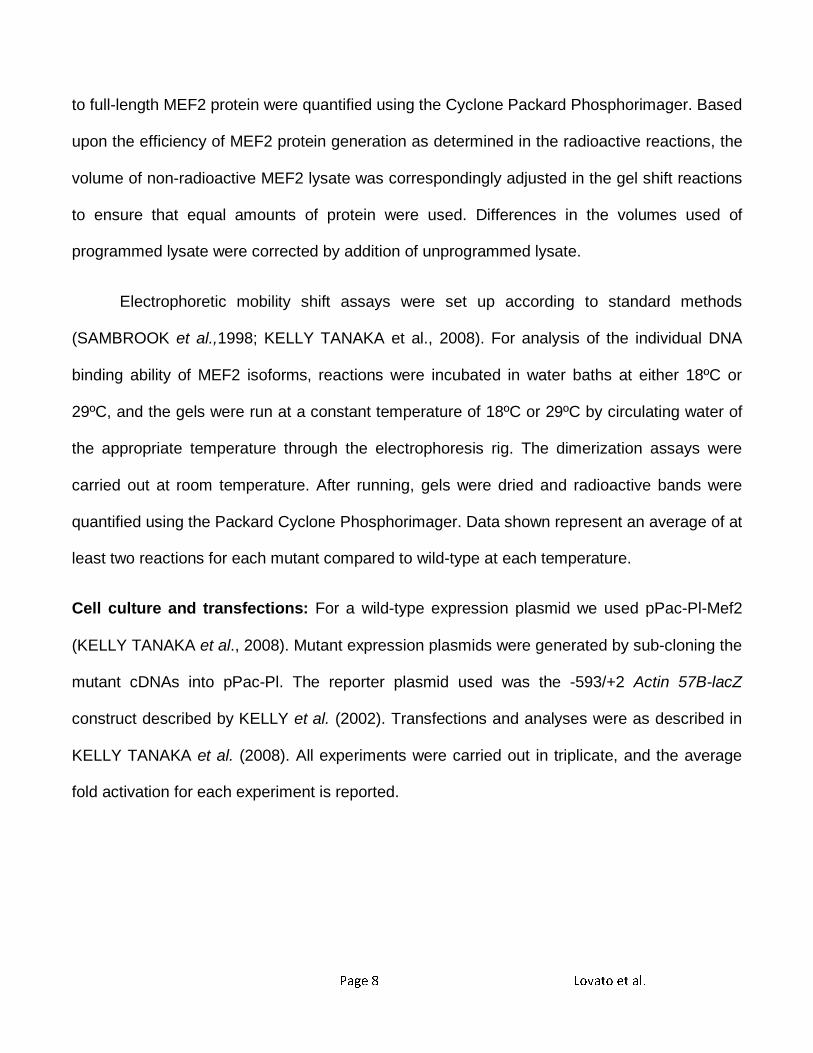
Figure 1
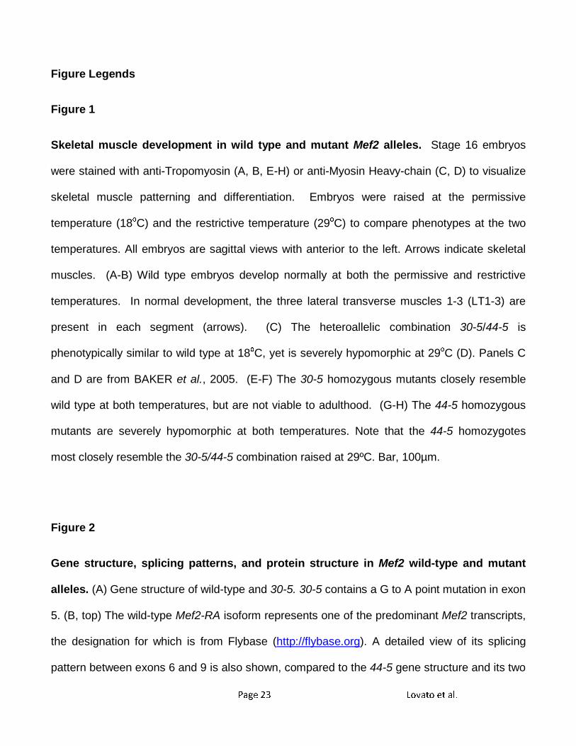
Skeletal muscle development in wild type and mutant Mef2 alleles. Stage 16 embryos
were stained with anti-Tropomyosin (A, B, E-H) or anti-Myosin Heavy-chain (C, D) to visualize
skeletal muscle patterning and differentiation. Embryos were raised at the permissive
temperature (18οC) and the restrictive temperature (29οC) to compare phenotypes at the two
temperatures. All embryos are sagittal views with anterior to the left. Arrows indicate skeletal
muscles. (A-B) Wild type embryos develop normally at both the permissive and restrictive
temperatures. In normal development, the three lateral transverse muscles 1-3 (LT1-3) are
present in each segment (arrows). (C) The heteroallelic combination 30-5/44-5 is
phenotypically similar to wild type at 18οC, yet is severely hypomorphic at 29οC (D). Panels C
and D are from BAKER et al., 2005. (E-F) The 30-5 homozygous mutants closely resemble
wild type at both temperatures, but are not viable to adulthood. (G-H) The 44-5 homozygous
mutants are severely hypomorphic at both temperatures. Note that the 44-5 homozygotes
most closely resemble the 30-5/44-5 combination raised at 29ºC. Bar, 100µm.
Figure 2
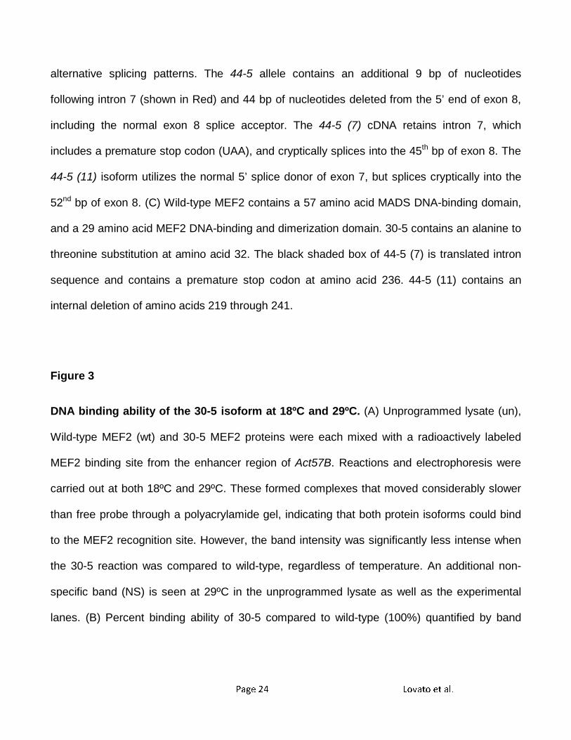
Gene structure, splicing patterns, and protein structure in Mef2 wild-type and mutant
alleles. (A) Gene structure of wild-type and 30-5. 30-5 contains a G to A point mutation in exon
5. (B, top) The wild-type Mef2-RA isoform represents one of the predominant Mef2 transcripts,
the designation for which is from Flybase (http://flybase.org). A detailed view of its splicing
pattern between exons 6 and 9 is also shown, compared to the 44-5 gene structure and its two
Page 24 Lovato et al.
alternative splicing patterns. The 44-5 allele contains an additional 9 bp of nucleotides
following intron 7 (shown in Red) and 44 bp of nucleotides deleted from the 5’ end of exon 8,
including the normal exon 8 splice acceptor. The 44-5 (7) cDNA retains intron 7, which
includes a premature stop codon (UAA), and cryptically splices into the 45th bp of exon 8. The
44-5 (11) isoform utilizes the normal 5’ splice donor of exon 7, but splices cryptically into the
52nd bp of exon 8. (C) Wild-type MEF2 contains a 57 amino acid MADS DNA-binding domain,
and a 29 amino acid MEF2 DNA-binding and dimerization domain. 30-5 contains an alanine to
threonine substitution at amino acid 32. The black shaded box of 44-5 (7) is translated intron
sequence and contains a premature stop codon at amino acid 236. 44-5 (11) contains an
internal deletion of amino acids 219 through 241.
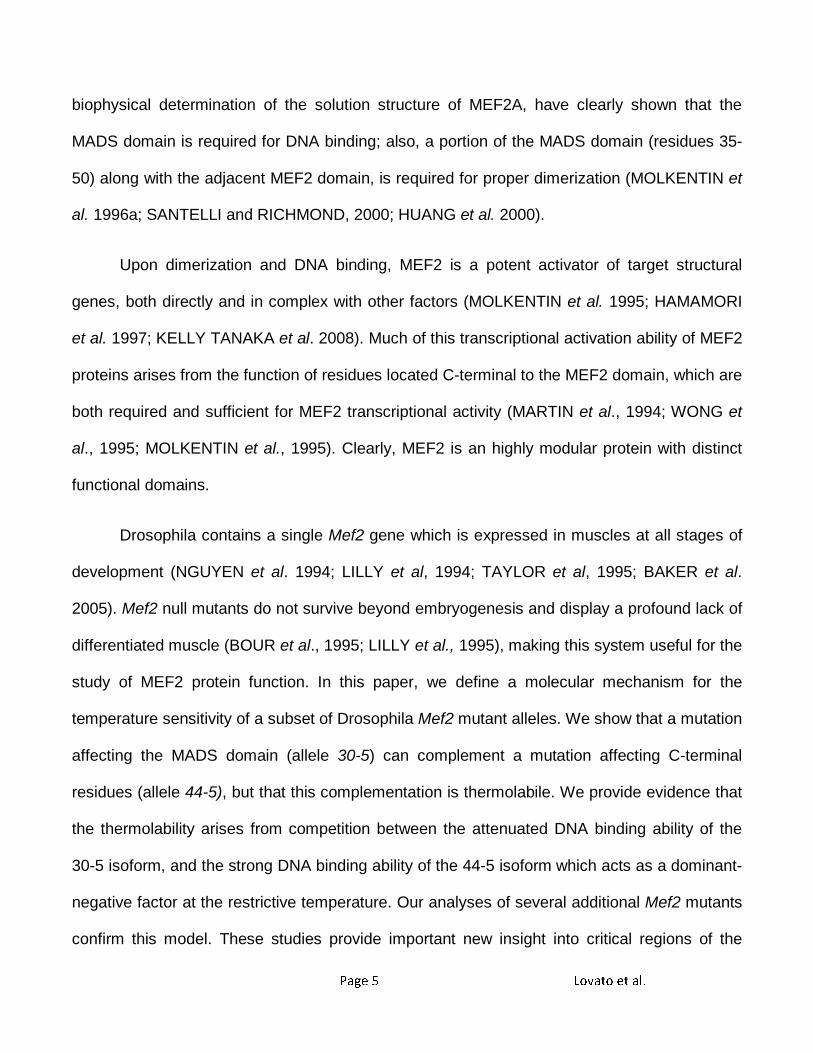
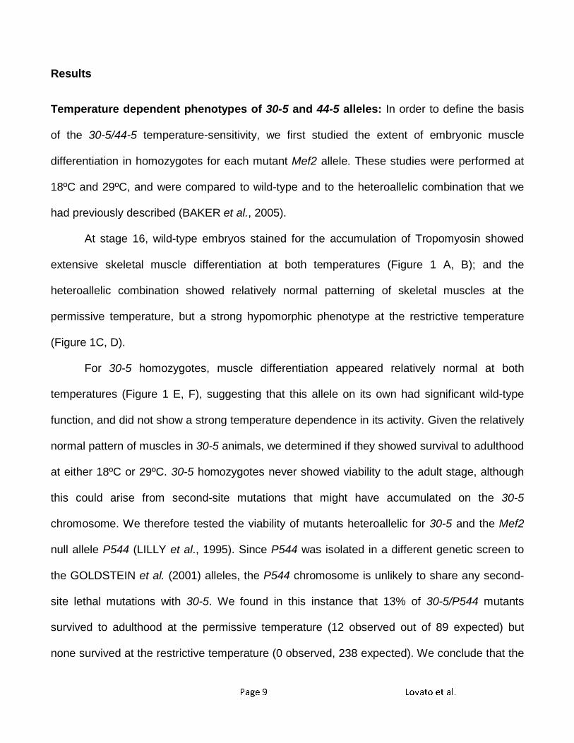
Figure 3
DNA binding ability of the 30-5 isoform at 18ºC and 29ºC. (A) Unprogrammed lysate (un),
Wild-type MEF2 (wt) and 30-5 MEF2 proteins were each mixed with a radioactively labeled
MEF2 binding site from the enhancer region of Act57B. Reactions and electrophoresis were
carried out at both 18ºC and 29ºC. These formed complexes that moved considerably slower
than free probe through a polyacrylamide gel, indicating that both protein isoforms could bind
to the MEF2 recognition site. However, the band intensity was significantly less intense when
the 30-5 reaction was compared to wild-type, regardless of temperature. An additional non-
specific band (NS) is seen at 29ºC in the unprogrammed lysate as well as the experimental
lanes. (B) Percent binding ability of 30-5 compared to wild-type (100%) quantified by band
Page 25 Lovato et al.
intensity (see Materials and Methods). Note that the 30-5 isoform consistently bound to DNA
less efficiently than wild-type.
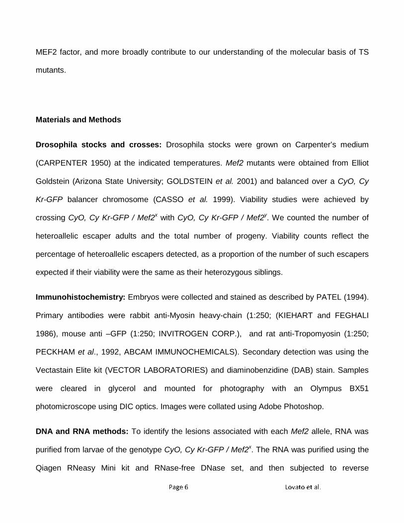
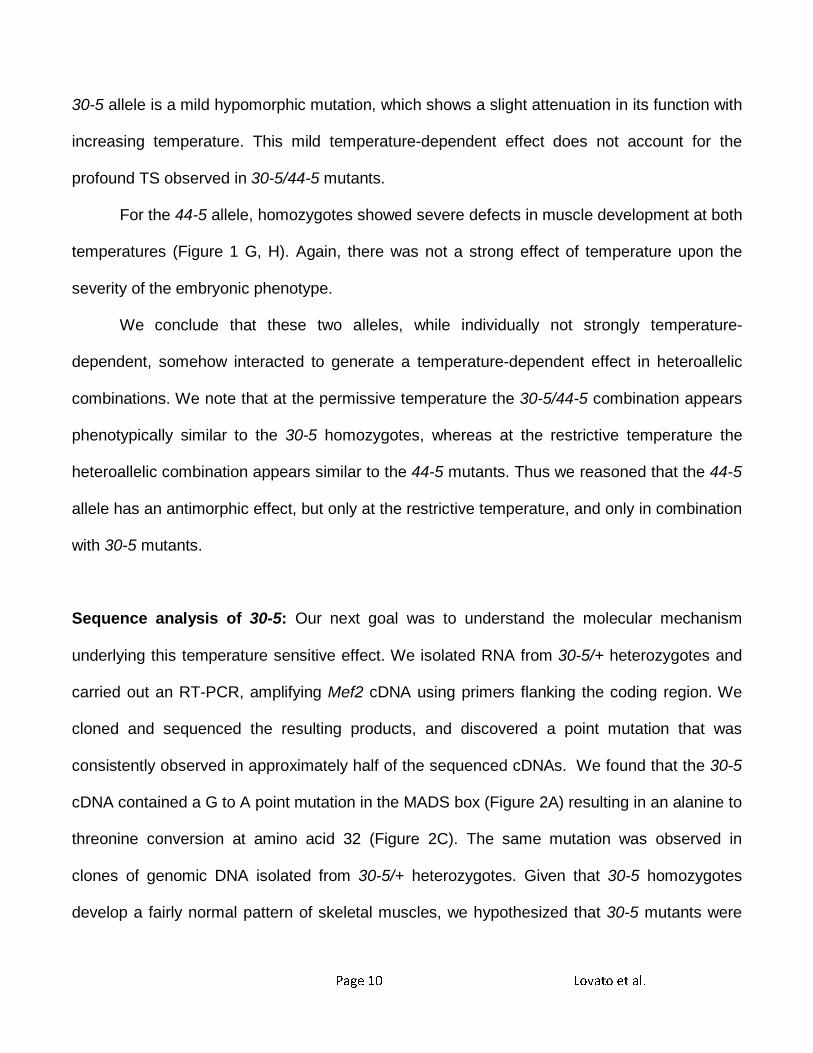
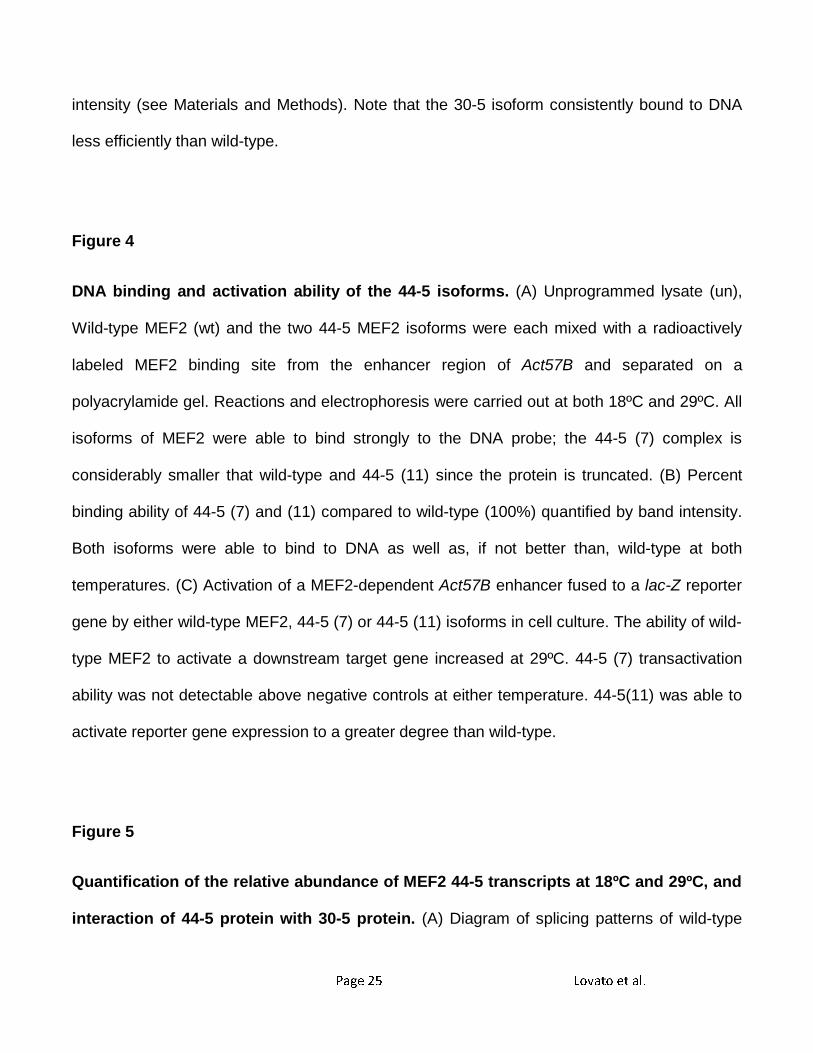
Figure 4
DNA binding and activation ability of the 44-5 isoforms. (A) Unprogrammed lysate (un),
Wild-type MEF2 (wt) and the two 44-5 MEF2 isoforms were each mixed with a radioactively
labeled MEF2 binding site from the enhancer region of Act57B and separated on a
polyacrylamide gel. Reactions and electrophoresis were carried out at both 18ºC and 29ºC. All
isoforms of MEF2 were able to bind strongly to the DNA probe; the 44-5 (7) complex is
considerably smaller that wild-type and 44-5 (11) since the protein is truncated. (B) Percent
binding ability of 44-5 (7) and (11) compared to wild-type (100%) quantified by band intensity.
Both isoforms were able to bind to DNA as well as, if not better than, wild-type at both
temperatures. (C) Activation of a MEF2-dependent Act57B enhancer fused to a lac-Z reporter
gene by either wild-type MEF2, 44-5 (7) or 44-5 (11) isoforms in cell culture. The ability of wild-
type MEF2 to activate a downstream target gene increased at 29ºC. 44-5 (7) transactivation
ability was not detectable above negative controls at either temperature. 44-5(11) was able to
activate reporter gene expression to a greater degree than wild-type.
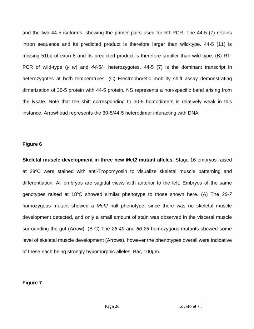
Figure 5
Quantification of the relative abundance of MEF2 44-5 transcripts at 18ºC and 29ºC, and
interaction of 44-5 protein with 30-5 protein. (A) Diagram of splicing patterns of wild-type
Page 26 Lovato et al.
and the two 44-5 isoforms, showing the primer pairs used for RT-PCR. The 44-5 (7) retains
intron sequence and its predicted product is therefore larger than wild-type. 44-5 (11) is
missing 51bp of exon 8 and its predicted product is therefore smaller than wild-type. (B) RT-
PCR of wild-type (y w) and 44-5/+ heterozygotes. 44-5 (7) is the dominant transcript in
heterozygotes at both temperatures. (C) Electrophoretic mobility shift assay demonstrating
dimerization of 30-5 protein with 44-5 protein. NS represents a non-specific band arising from
the lysate. Note that the shift corresponding to 30-5 homodimers is relatively weak in this
instance. Arrowhead represents the 30-5/44-5 heterodimer interacting with DNA.
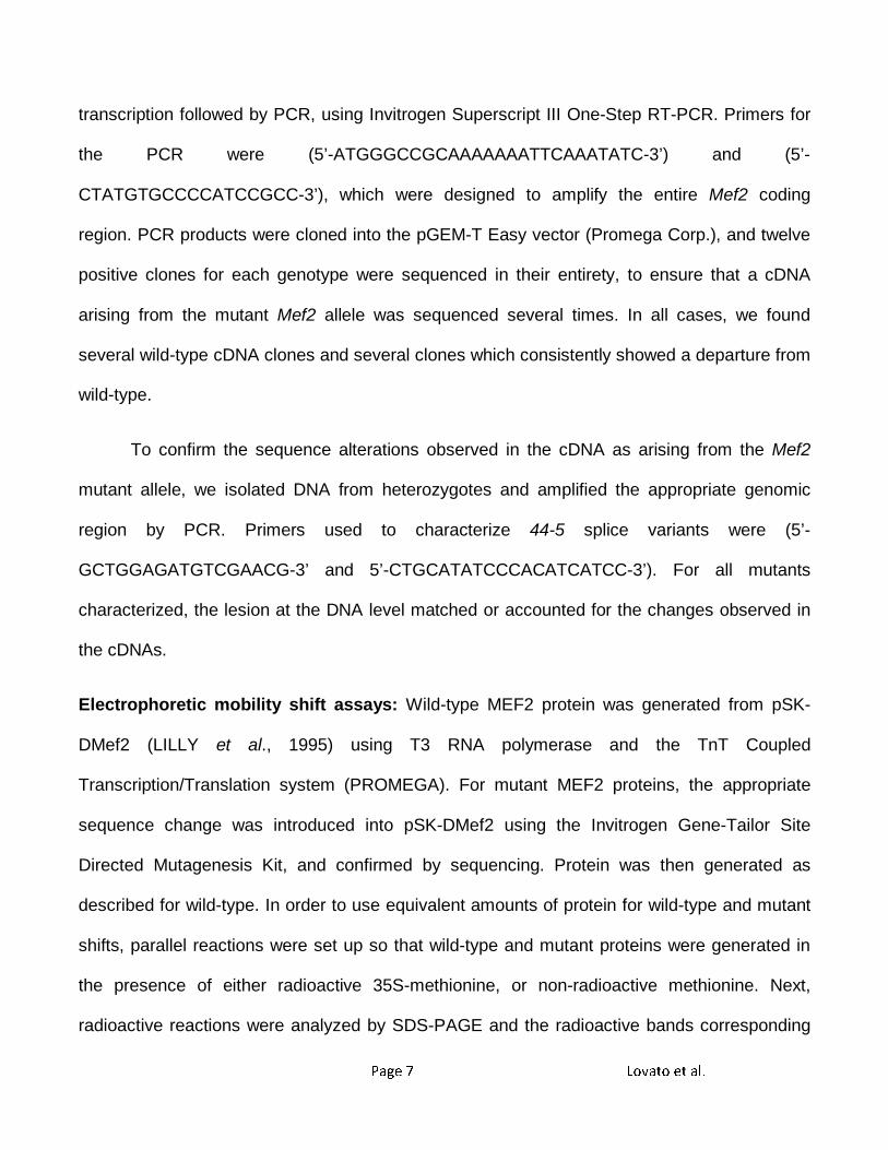
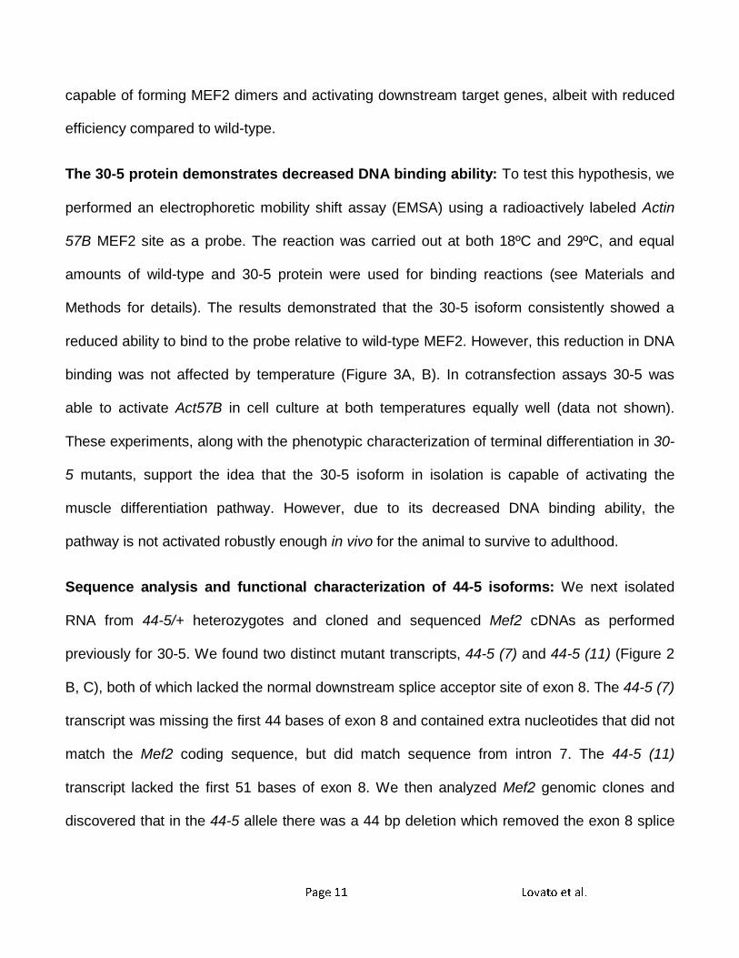
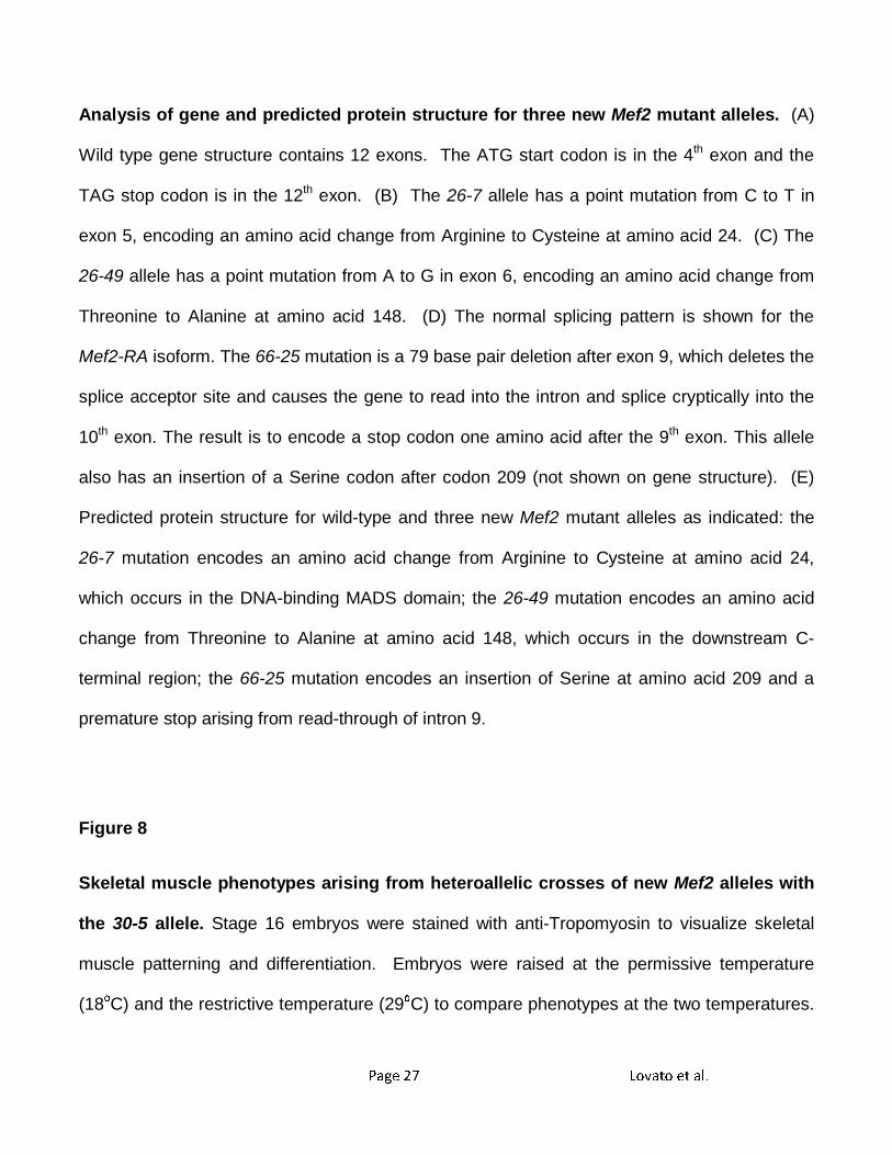
Figure 6
Skeletal muscle development in three new Mef2 mutant alleles. Stage 16 embryos raised
at 29ºC were stained with anti-Tropomyosin to visualize skeletal muscle patterning and
differentiation. All embryos are sagittal views with anterior to the left. Embryos of the same
genotypes raised at 18ºC showed similar phenotype to those shown here. (A) The 26-7
homozygous mutant showed a Mef2 null phenotype, since there was no skeletal muscle
development detected, and only a small amount of stain was observed in the visceral muscle
surrounding the gut (Arrow). (B-C) The 26-49 and 66-25 homozygous mutants showed some
level of skeletal muscle development (Arrows), however the phenotypes overall were indicative
of these each being strongly hypomorphic alleles. Bar, 100µm.
Figure 7
Page 27 Lovato et al.
Analysis of gene and predicted protein structure for three new Mef2 mutant alleles. (A)
Wild type gene structure contains 12 exons. The ATG start codon is in the 4th exon and the
TAG stop codon is in the 12th exon. (B) The 26-7 allele has a point mutation from C to T in
exon 5, encoding an amino acid change from Arginine to Cysteine at amino acid 24. (C) The
26-49 allele has a point mutation from A to G in exon 6, encoding an amino acid change from
Threonine to Alanine at amino acid 148. (D) The normal splicing pattern is shown for the
Mef2-RA isoform. The 66-25 mutation is a 79 base pair deletion after exon 9, which deletes the
splice acceptor site and causes the gene to read into the intron and splice cryptically into the
10th exon. The result is to encode a stop codon one amino acid after the 9th exon. This allele
also has an insertion of a Serine codon after codon 209 (not shown on gene structure). (E)
Predicted protein structure for wild-type and three new Mef2 mutant alleles as indicated: the
26-7 mutation encodes an amino acid change from Arginine to Cysteine at amino acid 24,
which occurs in the DNA-binding MADS domain; the 26-49 mutation encodes an amino acid
change from Threonine to Alanine at amino acid 148, which occurs in the downstream C-
terminal region; the 66-25 mutation encodes an insertion of Serine at amino acid 209 and a
premature stop arising from read-through of intron 9.
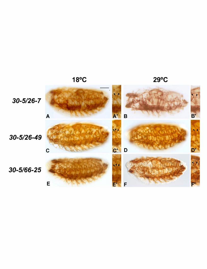
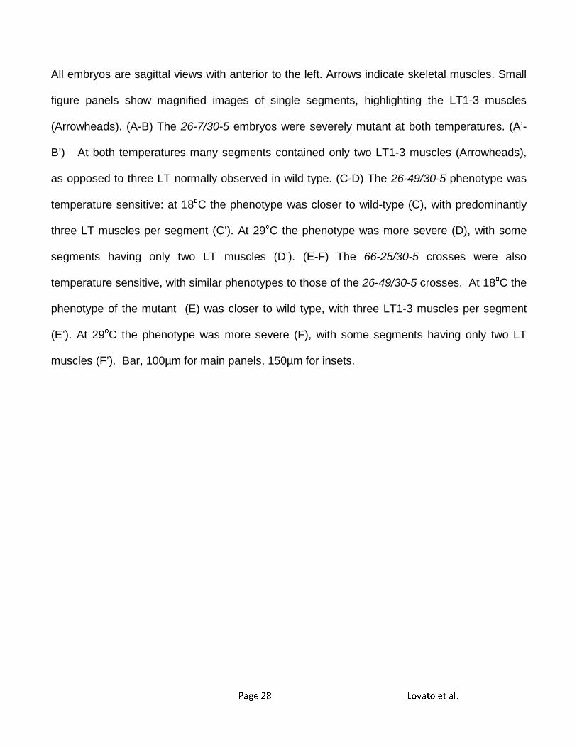
Figure 8
Skeletal muscle phenotypes arising from heteroallelic crosses of new Mef2 alleles with
the 30-5 allele. Stage 16 embryos were stained with anti-Tropomyosin to visualize skeletal
muscle patterning and differentiation. Embryos were raised at the permissive temperature
(18οC) and the restrictive temperature (29οC) to compare phenotypes at the two temperatures.
Page 28 Lovato et al.
All embryos are sagittal views with anterior to the left. Arrows indicate skeletal muscles. Small
figure panels show magnified images of single segments, highlighting the LT1-3 muscles
(Arrowheads). (A-B) The 26-7/30-5 embryos were severely mutant at both temperatures. (A’-
B’) At both temperatures many segments contained only two LT1-3 muscles (Arrowheads),
as opposed to three LT normally observed in wild type. (C-D) The 26-49/30-5 phenotype was
temperature sensitive: at 18οC the phenotype was closer to wild-type (C), with predominantly
three LT muscles per segment (C’). At 29οC the phenotype was more severe (D), with some
segments having only two LT muscles (D’). (E-F) The 66-25/30-5 crosses were also
temperature sensitive, with similar phenotypes to those of the 26-49/30-5 crosses. At 18οC the
phenotype of the mutant (E) was closer to wild type, with three LT1-3 muscles per segment
(E’). At 29οC the phenotype was more severe (F), with some segments having only two LT
muscles (F’). Bar, 100µm for main panels, 150µm for insets.
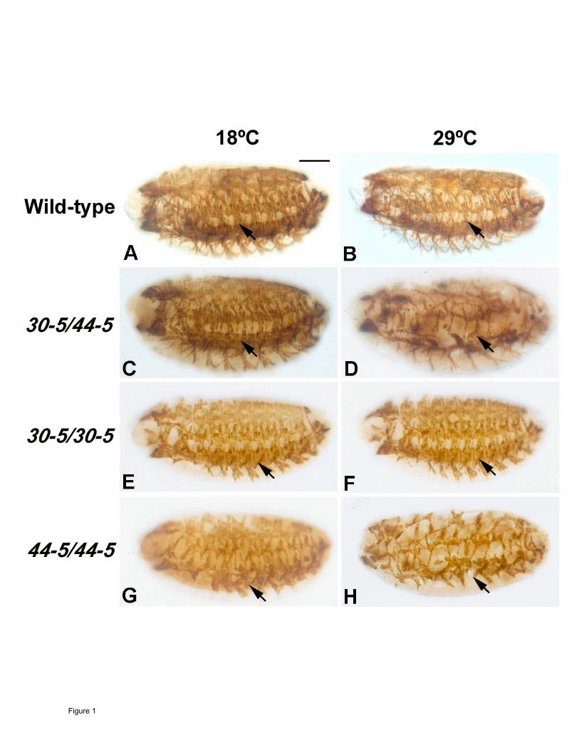
Figure 1
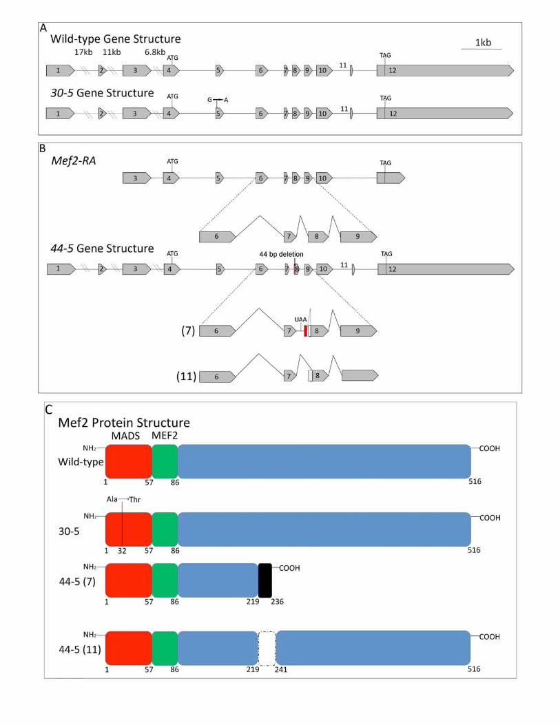
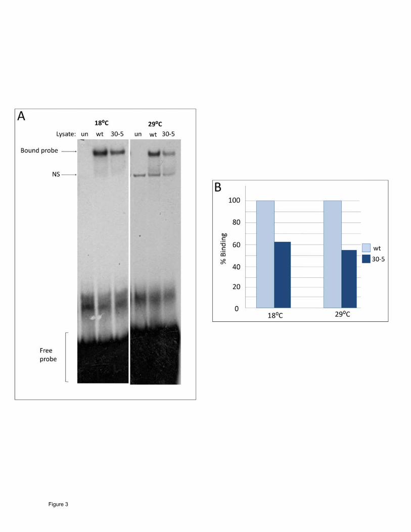
Figure 3
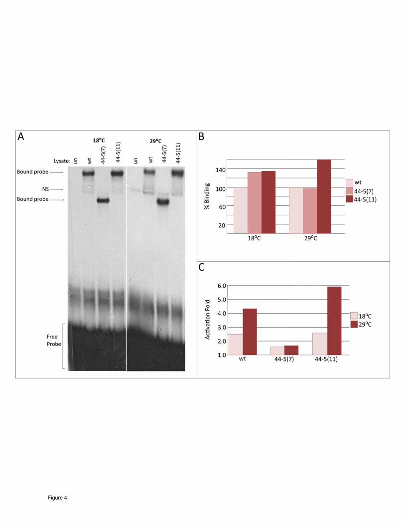
Figure 4
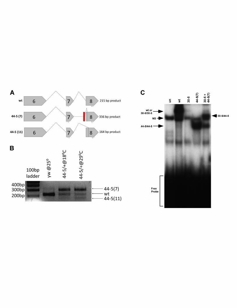
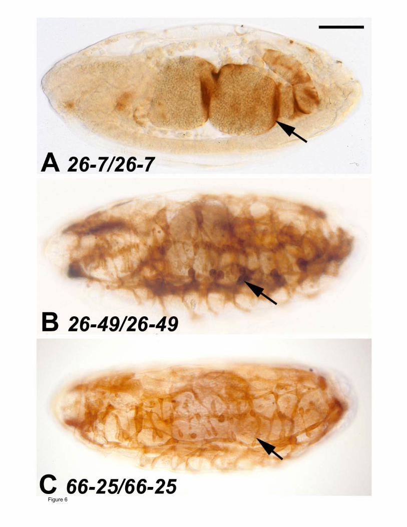
Figure 6