Embed Size (px)
Citation preview

TitleA new anatomical classification of the bronchial arteries basedon the spatial relationships to the esophagus and the tracheo-bronchus( Text_全文 )
Author(s) 早坂, 研
Citation Surgery Today, 47(7): 883-890
Issue Date 2016-11-23
URL http://hdl.handle.net/20.500.12000/36548
Rights

学位論文
A new anatomical classification of the bronchial arteries based on the
spatial relationships to the esophagus and the tracheo-bronchus
琉球大学大学院医学研究科医科学専攻
早坂 研


1
Ken Hayasaka1, Hajime Ishida2, Ryosuke Kimura2, Tadashi Nishimaki1
Title: A new anatomical classification of the bronchial arteries based on the spatial
relationships to the esophagus and the tracheo-bronchus
1 Department of Digestive and General Surgery, Graduate School of Medicine,
University of the Ryukyus, 207 Uehara, Nishihara-cho, Okinawa 903-0215, Japan
2 Department of Human Biology and Anatomy, Graduate School of Medicine,
University of the Ryukyus, 207 Uehara, Nishihara-cho, Okinawa 903-0215, Japan
Address to reprints and correspondence: Ken Hayasaka, MD.
Department of Digestive and General Surgery, Graduate School of Medicine, University
of the Ryukyus, 207 Uehara, Nishihara-cho, Okinawa 903-0215, Japan
E-mail: [email protected]
Tel: +81-98-895-1163
Fax: +81-98-895-1421
Article type: Original article (Experimental Original)
Keywords: bronchial artery, esophagectomy, anatomy
2
Abstract
Purpose: To reveal the patterns of the mediastinal course of bronchial arteries (BAs).
Methods: The BAs were dissected to determine the positional relationships of their
mediastinal courses with the tracheobronchus and the esophagus in 72 adult cadavers.
Results: The mediastinal courses of the 227 BAs found in the present study could be
classified into 4 types. There were 61 and 163 BAs passing the right side (Type I) and
the left side (Type II reaching dorsal surface (n = 98), or Type III reaching ventral
surface (n = 65) of the tracheobronchus) of the esophagus, respectively. Three BAs
originated from the subclavian artery (Type IV). All Type I BAs were the right BAs,
whereas 91.8% of the Type II BAs were the left BAs. However, 43.1% and 56.9% of the
Type III BAs were the right and left BAs, respectively.
Conclusion: The classification of the mediastinal course of the BAs determined by the
spatial relationships to the tracheobronchus and the esophagus may be clinically useful
because each category of this classification can be determined during esophagectomy,
indicates whether the BA is a right or left BA.
3
Introduction
En bloc esophagectomy with regional lymphadenectomy remains the mainstay
treatment for resectable esophageal cancer [1], although, radiotherapy with or without
chemotherapy has been used as the primary treatment in some cases of esophageal
cancer. Some recent studies have reported that higher numbers of removed lymph nodes
during esophagectomy are associated with better prognosis of esophageal cancer,
suggesting the necessity of thorough lymphadenectomy [2, 3]. In contrast, surgical
devascularization of the trachea and the main bronchi during extensive lymph node
dissection sometimes results in tracheobronchial ulcers or fistulae due to
tracheobronchial ischemia [4, 5]. Furthermore, high mortality rates have been reported
in patients with such postoperativecomplications [6-8]. Therefore, precise dissection of
lymph nodes while preserving the bronchial arteries (BAs) is mandatory for radical
esophagectomy, and the knowledge of diverse patterns of branching and routes of the
BAs is important for esophageal surgeons when performing radical esophagectomy.
Moreover, the importance of the BAs has also been recognized in the field of lung
transplantation and embolization for hemoptysis other than cancer surgery [9].
Recently introduced multi-detector row computed tomography (MDCT) with
the computed tomography angiography enables the visualization of the running courses
4
of the BAs [10, 11]. According to the accumulation of imaging studies using MDCT,
clinically useful information about the anatomy of the BAs including the precise site of
the ostia on the thoracic aorta and positional relationships between the BAs and the
esophagus or the tracheobronchus, has been revealed [12-17]. However, owing to the
small number of anatomical cadaver studies of the BAs so far, whether all the BAs in
individual patients can be fully visualized with MDCT remains unclear.
The aim of the present study is, therefore, 1) to precisely determine the
numbers, the origins, the distributions, and the running course patterns of the right and
the left BAs through a macro-anatomical study using cadavers; 2) to compare these data
with the findings previously reported in MDCT studies; and 3) to provide surgeons with
detailed anatomical features of the BAs for meticulous esophageal cancer surgery.
5
Materials and methods
Cadavers
We examined the anatomy of the BAs in 72 adult human cadavers at the University of
the Ryukyus, Japan; 44 were men and 28 were women. Their age at the time of death
ranged from 39 to 103 years. The mean age was 76.5 years. The most common cause of
their death was malignant neoplasm (20 cases), followed by infection (19 cases) and
cardiac disease (13 cases). This study was approved by the ethics committee of
University of the Ryukyus (approval number: 885).
Dissection
We detected the BAs macroscopically from the origin to the pulmonary root as follows.
First, the anterolateral chest wall was removed. The pericardium was incised to remove
the heart from the pericardial cavity. The hili of the lungs were transected with scissors
to remove the right and the left lungs from the thoracic cavity. The pericardium was
removed over the esophagus. The space between the thoracic aorta and the pulmonary
trunk was dissected carefully to remove the pulmonary trunk without injuring the BAs.
In this way, the deep mediastinal structures, including the thoracic aorta, the
tracheobronchus, and the esophagus, were exposed. After the BAs passing ventral to the
tracheobronchus were identified, dissection proceeded according to the process of
6
esophagectomy through right thoracotomy in esophageal cancer patients. The right side
of the mediastinal pleura was removed followed by the resection of the azygos vein arch.
At the hilus of the right lung, the BAs passing right side of the esophagus were
identified. The esophagus was lifted from the thoracic aorta to identify the origins of the
BAs passing the right side of the esophagus. The origins of the BAs from the thoracic
aorta and passing the left side of the esophagus were also identified. The pathways of
these arteries were confirmed by removing the left side of the mediastinal pleura.
Dissection of the BAs was performed macroscopically with the surgical instruments for
general use in surgery. The BAs less than 1mm in diameter were excluded in this study,
because these arteries are generally not preserved in esophageal surgery.
The BA running along the right main bronchus was defined as the right BA,
and the BA running along the left main bronchus was defined as the left BA.
Recording the anatomical findings of the BAs
1) Number of the BAs
The number of the BAs was determined as the number of their origins. In this study, we
found two cases in which a single left BA originating from the thoracic aorta ran to
divide into the ventral and dorsal branches of the trachea. We counted it as two left BAs
originating from a common trunk of the proper BA.

7
2) Origin of the BAs
Origins of the BAs were recorded as follows (Fig. 1): the thoracic aorta, the intercostal-
bronchial trunk (IBT), the common trunk of the right and the left BAs (CTB), and the
subclavian artery (SC). The level of origins of the BAs and the common trunks on the
aorta have been evaluated by the vertebral level in the previous studies [11, 12, 18, 19].
However, it is difficult to compare the aortic level of the origins with the vertebral body
from the narrow view of the surgical field. Therefore, we divided the thoracic aorta into
four portions by the three cross-sections: the cross-section through the ductus arteriosus,
the cross-section through the middle point of the origins of the first and the second
aortic intercostal arteries, and the cross-section through the middle point of the origin of
the second and the third aortic intercostal arteries. Thus, four parts of the aorta were
defined as the aortic arch, the upper portion (U), the middle portion (M), and the lower
portion (L), in order, from the proximal part (Fig.1).
3) Mediastinal courses of the BAs
In the present study, we classified the mediastinal courses of the BAs into 4 types
according to the positional relationships with the tracheobronchus and the esophagus
(Fig. 2). The BA running anteriorly along the right side of the esophagus to reach the
ventral or dorsal surface of the tracheobronchus was classified as Type I. The BA

8
running anteriorly along the left side of the esophagus to reach the dorsal surface of the
tracheobronchus was classified as Type II. The BA running anteriorly along the left side
of the esophagus, then passing the lateral side of the left main bronchus, to reach the
ventral surface of the tracheobronchus was classified as Type III. The BA originating
from the subclavian artery (SC) and descending to the ventral surface of the
tracheobronchus was classified as Type IV. We could apply the running courses of all
the BAs found in the 72 cadavers to this classification.
4) Distributions of the BAs
Based on the Kasai’s [20] report suggesting the presence of anatomical relationships
between the origin and the distribution of the BAs, we examined the distribution
patterns at the pulmonary hilus to clarify the presence or absence of relationships
between the distribution and the mediastinal course of the BAs. The distributions of the
BAs at the pulmonary hilus were classified into the superior branch (S), the superior and
inferior branch (SI), and the inferior branch (I).
9
Results
1) Number of the BAs
We identified 100 right and 127 left BAs in the 72 cadavers. The numbers of the BAs
per cadaver ranged from 1 to 5 (mean: 3.15). The mean number of the right and the left
BAs was 1.39 and 1.76, respectively, per cadaver. Cadavers with one right and two left
BAs were the most commonly observed (38.9%), followed by cadavers with two right
and two left BAs (18.1%) (Table 1). There were two cadavers (2.8%) in which either of
the right or the left BA was absent. The right BA was absent in one cadaver, and the left
BA was absent in another cadaver (Table 1).
2) Origins of the BAs
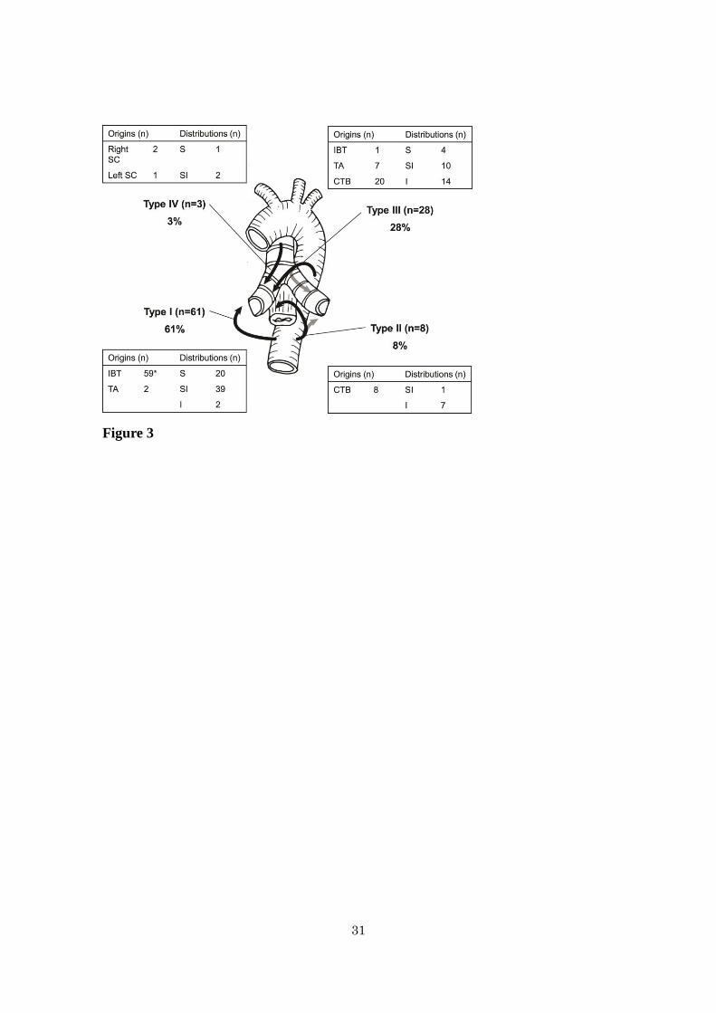
Of the 100 right BAs, 60 (60%), 28 (28%), 9 (9%), 2 (2%), and 1 (1%) originated from
the right intercostal- bronchial trunk (IBT), the common trunk of the right and the left
BAs (CTB), the thoracic aorta, the right SC, and the left SC, respectively (Fig. 3). Of
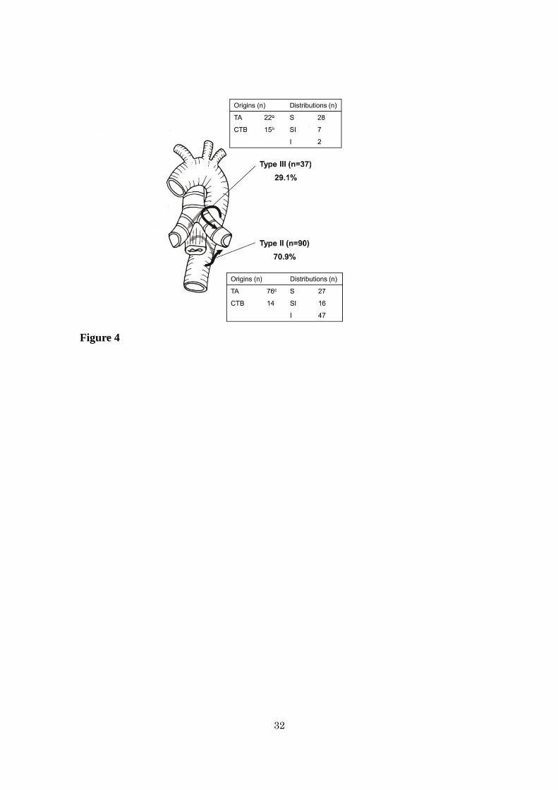
the 127 left BAs, 98 (77.2%) and 29 (22.8%) originated from the thoracic aorta and the
CTB, respectively (Fig. 4). The cadavers with one right BA from the right IBT and two
left BAs from the thoracic aorta were most commonly observed (20 bodies, 27.8%),
followed by the cadavers with one right BA from the right IBT and one left BA from the
thoracic aorta (9 bodies, 12.5%), and the cadavers with two right BAs from the right
10
IBT or CTB, and two left BAs from the thoracic aorta or CTB (8 bodies, 11.1%) (Table
1).
There were 192 arteries originating directly from the thoracic aorta (2.67/body). Of
the 192 arteries, 59 (30.7%), 28 (14.6%), 9 (4.7%), and 96 (50%) were the IBTs, the
CTBs, the right BAs, and the left BAs, respectively. Two right BAs originating from 1
IBT, 2 left BAs from 1 CTB, and 2 left BAs from 1 proper BA were found in 1, 1, and 2
cadavers, respectively. Of the 192 arteries including 105 proper BAs and 87 common
trunks, 164 (85.4%) originated from the U or M portion. Only 4 arteries (2.1%),
including 2 right BAs and 2 CTBs, originated from the aortic arch. The remaining 24
arteries (12.5%) originated from the L portion of the thoracic aorta, including 21 left
BAs and 3 CTBs.
3) Mediastinal courses of the BAs
Of the 227 BAs, Types I, II, III, and IV were found in 61 (26.9 %), 98 (43.2%), 65
(28.6%), and 3 (1.3%), respectively. However, Types I, II, III, and IV were present in 60
(83.3 %), 63 (87.5%), 44 (61.1%), 3 (4.2%) of the 72 cadavers, respectively. Of the 61
arteries showing Type I, 59 (96.7%) were the right BAs from the IBT, and the remaining
two were the right BAs originating directly from the thoracic aorta (Fig. 3). Thus, all of
the arteries showing Type I were right BAs. Of the 98 arteries showing Type II, 8
11
(8.2%), 76 (77.5%), and 14 (14.3%) were the right BAs from the CTB, the left BAs
from the thoracic aorta, and the left BAs from the CTB, respectively (Fig. 3,4). Of the
127 left BAs, 90 (70.9%) showed the Type II course (Fig. 4). Particularly, all of the left
BAs from the L portion ran along this course. Of the 65 arteries showing Type III, 7
(10.8%), 20 (30.8%), and 1 (1.5%) were the right BAs originating from the thoracic
aorta, the CTB, and the IBT, respectively (Fig. 3). However, 22 (33.8%), and 15
(23.1 %) were the left BAs from the thoracic aorta, and the CTB, respectively (Fig. 4).
Thus, the 65 arteries of Type III were composed of 28 right BAs (43.1%), and 37 left
BAs (56.9%). All of the BAs originating from the aortic arch showed the Type III
pattern. Of the 3 Type IV arteries, two and one were the right BAs from the right SC
and the left SC, respectively (Fig. 3). Any BAs showing mediastinal courses not
applicable to this classification were not found. All of the BAs originating from the
thoracic aorta, to reach the ventral surface of the tracheobronchus, passed the lateral
side of the right or the left main bronchi. Of the 28 CTBs, 20 arteries branched out into
the right and left BAs showing the same running course category: the Type II (n=7), and
the Type III (n=13) courses. The remaining 8 arteries branched out into the right and left
BAs showing different running course categories: one CTB had one right BA showing
the Type II with one left BA showing the Type III, and 7 CTBs had one right BA

12
showing the Type III with one left BAs showing the Type II.
4) Distribution of the BAs
The mediastinal courses of the BAs were related to their distributions. Of the 61 right
BAs showing Type I, 59 (96.7%) had the superior branches (S or SI) (Fig. 3).However,
all the right BAs showing Type II, and 24 (85.7%) right BAs showing Type III had the
inferior branches (SI or I) (Fig 3). In contrast with the right BAs, 35 (94.6%) of the 37
left BAs showing Type III, had the superior branches (Fig. 4).

13
Discussion
The BAs constitute the nutritive vascular system of the pulmonary tissues (the bronchial
walls and the glands, walls of the large vessels, and the visceral pleura). There are many
variations in the number, the origin, and the course of the BAs [18-21]. The underlying
anatomical study of the BAs was conducted by Cauldwell et al in 1948 [18]. After an
anatomical investigation in 150 cadavers, they have reported the classification of the
branching patterns of the BAs based on the origin and the number of the right and the
left BAs. In their study, the most commonly encountered pattern was the combination of
one right BA originating from the right IBT and two left BAs originating from the
thoracic aorta. After their report, this pattern has been described as normal in many
standard textbooks of anatomy and thoracic surgery. However, there are more
anatomical variations of the BAs taking into account the mediastinal courses and the
distributions as shown in this study. Those previous studies have classified the BAs into
9 or more subtypes based on the numbers, origins, and the branching patterns of this
artery. In contrast with such complex classifications of the BAs, we made the simple
classification of the BAs, based on the spatial relationships to the tracheobronchus and
the esophagus. Our classification is composed of the 4 categories (Type I-IV). However,
any BAs not applicable to this classification were not found in the present study. Thus,
14
our classification may be more convenient in the clinical practice, particularly during
esophageal surgery.
The BAs that originate outside the segment of the aorta between the T5 and T6
vertebrae are defined as ectopic in the prior studies [12]. Ectopic BAs include the BAs
originating from the aortic arch, the subclavian artery, the descending aorta, the
brachiocephalic trunk, the thyrocervical trunk, the internal mammary artery, and the
coronary artery. The frequencies of the right and the left ectopic BAs have been reported
to range from 14.0% to 17.2%, and 11.6% to 20.0%, respectively [12-14]. However, it is
difficult to compare the aortic level of the BA origin with the vertebral body in the
narrow view of the surgical field. Therefore, we divided the aorta into 4 portions using
the ductus arteriosus and the aortic origins of the intercostal arteries: the aortic arch,
upper (U), middle (M), and lower (L) portions. In this classification, the U and M
portions corresponded to the segment of the aorta between the T5 and T6 vertebrae. By
the aforementioned definition of the ectopic BA, i.e., the BAs originating outside the U
and M portion, 10 right BAs (10%) and 27 left BAs (21.3%) are regarded as ectopic in
our study. These frequencies found in the present study are consistent with those
reported by the previous studies.
We found more variations in the right BAs than in the left BAs. Embryological
15
development of the BAs may explain this phenomenon. Boyden dissected fetuses and
found that the BAs appear between the 9th and 12th week, and develop long after the
bronchopulmonary segments and their pulmonary arterial supply have been established
[22,23]. The BAs develop after the positional relationships of the aorta, the
tracheobronchus, and the esophagus were determined. As a result, the BAs extend
depending on favorable juxtaposition in each individual. Since the left main bronchus
crosses the aorta closely, the distance between the right main bronchus and the aorta is
larger than that between the left main bronchus and the aorta. Therefore, larger distance
from the aorta to the right main bronchus may allow the development of more variations
of the right BA as compared with the left BA. Boyden [23] also stated that the first BA
appears as the tracheobronchial artery which arises from the aorta just below the level of
the ductus arteriosus, and ascends between the aorta and the esophagus to reach the
ventral surface of the tracheobronchus supplying blood to the trachea and the proximal
parts of the right and the left main bronchus. This artery has a type III course in our
classification, which is the common type of the right BAs from the thoracic aorta and
the CTBs. The right BAs from the IBT and the left BAs from the thoracic aorta, which
is the most common branching pattern, develop later than the tracheobronchial artery
[23]. In our study, the cadavers without the right BAs from the IBT mostly have right
16
BAs from the CTB or the thoracic aorta (Table 1). We speculate that the
tracheobronchial artery may be persistent as the right BA, when the right BAs from the
IBT did not develop sufficiently.
Recent advancement of imaging technology enabled the BAs to be depicted in
a living body [10,11]. MDCT is one of such modality and is routinely used in clinical
practice. Anatomical characteristics of the right and the left BAs have been also reported
using MDCT [13-17]. The findings of the representative MDCT studies of the BA
anatomy are summarized in Table2, together with those from the cadaver dissection
studies [18,19], including the present study. The mean numbers of the right BAs per
body do not seem to be different among the 8 studies including 5 MDCT studies and 3
cadaver dissection studies. However, the mean numbers (0.95 – 1.44) of the left BAs
per body in the MDCT studies seem smaller than those (1.76 – 1.9) observed in the
cadaver dissection studies. Furthermore, the mean numbers of the left BA were smaller
than those of the right BA in all of the 5 MDCT studies. In contrast with the MDCT
studies, the former numbers are greater than the latter numbers in all of the 3 cadaver
dissection studies. As clearly shown in the Table2, the discrepancy in the mean number
of the left BA is mainly attributed to the smaller numbers (0.65 – 0.86) of the left BA
originating from the thoracic aorta in the MDCT studies as compared with those (1.36 –

17
1.46) found in the cadaver dissection studies. The numbers in the former studies are
almost half of those in the latter studies. These results indicate the tendency of MDCT
studies to underestimate the actual number of the left BAs. The shorter course of the left
BA and the beating movement of the heart may preclude clear visualization of the left
BA in MDCT studies.
In the present study, running courses of the BAs were classified into 4 types,
and all courses of the BAs observed in the present study were applicable to this
classification. All of the BAs passing the right side of the esophagus (Type I ) were the
right BAs, whereas, not all BAs passing the left side of the esophagus (Type II and Type
III ) were the left BAs. Of the 98 BAs showing the Type II course, 8 (8.2%) were the
right BAs. Of the 65 BAs showing the Type III course, 28 (43.1%) were the right BAs.
Thus, of the 163 BAs passing the left side of the esophagus, 36 (22.1%) were the right
BAs. The origin and the distribution of the BAs cannot be always identified during
esophagectomy. However, the running courses of the BAs determined by the spatial
relationships to the tracheobronchus and the esophagus can be easily identified even in
the narrow surgical field. Thus, the classification of the running courses of BAs may be
clinically of use as compared with the classification determined by the origin and
distribution of the BA. Moreover, the present study revealed the relationships between

18
the mediastinal courses and the distribution at the pulmonary hilus of the BAs.
In conclusion, the present study revealed more detailed anatomical variations
of the bronchial arteries using the three parameters including the origin, the distribution,
and the mediastinal running course of the artery. The number of the left bronchial artery
may be underestimated in the MDCT studies. The classification of the running course of
the bronchial artery determined by the spatial relationships to the tracheobronchus and
the esophagus may be clinically of use because this classification can be easily applied
during radical esophagectomy for esophageal cancer and each category of the
classification implies the possibilities of the right or left bronchial arteries in the
surgical field during esophagectomy.

19
Acknowledgments: The authors appreciate the contribution of the Deigo-Kai,
voluntary body donation organization of University of the Ryukyus, for allowing the
macro-anatomical study using the cadavers.
Conflict of interest: There is no conflict of interest.
20
References
1. Nishimaki T, Shimoji H, Sunagawa H. Recent changes and the future roles of
esophageal cancer surgery. Ann Thorac Cardiovasc Surg. 2004; 10:324–32
2. Peyre CG, Hagen JA, DeMeester SR, Altorki NK, Ancona E, Griffin SM, et al. The
number of lymph nodes removed predicts survival in esophageal cancer: an
international study on the impact of extent of surgical resection. Ann Surg. 2008;
248:549–56.
3. Altorki NK, Zhou XK, Stiles B, Port JL, Paul S, Lee PC, et al. Total number of
resected lymph nodes predicts survival in esophageal cancer. Ann Surg. 2008; 248:221–
6.
4. Maruyama K, Motoyama S, Sato Y, Hayashi K, Usami S, Minamiya Y, et al.
Tracheobronchial lesions following esophagectomy: erosions, ulcers, and fistulae, and
the predictive value of lymph node-related factors. World J Surg. 2009; 33:778–84.
5. Katayama H, Kurokawa Y, Nakamura K, Ito H, Kanemitsu Y, Masuda N, et al.
Extended Clavien-Dindo classification of surgical complications: Japan Clinical
Oncology Group postoperative complications criteria. Surg. Today. 2016; 46:668–85.
6. Matsuda S, Niihara M, Tsubosa Y, Sato H, Takebayashi K, Kawamorita K, et al.
Clinical significance of postoperative recovery of serum albumin levels in patients with
21
esophageal cancer who underwent transthoracic esophagectomy. Surg. Today. 2016;
46:1138–45.
7. Kato F, Takeuchi H, Matsuda S, Kawakubo H, Omori T, Kitagawa Y. Incidence of
and risk factors for venous thromboembolism during surgical treatment for esophageal
cancer: a single-institution study. Surg. Today. 2016; 46:445–52.
8. Bartels HE, Stein HJ, Siewert JR. Tracheobronchial lesions following
oesophagectomy: Prevalence, predisposing factors and outcome. Br J Surg. 1998;
85:403–6.
9. Osiro S, Wear C, Hudson R, Ma X-X, Zurada A, Michalak M, et al. A friend to the
airways: a review of the emerging clinical importance of the bronchial arterial
circulation. Surg Radiol Anat. 2012; 34:791–8.
10. Murayama S, Hashiguchi N, Murakami J, Sakai S, Matsumoto S, Mizushima A, et
al. Helical CT imaging of bronchial arteries with curved reformation technique in
comparison with selective bronchial arteriography: preliminary report. J Comput Assist
Tomogr. 1996; 20:749–55.
11. Remy-jardin M, Dumont P, Brillet P, Bruzzi J, Remy J. Bronchial and nonbronchial
systemic arteries at multi-detector row CT angiography: comparison with conventional
angiography. Radiology. 2004; 233:741–9.

22
12. Hartmann IJ, Remy-Jardin M, Menchini L, Teisseire A, Khalil C, Remy J. Ectopic
origin of bronchial arteries: assessment with multidetector helical CT angiography. Eur
Radiol. 2007; 17:1943–53.
13. Morita Y, Takase K, Ichikawa H, Yamada T, Sato A, Higano S, et al. Bronchial
artery anatomy: preoperative 3D simulation with multidetector CT. Radiology. 2010;
255:934–43.
14. Battal B, Akgun V, Karaman B, Bozlar U, Tasar M. Normal anatomical features and
variations of bronchial arteries: an analysis with 64-detector-row computed tomographic
angiography. J Comput Assist Tomogr. 2011; 35:253–9.
15. Ziyawudong J, Kawai N, Sato M, Ikoma A, Sanda H, Takeuchi T, et al. Aortic ostia
of the bronchial arteries and tracheal bifurcation: MDCT analysis. World J Radiol.
2012; 4:29–35.
16. Wada T, Takeuchi H, Kawakubo H, Nakamura R, Oyama T, Takahashi T, et al.
Clinical utility of preoperative evaluation of bronchial arteries by three-dimensional
computed tomographic angiography for esophageal cancer surgery. Dis Esophagus.
2013; 26:616–22.
17. Yener Ö, Türkvatan A, Yüce G, Yener AÜ. The normal anatomy and variations of
the bronchial arteries: evaluation with multidetector computed tomography. Can Assoc
23
Radiol J. 2015; 66:44–52.
18. Cauldwell EW, Siekert RG. The bronchial arteries; an anatomic study of 150 human
cadavers. Surg Gynecol Obstet. 1948; 86:395–412.
19. Liebow AA. Patterns of origin and distribution of the major bronchial arteries in
man. Am J Anat. 1965; 117:19–32.
20. Kasai T, Chiba S. Macroscopic anatomy of the bronchial arteries. Anat Anz. 1979;
145:166–81.
21. Nakamura N. Zur anatomie der bronchialarterien. Anat Anz. 1924; 58:508–17.
22. Boyden EA. The time lag in the development of bronchial arteries. Anat Rec. 1970;
166:611–4.
23. Boyden EA. The developing bronchial arteries in a fetus of the twelfth week. Am J
Anat. 1970; 129:357–68.

24
Table 1 Number and origins of the bronchial arteries (BAs) in 72 cadavers
Total number of the
BAs
Right BA Left BA Number of the cadavers
(%) Number Origin Number Origin
1
0 /
1 Aorta
1 (1.4)
1 IBT
0 /
1 (1.4)
2
1 IBTa
1 Aorta
9 (12.5)
12 (16.7)
1 CTB
1 CTB
2 (2.8)
1 Aorta
1 Aorta
1 (1.4)
3
1 IBT
2 Aorta×2b
20 (27.8)
28 (38.9)
1 CTB
2 Aorta, CTB
4 (5.6)
1 Aortac
2 Aorta×2
3 (4.2)
1 Right SC
2 Aorta×2
1 (1.4)
2 IBT, CTB
1 CTB
5 (6.9)
9 (12.5)
2 IBT×2d
1 Aorta
2 (2.8)
2 IBT, Aorta
1 Aorta
2 (2.8)
4
1 IBT
3 Aorta×3e
1 (1.4) 2 (2.8)
1 CTB
3 Aorta×2, CTB
1 (1.4)
2 IBT, CTB
2 Aorta, CTB
8 (11.1)
13 (18.1)
2 IBT, aorta
2 Aorta×2
2 (2.8)
2 IBT, CTB
2 Aorta, CTB
1 (1.4)
2 IBT, CTB
2 CTB×2f
1 (1.4)
2 IBT, Left SC
2 Aorta×2
1 (1.4)
5
2 IBT, CTB
3 Aorta×2, CTB
4 (5.6) 5 (6.9)
2 CTB, Right SC
3 Aorta×2, CTB
1 (1.4)
3 IBT, Aorta, CTB
2 Aorta, CTB
1 (1.4)
IBT: the intercostal- bronchial trunk, CTB: the common trunk of the right and the left
BAs, SC: the subclavian artery
a The right BA passing type III course: 1case, b Two left BAs originating from one
proper BA: 1case, c The right BAs passing type I course: 2 cases, d Two right BAs

25
originating from one IBT: 1 case, e Two left BAs originating from one proper BA, f Two
left BAs originating from one CTB

26
Table 2 Comparison of the anatomical features of the bronchial artery between MDCT
studies and cadaver dissection studies
Research
modality MDCT
Cadaver dissection
Author
Morita
[13]*
Battal
[14]*
Ziyawudong
[15]*
Wada
[16]*
Yener
[17]*
Cauldwell
[18]*
Liebow
[19]*
Present
study
No. of
bodies 73 163 100 64 208 150 50 72
Right BA
Total No.
118 229 132 69 291
203 89 100
No./body
1.62 1.4 1.32 1.08 1.4
1.35 1.78 1.39
Origin
IBT
0.84 0.64 0.69 0.64 0.67
0.91 / 0.83
CTB
0.52 0.47 0.34 0.13 0.37
0.27 / 0.39
TA
0.21 0.25 0.28 0.27 0.31
0.16 / 0.13
Other
0.05 0.05 0.01 0.05 0.05
0.01 / 0.04
Left BA
Total No.
105 203 103 61 240
264 95 127
No./ body
1.44 1.25 1.03 0.95 1.15
1.76 1.9 1.76
Origin
IBT
0.05 0.01 0.04 0.02 0.08
0.00 / 0.00
CTB
0.52 0.47 0.34 0.09 0.37
0.29 / 0.40
TA
0.86 0.75 0.65 0.84 0.70
1.46 / 1.36
Other 0.00 0.01 0.00 0.00 0.01 0.01 / 0.00
BA: the bronchial artery, MDCT: multi-detector row computed tomography, No.:
number, IBT: the intercostal-bronchial trunk, CTB: the common trunk of the right and
the left BAs, TA: the thoracic aorta
*The numbers in square brackets indicate the references.
27
Legends
Fig. 1 Classification of origins of the bronchial arteries (BAs).
Origins of the BAs were classified as the thoracic aorta, the intercostal- bronchial trunk
(IBT), the common trunk of the right and the left BAs (CTB) and the subclavian artery
(SC).
We divided the thoracic aorta into four portions by the three cross-sections: the
cross-section through the ductus arteriosus, the cross-section through the middle point
of the origins of the first and the second aortic intercostal arteries (ICs), and the
cross-section through the middle point of the origin of the second and the third aortic
ICs. Thus, four parts are defined as the aortic arch, the upper portion (U), the middle
portion (M), and the lower portion (L), in order, from the proximal part. *The ductus
arteriosus
Fig. 2 Classification of the running courses of the bronchial arteries (BAs).
The mediastinal courses of the BAs were classified into 4 types according to the
positional relationships with the tracheobronchus and the esophagus. Type I: The BAs
running anteriorly along the right side of the esophagus. Type II: The BAs running
anteriorly along the left side of the esophagus to reach the dorsal surface of the
tracheobronchus. Type III: The BAs running anteriorly along the left side of the
28
esophagus, and then passing the lateral side of the left main bronchus to reach the
ventral surface of the tracheobronchus. Type IV: The BAs originating from the SC to
descend the ventral surface of the tracheobronchus. Ao, Br, and E indicates the aorta,
bronchus, and esophagus, respectively.
Fig. 3 Number, origins, and distributions of the right bronchial arteries (BAs) according
to the classification of the running courses of the BAs (n=100).
IBT: the intercostal-bronchial trunk, TA: the thoracic aorta, CTB: the common trunk of
the right and the left BAs, SC: the subclavian artery, S: the superior branch, SI: the
superior and inferior branch, I: the inferior branch
*Two right BAs originating from one IBT were found in one body.
Fig. 4 Number, origins, and distributions of the left bronchial arteries (BAs) according
to the classification of the running courses of the BAs (n=127).
TA: the thoracic aorta, CTB: the common trunk of the right and the left BAs, S: the
superior branch, SI: the superior and inferior branch, I: the inferior branch
aTwo left BAs originating from one proper BA, btwo left BAs originating from one CTB,
and ctwo left BAs originating from one proper BA were found in 1, 1, and 1body,
respectively.

29
Figure 1

30
Figure 2
31
Figure 3
32
Figure 4