Embed Size (px)
Citation preview

Human Journals
Research Article
November 2017 Vol.:8, Issue:1
© All rights are reserved by Ivo Ilvan Kerppers et al.
A New Approach to Manual Therapy for the Immune System:
an Experimental Study
www.ijsrm.humanjournals.com
Keywords: Stress; Immune System; Interleukins; rats;
manual therapy
ABSTRACT
Objective: The aim of the present study was to analyze the
effects of micro-physiotherapy on the acute stress induced in
rats by analyzing the cytokines Th1 and Th2. Methods:
Forty-five wistar rats (weighing approximately 200 grams)
were divided into three groups (3, 14 and 21 days) and then
sub-divided into groups of five (control group, the placebo
group and treated group). The animals were deprived of sleep
for a period of four days. The treatment applied involved soft
touches on determined points of the referred organs and
tissues. Analysis of the Th1 and Th2 systems was performed
using flow cytometry. Results: Upon analysis of the pro-
inflammatory interleukin 2 cytokines and the Tumor Necrosis
Factor, the lowest concentration levels were observed in the
group that was treated for 21 days. The anti-inflammatory
cytokine, interleukin 4, recorded similar concentration levels
as the pro-inflammatory cytokines, with low values in the
group treated for 21 days. In the statistical analysis, a
significant difference was found between the 21-day control
group and the 21-day treated group for the concentration of
interleukin 4. Conclusion: Based on the analysis of serum
from rats submitted to micro-physiotherapy, the levels of pro-
inflammatory and anti-inflammatory cytokines remained
below the levels of the other groups. Thus, this technique
influenced the immune system in terms of treating the
mechanism of acute stress.
Daniel Grosjeana, Afonso Shiguemi Inoue Salgado
b,
Rodolfo Borges Parreirab, Lisandro Antônio Ceci
b,
Emerson Carraroc, Andressa Panegalli Hosni
d,
Andressa Leticia Mirid, Jossinelma Camargo
Gomesd, Ivo Ilvan Kerppers
*e
A Private Clinic - 30 r Foch, 57240 NILVANGE,
France.
B School of Postural and Manual Therapy, Salgado
Institute of Integral Health, Londrina-PR, Brazil. 86055-
240;
C Laboratory of Virology and Molecular Biology,
Universidade Estadual do Centro Oeste, Guarapuava,
Paraná, Brazil.
D Physiotherapy students, Universidade Estadual do
Centro Oeste, Guarapuava, Paraná, Brazil.
E Laboratory of Neuroanatomy and Neurophysiology,
Universidade Estadual do Centro Oeste, Guarapuava,
Paraná, Brazil.
Submission: 25 October 2017
Accepted: 2 November 2017
Published: 30 November 2017
www.ijsrm.humanjournals.com
Citation: Ivo Ilvan Kerppers et al. Ijsrm.Human, 2017; Vol. 8 (1): 137-148.
138
1. INTRODUCTION
All of the functions of a living organism depend on adequate equilibrium with the
environment. This balance is maintained by an adaptive response that consists of a series of
physical and emotional reactions that limit the deleterious effects of stressors in an attempt to
maintain homeostasis in the body. Homeostasis is constantly challenged or threatened by
intrinsic and extrinsic factors1. In order to maintain this equilibrium, an adaptive response is
necessary. According to Selye2, this depends on the quality (physical or emotional), intensity
and duration of the stimulus, in which any disturbance that alters homeostasis is considered a
stressor. This response to stress is necessary for survival. When the intensity of any stressor
goes above the body's threshold, a “stress syndrome” is the result1.
Therefore, stress can be defined as any situation capable of disturbing our physiological or
psychological homeostasis and can lead to repercussions that affect behavioral, endocrine and
immunological properties depending on the intensity and duration of the stressor3. Nowadays,
we are exposed to a wide range of adverse situations that have a significant impact on many
aspects of our day-to-day lives, depending on how the body reacts to a stressor. Homeostasis
in the immune system is completely dependent on the adequate interaction between regulator
cells and costimulatory molecules4. The main mediators involved in adaptive responses to
stressors are glucocorticoids and catecholamines, while the equilibrium between the
cytokines Th1 (pro-inflammatory) and Th2 (anti-inflammatory) is also significant5.
The Th1/Th2 equilibrium is fundamental to the protection of the body against external agents.
For example, Th1 cells become active in response to intracellular bacterial and viral attacks,
while also playing a role in the activation of macrophages and in the appearance of antigens
through the liberation of interferon-γ (IFN- γ), interleukin-2 (IL-2) and tumor necrosis factor-
α (TNF-α). Conversely, the immune cytokines that are characteristic of Th2, such as IL-4, IL-
5, IL-10 and IL-13, promote humoral defense by stimulating mastoids, eosinophils and B
cells against extracellular pathogens6.
Several previous studies have shown that adults submitted to acute stress exhibit increased
production of Th1 cytokines4, as well as greater Th2 activity, with more production of pro-
inflammatory agents, such as TNF-α, which are notable in Th1/Th2 imbalances. According to
Xiang et al.4 this imbalance may persist for up to a week after the end of a stressful event.
These mechanisms are partly responsible for abnormalities in the immune system, with an

www.ijsrm.humanjournals.com
Citation: Ivo Ilvan Kerppers et al. Ijsrm.Human, 2017; Vol. 8 (1): 137-148.
139
increase in pro-inflammatory activity leading to a significant risk of developing a disease or
disorder if it is not duly regulated.
A number of treatment options exist, including the use of drugs to reestablish the Th1/Th2
equilibrium7, 8
. However, conservative and non-pharmacological treatment protocols
represent a good alternative for patients, given that they have little or no side effects. Manual
therapy is one of these options and involves a wide range of techniques in which the therapist
uses their hands to make contact with the patient´s skin in order to assess, identify and treat a
variety of clinical conditions and reestablish the body´s normal function9. The literature
contains little information about the effects of manual therapy on the immune system,
particularly in relation to Th1 and Th2 cytokines10, 11
.
Micro-physiotherapy involves manual therapy that acts directly on the surface of the body to
identify and treat clinical conditions, with global results12, 13
. The technique involves
identifying the primary cause of the disease and/or symptoms and allowing the body to “heal
itself”. Thus, the body will recognize the aggressor (antigen) and begin the elimination
through cellular and tissue reprogramming14
. Assuming that micro-physiotherapy is a holistic
approach, and since stress affects the body as a whole, including several different systems,
we hypothesize that micro-physiotherapy would act on the protective systems, in this case,
the immune system, to promote a rebalance of the components such as the cytokines Th1 and
Th2. These cytokines respond to an acute or chronic stressful event by deregulating the
immune system. Therefore, the aim of the present study was to analyze the effects of micro-
physiotherapy on the acute stress induced in rats by analyzing the cytokines Th1 and Th2.
2. METHODS
2.1. Samples
In total, 45 wistar rats (weighing approximately 200 grams) were used in the present study.
Before the induction of acute stress, the rats had free access to food and water and were kept
in a light/dark cycle of 12 hours. They were randomly divided into three groups as follows:
Control Group (CG), which involved stress without treatment (n=15); the Placebo Group
(PG), which involved stress and caressing the animals back (n=15); and the Treated Group
(TG), in which the animals were treated using micro-physiotherapy (n=15). These groups
were sub-divided into CG3, CG7 and CG21; PG3, PG7 and PG21; TG3, TG7 and TG21;
www.ijsrm.humanjournals.com
Citation: Ivo Ilvan Kerppers et al. Ijsrm.Human, 2017; Vol. 8 (1): 137-148.
140
where 3, 7 and 21 means the analysis made after the treatment. The present study received
approval from the Animal Use Ethics Committee under protocol number 034/2014.
2.2. Induction of acute stress
A sleep deprivation model was used to induce acute stress15
. The animals were caged
individually and alternatively submitted to the following conditions: 16 hours without water;
two nights of continuous illumination; two periods (7 and 17 h) of 45°C inclination of the
cage; one period of 17 hours in a dirty cage (100 ml of water in a sawdust bedding); one
period (8 h) without food; and a period of 17 h paired with another animal in a cage (the
animals were always paired with the same partner).
2.3. Manual therapy procedure
2.3.1. Treated group
The application of the micro-physiotherapy begun with palpation assessment and the micro-
physiotherapy treatment were performed at the same time by one of the researchers (DG),
who had prior experience of this technique. The assessment involved a palpation examination
in which was carried out on the animals’ skin in an attempt to find a decrease in cutaneous
tissue mobility. Such method could be demonstrated by viscerosomatic reflex where this
reflex is initiated by afferent impulse from a visceral disorder to somatic tissues resulting in a
sensory and motor changes in skeletal muscle, and skin, for example16
. This was shown in a
study where the authors17
found consistent findings of viscerosomatic reflex where
osteopaths throughout palpation detected changes in somatic tissues such as decreased
mobility of the skin.
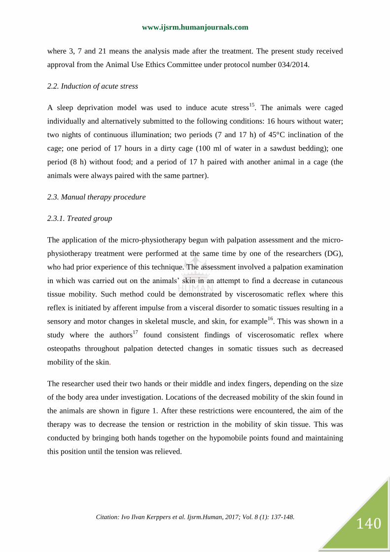
The researcher used their two hands or their middle and index fingers, depending on the size
of the body area under investigation. Locations of the decreased mobility of the skin found in
the animals are shown in figure 1. After these restrictions were encountered, the aim of the
therapy was to decrease the tension or restriction in the mobility of skin tissue. This was
conducted by bringing both hands together on the hypomobile points found and maintaining
this position until the tension was relieved.
www.ijsrm.humanjournals.com
Citation: Ivo Ilvan Kerppers et al. Ijsrm.Human, 2017; Vol. 8 (1): 137-148.
141
Figure 1. Localization of decrease mobility of the skin in the animals.
2.3.2. Placebo and Control Group
Each animal from the placebo group was removed from its cage, placed on the researcher´s
hand (AH), and with other hand had its neck and backstroked gently during 10 minutes in a 1
Hz cycle approximately. This researcher (AH) was not aware about the micro-physiotherapy
technique, to exclude any possible effects of this technique. In this manner, we were trying to
show two distinct effects. The control group was left undisturbed throughout the entire
protocol, except for regular cage cleaning, which was done for all animals by the same
researcher.
2.4. Flow Cytometry
In total, 1 ml of blood was collected from each rat for the flow cytometry analysis (BD
Accuri C6 - Becton Dickinson, USA). This content was deposited in microtubes of 1.5 ml
and kept in a water bath for 15 minutes, before being centrifuged at 1500 RPM for 5 minutes
at 18 degrees Celsius. After being centrifuged, the supernatant (serum) was separated for later
use. The BD™ cytometric bead array (CBA) mouse th1/th2 cytokine kit was used to detect
IL-2, IL-4, IL-6, TNFα and IFN, following the manufacturer’s instructions. Initially, we
added 50ul of the serum sample from each rat to 50ul of the mix containing an equal
proportion of the specific beads for each cytokine, with 50ul of detection reagent and 50ul of
standard diluent. after incubation for two hours in the dark, 1 ml of water was added and

www.ijsrm.humanjournals.com
Citation: Ivo Ilvan Kerppers et al. Ijsrm.Human, 2017; Vol. 8 (1): 137-148.
142
centrifuged for 2000 RPM for 5 minutes at 4 degrees Celsius. Subsequently, the supernatant
was discarded and resuspended in 300ul of water.
The theoretical limit of detection for each cytokine using the BD™ cytometric bead array
(CBA) mouse th1/th2 cytokine Kit was defined as the concentration corresponding to two
standard deviations above the mean fluorescence of 30 repetitions of the negative control
(0pg/ml). The following limits were applied: IL-2 = 0.1pg/ml; IL-4 = 0.03pg/ml and TNF =
0.9pg/ml. The cytometry reading was conducted manually, with the acquisition of 10,000
events from each sample.
2.5. Analysis of the cytometry data
FCap 3.0 Array software was used to analyze the samples after the performance of the flow
cytometry. The results were displayed in graphs, including the mean and standard deviations
values.
2.6. Euthanasia
All rats were euthanized 1 day after the procedures. The animals were anesthetized with 80
mg/kg ketamine and 15 mg/kg xylazine and euthanized with an intraperitoneal injection of a
lethal dose of thiopental. The procedure was performed in an experimental room without the
presence of other animals.
2.7. Statistical Analyses
The Shapiro-Wilk normality test was used and the sample was analyzed using the Kruskal-
Wallis test, with Dunn´s post-test.
3. RESULTS
Figure 2 displays the mean and standard deviation values for interleukin 2 in the control,
placebo and treated (micro-physiotherapy) groups. Note that these values were 648.8±144,2
pg/ml in CG3, 863.9±33.19 pg/ml in CG7 and 978.8±12.01 pg/ml in CG21. There was an
increase of interleukin 2 (pg/ml) in the CG. The mean values were 1191.0±591.8 pg/ml in
PG3, 635.6±284.3 pg/ml in PG7 and 1484.0±71,92 pg/ml in PG21, with a notable variance
between the interleukin values in PG3 and PG21. The mean values were 907.8±1156 pg/ml

www.ijsrm.humanjournals.com
Citation: Ivo Ilvan Kerppers et al. Ijsrm.Human, 2017; Vol. 8 (1): 137-148.
143
for TG3, 156.4±140.3 pg/ml for TG7 and 50.35±49.07 pg/ml for TG21. No statistically
significant values were found in the Kruskal Wallis test (P=0.1483).
Figure 2.
Figure 3 displays the mean and standard deviation values for IL-4: CG3 = 845.5±219.4; CG7
= 1204.0±381.0; and CG21 = 1718.0±197.8. Note that the presence of this interleukin
increased in the CG during the study period. The mean and standard deviation values in PG3
were 929.2±266.7, while those in PG7 were 755.4±70.9 and those in PG21 were
953.9±474.9. In the PG, there was a clear maintenance of the pg/ml of interleukin 4. In TG3,
the mean values were 49.15±36.23, while in TG7 they were 74.79±70.09 and in TG21, they
were 67.37±27.92. Note that the levels of interleukins increased slightly in TG7 and remained
low in the other two periods. A statistically significant difference was recorded in the
statistical analysis (p=0.0008). Dunn´s post-test confirmed significant differences between
the groups, the largest of which occurred between CG21 and TG21 (P=0.0078).
Figure 3.

www.ijsrm.humanjournals.com
Citation: Ivo Ilvan Kerppers et al. Ijsrm.Human, 2017; Vol. 8 (1): 137-148.
144
The pg/ml levels for TNF-α were 1788.0±85.50 pg/ml in CG3, 2464.0±295.3 pg/ml in CG7
and 2657.0±7.6 pg/ml in CG21. The TNF-α levels were found to increase in the CG and the
increase was continuous. The mean and standard deviation values were 3052.0±1496.0 pg/ml
in PG3, 2536.0±88.71pg/ml in PG7 and 2636.0±593.8pg/ml in PG21. In this case, for the PG,
the TNF-α levels exhibited a slight decrease in the concentration. The mean and standard
deviation values were 62.28±72.87pg/ml in TG3, 1039.0±670.7pg/ml in TG7 and
74.85±68.41 pg/ml in TG21. Notably, the concentration of TNF-α was higher in TG7 than in
TG3 and TG21. TG3 and TG21 exhibited similar levels. In the statistical analysis, the values
exhibited significant data, with a p-value of p=0.0033. A statistically significant difference
was recorded in Dunn´s test in CG21 ≠ TG21 and PG21 ≠ TG21, with p-values of 0.0078 and
0.0144, respectively (Fig 4).
Figure 4.
4. DISCUSSION
Chennoui et al.18
analyzed the presence of TNF-α during sleep deprivation and reported that
this cytokine plays a role in this situation. The same authors confirmed that the
concentrations of TNF-α and IL-6 were significantly higher among sedentary sleep-deprived
rats than among sedentary control rats. In addition, the IL-6 concentration was statistically
lower in sleep-deprived rats that performed exercise than among sedentary sleep-deprived
rats. In the present study, the TNF values were high in the CG and PG groups (3, 7 and 21
days), when compared with the TG groups. This suggests that the technique used to treat the
animals was effective in terms of controlling the liberation of TNF by the cells. No
significant correlation was found between the CG and the TG. The present study confirmed

www.ijsrm.humanjournals.com
Citation: Ivo Ilvan Kerppers et al. Ijsrm.Human, 2017; Vol. 8 (1): 137-148.
145
that TNF-α (pro-inflammatory cytokine) can activate immune-mediated inflammation in the
brain, thereby increasing the laboratory evidence that TNF-α activates the molecular
inflammatory mechanisms that induce neurotoxicity, for example.
The technique of micro-physiotherapy promoted immediate and delayed activity (for two
months). It is possible to confirm the influence of pro- and anti-inflammatories by comparing
TNF-α and IL-2 with IL-4, which increased in the 21-day period in the treated group. Subtle
tactile stimulation is capable of maintaining the homeostasis of the metabolism of the skin,
thereby demonstrating the interaction between the neuroendocrine system and cutaneous
stimulation19, 20
. This subtle contact (promoted in micro-physiotherapy) alters the HPA axis
through variations in the immune system, given that the skin and its appendages can generate
the same mediators used during the responses to systemic stress21
. Through the HPA axis of
the skin, the active stress of the mastoid cells leads to a selective suppression of the TH1
response, altering humoral immunity22
. A number of authors have demonstrated that the skin
and its appendages are richly innervated and their efferent signals are well represented by the
sensory cortex20,23
, which may explain the improvement in the regulation of cytokines in the
present study. Treatment protocols that involve subtle tactile stimulation have provided
favorable results among humans and animals in relation to stress24
, the autonomous nervous
system25
and neuro immunomodulatory systems26
.
Meltzer et al.27
and MacNeil et al.28
reported that the high SNS tone is a response to the
inflammatory alterations to the system. High circulating concentrations of TNF-α, IL-1 and
IL-6 act on the hypothalamus to stimulate the pathways of the central nervous system that
drive the sympathetic system to expend energy in the relevant target tissues, including
secondary lymphoid tissues and the inflammation sites. In the present study, the subtle
contact involved in the micro-physiotherapy identified restrictions in tissue mobility in areas
corresponding to the adrenal glands, pituitary gland, and thymus. After the intervention
involving light touch (approximately 5 to 10 grams), there was a significant reduction in the
quantity of the cytokines IL-4 and TNF-α. These responses to stress may occur as result of
the effective functions that these subtle touches promote when compared with discriminative
tactile functions. Type-C afferent fibers (Cf) respond with minimal contact in a pleasant
manner29
. Furthermore, Milne et al.30
have previously demonstrated that spinothalamic
neurons converge these nociceptive inputs towards the skin when faced with a visceral
stimulus. According to Craig31
, the Cf can be considered as a large extension of the afferent

www.ijsrm.humanjournals.com
Citation: Ivo Ilvan Kerppers et al. Ijsrm.Human, 2017; Vol. 8 (1): 137-148.
146
system that is involved in the monitoring of tissues (in skin, muscles, and viscera). This
system integrates several internal signals (of the body) and the cutaneous tissue, which is
vital for the maintenance of homeostatic balance.
In addition, psychological stress can impair several aspects of the immune system. Reiche et
al.32
reported that communication between the CNS, the endocrine system, and the immune
system involves chemical messengers, soluble mediators which are secreted by nerve cells,
cells from the endocrine organs or immune cells, and psychological stressors may disturb this
communication network. Palumbo et al.15
found that the concentration of IL-8 was
significantly lower among patients with post-traumatic stress disorder. In a second
assessment, they confirmed higher concentrations of IL-2 and IL-6. In the present study, it
was found that the level of IL-2 remained high in CG3, PG3 and TG3, when compared with
the level found in CG7, PG7 and most notably, TG7. It was notable that the level of IL-2 was
low in TG21. Based on these results, it is possible to hypothesize that the micro-
physiotherapy affected the liberation of IL-2 during the study period. This inhibition related
to the technique decreased over the 21-day period.
Some limitations may be raised in this study. Lack of another researcher to see the validity
and reliability of palpation in areas of poor skin mobility. Regarding the placebo group, the
same researcher who performed the technique in the experimental group could have
performed on the placebo, however, he would not be "blind" to the applied technique and this
could interfere with the results. In relation to analyzes, histochemical data from the
suprarenal and pituitary gland would be interesting to see the effects of the micro-
physiotherapy technique on cortisol and acetylcholine respectively. Future studies should
address these issues to better understand the mechanisms behind the micro-physiotherapy
technique, as well as research in humans where the effect of psychological stress is closest to
reality.
5. CONCLUSION
Based on the analysis of serum from rats submitted to micro-physiotherapy, the levels of pro-
inflammatory and anti-inflammatory cytokines remained below the levels of the other groups.
Thus, this technique influenced the immune system in terms of treating the mechanism of
acute stress.

www.ijsrm.humanjournals.com
Citation: Ivo Ilvan Kerppers et al. Ijsrm.Human, 2017; Vol. 8 (1): 137-148.
147
6. REFERENCES
1. Chrousos GP, Gold PW. The Concepts of Stress and Stress System Disorders Overview of. Jama. 1992;
267:1244–52.
2. Selye H. A syndrome produced by diverse nocuous agents. J Neuropsychiatry Clin Neurosci. 1998;
10(2):230–1.
3. McEwen BS, Seeman T. Protective and Damaging Effects of Mediators of Stress: Elaborating and Testing
the Concepts of Allostasis and Allostatic Load. Ann N Y Acad. 1999; 896(1):30–47.
4. Xiang L, Marshall GD. Immunomodulatory effects of in vitro stress hormones on FoxP3, Th1/Th2
cytokine, and costimulatory molecule mRNA expression in human peripheral blood mononuclear cells.
Neuroimmunomodulation. 2011; 18(1):1–10.
5. McEwen BS. Central effects of stress hormones in health and disease: Understanding the protective and
damaging effects of stress and stress mediators. Eur J Pharmacol. 2008; 583(2-3):174–85.
6. Ginaldi L, De Martinis M, D’Ostilio A, Marini L, Loreto MF, Corsi MP, Quaglino D. The immune system
in the elderly: I. Specific humoral immunity. Immunol Res. 1999; 20(2):101–8.
7. Martino M, Rocchi G, Escelsior A, Fornaro M. Immunomodulation Mechanism of Antidepressants:
Interactions between Serotonin/Norepinephrine Balance and Th1/Th2 Balance. Curr Neuropharmacol. 2012;
10(2):97–123.
8. Ahmad SF, Zoheir KMA, Ansari MA, Korashy HM, Bakheet SA, Ashour AE, Attia SM. Stimulation of
the histamine 4 receptor with 4-methylhistamine modulates the effects of chronic stress on the Th1/Th2 cytokine
balance. Immunobiology. 2015; 220(3):341–9.
9. Fitzgerald GK, McClure PW, Beattie P, Riddle DL. Issues in determining treatment effectiveness of manual
therapy. Phys Ther. 1994; 74(3):227–33.
10. Krohn M, Listing M, Tjahjono G, Reisshauer A, Peters E, Klapp BF, Rauchfuss M. Depression, mood,
stress, and Th1/Th2 immune balance in primary breast cancer patients undergoing classical massage therapy.
Support Care Cancer. 2011; 19(9):1303–11.
11. Rapaport MH, Schettler P, Bresee C. A preliminary study of the effects of repeated massage on
hypothalamic-pituitary-adrenal and immune function in healthy individuals: a study of mechanisms of action
and dosage. J Altern Complement Med. 2012; 18(8):789–97.
12. Salgado ASI, Parreira RB, Santos IR, Urbano JJ, Fonsêca NT, Bénini P, Grosjean D. Effects of
microkinesitherapie on heart rate variability. Man Ther Posturology & Rehabil J. 2013; 11:488–93.
13. Pereira A, Carvalho E, Knippers II, Furmann M, Pires J, Ribeiro L, et al. Assessment of heart rate
variability in fibromialgia after micro-physiotherapy. Man Ther Posturology Rehabil J. 2014; 12:191–5.
14. Menezes J. Inteligência quantica - por um mundo melhor. Rio de Janeiro: Novo ser; 110 p; 2006.
15. Palumbo ML, Canzobre MC, Pascuan CG, Ríos H, Wald M, Genaro AM. The stress induced cognitive
deficit is differentially modulated in BALB/c and C57Bl/6 mice: correlation with Th1/Th2 balance after stress
exposure. J Neuroimmunol. 2010; 218(1-2):12–20.
16. Beal MF, Mazurek MF, Tran VT, Chattha G, Bird ED, Martin JB. Reduced numbers of somatostatin
receptors in the cerebral cortex in Alzheimer's disease. Science. 1985; 229(4710):289-91.
17. Licciardone JC, Fulda KG, Stoll ST, Gamber RG, Cage AC. A case-control study of osteopathic palpatory
findings in type 2 diabetes mellitus. Osteopath Med Prim Care. 2007; 1:6.
18. Chennaoui M, Gomez-Merino D, Drogou C, Geoffroy H, Dispersyn G, Langrume C, Ciret S, Gallopin
T, Sauvet F. Effects of exercise on brain and peripheral inflammatory biomarkers induced by total sleep
deprivation in rats. J Inflamm (Lond). 2015; 12:56.
19. Kuhn CM, Schanberg SM, Field T, Symanski R, Zimmerman E, Scafidi F, Roberts J. Tactile-kinesthetic
stimulation effects on sympathetic and adrenocortical function in preterm infants. J Pediatr. 1991; 119(3):434–
40.
20. Mathai S, Fernandez A, Mondkar J, Kanbur W. Effects of tactile-kinesthetic stimulation in preterms: a
controlled trial. Indian Pediatr. 2001; 38(10):1091–8.
www.ijsrm.humanjournals.com
Citation: Ivo Ilvan Kerppers et al. Ijsrm.Human, 2017; Vol. 8 (1): 137-148.
148
21. Ito N, Ito T, Kromminga A, Bettermann A, Takigawa M, Kees F, Straub RH, Paus R. Human hair follicles
display a functional equivalent of the hypothalamic-pituitary-adrenal axis and synthesize cortisol. FASEB J.
2005; 19(10):1332–4.
22. Marshall JS, Gomi K, Blennerhassett MG, Bienenstock J. Nerve growth factor modifies the expression of
inflammatory cytokines by mast cells via a prostanoid-dependent mechanism. J Immunol. 1999; 162(7):4271–6.
23. Arck PC, Slominski A, Theoharides TC, Peters EMJ, Paus R. Neuroimmunology of stress: skin takes center
stage. J Invest Dermatol. 2006; 126(8):1697–704.
24. Henricson M, Ersson A, Määttä S, Segesten K, Berglund AL. The outcome of tactile touch on stress
parameters in intensive care: a randomized controlled trial. Complement Ther Clin Pract. 2008; 14(4):244–54.
25. Diego MA, Field T. Moderate Pressure Massage Elicits a Parasympathetic Nervous System Response. Int J
Neurosci. 2009; 119(5):630–8.
26. Waters-Banker C, Butterfield TA, Dupont-Versteegden EE. Immunomodulatory effects of massage on
nonperturbed skeletal muscle in rats. J Appl Physiol. 2014; 116(2):164–75.
27. Meltzer JC, MacNeil BJ, Sanders V, Pylypas S, Jansen AH, Greenberg AH, Nance DM. Contribution of the
adrenal glands and splenic nerve to LPS-induced splenic cytokine production in the rat. Brain Behav Immun.
2003; 17(6):482–97.
28. MacNeil BJ, Jansen AH, Greenberg AH, Nance DM. Activation and selectivity of splenic sympathetic
nerve electrical activity response to bacterial endotoxin. Am J Physiol. 1996; 270( 2):64–70.
29. Olausson H, Cole J, Rylander K, McGlone F, Lamarre Y, Wallin BG, Krämer H, Wessberg J, Elam
M, Bushnell MC, Vallbo A. Functional role of unmyelinated tactile afferents in human hairy skin: sympathetic
response and perceptual localization. Exp Brain Res. 2008; 184(1):135–40.
30. Milne RJ, Foreman RD, Giesler GJ, Willis WD. Convergence of cutaneous and pelvic visceral nociceptive
inputs onto primate spinothalamic neurons. Pain. 1981; 11(2):163–83.
31. Craig AD. How do you feel? Interoception: the sense of the physiological condition of the body. Nat Rev
Neurosci. 2002; 3(8):655–66.
32. Reiche EMV, Nunes SOV, Morimoto HK. Stress, depression, the immune system, and cancer. Lancet
Oncol. 2004; 5(10):617–25.

![Employee Attrition and Retention Strategies in Amara Raja ... ijsrm.pdf · DOI: 10.18535/ijsrm/v5i6.07 K. SreenivasMahesh, IJSRM Volume 5 Issue 06 June 2017 [] Page 5398 c. The costs](https://img.pdfslide.net/doc/110x75/5af0b2a07f8b9ac2468e63f3/employee-attrition-and-retention-strategies-in-amara-raja-ijsrmpdfdoi-1018535ijsrmv5i607.jpg)


![Structural Functionalism Analysis On Children … ijsrm.pdf · Bambang Soepeno, IJSRM Volume 05 Issue 09 September 2017 [] Page 7131 13-15 years are allowed to do light work, as long](https://img.pdfslide.net/doc/110x75/5b5851687f8b9a88698c0878/structural-functionalism-analysis-on-children-ijsrmpdf-bambang-soepeno-ijsrm.jpg)

![Challenges of Transformative-Strategic Leadership in … ijsrm.pdf · DOI: 10.18535/ijsrm/v5i8.10 Peter Njuguna Waichungo, IJSRM Volume 5 Issue 08 August 2017 [] Page 6639 world](https://img.pdfslide.net/doc/110x75/5ac4ab557f8b9a220b8cefb0/challenges-of-transformative-strategic-leadership-in-ijsrmpdfdoi-1018535ijsrmv5i810.jpg)