Embed Size (px)
Citation preview

Accepted by S. Carranza: 7 Mar. 2007; published: 12 Apr. 2007 1
ZOOTAXAISSN 1175-5326 (print edition)
ISSN 1175-5334 (online edition)Copyright © 2007 · Magnolia Press
Zootaxa 1446: 1–20 (2007) www.mapress.com/zootaxa/
A new species of Temple Pitviper (Tropidolaemus Wagler, 1830) from Sulawesi, Indonesia (Squamata: Viperidae: Crotalinae)
ULRICH KUCH1, ANDREAS GUMPRECHT2 & CHRISTIAN MELAUN3
1Sektion Herpetologie, Forschungsinstitut und Naturmuseum Senckenberg, Senckenberganlage 25, 60325 Frankfurt am Main, Germany. E-mail: [email protected] Str. 35, 53842 Troisdorf, Germany3Institut für Allgemeine Zoologie und Entwicklungsbiologie, Justus-Liebig-Universität Gießen, Stephanstr. 24, 35390 Gießen, Germany
Abstract
The Asian Temple Pitviper Tropidolaemus wagleri is a widespread polytypic species complex with a complicated taxo-nomic history, a lengthy species synonymy list, and a geographic distribution encompassing Vietnam, Thailand, Malay-sia, Singapore, Brunei, portions of Indonesia, and the Philippines. As a prelude to a comprehensive review of this speciescomplex, we describe a new species of Temple Pitviper based on five historic museum specimens from the Indonesianisland of Sulawesi. The new species can be distinguished from sympatric members of the Tropidolaemus subannulatuscomplex and other congeners on the basis of its conspicuous color pattern and scalation characters. Available collectingdata suggest that the new species has a wide distribution in rainforests and lower montane wet forests of Sulawesi Utaraand Sulawesi Tengah provinces.
Key words: Reptilia, Squamata, Viperidae, Crotalinae, Tropidolaemus, Tropidolaemus laticinctus sp. nov., Tropidolae-mus subannulatus, Tropidolaemus wagleri, Indonesia, morphological characters, pitviper, snake, Southeast Asia,Sulawesi, venomous
Introduction
The genus Tropidolaemus Wagler, 1830 currently comprises two species of pitvipers from mainland and insu-lar Asia (McDiarmid et al. 1999; Gumprecht et al. 2004). These small to medium-sized (about 35–100 cmtotal length) snakes are arboreal ambush predators with remarkable morphological features (Burger 1971;Hoge & Romano-Hoge 1983). Their venom contains neurotoxins called waglerins which are unique amongsnake venom toxins (Molles & Taylor 2002). Snakes of this genus are used in ceremonial contexts and tradi-tionally displayed in a Buddhist temple in Pulau Pinang, Malaysia (Manthey & Grossmann 1997), and conse-quently often referred to as Temple Pitvipers, or Wagler's Pitvipers.
After a long period of inclusion in the complex genus Trimeresurus (sensu lato), Tropidolaemus wasresurrected from synonymy and regarded as a subgenus of the latter by Brattstrom (1964) based on anatomicaland external characters. On the basis of morphological characteristics Burger (1971) considered Tropidolae-mus to be a distinct genus. This view was widely adopted in the literature (e.g., Hoge & Romano-Hoge 1981,1983; McDiarmid et al. 1999; Orlov et al. 2002; Gumprecht et al. 2004), and further supported by molecularstudies (Kraus et al. 1996; Malhotra & Thorpe 2000; Parkinson 1999; Parkinson et al. 2002; Vidal & Lecoin-tre 1998), which identified Tropidolaemus as an ancient lineage of Old World pitvipers without close relation-ships to the various genera now recognized from within the Trimeresurus complex (Malhotra & Thorpe 2004;

KUCH ET AL.2 · Zootaxa 1446 © 2007 Magnolia Press
Creer et al. 2006). Instead, most genetic studies suggested the possibility of a sister-group relationshipbetween Tropidolaemus and the terrestrial Indochinese pitviper genus Deinagkistrodon Gloyd, 1979 (Kraus etal. 1996; Parkinson 1999; Parkinson et al. 2002; Vidal & Lecointre 1998).
Temple Pitvipers are distributed in Southeast Asia, including southern Vietnam, peninsular Thailand,Malaysia and Singapore, the Philippines, the Indonesian islands of Sumatra, Sulawesi, and Borneo includingBrunei and the Malaysian states of Sabah and Sarawak, and on various smaller islands (Gumprecht et al.2004; Iskandar & Colijn 2001; Leviton 1964; Manthey & Grossmann 1997). However, David and Vogel(1998) referred the holotype of a rare pitviper from southern India (Trimeresurus huttoni Smith, 1949) toTropidolaemus based on similarity in its external phenotype, thereby greatly extending the known range ofthis genus. If confirmed by genetic or osteological data, this extended concept of Tropidolaemus will raiseinteresting biogeographical questions.
Temple Pitvipers are remarkably diverse in color pattern, and herpetologists in the nineteenth centurydescribed various taxa from this group (e.g., Gray 1842, 1849; Peters 1872). However, all of these were con-sidered to be conspecific, and synonymized with the species Tropidolaemus wagleri (F. Boie, 1827), by Bou-lenger (1896, as Lachesis wagleri). Instead of various nominal taxa, Boulenger (1896) recognized fiveunnamed color varieties ("A" to "E"). An attempt to revise Boulenger's view was made by Taylor (1917,1922a, b), who described a new subspecies (T. wagleri alboviridis Taylor, 1917) from Negros Island in thePhilippines, and recognized three additional taxa elsewhere in the Philippines: T. w. wagleri (restricted to Bal-abac and Palawan), Tropidolaemus wagleri subannulatus (type locality: “Philippines”; Gray, 1842), andTropidolaemus philippensis (type locality: “Philippine Islands”; Gray, 1842). All these were however againsynonymized with a monotypic T. wagleri by Leviton (1964). Boulenger's (1896) and Leviton's (1964) inter-pretation of T. wagleri as a single, widespread and morphologically variable species has been followed,although sometimes with hesitation, by most subsequent authors (e.g., David & Ineich 1999; David & Vogel1996, 1998; Gumprecht et al. 2004; Hoge & Romano-Hoge 1981; McDiarmid et al. 1999; Orlov et al. 2002).A common argument against the recognition of subspecies within T. wagleri, or several species of Tropidola-emus, has been the apparent difficulty of correlating differences in color pattern with geographic populationsor morphology (Leviton 1964).
Recent molecular studies on Tropidolaemus have allowed us to appreciate more fully the dramatic ontoge-netic color change and sexual dimorphism occurring in these snakes, thus unveiling phenotypic and geo-graphic congruence and helping define species boundaries (U. Kuch & N. Vidal, unpubl.). Followingconference presentations of this work, the availability of old names for Temple Pitviper populations was high-lighted in regional checklists for Southeast Asia, where various color morphs, subspecies, and species fromwithin T. wagleri were provisionally recognized (Iskandar & Colijn 2001; Vogel 2006) but without accompa-nying data or taxonomic justification.
In the course of revisions of museum specimens we found that green Temple Pitvipers with narrow bandsor spots are common in eastern Indonesia and the Philippines. Pending the completion of studies in progress,we will refer to these as an operational Tropidolaemus subannulatus complex. However, we also discoveredseveral unusual specimens of Tropidolaemus whose external phenotype differs substantially from that of T.subannulatus. Among the most divergent of these are several Temple Pitvipers from the Indonesian island ofSulawesi. The distinctiveness of these was first noticed by Boulenger (1896: 564) who established a separatevariety "E" within his concept of Lachesis wagleri for a specimen from "Minahasa, Celebes" (later specifiedas "Sonder, Minahassa" [Boulenger 1897: 227], now Sonder, Province of Sulawesi Utara, Indonesia) whosecoloration was green dorsally with large brick-red, black-edged spots, and white ventrally with black spotsand brick-red marblings, and a red tail tip (Boulenger 1896, 1897). A second, similarly colored specimen from"between L. Posso and Tomini Gulf, Central Celebes" (i.e., between Lake Poso and Tomini Bay, Province ofSulawesi Tengah, Indonesia) was illustrated by Boulenger (1897: Pl. XV; figs. 1–5, 6A, 7A), and contrasted tothree specimens from the Bone Valley (Province of Sulawesi Utara) which Boulenger referred to "the typical

Zootaxa 1446 © 2007 Magnolia Press · 3A NEW SPECIES OF TEMPLE PITVIPER
form" (i.e., variety "A" in Boulenger [1896]) based on their green coloration with white blue- or purple-edgedtransverse lines (Boulenger 1897: 227). Two additional specimens with this remarkable brick-red to brownringed pattern were reported by Ahl (1933) and illustrated in Gumprecht et al. (2004). The photograph of anapparently uncollected individual from Gunung Tangkoko (Province of Sulawesi Utara, Indonesia) is con-tained in de Lang and Vogel (2005, 2006) and Vogel (2006). Here we present evidence supporting the recogni-tion of this highly distinctive form as a species separate from T. wagleri and members of the T. subannulatuscomplex, employing the criterion of diagnosability (sensu de Queiroz 1998, 1999). As this conspicuous formis not represented by any of the available types and names for Temple Pitvipers, we introduce it as a new spe-cies, for which we provide a detailed description and diagnosis.
Material and methods
We examined preserved or live specimens of Tropidolaemus from the entire range of the genus except Viet-nam. Institutional abbreviations are listed in Leviton et al. (1985). Terminology follows Campbell and Lamar(2004). Ventral scale counts were performed according to the method proposed by Dowling (1951). Dorsalscale rows were counted one head length behind the angle of the jaw, at midbody, and one head length beforethe cloaca. Measurements of the head were made with calipers to the nearest 0.1 mm. A string was used todetermine the snout-vent length (from the tip of the snout to the posterior margin of the anal plate) and the taillength (from the posterior edge of the cloacal plate to the tip of the tail). The following head measurementswere recorded: distance from anteroventral corner of eye to caudal border of pit (EP), distance from antero-dorsal corner of eye to centre of naris (EN), horizontal distance across eye (ED), distance from tip of snout toangle of jaw (HL), height of rostral taken at midline (RH), width of rostral taken at widest point (RW). Sexwas determined by direct observation of the gonads or hemipenes in most specimens; in some cases sex wasdetermined based on sexually dimorphic characters of body and tail size and proportions in combination withsexually dimorphic color pattern characters, or by probing in live snakes. Color descriptions of the new spe-cies are based on the examination of alcohol-preserved specimens and photographs of two live specimens (deLang & Vogel 2005, 2006; this paper). Data on the color pattern of sympatric members of the T. subannulatuscomplex and of T. wagleri were obtained from both live and preserved specimens. Latitudes, longitudes andelevations were either taken from specimen tags, from the online database of the Koninklijk Instituut voor deTropen (2006), or from the Indonesia file of geographic names available from the GEOnet names server(National Geospatial-Intelligence Agency 2006).
Systematic account
Tropidolaemus laticinctus sp. nov. (figs. 1–6, 7A+B, 8A+B, 9–10, and 11A+B)
Lachesis wagleri variety E (Boulenger 1896) Lachesis wagleri (Boulenger 1897, in part)Trimeresurus wagleri, "bunte, rotgebänderte Form" (Ahl 1933)Trimeresurus wagleri (Leviton 1964, in part)Tropidolaemus wagleri (Hoge & Romano-Hoge 1981, in part)Tropidolaemus wagleri celebensis (Iskandar & Colijn 2001, in part)Tropidolaemus wagleri, "red form" (de Lang & Vogel 2005)Tropidolaemus subannulatus (celebensis morph 2) (Vogel 2006) [Date of publication: 10 March 2006]Tropidolaemus wagleri, "red form" (de Lang & Vogel 2006) [Date of publication: 16 December 2006]
Suggested English name: Broad-banded Temple Pitviper

KUCH ET AL.4 · Zootaxa 1446 © 2007 Magnolia Press
FIGURE 1. Color plate of Boulenger's variety "E" of Lachesis wagleri showing the holotype of Tropidolaemus laticinc-tus (BMNH 96.12.9.80 from "between L. Posso and Tomini Bay, Celebes" [= between Lake Poso and Tomini Bay, Prov-ince of Sulawesi Tengah, Indonesia]). Drawing and lithography by J. Green, from Boulenger (1897: Pl. XV).

Zootaxa 1446 © 2007 Magnolia Press · 5A NEW SPECIES OF TEMPLE PITVIPER
FIGURE 2. Dorsal view of male holotype of Tropidolaemus laticinctus (BMNH 96.12.9.80 from "between L. Posso andTomini Bay, Celebes" [= between Lake Poso and Tomini Bay, Province of Sulawesi Tengah, Indonesia]). Photograph byAndreas Gumprecht.
FIGURE 3. Ventral view of holotype of Tropidolaemus laticinctus (BMNH 96.12.9.80). Photograph by Andreas Gum-precht.

KUCH ET AL.6 · Zootaxa 1446 © 2007 Magnolia Press
FIGURE 4. Holotype of Tropidolaemus laticinctus (BMNH 96.12.9.80), detail of ringed color pattern on lateral body.Photograph by Andreas Gumprecht.
FIGURE 5. Holotype of Tropidolaemus laticinctus (BMNH 96.12.9.80), dorsal view of head. Photograph by AndreasGumprecht.
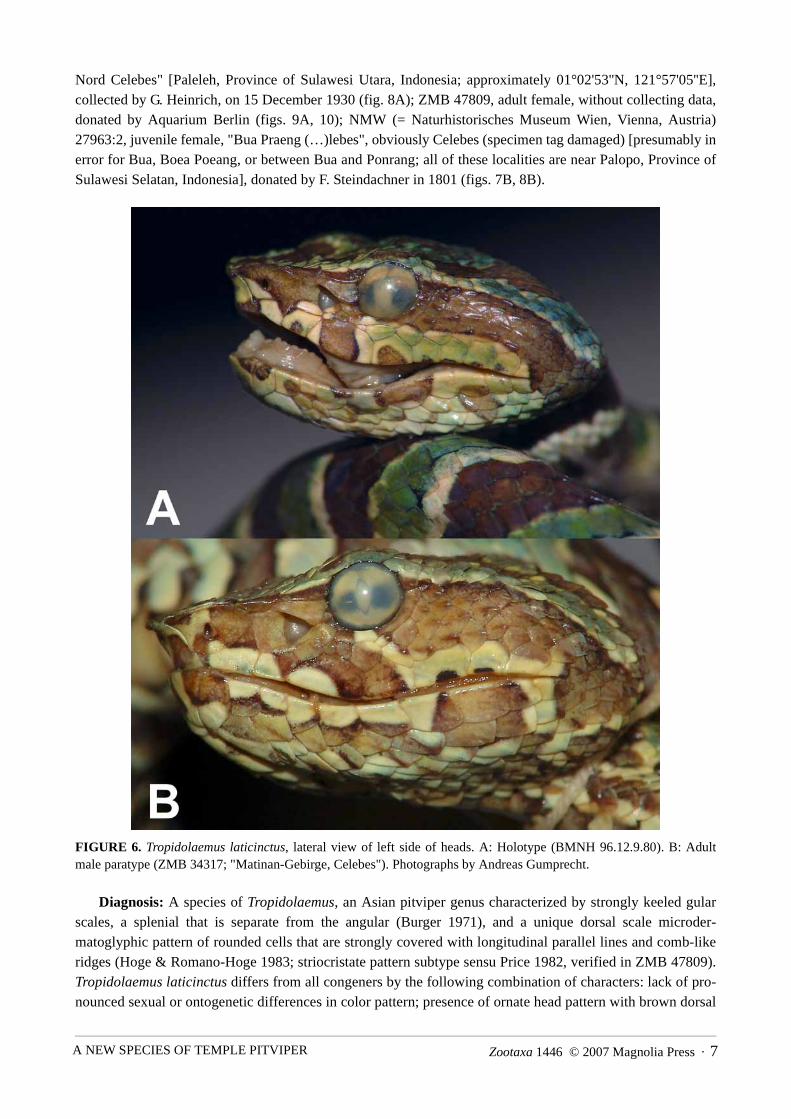
Holotype: BMNH (= The Natural History Museum, London, United Kingdom) 96.12.9.80, subadult or adultmale, "between L. Posso and Tomini Bay, Celebes" [= between Lake Poso and Tomini Bay, Province ofSulawesi Tengah, Indonesia], collected by P. & F. Sarasin (figs. 1–5, 6A, 7A).
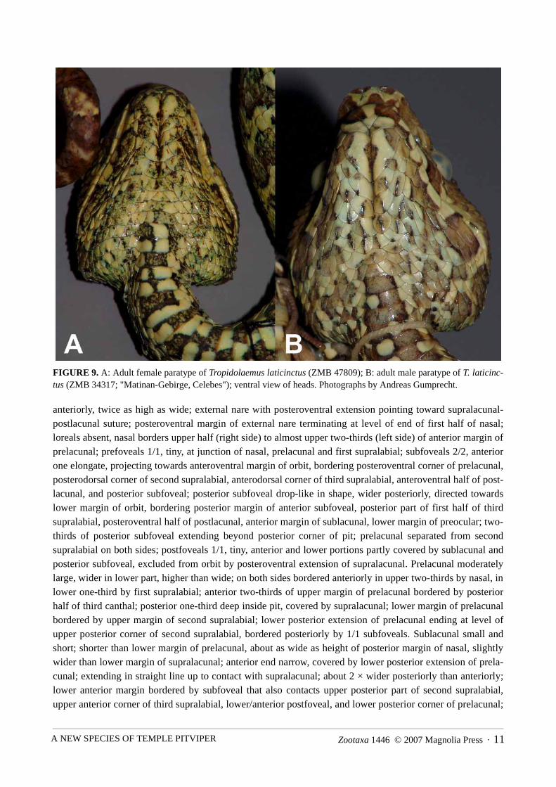
Paratypes (4): ZMB (= Institut für systematische Zoologie, Museum für Naturkunde der Humboldt-Uni-versität zu Berlin, Berlin, Germany) 34317, adult male, "Matinan-Gebirge, Celebes" [likely foothills ofGunung Tentolo Matinan, southwest of Paleleh, Province of Sulawesi Tengah, Indonesia], ca. 100 m abovesea level, collected by G. Heinrich, on 20 October 1930 (figs. 6B, 9B); ZMB 34318, adult female, "Paleleh,
Zootaxa 1446 © 2007 Magnolia Press · 7A NEW SPECIES OF TEMPLE PITVIPER
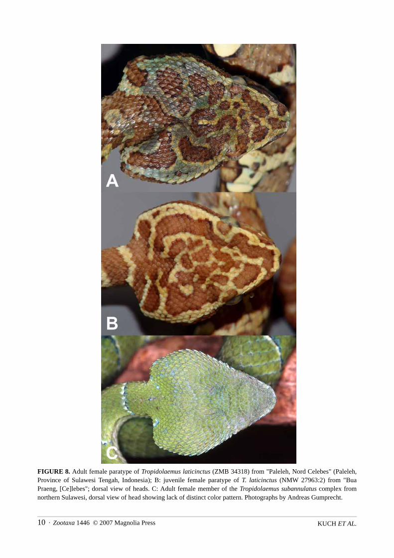
Nord Celebes" [Paleleh, Province of Sulawesi Utara, Indonesia; approximately 01°02'53''N, 121°57'05''E],collected by G. Heinrich, on 15 December 1930 (fig. 8A); ZMB 47809, adult female, without collecting data,donated by Aquarium Berlin (figs. 9A, 10); NMW (= Naturhistorisches Museum Wien, Vienna, Austria)27963:2, juvenile female, "Bua Praeng (…)lebes", obviously Celebes (specimen tag damaged) [presumably inerror for Bua, Boea Poeang, or between Bua and Ponrang; all of these localities are near Palopo, Province ofSulawesi Selatan, Indonesia], donated by F. Steindachner in 1801 (figs. 7B, 8B).
FIGURE 6. Tropidolaemus laticinctus, lateral view of left side of heads. A: Holotype (BMNH 96.12.9.80). B: Adultmale paratype (ZMB 34317; "Matinan-Gebirge, Celebes"). Photographs by Andreas Gumprecht.
Diagnosis: A species of Tropidolaemus, an Asian pitviper genus characterized by strongly keeled gularscales, a splenial that is separate from the angular (Burger 1971), and a unique dorsal scale microder-matoglyphic pattern of rounded cells that are strongly covered with longitudinal parallel lines and comb-likeridges (Hoge & Romano-Hoge 1983; striocristate pattern subtype sensu Price 1982, verified in ZMB 47809).Tropidolaemus laticinctus differs from all congeners by the following combination of characters: lack of pro-nounced sexual or ontogenetic differences in color pattern; presence of ornate head pattern with brown dorsal

KUCH ET AL.8 · Zootaxa 1446 © 2007 Magnolia Press
markings on green ground that are bordered by cream scales and are largest on the posterior head; wide brownpre- and postocular stripe that extends to lower side of head beyond angle of jaw; ornate labial pattern consist-ing of brown spots bordered by white or black, on green to cream ground color; conspicuous large brown spotextending from anteroventral corner of eye and posteroventral margin of pit to lower margin of supralabials;lower side of head with similar pattern as dorsal side of head, or mottled with numerous brown and blackspots and streaks; dorsal body pattern consisting of broad brick-red to brown rings that may be incompletedorsally or laterally and are about as wide as or wider than the cream to white interspaces; anterodorsal part oflight interspaces of body pattern containing wide green bands; brown rings across belly connected midven-trally to form midventral stripe (if brown rings incomplete ventrally, venter heavily spotted, with chessboard-like pattern); base of tail with whitish and brown rings, tip of tail lighter reddish brown with no or only indis-tinct bands.
Tropidolaemus wagleri differs from T. laticinctus in having distinct sexual differences in color pattern, infemales undergoing a dramatic ontogenetic color change from a bright green ground color to a largely blackand yellow coloration with or without green components, in lacking an extensively patterned venter, and inlacking an ornate labial pattern. Juvenile females of T. wagleri further differ from T. laticinctus in having adorsal pattern of very narrow red and white bands on bright green ground color and in lacking a dorsal headpattern; male T. wagleri of all ages differ from T. laticinctus in having a dorsal pattern of tiny red and whitespots and in lacking a dorsal head pattern. Members of the T. subannulatus complex (including the types ofTrimesurus [sic] philippensis Gray, 1842, Trimesurus [sic] subannulatus Gray, 1842, and Trigonocephaluswagleri var. celebensis Gray, 1849) differ from T. laticinctus in lacking a well-defined ornate head patternwith light-bordered dorsal markings and in lacking an ornate labial pattern, in lacking a ventral pattern ofbrown rings and/or midventral stripe, and in having a dorsal pattern of narrow bands or spots (vs. broad brick-red to brown rings). Both sexes of sympatric members of the T. subannulatus complex differ from T. laticinc-tus in having a uniform or near-uniform green dorsal head color (fig. 8C, vs. ornate brown head pattern), anarrower bluish (vs. wide brown) pre- and postocular stripe and largely patternless labial region (fig. 7C, vs.ornate labial pattern), the presence (vs. absence) of well-defined, black-edged ocelli on outer posterior mar-gins of ventral scales (fig. 7C), and an otherwise uniform green to bluish green venter (vs. heavily patternedventer with brown rings and midventral stripe). Sympatric members of the T. subannulatus complex furtherdiffer from T. laticinctus in having narrow white and bluish or reddish bands or spots on uniform green groundcolor (fig. 11C, vs. broad brick-red to brown rings), and in having a tail pattern consisting of narrow bluishbands and wide green interspaces at the base and nearly equidistant, distinct narrow black and white bands onbrownish to greyish ground towards the tail tip (fig. 11C, vs. well-defined, approximately equidistant light andbrown rings on base of tail, and light reddish brown tail tip with no or only indistinct bands). Adult females ofsympatric members of the T. subannulatus complex also differ from the adult female T. laticinctus illustratedby de Lang & Vogel (2005, 2006) in having a yellow to golden (figs. 7C, 11C; vs. reddish) eye color.
In characters of scalation, the specimens of the available small series of T. laticinctus (N=5) differ fromthe examined T. subannulatus complex members from Sulawesi (N=16) in having a slightly convex anteriormargin of the mental scale (vs. centrally concave when viewed from the same angle, or indented at the level ofthe tongue), and in having higher ventral scale counts (139–146 vs. 134–139, respectively). The females of T.laticinctus (N=3) also differ from the majority of examined T. subannulatus complex females from Sulawesi(11 of 13) in having a higher number of dorsal scale rows at midbody (25 vs. 23, respectively).
Description of holotype: A small pitviper with a subtriangular head that is very distinct from the neck,and a prehensile tail. Rostral overall trapezoidal, lower margin of rostral 2 × wider than upper margin, higherthan wide, almost vertical along suture with first supralabial on right side, only slightly concave alongsupralabial suture on left side; dorsal margin of rostral rounded, projecting anteriorly, forming part of verysharply upturned canthus rostralis; nasal very large, undivided, dorsal margin of nasal projecting dorsally andlaterally, forming major part of canthus rostralis; internal naris subelliptical, upper part turned slightly more

Zootaxa 1446 © 2007 Magnolia Press · 9A NEW SPECIES OF TEMPLE PITVIPER
FIGURE 7. A: Holotype of Tropidolaemus laticinctus (BMNH 96.12.9.80); B: juvenile female paratype of T. laticinctus(NMW 27963:2), lateral view of right head sides. C: Adult female member of the Tropidolaemus subannulatus complexfrom northern Sulawesi; lateral view of head showing bluish pre- and postocular stripe, largely patternless labial region,and presence of distinct black-edged blue ocelli on outer posterior margins of ventral scales. Photographs by AndreasGumprecht.
KUCH ET AL.10 · Zootaxa 1446 © 2007 Magnolia Press
FIGURE 8. Adult female paratype of Tropidolaemus laticinctus (ZMB 34318) from "Paleleh, Nord Celebes" (Paleleh,Province of Sulawesi Tengah, Indonesia); B: juvenile female paratype of T. laticinctus (NMW 27963:2) from "BuaPraeng, [Ce]lebes"; dorsal view of heads. C: Adult female member of the Tropidolaemus subannulatus complex fromnorthern Sulawesi, dorsal view of head showing lack of distinct color pattern. Photographs by Andreas Gumprecht.
Zootaxa 1446 © 2007 Magnolia Press · 11A NEW SPECIES OF TEMPLE PITVIPER
FIGURE 9. A: Adult female paratype of Tropidolaemus laticinctus (ZMB 47809); B: adult male paratype of T. laticinc-tus (ZMB 34317; "Matinan-Gebirge, Celebes"); ventral view of heads. Photographs by Andreas Gumprecht.
anteriorly, twice as high as wide; external nare with posteroventral extension pointing toward supralacunal-postlacunal suture; posteroventral margin of external nare terminating at level of end of first half of nasal;loreals absent, nasal borders upper half (right side) to almost upper two-thirds (left side) of anterior margin ofprelacunal; prefoveals 1/1, tiny, at junction of nasal, prelacunal and first supralabial; subfoveals 2/2, anteriorone elongate, projecting towards anteroventral margin of orbit, bordering posteroventral corner of prelacunal,posterodorsal corner of second supralabial, anterodorsal corner of third supralabial, anteroventral half of post-lacunal, and posterior subfoveal; posterior subfoveal drop-like in shape, wider posteriorly, directed towardslower margin of orbit, bordering posterior margin of anterior subfoveal, posterior part of first half of thirdsupralabial, posteroventral half of postlacunal, anterior margin of sublacunal, lower margin of preocular; two-thirds of posterior subfoveal extending beyond posterior corner of pit; prelacunal separated from secondsupralabial on both sides; postfoveals 1/1, tiny, anterior and lower portions partly covered by sublacunal andposterior subfoveal, excluded from orbit by posteroventral extension of supralacunal. Prelacunal moderatelylarge, wider in lower part, higher than wide; on both sides bordered anteriorly in upper two-thirds by nasal, inlower one-third by first supralabial; anterior two-thirds of upper margin of prelacunal bordered by posteriorhalf of third canthal; posterior one-third deep inside pit, covered by supralacunal; lower margin of prelacunalbordered by upper margin of second supralabial; lower posterior extension of prelacunal ending at level ofupper posterior corner of second supralabial, bordered posteriorly by 1/1 subfoveals. Sublacunal small andshort; shorter than lower margin of prelacunal, about as wide as height of posterior margin of nasal, slightlywider than lower margin of supralacunal; anterior end narrow, covered by lower posterior extension of prela-cunal; extending in straight line up to contact with supralacunal; about 2 × wider posteriorly than anteriorly;lower anterior margin bordered by subfoveal that also contacts upper posterior part of second supralabial,upper anterior corner of third supralabial, lower/anterior postfoveal, and lower posterior corner of prelacunal;

KUCH ET AL.12 · Zootaxa 1446 © 2007 Magnolia Press
posteriorly, sublacunal bordering 2/2 postfoveals. Supralacunal large, anterodorsal projection bordering poste-rolateral margin of third canthal, remaining dorsal margin of supralacunal covered by fourth canthal whichalso forms upper preocular; posterior margin of supralacunal almost vertical, but rounded in lower posteriorcorner, here covering upper anterior end of small, rounded lower preocular; lower margin anteriorly borderingsublacunal, posteriorly bordering upper postfoveal; lower anterior margin forming posterodorsal border of pit,extending upwards in an angle of approximately 45° from above posterior one-third of sublacunal to start ofposterolateral one-fourth of third canthal and posterior one-fourth of dorsal margin of prelacunal; lower ante-rior margin of supralacunal straight to slightly concave in upper part bordering pit. Preoculars 3/3, formed byentire posterior margin of small keeled scale that is completely fused to the posteriormost (fourth) canthal(upper preocular, bordering one-fourth of anterior margin of orbit), posterior margin of supralacunal (middlepreocular, bordering half of anterior margin of orbit), and a small lower preocular; lower preocular touchingthe eye, forming only one-fourth of anterior margin of orbit; lower preocular located between supralacunaland subocular, separated from third supralabial by subocular and one small interoculabial. Suboculars 1/1,long and narrow; extending from before anterior margin of lower preocular to level of posteriormost point oforbit, about as wide anteriorly as posteriorly, smooth anteriorly, slightly rugose posteriorly, upper posteriormargin covered by lower of two postoculars. Postoculars weakly keeled; upper postocular slightly larger, par-ticipating in orbit with about half of length of scale, about as much as lower postocular. Anterior end of leftsubocular in contact with central part of upper margin of third supralabial, posterior one-third of dorsal marginof third supralabial separated from subocular by one tiny triangular and one larger keeled interoculabial; rightsubocular separated from third supralabial; interoculabials 3/3, one row of scales between subocular andsupralabials: one scale each between subocular and suture of third/fourth and fourth/fifth supralabial; 2/2scales (in diagonal view) between posterior part of subocular and corner of sixth supralabials, the lowermostcovered anteriorly by posterodorsal margin of fifth supralabial, posteriorly covering anterodorsal margin ofsixth supralabial, both weakly keeled; 3/3 scales between posterior part of subocular and corner of seventhsupralabials. Upper lip curved; highest parts located at rostro-mental contact and the corners of the mouth;lowest parts at suture of third/fourth supralabials, corresponding approximately to level of fourth/fifth infrala-bials. Supralabials 9/10; first supralabial extending up to one-third of height of prelacunal, separate fromnasal; second supralabial about as large as first, separate from prelacunal; third supralabial largest, more than2 × size of first supralabial, about 2 × wider than high; fourth supralabial second largest, about as high aswide; supralabials 5–9/5–10 much smaller than fourth, but larger than dorsally adjacent scales; ninth/tenthsupralabials smallest. Internasals 1/1, small, about as wide as long; laterally covering anteromedian ends offirst canthals. Canthals 4/4, first pair widest, second shortest, third and fourth elongate and narrow, smooth butsome with uneven scale surface; first pair approximately 2 × longer than wide, entirely on top of head, bor-dered laterally by two intercanthals; second canthals ca. 1½ × longer than wide, bordered by three intercan-thals; third canthals about 2 × longer than wide, bordered by two intercanthals; fourth canthals approximately2 × longer than wide, bordered by two intercanthals and the supraocular; outer margins of third and fourthcanthals turned to side of head; posteror tip of fourth canthal on both sides fused to tiny keeled scale adjacentto anterolateral margin of supraoculars. Canthus rostralis sharply pronounced, projecting anteriorly and ante-rolaterally; nasals, first and second canthals and internasals contributing more to canthus rostralis than thirdand fourth canthals. Anterior and lateral intercanthals only weakly keeled, central intercanthals more dis-tinctly keeled, mostly narrower than internasals but often longer; three intercanthals between first pair of can-thals immediately posterior to internasals, the middle one directly posterior to the internasal suture being verysmall; increasing posteriorly to 5, 6, 7, 8 and 9–10 intercanthals, the latter between the fourth canthals.Supraoculars small, elongate and narrow, with uneven scale surface and poorly defined margins due to obvi-ous fusion with at least two adjacent scales each, on either side with 1–2 indistinct keels; supraoculars about1½–2 × longer and 2 × wider than fourth canthals; bordered by 8/8 scales including the fourth canthals withposteriorly fused scale and postoculars. Intersupraoculars keeled, minimum number 9 (between anterior ends

Zootaxa 1446 © 2007 Magnolia Press · 13A NEW SPECIES OF TEMPLE PITVIPER
FIGURE 10. Adult female paratype of Tropidolaemus laticinctus (ZMB 47809). A: dorsal view; B: ventral view. Photo-graphs by Andreas Gumprecht.
of supraoculars), maximum number 13 (between posterior ends), 11 between middle of supraoculars. Poste-rior head scales weakly to strongly keeled; approximately 32 interrictals. Scales on lower head (especiallymental, chinshields, also anterior infralabials and supralabials) with numerous tiny tubercular structures (pre-sumably mechanoreceptors). Mental large, anterior margin of convex appearance, slightly concave on bothsides, wider than long; bordered by first infralabials; posterior tip of mental separating first infralabials,

KUCH ET AL.14 · Zootaxa 1446 © 2007 Magnolia Press
extending between anterior end of chinshield suture. Infralabials 9/10; first infralabials 2 × higher than wide inupper part, in contact with mental, second infralabials, and chinshields; second infralabials small, rectangular,wider than high; on left side, third infralabial wider and ca. 1½ × higher than second infralabials; on right side,third infralabial small, 2 × higher than wide; infralabials 4/5+6 largest, infralabials 5–9/7–10 of similar sizeand shape, gradually becoming smaller posteriorly; on left side, ninth infralabial smallest (about ½ × smallerthan second infralabial); on right side, tenth infralabial second smallest, being slightly larger than third infrala-bial. One pair of chinshields, wide, with pointed posterior extension left and right of distinct mental groove,about 2 × longer than wide at longest part; bordered by mental, infralabials 1–2, two sublabials, and firstgular; chinshields followed by seven pairs of smooth to very slightly keeled gulars, the anterior of which arewider than long, followed posteriorly by one preventral that is mostly covered by laterally adjacent scales; 6–7 rows of sublabials between last infralabial and posteriormost gular; 146 ventrals; anal plate undivided; 52subcaudals, all divided, plus a terminal scale equal in length to two terminal subcaudals; dorsal body scalesarranged in 21/21/17 rows, only slightly keeled, most keels only on posterior half of scale, extending to termi-nus of scale; paraventrals smooth over most of body, slightly wider than adjacent dorsals; dorsal scales on tailand parasubcaudals without keels. Measurements of holotype (in millimeters): SVL 350, TL 66, EP 1.2, EN5.8, ED 3.5, HL 20.9, RH 2.69, RW 2.63.
FIGURE 11. A+B: Live Tropidolaemus laticinctus from the western side of Dumoga Bone National Park, about 30 kmnortheast of Gorontalo, Sulawesi Utara, Indonesia. Photographs by Tommy Ahnby. C: Live adult female member of theTropidolaemus subannulatus complex from northern Sulawesi showing narrow white and bluish bands and spots on uni-form green ground color, and tail tip with distinct pattern of nearly equidistant, narrow black and white bands on brown-ish to greyish ground. Photograph by Andreas Gumprecht.
Coloration of holotype (in preservative; figs. 1–5, 6A, 7A): Top of head with ornate head pattern com-posed of brown, green and cream elements; in dorsal view, outer margins of internasals and first canthals
Zootaxa 1446 © 2007 Magnolia Press · 15A NEW SPECIES OF TEMPLE PITVIPER
greenish cream, central portions as well as anteromedian parts of first intercanthals brown; posterior part ofresulting brown spot on right side connected to larger brown spot centered in anterior canthal region, coveringan area of approximately ten intercanthals; two brown spots of similar size and shape in posterior intercanthalregion, left one separate from anterior intercanthal spot and left supraocular but with small V-shaped centralextension, right one broadly fused to anterior intercanthal spot and just contacting anteromedian margin ofright supraocular; two smaller, irregularly shaped brown spots in anterior intersupraocular region; supraocu-lars centrally with brown area that is continuous (left) or near continuous (right) to large L-shaped (left) andinversely L-shaped (right) brown blotch; posteromedian extensions of these latter blotches pointing towardeach other but not in contact in occipital region, thereby framing a fronto-parietal area that contains a roughlytrapezoidal brown mark with central green spot; another brown spot involving about ten scales posterior towhere short legs of L-shaped blotches point toward each other; right (inversely) L-shaped blotch posteriorlyfused to large brown interrictal mark that is concave anteromedially and posterolaterally, convex anterolater-ally, has a posteromedian extension pointing toward the neck, and at its outer posterior margins connects tobrown pattern elements on the posterior sides of the head; brown spots on top of head in various parts withindistinct darker to blackish outer margins, in addition to more distinct, narrow cream to greenish cream bor-ders that set off the brown head pattern from much of the intervening light green (top of snout) to dark green(posterior head) areas. Rostral pale greenish yellow with brown upper and lower margins and small brownishcentral suffusion; anteroventral corner of nasal pale greenish yellow up to anteroventral margin of naris, restof nasal brown; first pair of supralabials brown along sutures with rostral and brown part of nasal and prelacu-nal, pale greenish yellow elsewhere; second supralabial pale yellow with greenish hue centered near postero-dorsal corner, and irregularly dark-edged brown spot in lower central part of scale; lower part of prelacunalcream, upper part greenish, finely dusted with brown; posterior two canthals with cream inner and brownouter margins; all scales bordering anterior orbit, including entire supralacunal, brown mixed with green pig-ment; posterior half of sublacunal brown mixed with green, anterior half cream; subfoveal cream; postfovealsbrown mixed with green; third supralabial with wide central brown area extending down from brown preocu-lar area, set off from cream anterior and posterior margins of third supralabial by narrow dark brown to blackmargins; anteriormost tip of subocular brown (part of brown preocular area), then cream up to about level ofcenter of eye, and brown posteriorly; lower postocular brown, upper postocular cream with brown lower cor-ner; from fourth supralabial posteriorly, ground color of supralabials and margins of adjacent scales pale yel-lowish green; anterior margin of fourth supralabial and anterior one-third of interoculabial between third andfourth supralabial and subocular cream; fourth supralabial with dorsally rounded brown spot on lower centralmargin; fifth to eighth supralabials with brown or black lower central margins; color of eye golden, withextensive black pigment anterior and posterior to vertically elliptical pupil, indicative of a continuation of thebrown pre-and postocular stripe; wide brown postocular stripe extending from posterior orbit to beyond angleof jaw, covering 3–4.5 scales in width, upper and lower margin delimited by black pigment only on a fewscales; upper margin of postocular stripe bordered by narrow cream line extending from upper postocular tobeyond angle of jaw. Mental and first infralabials greenish yellow, with indistinct brown spots on both uppermental-infralabial sutures and lower (posterior) end of mental; ground color of infralabials from cream andgreenish yellow anteriorly to pale yellowish green posteriorly, with variable degrees of brown dusting andspots; posterior part of second infralabial dusted with brown; third infralabial with poorly defined smallbrown streak in upper central part; fourth infralabial with brown spot in lower anterior corner and narrowdiagonal extension to upper posterior corner; brown posterior margin of fifth and brown anterior margin ofseventh supralabial forming dark-edged light brown infralabial spot together with brown-dusted sixth infrala-bial; upper posterior corner of seventh and upper anterior corner of eighth infralabial also with brown spot;posterior half of posteriormost supralabial and posterior one-third of posteriormost infralabial forming part ofdark brown margin of postocular stripe; ground color of scales on lower side of head cream to yellowishgreen; chinshields each with small black central spot and posterolateral black-edged brown spot that continues

KUCH ET AL.16 · Zootaxa 1446 © 2007 Magnolia Press
posteriorly as a poorly defined brown pattern element on adjacent scales; numerous gulars and other scaleswith small brown or black spots or streaks or dusted with brown or black pigment.
FIGURE 12. Relief map of Sulawesi, Indonesia, showing selected localities of Tropidolaemus laticinctus (rhomboids;filled: examined specimens; unfilled: photographs and literature records; area of the type locality marked by rhomboidwith thick black line) and Tropidolaemus subannulatus complex members (dots). Arrows designate localities of T. sub-annulatus complex members in the Siau and Sangihe Islands. An additional collecting locality of T. laticinctus ("BuaPraeng", presumably in error for Bua, Boea Poeang, or between Bua and Ponrang; all of these near Palopo, Province ofSulawesi Selatan) could not be reliably placed and is indicated by a question mark.

Zootaxa 1446 © 2007 Magnolia Press · 17A NEW SPECIES OF TEMPLE PITVIPER
Dorsal body pattern consisting of 35 brick-red to brown rings that are 2.5–4 dorsal scales long midverte-brally; ventrally, most of these rings are slightly directed toward the head; several rings laterally divided, jux-taposed, or incomplete dorsally or laterally (ring 1 split on right side; 2 split on left; 2+3 dorsally almost fused,3 on right side separated from ventral part; 4 split on left; 5 split on right; 7 split on left; 8 incomplete on right;13+30 dorsally almost interrupted, juxtaposed; 15 ventrally incomplete; 31+33 laterally divided on both sides;34 with anterior dorsal extension pointing toward 33; 35 present only on right side, dorsally pointing toward34); brown rings about as wide as or wider than interspaces; anterodorsal part of light interspaces of body pat-tern containing wide green bands; posterodorsal part of light interspaces with narrow cream bands; laterally,green color of light interspaces gradually fading towards venter; ventrolaterally, interspaces mostly cream towhite, on outer edges of ventrals with small dark (brown or black-edged brown) spots of variable number(usually 1–3), size, and shape; brown rings across belly connected midventrally to form midventral stripe;midventral stripe almost continuous along body, only four times interrupted at levels of first to sixth brownrings; midventral stripe about as wide as ventral part of brown rings, approximately as wide as 1–3 ventrals,with lateral extensions; midventral stripe and ventral part of brown rings tan to light brown, mostly black-edged; posterior margins of most ventrals at midventral stripe darker brown or black; ventral ground colorcream to light tan; base of tail with ten distinct brown rings; rings 2+3 on tail dorsally fused; rings 4+5 in con-tact dorsally; all other rings on tail separated by cream interspaces; green color on tail confined to dorsal partsof basal light interspaces; tip of tail light reddish brown with no rings or bands.
Variation: The number of ventral scales varies between 139 and 146 (males: 146; females: 139–140 ven-trals); the number of subcaudals ranges from 49–52 (males: 50–52; females: 49-51), all of which are divided.Supralabials 9–11 (males: 9–10; females: 10–11); interoculabials 3–4 (males: 3; females: 3–4); postoculars 2–3 (males: 2; females: 2–3), of which the lower 1–2 may be fused to the subocular; infralabials 9–11 (males: 9–10; females: 10–11), the first pair of which may or may not be in contact posterior to the mental; 4–7 gulars(males: 4–7; females: 7) between chinshield and first (pre-)ventral, arranged in pairs or (posteriorly) in rowsof three; preventrals 0–2 (males: 1–2; females: 0–2); intersupraoculars 9–13 (males: 9–10; females: 10–13);scales bordering intersupraoculars 8–9 (males: 8–9; females: 8–9); canthals 3–4 (males: 3–4; females: 3–4);intercanthals 3–7 (males: 3–6; females: 5–7) between first canthals, 9–12 (males: 9–11; females: 11–12)between posterior canthals; internasals 2, in contact, or separated by scale of approximately equal size, oranteromedian part of each internasal divided to form two separate scales; prefoveals 0–4 (males: 0–1;females: 2–4); dorsal scale rows 25/21/19 in males, 23–25/25/19 in females. Variation in color patternincludes about 30–42 dark brick-red to brown rings or bands on the body (males: 31–35; adult females: 30–38; juvenile female: ca. 40–42) which may be disintegrated, incomplete or juxtaposed vertebrally or laterally,interrupted laterally on one or both sides, and cover 2–5 dorsal scales in length midvertebrally; brown rings orbands bordered especially anteriorly, but sometimes also posteriorly, by narrow cream to white (fig. 11A+B)areas, or without distinct light borders (fig. 10A); some with narrow black borders along brown rings or bands(e.g., fig. 10A); 7–11 distinct brown rings or bands on basal part of tail (males: 10–11; adult females: 7–8;juvenile female: 11); light interspaces between brown rings or bands covering 1–3 dorsal scales in length mid-vertebrally; light interspaces very narrow in the juvenile specimen (NMW 27963:2), cream to tan after morethan 200 years in preservative (possibly green in life); light interspaces of the other specimens dorsally withwider green anterior part that may be nearly as wide as the brown rings, and with or without narrow cream towhite posterior part that borders the following brown ring; laterally, green color of light interspaces graduallyfading towards venter; ventrolaterally, interspaces mostly cream to white, on outer edges of ventrals withsmall dark (brown or black-edged brown) spots of variable number (usually 1–3), size, and shape that may beconnected to a brown midventral stripe; dorsal scales of green areas of many light interspaces also withbrown, black-edged brown, or black spots and streaks that may laterally extend up to the midvertebral region;green color on tail confined to dorsal parts of basal light interspaces; ventral ground color light tan to cream ordirty white; brown rings on venter connected to midventral stripe that may be incomplete and juxtaposed on
KUCH ET AL.18 · Zootaxa 1446 © 2007 Magnolia Press
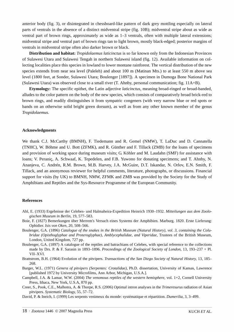
anterior body (fig. 3), or disintegrated in chessboard-like pattern of dark grey mottling especially on lateralparts of ventrals in the absence of a distinct midventral stripe (fig. 10B); midventral stripe about as wide asventral part of brown rings, approximately as wide as 1–3 ventrals, often with multiple lateral extensions;midventral stripe and ventral part of brown rings tan to light brown, mostly black-edged; posterior margins ofventrals in midventral stripe often also darker brown or black.
Distribution and habitat: Tropidolaemus laticinctus is so far known only from the Indonesian Provincesof Sulawesi Utara and Sulawesi Tengah in northern Sulawesi island (fig. 12). Available information on col-lecting localities place this species in lowland to lower montane rainforest. The vertical distribution of the newspecies extends from near sea level (Paleleh) and about 100 m (Matinan Mts.) to at least 550 m above sealevel (1800 feet, at Sonder, Sulawesi Utara; Boulenger [1897]). A specimen in Dumoga Bone National Park(Sulawesi Utara) was observed close to a small river (T. Ahnby, personal communication; fig. 11A+B).
Etymology: The specific epithet, the Latin adjective laticinctus, meaning broad-ringed or broad-banded,alludes to the color pattern on the body of the new species, which consists of comparatively broad brick-red tobrown rings, and readily distinguishes it from sympatric congeners (with very narrow blue or red spots orbands on an otherwise solid bright green dorsum), as well as from any other known member of the genusTropidolaemus.
Acknowledgments
We thank C.J. McCarthy (BMNH), F. Tiedemann and R. Gemel (NMW), T. LaDuc and D. Cannatella(TNHC), W. Böhme and U. Bott (ZFMK), and R. Günther and F. Tillack (ZMB) for the loans of specimensand provision of working space during museum visits; G. Köhler and M. Laudahn (SMF) for assistance withloans; V. Peraniç, A. Schwaaf, K. Tepedelen, and F.B. Yuwono for donating specimens; and T. Ahnby, N.Ananjeva, C. Andrén, R.M. Brown, M.B. Harvey, J.A. McGuire, D.T. Iskandar, N. Orlov, E.N. Smith, F.Tillack, and an anonymous reviewer for helpful comments, literature, photographs, or discussions. Financialsupport for visits (by UK) to BMNH, NMW, ZFMK and ZMB was provided by the Society for the Study ofAmphibians and Reptiles and the Sys-Resource Programme of the European Community.
References
Ahl, E. (1933) Ergebnisse der Celebes- und Halmaheira-Expedition Heinrich 1930–1932. Mitteilungen aus dem Zoolo-gischen Museum in Berlin, 19, 577–583.
Boie, F. (1827) Bemerkungen über Merrem's Versuch eines Systems der Amphibien. Marburg. 1820. Erste Lieferung:Ophidier. Isis von Oken, 20, 508–566.
Boulenger, G.A. (1896) Catalogue of the snakes in the British Museum (Natural History), vol. 3, containing the Colu-bridae (Opisthoglyphae and Proteroglyphae), Amblycephalidae, and Viperidae, Trustees of the British Museum,London, United Kingdom, 727 pp.
Boulenger, G.A. (1897) A catalogue of the reptiles and batrachians of Celebes, with special reference to the collectionsmade by Drs. P. & F. Sarasin in 1893–1896. Proceedings of the Zoological Society of London, 13, 193–237 + Pl.VII–XVI.
Brattstrom, B.H. (1964) Evolution of the pitvipers. Transactions of the San Diego Society of Natural History, 13, 185–268.
Burger, W.L. (1971) Genera of pitvipers (Serpentes: Crotalidae), Ph.D. dissertation, University of Kansas, Lawrence[published 1972 by University Microfilms, Ann Arbor, Michigan, U.S.A.].
Campbell, J.A. & Lamar, W.W. (2004) The venomous reptiles of the western hemisphere, vol. 1+2, Cornell UniversityPress, Ithaca, New York, U.S.A, 870 pp.
Creer, S., Pook, C.E., Malhotra, A. & Thorpe, R.S. (2006) Optimal intron analyses in the Trimeresurus radiation of Asianpitvipers. Systematic Biology, 55, 57–72.
David, P. & Ineich, I. (1999) Les serpents venimeux du monde: systématique et répartition. Dumerilia, 3, 3–499.

Zootaxa 1446 © 2007 Magnolia Press · 19A NEW SPECIES OF TEMPLE PITVIPER
David, P. & Vogel, G. (1996) The snakes of Sumatra. An annotated checklist and key with natural history notes. EditionChimaira, Frankfurt am Main, Germany, 260 pp.
David, P. & Vogel, G. (1998 [dated 1997]) Redescription of Trimeresurus huttoni Smith, 1949 (Serpentes, Crotalinae)with a discussion of its relationships. Hamadryad, 22, 73–87.
De Lang, R. & Vogel, G. (2005) The snakes of Sulawesi, Edition Chimaira, Frankfurt am Main, Germany, 312 pp.De Lang, R. & Vogel, G. (2006) The snakes of Sulawesi. In: Vences, M., Köhler, J., Ziegler, T., Böhme, W. (eds.) Herpe-
tologia bonnensis II. Proceedings of the 13th congress of the Societas Europaea Herpetologica, Bonn, Germany, pp.35–38.
De Queiroz, K. (1998) The general lineage concept of species, species criteria, and the process of speciation: A concep-tual unification and terminological recommendations. In: Howard, D.J. & Berlocher, S.H. (eds.) Endless forms: spe-cies and speciation, Oxford University Press, New York, New York, U.S.A., pp. 57–75.
De Queiroz, K. (1999) The general lineage concept of species and the defining properties of the species category. In:Wilson, R.A. (ed.) Species: new interdisciplinary essays, Massachusetts Institute of Technology Press, Cambridge,Massachusetts, U.S.A., pp. 49–89.
Dowling, H.G. (1951) A proposed standard system of counting ventrals in snakes. British Journal of Herpetology, 1, 97–99.
Gloyd, H.K. (1979) A new generic name for the hundred-pace viper. Proceedings of the Biological Society of Washing-ton, 91, 963–964.
Gray, J.E. (1842) Synopsis of the species of rattle-snakes, or family of Crotalidae. Zoological Miscellany, 2, 47–51. Gray, J.E. (1849) Catalogue of the specimens of snakes in the British Museum. British Museum (Natural History), Lon-
don, United Kingdom, 125 pp. Gumprecht, A., Tillack, F., Orlov, N.L., Captain, A. & Ryabov, S. (2004) Asian pitvipers, Geitje Books, Berlin, Germany,
368 pp. Hoge, A.R. & Romano-Hoge, S.A.R.W.L. (1981 [dated 1978/79]) Poisonous snakes of the world. Part I. Check list of the
pit vipers Viperoidea, Viperidae, Crotalinae. Memorias do Instituto Butantan, 42/43, 179–310. Hoge, A.R. & Romano Hoge, S.A. (1983 [dated 1980/81]) Notes on micro and ultrastructure of "Oberhäutschen" in
Viperoidea. Memorias do Instituto Butantan, 44/45, 81–118. Iskandar, D.T. & Colijn, E. (2001) A checklist of Southeast Asian and New Guinean reptiles. Part I. Serpentes, Biodiver-
sity Conservation Project, Indonesian Institute of Sciences, Japan International Cooperation Agency, The Ministryof Forestry, The Gibbon Foundation, and Institute of Technology Bandung, Jakarta, Indonesia, 195 pp.
Koninklijk Institut voor de Tropen (2006) KIT's historical colonial maps accessible online, Koninklijk Institut voor deTropen. Available from: http://www.kit.nl (accessed 27 March 2006).
Kraus, F., Mink, D.G. & Brown, W.M. (1996) Crotaline intergeneric relationships based on mitochondrial DNA sequencedata. Copeia, 1996, 763–773.
Leviton, A.E. (1964) Contributions to a review of Philippine snakes. V. The snakes of the genus Trimeresurus. PhilippineJournal of Science, 93, 251–276.
Leviton, A.E., Gibbs Jr., R.H., Heal, E. & Dawson, C.E. (1985) Standards in herpetology and ichthyology: Part I. Stan-dard symbolic codes for institutional resource collections in herpetology and ichthyology. Copeia, 1985, 802–832.
Malhotra, A. & Thorpe, R.S. (2000) A phylogeny of the Trimeresurus group of pit–vipers: new evidence from a mito-chondrial gene tree. Molecular Phylogenetics and Evolution, 16, 199–211.
Malhotra, A. & Thorpe, R.S. (2004) A phylogeny of four mitochondrial gene regions suggests a revised taxonomy forAsian pit vipers (Trimeresurus and Ovophis). Molecular Phylogenetics and Evolution, 31, 83–100.
Manthey, U. & Grossmann, W. (1997) Amphibien & Reptilien Südostasiens. Natur und Tier-Verlag, Münster, Germany,512 pp.
McDiarmid, R.W., Campbell, J.A. & Touré, T.A. (1999) Snake species of the world. A taxonomic and geographic refer-ence, vol. 1, The Herpetologists' League, Washington, D.C., U.S.A., 511 pp.
Molles, B.E. & Taylor, P. (2002) Structure and function of the waglerins, peptide toxins from the venom of Wagler's PitViper, Tropidolaemus wagleri. Journal of Toxicology – Toxin Reviews, 21, 273–292.
National Geospatial-Intelligence Agency (2006) GEOnet names server (GNS), geographic names data base, containingofficial standard names approved by the United States Board on Geographic Names, National Geospatial-Intelli-gence Agency, USA. Available from: http://earth-info.nga.mil/gns/html/ (accessed 30 April 2006).
Orlov, N.L., Ananjeva, N., Barabanov, A., Ryabov, S., & Khalikov, R. (2002) Diversity of vipers (Azemiopinae, Crotali-nae) in East, Southeast and South Asia: Annotated checklist and natural history data (Reptilia: Squamata: Serpentes:Viperidae). Faunistische Abhandlungen des Museums für Tierkunde Dresden, 23, 177–218.
Parkinson, C.L. (1999) Molecular systematics and biogeographical history of pitvipers as determined by mitochondrialribosomal DNA sequences. Copeia, 1999, 57–586.
Parkinson, C.L., Campbell, J.A. & Chippindale, P.T. (2002) Multigene phylogenetic analysis of pitvipers, with commentson their biogeography. In: Schuett, G.W., Höggren, M., Douglas, M.E. & Greene, H.W. (eds.) Biology of the vipers,Eagle Mountain Publishing, Eagle Mountain, Utah, U.S.A., pp. 93–110.

KUCH ET AL.20 · Zootaxa 1446 © 2007 Magnolia Press
Peters, W.C.H. (1872) Mittheilung ueber einige von Hrn. Dr. A. B. Meyer bei Gorontalo und auf den Togian-Inseln ges-ammelte Amphibien. Monatsberichte der Königlichen Akademie der Wissenschaften zu Berlin, 1872, 581–585.
Price, R.M. (1982) Dorsal scale microdermatoglyphics: ecological indicator or taxonomic tool? Journal of Herpetology,16, 294–306.
Smith, M.A. (1949) A new species of pit viper from south India: Trimeresurus huttoni sp. nov. Journal of the BombayNatural History Society, 48, 596.
Taylor, E.H. (1917) Snakes and lizards known from Negros, with descriptions of new species and new subspecies. Phil-ippine Journal of Science (D), 13, 353–381.
Taylor, E.H. (1922a) The snakes of the Philippine Islands. Monographs of the Bureau of Science, Manila, 16, 1–312. Taylor, E.H. (1922b) Additions to the herpetological fauna of the Philippine Islands. II. Philippine Journal of Science
(D), 21, 257–303. Vidal, N. & Lecointre, G. (1998) Weighting and congruence: a case study based on three mitochondrial genes in pitvi-
pers. Molecular Phylogenetics and Evolution, 9, 366–374. Vogel, G. (2006) Venomous snakes of Asia/Giftschlangen Asiens, Edition Chimaira/Aqualog Verlag, Frankfurt am Main/
Rodgau, Germany, 148 pp.Wagler, J.G. (1830) Natürliches System der Amphibien, mit vorangehender Classification der Säugethiere und Vögel. Ein
Beitrag zur vergleichenden Zoologie, J. G. Cotta Buchhandlung, München, Germany, 354 pp.
Appendix 1. Selected additional specimens examined
Tropidolaemus subannulatus complex: No locality: ZMB 63864. PHILIPPINES: "Philippine Islands": BMNH1946.1.17.67; "Philippines": 1946.1.19.32, BMNH 1946.1.19.33. INDONESIA: "Celebes": BMNH 49.3.2.39;"Sulawesi": SMF 86850–86856, ZFMK 68525, ZFMK 76337, ZFMK 76338; "Siau Langi Ins." (likely SULAWESI
UTARA PROVINCE: Sangihe Islands: Siau Island): ZMB 7427; SULAWESI SELATAN PROVINCE, Bantimurung: SMF75739, SMF 77910; SULAWESI TENGAH PROVINCE, Kabupaten Donggala, Kulani, Torro, 600 m: BMNH 1980.936;Moronali Nature Reserve, Ranu River: BMNH 1980.1718; [SULAWESI UTARA PROVINCE], "N. Celebes, Bone Val-ley", 650–1000 feet (fide Boulenger 1897): BMNH 96.12.9.79; Sangihe Islands, Tamako, Tankoko National Park:ZFMK 73918; SULAWESI UTARA PROVINCE: Kabupaten Gorontalo: Kecamatan Suawa: Desa Lombongo: DusunDua: Kampung Lompongo: TNHC 59994.