Embed Size (px)
Citation preview

Dynamic Article LinksC<Analyst
Cite this: Analyst, 2012, 137, 4262
www.rsc.org/analyst PAPER
Dow
nloa
ded
by U
NIV
ER
SIT
Y O
F SO
UT
H A
UST
RA
LIA
on
27 A
ugus
t 201
2Pu
blis
hed
on 2
6 Ju
ne 2
012
on h
ttp://
pubs
.rsc
.org
| do
i:10.
1039
/C2A
N35
520E
View Online / Journal Homepage / Table of Contents for this issue
A new strategy for the detection of adenosine triphosphate by aptamer/quantum dot biosensor based on chemiluminescence resonance energy transfer
Zi-Ming Zhou, Yong Yu and Yuan-Di Zhao*
Received 20th April 2012, Accepted 26th June 2012
DOI: 10.1039/c2an35520e
We designed an aptasensor for the detection of adenosine triphosphate (ATP) based on
chemiluminescence resonance energy transfer (CRET). An adenosine aptamer was cut into two pieces
of ssDNA, which were attached to quantum dots (QDs) and horse radish peroxidase (HRP),
respectively. They could reassemble into specific structures in the presence of ATP and then decrease
the distance of HRP and QDs. ATP detection can be easily realized according to the fluorescent
intensity of QDs, which is excited by CRET between luminol and QDs. Results show that the
concentration of ATP is linear relation with the fluorescent intensity of the peak of QDs emission and
the linear range for the linear equation is from 50 mM to 231 mM and the detection limit was 185 nM.
When the concentration of ATP was 2 mM, the efficiency of CRET is 13.6%. Good specificity for ATP
had been demonstrated compared to thymidine triphosphate (TTP), cytidine triphosphate (CTP) and
guanosine triphosphate (GTP), when 1 mM of each was added, respectively. This method needs no
external light source and can avoid autofluorescence and photobleaching, and ATP can be detected
selectively, specifically, and sensitively in a low micromolar range, which means that the strategy
reported here can be applicable to the detection of several other target molecules.
Introduction
ATP is the basic biological energy source in life, which sustains
every biological process. Existing research shows that the content
of ATP in vivo has great influence on cell metabolism and death.1
Moreover, many diseases demonstrate an abnormal content of
ATP, such as cardiovascular disease,2 parkinsonism,3 and Alz-
heimer’s.4 Therefore, it’s important to detect ATP in life science
and clinical medical research.
At present, there is lots of methods for detection of ATP, like
chromatography, bioluminescence assay, electrochemical, and
fluorescent biosensor.5 Chromatography is restricted to the
tedious separation of sample and low accuracy detection. A
broad method for detection of ATP at present is based on
luciferase, which has a good sensitivity but its accuracy some-
times could be affected by quenching in matrix. Low specificity
and sensitivity are always the challenges in electrochemical
sensors. In a word, how to detect ATP with high specificity and
sensitivity and more simply is a hot issue.
Aptamers, first reported by three groups independently in
1990,6 are the artificial single-stranded DNA or RNA sequences,
which are screened using the systematic evolution of ligands by
Britton Chance Center for Biomedical Photonics, Wuhan NationalLaboratory for Optoelectronics, Key Laboratory of BiomedicalPhotonics of Ministry of Education, Department of BiomedicalEngineering, Huazhong University of Science and Technology, Wuhan430074, People’s Republic of China. E-mail: [email protected];Fax: +86 27-8779-2202
4262 | Analyst, 2012, 137, 4262–4266
the exponential enrichment (SELEX) approach. They could fold
into special structures, and bind to certain targets with extremely
high specificity, such as metal ions,7 small molecules8 and
proteins.9 Owing to its high specificity, thermostability, easy
synthesis, and modification, aptamers have been used in many
biosensors for the detection of thrombin,10 cocaine11 or ATP,12
etc., which are known as aptasensors. Fan’s group has developed
a target-responsive electrochemical aptamer switch technology
(TREAS). In their research, ferrocene was used as a signal
molecule to observe the change of current before and after
aptamer bound to ATP to achieve electrochemical detection of
ATP.13 Plaxco and coworkers cut aptamers into two pieces.14
When targets, like ATP or cocaine, were added, they worked as
molecular linkers forming sandwich structures with the two
pieces of aptamer, making signal molecules close to the electrode,
which then induced the change of current.
Compared with other methods, the traditional fluorescence
method needs small samples and has high sensitivity, high
selectivity, and is easy to operate. Many works have been
reported using this method. Brennan’s group fixed aptamers in
sol–gel-derived silica material,15 using fluorescein as a signal
molecule. The detection of ATP was realized after 4,4-dimethy-
lamino-azobenzene-40-carboxylic acid (DABCYL), the quencher
molecule, was separated by adding ATP, recovering the fluo-
rescence. Yamana’s group reported the use of pyrene fluo-
rophore labelled anti-ATP aptamer to detect ATP.16 The light
intensity of pyrene fluorophores is sensitive to conformational
change of aptamer by ATP.
This journal is ª The Royal Society of Chemistry 2012

Dow
nloa
ded
by U
NIV
ER
SIT
Y O
F SO
UT
H A
UST
RA
LIA
on
27 A
ugus
t 201
2Pu
blis
hed
on 2
6 Ju
ne 2
012
on h
ttp://
pubs
.rsc
.org
| do
i:10.
1039
/C2A
N35
520E
View Online
In contrast to organic dye, quantum dots (QDs) have wide
excitation, narrow emission, size-dependent character and
resistance to light bleaching. As a new fluorescent probe, QDs
have been used in aptamer biosensors to detect biomolecules and
metal ions. Deng’s group reported a biosensor based on aptamer/
QDs, where QDs were used as donor and Cy5 as acceptor.17 In
their system, the hybridization of ATP aptamer and its comple-
mentary strand produced fluorescence resonance energy transfer
(FRET) from QDs to Cy5. When ATP was added, the confor-
mational change of aptamer led to weakening of FRET to ach-
ieve the detection of ATP. Aldissi and Bogomolova also used the
specific affinity of ATP with its aptamer to replace the comple-
mentary strand, which induced fluorescence change to detect
ATP.18 Recently, Willner and coworkers designed an aptamer
biosensor based on CRET.19 When ATP or Hg2+ were present,
the fragments of aptamer self-assembled into the active hemin-G-
quadruplex DNAzyme structure, which could catalyze luminol
by hydrogen peroxide inducing chemiluminescence. Moreover,
the energy of chemiluminescence could be transferred to QDs
labeled on aptamers, inducing the CRET signal which achieved
the detection of ATP or Hg2+. Compared with FRET, CRET
needs no external light source, thus can avoid autofluorescence
and photobleaching. In this paper, we propose a new strategy for
the detection for ATP based on CRET of aptamer/quantum
dots. Adenosine aptamer was cut into two pieces of ssDNA,
which were attached to QDs and horse radish peroxidase (HRP),
respectively. When ATP was added, the two fragments were
reassembled by ATP that decrease the distance of QDs andHRP,
which induced CRET. Therefore, the quantity of ATP can be
estimated by the fluorescence intensity of QDs. This method
needs no external light source and can avoid autofluorescence
and photobleaching, and ATP can be detected selectively,
specifically and sensitively in a low micromolar range.
Experimental
Materials
Chromium chloride (99%), tellurium powder (200 mesh,
99.99%), sodium borate and hydrogen peroxide were all
purchased from Sinopharm Chemical Reagent Co., Ltd.
(Shanghai, China). Sulfo-SMCC, EDC$HCl, luminol, para-
iodophenol, and reduced glutathione (GSH) were all obtained
from Sigma-Aldrich Fine Chemicals (St. Louis, MO, USA).
Sodium thioglycollate and HRP were purchased from Shanghai
Sangon Biological Engineering Technology and Services Co.,
Ltd. (Shanghai, China). Ultrafiltration membrane was purchased
fromMicom (Millipore, Bedford, MA, USA). Zeba desalination
columns were supplied by Thenno (USA). All other materials
and reagents were of analytical grade. All oligonucleotides used
in the present study were synthesized and purified by Shanghai
Sangon Biological Engineering Technology and Services Co.,
Ltd. (Shanghai, China). Their base sequences are as follows:
fragment A: 50-NH2–(CH2)6–ACCTGGGGGAGTAT-30; frag-ment B: 50-TGCGGAGGAAGGT–(CH2)3–SH-30.
Characterization
Capillary electrophoresis analysis with fluorescence detection
was carried out on a home-built system, consisting of a high-
This journal is ª The Royal Society of Chemistry 2012
voltage supply (0–30 kV) (Shanghai Nucleus Research Institute,
China), and an inverted fluorescence microscope (IX71,
Olympus, Japan) equipped with a 100 W mercury lamp, and a
fiber optic spectrometer (QE65000, Ocean Optics, USA). The
absorption spectrum was measured by UV-vis spectrophotom-
eter (UV-2550, Shimadzu, Japan). The fluorescence spectrum
was measured by a luminescence spectrometer (LS-55, Perkin-
Elmer, USA).
Preparation of CdTe quantum dots
CdTe quantum dots capped with GSH were synthesized
according to previous reports.20 Briefly, a chromium precursor
solution was made by mixing a solution of chromium chloride
and GSH in ultrapure water, and then adjusting the pH to 11.2
by NaOH. NaHTe solution was prepared by the reaction of
Te powder with NaBH4 in an oxygen-free condition immediately
and then was added to the nitrogen-saturated chromium
precursor solution under vigorous stirring. The resulting
mixture solution with a typical molar ratio of 5 : 6 : 1
(Cd2+ : GSH : Te2+) was heated to 100 �C. Different size of
water-soluble CdTe QDs were obtained by controlled reaction
time. The GSH-coated CdTe QDs were precipitated with iso-
propanol and then isolated by centrifugation and decantation to
remove free GSHmolecules. Finally, CdTe Qds were dissolved in
0.01 M phosphate buffered saline (PBS) (10 mM, pH 7.4). The
obtained concentration of QDs and quantum yield was estimated
followed ref. 21 and 22, respectively.
Preparation of aptamer-ATP biosensor
Water-soluble QDs were conjugated to fragment A using EDC as
a coupling reagent.23 Firstly, a reaction mixture containing QDs,
DNA and EDCwith a ratio of 1 : 5 : 200 in PBS (10 mM, pH 7.4)
was prepared and left to stand for 8–10 h (in fact, 2 h is enough
for the conjugation; we chose 8–10 h overnight simply to be
compatible with working hours) at room temperature. Then, the
mixture was purified using an ultra-filtration membrane. The
conjugates were obtained and dissolved in 500 mL PBS. HRP
were conjugated to fragment B using sulfo-SMCC as a coupling
reagent.24 First of all, a reaction mixture containing 3 mg HRP
and 2 mg sulfo-SMCC in PBS (10 mM, pH 7.2; containing
0.15 M NaCl) was prepared, stirred for 30 min at room
temperature, then filtered by Sephadex G-100 column (35 cm
long and 1 cm diameter). The filtrate was diluted to 3 mg mL�1
and fragment B was added with a ratio of 1 : 3 (HRP : DNA),
shook for 3 h, then purified using an ultra-filtration membrane.
ATP detection
Chemiluminescence (CL) spectra were measured with a fluores-
cence microscope with a closed mercury lamp using a 3 mL
quartz cuvette (1 cm optical path) in a darkroom. ATP with
different concentrations was added to a solution containing QD–
fragment A (1 mM) and HRP–fragment B (10 mM). After
incubation for 1 h, 0.5 mL CL reaction buffer (containing
Na2B4O7 50 mM, 1 � 10�4 M luminol, 5 � 10�4 M H2O2, 5 �10�4 M p-IP) was added to the quartz cuvette. CL reactions were
initiated, and CL spectra were recorded immediately.
Analyst, 2012, 137, 4262–4266 | 4263

Fig. 2 UV-vis absorption of QDs (a), luminol CL spectrum (b), emission
spectrum of QDs (c).
Dow
nloa
ded
by U
NIV
ER
SIT
Y O
F SO
UT
H A
UST
RA
LIA
on
27 A
ugus
t 201
2Pu
blis
hed
on 2
6 Ju
ne 2
012
on h
ttp://
pubs
.rsc
.org
| do
i:10.
1039
/C2A
N35
520E
View Online
Results and discussion
ATP detection strategy
An illustration of aptamer/quantum dots biosensor based on
CRET to detect ATP is shown in Fig. 1. Firstly, the ATP
aptamer is cut into two pieces of ssDNA: fragment A has an
amino group at the 50 end and fragment B has a thiol group at the
30 end. QD is conjugated to the former using EDC as a coupling
reagent (Fig. 1(1)), and HRP is conjugated to the latter using
sulfo-SMCC (Fig. 1(2)). The ATP, working as a molecular
linker, reassembles the two pieces of ssDNA into the intact
aptamer tertiary structure, and then the distance between QDs
and HRP is decreased (Fig. 1(3)). If CL buffer is added, the
oxidation of luminol can be catalyzed by HRP and induce
chemiluminescence. The CL spectrum (the peak at 470 nm,
Fig. 2, curve b) of luminol overlaps well with the absorption of
QDs (Fig. 2, curve a). Therefore, CRET happens between
luminol and QDs.
Characterization of conjugation of QD–fragment A
In our study, water-soluble CdTe QDs with an emission
wavelength of 650 nm (Fig. 2, curve c) and quantum yield of
53% was adopted. Capillary electrophoresis was used to inspect
the conjugation of QDs and fragment A (Fig. 3A). It was found
that the QDs have a strong fluorescent peak at about 380 �0.6 s (Fig. 3A, curve b). After coupling with fragment A, the
peak shifted to about 338 � 4.5 s (Fig. 3A, curve a). This might
be caused by the decrease of the charge–mass ratio after the
conjugation of QDs and fragment A. Meanwhile, it could be
seen that the QD–fragment A had a smaller peak width
than QDs (Fig. 3A), indicating that the uniformity of charge
distribution of QDs was optimized to some extent during the
process of conjugation. UV-vis spectrophotometer was used to
confirm whether fragment A was conjugated completely to QDs
(the ratio of QDs to DNA is 1 : 5). It was found that free
fragment A could get through the ultrafiltration membrane
(Fig. 3B), but free QDs could not pass through it (Fig. 3D).
When QDs were incubated with fragment A at that ratio, no
peak was found in the filtrate (Fig. 3C), suggesting that there is
no free DNA in the filtrate, and fragment A was completely
conjugated to QDs.
Characterization of conjugation of HRP–fragment B
UV-vis spectrophotometer was also used to confirm whether
fragment B was conjugated completely to HRP (the ratio of
HRP to DNA is 1 : 3, figures were not given out). When there
Fig. 1 Schematic depiction of the aptamer/QD biosensor based on
CRET for detection of ATP.
4264 | Analyst, 2012, 137, 4262–4266
was no fragment B in solution, little absorption of free HRP in
filtrate was found, but one peak at about 400 nm was found for
the filtration residue on the ultrafiltration membrane, which
was the same as the absorption of HRP,25 indicating free HRP
cannot get through the membrane. When HRP was incubated
with fragment B at that ratio, no absorption of free HRP in
the filtrate was found, but for the filtration residue on the
ultrafiltration membrane, in addition to the peak at 400 nm
due to HRP, there was another peak at 260 nm. According to
the intensity of peak at 260 nm, it was calculated that almost
all of the added fragment B was found on the ultrafiltration
membrane, indicating fragment B was conjugated completely
to HRP.
Fig. 3 Capillary electrophoresis for QDs incubated with (a) and without
(b) fragment A (A); UV-vis spectrophotometer for the filtrate of fragment
A (B); UV-vis spectrophotometer for the filtrate of QDs incubated with
(C), or without (D) fragment A. A reaction mixture containing QDs,
DNA and EDC with a ratio of 1 : 5 : 200 in PBS (10 mM, pH 7.4) was
prepared and left to stand for 8–10 h at room temperature, then filtered
using an ultrafiltration membrane. The intensities of electrophoresis
peaks shown in A were normalized.
This journal is ª The Royal Society of Chemistry 2012

Dow
nloa
ded
by U
NIV
ER
SIT
Y O
F SO
UT
H A
UST
RA
LIA
on
27 A
ugus
t 201
2Pu
blis
hed
on 2
6 Ju
ne 2
012
on h
ttp://
pubs
.rsc
.org
| do
i:10.
1039
/C2A
N35
520E
View Online
Characterization of conjugation of QD–fragment A with HRP–
fragment B by ATP
Capillary electrophoresis was used to inspect the conjugation of
QD–fragment A with HRP–fragment B in the absence/presence
of ATP (Fig. 4). It was found that there was a peak at about
323 � 2.7 s (Fig. 4, curve b) for QD–fragment A conjugated with
HRP–fragment B in absence of ATP, which was closed to the
peak of QD-fragment A. When ATP was added to the mixture of
QD–fragment A and HRP–fragment B, the peak appears at
about 301 � 3.8 s (Fig. 4, curve a), which meant ATP linked
HRP–fragment B to QD–fragment A.
Fig. 5 CRET spectrum of QD–fragment A and HRP–fragment B in
different concentrations of ATP: (a) 2 mM, (b) 400 mM, (c) 200 mM, (d)
100 mM, and (e) 0 M. The inset shows fluorescence intensity of QDs in
different concentrations of ATP: 2 mM, 1 mM, 400 mM, 200 mM,
100 mM, 50 mM and 0 M from top to bottom.
Aptamer/QDs biosensor based CRET ATP detection
In our system, the CL spectrum of luminol/hydrogen peroxide
(about 470 nm) overlaps well with the absorption of QDs used in
this manuscript. This can induce resonance energy transfer
under certain conditions if QDs are close to HRP. Then, when
ATP was added, one peak was observed at 650 nm, which is
emission peak of the QDs (Fig. 5). With the increase of
concentration of ATP, the CL intensity of luminol decreased and
emission intensity of QDs increased, suggesting the efficiency of
CRET was increasing (inset in Fig. 5). The efficiency of CRET
was determined according to the literature method26 by the
formula, E ¼ S/(S0 + S), where E is the CRET efficiency, S is the
area of QD emission spectrum, and S0 is the area of luminol
chemiluminescence spectrum. When the concentration of ATP
was 2 mM, the efficiency was ca. 13.6%. It was found that when
the concentration of ATP was at a low level, the emission
intensity of QDs at the peak increased with increasing concen-
tration of ATP. The relationship of ATP concentration and the
fluorescence intensity of QDs at the peak was studied (Fig. 6).
The concentration of ATP can be estimated by the formula, F ¼kC + 7.53, where C is the concentration of ATP, F is the fluo-
rescence intensity of QD at the peak, and k is a constant with a
value of 7.0. The linear range for the linear equation is from
50 mM to 231 mM and the detection limit was 185 nM based on a
signal-to-noise ratio of 3.
Fig. 4 Capillary electrophoresis for QD–fragment A with HRP–frag-
ment B in the presence (a) and absence (b) of ATP. The intensity of
electrophoresis was normalized.
This journal is ª The Royal Society of Chemistry 2012
ATP detection specificity
In this study, under optimized conditions, TTP, CTP and GTP
were selected to study the specificity of aptamer/QDs biosensor
based on CRET. It was found that ATP analogues such as TTP,
CTP or GTP did not induce similar structural switching. Thus
they did not lead to CRET. As shown in Fig. 7, the fluorescence
intensity of QDs showed little change after the addition of TTP,
CTP, or GTP at a concentration of 1 mM, but ATP caused a
dramatic fluorescence of QDs with the same concentrations. The
good selectivity of aptamer/QDs biosensor based on CRET is
attributed to the high specificity of aptamer. Therefore, the
biosensor can be applied to highly sensitive detection of ATP
with high specificity.
ATP detection sensitivity
The biosensor for the detection of adenosine triphosphate based
on chemiluminescence resonance energy transfer is sensitive in a
low micromolar range, and the sensitivity is influenced by several
factors, such as the inspection instrument, the physical property
Fig. 6 The relationship of ATP concentration and the fluorescence
intensity of QDs.
Analyst, 2012, 137, 4262–4266 | 4265
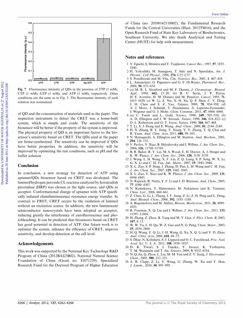
Fig. 7 Fluorescence intensity of QDs in the presence of TTP (1 mM),
CTP (1 mM), GTP (1 mM), and ATP (1 mM), respectively. Other
conditions are the same as in Fig. 5. The fluorescence intensity of each
solution was normalized.
Dow
nloa
ded
by U
NIV
ER
SIT
Y O
F SO
UT
H A
UST
RA
LIA
on
27 A
ugus
t 201
2Pu
blis
hed
on 2
6 Ju
ne 2
012
on h
ttp://
pubs
.rsc
.org
| do
i:10.
1039
/C2A
N35
520E
View Online
of QD and the concentration of materials used in the paper. The
inspection instrument to detect the CRET was a home-built
system, which is simple and crude. The sensitivity of the
biosensor will be better if the property of the system is improved.
The physical property of QD is an important factor to the bio-
sensor’s sensitivity based on CRET. The QDs used in the paper
are home-synthesized. The sensitivity can be improved if QDs
have better properties. In addition, the sensitivity will be
improved by optimizing the test conditions, such as pH and the
buffer solution.
Conclusion
In conclusion, a new strategy for detection of ATP using
aptamer/QDs biosensor based on CRET was developed. The
luminol/hydrogen peroxide CL reaction catalyzed by horseradish
peroxidase (HRP) was chosen as the light source, and QDs as
acceptor. Conformational change of aptamer with ATP specifi-
cally induced chemiluminescence resonance energy transfer. In
contrast to FRET, CRET occurs by the oxidation of luminol
without an excitation source. In addition, the new luminescent
semiconductor nanocrystals have been adopted as acceptor,
reducing greatly the interference of autofluorescence and pho-
tobleaching. It can be predicted that biosensors based on CRET
has good potential in detection of ATP. Our future work is to
optimize the system, enhance the efficiency of CRET, improve
sensitivity, and develop detection at the cell level.
Acknowledgements
This work was supported by the National Key Technology R&D
Program of China (2012BAI23B02), National Natural Science
Foundation of China (Grant no. 81071229), Specialized
Research Fund for the Doctoral Program of Higher Education
4266 | Analyst, 2012, 137, 4262–4266
of China (no. 20100142110002), the Fundamental Research
Funds for the Central Universities (Hust, 2012TS016), and the
Open Research Fund of State Key Laboratory of Bioelectronics,
Southeast University. We also thank Analytical and Testing
Center (HUST) for help with measurement.
Notes and references
1 Y. Eguchi, S. Shimizu and Y. Tsujimoto,Cancer Res., 1997, 57, 1835–1840.
2 H. Yokoshiki, M. Sunagawa, T. Seki and N. Sperelakis, Am. J.Physiol.: Cell Physiol., 1998, 274, C25–C37.
3 S. Przedborski and M. Vila, Clin. Neurosci. Res., 2001, 1, 407–418.4 L. Annunziato, G. Pignataro and G. F. Di Renzo, Pharmacol. Rev.,2004, 56, 633–654.
5 (a) M. R. L. Stratford and M. F. Dennis, J. Chromatogr., Biomed.Appl., 1994, 662, 15–20; (b) B. U. Sevin, J. P. Perras,H. E. Averette, D. M. Donato and M. Penalver, Cancer, 1993, 71,1613–1620; (c) W. Li, Z. Nie, X. H. Xu, Q. P. Shen, C. Y. Deng,J. H. Chen and S. Z. Yao, Talanta, 2009, 78, 954–958; (d)A. J. Moro, J. Schmidt, T. Doussineau, A. Lapresta-Fernandez,J. Wegener and G. J. Mohr, Chem. Commun., 2011, 47, 6066–6068.
6 (a) C. Tuerk and L. Gold, Science, 1990, 249, 505–510; (b)A. D. Ellington and J. W. Szostak, Nature, 1990, 346, 818–822; (c)D. L. Robertson and G. F. Joyce, Nature, 1990, 344, 467–468.
7 T. Li, S. J. Dong and E. Wang, Anal. Chem., 2009, 81, 2144–2149.8 H. X. Zhang, B. Y. Jiang, Y. Xiang, Y. Y. Zhang, Y. Q. Chai andR. Yuan, Anal. Chim. Acta, 2011, 688, 99–103.
9 N. Hamaguchi, A. Ellington and M. Stanton, Anal. Biochem., 2001,294, 126–131.
10 V. Pavlov, Y. Xiao, B. Shlyahovsky and I.Willner, J. Am. Chem. Soc.,2004, 126, 11768–11769.
11 B. R. Baker, R. Y. Lai, M. S. Wood, E. H. Doctor, A. J. Heeger andK. W. Plaxco, J. Am. Chem. Soc., 2006, 128, 3138–3139.
12 J. Wang, L. H. Wang, X. F. Liu, Z. Q. Liang, S. P. Song, W. X. Li,G. X. Li and C. H. Fan, Adv. Mater., 2007, 19, 3943–3946.
13 X. L. Zuo, S. P. Song, J. Zhang, D. Pan, L. H. Wang and C. H. Fan,J. Am. Chem. Soc., 2007, 129, 1042–1043.
14 X. L. Zuo, Y. Xiao and K. W. Plaxco, J. Am. Chem. Soc., 2009, 131,6944–6945.
15 N. Rupcich, R. Nutiu, Y. F. Li and J. D. Brennan,Anal. Chem., 2005,77, 4300–4307.
16 N. Kamekawa, Y. Shimomura, M. Nakamura and K. Yamana,Chem. Lett., 2006, 660–661.
17 Z. Chen, G. Li, L. Zhang, J. F. Jiang, Z. Li, Z. H. Peng and L. Deng,Anal. Bioanal. Chem., 2008, 392, 1185–1188.
18 A. Bogomolova and M. Aldissi, Biosens. Bioelectron., 2011, 26, 4099–4103.
19 R. Freeman, X. Q. Liu and I. Willner, J. Am. Chem. Soc., 2011, 133,11597–11604.
20 H. Zhang, Z. Zhou, B. Yang and M. Y. Gao, J. Phys. Chem. B, 2003,107, 8–13.
21 W.W. Yu, L. H. Qu, W. Z. Guo and X. G. Peng, Chem.Mater., 2003,15, 2854–2860.
22 H. Q. Wang, Y. Q. Li, J. H. Wang, Q. Xu, X. Q. Li and Y. D. Zhao,Anal. Chim. Acta, 2008, 610, 68–73.
23 S. Dhar, N. Kolishetti, S. J. Lippard and O. C. Farokhzad,Proc. Natl.Acad. Sci. U. S. A., 2011, 108, 1850–1855.
24 D. K. Tiwari, S. I. Tanaka, Y. Inouye, K. Yoshizawa,T. M. Watanabe and T. Jin, Sensors, 2009, 9, 9332–9354.
25 N. Q. Jia, Q. Zhou, L. Liu, M.M. Yan and Z. Y. Jiang, J. Electroanal.Chem., 2005, 580, 213–221.
26 A. R. Clapp, Z. Li, Y. Wang, G. Zhang, W. Xu and Y. Han,J. Lumin., 2010, 30, 995–999.
This journal is ª The Royal Society of Chemistry 2012