Embed Size (px)
Citation preview
A novel affinity-based method for the isolation
of highly purified extracellular vesicles
Wataru Nakai1, Takeshi Yoshida1,2, Diego Diez3, Yuji Miyatake1,2, Takahiro Nishibu4, Ryo Ukekawa4, Naoko Imawaka4, Ken Naruse4, Yoshifusa Sadamura4 & Rikinari Hanayama1,2,5
1 Laboratory of Immune Network, WPI Immunology Frontier Research Center (IFReC), Osaka University, Japan 2 Department of Immunology, Kanazawa University Graduate School of Medical Sciences, Japan 3 Quantitative Immunology Research Unit, WPI Immunology Frontier Research Center (IFReC), Osaka University, Japan 4 Life Science Research Laboratories, Wako Pure Chemical Industries Ltd, Japan 5 PRESTO, Japan Science and Technology Agency (JST), Japan
Cell Culture
Supertanant
Serum Urine
EDTA
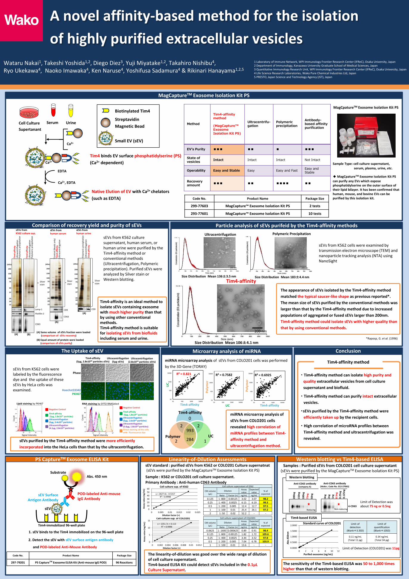
Tim4 binds EV surface phosphatidylserine (PS)
(Ca2+ dependent)
Native Elution of EV with Ca2+ chelators
(such as EDTA)
Biotinylated Tim4
Streptavidin
Magnetic Bead
Small EV (sEV)
MagCaptureTM Exosome Isolation Kit PS
Ca2+
Method
Tim4-affinity method (MagCaptureTM Exosome Isolation Kit PS)
Ultracentrifu-gation
Polymeric precipitation
Antibody-based affinity purification
EV’s Purity ■■■ ■■ ■ ■■■
State of vesicles
Intact Intact Intact Not Intact
Operability Easy and Stable Easy Easy and Fast Easy and Stable
Recovery amount
■■■ ■■ ■■■■ ■■
Code No. Product Name Package Size
299-77603 MagCapture™ Exosome Isolation Kit PS 2 tests
293-77601 MagCapture™ Exosome Isolation Kit PS 10 tests
MagCpatureTM Exosome Isolation Kit PS
Sample Type: cell culture supernatant,
serum, plasma, urine, etc. ◆ MagCpatureTM Exosome Isolation Kit PS
can purify any EVs which expose phosphatidylserine on the outer surface of their lipid bilayer. It has been confirmed that human, mouse, and bovine EVs can be purified by this isolation kit.
Comparison of recovery yield and purity of sEVs Particle analysis of sEVs purifeid by the Tim4-affinity methods sEVs from K562 culture sup.
Po
lym
eric
pre
cip
itat
ion
U
ltra
cen
trif
uga
tio
n
Tim
4-a
ffin
ity
Lamp-1
Flotillin-2
CD63
Silver stain
Po
lym
eric
pre
cip
itat
ion
U
ltra
cen
trif
uga
tio
n
Tim
4-a
ffin
ity
Co
ntr
ol (
Bea
ds
on
ly)
Tim
4-a
ffin
ity
Ult
race
ntr
ifu
gati
on
CD9
CD63
Silver stain
sEVs from human serum
CD9
Po
lym
eric
pre
cip
itat
ion
Tim
4-a
ffin
ity
Ult
race
ntr
ifu
gati
on
sEVs from human urine
Silver stain
sEVs from K562 culture supernatant, human serum, or human urine were purified by the Tim4-affinity method or conventional methods (Ultracentrifugation, Polymeric precipitation). Purified sEVs were analyzed by Silver stain or Western blotting.
Tim4-affinity is an ideal method to isolate sEVs containing exosome with much higher purity than that by using other conventional methods. Tim4-affinity method is suitable for isolating sEVs from biofluids including serum and urine.
(A)
(A)
(A)
(B) (A) Same volume of sEVs fraction were loaded (comparison of sEVs recovery)
(B) Equal amount of protein were loaded (comparison of sEVs purity)
W
The Uptake of sEV
W
Microarray analysis of miRNA
Linearity-of-Dilution Assessments Western blotting vs Tim4-based ELISA
Ultracentrifugation
200 nm
Size Distribution Mean 136±3.5 nm
Polymeric Precipitation
200 nm
Size Distribution Mean 183±4.4 nm
Tim4-affinity
200 nm
Size Distribution Mean 106±4.1 nm
sEVs from K562 cells were examined by transmission electron microscope (TEM) and nanoparticle tracking analysis (NTA) using NanoSight
The appearance of sEVs isolated by the Tim4-affinity method
matched the typical saucer-like shape as previous reported*.
The mean size of sEVs purified by the conventional methods was
larger than that by the Tim4-affinity method due to increased
populations of aggregated or fused sEVs larger than 200nm.
Tim4-affinity method could isolate sEVs with higher quality than
that by using conventional methods.
*Raposp, G. et al. (1996)
Hoechst33342 PKH67
Phase
Tim4-affinity (5μg, 2.8x10¹⁰ particles sEVs)
Ultracentrifugation (5μg sEVs)
Ultracentrifugation (2.8x10¹⁰ particles sEVs)
sEVs from K562 cells were labeled by the fluorescence dye and the uptake of these sEVs by HeLa cells was examined.
sEVs purified by the Tim4-affinity method were more efficiently
incorporated into the HeLa cells than that by the ultracentrifugation.
R² = 0.7582
50
500
5000
50000
50 500 5000 50000
Po
lym
er
UC
R² = 0.6925
50
500
5000
50000
50 500 5000 50000
Po
lym
er
Tim4-affinity
R² = 0.821
50
500
5000
50000
50 500 5000 50000
UC
Tim4-affinity W
Conclusion
miRNA microarray analysis of sEVs from COLO201 cells was performed
by the 3D-Gene (TORAY) Tim4-affinity method
・ Tim4-affinity method can isolate high purity and
quality extracellular vesicles from cell culture
supernatant and biofluid.
・ Tim4-affinity method can purify intact extracellular
vesicles.
・ High correlation of microRNA profiles between
Tim4-affinity method and ultracentrifugation was
revealed.
・sEVs purified by the Tim4-affinity method were
efficiently taken up by the recipient cells. miRNA microarray analysis of
sEVs from COLO201 cells
revealed high correlation of
miRNA profiles between Tim4-
affinity method and
ultracentrifugation method.
2
1 2
993
284
2
0 Tim4-affinity
Polymer UC
1. sEV binds to the Tim4 immobilized on the 96-well plate
2. Detect the sEV with sEV surface antigen antibody
and POD-labeled Anti-Mouse Antibody
sEV Surface Antigen Antibody
POD-labeled Anti-mouse IgG Antibody
Abs. 450 nm
Tim4-immobilized 96-well plate
sEV
Substrate
PS CaptureTM Exosome ELISA Kit
Code No. Product Name Package Size
297-79201 PS CaptureTM Exosome ELISA Kit (Anti-mouse IgG POD) 96 Reactions
sEV standard : purified sEVs from K562 or COLO201 Culture supernatnat (sEVs were purified by the MagCaptureTM Exosome Isolation Kit PS)
Sample : K562 or COLO201 cell culture supernatant.
y = 1351.7x + 0.113 R² = 0.9996
0
5
10
15
0 0.002 0.004 0.006 0.008 0.01 0.012
Ass
ay
valu
e (n
g/m
L)
Dilution factor (×)
Cell culture sup. of COLO201 Cell culture supernatant of COLO201
CM volume (μL)
Dilution Assay value
Expected value % of
expected Ratio Factor (×) ng/mL ng/mL
0.0625 1:1600 0.000625 0.89 0.91 98.4
0.125 1:800 0.00125 1.82 1.72 105.6
0.25 1:400 0.0025 3.44 3.52 97.8
0.5 1:200 0.005 7.04 6.78 103.9
1 1:100 0.01 13.6 - -
Cell culture supernatant of K562
CM volume (μL)
Dilution Assay value
Expected value % of
expected Ratio Factor (×) ng/mL ng/mL
0.125 1:800 0.00125 3.02 3.07 98.2
0.25 1:400 0.0025 6.15 6.19 99.3
0.5 1:200 0.005 12.4 12.7 97.5
1 1:100 0.01 25.4 26.1 97.2
2 1:50 0.02 52.2 - -
The linearity-of-dilution was good over the wide range of dilution of cell culture supernatant. Tim4-based ELISA Kit could detect sEVs included in the 0.1μL Culture Supernatant.
y = 2627.4x - 0.5317 R² = 0.9998
0
10
20
30
40
50
60
0 0.005 0.01 0.015 0.02 0.025
Ass
ay
valu
e (n
g/m
L)
Dilution factor (×)
Cell culture sup. of K562
Primary Antibody : Anti-human CD63 Antibody
Samples : Purified sEVs from COLO201 cell culture supernatant (sEVs were purified by the MagCaptureTM Exosome Isolation Kit PS)
Limit of detection
(Blank+3.3SD)
Limit of quantification (Blank+10SD)
0.11 ng/mL (Total 11 pg)
0.34 ng/mL (Total 34 pg)
The sensitivity of the Tim4-based ELISA was 50 to 1,000 times higher than that of western blotting.
Limit of Detection (COLO201) was 11pg
Anti-CD63 antibody (company A)
←CD63
Limit of Detection was about 75 ng or 0.5ng
Western blotting
200n
g
150n
g
100
ng
75n
g
50n
g
Reducing
4n
g
2n
g
1n
g
0.5
ng
0.2
5ng
0.12
5ng
Non-reducing
Anti-CD63 antibody (Wako ; Code No. 012-27063)
0.0000
0.5000
1.0000
1.5000
2.0000
0 2 4 6 8 10 12
Ab
s.4
50n
m
Purifed exosome (ng/mL)
Standard curve of COLO201
Tim4-based ELISA
(kDa)
50
37
25
75
100
150
250
20
15
(kDa)
50
37
25
75
100
150
250
20
15
(kDa)
50
37
25
75
100
150
250
20
15
Signal Intensity
Co
un
t
Lipid staining by PKH67
Negative Control
Tim4-affinity (5μg, 2.8x10¹⁰ particles) Ultracentrifugation (5μg, 3.4x10¹⁰ particles)
Ultracentrifugation (4.1μg, 2.8x10¹⁰ particles)
Signal Intensity
Co
un
t
RNA staining by SYTO RNASelect
Negative Control
Tim4-affinity (5μg, 1.8x10¹⁰ particles)
Ultracentrifugation (5μg, 1.6x10¹⁰ particles)
Ultracentrifugation (5.6μg, 1.8x10¹⁰ particles)
Direct staining of the cells with fluorescence dye
Ca2+, EDTA
Ca2+