Embed Size (px)
Citation preview
Research ArticleA Novel Evaluation Method for Detecting Defects of the BondedOrthodontic Bracket-Tooth Interface
Mona Aly Abbassy ,1,2 Turki A. Bakhsh ,3 and Ahmed Samir Bakry 3,4
1Department of Orthodontics, Faculty of Dentistry, King Abdulaziz University, Jeddah, Saudi Arabia2Alexandria University, Alexandria, Egypt3Operative and Esthetic Dentistry Department, Faculty of Dentistry, King Abdulaziz University, Jeddah, Saudi Arabia4Conservative Dentistry Department, Faculty of Dentistry, Alexandria University, Alexandria, Egypt
Correspondence should be addressed to Ahmed Samir Bakry; [email protected]
Received 13 December 2020; Revised 12 February 2021; Accepted 20 February 2021; Published 16 March 2021
Academic Editor: Vincenzo Grassia
Copyright © 2021 Mona Aly Abbassy et al. This is an open access article distributed under the Creative Commons AttributionLicense, which permits unrestricted use, distribution, and reproduction in any medium, provided the original work isproperly cited.
Background. Orthodontic patients are at high risk to develop caries. This study is introducing a clinical method detecting interfacialdefects between ceramic brackets and enamel utilizing optical coherent tomography in addition to using the nanoleakageexpression in vitro test. Methods. Transbond XT primer and moisture insensitive primer (MIP) were bonded to 75 humanpremolar enamel surfaces and divided into (XTD), (MIPD), and (MIPW) groups. The (XTD) and (MIPD) groups had ceramicbrackets bonded to dry enamel surfaces using TransBond and moisture insensitive primers, respectively, while the (MIPW)samples were bonded to moist enamel using moisture insensitive primer. All specimens were examined under crosspolarizationoptical coherence tomography. Debonding forces of the brackets to 45 teeth (15 teeth/group). 30 bonded specimens (15specimens/group) were cross-sectioned to detect the nanoleakage expression using scanning electron microscope equipped withenergy-dispersive spectroscopy (SEM/EDS). The degree of conversion of the specimens in the experimental groups was testedusing attenuated total reflectance Fourier transform infrared spectroscopy (FTIR/ATR). Results. Optical coherence tomographydetected the interfacial defects between the ceramic brackets and tooth structure. One way ANOVA showed that (XTD) and(MIPD) groups recorded significantly higher bond strength values and less nanoleakage expression when compared to MIPW(p > 0:05). Conclusions. Optical coherence tomography can be utilized to detect interfacial adhesive-tooth defects. Dry enamelsurfaces improve the quality of the enamel/primer interface (200 words).
1. Introduction
One of the chief goals of orthodontic treatment is to achieve astable interface between orthodontic brackets and toothenamel with low susceptibility to degradation over time [1].It was previously recommended that all measures should betaken to keep the outer enamel surface intact as it containsthe highest degree of mineral and mechanical properties tominimize the enamel damage during the bracket debondingphase [1]. However, the high rate of bacterial biofilm forma-tion observed in orthodontic patients [2] exposes thebracket-resin-enamel interface to continuous acidic chal-lenge [2]; thus, the proper sealing of the aforementionedinterface is an essential element in preventing the ingress of
bacterial toxins and acids under the bracket region whichmay lead to enamel demineralization [3]. Such lesions areextremely difficult to be controlled or remineralized becauseit is located under the bracket region [3]. The detection ofsuch lesions in an early stage may allow its remineralizationinstead of restoring such lesions after debonding of theorthodontic brackets, which agree with the principals ofminimal interventions in modern dentistry that advocateremineralization rather than restoration of enamel and den-tin lesions [4, 5].
Optical coherence tomography (OCT) is an imaging sys-tem that is utilized in medical fields to diagnose variouslesions, especially in ophthalmology [6]. It provides volumet-ric and cross-sectional images safely, noninvasively, and
HindawiBioMed Research InternationalVolume 2021, Article ID 6634595, 8 pageshttps://doi.org/10.1155/2021/6634595
nondestructively [6]. Previous research showed encouragingresults for utilizing Swept-source OCT for detecting whitespot demineralization lesions around orthodontic brackets[6]. However, the ability of evaluation of adhesive interfacialdefects under ceramic orthodontic brackets is still anuntested potential for the OCT technology [6].
Moreover, bonding of brackets to malaligned teeth,partially or completely unerupted teeth, is a challengefaced by many orthodontists that render the field isolationprior to bonding an extremely difficult process. This maynegatively affect the brackets’ bond strength [7] causingearly and repeated debonding of the orthodontic brackets.This challenging environment renders the sealing ability ofthe orthodontic bonding resin a questionable matter.
The nanoleakage examination can be a suitable candidatefor accurate determination of the sealing properties of ortho-dontic resins [8]. Nanoleakage studies are different frommicroleakage because nanoleakage can give a clear imagefor any porosity or defects resulting from deficiency in resinpolymerization or infiltration within the substrate that it isbonded to [1, 8].
Orthodontic bonding systems capable of polymerizing inchallenging the oral environment including enamel surfacesthat are difficult to isolate prior to bonding were introducedin the market, such as moisture insensitive primer (MIP)[9–11]. However, controversial results were obtained regard-ing its bond strength [7, 9–12], and there is scant informationregarding the efficiency of this primer in sealing the resin-enamel interface of the orthodontic brackets under moistconditions.
The current experiments aimed at examining the capabil-ity of OCT technology to detect interfacial defects underorthodontic brackets and correlate the obtained results tonanoleakage, debonding, and degree of conversion results.
The null hypotheses adopted in the current researchare that OCT will not be able to detect any interfacialdefects under the ceramic orthodontic brackets, (MIP) willnot seal the enamel-resin interface under moist condition,and will score low bond strength values when bonded tomoist enamel.
2. Materials and Methods
2.1. Specimen Preparation. Extracted human sound premolarteeth that were extracted for orthodontic reasons were col-lected from the oral surgery department after obtainingthe permission of the ethical committee of the faculty055-02-19. The teeth were hand scaled from any calculusor soft tissues. The teeth were stored in 0.1% thymol untilthe start of the experiment according to the guidelinesapproved by University and in accordance with the princi-ples of the Declaration of Helsinki and its later amendmentsor comparable ethical standards. The number of specimensassigned to each group was adopted according to previousliterature and according to the 80% power of test. Randomi-zation of the specimens was done using a computer program(Excel 2007, Microsoft, Redmond, WA, USA). All teeth wereexamined by light microscope to exclude any teeth havingcracks, demineralization, or any defects. Intra and interexa-
miners calibrations were conducted before the actual record-ing of the obtained results.
2.2. Materials Used in This Study. Two different adhesiveprimers were used on the enamel surface: Transbond XTprimer (XTP; 3M ESPE, USA) and TransbondMIPMoistureInsensitive Primer (MIP; 3M ESPE, USA). Transbond PLUScolor change adhesive (3M ESPE, USA) was used to bond theorthodontic ceramic brackets (Victory series, 3M Unitek,USA). The lot number and chemical composition of eachmaterial are according to the information provided by themanufacturers (Table 1).
2.3. Bonding of Orthodontic Brackets. Forty five caries-freehuman premolar teeth extracted due to orthodontic reasonswere used in this study. The teeth were divided into threegroups with the buccal surface of each group being treatedwith XTP group or MIPD group in which XTP and MIPprimer adhesives were applied on the dry enamel surfaceaccording to the manufacturer instructions, while teeth inthe MIPW group 0.02ml distilled water [12] was added tothe surface, and the water was not removed. TransbondPLUS color change adhesive resin was used to bond theorthodontic ceramic brackets according to the manufacturerinstruction. Schematic illustration of the sample preparationand the experimental design are described in Figure 1.
2.4. CP-OCT Assessment. Crosspolarization optical coher-ence tomography (CP-OCT; IVS300, Santec, Japan) was usedin this study. Technological specifications of the utilized CP-OCT are described in Table 2. This noninvasive imagingsystem uses a continuous wavelength diode laser centerednear 1310 nm with a 30 kHz scanning rate. CP-OCT.
All specimens assigned to the shear bond strength testswere examined under the CP-OCT system after the bondingprocedures to exclude misplaced brackets and defectivebonded specimens; this was followed by examining the spec-imens by the CP-OCT to determine the quality of the ortho-dontic ceramic bracket-tooth interface.
Due to the different absorption of the OCT rays in theceramic brackets, the examined interface between theceramic brackets and enamel appeared on two levels. Forsimplification of the obtained results, the authors decidedto conduct an experiment in which Transbond XT resinwas condensed in Teflon 2mm diameter×2mm height moldand light cured to obtain 15 composite cylinders. The com-posite cylinders were divided into three groups and werebonded to lingual surfaces utilizing the same condition ofthe three experimental groups. These group of samples hadtheir enamel-ceramic brackets interfaces on one level to elab-orate the capability of examining interface and in the sametime clarify the effect of orthodontic bracket material onthe CP-OCT observation.
To conduct the CP-OCT test, each bonded specimen wasplaced on a micrometer stage with a 3D axis (x, y, z), and theOCT probe was positioned perpendicularly on the buccalsurface to scan the bonded interface across the bracket. Con-secutive scans were accomplished at 500μm intervals. The
2 BioMed Research International
size of each image was 500 × 924 pixels corresponding to5mm × 8:2mm (x, z).
2.5. Shear Bond Strength Test (SBS). 15 bonded specimensfrom each group were mounted on a universal testingmachine (ElectroPlus E1000, Instron, Canton, MA, USA)and subjected to a shear force at the interface between theenamel and the orthodontic bracket at a crosshead speed of0.5mm/minute.
2.6. Nanoleakage Test. The nanoleakage solution was pre-pared in a dark room by dissolving 25 g of silver nitrate crys-tals (Sigma Chemical Co., St. Louis, MO, USA) in 25ml of
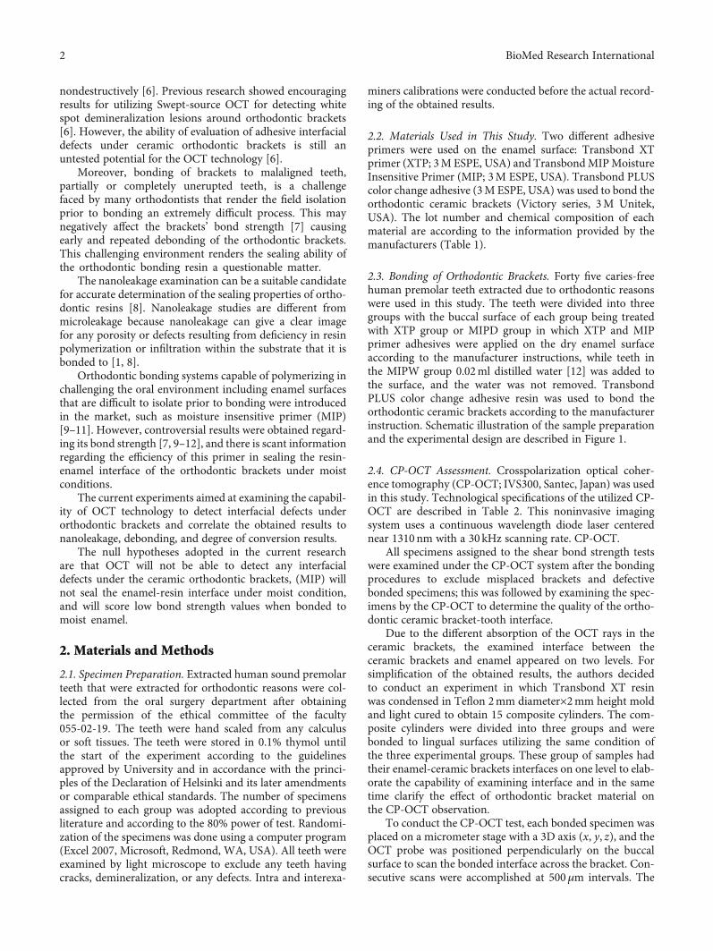
Table 1: Composition of the materials used in the study.
Material (manufacturer) code Ingredients Lot No.
Transbond XT light cure adhesiveprimer (3M Unitek, USA) XTP
(i) Bisphenol A diglycidyl ether dimethacrylate (45–55%Wt)(ii) Triethylene glycol dimethacrylate(45–55%Wt)
N611932
Transbond MIP moisture insensitive(3M Unitek, USA)MIP
(i) Ethyl alcohol (30–40% Wt)(ii) Bisphenol A diglycidyl ether dimethacrylate (15-25% Wt)
(iii) 2-Hydroxyethyl methacrylate (10-20% Wt)(vi) 2-Hydroxy-1,3-dimethacryloxypropane (5 15% Wt)(v) Copolymer of itaconic and acrylic acid (5-15% Wt)
(vi) Diurethane dimethacrylate (1-10% Wt)
N141377
Transbond PLUS color change adhesive(3M Unitek, USA)
(i) Silane treated glass (35–45%Wt)(ii) Silane treated quartz (35–45%Wt)
(iii) 1,2,3-Propanetricarboxylic acid, 2-hydroxy-, reaction products with 2-isocyanatoethyl methacrylate (5-15%Wt)
(iv) Polyethylene glycol dimethacrylate (PEGDMA) (5–15%Wt)(v) Bisphenol A diglycidyl ether dimethacrylate (BISGMA)<2Wt
(vi) Silane treated silica <2Wt(vii) Diphenyliodonium hexafluorophosphate <1 Wt
N576253
Enam
el
Cross sectioning of the bonded interface between the bonded
brackets and the enamel.
Sectioning of the crowns of premolar
Bonding of the orthodontic
brackets
MIPW group
MIPD group
XTD group
Moist enamel
Dry enamel
Dry enamel
Shear bondstrength test
Storing of specimens in silver nitrate
Storing of specimens in
developer solution under fluorescent light
Nanoleakagetest
SEM observation
Testing specimens byoptical coherence
tomography
FTIR/ATR test
EnamelEn
amel
Cross sectioning of the bondinterface between the bonde
gg
brackets and the enamel.
Sectioning of the crownsof premolar
Bonding of theorthodontic
brackets
MIPD group
XTD group
Moist enamel
Dry enamel
Dry enamel
Shear bondstrength test
Storing of specimens in silver nitrate
Storing of specimens in
developer solution underfluorescent light
Nanoleakagetest
SEM observation
Testing specimens byoptical coherence
tomography
FTIR/ATR test
Enamel
Figure 1: Summary of experimental procedures.
Table 2: Means and standard deviation for shear bond strength and% degree of conversion results. Similar superscripts in horizontalrows are not statistically significant, p > 0:05.
XTPD(TransbondXT, dry)
MIPD (moistureinsensitive, dry)
MIPW (moistureinsensitive, wet)
Shear bondstrength
21:34 ± 3:81a 17:9 ± 6:69a 10:74 ± 2:96b
%degree ofconversion
67:8 ± 6:8a 49:8 + 7:3b 31:3 + 3:8c
3BioMed Research International
distilled water [13]. Dilution of ammonium hydroxide(Sigma Chemical Co., St. Louis, MO, USA) was conductedunder proper suction in a specialized hood to obtain 28%ammonium hydroxide solution. The black silver nitrate solu-tion was titrated by 28% ammonium hydroxide that wasstirred using magnetic stirrer until it became clear. 50ml ofdistilled water was added to obtain a 50% wt ammoniacalsilver nitrate pH = 9:5 solution [13, 14]. All measures werecarried out to avoid hand or surface contamination by thesilver nitrate solution due to its staining properties. 10bonded specimens from each group were vertically cross-sectioned with a diamond saw under water cooling throughthe bracket-enamel interface. The central slab was chosenfrom each tooth, forming a total of 10 specimens per group,n = 10. Bonded slabs were ground and polished using wet#1000 silicon carbide paper, then coated with two layers offast-drying nail varnish applied up to 1mm from the bondedinterface [13, 14]. The specimens were stored in ammoniacalsilver nitrate in total darkness for 18 h, rinsed thoroughly,and immersed in photo developing solution (Kodak, NY,USA) for 6 h under fluorescent light to reduce silver ions intometallic silver [13]. The silver-stained resin-bonded speci-mens were lightly polished to remove the superficial silverremnants [8, 13, 14], followed by drying the specimens andgold sputter coating. The specimens were observed usingSEM/EDS (JCM-6000, NeoScope, JEOL, Tokyo, Japan), andline scans were examined across the resin-enamel [8, 13, 14].
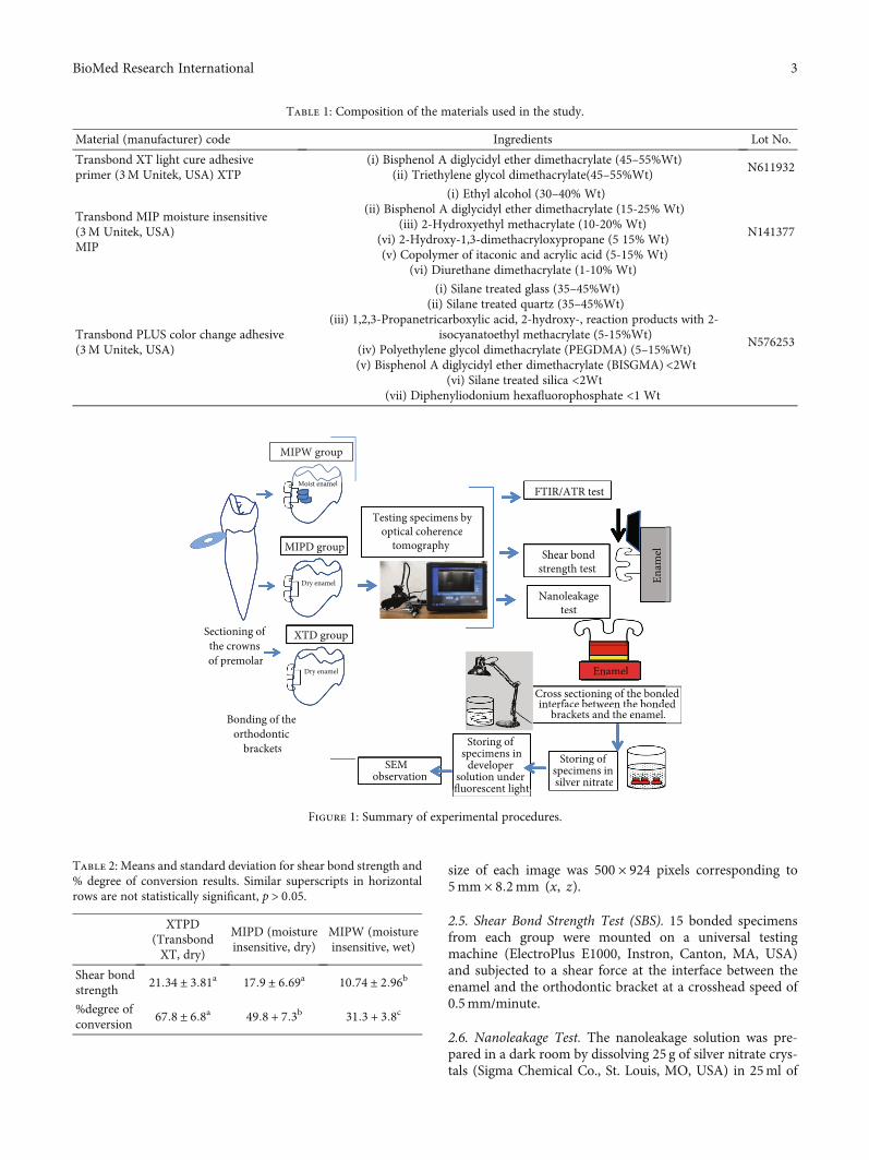
2.7. Attenuated Total Reflectance Fourier Transform InfraredSpectroscopy (FTIR/ATR) Degree of Conversion Analysis.Transbond PLUS Color Change Adhesive (3M Unitek) wasapplied to the base of 30 metallic brackets. A thin layer ofthe unpolymerized moisture insensitive primer was appliedin groups MIPD and MIPW, while Transbond XT primerwas applied in group XTP n = 10. The brackets in groupMIPW had 3μl of distilled water applied on top of the FTIRexamination window using a microbrush [10]. All specimenswere placed under a static load of 100 grams on the FTIR/-ATR examination window [15] as shown in Figure 2
(Thermo Scientific Nicolet iS5 FTIR Spectrometer; ThermoFisher Scientific, Waltham, MA, USA).
The specimens were light cured by a LED unit (Ortholux;3M Unitek) with an output of 1,600mW/cm2. The light cur-ing unit was calibrated using the Demetron™ LED radiome-ter. The LED unit was slightly touching the bracket, and thecuring time was 3 seconds mesial and 3 seconds distal tothe brackets.
All FTIR/ATR measurements were obtained under thefollowing conditions: a resolution of 4 cm−1 and four internalscans per reading [15]. The uncured resin in each groupserved as the control for the cured resin.
The spectra of the monomers and their respective poly-mers were compared to determine the conversion rate ofthe double bonds into simple carbon bonds. The peaks weremeasured at the frequencies of 1,715/cm−1 (corresponding tothe aromatic ring bonds) and 1,637/cm−1 (corresponding tothe bonds between carbons of the methacrylate groups)[15]. The following formula was used to calculate the conver-sion rate of the double carbon bonds into simple bonds.
%Conversion = 100 × 1 − Polymer C = Cð Þ ×monomer C − Cð ÞMonomer C = Cð Þ × polymer C − Cð Þ
ð1Þ
2.8. Statistical Analysis. Shear bond strength and the degreeof conversion results were analyzed using one-way ANOVA,p ≤ 0:05. Evaluation of nanoleakage locations was carried outthrough analysis of the obtained images. The infiltration ofsilver nitrate into the interface was evaluated as follows:enamel-hybrid layer, adhesive–hybrid interface, and adhe-sive layer were evaluated and graded as (no) (no leakage,score 0), (slight) (slight leakage, score 1), and (distinct) (dis-tinct leakage, score 2). Analysis was carried out using theKruskal–Wallis test p ≤ 0:05 [[8]], and the differences wereconsidered statistically significant at the level of 0.05 5%(SPSS v24, IBM, Armonk, US).
3. Results
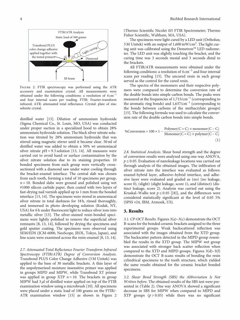
3.1. CP-OCT Results. Figures 3(a)–3(c) demonstrate the OCTB-scans for the bonded ceramic brackets assigned to the threeexperimental groups. Weak backscattered reflection wasassociated with the images obtained from the XTD group.The backscatter pattern detected in the MIPD group resem-bled the results in the XTD group. The MIPW wet groupwas associated with stronger back scatter reflection whencompared to the XTD and MIPD groups. Figures 3(d)–3(f)demonstrate the OCT B-scans results of bonding the resincylindrical specimens to the tooth structure, which yieldedthe same results obtained for the ceramic bracket bondedspecimens.
3.2. Shear Bond Strength (SBS) the Abbreviation Is NotWritten before. The obtained results of the SBS test were pre-sented in (Table 2). One way ANOVA showed a significantdecrease in SBS for MIPW when compared to MIPD andXTP groups (p > 0:05) while there was no significant
IR beam
Static load of 500 grams
Transbond PLUScolor change adhesiveapplied together with
the tested primers Diamond
FTIR/ATR Analysis
Light curing
Figure 2: FTIR spectroscopy was performed using the ATRaccessory and examination crystal. All measurements wereobtained under the following conditions: a resolution of 4 cm−1
and four internal scans per reading. FTIR: Fourier-transforminfrared; ATR: attenuated total reflectance. Crystal: plate of zincselenite crystal.
4 BioMed Research International
difference in shear bond strength between MIPD and XTP(p > 0:05).
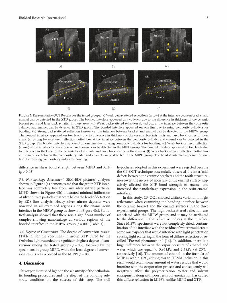
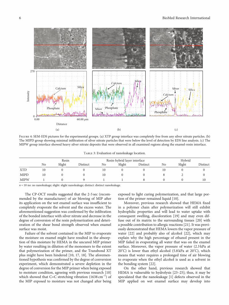
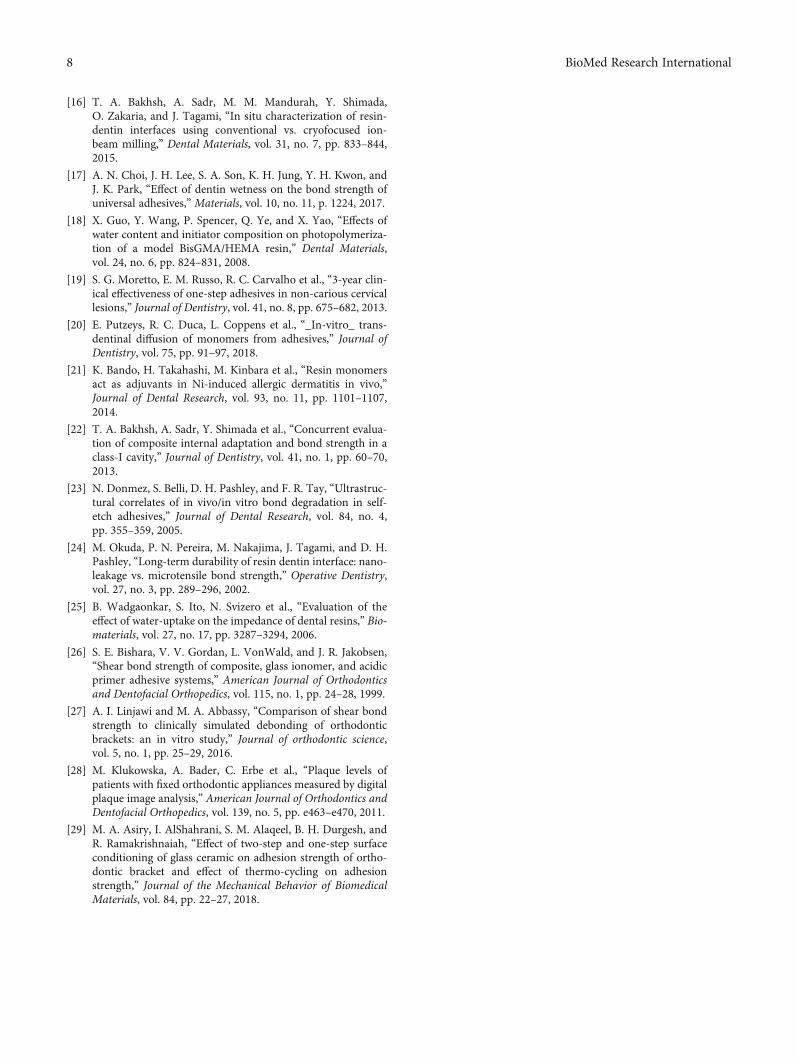
3.3. Nanoleakage Assessment. SEM-EDS pictures’ analysesshown in Figure 4(a) demonstrated that the group XTP inter-face was completely free from any silver nitrate particles.MIPD shown in Figure 4(b) illustrated minimal infiltrationof silver nitrate particles that were below the level of detectionby EDS line analysis. Heavy silver nitrate deposits wereobserved in all examined regions along the enamel-resininterface in the MIPW group as shown in Figure 4(c). Statis-tical analysis showed that there was a significant number ofsamples showing nanoleakage at various regions of thebonded interface in the MIPW group, p = 000 (Table 3).
3.4. Degree of Conversion. The degree of conversion results(Table 3) for the specimens in group XTP cured by theOrtholux light recorded the significant highest degree of con-version among the tested groups p = 000, followed by thespecimens in group MIPD, while the least degree of conver-sion results was recorded in the MIPW p = 000.
4. Discussion
This experiment shed light on the sensitivity of the orthodon-tic bonding procedures and the effect of the bonding sub-strate condition on the success of this step. The null
hypotheses adopted in this experiment were rejected becausethe CP-OCT technique successfully observed the interfacialdefects between the ceramic brackets and the tooth structure;moreover, the increased moisture of the enamel surface neg-atively affected the MIP bond strength to enamel andincreased the nanoleakage expression in the resin-enamelinterface.
In this study, CP-OCT showed distinct variation in lightreflectance when examining the bonding interface betweenthe ceramic bracket and the enamel surfaces in the threeexperimental groups. The high backscattered reflection wasassociated with the MIPW group, and it may be attributedto the difference in the refractive indices at the interface.Since MIPW specimens were not completely dried, contam-ination of the interface with the residue of water would createsome microspaces that would interfere with light penetrationcausing light scattering in the form of diffuse reflection or so-called “Fresnel phenomenon” [16]. In addition, there is ahuge difference between the vapor pressure of ethanol andwater which are equal to 5.95 kPa and 2.3 kPa (at 20°C),respectively [16]. The amount of ethanol in the formula ofMIIP is within 40%, adding this to HEMA inclusion in thisresin would retain some amount of water residue that wouldinterfere with the evaporation process and consequently willnegatively affect the polymerization. Water and solvententrapment along with poor resin polymerization has causedthis diffuse reflection in MIPW, unlike MIPD and XTP.
Air
Bracket
E
(a)
Air
Bracket
E
(b)
Air
Bracket
E
(c)
Composite
Enamel
(d)
Air
Composite
Enamel
(e)
AirComposite
Enamel
(f)
Figure 3: Representative OCT B-scans for the tested groups. (a) Weak backscattered reflections (arrow) at the interface between bracket andenamel can be detected in the XTD group. The bonded interface appeared on two levels due to the difference in thickness of the ceramicbracket parts and laser back schatter in these areas. (d) Weak backscattered reflection dotted box at the interface between the compositecylinder and enamel can be detected in XTD group. The bonded interface appeared on one line due to using composite cylinders forbonding. (b) Strong backscattered reflection (arrows) at the interface between bracket and enamel can be detected in the MIPW group.The bonded interface appeared on two levels due to difference in thickness of the ceramic brackets parts and laser back scatter in theseareas. (e) Strong backscattered reflection dotted box at the interface between the composite cylinder and enamel can be detected in theXTD group. The bonded interface appeared on one line due to using composite cylinders for bonding. (c) Weak backscattered reflection(arrow) at the interface between bracket and enamel can be detected in the MIPD group. The bonded interface appeared on two levels dueto difference in thickness of the ceramic brackets parts and laser back scatter in these areas. (f) Weak backscattered reflection dotted boxat the interface between the composite cylinder and enamel can be detected in the MIPD group. The bonded interface appeared on oneline due to using composite cylinders for bonding.
5BioMed Research International
The CP-OCT results suggested that the 2-5 sec (recom-mended by the manufacturer) of air blowing of MIP afterits application on the wet enamel surface was insufficient tocompletely evaporate the solvent and the excess water. Theaforementioned suggestion was confirmed by the infiltrationof the bonded interface with silver nitrate and decrease in thedegree of conversion of the resin polymerization and deteri-oration of the shear bond strength observed when enamelsurface was moist.
Failure of the solvent contained in the MIP to evaporatethe moisture on enamel might have resulted in the absorp-tion of this moisture by HEMA in the uncured MIP primerby water resulting in dilution of the monomers to the extentthat polymerization of the primer, and the Transbond XTplus might have been hindered [10, 17, 18]. The aforemen-tioned hypothesis was confirmed by the degree of conversionexperiment, which demonstrated a severe depletion in thedegree of conversion for the MIP primer when being exposedto moisture condition, agreeing with previous research [10]which showed that C=C stretching vibration (1638 cm–1) ofthe MIP exposed to moisture was not changed after being
exposed to light curing polymerization, and that large por-tion of the primer remained liquid [10].
Moreover, previous research showed that HEMA fixedin a polymer chain after polymerization will still exhibithydrophilic properties and will lead to water uptake withconsequent swelling, discoloration [19] and may even dif-fuse out of its matrix to the surrounding tissues [20] witha possible contribution to allergic reactions [21]. It was previ-ously demonstrated that HEMA lowers the vapor pressure ofwater [22] and probably also of alcohol [22], which mayexplain why the high percentage of ethanol present in theMIP failed in evaporating all water that was on the enamelsurface. Moreover, the vapor pressure of water (2.3 kPa at20°C) is lower than ethyl alcohol (5.8 kPa at 20°C), whichmeans that water requires a prolonged time of air blowingto evaporate when the ethyl alcohol is used as a solvent inthe bonding system [22].
On the other hand, previous research showed thatHEMA is vulnerable to hydrolysis [23–25]; thus, it may bespeculated that the nanoleakage [1] defects observed in theMIP applied on wet enamel surface may develop into
Distance0.00 60.00 𝜇m
Calcium
Phosphate
Silica
Inte
nsity
270
0
10 𝜇m
(a)
Calcium
PhosphateSilver
SilicaCalciucccc m
PhosphateSilver
SilSSSSSSSSSSSSSSS icacaccccccccccccccac
10 𝜇m
(b)
Calcium
Phosphate
SilicaSilver
Calcium
Phosphate
SilicacaccaaacaacaacccacaaccacSilver
10 𝜇m
(c)
Figure 4: SEM-EDS pictures for the experimental groups. (a) XTP group interface was completely free from any silver nitrate particles. (b)The MIPD group showing minimal infiltration of silver nitrate particles that were below the level of detection by EDS line analysis. (c) TheMIPW group interface showed heavy silver nitrate deposits that were observed in all examined regions along the enamel-resin interface.
Table 3: Evaluation of nanoleakage location.
Resin Resin-hybrid layer interface HybridNo Slight Distinct No Slight Distinct No Slight Distinct
XTD 10 0 0 10 0 0 10 0 0
MIPD 10 0 0 10 0 0 8 2 0
MIPW 1 0 9 1 1 8 0 0 10
n = 10 no: no nanoleakage; slight: slight nanoleakage; distinct: distinct nanoleakage.
6 BioMed Research International
microleakage [3] and increase the chance of caries incidenceunder the cemented orthodontic brackets.
Our results confirmed previous literature [7, 9–11] thatreported the deterioration of the shear bond strength of theMIP when bonded to moist enamel or contaminated byblood or saliva; however, observation of the sealing abilityof the interface in the current experiment shed light on a pos-sible mechanism that explains the decrease of the bondstrength of MIP to moist enamel.
Although there was a significant decrease in the bondstrength of MIP to moist enamel, however, the MIP bondstrength values were still within the acceptable range for clin-ical bonding of orthodontic brackets [26, 27].
It is of prime importance to state that any resultsobtained from the current in vitro experiment should beinterpreted cautiously. This is due to the fact that all attemptswere done to simulate the clinical situation in the currentexperiment; however, using the MIP in the oral environmentmay exert more challenges like the continuous acidic attacksexerted by the bacterial biofilm that is abundant around theorthodontic brackets [2, 28]. Moreover, the variable thermalchanges [29] in the oral cavity and the various occlusalstresses exerted on the brackets bonded to the malalignedteeth may aggravate the deterioration of the bond interfaceinside the patients’ oral cavity.
Further studies are required to quantify the degree ofreflectance and directly correlate it to the interfacial defectsobserved in the orthodontic ceramic-enamel interface.
5. Conclusions
It is of prime importance to obtain a clean, dry enamelsurface prior to orthodontic bonding procedures to obtainreliable results regarding the bond strength and sealingability of the bonding orthodontic resin under orthodonticbrackets. CP-OCT may be a valuable tool to examine andfollow up the quality of the bonded orthodontic ceramicbracket-enamel interface in clinical situations.
Data Availability
All data were supplied in the submitted manuscript.
Conflicts of Interest
The authors certify the research is original, not under publi-cation consideration elsewhere, and free of conflict ofinterest.
Acknowledgments
This Project was funded by the Deanship of ScientificResearch (DSR) at King Abdulaziz University, Jeddah, undergrant no. (G: 607-165-1441). The authors, therefore, acknowl-edge with thanks DSR for technical and financial support.
References
[1] S. Arslan, L. Lipski, K. Dubbs, F. Elmali, and F. Ozer, “Effectsof different resin sealing therapies on nanoleakage within arti-
ficial non-cavitated enamel lesions,” Dental Materials Journal,vol. 37, no. 6, pp. 981–987, 2018.
[2] S. M. Al-Bazi, M. A. Abbassy, A. S. Bakry, L. A. Merdad, andA. H. Hassan, “Effects of chlorhexidine (gel) application onbacterial levels and orthodontic brackets during orthodontictreatment,” Journal of Oral Science, vol. 58, no. 1, pp. 35–42,2016.
[3] R. Atash, A. Fneiche, S. Cetik et al., “In vitro evaluation ofmicroleakage under orthodontic brackets bonded with differ-ent adhesive systems,” European journal of dentistry, vol. 11,no. 2, pp. 180–185, 2017.
[4] A. S. Bakry and M. A. Abbassy, “Increasing the efficiency ofCPP-ACP to remineralize enamel white spot lesions,” Journalof Dentistry, vol. 76, pp. 52–57, 2018.
[5] J. E. Frencken, M. C. Peters, D. J. Manton, S. C. Leal, V. V. Gor-dan, and E. Eden, “Minimal intervention dentistry for manag-ing dental caries - a review:,” International Dental Journal,vol. 62, no. 5, pp. 223–243, 2012.
[6] T. A. Bakhsh, A. S. Bakry, M. M. Mandurah, and M. A.Abbassy, “Novel evaluation and treatment techniques forwhite spot lesions. An in vitro study,” Orthodontics & cranio-facial research, vol. 20, no. 3, pp. 170–176, 2017.
[7] A. Faltermeier, M. Behr, M. Rosentritt, C. Reicheneder, andD. Mussig, “An in vitro comparative assessment of differ-ent enamel contaminants during bracket bonding,” Euro-pean Journal of Orthodontics, vol. 29, no. 6, pp. 559–563,2007.
[8] Y. Yuan, Y. Shimada, S. Ichinose, and J. Tagami, “Qualitativeanalysis of adhesive interface nanoleakage using FE-SEM/EDS,”Dental Materials, vol. 23, no. 5, pp. 561–569, 2007.
[9] T. Endo, R. Ozoe, S. Sanpei, K. Shinkai, Y. Katoh, andS. Shimooka, “Effects of moisture conditions of dental enamelsurface on bond strength of brackets bonded with moisture-insensitive primer adhesive system,” Odontology, vol. 96,no. 1, pp. 50–54, 2008.
[10] T. Eliades, E. Katsavrias, and G. Eliades, “Moisture-insensitiveadhesives: reactivity with water and bond strength to wet andsaliva-contaminated enamel,” European Journal of Orthodon-tics, vol. 24, no. 1, pp. 35–42, 2002.
[11] I. L. Zeppieri, C. H. Chung, and F. K. Mante, “Effect of saliva onshear bond strength of an orthodontic adhesive used withmoisture-insensitive and self-etching primers,” American Jour-nal of Orthodontics and Dentofacial Orthopedics, vol. 124,no. 4, pp. 414–419, 2003.
[12] F. Bazargani, A. Magnuson, H. Lothgren, and A. Kowalczyk,“Orthodontic bonding with and without primer: a randomizedcontrolled trial,” European Journal of Orthodontics, vol. 38,no. 5, pp. 503–507, 2016.
[13] F. R. Tay, D. H. Pashley, and M. Yoshiyama, “Two modes ofnanoleakage expression in single-step adhesives,” Journal ofDental Research, vol. 81, no. 7, pp. 472–476, 2002.
[14] A. S. Bakry, M. Nakajima, M. Otsuki, and J. Tagami, “Effect ofEr:YAG laser on dentin bonding durability under simulatedpulpal pressure,” The Journal of Adhesive Dentistry, vol. 11,no. 5, pp. 361–368, 2009.
[15] T. M. Masood, M. A. Abbassy, A. S. Bakry, N. Y. Matar, andA. H. Hassan, “Fourier-transform infrared spectroscopy/atte-nuated total reflectance analysis for the degree of conversionand shear bond strength of Transbond XT adhesive system,”Clinical, Cosmetic and Investigational Dentistry, vol. Volume10, pp. 275–280, 2018.
7BioMed Research International
[16] T. A. Bakhsh, A. Sadr, M. M. Mandurah, Y. Shimada,O. Zakaria, and J. Tagami, “In situ characterization of resin-dentin interfaces using conventional vs. cryofocused ion-beam milling,” Dental Materials, vol. 31, no. 7, pp. 833–844,2015.
[17] A. N. Choi, J. H. Lee, S. A. Son, K. H. Jung, Y. H. Kwon, andJ. K. Park, “Effect of dentin wetness on the bond strength ofuniversal adhesives,” Materials, vol. 10, no. 11, p. 1224, 2017.
[18] X. Guo, Y. Wang, P. Spencer, Q. Ye, and X. Yao, “Effects ofwater content and initiator composition on photopolymeriza-tion of a model BisGMA/HEMA resin,” Dental Materials,vol. 24, no. 6, pp. 824–831, 2008.
[19] S. G. Moretto, E. M. Russo, R. C. Carvalho et al., “3-year clin-ical effectiveness of one-step adhesives in non-carious cervicallesions,” Journal of Dentistry, vol. 41, no. 8, pp. 675–682, 2013.
[20] E. Putzeys, R. C. Duca, L. Coppens et al., “_In-vitro_ trans-dentinal diffusion of monomers from adhesives,” Journal ofDentistry, vol. 75, pp. 91–97, 2018.
[21] K. Bando, H. Takahashi, M. Kinbara et al., “Resin monomersact as adjuvants in Ni-induced allergic dermatitis in vivo,”Journal of Dental Research, vol. 93, no. 11, pp. 1101–1107,2014.
[22] T. A. Bakhsh, A. Sadr, Y. Shimada et al., “Concurrent evalua-tion of composite internal adaptation and bond strength in aclass-I cavity,” Journal of Dentistry, vol. 41, no. 1, pp. 60–70,2013.
[23] N. Donmez, S. Belli, D. H. Pashley, and F. R. Tay, “Ultrastruc-tural correlates of in vivo/in vitro bond degradation in self-etch adhesives,” Journal of Dental Research, vol. 84, no. 4,pp. 355–359, 2005.
[24] M. Okuda, P. N. Pereira, M. Nakajima, J. Tagami, and D. H.Pashley, “Long-term durability of resin dentin interface: nano-leakage vs. microtensile bond strength,” Operative Dentistry,vol. 27, no. 3, pp. 289–296, 2002.
[25] B. Wadgaonkar, S. Ito, N. Svizero et al., “Evaluation of theeffect of water-uptake on the impedance of dental resins,” Bio-materials, vol. 27, no. 17, pp. 3287–3294, 2006.
[26] S. E. Bishara, V. V. Gordan, L. VonWald, and J. R. Jakobsen,“Shear bond strength of composite, glass ionomer, and acidicprimer adhesive systems,” American Journal of Orthodonticsand Dentofacial Orthopedics, vol. 115, no. 1, pp. 24–28, 1999.
[27] A. I. Linjawi and M. A. Abbassy, “Comparison of shear bondstrength to clinically simulated debonding of orthodonticbrackets: an in vitro study,” Journal of orthodontic science,vol. 5, no. 1, pp. 25–29, 2016.
[28] M. Klukowska, A. Bader, C. Erbe et al., “Plaque levels ofpatients with fixed orthodontic appliances measured by digitalplaque image analysis,” American Journal of Orthodontics andDentofacial Orthopedics, vol. 139, no. 5, pp. e463–e470, 2011.
[29] M. A. Asiry, I. AlShahrani, S. M. Alaqeel, B. H. Durgesh, andR. Ramakrishnaiah, “Effect of two-step and one-step surfaceconditioning of glass ceramic on adhesion strength of ortho-dontic bracket and effect of thermo-cycling on adhesionstrength,” Journal of the Mechanical Behavior of BiomedicalMaterials, vol. 84, pp. 22–27, 2018.
8 BioMed Research International