Embed Size (px)
Citation preview

Organic Mass Spectrometry, 1973, Vol. 7, pp. 277 to 281. Heyden & Son Limited. Printed in Northern Ireland
A NOVEL FRAGMENTATION OF TRIMETHYLSILYL ETHERS OF 3p-HYDROXY-A5-STEROIDS
INGEMAR BJORKHEM, JAN-AKE GUSTAFSSON and JAN SJOVALL Kemiska Institutionen, Karolinska Institutet, S-104 01 Stockholm 60, Sweden
(Received 2 May 1972; accepted 30 August 1972)
Abstract-An ion formed by loss of 56 mass units from the molecular ion is often seen in mass spectra of trimethylsilyl ethers of C19 and C,, steroids having a 3b-hydroxy-A5 structure and an 0x0 group at C-17 or C-20. The nature of this fragment was investigated by the use of perdeuteriotrimethylsilyl ether derivatives and of [4-l*C], [3J80], [4,4JHa] and [2,2,4,4-,H] labelled derivatives of 3/5’-hydroxy- 5-androsten-17-one and 3b-hydroxy-5-pregnen-20-one. Evidence is presented to show that the neutral fragment of mass 56 is composed of carbon atoms 1 , 2 and 3, the oxygen at C-3 and four hydrogen atoms. During the fragmentation process, the trimethylsilyl group and one of the hydrogens at C-2 are transferred to the fragment that carries the charge.
I N T R O D U C T I O N
STEROIDS with an 0x0 group at C-17 give mass spectra with peaks at m/e [M - 561 and [M - 57]1-2 representing loss of ring D. Loss of 56 mass units is also charac- teristic of the trimethylsilyl ether of 3b-hydroxy-5-androsten-17-one and this fragmen- tation was originally thought to be identical to that in other 17-0x0 steroids? Mass spectra of trimethylsilyl ethers of C,, steroids with a 3B-hydroxy-A5 structure, e.g. 3/3-hydroxy-5-pregnen-20-one, also show a peak at mje [M - 561, however. When the mass spectrum of 3j3-hydroxy-5-[3~+~H]pregnen-20-one trimethylsilyl ether was recorded, the mass spectrum showed a peak at m/e [M - 571 instead of [M - 561. This indicated that the fragment lost originated from ring A. The nature of this frag- ment in trimethylsilyl ethers of 38-hydroxy-A5 steroids was therefore studied in greater detail and the results are reported in this paper.
RESULTS A N D DISCUSSION
High resolution mass measurement gave values of 304.2217 (C,,H3,0Si = 304-2222) and 332.2548 (C,,H,,OSi = 332-2535) for the ions formed by loss of 56 mass units from the molecular ions of the trimethylsilyl ethers of 3p-hydroxy-5- androsten-17-one and 3p-hydroxy-5-pregnen-20-one, respectively. Thus the elimi- nated fragment had the composition C3H4O. Low resolution spectra of the perdeu- teriotrimethylsilyl ethers showed that none of the three methyl groups of the trimethylsilyl moiety were eliminated.
The trimethylsilyl ether of 316-hydroxy-5- [3~-~H]pregnen-20-one, having a deu- terium excess of 79.2 atoms percent in the 3a position, gave a peak at m/e 332 [M - 571 with only an insignificant peak at m/e 333 (corresponding to a deuterium excess of 1.3 atoms percent in this ion). Thus the 3ct hydrogen atom was present in the eliminated fragment. The mass spectrum of the trimethylsilyl ether of 3p-hydroxy- 5-[4-14C]pregnen-20-one (I4C excess at C-4,gO.O atoms percent) had a peak at m/e 334 that was ten times more intense than that at mle 332, showing that C-4 was not one of the carbon atoms lost. As is the case in the spectrum of the unlabelled compound there was a peak at mle 129. A corresponding ion with I4C at m/e 131 was not present, which is in agreement with the conclusions by Diekman and Djerassi4 regarding the origin of the ion of mass 129.
211
278 1. BJORKHEM, J.-A. GUSTAFSSON and J. SJOVALL
The presence of the 3ar-hydrogen in the fragment of mass 56 suggested that the oxygen lost might be that at C-3. The mass spectrum of the trimethylsilyl ether of 3~-[3-180]hydroxy-5-androsten-17-one (l80 excess at C-3, 19.9 atoms percent) showed that this was the case. Thus the peak at m/e 304 in the spectrum of the unlabelled compound remained at the same mass in the spectrum of the labelled compound. The intensity of a peak at m/e 306 corresponded to an l 8 0 excess of less than 1 atoms percent. As expected: the peak at mle 129 moved to nile 131, whereas a peak at mle 231 was found in both spectra, corresponding to loss of 129 and 131 mass units from the molecular ions of unlabelled and labelled compound, respectively.
The data presented above indicated that the [M - 56]+. ion was formed by loss of carbon atoms 1-3, the oxygen at C-3 and four hydrogen atoms. This was supported by the mass spectra of the trimethylsilyl ethers of 3B-hydro~y-5-[3a-~H] androsten-17- one, 3B-hydro~y-5-[4,4-~H,1 androsten-17-one and 3/3-hydroxy-5-[2,2,4,4-2H,1 androsten-17-one. The isotopic compositions of relevant ions in the spectra of these compounds are summarised in Table 1. The results indicate that the fragment of mass 56 lost from the molecular ion of the unlabelled compound contains the 3ar hydrogen and one hydrogen at C-2, and does not contain any of the hydrogens at C-4. The results regarding the ions of mass 129 and [M - 1291 are in good agreement with those of Diekman and Djera~si .~ The conclusion by these authors that some of the hydrogen at C-4 is lost with trimethylsilanol could not be confirmed with certainty.
TABLE 1. ISOTOPIC COMPOSITION OF IONS IN THE MASS SPECTRA OF TRIMETHYLSILYL ETHERS OF 38- HYDROXY-5- [3a-2H]ANDROSTEN-1 7-ONE, 3P-HYDROXY-5- [4,4-ZHz]ANDROSTEN-17-ONE AND 3P-HYDROXY-
5- [2,2,4,4-*H,]ANDROSTEN- 17-ONE
Labelled m/e of unlabelled Per cent isotopic composition8 positions ion 'H 2H, 'Hz 2H3 'Ha
~ u - ~ H 360 [MI 4.8 93.3 304 [M - 561 97.5 1.6 270 [M - 901 7.3 90.0 231 [M - 1291 96.6 3.0 129 5.2 92.1
4,4-'H* 360 [MI 34.4 41.8 22.8 345 [M - 151 40.2 34.4 20.6
270 [M - 901 41.0 42.5 15.7 231 [M - 1291 47.0 36.7 15.4 129 91.4 1.6 0.5
304 [M - 561 41.4 38.1 20.1
2,2,4,4-aH4 360 [MI 4.1 6.2 18.0 38.2 32.7 345 [M - 151 2.5 3.3 17-1 37.3 35.4 304 [M - 561 5.1 13.9 35-6 41.6 3.7 270 [M - 901 1.7 5.0 17.6 37-8 37.9 231 [M - 1291 11.8 16.8 40.0 29.0 1.8 129 26.0 73.4 0.2 0 0
a Calculations were based on the assumption that a maximum of five deuterium atoms were present in the molecules.
A novel fragmentation of trimethylsilyl ethers of 3p-hydroxy-h5-steroids 279
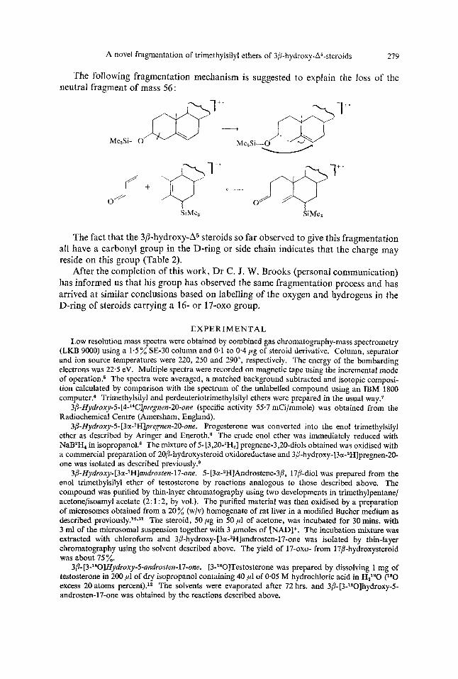
The following fragmentation mechanism is suggested to explain the loss of the neutral fragment of mass 56 :
-l
The fact that the 3/3-hydroxy-A5 steroids so far observed to give this fragmentation all have a carbonyl group in the D-ring or side chain indicates that the charge may reside on this group (Table 2).
After the completion of this work, Dr C. J. W. Brooks (personal communication) has informed us that his group has observed the same fragmentation process acd has arrived at similar conclusions based on labelling of the oxygen and hydrogens in the D-ring of steroids carrying a 16- or 17-0x0 group.
EXPERIMENTAL Low resolution mass spectra were obtained by combined gas chromatography-mass spectrometry
(LKB 9000) using a 1.5 % SE-30 column and 0.1 to 0.4 pg of steroid derivative. Column, separator and ion source temperatures were 220, 250 and 290", respectively. The energy of the bombarding electrons was 22.5 eV. Multiple spectra were recorded on magnetic tape using the incremental mode of o~era t ion .~ The spectra were averaged, a matched background subtracted and isotopic composi- tion calculated by comparison with the spectrum of the unlabelled compound using an IBM 1800 computer.6 Trimethylsilyl and perdeuteriotrimethylsilyl ethers were prepared in the usual way.'
3B-Hy~roxy-5-[4-'*C]pregne~-2o-one (specific activity 55.7 mCi/mmole) was obtained from the Radiochemical Centre (Amersham, England).
3p-Hydroxy-5- [3cr-2H]pregnen-20-one. Progesterone was converted into the enol trimethylsilyl ether as described by Aringer and Eneroth.8 The crude enol ether was immediately reduced with NaB2H4 in isopropanol.* The mixture of 5-[3,20-2H2] pregnene-3,20-diols obtained was oxidised with a commercial preparation of 20p-hydroxysteroid oxidoreductase and 3P-hydroxy- [3~-~H]pregnen-20- one was isolated as described previou~ly.~
3B-Hydroxy- [3cr-2H]androsten-17-one. 5- [3cPHJAndrostene-3/3, 17p-diol was prepared from the enol trimethylsilyl ether of testosterone by reactions analogous to those described above. The compound was purified by thin-layer chromatography using two developments in trimethylpentanel acetone/isoamyl acetate (2: 1 :2, by vol.). The purified material was then oxidised by a preparation of microsomes obtained from a 20% (w/v) homogenate of rat liver in a modified Bucher medium as described previously.l0*l1 The steroid, 50 ,ug in 50 ,ul of acetone, was incubated for 30 mins. with 3 ml of the microsomal suspension together with 3 pmoles of INAD]+. The incubation mixture was extracted with chloroform and 3B-hydro~y-[3a-~H]androsten-17-one was isolated by thin-layer chromatography using the solvent described above. The yield of 17-0x0- from 17B-hydroxysteroid was about 75 %.
3B-[3-180]Hydroxy-5-undsten-17-one. [3-*80]Testosterone was prepared by dissolving 1 rng of testosterone in 200 p1 of dry isopropanol containing 40 ,ul of 0.05 M hydrochloric acid in HZ1*0 (180
excess 20 atoms percent).12 The solvents were evaporated after 72 hrs. and 38- [3-180]hydroxy-5- androsten-17-one was obtained by the reactions described above.
280 I. BJORKHEM, J.-A. GUSTAFSSON and J. SJOVALL
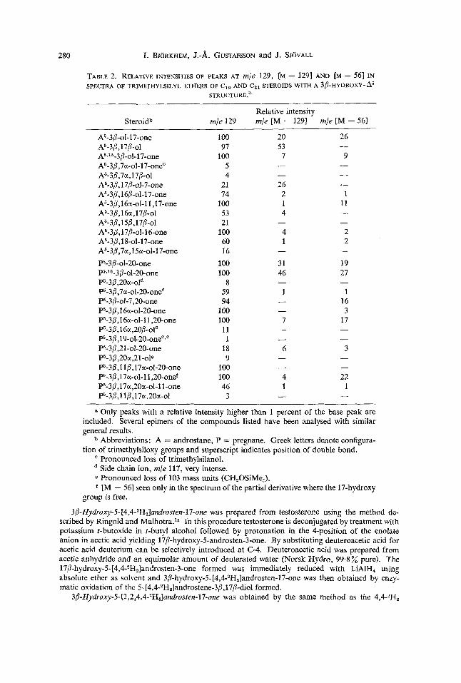
TABLE 2. RELATIVE INTENSITIES OF PEAKS AT m/e 129, [M - 1291 AND [M - 561 IN SPECTRA OF TRIMETHYLSILYL ETHERS OF C,, AND C,, STEROIDS WITH A 3P-HYDROXY-fi
STRUCTURE."
Relative intensity Steroid" mle 129 mle [M - 1291 mle [M - 561
A5-3~-ol-17-one A5-3B,17P-ol A5J5-3B-ol-17-one A5-3B,7c(-ol-17-oneC
A5-3B,1 7B-Ol-7-01~ A5-3/l,16P-ol-17-one A5-3P,16cr-ol-l 1,17-one
A5-3/3,7a,1713-ol
A5-3,B,16~,l7/3-ol A5-3B,1 5b,17P-0l A5-3,9,1 7B-oI-16-0ne A5-3B,18-ol-17-one A5-3P,7a,l 5a-ol-17-one P5-3,5-ol-20-one P5J6-3P-ol-20-one P5-3P,200(-oln P5-3j3,7a-ol-20-oneC P5-3b-oI-7,20-one P5-3~,16c(-ol-20-one P5-3p,16c(-ol-1 1,20-one
P5-3~,19-ol-20-oneC~e P5-3b,21-ol-20-one P5-3b,20c(,21-ole P5-3b, 1 1 P,17c(-oI-20-one P5-3B,17a-ol-1 1,20-one' P5-3b,17c(,20a-ol-1 1-one
P5-3B,1 ~CC,~O/I -OI~
P5-3/?,1 1/!?,17a,20~t-01
100 97
100 5 4
21 74
100 53 21
100 60 16
100 100
8 59 94
100 100 11
1 18 9
100 100 46 3
20 53 7 - - 26 2 1 4
4 1
-
31 46
1 -
4 1
2 2
19 27
1 16 3
17
-
- 3
- 22 1
a Only peaks with a relative intensity higher than 1 percent of the base peak are included. Several epimers of the compounds listed have been analysed with similar general results.
b Abbreviations: A = androstane, P = pregnane. Greek letters denote configura- tion of trimethylsiloxy groups and superscript indicates position of double bond.
Pronounced loss of trimethylsilanol. Side chain ion, m/e 117, very intense. Pronounced loss of 103 mass units (CH,OSiMe,).
f [M - 561 seen only in the spectrum of the partial derivative where the 17-hydroxy
38-Hydroxy-5- [4,4-2H,]androsten-17-one was prepared from testosterone using the method de- scribed by Ringold and Ma1h0tra.l~ In this procedure testosterone is deconjugated by treatment with potassium t-butoxide in t-butyl alcohol followed by protonation in the 4-position of the enolate anion in acetic acid yielding 17/3-hydroxy-5-androsten-3-0ne. By substituting deuteroacetic acid for acetic acid deuterium can be selectively introduced at C-4. Deuteroacetic acid was prepared from acetic anhydride and an equimolar amount of deuterated water (Norsk Hydro, 99.8% pure). The 17P-hydro~y-5-[4,4-~H,]androsten-3-one formed was immediately reduced with LiAIH, using absolute ether as solvent and 3P-hydroxy-5- [4,4-2H,]androsten-17-one was then obtained by enzy- matic oxidation of the 5-[4,4-*H2]androstene-3B,17P-dioI formed.
3P-Hyduoxy-5-[2,2,4,4-ZH,]androsten-17-one was obtained by the same method as the 4,4-?H,
group is free.
A novel fragmentation of trimethylsilyl ethers of 3@-hydroxy-A5-steroids 281
derivative except that (CH3),COaH was used. This was obtained by treatment of potassium t-butylate with an excess of deuterated water. The mixture yielded a two-phase system and the last traces of deuterated water could be removed from the separated t-butyl alcohol with Drierite.
Acknowledgements-We are grateful to Dr R. Ryhage for the high resolution mass spectra measure- ments. This work was supported by the Swedish Medical Research Council (Project No. 13X-219) and Knut and Alice Wallenbergs Stiftelse.
R E F E R E N C E S 1. L. TokCs, R. T. LaLonde and C. Djerassi, J. Org. Chem. 32, 1012 (1967). 2. G. Jones and C. Djerassi, Steroids 10, 653 (1967). 3. R. Vihko, Actu Endocrinol. Suppl. 109, 35 (1966). 4. J. Diekman and C. Djerassi, J. Amer. Chem. SOC. 32, 1005 (1967). 5. B. Hedfjall, P.-A, Jansson, Y. Mgrde, R. Ryhage and S. Wikstrom, J. Sci. Znstr. 2, 1031 (1969). 6. R. Reimendal and J. Sjovall, Anal. Chem. 44, 21 (1972). 7. J.-A. Gustafsson and J. Sjovall, Eur. J . Biochem. 8, 467 (1969). 8. L. Aringer and P. Eneroth, Steroids 18, 381 (1971). 9. I. Bjorkhem, J.-A. Gustafsson and S. Gustafsson, Eur. J. Biochem. 16, 557 (1970).
10. I. Bjorkhem, K. Einarsson, J.-A. Gustafsson and A. Some11 Acta Endocrinol. 71, 569 (1972). 11. G. Johansson, Eur. J . Biochem. 21, 68 (1971). 12. A. M. Lawson, F. A. J. M. Leemans and J. A. McCloskey, Steroids 14,603 (1969). 13. H. J. Ringold and S. K. Malhotra, Tetrahedron Letters 15, 669 (1962).


![Synthesis and structure of 1,4,5,8-tetraethynylnaphthalene ... · Cartesian coordinate of optimized structure of 4a ... (trimethylsilyl)ethynyl]-1,4-dihydronaphthalene-1,4-diol (6)](https://img.pdfslide.net/doc/110x75/5b41cf3e7f8b9af3388b4b9e/synthesis-and-structure-of-1458-tetraethynylnaphthalene-cartesian-coordinate.jpg)


![Syntheses, X-ray crystal structures and reactivity of ... · Attempted synthesis of 1-trimethylsilyl-2-dibenzo[a,d]cycloheptylidene-ethene (21b) As for Method A above, 1-bromo-1-trimethylsilyl-2-dibenzo[](https://img.pdfslide.net/doc/110x75/5f890aa86bf1eb0265155785/syntheses-x-ray-crystal-structures-and-reactivity-of-attempted-synthesis-of.jpg)