Embed Size (px)
Citation preview

A novel peptide motif binding to and blockingthe intracellular activity of the human papillomavirusE6 oncoprotein
Susanne Dymalla & Martin Scheffner & Elvira Weber &
Peter Sehr & Claudia Lohrey & Felix Hoppe-Seyler &
Karin Hoppe-Seyler
Abstract Specific types of human papillomaviruses (HPVs)cause cervical cancer. The viral E6 oncogene is a criticalfactor for maintaining the malignant phenotype of HPV-positive tumour cells. By yeast two-hybrid screening of arandomised peptide expression library, we isolated linearshort peptides, which specifically bind to the HPV16 E6oncoprotein. Sequence alignments and mutational analysesof the peptides identified a hitherto undiscovered E6-bindingmotif. Intracellular expression of a peptide containing thenovel E6-binding motif resulted in inhibition of colonyformation capacity, specifically of HPV16-positive cancercells. A solubility-optimised variant of the peptide wascreated, which binds to HPV16 E6 with high affinity. Itsintracellular expression efficiently induced apoptosis inHPV16-positive cancer cells. This was linked to restorationof intracellular p53 activities. Thus, this newly identified E6-binding motif could form a novel basis for the development
of rational strategies for the treatment of HPV16-positivepreneoplastic and neoplastic lesions.
Keywords Human papillomavirus . Cervical cancer .
Cancer therapy . Oncogenes
Introduction
Specific types of human papillomaviruses (HPVs) causecervical cancer, the second most common cancer in femalesworldwide. In addition, oncogenic HPV types are associat-ed with other cancers, e.g. in the anogenital region ororopharynx [1]. The most prevalent HPV type found inthese cancers is HPV16, which, for example, accounts for50–60% of all cervical carcinoma cases worldwide [2].Recently developed prophylactic HPV vaccines hold greatpromise for a future prevention of HPV-associated cancers[3]. Yet, there are still many issues to be solved for awidespread application, which include high costs of thevaccine as well as problems related to medical infrastruc-ture and health education [4]. The incidence of cervicalcancer is estimated to increase by over 40% until the year2020, mostly due to the population dynamics in thedeveloping countries [5]. Thus, novel treatment strategiesare urgently required.
The viral E6 oncogene is known to play a crucial role forthe maintenance of the malignant phenotype of HPV-positive cancer cells [1]. Therefore, inhibition of E6function could be ultimately developed into a rationalanti-cancer strategy. Indeed, interference with E6 activityhas been found to exert strong anti-oncogenic effects inHPV-positive cancer cells in vitro and in vivo, resultingeither in apoptosis [6–10], growth arrest [11, 12] or both[13]. Since E6, as a non-cellular protein, is not expressed in
S. Dymalla : C. Lohrey : F. Hoppe-Seyler (*) :K. Hoppe-Seyler (*)Molecular Therapy of Virus-Associated Cancers (F065),German Cancer Research Center,Im Neuenheimer Feld 242,69120 Heidelberg, Germanye-mail: [email protected]
M. Scheffner : E. WeberDepartment of Biology, University of Konstanz,Konstanz, Germany
P. SehrChemical Biology Core Facility, EMBL,Heidelberg, Germany
e-mail: [email protected]
Konstanzer Online-Publikations-System (KOPS) URL: http://nbn-resolving.de/urn:nbn:de:bsz:352-opus-84926
Erschienen in: Journal of Molecular Medicine ; 87 (2009), 3. - S. 321-331 https://dx.doi.org/10.1007/s00109-008-0432-1

undiseased cells, E6 inhibitors should principally allow aselective attack on HPV-positive cells. As proof of concept,previous studies have shown that specific targeting of E6 atthe protein level by peptide aptamers or intracellularantibodies (“intrabodies”) induced apoptosis in HPV-positivecancer cells, without affecting HPV-negative cells [6, 8, 10].However, both peptide aptamers and intrabodies are rela-tively large and complex molecules, which, at current,restricts their further development into drugs [14, 15]. Onthe other hand, intracellular expression of peptides, whichwere rationally derived from the binding domains of naturalE6 interaction partners, such as E6AP, failed to exert anti-oncogenic effects in HPV-positive cells [8].
Here, we screened a randomised expression library forsmall linear peptides targeting the HPV16 E6 oncoprotein.A hitherto unrecognised peptide-binding motif was identi-fied, which specifically binds to the HPV16 E6 protein.Intracellular expression of peptides harboring the novel E6-binding motif efficiently induced apoptosis, selectively inHPV16-positive cancer cells. On the basis of this bioactiv-ity, this novel peptide motif should serve as a valuable newtool for the development of therapeutic strategies tospecifically target HPV-positive preneoplastic and neoplas-tic lesions.
Materials and methods
Plasmids
Yeast expression vectors encoding full-length E6 proteinsfused to the GAL4-DNA-binding domain were obtained bysubcloning HPV16 E6, HPV18 E6, HPV6 E6 and HPV11 E6 coding sequences into pPC97 [6]. For CheckMate™(Promega, Mannheim, Germany) analyses, the completeHPV16 E6 coding sequence was fused to GAL4-BD inpBIND; peptide coding sequences were fused to VP16-ADin pACT. To monitor expression in mammalian cells,peptide sequences were fused to human recombinant greenfluorescent protein (hrGFP; Stratagene, Heidelberg, Ger-many) in pCEP4 (Invitrogen, Karlsruhe, Germany). Mutantderivatives of pep11 were generated either by introducingdouble-stranded synthetic oligonucleotides of the desiredsequence or by site directed mutagenesis. For ectopicexpression in mammalian cells, HPV16 E6 was expressedfrom pcDNA3 (Invitrogen) and p53 from p53wt [16].
Cell lines and transfections
HPV16-positive SiHa, CaSki and HPV18-positive HeLacervical carcinoma cells, H1299 lung cancer cells, U2OSosteosarcoma cells and MCF-7 breast cancer cells weremaintained in Dulbecco’s minimal essential medium
(pH 7.2), supplemented with 10% foetal calf serum (FCS).HPV16-positive MRI-H-186 cervical carcinoma cells [17]were grown in RPMI-1640 medium, supplemented with10% FCS. Plasmids were transfected by calcium phosphateco-precipitation, as described [17], or with Fugene HD(Roche Diagnostics, Mannheim, Germany).
Peptide screening
Yeast strain KF1 (MATa trp1-901 leu2-3,112 his3-200gal4Δ gal80Δ LYS2::GAL1-HIS3 GAL2-ADE2 met2::GAL7-lacZ SPAL10-URA3) contains three selectable nutri-tional marker genes under the control of GAL4-bindingsites [6]. A peptide expression library was generated byfusing randomised 60mer oligonucleotides to GAL-AD inpADtrx [6], thereby replacing its trx insert. Oligonucleo-tides contained triplets of the sequence NNK (where N = G,A, T or C; K = G or C), which encode for all 20 aminoacids but result in only one stop codon. KF1 transformantsexpressing GAL4-BD-HPV16 E6 and randomised peptideswere initially selected for activation of the intermediatestringent [6] marker GAL2-ADE. Subsequently, they wereanalysed by replica plating for activation of the GAL4-dependent GAL1-HIS3, GAL2-ADE and SPO13-URA3genes. Peptide expression vectors from clones, whichactivated at least two marker genes, were rescued, andactivation of the selectable markers was verified by re-screening. E6APpep corresponds to amino acids 391–408of E6AP [18] and served as a positive control for E6binding.
Mammalian two-hybrid assays
The CheckMate™ system (Promega) was used to investi-gate the binding capacities of pep11 and pep11 mutants toHPV16 E6 in mammalian cells. Both pBIND and pACTfusion constructs were transfected into HeLa cells alongwith the GAL4-responsive luciferase reporter constructpG5luc and the internal standard pCMV-Gal [7]. Two daysafter transfection, cells were harvested. Luciferase activitieswere determined as duplicates in at least three independentexperiments and normalised for pCMV-Gal activities, asdescribed [19].
Protein purification, synthetic peptides
Recombinant GST-HPV16 E6 was expressed in Escherichiacoli strain BL21 and purified as described [20]. Peptideswere chemically synthesised with or without an N-terminalfluorescein label at the Peptide Synthesis Core Facility(German Cancer Research Center), using Fmoc chemistry.Crude peptides were purified by HPLC and checked by massspectrometry. Peptides were dissolved in DMSO to obtain
322

10 mM solutions, supplemented with equimolar DTT andstored at −20°C.
Fluorescence polarisation
For equilibrium binding studies, twofold serial dilutions ofGST-HPV16 E6 were prepared in fluorescence polarisation(FP) buffer (PBS, 0.05% Tween 20, 1 mM DTT) withvarying glycerol contents to maintain a continuous glycerolconcentration (14% in the assay). Fluorescein-labelledpeptides (30 nM) were mixed with protein samples inblack 384-well ProxiPlates (PerkinElmer, Boston, USA) toa final volume of 10 μl and incubated for 90 min at roomtemperature. For competition studies, 1:3 serial dilutions ofunlabelled peptides were co-incubated with 30 nM offluorescein-labelled peptides and 500 nM of GST-HPV16E6 in a final volume of 15 μl for 90 min. FP signals weremeasured in an Envision™ Multilabel Plate Reader(PerkinElmer). Background corrected signals were plottedagainst protein concentrations and fitted with a one-sitesaturation model (binding) or a non-linear regression model(competition) using SigmaPlot (Systat Software, San Jose,USA).
Colony formation assays
Cell lines transfected with hrGFP-peptide expressionvectors were selected for the presence of pCEP4 byhygromycin B treatment. Colonies were fixed and stainedwith formaldehyde-crystal violet after 7–14 days ofselection.
p53-Responsive reporter assays
hrGFP-peptide expression vectors were transfected intoSiHa, MRI-H-186 or HeLa cells, together with the p53-responsive reporter construct p53CON-pGUP.PA.8 [21] andpCMV-Gal as internal standard. Two days after transfec-tion, cells were harvested and luciferase activities weredetermined as duplicates in at least three independentexperiments, normalised for pCMV-Gal activities, asfurther described [19].
Apoptosis assays
Cells grown on coverslips were transfected with pCEP4-hrGFP-peptide expression vectors and fixed with parafor-maldehyde (4% in PBS), 2 days after transfection. Terminaldeoxynucleotidyltransferase-mediated UTP end labelling(TUNEL) analysis was performed, utilising the in situ celldeath detection kit TMR red (Roche Diagnostics). DNAwas stained with 4′,6-diamidino-2-phenylindole (RocheDiagnostics). Apoptotic DNA strand breaks, peptide ex-
pression and nuclei were visualised with a Vanox-T AH-2epifluorescence microscope (Olympus, Center Valley,USA). Pictures were captured with an F-View camera (SoftImaging System, Olympus) and analysed with analySIS^Bsoftware (Soft Imaging System).
Western blot analysis
Two days after transfection, cellular protein was extractedin RIPA buffer (10 mM Tris–HCl pH 7.5, 150 mM NaCl,1 mM EDTA, 1% NP40, 0.5% sodium deoxycholate, 0.1%SDS). Approximately 30 μg of protein was separated by12.5% SDS-PAGE, transferred to an Immobilon-P mem-brane (Millipore, Eschborn, Germany) and analysed byenhanced chemiluminescence (Amersham Biosciences,Freiburg, Germany). The following primary antibodieswere used for protein detection: anti-β-galactosidase(Promega), anti-α-tubulin CP06 (Calbiochem, Schwalbach,Germany), anti-hrGFP (kind gift of H. Zentgraf, Heidel-berg), anti-p53 DO-1 (BD PharMingen, San Diego, USA),anti-p21 OP64 (Calbiochem) and anti-PUMA (Cell Signal-ing, Danvers, USA).
Ubiquitination assay
The HPV16 E6 protein was expressed as glutathioneS-transferase fusion proteins in E. coli DH5α as described[18]. The ubiquitin-activating enzyme E1, E6AP, and theubiquitin-conjugating enzyme UbcH5b were expressed inthe baculovirus system or in E. coli BL21 by using the pETexpression system as described [22]. For in vitro ubiquiti-nation of human Dlg [23], 1 μl of wheat germ extract-translated 35S-labelled Dlg was incubated with 50 ng E1,50 ng UbcH5, 50 ng E6AP and 10 μg ubiquitin (Sigma,Taufkirchen, Germany) in the absence or in the presence ofbacterially expressed E6 (50 ng) and increasing amounts(micrograms) of the peptides indicated in 50-μl volumes. Inaddition, reactions contained 25 mM Tris–HCl (pH 7.5),60 mM NaCl, 3 mM DTT, 2 mM ATP and 4 mM MgCl2.After incubation at 30°C for 2 h, total reaction mixtureswere electrophoresed in 10% SDS-polyacrylamide gels andthe 35S-labelled substrates detected by fluorography usingthe LAS-3000 imaging system (Fujifilm, Düsseldorf,Germany). Quantitation of the intensity of the signals wasperformed with the Aida 4.08 Software package (Raytest,Straubenhardt, Germany).
Results
An expression library encoding randomised linear 20-merpeptides linked to the transcriptional activation domain ofGAL4 was created (complexity approximately 8.3×10⁶)
323
and screened by yeast two-hybrid for molecules binding tothe HPV16 E6 oncoprotein. Among 2.1×10⁷ transformantsinitially screened for their ability to activate the ADE2marker, we isolated six different clones, which activated inreplica analyses the HIS3 and ADE2 selection markers inyeast test strain KF1. In addition, three of them alsostimulated the URA3 marker, which is activated in KF1only by relatively strong protein/protein interactions [6].
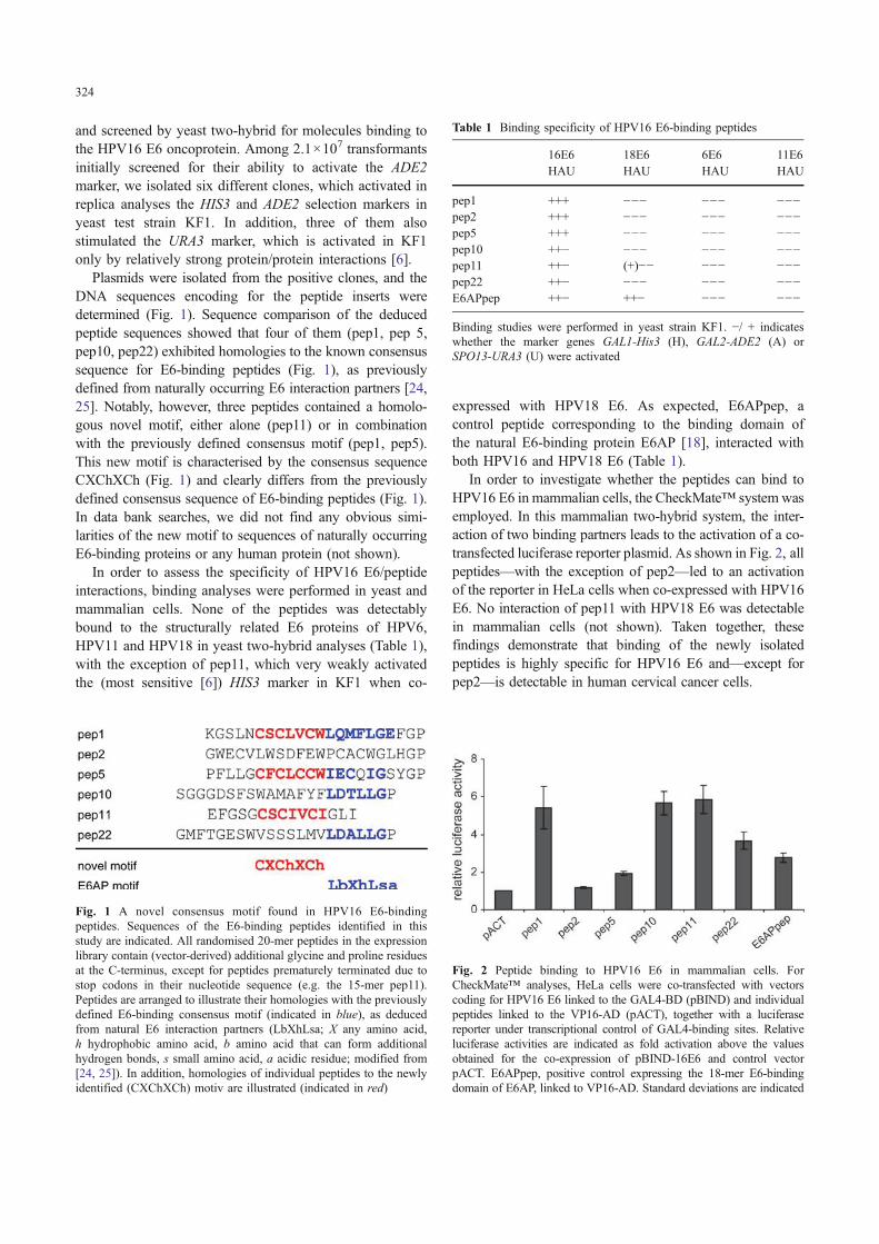
Plasmids were isolated from the positive clones, and theDNA sequences encoding for the peptide inserts weredetermined (Fig. 1). Sequence comparison of the deducedpeptide sequences showed that four of them (pep1, pep 5,pep10, pep22) exhibited homologies to the known consensussequence for E6-binding peptides (Fig. 1), as previouslydefined from naturally occurring E6 interaction partners [24,25]. Notably, however, three peptides contained a homolo-gous novel motif, either alone (pep11) or in combinationwith the previously defined consensus motif (pep1, pep5).This new motif is characterised by the consensus sequenceCXChXCh (Fig. 1) and clearly differs from the previouslydefined consensus sequence of E6-binding peptides (Fig. 1).In data bank searches, we did not find any obvious simi-larities of the new motif to sequences of naturally occurringE6-binding proteins or any human protein (not shown).
In order to assess the specificity of HPV16 E6/peptideinteractions, binding analyses were performed in yeast andmammalian cells. None of the peptides was detectablybound to the structurally related E6 proteins of HPV6,HPV11 and HPV18 in yeast two-hybrid analyses (Table 1),with the exception of pep11, which very weakly activatedthe (most sensitive [6]) HIS3 marker in KF1 when co-
expressed with HPV18 E6. As expected, E6APpep, acontrol peptide corresponding to the binding domain ofthe natural E6-binding protein E6AP [18], interacted withboth HPV16 and HPV18 E6 (Table 1).
In order to investigate whether the peptides can bind toHPV16 E6 in mammalian cells, the CheckMate™ systemwasemployed. In this mammalian two-hybrid system, the inter-action of two binding partners leads to the activation of a co-transfected luciferase reporter plasmid. As shown in Fig. 2, allpeptides—with the exception of pep2—led to an activationof the reporter in HeLa cells when co-expressed with HPV16E6. No interaction of pep11 with HPV18 E6 was detectablein mammalian cells (not shown). Taken together, thesefindings demonstrate that binding of the newly isolatedpeptides is highly specific for HPV16 E6 and—except forpep2—is detectable in human cervical cancer cells.
Fig. 1 A novel consensus motif found in HPV16 E6-bindingpeptides. Sequences of the E6-binding peptides identified in thisstudy are indicated. All randomised 20-mer peptides in the expressionlibrary contain (vector-derived) additional glycine and proline residuesat the C-terminus, except for peptides prematurely terminated due tostop codons in their nucleotide sequence (e.g. the 15-mer pep11).Peptides are arranged to illustrate their homologies with the previouslydefined E6-binding consensus motif (indicated in blue), as deducedfrom natural E6 interaction partners (LbXhLsa; X any amino acid,h hydrophobic amino acid, b amino acid that can form additionalhydrogen bonds, s small amino acid, a acidic residue; modified from[24, 25]). In addition, homologies of individual peptides to the newlyidentified (CXChXCh) motiv are illustrated (indicated in red)
Table 1 Binding specificity of HPV16 E6-binding peptides
16E6 18E6 6E6 11E6HAU HAU HAU HAU
pep1 +++ −−− −−− −−−pep2 +++ −−− −−− −−−pep5 +++ −−− −−− −−−pep10 ++− −−− −−− −−−pep11 ++− (+)−− −−− −−−pep22 ++− −−− −−− −−−E6APpep ++− ++− −−− −−−
Binding studies were performed in yeast strain KF1. −/ + indicateswhether the marker genes GAL1-His3 (H), GAL2-ADE2 (A) orSPO13-URA3 (U) were activated
Fig. 2 Peptide binding to HPV16 E6 in mammalian cells. ForCheckMate™ analyses, HeLa cells were co-transfected with vectorscoding for HPV16 E6 linked to the GAL4-BD (pBIND) and individualpeptides linked to the VP16-AD (pACT), together with a luciferasereporter under transcriptional control of GAL4-binding sites. Relativeluciferase activities are indicated as fold activation above the valuesobtained for the co-expression of pBIND-16E6 and control vectorpACT. E6APpep, positive control expressing the 18-mer E6-bindingdomain of E6AP, linked to VP16-AD. Standard deviations are indicated
324

Next, we tested whether intracellular expression of the E6-binding peptides affects the phenotype of HPV16-positivecancer cells. As shown in Fig. 3, pep11, which contains thenewly defined E6-binding motif, strongly interfered with thecolony formation capacity of HPV16-positive CaSki or SiHacells. On the contrary, growth of control cells, such asHPV18-positive HeLa and HPV-negative MCF-7 (Fig. 3) orH1299 cells (not shown), was not affected by pep11. Thesefindings show that the detrimental effect of pep11 expressionon colony formation capacity is HPV16-specific and is notdue to unspecific toxicity induced by pep11 expression. Incontrast to pep11, expression of E6APpep did not affect thegrowth of either HPV16- or HPV18-positive cancer cells,under the same experimental conditions.
In view of its pronounced bioactivity in HPV16-positivecancer cells, further work concentrated on pep11. In orderto investigate whether pep11 can bind to HPV16 E6 invitro, several attempts were made to chemically synthesisepep11. However, yields were very poor, and the peptidewas hardly soluble in aqueous solution. Since pep11 isrelatively hydrophobic, we decided to create a solubility-improved pep11 variant, which retains binding to HPV16E6. First, we defined the critical amino acids required forbinding to HPV16 E6. We systematically replaced allamino acids within pep11 by alanine. Mammalian two-hybrid analyses showed that mutation of pep11 amino acidresidues C6, C8, I9, C11 and I12 within the newly definedE6-binding consensus sequence were strongly detrimental
to its binding capacity to E6 (Fig. 4a). In addition, thehydrophobic C-terminus of pep11 contributed to binding,whereas mutations of the amino acids N-terminal of theconsensus motif were generally well tolerated.
Next, we replaced several amino acids by more hydro-philic residues in order to improve solubility. Exchangedresidues were selected for their similar side chain volumes,chemical similarity and maintenance of overall peptidecharge. For example, cysteine was replaced by serine,which differs from cysteine only by a hydroxyl groupinstead of a thiol group, and isoleucine was replaced byglutamine, which both have similar side chain volumes.Figure 4b summarises which amino acid replacementsconserved E6 binding. Based on these data, all functionalmutations were merged to yield a solubility-improved variantpep11* (Fig. 4c and d). In addition, we created a secondpep11 variant, termed pep11**, by adding a four amino acidsolubility enhancement tag [26] at the N-terminus of pep11*,KEKE, maintaining the overall peptide charge (Fig. 4d).Both solubility-improved pep11 variants, pep11* andpep11**, retained E6-binding activity (Fig. 4c), whereasthe respective E6-binding-defective mutants pep11*m andpep11**m, containing three cysteine to serine conversions,served as negative controls for further experiments.
To address the question whether pep11** binds HPV16E6 in vitro, the solubility-improved variant pep11** waschemically synthesised and investigated for binding topurified HPV16 E6 by FP. FP-based equilibrium binding
Fig. 3 pep11 specifically interferes with the colony formationcapacity of HPV16-positive cells. Colony formation assays inHPV16-positive CaSki and SiHa as well as in HPV18-positive HeLa
or HPV-negative MCF-7 cells, following transfection with hrGFP-peptide expression vectors. hrGFP negative control
325

studies (Fig. 5) revealed that pep11** associates withHPV16 E6 with high affinity (Kd 118 nM). E6APpepbounds to E6 with an approximately five times loweraffinity (612 nM), comparable to the Kd value of 4 μM,determined previously by surface plasmon resonance [27].
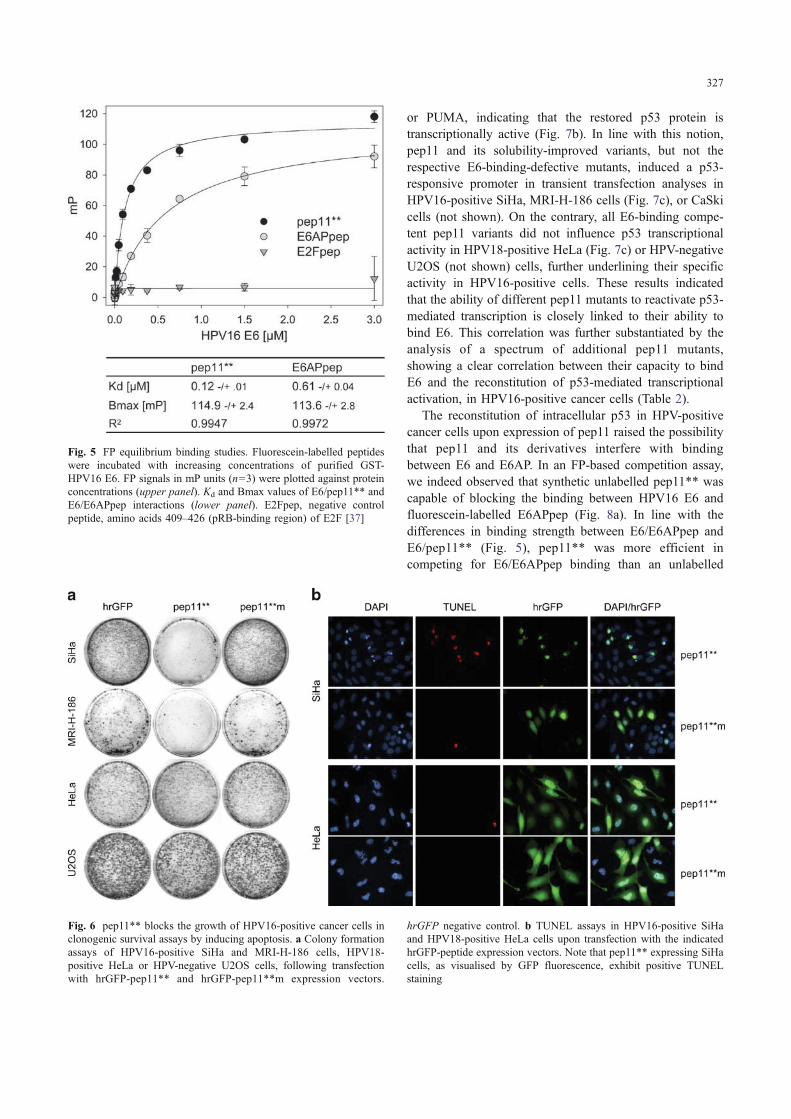
As shown in Fig. 6a, alike pep11, the solubility-improvedpep11** efficiently blocked colony growth of HPV16-positive cancer cells (SiHa, MRI-H-186) but not ofHPV16-negative control cells (HeLa, U2OS), showing thatit is functional at the intracellular level. On the contrary,control peptide pep11**m did not affect colony formationcapacity of any of the tested cell lines. In order to assesswhether pep11**-mediated inhibition of colony growth islinked to induction of apoptosis, TUNEL analyses wereperformed. As shown in Fig. 6b, expression of pep11**, butnot of pep11**m, induced apoptosis. Again, this effect wasspecific for HPV16-positive cells in that HPV18-positiveHeLa cells did not show signs of apoptosis upon intracellularpep11** expression. Corresponding experiments analysingpep11* and pep11*m yielded identical results (not shown).
Since E6 induces the proteolytic degradation of p53 viaformation of a trimeric complex with E6AP [18, 28], weinvestigated whether the pep11-induced phenotypic effectscorrelate with alterations of p53 protein levels. As shown inFig. 7a, ectopic co-expression of p53 and HPV16 E6 inH1299 cells (in which the endogenous p53 gene is deleted)resulted in a strong reduction of p53 protein concentrations,as expected. The additional co-expression of pep11 or thesolubility-optimised variants pep11* and pep11** restoredp53 protein levels, indicating interference with E6-mediatedp53 degradation. In contrast, the E6-binding-defectivemutant pep11*m did not replenish intracellular p53 levels.Corresponding results were obtained, analysing endoge-nous p53 levels in HPV16-positive MRI-H-186 cells.Again, p53 levels were increased by pep11 and evengreater by the solubility-optimised pep11* and pep11**variants, but not by pep11*m (Fig. 7b). Identical findingswere obtained in HPV16-positive SiHa cells (not shown).
The increase in p53 upon expression of pep11 waslinked to induction of known p53 target genes, such as p21
Fig. 4 Binding behaviour of pep11 variants to HPV16 E6. Check-Mate™ analyses in HeLa cells, co-transfected with expression vectorscoding for GAL4-BD-HPV16 E6, VP16-AD-peptide variants and aGAL4-responsive luciferase reporter plasmid. Values represent foldluciferase activation above co-transfections with basic vector (pACT).n=3±SD. a Alanin scanning of pep11 residues critical for binding toHPV16 E6. b Solubility optimisation of pep11 by replacement of
individual amino acids by more hydrophilic residues. c Bindingbehaviour and d peptide sequences of solubility-optimised variantspep11* and pep11** and non-functional mutants pep11*m andpep11**m. Residues highlighted in grey indicate replacements inpep11 by more hydrophilic amino acids, which were not detrimentalfor binding. Circled residues indicate cysteine to serine conversions inthe binding-defective mutants
326
or PUMA, indicating that the restored p53 protein istranscriptionally active (Fig. 7b). In line with this notion,pep11 and its solubility-improved variants, but not therespective E6-binding-defective mutants, induced a p53-responsive promoter in transient transfection analyses inHPV16-positive SiHa, MRI-H-186 cells (Fig. 7c), or CaSkicells (not shown). On the contrary, all E6-binding compe-tent pep11 variants did not influence p53 transcriptionalactivity in HPV18-positive HeLa (Fig. 7c) or HPV-negativeU2OS (not shown) cells, further underlining their specificactivity in HPV16-positive cells. These results indicatedthat the ability of different pep11 mutants to reactivate p53-mediated transcription is closely linked to their ability tobind E6. This correlation was further substantiated by theanalysis of a spectrum of additional pep11 mutants,showing a clear correlation between their capacity to bindE6 and the reconstitution of p53-mediated transcriptionalactivation, in HPV16-positive cancer cells (Table 2).
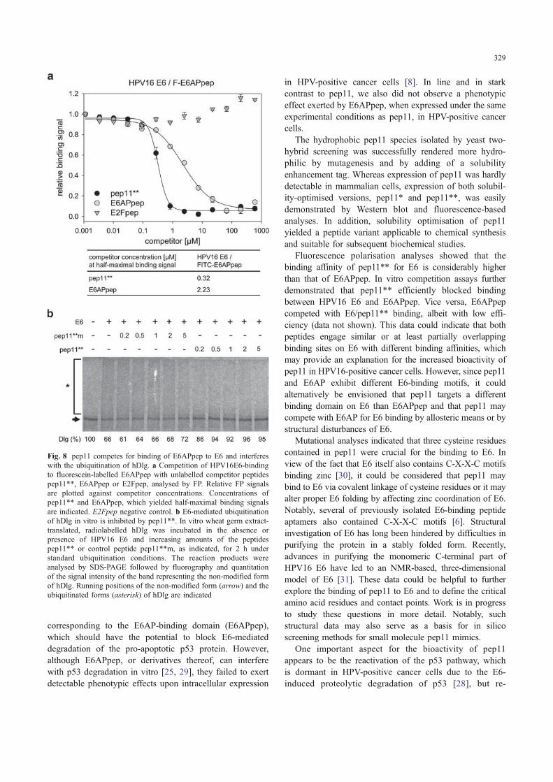
The reconstitution of intracellular p53 in HPV-positivecancer cells upon expression of pep11 raised the possibilitythat pep11 and its derivatives interfere with bindingbetween E6 and E6AP. In an FP-based competition assay,we indeed observed that synthetic unlabelled pep11** wascapable of blocking the binding between HPV16 E6 andfluorescein-labelled E6APpep (Fig. 8a). In line with thedifferences in binding strength between E6/E6APpep andE6/pep11** (Fig. 5), pep11** was more efficient incompeting for E6/E6APpep binding than an unlabelled
Fig. 6 pep11** blocks the growth of HPV16-positive cancer cells inclonogenic survival assays by inducing apoptosis. a Colony formationassays of HPV16-positive SiHa and MRI-H-186 cells, HPV18-positive HeLa or HPV-negative U2OS cells, following transfectionwith hrGFP-pep11** and hrGFP-pep11**m expression vectors.
hrGFP negative control. b TUNEL assays in HPV16-positive SiHaand HPV18-positive HeLa cells upon transfection with the indicatedhrGFP-peptide expression vectors. Note that pep11** expressing SiHacells, as visualised by GFP fluorescence, exhibit positive TUNELstaining
Fig. 5 FP equilibrium binding studies. Fluorescein-labelled peptideswere incubated with increasing concentrations of purified GST-HPV16 E6. FP signals in mP units (n=3) were plotted against proteinconcentrations (upper panel). Kd and Bmax values of E6/pep11** andE6/E6APpep interactions (lower panel). E2Fpep, negative controlpeptide, amino acids 409–426 (pRB-binding region) of E2F [37]
327

E6APpep peptide (Fig. 8a). In order to investigate whetherpep11 is also functional under in vitro conditions and mayaffect other E6 targets, we tested its effect on E6-inducedubiquitination of the human homologue of the Drosophiladiscs large (hDlg) tumour suppressor protein, which ismediated by E6AP [23]. To this end, E6 was preincubatedwith increasing amounts of pep11**, and then, the mixturewas added to a ubiquitination reaction containing E6APand radiolabelled hDlg. As shown in Fig. 8b, the presenceof pep11** efficiently interfered with E6-mediated ubiq-uitination of hDlg. In contrast, the non-functional mutantpep11**m had no significant effect on hDlg ubiquitination,further corroborating the specificity of pep11** activity.
Discussion
The present work identifies a novel peptide-binding motif,which specifically binds to the HPV16 E6 protein.Moreover, expression of this motif in the pep11 peptide
restored functional p53 protein and specifically killedHPV16-positive cervical cancer cells by inducing apoptoticcell death. These findings support the idea that specific E6inhibitors can efficiently induce cell death in HPV-positivecancer cells. Moreover, pep11 should provide a novel basisto generate pharmacologically useful molecules for thetreatment of HPV-associated lesions.
The pronounced phenotypic effects observed in ourstudy indicate that pep11 targets a surface domain on E6,which is critical for the anti-apoptotic activity of E6. Inprinciple, this could be also expected for peptides
Fig. 7 Reconstitution of p53 by pep11 in HPV16 E6 expressing cells.a Western blot analysis upon co-expression of HPV16 E6 and p53 inH1299 cells—with an additional co-expression of hrGFP-pep11variants, as indicated. β-Gal transfection control. b HPV16-positiveMRI-H-186 cells expressing pep11 derivatives. Analysis of endoge-nous p53 levels as well as p21 and PUMA expression by Western blot.Tubulin loading control. c Luciferase reporter assays in HPV16-positive MRI-H-186 and SiHa or in HPV18-positive HeLa controlcells, upon co-transfection of a p53-responsive reporter plasmidtogether with expression vectors coding for different hrGFP-pep11variants or hrGFP-E6APpep
Table 2 Binding specificity and p53 reactivation pattern of pep11variants
HPV16 E6 binding p53 reactivation
pep11 wt 5.87±0.76 4.07±0.55E1A 3.44±0.43 3.70±0.53F2A 4.92±0.25 3.93±1.18G3A 5.85±0.78 4.15±1.55S4A 5.51±0.86 4.07±0.58G5A 7.05±1.35 3.80±0.51C6A 0.97±0.06 1.09±0.11S7A 5.57±1.75 4.94±0.96C8A 0.99±0.08 1.10±0.16I9A 1.80±0.15 3.15±0.44V10A 6.25±1.49 8.54±2.43C11A 1.02±0.09 0.94±0.19I12A 2.07±0.24 2.12±0.23G13A 1.21±0.11 1.16±0.16L14A 2.26±0.29 1.98±0.35I15A 2.27±0.36 1.95±0.25KEKE 9.27±1.15 5.06±0.92F2Y 5.18±0.44 5.17±0.32G3N 7.04±0.96 4.60±1.11G5N 7.03±0.79 4.07±0.90C6S 1.03±0.15 0.94±0.42C8S 1.02±0.12 1.05±0.57I9Q 1.12±0.06 1.43±0.10V10T 4.39±0.45 4.69±0.78C11S 1.01±0.12 1.11±0.54I12Q 1.95±0.18 2.86±0.88G13N 1.36±0.11 1.29±0.16L14N 1.34±0.02 1.23±0.14I15Q 1.35±0.02 1.19±0.17pep11* 8.07±1.82 11.44±2.09pep11*m 0.94±0.03 1.09±0.41pep11** 9.97±1.61 7.68±1.31pep11**m 1.10±0.06 0.98±0.01E6APpep 2.77±0.23 2.99±0.52
Binding of pep11 variants to HPV16 E6, as determined by Check-Mate™ analyses. Reactivation of endogenous p53 in HPV16-positiveMRI-H-186 cells, as determined by p53-responsive luciferase assaysupon expression of hrGFP-peptide fusions. Values indicate foldactivation compared to co-transfecting the basic vectors (pACT forCheckMate™ analyses, pCEP4-hrGFP for p53 reactivation assays).n=3±SD
328
corresponding to the E6AP-binding domain (E6APpep),which should have the potential to block E6-mediateddegradation of the pro-apoptotic p53 protein. However,although E6APpep, or derivatives thereof, can interferewith p53 degradation in vitro [25, 29], they failed to exertdetectable phenotypic effects upon intracellular expression
in HPV-positive cancer cells [8]. In line and in starkcontrast to pep11, we also did not observe a phenotypiceffect exerted by E6APpep, when expressed under the sameexperimental conditions as pep11, in HPV-positive cancercells.
The hydrophobic pep11 species isolated by yeast two-hybrid screening was successfully rendered more hydro-philic by mutagenesis and by adding of a solubilityenhancement tag. Whereas expression of pep11 was hardlydetectable in mammalian cells, expression of both solubil-ity-optimised versions, pep11* and pep11**, was easilydemonstrated by Western blot and fluorescence-basedanalyses. In addition, solubility optimisation of pep11yielded a peptide variant applicable to chemical synthesisand suitable for subsequent biochemical studies.
Fluorescence polarisation analyses showed that thebinding affinity of pep11** for E6 is considerably higherthan that of E6APpep. In vitro competition assays furtherdemonstrated that pep11** efficiently blocked bindingbetween HPV16 E6 and E6APpep. Vice versa, E6APpepcompeted with E6/pep11** binding, albeit with low effi-ciency (data not shown). This data could indicate that bothpeptides engage similar or at least partially overlappingbinding sites on E6 with different binding affinities, whichmay provide an explanation for the increased bioactivity ofpep11 in HPV16-positive cancer cells. However, since pep11and E6AP exhibit different E6-binding motifs, it couldalternatively be envisioned that pep11 targets a differentbinding domain on E6 than E6APpep and that pep11 maycompete with E6AP for E6 binding by allosteric means or bystructural disturbances of E6.
Mutational analyses indicated that three cysteine residuescontained in pep11 were crucial for the binding to E6. Inview of the fact that E6 itself also contains C-X-X-C motifsbinding zinc [30], it could be considered that pep11 maybind to E6 via covalent linkage of cysteine residues or it mayalter proper E6 folding by affecting zinc coordination of E6.Notably, several of previously isolated E6-binding peptideaptamers also contained C-X-X-C motifs [6]. Structuralinvestigation of E6 has long been hindered by difficulties inpurifying the protein in a stably folded form. Recently,advances in purifying the monomeric C-terminal part ofHPV16 E6 have led to an NMR-based, three-dimensionalmodel of E6 [31]. These data could be helpful to furtherexplore the binding of pep11 to E6 and to define the criticalamino acid residues and contact points. Work is in progressto study these questions in more detail. Notably, suchstructural data may also serve as a basis for in silicoscreening methods for small molecule pep11 mimics.
One important aspect for the bioactivity of pep11appears to be the reactivation of the p53 pathway, whichis dormant in HPV-positive cancer cells due to the E6-induced proteolytic degradation of p53 [28], but re-
Fig. 8 pep11 competes for binding of E6APpep to E6 and interfereswith the ubiquitination of hDlg. a Competition of HPV16E6-bindingto fluorescein-labelled E6APpep with unlabelled competitor peptidespep11**, E6APpep or E2Fpep, analysed by FP. Relative FP signalsare plotted against competitor concentrations. Concentrations ofpep11** and E6APpep, which yielded half-maximal binding signalsare indicated. E2Fpep negative control. b E6-mediated ubiquitinationof hDlg in vitro is inhibited by pep11**. In vitro wheat germ extract-translated, radiolabelled hDlg was incubated in the absence orpresence of HPV16 E6 and increasing amounts of the peptidespep11** or control peptide pep11**m, as indicated, for 2 h understandard ubiquitination conditions. The reaction products wereanalysed by SDS-PAGE followed by fluorography and quantitationof the signal intensity of the band representing the non-modified formof hDlg. Running positions of the non-modified form (arrow) and theubiquitinated forms (asterisk) of hDlg are indicated
329

inducible upon repression of E6 expression [32, 33].Indeed, apoptosis induction by pep11 was linked to areconstitution of endogenous p53 protein levels in HPV-positive cancer cells, induction of p53 target genes andtranscriptional stimulation of a p53-responsive reporterplasmid. Furthermore, analyses of a comprehensive seriesof pep11 mutants exhibited a clear correlation between theability of individual peptides to bind HPV16 E6 and p53-mediated transcriptional stimulation. The observation thatpep11 interfered with E6-induced ubiquitination of hDlgindicates that pep11 also can affect additional tumourrelevant E6 interactions, besides p53.
In view of its pronounced pro-apoptotic activity, it isconceivable that drugs acting similarly to pep11 could be oftherapeutic benefit for the treatment of HPV16-associateddiseases. At least two avenues could be pursued in order tofurther develop the therapeutic potential of pep11 into anapplication perspective. Firstly, it could be envisioned todevelop pep11 itself into a proteinaceous drug. Like proteintherapy in general, this endeavour will require optimisationof both biopharmaceutic and pharmacokinetic parameters.For example, peptide stability could be increased byemploying modified L-amino acids [34], stereochemicalmodifications of amino acids [35] or enhancement ofpeptide folds via intramolecular stabilisation [36].
Secondly, it will be interesting to explore whether it ispossible to identify small, drug-like molecules fromcompound libraries, which can act as functional pep11mimics. In this scenario, the pep11/E6 interaction shouldform a valuable basis for developing in vitro screeningsystems to identify compounds, which bind to the same,functionally critical E6 surface domain as targeted bypep11. The general feasibility of such “displacementscreening” approaches has been demonstrated, for example,by identifying small molecules interfering with the interac-tion of peptide aptamers with their target proteins [15]. Asfor other in vitro analyses, such as NMR studies, thesolubility-improved pep11 variant designed in the presentstudy should also be useful for screening purposes.Conceivably, small molecules targeting the surface regiondefined by pep11 could have the potential to also efficientlyinterfere with the anti-apoptotic activity of E6.
Acknowledgements This work was supported by the DeutscheKrebshilfe. We thank Angela Heilig for excellent technical assistance,Dr. H. Zentgraf for providing anti-hrGFP antibodies and Dr. JenniferReed for helpful discussions.
References
1. zur Hausen H (2002) Papillomaviruses and cancer: from basicstudies to clinical application. Nat Rev Cancer 2:342–350
2. Schiffman M, Castle PE, Jeronimo J, Rodriguez AC, Wacholder S(2007) Human papillomavirus and cervical cancer. Lancet370:890–907
3. Lowy DR, Schiller JT (2006) Prophylactic human papillomavirusvaccines. J Clin Invest 116:1167–1173
4. Agosti JM, Goldie SJ (2007) Introducing HPV vaccine indeveloping countries—key challenges and issues. New Engl JMed 356:1908–1910
5. Parkin DM, Bray F (2006) The burden of HPV-related cancers.Vaccine 24(Suppl. 3):S11–S25
6. Butz K, Denk C, Ullmann A, Scheffner M, Hoppe-Seyler F(2000) Induction of apoptosis in human papillomavirus-positivecancer cells by peptide aptamers targeting the viral E6 oncopro-tein. Proc Natl Acad Sci U S A 97:6693–6697
7. Butz K, Ristriani T, Hengstermann A, Denk C, Scheffner M,Hoppe-Seyler F (2003) siRNA targeting of the viral E6 oncogeneefficiently kills human papillomavirus-positive cancer cells.Oncogene 22:5938–5945
8. Griffin H, Elston R, Jackson D, Ansell K, Coleman M, Winter G,Doorbar J (2006) Inhibition of papillomavirus protein function incervical cancer cells by intrabody targeting. J Mol Biol 355:360–378
9. Yamato K, Fen J, Kobuchi H, Nasu Y, Yamada T, Nishihara T,Ikeda Y, Kizaki M, Yoshinouchi M (2006) Induction of cell deathin human papillomavirus 18-positive cervical cancer cells by E6siRNA. Cancer Gene Ther 13:234–241
10. Lagrange M, Boulade-Ladame C, Mailly L, Weiss E, OrfanoudakisG, Deryckere F (2007) Intracellular scFvs against the viral E6oncoprotein provoke apoptosis in human papillomavirus-positivecancer cells. Biochem Biophys Res Commun 361:487–492
11. Koivusalo R, Mialon A, Pitkänen H, Westermarck J, Hietanen S(2006) Activation of p53 in cervical cancer cells by humanpapillomavirus E6 RNA interference is transient, but can besustained by inhibiting endogenous nuclear export-dependent p53antagonists. Cancer Res 66:11817–11824
12. Courtete J, Sibler AP, Zeder-Lutz G, Dalkara D, Oulad-AbdelghaniM, Zuber G, Weiss E (2007) Suppression of cervical carcinoma cellgrowth by intracytoplasmic codelivery of anti-oncoprotein E6antibody and small interfering RNA. Mol Cancer Ther 6:1728–1735
13. DeFilippis RA, Goodwin EC, Wu L, DiMaio D (2003) Endoge-nous human papillomavirus E6 and E7 proteins differentiallyregulate proliferation, senescence, and apoptosis in HeLa cervicalcarcinoma cells. J Virol 77:1551–1563
14. Lobato MN, Rabbitts TH (2004) Intracellular antibodies asspecific reagents for functional ablation: future therapeuticmolecules. Curr Mol Med 4:519–528
15. Baines IC, Colas P (2006) Peptide aptamers as guides for small-molecule drug discovery. Drug Discov Today 7–8:334–341
16. Hoppe-Seyler F, Butz K (1993) Repression of endogenous p53transactivation function in HeLa cervical carcinoma cells byhuman papillomavirus type 16 E6, human mdm-2, and mutantp53. J Virol 67:3111–3117
17. Vogt M, Butz K, Dymalla S, Semzow J, Hoppe-Seyler F (2006)Inhibition of bax activity is crucial for the anti-apoptotic function ofthe human papillomavirus E6 oncoprotein. Oncogene 25:4009–4015
18. Huibregtse JM, Scheffner M, Howley PM (1993) Cloning andexpression of the cDNA for E6-AP, a protein that mediates theinteraction of the human papillomavirus E6 oncoprotein with p53.Mol Cell Biol 13:775–784
19. Butz K, Denk C, Fitscher B, Crnković-Mertens I, Ullmann A,Schröder CH, Hoppe-Seyler F (2001) Peptide aptamers targetingthe hepatitis B virus core protein: a new generation of moleculeswith antiviral activity. Oncogene 20:6579–6586
20. Sehr P, Zumbach K, Pawlita M (2001) A generic capture ELISAfor recombinant proteins fused to glutathione S-transferase:validation for HPV serology. J Immunol Methods 253:153–162
330

21. Funk WD, Pak DT, Karas RH, Wright WE, Shay JW (1992) Atranscriptionally active DNA-binding site for human p53 proteincomplexes. Mol Cell Biol 12:2866–2871
22. Nuber U, Schwarz S, Kaiser P, Schneider R, Scheffner M (1996)Cloning of human ubiquitin-conjugating enzymes UbcH6 andUbcH7 (E2-F1) and characterization of their interaction with E6-AP and RSP5. J Biol Chem 271:2795–2800
23. Kuballa P, Matentzoglu K, Scheffner M (2007) The role of theubiquitin ligase E6-AP in human papillomavirus E6-mediateddegradation of PDZ domain-containing proteins. J Biol Chem282:65–71
24. Chen JJ, Hong Y, Rustamzadeh E, Baleja JD, Androphy EJ (1998)Identification of an alpha helical motif sufficient for associationwith papillomavirus E6. J Biol Chem 273:13537–13544
25. Elston RC, Naphtine S, Doorbar J (1998) The identification of aconserved binding motif within human papillomavirus type 16 E6binding peptides, E6AP and E6BP. J Gen Virol 79:371–374
26. Kato A, Maki K, Ebina T, Kuwajima K, Soda K, Kuroda Y (2007)Mutational analysis of protein solubility enhancement using shortpeptide tags. Biopolymers 85:12–18
27. Zanier K, Charbonnier S, Baltzinger M, Nominé Y, Altschuh D,Travé G (2005) Kinetic analysis of the interactions of humanpapillomavirus E6 oncoproteins with the ubiquitin ligase E6APusing surface plasmon resonance. J Mol Biol 349(2):401–412
28. Scheffner M, Werness BA, Huibregtse JM, Levine AJ, HowleyPM (1990) The E6 oncoprotein encoded by human papillomavirustypes 16 and 18 promotes the degradation of p53. Cell 63:1129–1136
29. Sterlinko Grm H, Weber M, Elston R, McIntosh P, Griffin H,Banks L, Doorbar J (2004) Inhibition of E6-induced degradation
of its cellular substrates by novel blocking peptides. J Mol Biol335:971–985
30. Barbosa MS, Lowy DR, Schiller JT (1989) Papillomaviruspolypeptides E6 and E7 are zinc-binding proteins. J Virol63:1404–1407
31. Nominé Y, Masson M, Charbonnier S, Zanier K, Ristriani T,Deryckère F, Sibler AP, Desplancq D, Atkinson RA, Weiss E,Orfanoudakis G, Kieffer B, Travé G (2006) Structural and functionalanalysis of E6 oncoprotein: insights in the molecular pathways ofhuman papillomavirus-mediated pathogenesis. Mol Cell 21:665–678
32. Butz K, Shahabeddin L, Geisen C, Spitkovsky D, Ullmann A,Hoppe-Seyler F (1995) Functional p53 protein in humanpapillomavirus-positive cancer cells. Oncogene 10:927–936
33. Goodwin EC, DiMaio D (2000) Repression of human papilloma-virus oncogenes in HeLa cervical carcinoma cells causes theorderly reactivation of dormant tumor suppressor pathways. ProcNatl Acad Sci U S A 97:12513–12518
34. Fauchere JL, Thurieau C (1992) Evaluation of the stability ofpeptides and pseudopeptides as a tool in peptide drug design. AdvDrug Res 23:127–159
35. Fischer PM (2003) The design, synthesis and application ofstereochemical and directional peptide isomers: a critical review.Curr Protein Pept Sci 4:339–356
36. Walensky LD, Kung AL, Escher I, Malia TJ, Barbuto S, WrightRD, Wagner G, Verdine GL, Korsmeyer SJ (2004) Activation ofapoptosis in vivo by a hydrocarbon-stapled BH3 helix. Science305:1466–1470
37. Helin K, Lees JA, Vidal M, Dyson N, Harlow E, Fattaey A (1992)A cDNA encoding a pRB-binding protein with properties of thetranscription factor E2F. Cell 70:337–350
331


![MOTIF XF Editor VST Owner’s Manual7.Sélectionnez « MOTIF XF6 (MOTIF XF7 ou MOTIF XF8) » dans la colonne [FW Device] (Périphérique FW). 8.Sélectionnez « MOTIF XF6 (MOTIF XF7](https://img.pdfslide.net/doc/110x75/611158b13f31404d2d274378/motif-xf-editor-vst-owneras-manual-7slectionnez-motif-xf6-motif-xf7-ou.jpg)