Embed Size (px)
Citation preview

Trends in Biotechnology
TIBTEC 1807 No. of Pages 15
Review
A Physiology-Inspired Multifactorial Toolbox inSoft-to-Hard Musculoskeletal InterfaceTissue Engineering
Isabel Calejo,1,2,4,@ Raquel Costa-Almeida ,1,2,4,@ Rui L. Reis,1,2,3,@ and Manuela E. Gomes1,2,3,*,@
HighlightsSoft-to-hard interfaces exhibit uniquecompositional and structural gradientsthat are difficult to heal and have limitedregenerative abilities.
From a biomimetic perspective, combin-ing biological, biophysical, and biochem-ical cues is likely to enable the generationof physiologically relevant tissueengineered constructs emulating suchcomplex multitissue transitions.
Progresses on cell sheet engineering areintegrating cell patterning and mechani-
Musculoskeletal diseases are increasing the prevalence of physical disabilityworldwide. Within the body, musculoskeletal soft and hard tissues integratethrough specific multitissue transitions, allowing for body movements. Owingto their unique compositional and structural gradients, injuries challenge the na-tive interfaces and tissue regeneration is unlikely to occur. Tissue engineeringstrategies are emerging to emulate the physiological environment of soft-to-hard tissue interfaces. Advances in biomaterial design enable control over bio-physical parameters, but biomaterials alone are not sufficient to provide ade-quate support and guide transplanted cells. Therefore, biological, biophysical,and biochemical tools can be integrated into a multifactorial toolbox, steeringprospective advances toward engineering clinically relevant soft-to-hard tissueinterfaces.
cal stimulation, together with cells andtheir matrices as native orchestrators oftissue regeneration.
Advances in biomaterial design offer aprecise tuning of architectural, topo-graphical, and mechanical properties torecreate cell-specific niches.
Together with gradients of biochemicalcues (including oxygen and growth fac-tors), multifactorial strategies allow stra-tegic control of stem cell differentiationalong a single unit.
13B’s Research Group, I3Bs – ResearchInstitute on Biomaterials, Biodegradablesand Biomimetics, University of Minho,Headquarters of the European Institute ofExcellence on Tissue Engineering andRegenerative Medicine, AvePark, Parquede Ciência e Tecnologia, Zona Industrialda Gandra, 4805-017 Barco, Guimarães,Portugal2ICVS/3B’s–PT Government AssociateLaboratory, Braga/Guimarães, Portugal3The Discoveries Centre for Regenerativeand Precision Medicine, Headquarters atUniversity of Minho, Avepark, 4805-017Barco, Guimarães, Portugal
Musculoskeletal Interfaces and Regeneration Requirements: A Global BurdenBody tissues and organs are inherently composed of multiple tissues interfacing each other andallowing extremely complex biological functions to take place. In the musculoskeletal system,these tissue interfaces integrate extremely dissimilar tissues with distinctive characteristics, rang-ing from a hard and highly vascularized tissue with lightweight stiffness and strength, such as thebone, to extremely viscoelastic and avascular tissues, such as articular cartilage, or to tough, re-silient, and elastic tissue, such as the tendons (Box 1). Interestingly, the defining characteristic ofmusculoskeletal interfaces is their primary load-bearing function while mediating the multiple tran-sition in tissues stiffness [1,2], which structurally requires the presence of hierarchically assembledproteins in highly specialized extracellular matrices (Box 1). Examples of soft-to-hard tissue inter-faces are the tendon/ligament-to-bone interface, commonly found in the rotator cuff and anteriorcruciate ligament, and osteochondral unit (cartilage-to-bone interface) found in the knee joints.However, these tissue interfaces are commonly affected by diseases and disorders at all stagesof life. Strikingly, musculoskeletal diseases have been estimated to correspond to the majorcause of disability worldwide, with significant healthcare and social support costs [3]. Rotatorcuff and anterior cruciate ligament tears or detachment affect the daily life of both adults andthe elderly, resulting in pain and movement impairment [4]. Similarly, in adulthood, osteoarthritis,a degenerative process resulting in progressive articular cartilage and joint destruction, especiallyat the osteochondral interface, is one of the major contributors to immobility, pain, and productiv-ity loss [3]. Current strategies used in the clinics to manage ligament/tendon injuries include theapplication of grafts (see Glossary) (auto-, allo-, and synthetic grafts) and, alternatively,microfracture surgery, where subchondral bone is perforated to enable a localized transport ofbone marrow and blood into contact with the degenerate tissue in the osteochondral unit. How-ever, unsatisfactory functional outcomes of repaired tissues due to previous degeneration, to-gether with increased risk of re-injury, lay the foundation for the development of soft-to-hard
Trends in Biotechnology, Month 2019, Vol. xx, No. xx https://doi.org/10.1016/j.tibtech.2019.06.003 1© 2019 Elsevier Ltd. All rights reserved.
4These authors contributed equally to thiswork
*Correspondence:[email protected](M.E. Gomes).URL: http://www.3bs.uminho.pt@Twitter: @3bsuminho (3B’s ResearchGroup), @calejo_isabel (I. Calejo),@raquelccalmeida (R. Costa-Almeida),@RLGReis (R.L. Reis), and@ManuelaEGomes (M.E. Gomes).
Trends in Biotechnology
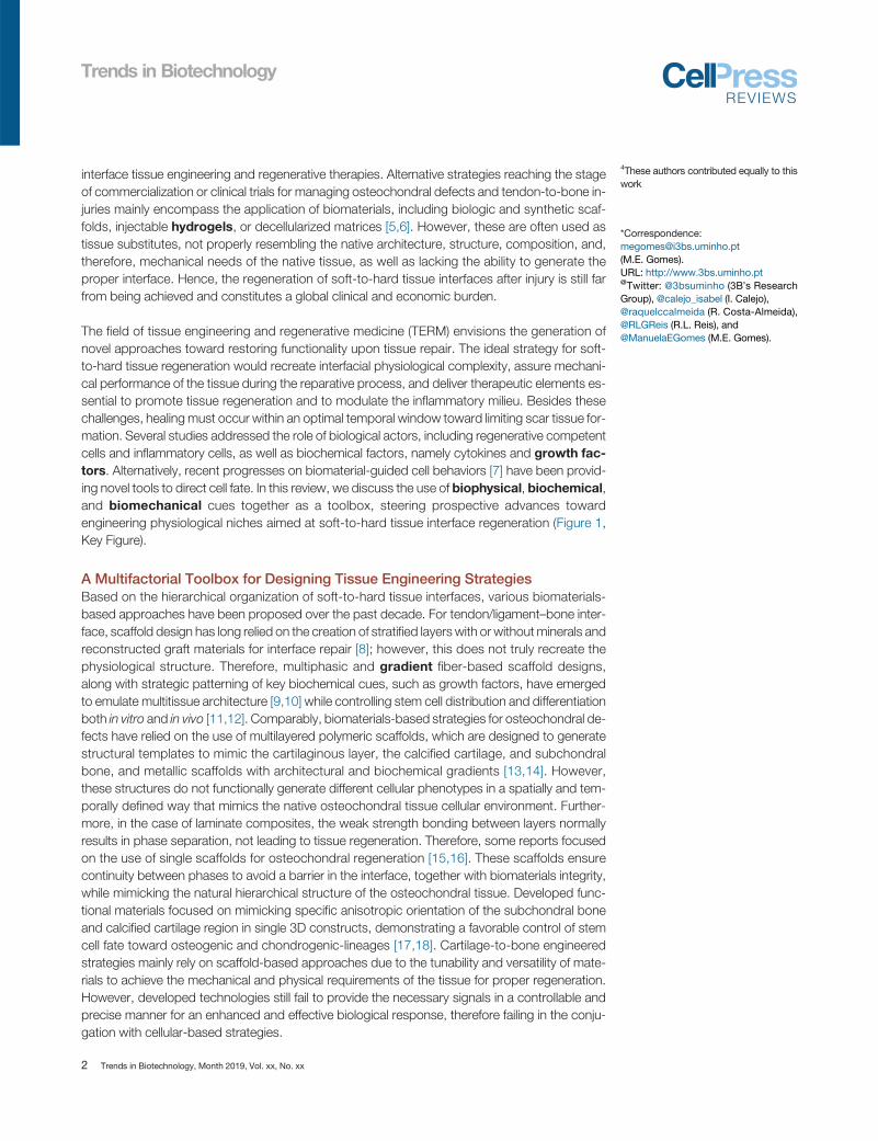
interface tissue engineering and regenerative therapies. Alternative strategies reaching the stageof commercialization or clinical trials for managing osteochondral defects and tendon-to-bone in-juries mainly encompass the application of biomaterials, including biologic and synthetic scaf-folds, injectable hydrogels, or decellularized matrices [5,6]. However, these are often used astissue substitutes, not properly resembling the native architecture, structure, composition, and,therefore, mechanical needs of the native tissue, as well as lacking the ability to generate theproper interface. Hence, the regeneration of soft-to-hard tissue interfaces after injury is still farfrom being achieved and constitutes a global clinical and economic burden.
The field of tissue engineering and regenerative medicine (TERM) envisions the generation ofnovel approaches toward restoring functionality upon tissue repair. The ideal strategy for soft-to-hard tissue regeneration would recreate interfacial physiological complexity, assure mechani-cal performance of the tissue during the reparative process, and deliver therapeutic elements es-sential to promote tissue regeneration and to modulate the inflammatory milieu. Besides thesechallenges, healing must occur within an optimal temporal window toward limiting scar tissue for-mation. Several studies addressed the role of biological actors, including regenerative competentcells and inflammatory cells, as well as biochemical factors, namely cytokines and growth fac-tors. Alternatively, recent progresses on biomaterial-guided cell behaviors [7] have been provid-ing novel tools to direct cell fate. In this review, we discuss the use of biophysical, biochemical,and biomechanical cues together as a toolbox, steering prospective advances towardengineering physiological niches aimed at soft-to-hard tissue interface regeneration (Figure 1,Key Figure).
A Multifactorial Toolbox for Designing Tissue Engineering StrategiesBased on the hierarchical organization of soft-to-hard tissue interfaces, various biomaterials-based approaches have been proposed over the past decade. For tendon/ligament–bone inter-face, scaffold design has long relied on the creation of stratified layers with or withoutminerals andreconstructed graft materials for interface repair [8]; however, this does not truly recreate thephysiological structure. Therefore, multiphasic and gradient fiber-based scaffold designs,along with strategic patterning of key biochemical cues, such as growth factors, have emergedto emulate multitissue architecture [9,10] while controlling stem cell distribution and differentiationboth in vitro and in vivo [11,12]. Comparably, biomaterials-based strategies for osteochondral de-fects have relied on the use of multilayered polymeric scaffolds, which are designed to generatestructural templates to mimic the cartilaginous layer, the calcified cartilage, and subchondralbone, and metallic scaffolds with architectural and biochemical gradients [13,14]. However,these structures do not functionally generate different cellular phenotypes in a spatially and tem-porally defined way that mimics the native osteochondral tissue cellular environment. Further-more, in the case of laminate composites, the weak strength bonding between layers normallyresults in phase separation, not leading to tissue regeneration. Therefore, some reports focusedon the use of single scaffolds for osteochondral regeneration [15,16]. These scaffolds ensurecontinuity between phases to avoid a barrier in the interface, together with biomaterials integrity,while mimicking the natural hierarchical structure of the osteochondral tissue. Developed func-tional materials focused on mimicking specific anisotropic orientation of the subchondral boneand calcified cartilage region in single 3D constructs, demonstrating a favorable control of stemcell fate toward osteogenic and chondrogenic-lineages [17,18]. Cartilage-to-bone engineeredstrategies mainly rely on scaffold-based approaches due to the tunability and versatility of mate-rials to achieve the mechanical and physical requirements of the tissue for proper regeneration.However, developed technologies still fail to provide the necessary signals in a controllable andprecise manner for an enhanced and effective biological response, therefore failing in the conju-gation with cellular-based strategies.
2 Trends in Biotechnology, Month 2019, Vol. xx, No. xx
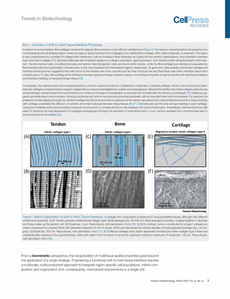
Box 1. Overview on Soft-to-Hard Tissue Interfaces Physiology
In tendon-to-bone interface, fibrocartilage connects the aligned, fibrous tendon to the stiff and calcified bone (Figure I). This tissue is characterized by the presence of anonmineralized and mineralized region, where normally an abrupt transition from cartilaginous to calcified fibrocartilage, often called a tidemark, is observed. This regionis also characterized by a gradient of collagen fiber distribution, with an increase in fiber dispersion as it goes far from tendon midsubstance, and a transition betweentype I and type II collagen [10]. Several studies also demonstrated variations in cellular composition, gene expression, and mineral content along the length of the inser-tion. Tendon-derived cells, including tenocytes and tendon stem/progenitor cells, are found within tendon, while the fibrocartilaginous interface is populated byfibrochondrocytes and hypertrophic chondrocytes, in the nonmineralized and mineralized regions, respectively. At gene level, data analysis of enriched cartilage andinterface transcriptomes suggested that cells found at the interface are more chondrocyte-like than tenocyte-like and that these cells within interface derive fromchondrocytes [77]. Also, fibrocartilage at the enthesis endorses a gradual change of elastic modulus, smoothing the transfer of load across the soft–hard tissue interfaceand therefore resisting compression/shear stress [78].
Comparably, the osteochondral unit is characterized by a chondro-osseous continuum, interfaced by a tidemark, a calcified cartilage, and the subchondral bone plate.Articular cartilage is characterized by a type II collagen fibrous network interdigitated by sulfate-rich proteoglycans. Beyond themiddle zone, these collagen networks areperpendicularly oriented toward the subchondral bone, where an increase in mineralization is extended from the tidemark into the bony counterpart [79]. Evidence sug-gests a possible direct communication, forming a functional unit, both at mechanical and biochemical levels, with an important role in joint homeostasis. For example, thepresence of holes dipping through the calcified cartilage into the bone and marrow spaces and the dense vasculature from subchondral bone found in close proximitywith cartilage potentiates the diffusion of nutrients and small molecules between these tissues [80,81]. Chondrocytes are the only cell type residing in adult cartilage,owing low metabolic activity and surviving in a hypoxic environment, in contrast with bone cells residing in the subchondral region. Interestingly, subchondral bone cellsseem to influence the loss/degradation of cartilage proteoglycans through the secretion of local factors and, in turn, factors secreted from chondrocytes lead tosubchondral bone remodeling [82].
Fibrils: collagen type I Fibrils: collagen type I Aligned to random mesh: collagen type II
(A) (C)
(B) (D)
(E)
(F)
TrendsTrends inin BiotechnologyBiotechnology
Figure I. Matrix Organization of Soft-to-Hard Tissue Interfaces. A collagen rich composition is observed in musculoskeletal tissues, although with differentarchitectural assembly. (A,B) Tendon presents unidirectional collagen type I fibrils (average size, 35–500 nm, blue) arranged in bundles, coupled together in fasciclesand these make up the tendon unit. (B) Scale bar, 2 μm. Reproduced, with permission, from [76]. (C,D) In contrast, bone is constituted by a type I collagenousmatrix comprised into stacked fibrils with diameters between 20-45 nm (blue), which are intercepted by mineral spindles of hydroxyapatite (average size ~20 nm,grey). (D) Scale bar, 200 nm. Reproduced, with permission, from [77]. (E,F) Mature cartilage owns depth-dependent architecture where collagen type II fibers areunidirectionally oriented at the superficial layer, while with depth it the formation of randomly organized meshes is observed. (F) Scale bar, 100 μm. Reproduced,with permission, from [78].
Trends in Biotechnology
From a biomimetic perspective, the recapitulation of multitissue spatial properties goes beyondthe application of a single strategy. Engineering a functional soft-to-hard tissue interface requiresa multiscale, multicomponent approach to integrate region-specific cell populations, matrix com-position and organization and, consequently, mechanical requirements in a single unit.
Trends in Biotechnology, Month 2019, Vol. xx, No. xx 3
GlossaryBiochemical cues:molecules involved
Trends in Biotechnology
Biological ToolsUpon injury, hypovascular dense regular connective tissues (e.g., meniscus, tendons, and liga-
in chemical reactions within livingorganisms that have the ability ofinitiating or modifying a biochemical orsignaling cascade; such signals can bemimicked in vitro by culturesupplementation or biofunctionalization.Biofunctionalization:modification of amaterial surface for either specific ornonspecific immobilization of definedmotifs or biomolecules that addbiological functionality in addition tobiocompatibility/tolerability by the body.Biomechanical/biophysical cues:physical signals or forces from theexternal environment that are perceivedthrough themechanosensorymachineryof cells and activate signaling cascades;such signals can be mimicked in vitro byspecific mechanical properties orculturing systems, as well as substrateproperties like elasticity, rigidity, andtopography.Biomimetic: replication/imitation ofbiological systems.Bioreactors: mechanical devices orsystems used for providing regulatorybiochemical and physical signals to cells,in a dynamic manner, toward enablingstem cell differentiation and/orextracellular matrix deposition andreplicating tissue-specific requirements.Cell sheet: confluent cell layer withintact cell–cell junctions and extracellularmatrix resulting from the detachment ofcell cultures commonly from thermo-responsive surfaces or using alternativeapproaches (e.g., light-induced,magnetic force).Engraftment: response given by thebody in which cells are accepted aftertransplantation.Epigenomic modifications: in vitromanipulation of cellular processes thatregulate the transcription of geneticinformation through pharmacologicaltools, genetic editing, and precisionepigenetic editing.Extracellular matrix: 3D network ofstructural macromolecules produced bycells into the surrounding environment,including collagens and other proteins,proteoglycans, and glycoproteins.Gradient: an increase or decrease inthe magnitude of a biochemical/biophysical property or a variation incellular content/type.Graft: a small sample of living orsynthetic tissue that is surgicallytransplanted.
ments) have a limited self-repair ability and depend on resident or neighboring cells to orchestratetissue repair, as opposed to the majority of tissues that are supplied by a vascular network,benefiting from circulating progenitor cells. To date, the identification of regeneration compe-tent cells within soft-to-hard tissue interfaces remains a mystery, particularly for tendon/liga-ment-to-bone interfaces, since resident cells and their heterogeneity are not fully understood.
Different cell populations, particularly bonemarrow-derivedmesenchymal stem cells (MSCs), andother cell sources ranging from resident/tissue-specific cells to induced pluripotent stemcells,are explored for musculoskeletal tissue regeneration using various delivery strategies(e.g., injection, arthroscopy, implantation), as reviewed elsewhere [19]. Autologous chondrocyteimplantation aimed at osteochondral repair was one of the first cell-based tissue engineeringinterventions to reach clinical application. Similarly, autologous tenocyte implantation is alsoexplored (Phase II–III clinical study NCT01343836). Nonetheless, the need for cell supporthas been recognized and the combination with matrix-based cell implantation is pursuednowadays.
Scaffold-Free Cell Delivery TechnologiesCell sheet engineering (Figure 2) has been proposed to overcome shortcomings of single cellsuspension injection in tissue reconstruction through preservation of cell–cell contacts and de-posited extracellular matrix (ECM) [20]. Autologous cell sheet transplantation has been ex-plored for regenerating thin-layered tissues (e.g., cornea, esophagus, periodontal tissues), butadvances in 3D cell sheet manipulation enable the reconstruction of thicker and more complextissue architectures (Figure 2A–E). Layered chondrocyte sheets alone and in combination with sy-novial cells or even further combined with scaffolds have been reported to facilitate osteochondralregeneration through barrier functionality and support chondrocyte phenotype and chondrogenicdifferentiation [21–23]. In the case of tendon and tendon-to-bone repair, the use of native tissuecells and derived cell sheets has been defied by the phenotypic drift and senescence of tendoncells upon in vitro expansion, leading to limited healing capacity. Recently, this has been associ-ated with increased activity of histone deacetylase (HDAC) [24]. Hence, epigenomic modifica-tions targeting the inhibition of different HDAC subtypes allowed the recovery of tendon-markerscleraxis expression, supporting the use of tendon stem/progenitor cells (TSPC) sheets(Figure 2F) in accelerating tendon repair [24]. Further combining stem cell sheets with native ten-don–fibrocartilage–bone composite as a biological patch to augment rotator cuff healing resultedin enhanced fibrocartilage formation and collagen fiber organization, while providing biomechan-ical support during tissue repair [25]. The value of cell sheets in tissue repair is well recognized, asthey overcome cell loss and reduced engraftment upon transplantation. Nevertheless, engi-neering functional soft-to-hard tissue interfaces is postulated to require an integration of physio-logically relevant ECM signals and mechanical stimuli toward guiding stem cell fate.
Next-generation cell sheet engineering is taking this technology to a higher level of organization inrecreating complex tissues. The development of anisotropic cell sheets using stripe-likemicropatterned thermoresponsive surfaces [26] (Figure 2A) and, recently, light-induced cell align-ment and cell sheet harvest [27] (Figure 2B–D) hold promising results to be explored for interfacialtissue engineering. More recent trends have been exploringmagnetic force-based tissue en-gineering. The generation of magnetic cell sheets by cellular internalization of magnetic nanopar-ticles enabled the fabrication of tenogenic living ECM-rich patches (Figure 2E) with potential forremote control upon application of an external magnetic field as mechano-magnetic stimulus[28].
4 Trends in Biotechnology, Month 2019, Vol. xx, No. xx
Growth factors: naturally occurringmolecules, usually proteins, whichstimulate cell growth, proliferation,differentiation, survival, and regulatevarious cellular processes, includingtissue homeostasis and healing.Homeostasis: a relatively constantequilibrium in the internal physical andchemical conditions maintained atdifferent levels (cellular, tissue, organ,system) by living organisms throughphysiological processes.Hydrogel: crosslinked 3D network ofnatural or synthetic polymers that canabsorb and retain large volumes of wateror biological fluids without polymerdissolution.Hypoxia: condition in which the oxygensupply is lower than the normal arterialblood concentration.Induced pluripotent stem cells:pluripotent stem cells generated fromadult somatic cells that werereprogrammed back into theirembryonic-like state through the
Trends in Biotechnology
Biotechnological advances are pushing forward the complexity of cell-based therapies, pavingthe way to engineer living constructs with physiological and clinical relevance. Despite the prom-ising reported outcomes, unsolved issues remain that challenge the field, including the develop-ment of off-the-shelf cellular therapies. Further, the refinement of cellular approaches incombination with biophysical and biochemical tools to manipulate cell fate is a current need to-ward generating physiologically relevant cellular gradients to emulate the cellular niche from thedifferent musculoskeletal interfaces.
Biophysical ParametersSoft-to-hard musculoskeletal interfaces are highly structured nanocomposites arranged intomicroarchitectures with unique directionalities, gradients, and cellular environments. Attachmentof cells to the ECM regulates diverse cellular functions. It is well recognized that a precise controlover nano-to-macro structural features of biomaterials is of major importance to recreate keyproperties of the native ECM. However, the development of multitissue transitions is still achallenge.
Advances in biomaterials design allow for refining microenvironmental properties, paving the wayto generate physiologically relevant niches and engineering soft-to-hard tissue interfaces. Overthe years, different fabrication methods have enabled control of a panoply of interdependentbiophysical parameters (Table 1).
addition of a transcription factor cocktail
Tailoring Biomaterial Architecture, Surface Topography, and Mechanical Propertiesunder embryonic stem cell cultureconditions.
Magnetic force-based tissue engi-neering: tissue engineering strategiesthat rely on the use of magneticnanoparticles and external magneticactuation to direct cell positioning/behavior.Matrix stiffness: property of a materialto resist deformation in response to anapplied force and defined as the ratiobetween the applied force anddeformation.Mechanosensing: ability of a cell tofeel the mechanical/physical cues of thesurrounding environment, includingforce components, substratedeformation, and properties (see‘Biochemical/biophysical cues’).Multilineage differentiation: potentialof stem cells to develop into a multiplenumber of cells types.Nanocomposites: materials in whichone of the phases is within thenanometer range (b10 nm).Progenitor cells: cells that are typicallydescendants of stem cells but with moreconstrained differentiation potential andself-renewal capacity.As biophysical properties of native ECM are difficult to emulate, synthetic fibrous scaffolds havebeen essential to study and regulate specific matrix properties, important for cellular proliferationand function. From electrospinning to hydrogel systems, tendon-to-bone regeneration has beenrelying on the fabrication of variable scaffolds for the replication of architectural and mechanicalproperties of the graded tissue. Nanofibrous scaffolds have been widely used for tendon-to-bone regeneration as these biomaterials act as a physical platform mimicking 3D fibrous collag-enous hierarchical structure [29,30]. In contrast, 3D printing and hydrogels have been used formimicking physical properties of the osteochondral unit, as this combination demonstratesgood physical and mechanical performance [12,31].
The development of injectable hydrogel systems with control over the architecture of the fibrillarnetwork has been also demonstrated to have biological relevance [32]. These hydrogels have acontrollable and precise internal fibrous structure, which determines their pore size and mechan-ical properties, while replicating the filamentous architecture of the ECM. Moreover, hydrogelscan also be tuned to present an anisotropic architecture, for instance, taking advantage of mag-netic stimulation to nanoparticle alignment [33]. Strikingly, these hydrogels provide biochemicaland physical cues, enabling us to tune the behavior of encapsulated stem cells, with prospectiveapplications in minimally invasive defect filling surgeries.
Within every tissue and organ, cells sense the properties of their supporting environment at mul-tiple length scales. Hence, new biomaterial designs must consider the role ofmechanosensing.By generating methacrylated dextran fibrillar matrices resembling collagen type I networks, Bakerand colleagues have demonstrated the role of fibrillar topography in directing cellular morphology,impacting cellular alignment and matrix remodeling by cellular traction forces [34]. Remarkably,cellular forces at the microscale, together with tissue-generated forces at the macroscale, aretwo essential parameters guiding numerous tissue differentiation and maturation during soft-to-hard tissue development [35]. In turn, cellular processes, including stem cell lineage specification,can be guided by contact with the surrounding physical microenvironment.Matrix stiffness as a
Trends in Biotechnology, Month 2019, Vol. xx, No. xx 5
Key Figure
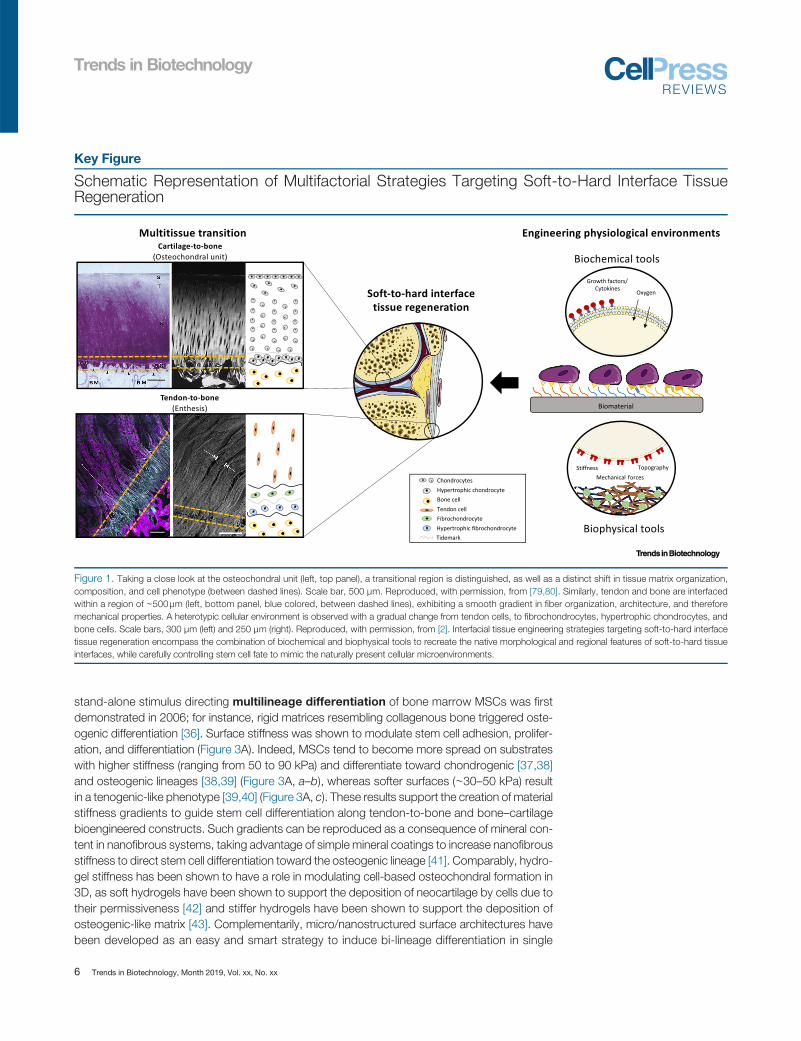
Schematic Representation of Multifactorial Strategies Targeting Soft-to-Hard Interface TissueRegeneration
TrendsTrends inin BiotechnologyBiotechnology
Figure 1. Taking a close look at the osteochondral unit (left, top panel), a transitional region is distinguished, as well as a distinct shift in tissue matrix organization,composition, and cell phenotype (between dashed lines). Scale bar, 500 μm. Reproduced, with permission, from [79,80]. Similarly, tendon and bone are interfacedwithin a region of ∼500μm (left, bottom panel, blue colored, between dashed lines), exhibiting a smooth gradient in fiber organization, architecture, and thereforemechanical properties. A heterotypic cellular environment is observed with a gradual change from tendon cells, to fibrochondrocytes, hypertrophic chondrocytes, andbone cells. Scale bars, 300 μm (left) and 250 μm (right). Reproduced, with permission, from [2]. Interfacial tissue engineering strategies targeting soft-to-hard interfacetissue regeneration encompass the combination of biochemical and biophysical tools to recreate the native morphological and regional features of soft-to-hard tissueinterfaces, while carefully controlling stem cell fate to mimic the naturally present cellular microenvironments.
Trends in Biotechnology
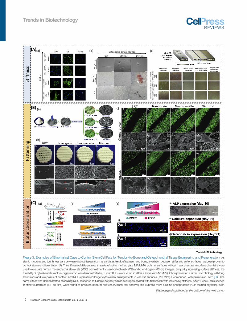
stand-alone stimulus directing multilineage differentiation of bone marrow MSCs was firstdemonstrated in 2006; for instance, rigid matrices resembling collagenous bone triggered oste-ogenic differentiation [36]. Surface stiffness was shown to modulate stem cell adhesion, prolifer-ation, and differentiation (Figure 3A). Indeed, MSCs tend to become more spread on substrateswith higher stiffness (ranging from 50 to 90 kPa) and differentiate toward chondrogenic [37,38]and osteogenic lineages [38,39] (Figure 3A, a–b), whereas softer surfaces (∼30–50 kPa) resultin a tenogenic-like phenotype [39,40] (Figure 3A, c). These results support the creation of materialstiffness gradients to guide stem cell differentiation along tendon-to-bone and bone–cartilagebioengineered constructs. Such gradients can be reproduced as a consequence of mineral con-tent in nanofibrous systems, taking advantage of simple mineral coatings to increase nanofibrousstiffness to direct stem cell differentiation toward the osteogenic lineage [41]. Comparably, hydro-gel stiffness has been shown to have a role in modulating cell-based osteochondral formation in3D, as soft hydrogels have been shown to support the deposition of neocartilage by cells due totheir permissiveness [42] and stiffer hydrogels have been shown to support the deposition ofosteogenic-like matrix [43]. Complementarily, micro/nanostructured surface architectures havebeen developed as an easy and smart strategy to induce bi-lineage differentiation in single
6 Trends in Biotechnology, Month 2019, Vol. xx, No. xx
Trends in Biotechnology
scaffolds (Figure 3B). Interestingly, tailoring the surface topography of scaffolds by increasing ei-ther the roughness or the use of nano-scaled matrices has been shown to allow a better cell ad-hesion to the matrix, followed by subsequent tenogenic, osteogenic, and chondrogeniccommitment of stem cells when in contact with oriented groove materials or just by creatingdense or fibrous topologies in scaffolds, respectively [10,18,44–47].
In this sense, it is worth mentioning that phenotypic alterations induced by biophysical sensinghave been shown to have a dose-dependent response and to be cell type-specific and contextdependent [48]. It has been long recognized that material properties (e.g., matrix stiffness, topog-raphy) can sensitize cells to other microenvironmental features, impacting cell response [49–52].Notwithstanding, recent studies using RNA sequencing and differential expression analyses havedemonstrated that one specific parameter has the power to contextualize the response to otherfeatures, through a dose-dependent effect [48,52]. Besides this context-dependence of couplingbiophysical cues, the type of cell also determines the downstream response and the way the bio-physical sensing happens. Indeed, the ability of cells to cluster adhesion ligands in response to aspecific material parameter has been demonstrated to occur in a cell type-specific manner and tobe dependent on cellular intrinsic characteristics (lineage, species) [48].
These findings raise several questions, particularly concerning the interplay of downstream cellregulatory networks in response to specific combinations of biophysical features. Hence, chal-lenges emerge regarding the establishment of adequate gradient biomaterials for interfacial tissueengineering. Other challenges include the impact on selection of cell sources and cell history fordeveloping adequate tissue mimetics. Biophysical gradients are expected to guide the behaviorof different cells types, which will, in turn, establish a cross-communication and influence eachother, resulting in a highly complex system.
The Role of Mechanotransduction
Since the function of musculoskeletal interfaces is to bear and transmit loads between mechan-ically different tissues, it is not a surprise that mechanical loading contributes to the developmentand function (and even pathology onset) of suchmultitissue interfaces, contributing to a gradationin tissue cellularity and structure. During the last few years, growing evidence has demonstratedthat cells sense the mechanical forces in different ways, transducing these mechanosignals intogene regulation that will impact not only cell migration or ECM adhesion but also proliferationand differentiation [53]. Therefore, understanding cellular responses upon stimulation bymechan-ical inputs from the surrounding environment may provide key information for manipulatingcellular behaviors toward proregenerative phenotypes.Active loading has been used in vivo to guide tissue formation upon construct implantation, butissues remain regarding the lack of control over mechanical loading regimes. Alternatively, effectsof mechanical stresses have been increasingly explored in vitro using dynamic systems as biore-actors. The potential regeneration of tendon/ligament–bone has been evaluated mainly throughthe use of cyclic tension, whereas bone–cartilage regeneration has relied on the use of compres-sive stress. For instance, a dynamic compression bioreactor was used in a semi-confined com-pression model to direct MSC differentiation throughout the depth of a hydrogel to resemble thespatial endochondral progression [54]. The application of dynamic compression increased strainsacross the top of the construct, while the confinement reduced oxygen levels (see section‘Biochemical Tools’) at the bottom of the construct, resulting in increased glycosaminoglycan ac-cumulation in the bottom, increased collagen accumulation in the top along with a suppression ofhypertrophy and calcification throughout the construct [54]. In contrast, mechanical and biolog-ical properties of an engineered tendon-to-bone composite have been investigated after culturing
Trends in Biotechnology, Month 2019, Vol. xx, No. xx 7

(A) (B)
(C)(D)
(E) (F)
TrendsTrends inin BiotechnologyBiotechnology
(See figure legend at the bottom of the next page.)
Trends in Biotechnology
8 Trends in Biotechnology, Month 2019, Vol. xx, No. xx
Trends in Biotechnology
bone marrow-derived MSC sheets under cyclic tension for 7 days [55]. Mechanical tension led toincreased cell migration and aligned distribution of cells within the scaffold, resulting in the upreg-ulation of tenogenic genes as scleraxis [55].
While there are some challenges to overcome in engineering a native-like soft-to-hard tissue inter-face, whether bioreactors systems will be ultimately used for tissue maturation for clinics remainsan open question. However, these systems present an advantage over current in vitro systems asthey allow fine control over mechanical cues, improving both local cell modulation and, ultimately,tissue regeneration after implantation.
Biochemical ToolsSupplementation of cell cultures using biomolecules has been explored to maintain the phenotypeof permanently differentiated cells or to modulate stem cell fate and induce differentiation. Amongdifferent biochemical factors, oxygen is a crucial molecule and we will discuss the influence of ox-ygen tension on cellular behavior. Additionally, small molecules, like ascorbic acid, can beemployed to accelerate ECM deposition, particularly for cell sheet engineering [56–59], or to pro-mote cellular proliferation in cell expansion protocols (e.g., glucose and essential amino acids).Other signals include hormones, growth factors, and cytokines. Although high-throughput analysisof combinatorial approaches has been troublesome, the use of such biochemical cues has been atthe forefront of cell-based therapy development and is herein addressed.
Oxygen Tension and Hypoxic Niches
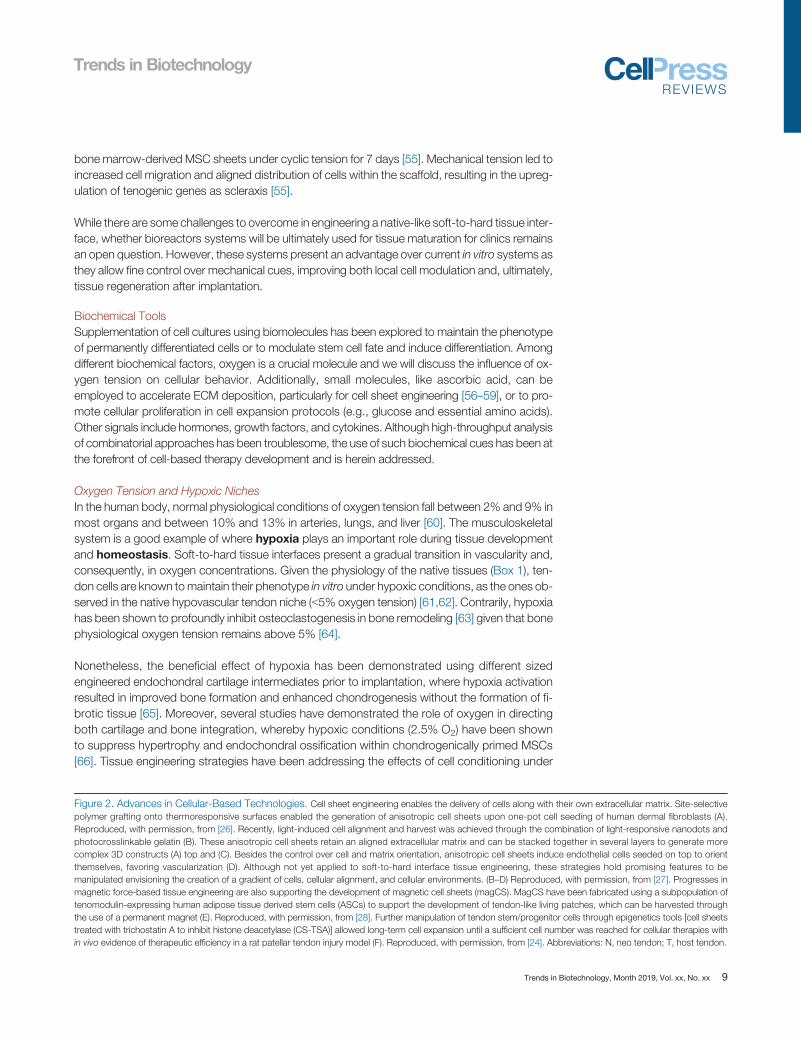
In the human body, normal physiological conditions of oxygen tension fall between 2% and 9% inmost organs and between 10% and 13% in arteries, lungs, and liver [60]. The musculoskeletalsystem is a good example of where hypoxia plays an important role during tissue developmentand homeostasis. Soft-to-hard tissue interfaces present a gradual transition in vascularity and,consequently, in oxygen concentrations. Given the physiology of the native tissues (Box 1), ten-don cells are known tomaintain their phenotype in vitro under hypoxic conditions, as the ones ob-served in the native hypovascular tendon niche (b5% oxygen tension) [61,62]. Contrarily, hypoxiahas been shown to profoundly inhibit osteoclastogenesis in bone remodeling [63] given that bonephysiological oxygen tension remains above 5% [64].Nonetheless, the beneficial effect of hypoxia has been demonstrated using different sizedengineered endochondral cartilage intermediates prior to implantation, where hypoxia activationresulted in improved bone formation and enhanced chondrogenesis without the formation of fi-brotic tissue [65]. Moreover, several studies have demonstrated the role of oxygen in directingboth cartilage and bone integration, whereby hypoxic conditions (2.5% O2) have been shownto suppress hypertrophy and endochondral ossification within chondrogenically primed MSCs[66]. Tissue engineering strategies have been addressing the effects of cell conditioning under
Figure 2. Advances in Cellular-Based Technologies. Cell sheet engineering enables the delivery of cells along with their own extracellular matrix. Site-selectivepolymer grafting onto thermoresponsive surfaces enabled the generation of anisotropic cell sheets upon one-pot cell seeding of human dermal fibroblasts (A).Reproduced, with permission, from [26]. Recently, light-induced cell alignment and harvest was achieved through the combination of light-responsive nanodots andphotocrosslinkable gelatin (B). These anisotropic cell sheets retain an aligned extracellular matrix and can be stacked together in several layers to generate morecomplex 3D constructs (A) top and (C). Besides the control over cell and matrix orientation, anisotropic cell sheets induce endothelial cells seeded on top to orientthemselves, favoring vascularization (D). Although not yet applied to soft-to-hard interface tissue engineering, these strategies hold promising features to bemanipulated envisioning the creation of a gradient of cells, cellular alignment, and cellular environments. (B–D) Reproduced, with permission, from [27]. Progresses inmagnetic force-based tissue engineering are also supporting the development of magnetic cell sheets (magCS). MagCS have been fabricated using a subpopulation oftenomodulin-expressing human adipose tissue derived stem cells (ASCs) to support the development of tendon-like living patches, which can be harvested throughthe use of a permanent magnet (E). Reproduced, with permission, from [28]. Further manipulation of tendon stem/progenitor cells through epigenetics tools [cell sheetstreated with trichostatin A to inhibit histone deacetylase (CS-TSA)] allowed long-term cell expansion until a sufficient cell number was reached for cellular therapies within vivo evidence of therapeutic efficiency in a rat patellar tendon injury model (F). Reproduced, with permission, from [24]. Abbreviations: N, neo tendon; T, host tendon.
Trends in Biotechnology, Month 2019, Vol. xx, No. xx 9
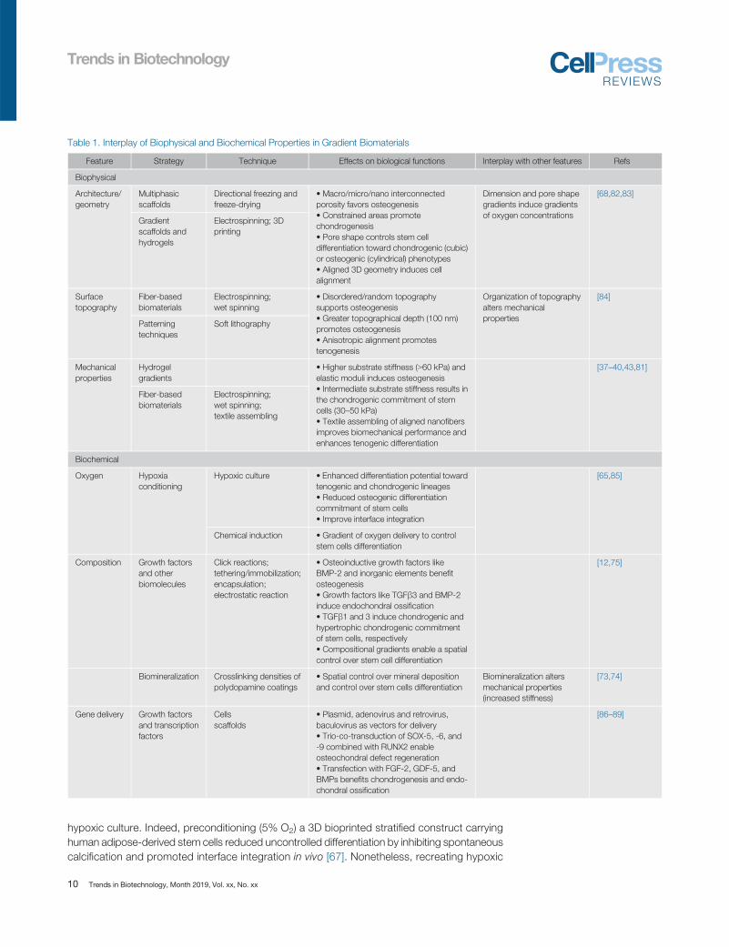
Table 1. Interplay of Biophysical and Biochemical Properties in Gradient Biomaterials
Feature Strategy Technique Effects on biological functions Interplay with other features Refs
Biophysical
Architecture/geometry
Multiphasicscaffolds
Directional freezing andfreeze-drying
• Macro/micro/nano interconnectedporosity favors osteogenesis• Constrained areas promotechondrogenesis• Pore shape controls stem celldifferentiation toward chondrogenic (cubic)or osteogenic (cylindrical) phenotypes• Aligned 3D geometry induces cellalignment
Dimension and pore shapegradients induce gradientsof oxygen concentrations
[68,82,83]
Gradientscaffolds andhydrogels
Electrospinning; 3Dprinting
Surfacetopography
Fiber-basedbiomaterials
Electrospinning;wet spinning
• Disordered/random topographysupports osteogenesis• Greater topographical depth (100 nm)promotes osteogenesis• Anisotropic alignment promotestenogenesis
Organization of topographyalters mechanicalproperties
[84]
Patterningtechniques
Soft lithography
Mechanicalproperties
Hydrogelgradients
• Higher substrate stiffness (N60 kPa) andelastic moduli induces osteogenesis• Intermediate substrate stiffness results inthe chondrogenic commitment of stemcells (30–50 kPa)• Textile assembling of aligned nanofibersimproves biomechanical performance andenhances tenogenic differentiation
[37–40,43,81]
Fiber-basedbiomaterials
Electrospinning;wet spinning;textile assembling
Biochemical
Oxygen Hypoxiaconditioning
Hypoxic culture • Enhanced differentiation potential towardtenogenic and chondrogenic lineages• Reduced osteogenic differentiationcommitment of stem cells• Improve interface integration
[65,85]
Chemical induction • Gradient of oxygen delivery to controlstem cells differentiation
Composition Growth factorsand otherbiomolecules
Click reactions;tethering/immobilization;encapsulation;electrostatic reaction
• Osteoinductive growth factors likeBMP-2 and inorganic elements benefitosteogenesis• Growth factors like TGFβ3 and BMP-2induce endochondral ossification• TGFβ1 and 3 induce chondrogenic andhypertrophic chondrogenic commitmentof stem cells, respectively• Compositional gradients enable a spatialcontrol over stem cell differentiation
[12,75]
Biomineralization Crosslinking densities ofpolydopamine coatings
• Spatial control over mineral depositionand control over stem cells differentiation
Biomineralization altersmechanical properties(increased stiffness)
[73,74]
Gene delivery Growth factorsand transcriptionfactors
Cellsscaffolds
• Plasmid, adenovirus and retrovirus,baculovirus as vectors for delivery• Trio-co-transduction of SOX-5, -6, and-9 combined with RUNX2 enableosteochondral defect regeneration• Transfection with FGF-2, GDF-5, andBMPs benefits chondrogenesis and endo-chondral ossification
[86–89]
Trends in Biotechnology
hypoxic culture. Indeed, preconditioning (5% O2) a 3D bioprinted stratified construct carryinghuman adipose-derived stem cells reduced uncontrolled differentiation by inhibiting spontaneouscalcification and promoted interface integration in vivo [67]. Nonetheless, recreating hypoxic
10 Trends in Biotechnology, Month 2019, Vol. xx, No. xx
Outstanding QuestionsWhat are themost critical parameters tocontrol while engineering soft-to-hardtissue physiological environments?
How can such small sized multitissuetransitions be precisely engineered?
Can a soft-to-hard tissue engineeredconstruct be produced in clinicallytranslatable setups?
With which resolution can tissue-specific microenvironments be emu-lated regarding the spatiotemporal
Trends in Biotechnology
niches through culture conditions limits the maintenance of this biomimetic feature to in vitrosettings. Strikingly, a minute control over physical features of produced scaffolds has beenshown to have impact on oxygen supply. Through variations in pore size (higher to smaller),oxygen diffusion will decrease, resulting in the recreation of a gradient of hypoxic environmentswithin a single structure. This enables a control over stem cell differentiation toward osteogenicand chondrogenic lineages [68].
The role of oxygen tension is frequently disregarded in tissue engineering strategies, but ithas been increasingly recognized as a critical biochemical parameter to address in soft-to-hard tissue interface regeneration. Novel approaches deploying a control over oxygengradients are likely to provide a strategic management of multidifferentiation of a singlestem cell source.
Growth Factors and Biofunctionalization Strategiesdynamics of cellular processes?
Musculoskeletal interface tissue repair relies on the use of growth factors to elicit a desired phe-notypic response from a host tissue or when co-delivered with cells, through localized and con-trolled multifunctional delivery systems. Interestingly, growth factors involved in growth platedevelopment have been elucidated as possible targets for both tendon-to-bone andosteochondral tissue regeneration, particularly bone morphogenetic proteins and transforminggrowth factor beta superfamily [69–71].
Biofunctionalization of biomaterial surfaces have demonstrated good results toward the in-duction of different phenotypes in single structures. Multifunctional gradients using controllableand reversed click reactions [72] or polymerization of dopamine [73,74] have been explored fortendon–bone interface regeneration. Resulting available groups provide accessibility for bio-molecule immobilization and biomineralization [74], allowing a gradual differentiation of stemcells (Figure 3C). Comparably, strategies aimed at the delivery of growth factors forosteochondral unit regeneration have focused on bulk phase delivery, where the release of bio-active factors is dependent on the interaction between the growth factor and the matrix, eitherby tethering/immobilization [75] or encapsulation [12], resulting in the chondrogenic and oste-ogenic differentiation of stem cells in single units. In this regard, gradients of growth factors andrate of release have been demonstrated to affect tissue formation. Even though immobilizationof growth factors has shown satisfactory results in in vitro applications, this strategy facessome challenges in vivo, such as ion exchange with physiological fluids. Therefore, the unde-fined and negative cross-effects in in vivo defect studies suggest that these methodologiesstill require a fine-tuning. Effectively, the need for an ‘ideal’ spatial and temporal delivery ofgrowth factors in order to potentiate their highest therapeutic efficacy in future tissueengineered approaches is well acknowledged.
Concluding Remarks and Future PerspectivesSoft-to-hard tissue interfaces have primary mechanical roles. Thus, tissue engineering dedicatesa considerable effort toward recapitulating these structures through biomaterial design. Nonethe-less, the characteristic complexity of interfacial tissues requires integrative tissue engineering ap-proaches that combine a set of tools (biological, biophysical, and biochemical) toward guidingnative or transplanted cells.
Over the years, advances in biotechnological tools have refined TERM strategies. The develop-ment of adequate constructs for soft-to-hard tissue interface regeneration is challenged by limitedknowledge of the biology of these multitissue transitions.
Trends in Biotechnology, Month 2019, Vol. xx, No. xx 11
differen�a�on
substrate substrate
TrendsTrends inin BiotechnologyBiotechnology
(A)
(B)
(C)
Figure 3. Examples of Biophysical Cues to Control Stem Cell Fate for Tendon-to-Bone and Osteochondral Tissue Engineering and Regeneration. Aselastic modulus and toughness vary between distinct tissues such as cartilage, tendon/ligament, and bone, a variation between stiffer and softer surfaces has been proven tocontrol stem cell differentiation (A). The stiffness of different methyl acrylate/methyl methacrylate (MA/MMA) polymer surfaces without major changes in surface chemistry wereused to evaluate human mesenchymal stem cells (MSC) commitment toward osteoblastic (OB) and chondrogenic (Chon) lineages. Simply by increasing surface stiffness, thetunability of cytoskeletal structural organization was demonstrated (a). Round OBs were found in stiffer substrates (N10MPa), Chon presented a similar morphology with longextensions and few points of contact, and MSCs presented longer cytoskeletal arrangements in less stiff surfaces (b10 MPa). Reproduced, with permission, from [38]. Thesame effect was demonstrated assessing MSC response to tunable polyacrylamide hydrogels coated with fibronectin with increasing stiffness. After 1 week, cells seededin stiffer substrates (62–68 kPa) were found to produce calcium nodules (Alizarin red-positive) and express more alkaline phosphatase (ALP-stained crystals), even
(Figure legend continued at the bottom of the next page.)
Trends in Biotechnology
12 Trends in Biotechnology, Month 2019, Vol. xx, No. xx
Trends in Biotechnology
Taking advantage of the body’s self-healing ability, advanced cell therapies have been increas-ingly explored. Cell injections rely on taking cells out of their ‘comfort zone’ and leaving them toface a very harsh environment that is the injury site, leading to cell loss and poor cell engraftment.Although with disadvantages associated with in vitro cellular expansion and extensive manipula-tion, advanced cell therapies, either based on scaffold-free cell delivery technologies or on cellularactuation through mechanical/magnetic forces, hold potential to change the clinical landscape.Indeed, biotechnological advances enable the generation of regeneration-competent cellularphenotypes and tissue-specific living patches aimed at shifting the profibrotic healing toward aproregenerative environment, which is of particular importance in the case of nonhealing andchronic injuries.
Furthermore, the combination of cell therapies with a support matrix is undoubtedly important,but the reduced size of soft-to-hard tissue insertions also defies the design of gradientbiomaterials. Alternative biomaterial designs may focus on aligned-random fibrous scaffoldsassembled through textile techniques to guide different cellular phenotypes within the twodistinct regions and rely on the cellular crosstalk to recreate an interfacial phenotype whenthe two cell types merge. Although particularly unpredictable, evidence has been supportingthe role of cellular communication in recreating tissue-specific zones. However, future researchcould focus on improving the resolution of material processing techniques to achieve abetter spatial control over physical and biochemical properties and, consequently, cell fatedetermination.
High-throughput screening technologies have elucidated cellular responses to changes inbiophysical and biochemical properties, both at the single cell level and on long-range cellbehaviors (see Outstanding Questions). Combining these transcriptomics and proteomics in-sights with high-resolution fabrication techniques may help to develop more physiologicallyrepresentative tissue engineered products.
An important aspect of future advanced tissue engineering therapies for soft-to-hard interface re-generation is the establishment of adequate regimes of mechanical stimulation to promoteproregenerative cellular responses. For instance, walking steps stimulate soft-to-hard musculo-skeletal systems at 1.5 Hz frequency, under 30.6 kg/m2 according to the average walkingspeed and body mass index. To replicate this scenario, dynamic cell culture systems, as bioreac-tors, have been refined. However, a spatial control over vascularization and innervation in distinctbut integrated microtissues is still a main challenge in the field, due to a lack of simultaneous con-trol over biochemical (e.g., growth factors and oxygen tension) and mechanical cues. This wayalready reported mechanical and biochemical stimulating bioreactors should be combined in asingle concept as a means to stimulate distinct microenvironments, allowing a biomimeticregenerative response in soft-hard interfaced tissues.
compared with substrates such as tissue culture plastic (TCP) (b). Reproduced, with permission, from [81]. Materials stiffness can also influence tendon differentiation.Polyacrylamide hydrogels with mechanical gradient comprising a moderately rigid collagen type I substrate (∼30–50 kPa) was found to induce MSC differentiation intotenogenic lineage, while MSCs differentiate into osteogenic cells on more rigid substrates (70–90 kPa) (c). Reproduced, with permission, from [40]. (B) Surface patterning of3D scaffolds with different morphological micro/nanostructured surface has demonstrated potential use for cartilage and subchondral bone application (a). Pure bredigite, abioactive composite made of silica, magnesium, and calcium, with good bioactivity, biodegradability, and mechanical properties, was used to produce the 3D scaffolds.Structured surfaces distinctly facilitated the spread and differentiation of chondrocytes, regulated cell morphology, and promoted osteogenic differentiation of rBMSCs (b,c).Reproduced, with permission, from [46]. (C) Surface modification by dual reverse click reactions producing a continuous and gradient biofunctionalization to control stem celldifferentiation for tendon-to-bone regeneration. (a) Seeding with 3T3 fibroblasts on surfaces containing (a) only PEG immobilized to the first gradient and (b) two gradients ofPEG and RGD contrary gradients, demonstrated a cell-dependent behavior for PEG. After 21 days with human adipose tissue-derived stem cells (ASCs) in functionalizedsurfaces with BMP2 and FGF-2, a gradient was observed of (iii) ALP expression, (iv) calcium deposition, and (v) osteocalcin expression, showing the osteogenic commitmentof hASCs in a continuous and gradient manner. Reproduced, with permission, from [72]. Abbreviations: rBMSC: rat bone marrow mesenchymal stem cells.
Trends in Biotechnology, Month 2019, Vol. xx, No. xx 13

Trends in Biotechnology
Acknowledgments
The authors acknowledge the financial support from the European Union Framework Programme for Research and InnovationHORIZON2020 (TEAMING Grant agreement, No 739572 - The Discoveries CTR), the ERC Grant CoG MagTendon
(nr 772817), Fundação para a Ciência e a Tecnologia (FCT) for the PhD grant of I.C. (PD/BD/128088/2016), and the Project
NORTE-01-0145-FEDER-000021 through the European Regional Development Fund (ERDF).
References1. Goldring, S.R. and Goldring, M.B. (2016) Changes in the
osteochondral unit during osteoarthritis: structure, function andcartilage–bone crosstalk. Nat. Rev. Rheumatol. 12, 632
2. Rossetti, L. et al. (2017) The microstructure and micromechanicsof the tendon–bone insertion. Nat. Mater. 16, 664
3. Briggs, A.M. et al. (2016) Musculoskeletal health conditions rep-resent a global threat to healthy aging: a report for the 2015World Health Organization World Report on Ageing and Health.Gerontologist 56, S243–S255
4. Sambandam, S.N. et al. (2015) Rotator cuff tears: an evidencebased approach. World J. Orthop. 6, 902–918
5. Chen, J. et al. (2009) Scaffolds for tendon and ligament repair:review of the efficacy of commercial products. Expert Rev.Med. Devices 6, 61–73
6. Bicho, D. et al. (2018) Commercial products for osteochondral tis-sue repair and regeneration. Adv. Exp. Med. Biol. 1058, 415–428
7. Li, Y. et al. (2017) The horizon of materiobiology: a perspectiveon material-guided cell behaviors and tissue engineering.Chem. Rev. 117, 4376–4421
8. Kim, B.S. et al. (2014) Human collagen-based multilayer scaf-folds for tendon-to-bone interface tissue engineering.J. Biomed. Mater. Res. A 102, 4044–4054
9. Huang, G.X. et al. (2015) Modeling and validation of multilayerpoly(lactide-co-glycolide) scaffolds for in vitro directed differenti-ation of juxtaposed cartilage and bone. Tissue Eng. Part A 21,2228–2240
10. Perikamana, M. et al. (2018) Harnessing biochemical andstructural cues for tenogenic differentiation of adipose derivedstem cells (ADSCs) and development of an in vitro tissueinterface mimicking tendon-bone insertion graft. Biomaterials165, 79–93
11. Tellado, S.F. et al. (2018) Heparin functionalization increases re-tention of TGF-β2 and GDF5 on biphasic silk fibroin scaffolds fortendon/ligament-to-bone tissue engineering. Acta Biomater. 72,150–166
12. Gao, F. et al. (2018) Direct 3D printing of high strength biohybridgradient hydrogel scaffolds for efficient repair of osteochondraldefect. Adv. Funct. Mater. 28, 1706644
13. Levingstone, T.J. et al. (2016) Multi-layered collagen-based scaf-folds for osteochondral defect repair in rabbits. Acta Biomater.32, 149–160
14. Chen, J. et al. (2011) Simultaneous regeneration of articular car-tilage and subchondral bone in vivo using MSCs induced by aspatially controlled gene delivery system in bilayered integratedscaffolds. Biomaterials 32, 4793–4805
15. Bunpetch, V. et al. (2019) Silicate-based bioceramic scaffolds fordual-lineage regeneration of osteochondral defect. Biomaterials192, 323–333
16. Zhu, Y. et al. (2019) An injectable continuous stratified structur-ally and functionally biomimetic construct for enhancingosteochondral regeneration. Biomaterials 192, 149–158
17. Canadas, R.F. et al. (2018) Biochemical gradients to generate3D heterotypic-like tissues with isotropic and anisotropic archi-tectures. Adv. Funct. Mater. 28, 1804148
18. Radhakrishnan, J. et al. (2018) Gradient nano-engineered in situforming composite hydrogel for osteochondral regeneration.Biomaterials 162, 82–98
19. Loebel, C. and Burdick, J.A. (2018) Engineering stem and stro-mal cell therapies for musculoskeletal tissue repair. Cell StemCell 22, 325–339
20. Yamato, M. and Okano, T. (2004) Cell sheet engineering.Mater.Today 7, 42–47
21. Ebihara, G. et al. (2012) Cartilage repair in transplanted scaffold-free chondrocyte sheets using a minipig model. Biomaterials 33,3846–3851
22. Ito, S. et al. (2012) Repair of articular cartilage defect with layeredchondrocyte sheets and cultured synovial cells. Biomaterials 33,5278–5286
23. Wang, F. et al. (2018) Scaffold-free cartilage cell sheetcombined with bone-phase BMSCs-scaffold regenerateosteochondral construct in mini-pig model. Am. J. Transl.Res. 10, 2997–3010
24. Zhang, C. et al. (2018) Histone deacetylase inhibitor treated cellsheet from mouse tendon stem/progenitor cells promotes ten-don repair. Biomaterials 172, 66–82
25. Liu, Q. et al. (2019) Engineered tendon-fibrocartilage-bone com-posite and bone marrow-derived mesenchymal stem cell sheetaugmentation promotes rotator cuff healing in a non-weight-bearing canine model. Biomaterials 192, 189–198
26. Takahashi, H. et al. (2011) Anisotropic cell sheets for construct-ing three-dimensional tissue with well-organized cell orientation.Biomaterials 32, 8830–8838
27. Liu, C. et al. (2017) Light-induced cell alignment and harvest foranisotropic cell sheet technology. ACS Appl. Mater. Interfaces 9,36513–36524
28. Gonçalves, A.I. et al. (2017) Tissue-engineered magnetic cellsheet patches for advanced strategies in tendon regeneration.Acta Biomater. 63, 110–122
29. Sant, S. et al. (2017) Self-assembled hydrogel fiber bundles fromoppositely charged polyelectrolytes mimic micro-/nanoscale hi-erarchy of collagen. Adv. Funct. Mater. 27, 1606273
30. Calejo, I. et al. (2019) A textile platform using continuous alignedand textured composite microfibers to engineer tendon. Adv.Healthc. Mater. https://doi.org/10.1002/adhm.201900200Published online June 13, 2019
31. Li, L. et al. (2019) 3D molecularly functionalized cell-free biomi-metic scaffolds for osteochondral regeneration. Adv. Funct.Mater. 29, 1807356
32. Mendes, B.B. et al. (2018) Human-based fibrillar nanocompositehydrogels as bioinstructive matrices to tune stem cell behavior.Nanoscale 10, 17388–17401
33. Araújo-Custódio, S. et al. (2019) Injectable and magnetic re-sponsive hydrogels with bioinspired ordered structures. ACSBiomater. Sci. Eng. 5, 1392–1404
34. Baker, B.M. et al. (2015) Cell-mediated fibre recruitment drivesextracellular matrix mechanosensing in engineered fibrillar micro-environments. Nat. Mater. 14, 1262–1268
35. Felsenthal, N. and Zelzer, E. (2017) Mechanical regulation of mus-culoskeletal system development. Development 144, 4271–4283
36. Engler, A. et al. (2006) Matrix elasticity directs stem cell lineagespecification. Cell 126, 677–689
37. Wu, Y. et al. (2017) The combined effect of substrate stiffnessand surface topography on chondrogenic differentiation of mes-enchymal stem cells. Tissue Eng. Part A 23, 43–54
38. Olivares-Navarrete, R. et al. (2017) Substrate stiffnesscontrols osteoblastic and chondrocytic differentiation of mesenchy-mal stem cells without exogenous stimuli. PLoS One 12, e0170312
39. Sharma, R.I. and Snedeker, J.G. (2010) Biochemical andbiomechanical gradients for directed bone marrow stromal celldifferentiation toward tendon and bone. Biomaterials 31,7695–7704
40. Sharma, R.I. and Snedeker, J.G. (2012) Paracrine interactions be-tweenmesenchymal stem cells affect substrate driven differentiationtoward tendon and bone phenotypes. PLoS One 7, e31504
41. Liu, W. et al. (2014) Nanofiber scaffolds with gradients in mineralcontent for spatial control of osteogenesis. ACS Appl. Mater. In-terfaces 6, 2842–2849
42. Liu, S.Q. et al. (2010) Biomimetic hydrogels for chondrogenic dif-ferentiation of human mesenchymal stem cells to neocartilage.Biomaterials 31, 7298–7307
14 Trends in Biotechnology, Month 2019, Vol. xx, No. xx
Trends in Biotechnology
43. Wang, T. et al. (2016) Effects of hydrogel stiffness and extracel-lular compositions on modulating cartilage regeneration bymixed populations of stem cells and chondrocytes in vivo. TissueEng. Part A 22, 1348–1356
44. Shi, Y. et al. (2017) Microgrooved topographical surface directstenogenic lineage specific differentiation of mouse tendon de-rived stem cells. Biomed. Mater. 12, 015013
45. Mahapatra, C. et al. (2019) Differential chondro- and osteo-stimulation in three-dimensional porous scaffolds with different to-pological surfaces provides a design strategy for biphasicosteochondral engineering. J. Tissue Eng. 10, 2041731419826433
46. Deng, C. et al. (2018) Micro/nanometer-structured scaffolds forregeneration of both cartilage and subchondral bone. Adv.Funct. Mater. 29, 1806068
47. Nowlin, J. et al. (2018) Engineering the hard-soft tissue interfacewith random-to-aligned nanofiber scaffolds. Nanobiomedicine(Rij) 5, 1849543518803538
48. Darnell, M. et al. (2018) Material microenvironmental propertiescouple to induce distinct transcriptional programs in mammalianstem cells. Proc. Natl. Acad. Sci. U. S. A. 115, E8368–E8377
49. Dalby, M.J. et al. (2014) Harnessing nanotopography andintegrin–matrix interactions to influence stem cell fate. Nat.Mater. 13, 558
50. Wen, J.H. et al. (2014) Interplay of matrix stiffness and proteintethering in stem cell differentiation. Nat. Mater. 13, 979
51. Chaudhuri, O. et al. (2014) Extracellular matrix stiffness andcomposition jointly regulate the induction of malignant pheno-types in mammary epithelium. Nat. Mater. 13, 970
52. Darnell, M. et al. (2018) RNA-seq reveals diverse effects of substratestiffness on mesenchymal stem cells. Biomaterials 181, 182–188
53. Yang, C. et al. (2014) Mechanical memory and dosing influencestem cell fate. Nat. Mater. 13, 645–652
54. Thorpe, S.D. et al. (2013)Modulating gradients in regulatory signalswithin mesenchymal stem cell seeded hydrogels: a novel strategyto engineer zonal articular cartilage. PLoS One 8, e60764
55. Liu, Q. et al. (2018) Novel engineered tendon-fibrocartilage-bonecomposite with cyclic tension for rotator cuff repair. J. TissueEng. Regen. Med. 12, 1690–1701
56. Ni, M. et al. (2013) Engineered scaffold-free tendon tissueproduced by tendon-derived stem cells. Biomaterials 34,2024–2037
57. Lui, P.P.Y. et al. (2016) Transplantation of tendon-derived stemcells pre-treated with connective tissue growth factor and ascor-bic acid in vitro promoted better tendon repair in a patellar ten-don window injury rat model. Cytotherapy 18, 99–112
58. Hsieh, C.-F. et al. (2018) In vitro comparison of 2D-cell cultureand 3D-cell sheets of scleraxis-programmed bone marrowderived mesenchymal stem cells to primary tendon stem/progenitor cells for tendon repair. Int. J. Mol. Sci. 19, 2272
59. Shimizu, R. et al. (2015) Repair mechanism of osteochondral de-fect promoted by bioengineered chondrocyte sheet. Tissue Eng.Part A 21, 1131–1141
60. Koh, M.Y. and Powis, G. (2012) Passing the baton: the HIFswitch. Trends Biochem. Sci. 37, 364–372
61. Zhang, J. and Wang, J.H. (2013) Human tendon stem cells bet-ter maintain their stemness in hypoxic culture conditions. PLoSOne 8, e61424
62. Yu, Y. et al. (2017) Effect of hypoxia on self-renewal capacity anddifferentiation in human tendon-derived stem cells. Med. Sci.Monit. 23, 1334–1339
63. Kang, H. et al. (2017) Osteoblast hypoxia-inducible factor-1alphapathway activation restrains osteoclastogenesis via the interleukin-33-microRNA-34a-Notch1 pathway. Front. Immunol. 8, 1312
64. Marenzana, M. and Arnett, T.R. (2013) The key role of the bloodsupply to bone. Bone Res. 1, 203–215
65. Stiers, P.J. et al. (2018) Inhibition of the oxygen sensor PHD2 en-hances tissue-engineered endochondral bone formation.J. Bone Miner. Res. 34, 333–348
66. Huang, X. et al. (2018) Promoted chondrogenesis of coculturedchondrocytes and mesenchymal stem cells under hypoxia usingin-situ forming degradable hydrogel scaffolds.Biomacromolecules19, 94–102
67. Wang, Y. et al. (2017) Effects of hydroxyapatite and hypoxia onchondrogenesis and hypertrophy in 3D bioprinted ADMSCladen constructs. Biomater. Sci. Eng. 3, 826–835
68. Di Luca, A. et al. (2016) Tuning cell differentiation into a 3D scaf-fold presenting a pore shape gradient for osteochondral regen-eration. Adv. Healthc. Mater. 5, 1753–1763
69. Liao, J. et al. (2014) Sox9 potentiates BMP2-inducedchondrogenic differentiation and inhibits BMP2-induced osteo-genic differentiation. PLoS One 9, e89025
70. Mendes, L.F. et al. (2018) Advancing osteochondral tissue engi-neering: bone morphogenetic protein, transforming growth fac-tor, and fibroblast growth factor signaling drive ordereddifferentiation of periosteal cells resulting in stable cartilage andbone formation in vivo. Stem Cell Res Ther 9, 42
71. Zhou, N. et al. (2016) BMP2 induces chondrogenic differentia-tion, osteogenic differentiation and endochondral ossification instem cells. Cell Tissue Res. 366, 101–111
72. Guan, Z.-Y. et al. (2016) Multifunctional and continuous gradi-ents of biointerfaces based on dual reverse click reactions.ACS Appl. Mater. Interfaces 8, 13812–13818
73. Perikamana, S.K.M. et al. (2017) Graded functionalizationof biomaterial surfaces using mussel-inspired adhesive coatingof polydopamine. Colloids Surf. B Biointerface 159, 546–556
74. Madhurakkat Perikamana, S.K. et al. (2017) Oxygen-dependentgeneration of a graded polydopamine coating on nanofibrousmaterials for controlling stem cell functions. J. Mater. Chem. B5, 8865–8878
75. Stüdle, C. et al. (2018) Spatially confined induction ofendochondral ossification by functionalized hydrogels for ectopicengineering of osteochondral tissues. Biomaterials 171, 219–229
76. Provenzano, P. and Vanderby, R. (2006) Collagen fibril morphol-ogy and organization- implications for force transmission in liga-ment and tendon. Matrix Biol. 25, 71–84
77. McNally, E.A. et al. (2012) A model for the ultrastructure of bonebased on electron microscopy of ion-milled sections. PLoS One7, e29258.
78. Fujie, H. and Imade, K. (2015) Effects of low tangential perme-ability in the superficial layer on the frictional property of articularcartilage. Biosurf. Biotribol. 1, 124–129
79. Hunziker, E.B. et al. (2002) Quantitative structural organization ofnormal adult human articular cartilage. Osteoarthr. Cartil. 10,564–572
80. Rieppo, J. et al. (2009) Changes in spatial collagen content andcollagen network architecture in porcine articular cartilage duringgrowth and maturation. Osteoarthr. Cartil. 17, 448–455
81. Sun, M. et al. (2018) Extracellular matrix stiffness controls osteo-genic differentiation of mesenchymal stem cells mediated byintegrin alpha5. Stem Cell Res. Ther. 9, 52
82. Park, K.M. and Gerecht, S. (2014) Hypoxia-inducible hydrogels.Nat. Commun. 5, 4075
83. Ferlin, K.M. et al. (2016) Influence of 3D printed porous architec-ture on mesenchymal stem cell enrichment and differentiation.Acta Biomater. 32, 161–169
84. Zouani, O.F. et al. (2012) Altered nanofeature size dictates stemcell differentiation. J. Cell Sci. 125, 1217
85. Farris, A.L. et al. (2016) Oxygen delivering biomaterials for tissueengineering. J. Mater. Chem. B 4, 3422–3432
86. Lee, J.-M. and Im, G.-I. (2012) SOX trio-co-transducedadipose stem cells in fibrin gel to enhance cartilage repair anddelay the progression of osteoarthritis in the rat. Biomaterials 33,2016–2024
87. Needham, C.J. et al. (2014) Osteochondral tissue regenerationthrough polymeric delivery of DNA encoding for the SOX trioand RUNX2. Acta Biomater. 10, 4103–4112
88. An, C. et al. (2010) IGF-1 and BMP-2 induces differentiation ofadipose-derived mesenchymal stem cells into chondrocytes-like cells. Ann. Biomed. Eng. 38, 1647–1654
89. Cucchiarini, M. et al. (2011) Metabolic activities andchondrogenic differentiation of human mesenchymal stem cellsfollowing recombinant adeno-associated virus–mediated genetransfer and overexpression of fibroblast growth factor 2. TissueEng. Part A 17, 1921–1933
Trends in Biotechnology, Month 2019, Vol. xx, No. xx 15