Embed Size (px)
Citation preview
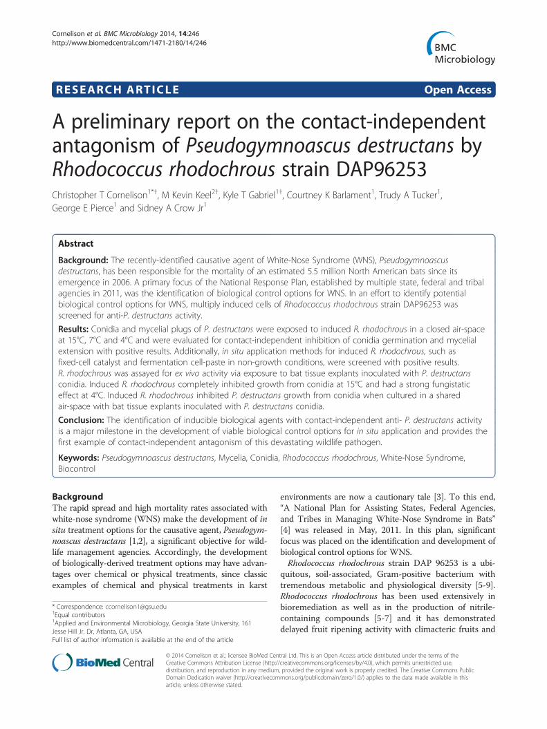
Cornelison et al. BMC Microbiology 2014, 14:246http://www.biomedcentral.com/1471-2180/14/246
RESEARCH ARTICLE Open Access
A preliminary report on the contact-independentantagonism of Pseudogymnoascus destructans byRhodococcus rhodochrous strain DAP96253Christopher T Cornelison1*†, M Kevin Keel2†, Kyle T Gabriel1†, Courtney K Barlament1, Trudy A Tucker1,George E Pierce1 and Sidney A Crow Jr1
Abstract
Background: The recently-identified causative agent of White-Nose Syndrome (WNS), Pseudogymnoascusdestructans, has been responsible for the mortality of an estimated 5.5 million North American bats since itsemergence in 2006. A primary focus of the National Response Plan, established by multiple state, federal and tribalagencies in 2011, was the identification of biological control options for WNS. In an effort to identify potentialbiological control options for WNS, multiply induced cells of Rhodococcus rhodochrous strain DAP96253 wasscreened for anti-P. destructans activity.
Results: Conidia and mycelial plugs of P. destructans were exposed to induced R. rhodochrous in a closed air-spaceat 15°C, 7°C and 4°C and were evaluated for contact-independent inhibition of conidia germination and mycelialextension with positive results. Additionally, in situ application methods for induced R. rhodochrous, such asfixed-cell catalyst and fermentation cell-paste in non-growth conditions, were screened with positive results.R. rhodochrous was assayed for ex vivo activity via exposure to bat tissue explants inoculated with P. destructansconidia. Induced R. rhodochrous completely inhibited growth from conidia at 15°C and had a strong fungistaticeffect at 4°C. Induced R. rhodochrous inhibited P. destructans growth from conidia when cultured in a sharedair-space with bat tissue explants inoculated with P. destructans conidia.
Conclusion: The identification of inducible biological agents with contact-independent anti- P. destructans activityis a major milestone in the development of viable biological control options for in situ application and provides thefirst example of contact-independent antagonism of this devastating wildlife pathogen.
Keywords: Pseudogymnoascus destructans, Mycelia, Conidia, Rhodococcus rhodochrous, White-Nose Syndrome,Biocontrol
BackgroundThe rapid spread and high mortality rates associated withwhite-nose syndrome (WNS) make the development of insitu treatment options for the causative agent, Pseudogym-noascus destructans [1,2], a significant objective for wild-life management agencies. Accordingly, the developmentof biologically-derived treatment options may have advan-tages over chemical or physical treatments, since classicexamples of chemical and physical treatments in karst
* Correspondence: [email protected]†Equal contributors1Applied and Environmental Microbiology, Georgia State University, 161Jesse Hill Jr. Dr, Atlanta, GA, USAFull list of author information is available at the end of the article
© 2014 Cornelison et al.; licensee BioMed CenCreative Commons Attribution License (http:/distribution, and reproduction in any mediumDomain Dedication waiver (http://creativecomarticle, unless otherwise stated.
environments are now a cautionary tale [3]. To this end,“A National Plan for Assisting States, Federal Agencies,and Tribes in Managing White-Nose Syndrome in Bats”[4] was released in May, 2011. In this plan, significantfocus was placed on the identification and development ofbiological control options for WNS.Rhodococcus rhodochrous strain DAP 96253 is a ubi-
quitous, soil-associated, Gram-positive bacterium withtremendous metabolic and physiological diversity [5-9].Rhodococcus rhodochrous has been used extensively inbioremediation as well as in the production of nitrile-containing compounds [5-7] and it has demonstrateddelayed fruit ripening activity with climacteric fruits and
tral Ltd. This is an Open Access article distributed under the terms of the/creativecommons.org/licenses/by/4.0), which permits unrestricted use,, provided the original work is properly credited. The Creative Commons Publicmons.org/publicdomain/zero/1.0/) applies to the data made available in this
Cornelison et al. BMC Microbiology 2014, 14:246 Page 2 of 7http://www.biomedcentral.com/1471-2180/14/246
vegetables [8]. Several enzymes have been shown to haveincreased activity and prevalence in bacteria induced todelay fruit ripening and these enzymes may play a role inthe observed antifungal activity [8]. Initial investigation ofthe potential antagonism of P. destructans by R. rhodo-chrous indicated that, when induced under the protocoloutlined in US patents 7,531,343, and 7,531,344 [10,11],R. rhodochrous strain DAP 96253 demonstrated significantcontact-independent antagonism of P. destructans in vitro.As a result, the principal objective of this was evalu-ation of R. rhodochrous induced with urea for potentialin situ application as a biological control agent forP. destructans.In addition to the strong evidence established via
in vitro analysis of the observed antagonism, the eva-luation of the efficacy of induced R. rhodochrous waspursued in order to establish in vivo efficacy at preven-ting fungal invasion of bat tissue. This goal was accom-plished using a bat-skin explant assay. The evaluation ofinduced R. rhodochrous to prevent or reduce the infect-ive potential of P. destructans conidia was demonstratedby the inhibition of P. destructans growth on living battissue. This is the first example of antifungal efficacy onliving bat skin for any biological control agent of WNSand represents a major milestone in this effort.In order to optimize biocontrol efficacy and reduce
potential cross-contamination of karst environments,various whole- and fixed-cell applications were investi-gated. The evaluation of various application methods ofinduced cells of R. rhodochrous for potential in situ ap-plication, including whole-cell application, non-growthfermentation cell-paste, and fixed-cell catalyst [8,12,13],were conducted. Non-growth fermentation cell-paste de-monstrated persistent inhibitory activity and representsthe most promising application method evaluated. Theassociated cell-paste activity is a significant developmentas it represents multiple hallmarks of ideal biocontrolagents.
MethodsCulture acquisition and maintenanceAll P. destructans isolates used in the project were acquiredfrom the WNS diagnostic lab at The University of GeorgiaSoutheastern Cooperative Wildlife Disease Study (UGASCWDS). Initial investigations have shown very low ge-netic and physiological variability amongst P. destructansisolates [14]. Accordingly, all assays were conducted witha small isolate sample size (n ≤ 3). P. destructans cultureswere maintained on Sabouraud Dextrose Agar (SDA,Difco) or in Sabouraud Dextrose Broth (SDB, Difco) at4°C, 7°C, or 15°C depending on anticipated usage.P. destructans conidia were harvested from fungal lawnson SDA plates by adding 10 ml of conidia harvesting solu-tion (CHS; 0.05% Tween 80, 0.9% NaCl) to the surface of
the plate and gently scrapping with a sterile loop to dis-lodge conidia. The resulting solution was filtered throughglass wool and centrifuged at 5000 rpm for 10 minutes.The resulting supernatant was removed and the spore pel-let washed with 5 mL of sterile phosphate buffered saline(PBS, pH = 7), re-suspended, and filtered through glasswool. Conidia were stored in sterile PBS at −20°C. Conidiawere stored no longer than six weeks prior to use basedon in-house assessment of conidial viability under theseconditions (unpublished data). R. rhodochrous strain DAP96253 cells were maintained as glycerol stock aliquots(30% v/v) from 10 l fermentations carried out at GSU.Fresh glycerol stocks were used as the source of cells atthe onset of each assay. The induction process was per-formed using the addition of urea or urea and cobalt asdescribed in US patents 7,531,343 and 7,531,344 [8,10,11].
Co-culture assays with R. rhodochrousA single-compartment Petri plate (150 mm × 15 mm) wasused for a contained air-space to assess P. destructansgrowth characteristics in the presence of induced cells ofR. rhodochrous. A 10 μl inoculum of P. destructans conidiasolution (106 ml−1) in a phosphate buffer solution wasspread onto SDA in Petri plates (35 mm x 10 mm). Multi-ply induced cells of R. rhodochrous [10,11] were inocu-lated onto Petri plates (35 mm × 10 mm) containing YeastExtract/Malt Extract agar (YEMEA) with or without urea(7.5 g/l) [8], and cultured in the contained air-space for upto 30 days. All assays were conducted in triplicate. Theability of induced R. rhodochrous to inhibit healthy estab-lished hyphae of P. destructans was assessed using myce-lial plug assays. A lawn of P. destructans was allowed togrow for up to 20 days at which time a 5-mm-diametertransfer tube was used to remove a plug from the mat offungus. The plugs were then inserted into a similarly sizedcore removed from an uninoculated culture plate. Theplates were co-incubated in a shared air-space as describedpreviously and radial growth from the plug was assessedover time.
Induced R. rhodochrous germule suppression assayThin layers (~750 μl) of 10% SDA were applied to stan-dard microscope slides (24.5 × 76.2 mm) and 100 μl ofP. destructans conidia solution (106 ml−1) were spreadacross the agar surface. R. rhodochrous-inoculated Petriplates (35 mm × 10 mm) were placed in larger Petriplates (150 mm × 15 mm) and sealed with parafilm.Negative controls consisted of similarly-cultured conidiawith no R. rhodochrous exposure. All trials were con-ducted in triplicate. At 4 and 7 days post-inoculation,conidia were observed in a light microscope at 200Xmagnification for the presence of germule formation.Germules were defined as single mycelial extensions em-anating from conidia with a length equal to or greater
Cornelison et al. BMC Microbiology 2014, 14:246 Page 3 of 7http://www.biomedcentral.com/1471-2180/14/246
than the intact conidia. Control and exposed slides wereretained and examined daily for up to 21 days aftergermule formation was first observed on control slides.Recovery of conidia was determined by removing theR. rhodochrous after 24 hours, 72 hours, and 7 days.Slides were observed for 21 days after removal of controlagent to assess recovery.
Preparation and evaluation of fixed-cell catalyst andfermentation cell-paste in non-growth conditionsImmobilization of whole bacteria was carried out basedon the methods of DeFilippi [12] and Lopez-Gallego et al.[13]. Refinement of immobilized cells to produce activecatalyst was carried out according to the methods ofPierce et al. [10,11]. Evaluation of anti-P. destructans ac-tivity of fixed-cell catalyst and fermentation cell-paste wasdetermined in co-culture assays with P. destructans co-nidia and mycelial plugs with various amounts of controlagent (<1.0 g), as described previously. Efficacy was deter-mined by observation of germule formation as comparedto unexposed controls for growth from conidia, and aspercent reduction in radial growth of mycelial plugs.
Ex vivo anti-infectivity assayThe potential for induced R. rhodochrous to inhibitfungal growth on bat skin explants was evaluated usingan ex vivo model of WNS. A 10-mm-diameter biopsypunch was used to collect full-thickness samples of skin(n = 40) from the patagium of bats (n = 2) immediatelyafter euthanasia. The explants were adhered to a meshsupport with tissue adhesive (TissueTek®) so that theywould retain their shape and could be supported at the
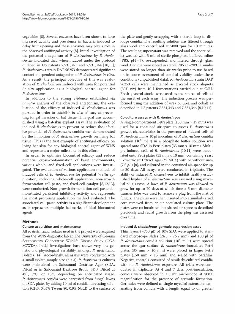
Figure 1 Shared air-space co-culture of P. destructans conidia with R.P. destructans control (a, d) were incubated in a shared air-space at 15°C (tinhibit growth from conidia when activated carbon is included in the head
medium surface without allowing media to come in con-tact with the inoculated surface of the skin. The skinexplants were then maintained on Eagle’s modified mi-nimal essential medium supplemented with antibiotics(kanamycin, 100 μg/ml: amikacin, 20 μg/ml; and vanco-mycin 50 μg/ml). A suspension of spores was placed ontothe center of the explant and allowed to dry. The inocu-lated explants were incubated in a shared air-space withinduced R. rhodochrous. Uninoculated control explantswere incubated alone or with uninduced R. rhodochrous.Initial experiments were conducted at 7°C. Anti-infectiveefficacy was determined by visual and microscopic eva-luation of bat wing membrane tissue cultures exposed toinduced R. rhodochrous as compared to unexposed anduninduced controls.
ResultsAnti-P. destructans activity of induced R. rhodochrousInitial experiments with induced cells of R. rhodochrousdemonstrated complete inhibition of growth from conidiaof P. destructans when cultured with a shared air-space at15°C (Figure 1a-c). Uninduced cells of R. rhodochrousshowed no signs of inhibition, and were comparable tounexposed controls. Subsequent testing at 4°C demon-strated fungistatic activity of induced cells of R. rhodo-chrous and resulted in slower germination and reducedtotal mycelial growth as compared to uninduced cells ofR. rhodochrous and unexposed controls (Figure 1d-f).Inclusion of activated carbon into the shared air-spaceabolished the anti-P. destructans activity of induced R.rhodochrous (Figure 1c). Mycelial plugs of P. destructanscultured in a shared air-space with induced R. rhodochrous
rhodochrous. Uninduced cells (e), induced cells (b, c and f) andop panel) and 4°C (bottom panel). Induced R. rhodochrous fails to-space (c).
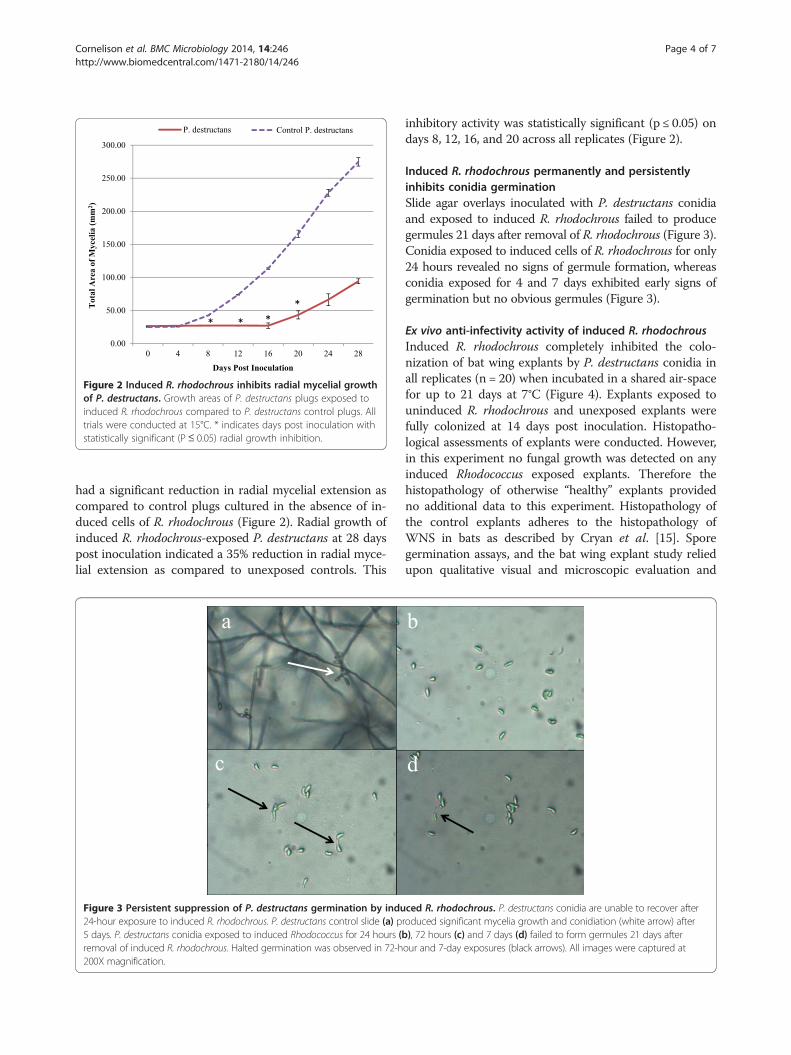
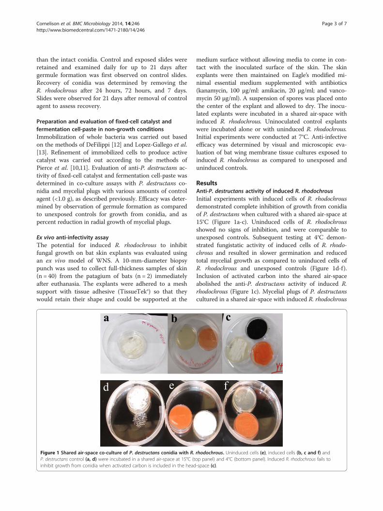
Figure 2 Induced R. rhodochrous inhibits radial mycelial growthof P. destructans. Growth areas of P. destructans plugs exposed toinduced R. rhodochrous compared to P. destructans control plugs. Alltrials were conducted at 15°C. * indicates days post inoculation withstatistically significant (P≤ 0.05) radial growth inhibition.
Cornelison et al. BMC Microbiology 2014, 14:246 Page 4 of 7http://www.biomedcentral.com/1471-2180/14/246
had a significant reduction in radial mycelial extension ascompared to control plugs cultured in the absence of in-duced cells of R. rhodochrous (Figure 2). Radial growth ofinduced R. rhodochrous-exposed P. destructans at 28 dayspost inoculation indicated a 35% reduction in radial myce-lial extension as compared to unexposed controls. This
Figure 3 Persistent suppression of P. destructans germination by indu24-hour exposure to induced R. rhodochrous. P. destructans control slide (a) p5 days. P. destructans conidia exposed to induced Rhodococcus for 24 hours (bremoval of induced R. rhodochrous. Halted germination was observed in 72-h200X magnification.
inhibitory activity was statistically significant (p ≤ 0.05) ondays 8, 12, 16, and 20 across all replicates (Figure 2).
Induced R. rhodochrous permanently and persistentlyinhibits conidia germinationSlide agar overlays inoculated with P. destructans conidiaand exposed to induced R. rhodochrous failed to producegermules 21 days after removal of R. rhodochrous (Figure 3).Conidia exposed to induced cells of R. rhodochrous for only24 hours revealed no signs of germule formation, whereasconidia exposed for 4 and 7 days exhibited early signs ofgermination but no obvious germules (Figure 3).
Ex vivo anti-infectivity activity of induced R. rhodochrousInduced R. rhodochrous completely inhibited the colo-nization of bat wing explants by P. destructans conidia inall replicates (n = 20) when incubated in a shared air-spacefor up to 21 days at 7°C (Figure 4). Explants exposed touninduced R. rhodochrous and unexposed explants werefully colonized at 14 days post inoculation. Histopatho-logical assessments of explants were conducted. However,in this experiment no fungal growth was detected on anyinduced Rhodococcus exposed explants. Therefore thehistopathology of otherwise “healthy” explants providedno additional data to this experiment. Histopathology ofthe control explants adheres to the histopathology ofWNS in bats as described by Cryan et al. [15]. Sporegermination assays, and the bat wing explant study reliedupon qualitative visual and microscopic evaluation and
ced R. rhodochrous. P. destructans conidia are unable to recover afterroduced significant mycelia growth and conidiation (white arrow) after), 72 hours (c) and 7 days (d) failed to form germules 21 days afterour and 7-day exposures (black arrows). All images were captured at

Figure 4 Induced R. rhodochrous prevents fungal colonizationof bat tissue when contained in a shared air-space. Bat wingtissue explants in a shared air-space with induced R. rhodochrous21 days post-inoculation with P. destructans conidia (a). Magnifiedimage of a control explant with visible fungal colonization 21 dayspost-inoculation (b).
Figure 5 Non-growth cell-paste of R. rhodochrous inhibitsgrowth from conidia of P. destructans. Non-growth fermentationcell-paste of induced R. rhodochrous was incubated in a shared air-spacewith P. destructans conidia inoculated plates. Quantities of 1.0 g, 0.5 g,and 0.25 g (a, b, and c) all demonstrated complete inhibition of growthfrom conidia of P. destructans as compared to unexposed controls(d, white arrow). Image taken 21 days post-inoculation.
Cornelison et al. BMC Microbiology 2014, 14:246 Page 5 of 7http://www.biomedcentral.com/1471-2180/14/246
produced definitive results (i.e. no exposed explants de-veloped fungal growth) therefore a statistical evaluation isunwarranted and omitted.
Evaluation of fixed-cell catalyst and fermentationcell-pasteFixed-cell catalyst [8,10,11] failed to inhibit or slow growthfrom conidia of P. destructans when grown in a sharedair-space. Fermentation cell-paste in quantities of 1.0 g,0.5 g, and 0.25 g completely inhibited growth from conidiaof P. destructans for greater than 80 days (Figure 5a-c).
Discussion and conclusionSince its initial documentation in 2006, WNS has spreadto twenty-four states and four provinces and has beenimplicated in the mortality of millions of North Americanbats [16-18] which may have a significant impact onNorth American agricultural practices [19]. WNS is char-acterized by invasive mycelial growth on the wings, muzzleand ears of hibernating bats that perturbs physiological
functions of the host tissues leading to mortality [15]. Caveclosures and culling of infected individuals appears to havelittle to no impact on the spread and mortality associatedwith this devastating disease [20]. Classic disease manage-ment practices applied in agriculture, such as vaccinationand broad-spectrum dissemination of antibiotics, presentmany challenges in the management of disease in wild,highly disseminated, and migratory animal populations.Consequently, the development of novel treatment optionsare needed to avert the spread of WNS and reduce themortality associated with currently infected hibernacula.To this end, the development of biologically-based controltools is the preferred option for application in karstenvironments.Since the publication of the national response plan [4],
several groups have initiated investigations to identifypotential biological control agents for P. destructans[21-23]. Several of the investigations have relied on tra-ditional sources of biocontrol agents or probiotics suchas bacilli and lactobacilli, or competitive exclusion fungisuch as Trichoderma sp., as well as attempts to isolatebat-skin-associated microbes with anti-P. destructansactivity [21-23]. While these approaches have provensuccessful in agricultural and human health applications[24-27], their application in the attempted remediation
Cornelison et al. BMC Microbiology 2014, 14:246 Page 6 of 7http://www.biomedcentral.com/1471-2180/14/246
of WNS in bats has not been demonstrated. The require-ment for contact with P. destructans and the bat hosts is amajor hurdle for any agents reliant on competitive exclu-sion or non-volatile antimicrobial compound production.These potential control agents may prove to have limitedefficacy against P. destructans in situ and potentially beharmful to the bat hosts. In contrast, the evaluation of in-duced R. rhodochrous strain DAP 96253 for application as abiological control agent of P. destructans aligns ideally withthe needs of wildlife management agencies tasked withcombatting WNS and is the first documented contact-independent microbial antagonism of P. destructans.The evolutionary lineage of R. rhodochrous lends itself
to VOC-based fungistasis due to its terrestrial ancestry[28-30]. The global prevalence of fungistatic soils is ameasure of the natural antagonisms that exists in thesecomplex environments [28-32]. Due to the ubiquity ofR. rhodochrous in soils [5], it can be expected thatR. rhodochrous as well as many other soil-dwellingbacteria have the potential to contribute to VOC-basedfungistasis observed in these environments [29,30]. How-ever, the development of induction methodologies is re-quired to optimize this activity for biocontrol applicationsand is a decidedly advantageous quality of R. rhodochrousstrain DAP 96253 as a potential biological control agent ofWNS [33]. Leveraging this naturally evolved antagonismfor control efforts has many benefits, particularly in thecase of WNS. The complexity of soil ecology selects forantagonisms that are effective at low concentrations indiverse, compartmentalized environments where solublediffusion may be limited [29]. Therefore, the productionof antagonistic VOCs provides a viable means for soil-dwelling bacteria to compete with soil-dwelling fungi forresources and equates favorably with the environmentalconditions of susceptible bat hibernacula. The ability ofR. rhodochrous to detect and interfere with volatile signalshas also been demonstrated in its delayed fruit-ripeningactivity [8] and is hypothesized to mediate the observedanti-P. destructans activity.While the efficacy of urea-induced R. rhodochrous
under growth conditions is promising for in situ man-agement of WNS, the need for growth media supple-mentation poses problems for field application. The longterm in vitro efficacy of non-growth-condition cell-pasteat 4°C allows for increased confidence in forecasting theefficacy of this biocontrol agent in managing WNS inthe field as this temperature is a sound approximation ofaverage winter temperature of North American bathibernacula [34]. The lack of growth media reduces thecosts associated with application as well as reduces thelikelihood of cross-contamination of control agent mediawith native cave microflora. In addition, the contact-independent basis of the non-growth antagonism willallow for in situ application methods that will reduce the
potential for ecological impacts associated with intro-ducing exogenous organisms to karst environments. Theecological impacts of any potential control agent are ofsignificant concern for wildlife management agenciesand the evaluation of potential ecological impacts mustbe assessed in order to circumvent ecological disastersassociated with augmenting cave microflora (e.g. Lascauxcave) [4].The evaluation of R. rhodochrous using ex vivo bat
tissue explants as an indicator of anti-infective activitywas paramount to establishing R. rhodochrous as a viablebiocontrol agent of P. destructans. This was the firstdemonstration of inhibition of fungal colonization of battissue by a biological control agent. This ex vivo efficacyjustifies further in vivo studies with live bats and shouldbe pursued vigorously.The ability of dormant conidia to remain viable in
host-free environments increases long-term impacts offungal pathogens and renders contaminated environ-ments inhospitable to re-colonization [35]. The impactof WNS in locations such as New York has been tre-mendous, vastly reducing the populations of insectivor-ous bats over a broad geographic range. The permanentand persistent inhibition of conidia germination is apromising result and indicates that treatment of pre-viously decimated hibernacula to inactivate resident co-nidia prior to re-colonization attempts may be feasibleby applying induced R. rhodochrous in these environ-ments. However further investigations are needed toconfirm the applicability of this approach.The evaluation of R. rhodochrous strain DAP 96253
has demonstrated the tremendous potential of this or-ganism for application as a biological control agent ofP. destructans. This is the first and only demonstrationof contact-independent antagonism of P. destructansand represents a significant step toward the develop-ment of biologically-based treatment tools for WNS.
Competing interestsGEP and SAC are contributing authors on the seminal patents for theinduction and application of Rhodococcus rhodochrous DAP 96253 cited inthe text. These patents are held by Georgia State University ResearchFoundation.
Authors’ contributionsCTC, SAC, and GEP conceived and designed the experiments conducted atGSU. KTG TAT and CKB developed and carried out the methodology toassess and produce induced R. rhodochrous as well as collected andanalyzed data. MKK provided P. destructans isolates from his diagnostic workas well as designed and conducted the experiments with bat tissue explantsat UGA. CTC, KTG, and MKK wrote the manuscript. All authors read andapproved the final version of the manuscript.
AcknowledgementsThis work was funded by Bat Conservation International through a WNSresearch grant as well as the Georgia State University EnvironmentalResearch Program. The authors would like to thank Lisa Last and PageLuttrell for assistance with the maintenance of tissue explants. The authors
Cornelison et al. BMC Microbiology 2014, 14:246 Page 7 of 7http://www.biomedcentral.com/1471-2180/14/246
would also like to thank Ian Sarad, Blake Cherney, and Ben Poodiak for theircontributions to this effort.
Author details1Applied and Environmental Microbiology, Georgia State University, 161Jesse Hill Jr. Dr, Atlanta, GA, USA. 2Department of Pathology Microbiology &Immunology, University of California Davis, One Shields Avenue, Davis, CA,USA.
Received: 3 June 2014 Accepted: 11 September 2014
References1. Lorch JM, Meteyer CU, Behr MJ, Boyles JG, Cryan JM, Hicks AC, Ballmann AE,
Coleman JTH, Redell DN, Reeder DM, Blehert DS: Experimental infection ofbats with Geomyces destructans causes white-nose syndrome. Nature2011, 10:90–105.
2. Warnecke L, Turner JM, Bollinger TK, Lorch JM, Misra V, Cryan PM, WibbeltG, Blehert DS, Willis CKR: Inoculation of bats with European Geomycesdestructans supports the novel pathogen hypothesis for the origin ofwhite-nose syndrome. PNAS 2012, 109:6999–7003.
3. Bastian F, Jurado V, Navakova A, Alabouvette C, Saiz-Jiminez C: The microbiologyof Lascaux cave. Microbiology 2010, 156:644–652.
4. Ballman A, Benedict L, Britzke E, Castle K, Cottrell W, Cryan P, DeLiberto T,Elliot A, Ewing R, Hicks A, Reynolds R, Rubado J, Slack B, Williams L,Coleman J: A national plan for assisting states, federal agencies, andtribes in managing white-nose syndrome in bats. 2011.http://www.WhiteNoseSyndrome.org.
5. Bell KS, Philp JC, Aw DWJ, Christofi N: The genus Rhodococcus. J ApplMicrobiol 1998, 85(2):195–210.
6. Larkin MJ, Kulakov LA, Allen CC: Biodegradation and R. rhodochrous –masters of catabolic versatility. Curr Opin Biotechnol 2005, 16(3):282–290.
7. Nagasawa T, Shimizu H, Yamada H: The superiority of the third-generationcatalyst, Rhodococcus rhodochrous J1 nitrile hydratase, for industrialproduction of acrylamide. Appl Microbiol Biotechnol 1993, 40:189–195.
8. Pierce GE, Drago GK, Ganguly S, Tucker T, Hooker JW, Jones S, Crow SA Jr:Preliminary report on a catalyst derived from induced cells ofRhodococcus rhodochrous DAP 96253 that delays the ripening ofselected climacteric fruit: bananas, avocados, and peaches. J Ind MicrobiolBiotechnol 2011, 38:1567–1573.
9. Sunairi M, Iwabuchi N, Yoshizawa Y, Murooka H, Morisaki H, Nakajima M:Cell-surface hydrophobicity and scum formation of Rhodococcusrhodochrous strains with different colonial morphologies. J Appl Microbiol1997, 82(2):204–210.
10. Pierce GE, Drago GK, Ganguly S: Induction and stabilization of enzymaticactivity in microorganisms. 2009. US Patent 7,531,343.
11. Pierce GE, Drago GK, Ganguly S: Induction and stabilization of enzymaticactivity in microorganisms. 2009. US Patent 7,531,344.
12. De Filippi LJ: Process for preparing immobilized enzymes. 1980. US Patent4,229,536.
13. Lopez-Gallaego F, Betancor L, Mateo C, Hidalgo A, Alonso-Morales N,Dellamora-Ortiz G, Gusian JM, Fernandez-Lafuente R: Enzyme stabilizationby gluteraldehyde crosslinking of absorbed proteins on aminatedsupports. J Biotechnol 2005, 119:70–75.
14. Rajkumar SS, Li X, Rudd RJ, Okoniewski JC, Xu J, Chaturvedi S, Chaturvedi V:Clonal genotype of Geomyces destructans among bats with White-NoseSyndrome, New York, USA. Emerg Infect Dis 2011, 17:1273–1276.
15. Cryan PM, Meteyer CU, Blehert DS, Boyles JG: Wing pathology of white-nose syndrome in bats suggests life-threatening disruption ofphysiology. BMC Biol 2010, 8:135.
16. Blehert DS, Lorch JM, Ballman AE, Cryan PM, Meteyer CU: Since 2007,infections by a previously unrecognized, perhaps imported fungus killedand estimated 1 million bats in North America. Microbe 2011.
17. Froschauer A, Coleman J: North American bat death toll exceeds 5.5million from white-nose syndrome. 2012. U.S. Fish & Wildlife ServicePress release.
18. Gargas A, Trest MT, Christensen M, Volk TJ, Blehert DS: Geomycesdestructans sp. Nov. associated with white-nose syndrome. Mycotaxon2009, 108:147–154.
19. Boyles JG, Cryan PM, McCracken GF, Kunz TH: Economic importance ofbats in agriculture. Science 2011, 332:41–42.
20. Hallam TG, McCracken GF: Management of the panzootic white-nosesyndrome through culling of bats. Conserv Biol 2011, 25(1):189–194.
21. Amelon S, Knudsen G: Identification and evaluation of potentialbiological control agents towards Geomyces destructans. WNS ResearchTracking-Draft 2011. WNS Research Tracking- Draft.
22. Chaturvedi V, Chatuvedi S: Fungal biocontrol agents for alleviation orremediation of Geomyces destructans. Fiscal Year 2012 U.S. Fish andWildlife Service-funded projects. FY 2012 USFWS-funded projects.
23. Frick WF, Kilpatrick AM: Antifungal skin microbes as tools for WNSmanagement. Fiscal Year 2012 U.S. Fish and Wildlife Service-fundedprojects. FY 2012 USFWS-funded projects.
24. Berg G: Plant-Microbe interactions promoting plant growth and health:perspectives for controlled use of microorganisms in agriculture.Appl Microbiol And Biotech 2009, 84:8–11.
25. Pascual LM, Daniele MB, Ruiz F, Giordano W, Pajaro C, Barberis L:Lactobacillus rhamnosus L60, a potential probiotic isolated from thehuman vagina. J Gen Appl Microbiol 2008, 54:141–148.
26. Pitt JI, Hocking AD: Mycotoxins in Australia: biocontrol of aflatoxin inpeanuts. Mycopathologia 2006, 162:233–243.
27. Wisniewski ME, Wilson CL: Biological control of postharvest diseases offruits and vegetables: recent advances. Hort Science 1992, 27:94–98.
28. Chuankun X, Minghe M, Zhang L, Zhang K: Soil volatile fungistasis andvolatile fungistatic compounds. Soil Biol Biochem 2004, 36:1997–2004.
29. Garbeva P, Hol WHG, Termorshuizen AJ, Kowalchuk GA, Boer WD:Fungistasis and general soil biostasis – a new synthesis. Soil Biol Biochem2001, 43:469–477.
30. Kerr JR: Bacterial inhibition of fungal growth and pathogenicity. MicrobEcol Health Dis 1999, 11:129–142.
31. Ezra D, Strobel GA: Effect of substrate on the bioactivity of volatileantimicrobials produced by Muscodor albus. Plant Sci 2003, 165(2):1229–1238.
32. Fernando WG, Ramarathnam R, Krichnamoorthy AS, Savchuk SC:Identification and use of potential bacterial organic antifungal volatilesin biocontrol. Soil Biol Biochem 2005, 37:955–964.
33. Strobel GA, Kluck K, Hess WM, Sears J, Ezra D, Vargas PN: Muscodor albusE-6, an endophyte of Guazuma ulmifolia making volatile antibiotics:isolation, characterization and experimental establishment in the hostplant. Microbiology 2007, 153:2613–2620.
34. Webb PI, Speakman JR, Racey PA: How hot is a hibernaculum? A review ofthe temperatures at which bats hibernate. Can J Zool 1996, 74:761–765.
35. Fisher MC, Henk DA, Briggs CJ, Brownstein JS, Madoff LC, McCraw SL, GurrSJ: Emerging fungal threats to animal, plant and ecosystem health.Nature 2012, 484:186–194. Doi:10.1038/nature10947.
doi:10.1186/s12866-014-0246-yCite this article as: Cornelison et al.: A preliminary report on the contact-independent antagonism of Pseudogymnoascus destructans by Rhodococcusrhodochrous strain DAP96253. BMC Microbiology 2014 14:246.
Submit your next manuscript to BioMed Centraland take full advantage of:
• Convenient online submission
• Thorough peer review
• No space constraints or color figure charges
• Immediate publication on acceptance
• Inclusion in PubMed, CAS, Scopus and Google Scholar
• Research which is freely available for redistribution
Submit your manuscript at www.biomedcentral.com/submit