Embed Size (px)
Citation preview
American Journal of Pediatrics 2018; 4(4): 80-83
http://www.sciencepublishinggroup.com/j/ajp
doi: 10.11648/j.ajp.20180404.11
ISSN: 2472-0887 (Print); ISSN: 2472-0909 (Online)
A Rare Case of PeutzJegher Syndrome Presenting with a Solitary Polyp in the Jejunum – A Case Report
Prabakaran Sundararajan1, *
, Saravanan Natarajan1, Kasthuri Thilagam Kannaian
2
1Department of Pediatric Surgery, Government Mohan Kumaramangalam Medical College, Salem, India 2Department of Pathology, Government Mohan Kumaramangalam Medical College, Salem, India
Email address:
*Corresponding author
To cite this article: Prabakaran Sundararajan, Saravanan Natarajan, Kasthuri Thilagam Kannaian. A Rare Case of PeutzJegher Syndrome Presenting with a
Solitary Polyp in the Jejunum – A Case Report. American Journal of Pediatrics. Vol. 4, No. 4, 2018, pp. 80-83.
doi: 10.11648/j.ajp.20180404.11
Received: August 17, 2018; Accepted: September 12, 2018; Published: October 19, 2018
Abstract: Background: Intussusception is a common abdominal emergency requiring surgical intervention in newborns and
infants. They commonly present in babies less than 2 years of age. A vast majority of intussusceptions are idiopathic. They
rarely present in older age groups and in adults. Certain autosomal disorders may be linked to intussusceptions. Peutz Jegher
Syndrome is a rare autosomal dominant disorder resulting due to mutation in the SPK 11 gene located in chromosome 19q13.3.
The disease is characterized by multiple hamartomatous polyposis, jejuna intussusceptions and hyperpigmented melanotic
spots in the mucocutaneous regions like mouth, etc. Case report: We present a case of 12 year old girl with chronic stomach
pain and occasional vomiting. General examination showed presence of hyperpigmented spots in the soles, cheeks, lips and
mouth. CECT abdomen showed presence of jejunal intussusception. The patient was taken up for laproscopy and laparotomy
was performed after reducing the intussusceptions. A soliatary sessile polyp was removed and histopathological examination
confirmed the presence of hamartomatous polyp. There is an increased susceptibility of cancers of gastrointestinal system and
several other organs like breast, ovaries, etc. Conclusion: Further research in this area may be carried out to explore the risk
factors and genetic mechanisms which may help in early detection and prevention of such rare syndromes.
Keywords: Hamartomatous Polyp, Jejunal Intussusception, Laprotomy, Peutz Jegher Syndrome
1. Introduction
Intussusception is a surgical emergency occurring as a
result of invagination of bowel into an immediately adjacent
section of the bowel. It is characterized by telescoping of a
part of intestine into itself. [1]Intussusception is a common
cause of abdominal pain in the pediatric age group. It is
however, the most common surgical emergency among the
children all over the world. High incidence rates are seen in
infants aged 4-10 months. [2] The diagnosis is often clinical,
although radiological procedures like CT and MRI of the
abdomen might help. [3] There are several risk factors
attributed in children namely infections, cystic fibrosis and
intestinal polyps. Some of the other risk factors include
anatomical abnormalities, Meckel’s diverticulum, altered
mobility, duplication, appendicitis and hyperplasia of Peyer’s
patches.
Intussusception often presents at five months of life, peaks
at around nine months with the lowest incidence rates
reported at 18 months. [4] The most common end point
pathology in intussusceptions is intestinal obstruction.
Peristaltic movements play a major role in the
pathophysiology of intussusception. The peristaltic action
pulls the proximal segment into the distal segment. There is
often an anatomical lead point noted in 10% of the
intussusceptions. Intussusception presents with vomiting,
abdominal pain and rectal bleeding. There are several
maneuvers to reduce the intussusception. However, in most
cases, surgical correction is often warranted.
In rare situations, intussusception is caused by certain
genetic and autoimmune disorders. Peutz Jegher Syndrome is
an autosomal dominant disorder characterized by various
presentations like mucocutaneous hyperpigmentation and
81 Prabakaran Sundararajan et al.: A Rare Case of PeutzJegher Syndrome Presenting with a Solitary
Polyp in the Jejunum – A Case Report
hamartomatous polyp in the gastrointestinal tract. They are
also associated with intussusceptions. [5] In this case report
we present a case of Peutz Jegher syndrome which presented
as jejuna intussusception in a 12 year old girl.
2. Case Report
A 12 years old female, presented with history of chronic,
recurrent pain abdomen on and off. There were also
complaints of vomiting, which was bilious in nature, on and
off for 10 days. The patient did not have any other abdominal
symptoms. On examination, the general condition was good,
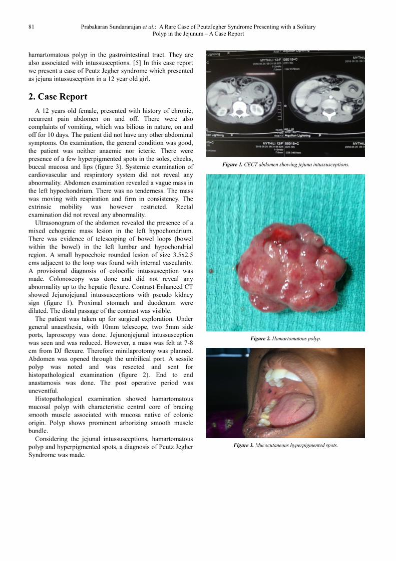
the patient was neither anaemic nor icteric. There were
presence of a few hyperpigmented spots in the soles, cheeks,
buccal mucosa and lips (figure 3). Systemic examination of
cardiovascular and respiratory system did not reveal any
abnormality. Abdomen examination revealed a vague mass in
the left hypochondrium. There was no tenderness. The mass
was moving with respiration and firm in consistency. The
extrinsic mobility was however restricted. Rectal
examination did not reveal any abnormality.
Ultrasonogram of the abdomen revealed the presence of a
mixed echogenic mass lesion in the left hypochondrium.
There was evidence of telescoping of bowel loops (bowel
within the bowel) in the left lumbar and hypochondrial
region. A small hypoechoic rounded lesion of size 3.5x2.5
cms adjacent to the loop was found with internal vascularity.
A provisional diagnosis of colocolic intussusception was
made. Colonoscopy was done and did not reveal any
abnormality up to the hepatic flexure. Contrast Enhanced CT
showed Jejunojejunal intussusceptions with pseudo kidney
sign (figure 1). Proximal stomach and duodenum were
dilated. The distal passage of the contrast was visible.
The patient was taken up for surgical exploration. Under
general anaesthesia, with 10mm telescope, two 5mm side
ports, laproscopy was done. Jejunonjejunal intussusception
was seen and was reduced. However, a mass was felt at 7-8
cm from DJ flexure. Therefore minilaprotomy was planned.
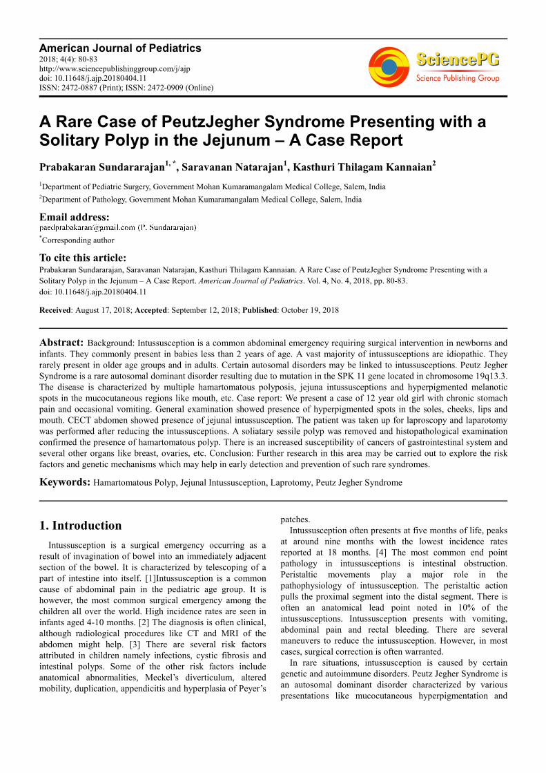
Abdomen was opened through the umbilical port. A sessile
polyp was noted and was resected and sent for
histopathological examination (figure 2). End to end
anastamosis was done. The post operative period was
uneventful.
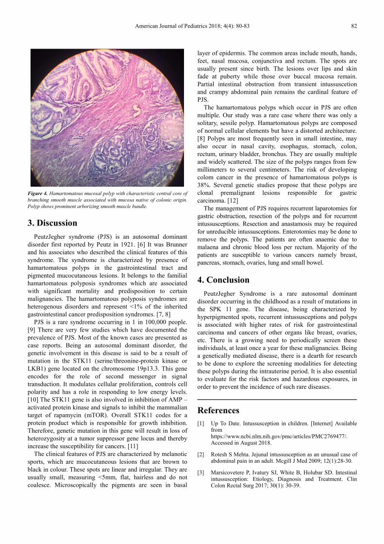
Histopathological examination showed hamartomatous
mucosal polyp with characteristic central core of bracing
smooth muscle associated with mucosa native of colonic
origin. Polyp shows prominent arborizing smooth muscle
bundle.
Considering the jejunal intussusceptions, hamartomatous
polyp and hyperpigmented spots, a diagnosis of Peutz Jegher
Syndrome was made.
Figure 1. CECT abdomen showing jejuna intussusceptions.
Figure 2. Hamartomatous polyp.
Figure 3. Mucocutaneous hyperpigmented spots.
American Journal of Pediatrics 2018; 4(4): 80-83 82
Figure 4. Hamartomatous mucosal polyp with characteristic central core of
branching smooth muscle associated with mucosa native of colonic origin.
Polyp shows prominent arborizing smooth muscle bundle.
3. Discussion
PeutzJegher syndrome (PJS) is an autosomal dominant
disorder first reported by Peutz in 1921. [6] It was Brunner
and his associates who described the clinical features of this
syndrome. The syndrome is characterized by presence of
hamartomatous polyps in the gastrointestinal tract and
pigmented mucocutaneous lesions. It belongs to the familial
hamartomatous polyposis syndromes which are associated
with significant mortality and predisposition to certain
malignancies. The hamartomatous polyposis syndromes are
heterogenous disorders and represent <1% of the inherited
gastrointestinal cancer predisposition syndromes. [7, 8]
PJS is a rare syndrome occurring in 1 in 100,000 people.
[9] There are very few studies which have documented the
prevalence of PJS. Most of the known cases are presented as
case reports. Being an autosomal dominant disorder, the
genetic involvement in this disease is said to be a result of
mutation in the STK11 (serine/threonine-protein kinase or
LKB1) gene located on the chromosome 19p13.3. This gene
encodes for the role of second messenger in signal
transduction. It modulates cellular proliferation, controls cell
polarity and has a role in responding to low energy levels.
[10] The STK11 gene is also involved in inhibition of AMP –
activated protein kinase and signals to inhibit the mammalian
target of rapamycin (mTOR). Overall STK11 codes for a
protein product which is responsible for growth inhibition.
Therefore, genetic mutation in this gene will result in loss of
heterozygosity at a tumor suppressor gene locus and thereby
increase the susceptibility for cancers. [11]
The clinical features of PJS are characterized by melanotic
sports, which are mucocutaneous lesions that are brown to
black in colour. These spots are linear and irregular. They are
usually small, measuring <5mm, flat, hairless and do not
coalesce. Microscopically the pigments are seen in basal
layer of epidermis. The common areas include mouth, hands,
feet, nasal mucosa, conjunctiva and rectum. The spots are
usually present since birth. The lesions over lips and skin
fade at puberty while those over buccal mucosa remain.
Partial intestinal obstruction from transient intussuscetion
and crampy abdominal pain remains the cardinal feature of
PJS.
The hamartomatous polyps which occur in PJS are often
multiple. Our study was a rare case where there was only a
solitary, sessile polyp. Hamartomatous polyps are composed
of normal cellular elements but have a distorted architecture.
[8] Polyps are most frequently seen in small intestine, may
also occur in nasal cavity, esophagus, stomach, colon,
rectum, urinary bladder, bronchus. They are usually multiple
and widely scattered. The size of the polyps ranges from few
millimeters to several centimeters. The risk of developing
colom cancer in the presence of hamartomatous polyps is
38%. Several genetic studies propose that these polyps are
clonal premalignant lesions responsible for gastric
carcinoma. [12]
The management of PJS requires recurrent laparotomies for
gastric obstruction, resection of the polyps and for recurrent
intussusceptions. Resection and anastamosis may be required
for unreducible intussusceptions. Enterotomies may be done to
remove the polyps. The patients are often anaemic due to
malaena and chronic blood loss per rectum. Majority of the
patients are susceptible to various cancers namely breast,
pancreas, stomach, ovaries, lung and small bowel.
4. Conclusion
PeutzJegher Syndrome is a rare autosomal dominant
disorder occurring in the childhood as a result of mutations in
the SPK 11 gene. The disease, being characterized by
hyperpigmented spots, recurrent intussusceptions and polyps
is associated with higher rates of risk for gastrointestinal
carcinoma and cancers of other organs like breast, ovaries,
etc. There is a growing need to periodically screen these
individuals, at least once a year for these malignancies. Being
a genetically mediated disease, there is a dearth for research
to be done to explore the screening modalities for detecting
these polyps during the intrauterine period. It is also essential
to evaluate for the risk factors and hazardous exposures, in
order to prevent the incidence of such rare diseases.
References
[1] Up To Date. Intussusception in children. [Internet] Available from https://www.ncbi.nlm.nih.gov/pmc/articles/PMC2769477/. Accessed in August 2018.
[2] Rotesh S Mehta. Jejunal intussusception as an unusual case of abdominal pain in an adult. Mcgill J Med 2009; 12(1):28-30.
[3] Marsicovetere P, Ivatury SJ, White B, Holubar SD. Intestinal intussusception: Etiology, Diagnosis and Treatment. Clin Colon Rectal Surg 2017; 30(1): 30-39.
83 Prabakaran Sundararajan et al.: A Rare Case of PeutzJegher Syndrome Presenting with a Solitary
Polyp in the Jejunum – A Case Report
[4] Fusco EE, Bhimji SS. Intussusception, child. Bookshelf StatPearls [Internet]. Available fromhttps://www.ncbi.nlm.nih.gov/books/NBK431078/.
[5] Thakker HH, Joshi A, Deshpande A. Peutz-Jegher’s Syndrome presenting as jejunoileal intussusception in an adult male: a case report. Cases J 2009; 2:8865.
[6] Peutz JLA. Over eenzeermerkwaardige, gecombineerdefamiliairepollyposis van de sligmliezen van den tractusintestinalis met die van de neuskeelholte en gepaard met eigenaardigepigmentaties van huid-en slijmvliezen (Very remarkable case of familial polyposis of mucous membrane of intestinal tract and nasopharynx accompanied by peculiar pigmentations of skin and mucous membrane; in Dutch) Nederl Maandschr v Geneesk. 1921; 10:134–146.
[7] Calva D, Howe JR Hamartomatous polyposis syndromes. SurgClin North Am. 2008 Aug; 88(4):779-817, vii.
[8] Chen HM, Fang JY. Genetics of the hamartomatous polyposis syndromes: a molecular review. Int J Colorectal Dis. 2009 Aug; 24(8):865-74.
[9] Gammon A, Jasperson K, Kohlmann W, Burt RWH amartomatous polyposis syndromes. Best Pract Res Clin Gastroenterol. 2009; 23(2):219-31.
[10] Zbuk KM, Eng C. Hamartomatous polyposis syndromes. Nat Clin Pract Gastroenterol Hepatol. 2007 Sep; 4(9):492-502.
[11] Katajisto P, Vallenius T, Vaahtomeri K, Ekman N, Udd L, Tiainen M, Mäkelä TP The LKB1 Tumor suppressor kinase in human disease. Biochim Biophys Acta. 2007 Jan; 1775(1):63-75.
[12] deLeng WW J, Jansen M, Keller JJ, de Gijsel M, Milne ANA, Morsink FHM, et al. Peutz Jegher Syndrome polyps are polyclonal with expanded progenitor cell compartment. Gut 2007;56:1475-1476.