Embed Size (px)
Citation preview
REVIEW Open Access
A rather dry subject; investigating thestudy of arid-associated microbialcommunitiesPeter Osborne1* , Lindsay J. Hall2,3, Noga Kronfeld-Schor4, David Thybert5 and Wilfried Haerty1
Abstract
Almost one third of Earth’s land surface is arid, with deserts alone covering more than 46 million square kilometres.Nearly 2.1 billion people inhabit deserts or drylands and these regions are also home to a great diversity of plant andanimal species including many that are unique to them. Aridity is a multifaceted environmental stress combining a lackof water with limited food availability and typically extremes of temperature, impacting animal species across theplanet from polar cold valleys, to Andean deserts and the Sahara. These harsh environments are also home to diversemicrobial communities, demonstrating the ability of bacteria, fungi and archaea to settle and live in some of thetoughest locations known. We now understand that these microbial ecosystems i.e. microbiotas, the sum total ofmicrobial life across and within an environment, interact across both the environment, and the macroscopic organismsresiding in these arid environments. Although multiple studies have explored these microbial communities in differentarid environments, few studies have examined the microbiota of animals which are themselves arid-adapted. Here weaim to review the interactions between arid environments and the microbial communities which inhabit them,covering hot and cold deserts, the challenges these environments pose and some issues arising from limitations in thefield. We also consider the work carried out on arid-adapted animal microbiotas, to investigate if any shared patterns ortrends exist, whether between organisms or between the animals and the wider arid environment microbialcommunities. We determine if there are any patterns across studies potentially demonstrating a general impact ofaridity on animal-associated microbiomes or benefits from aridity-adapted microbiomes for animals. In the context ofincreasing desertification and climate change it is important to understand the connections between the three pillarsof microbiome, host genome and environment.
IntroductionWater is essential for all known forms of life [1] and astable arid environment is characterised by low annualprecipitation (depending on location from 500- < 10mmprecipitation annually), with desertification occurringwith greater loss than gain [2]. Approximately 30% [3] ofthe land surface area of the Earth is classified as ‘arid’.However even extreme deserts, including both hot and coldextremes and areas with exceptionally little precipitation,
are still home to a wide diversity of life from microscopic[4, 5], to large mammals [6, 7] and long lived charismaticflora [8]. It is important therefore to understand how life isable to survive and adapt to the challenge of obtaining andretaining water, along with the other threats to animal lifelisted in Table 1. Aridity driven adaptations in plants[17, 18] as well as behavioural adaptations in animals[19] have already been well described and will not beaddressed in this review. Research has demonstratedthe importance of microbial organisms when living asmembers of associated communities on and in animals;for growth and development [20], dietary necessity [21]and for maintaining expected behaviour [22]. It is reasonable
© The Author(s). 2020 Open Access This article is licensed under a Creative Commons Attribution 4.0 International License,which permits use, sharing, adaptation, distribution and reproduction in any medium or format, as long as you giveappropriate credit to the original author(s) and the source, provide a link to the Creative Commons licence, and indicate ifchanges were made. The images or other third party material in this article are included in the article's Creative Commonslicence, unless indicated otherwise in a credit line to the material. If material is not included in the article's Creative Commonslicence and your intended use is not permitted by statutory regulation or exceeds the permitted use, you will need to obtainpermission directly from the copyright holder. To view a copy of this licence, visit http://creativecommons.org/licenses/by/4.0/.The Creative Commons Public Domain Dedication waiver (http://creativecommons.org/publicdomain/zero/1.0/) applies to thedata made available in this article, unless otherwise stated in a credit line to the data.
* Correspondence: [email protected] Institute, Norwich Research Park Innovation Centre, Colney Lane,Norwich NR4 7UZ, UKFull list of author information is available at the end of the article
Environmental MicrobiomeOsborne et al. Environmental Microbiome (2020) 15:20 https://doi.org/10.1186/s40793-020-00367-6
therefore to infer that the microbiota of animals resident inarid environments contributes to their host’s fitness in suchharsh conditions. This may be through mechanisms alreadyknown from the study of microbiota in model organisms orby unique mechanisms.Here we review the interactions between arid environ-
ments and the microbial communities which inhabitthem along with the typical stresses these environmentspresent. We specifically highlight and compare studiesin arid-adapted animal microbiota, investigating patternsacross studies, potentially demonstrating a general im-pact of aridity on animal-associated microbiomes orbenefits from hosting aridity-adapted bacteria, fungi andarchaea. We use the terms ‘desert’ and ‘arid’ inter-changeably throughout the piece (as naturally occurringarid environments are referred to as deserts), typicallynaming the desert in question when relevant and indi-cating if a hot or cold desert is being discussed.In this review we:
I. Review environmental microbiology studies in aridenvironments through the different environmentalfactors acting on bacteria, fungi and archaea
II. Describe animal physiological adaptations to aridity& highlight animal-associated microbiomes andtheir roles
III. Review arid-associated animal microbiomes,examining two camels in particular and the impacton microbiomes of environmental factorsassociated with aridity
IV. Discuss some of the challenges and opportunitiesaround studying arid animal and environmentalmicrobiomes
Increased global desertification [23–25], acceleratingclimate change [26] and changing land management [27]highlight the importance of better understanding aridecosystems and animal adaptations to them. There havebeen comparatively few environmental microbiologystudies on arid environments though in the hunt forextremophiles and pharmaceutically useful compoundsthere have been some focussed investigations of particularlocations [28–31]. Animal-associated microbiota researchhas often been a component of a larger investigation intoa particular host species [32] rather than a systemic ap-proach considering the microbiome as a feature of animallife in arid environments.
Aridity and environmental microbiologyMicroorganisms can be found in almost all environments,adapting over millions of years to survive and thrive inconditions ranging from extremely hostile [33–35] to
Table 1 Examples of environmental factors which make arid environments inhospitable for animal life, and associated challengesfaced by living organisms
Arid environmental factor Challenges Example animal adaptations
Lack of food Extreme seasonality of food sourcesRequirement for multiple different food sources,or in contrast, to specialise to a single food sourceTravel longer distances to find foodIncreased exposure to predatorsDepressed metabolism
Switch lifestyle to nocturnal to access more differentfood sources - potentially also with greater watercontent [9]
Lack of water DehydrationReduced metabolic rateReduced ability to manage body temperatureBehavioural changes increase risk of predation i.e.sheltering to reduce water loss leading to exposureto predators
Reduce urine production and concentrate anyproduced [10]Store greater amounts of water in the body [11]Changes in activity patterns [12]
Extremes of temperature Hyperthermia● Protein denaturation● Dehydration from increased panting or sweating● Multiple organ failure
Hypothermia● Frostbite● Water in body freezing● Metabolic rate falling below survival baseline● Loss of heat from extremities
Tolerate increased temperatures through seekingshelter from the heat of the sun [13]Tolerate freezing through production of specialisedcompounds and antifreeze proteins [14]
Extremes of salinity Herbivores need ability to digest salty plant matterwithout losingexcess water during digestionNeed to maintain water balance in face of osmoticchallenges from consuming salty water
Salt glands which can excrete salt from the bodydepending on dietary intake and internal osmoticbalance [15]
Elevated UV-C andUV-B exposure
Increased risk of genetic damage from UV irradiationof external body surfaces
Increased skin, fur or carapace pigmentation [16]
Osborne et al. Environmental Microbiome (2020) 15:20 Page 2 of 14
resource rich [36, 37]. Prior researchers were limited bythe need to culture microorganisms, significantly impact-ing the scope of investigations on microbial diversity.Recent studies have indicated the importance of ‘culture-omics’ [38, 39] for creating large-scale microbial collec-tions for mechanistic analysis and classifying metagenomicresults which could not be assigned purely from bioinfor-matics. This has allowed investigation of environmental[40] and animal-associated [41] bacteria, fungi and ar-chaea detected through bioinformatics and sequencing,but previously difficult to culture. The ability to sequenceenvironmental samples and analyse genetic material with-out a need to grow microorganisms has allowed moreaccurate and large-scale accounting of existing diversity[42–44]. With the advent of new techniques and reducedsequencing cost, came a greater understanding of theinfluence of abiotic and biotic factors acting on micro-bial communities. Temperature [45–48], UV exposure[49, 50], salinity [51, 52], humidity [53], pH [54], irradi-ation [55], pressure [56], pollution [57] and oxygenconcentration [58] have all been demonstrated to influ-ence microbiomes.A similar suite of challenges is encountered in almost all
arid environments, foremost amongst these being the lackof free water. These environmental stresses may lead tothe establishment of new species, affect the compositionof a microbial community (presence or absence of givenmembers) and the relative abundances of species in acommunity (the level at which members are present).
Desiccation - lack of waterLack of free water is the defining trait of arid environ-ments, irrespective of temperature [59]. From a micro-bial perspective the lack of water presents the samedangers as those faced by macroscopic organisms alongwith uniquely microscopic challenges. In deserts wherefree water availability is low and the medium in whichactivity occurs is largely absent, Ho-Kyung Song et. al.[60] found that desiccation led to selection against mo-tility associated proteins within their studied bacteria.They note that these proteins are associated with fla-gella; and this selective pressure may not be seen withalternative methods of motility. Multiple studies acrossdifferent natural environments demonstrate reduced mi-crobial diversity with desiccation [61–63] compared tosites with greater water availability. This may come fromthe impaired ability to obtain nutrients from free-moving water [64]. Desiccation can also lead to de-creased production of anti-competition compounds.Fierer et. al. hypothesised that the significantly lowerproduction of antimicrobial murein hydrolases, alongwith reduced abundance of antibiotic resistance genesare associated with the greater environmental pressureon prokaryotic and eukaryotic microorganisms over
competition [65] as those compounds would requirewater for distribution. Le et. al. reported the productionof potential osmoprotectants (such as osmoprotectiveproline) to be upregulated in the microbial communitiesof hypoliths from the Namib Desert and Antarctica [66];similar adaptations were observed amongst bacteria liv-ing on dry city surfaces like metal and glass in New York[67]. Anderson et. al. found desiccation tolerance of anArchaeon increased when EPS (extracellular polysac-charide) production increased; additionally reportingincreased tolerance of heat and oxygen stresses in desic-cated versus control cells [68]. Due to environmentalchallenges, specific reproductive strategies may beemployed. For instance, fungi living on extremely drysurfaces using meristematic development in order to re-produce without requiring water for dispersal [69, 70].Within broader arid environments, relatively moist sitestend towards richer and larger microbial communities[71, 72], demonstrating the intensive selective pressureof aridity. It is worth noting that seemingly desiccatedenvironments can in fact contain tiny water dropletshome to bacteria, fungi and archaea surviving in other-wise deadly conditions [73], as well as dormant bacteriaand fungi which revive and become metabolically activeafter an increase in moisture [74].
Temperature - bake or freezeAridity and extremes of temperature are commonlyfound together. Antarctica and the Sahara are two clearexamples of desiccation and dangerous temperaturesmaking survival extremely difficult [75]. Multiple studieshave investigated microorganisms surviving and thrivingin locations with extreme temperatures [76–81]. In a hotenvironment, Armstrong et. al. found that the soil mi-crobial communities of gravel plains in the Namib desertremained constant over time [82], speculating that thisis likely due to the stable (though hostile) environmentalconditions experienced throughout the majority of thestudy period. The stress of high temperature has beenshown to lead to increased production of heat shockproteins [83]; high temperatures in geothermically activesoils in Antarctica have also been shown to influencemicrobial community composition and may help explainthe presence of thermophilic Archaea closely related tothose found in similar hot environments thousands ofmiles away [84]. Cockell et. al. found that higher tem-peratures limited microbial diversity when other condi-tions were well-suited for life in and around Hawaiianfumaroles [85]. As expected, high temperatures havebeen shown to interact with the other stresses associatedwith aridity to influence microbial community compos-ition - such as favouring endospore forming Firmicutes[86]. This supports the findings by Savage et. al. that ele-vated temperatures still allowed more diverse microbial
Osborne et al. Environmental Microbiome (2020) 15:20 Page 3 of 14
communities than those possible when temperatureswere combined with other abiotic stresses [87]. In coldenvironments, production of cold shock proteins hasbeen documented [88]. Antarctic bacteria are known toproduce antifreeze proteins [89], Liljeqvist et. al. found agene predicted to code for production of an antifreezeprotein in their metagenomic study of an acid minedrainage stream in northern Sweden [90]. Adaptationsto cold temperatures have also been noted in fungithrough increased production of unsaturated lipids inthe cell membrane [91], maintaining membrane fluidity.Cryotolerant fungi may also accumulate cryoprotectantslike glycerol [92].
RadiationDrastically reduced coverage from clouds or vegetationin arid environments means exposure to damaging UVlight [93, 94]. UV exposure levels have been shown toinfluence microbial community composition [95]. Thisincludes dry environments where desiccation can lead togreater difficulties in tolerating UV irradiation [96];though some other studies disagree [97]. Adaptation forUV exposure is typically through pigmentation. As such,environmental sampling of UV irradiated sites in Antarctica[98] and Tibet [99] identified increased pigmentation in UVirradiation tolerant Hymenobacter. A number of studieslooking at the microbial residents of solar panels [100, 101];including panels in Antarctica, the Arctic and the Mediter-ranean all found Hymenobacter as the most abundantgenus [102]. Greater numbers of genes associated with anti-oxidant production and DNA repair have been noted inArchaea and Bacteria in heavily UV exposed environments[103–105]. These observations raise the possibility thatextremely effective DNA repair mechanisms may also be ameans of adapting to aridity. Pacelli et. al. subjected desic-cation resistant Antarctic fungi to gamma radiation levelsmuch greater than any found in nature and speculate thatobserved tolerance may use known DNA repair mecha-nisms associated with UV irradiation and dehydration[106]. Investigation by Selbmann et. al. in Antarctic fungiresistant to UV-B exposure led them to suggest that thickand highly melanised cell walls rather than enhanced DNArepair systems were the principal factor in tolerance of UVirradiation [107]. Jones and Baxter review in depth somemethods of tolerating UV stress in Archaea which may betranslatable into work with other microorganisms [108].
SalinitySome arid and semi-arid environments are heavily sub-jected to salt stress [109]. Intertidal zones and beachesexperience either daily coverage with seawater or largequantities of salt deposition from wind off the ocean.Typically, the adaptations employed to protect microor-ganisms against desiccation also confer protection against
salinity - the loss of water being a shared peril. Salinity inarid or other extreme environments can act as an inde-pendent factor controlling community composition; withmicroorganisms needing to be halotolerant in addition tobeing capable of dealing with other stresses. Managementof osmotic potential in a saline environment is a long-term issue and so may require greater dedication ofresources than acute salt stress caused by decreasingvolumes of water; or employ particular pathways. Thiswas noted by Molina-Menor, et. al. when examining themicrobial community of rocks in the intertidal zone of theMediterranean [110], and distinct microbiomes frommore saline areas of larger environments have been re-corded [111]. Production of hydrophobins [112] and accu-mulation of salt-stress specific solutes [113] were observedin fungi from saline environments.
From environmental to animal microbiomesComparisons of animal microbiomes to those of theirsurroundings within extreme environments, such as hotand cold deserts, are currently few in number and notcovered in detail in this review. The external conditionsof animals in arid environments are liable to be similarto their wider environment. Although behavioural adap-tation will alleviate some of the effects of environmentalstresses, members of the skin microbiota will be subjectto similar stresses as other environmental communities.Salt levels in the intestinal tract of the hot-desert dwell-ing Fat Sand rat (Psammomys obesus) are similar -atleast initially- to those of the saltbush they consume[114], and water content in the faces of desert species isvery low. Internal conditions in an animal will differfrom the external environment, but potentially not asgreatly as between an arid and a wet environment.Therefore arid-adapted animals may host extreme pointson different gradients within the larger arid environment,in terms of water availability, temperature (for ectotherms)or salinity; whilst being within the standard range forother environmental variables (e.g. temperature on andwithin an ectothermic cold desert insect).
Animal adaptation to aridity & animal-associatedmicrobiomesThe same factors (e.g. lack of water, extremes oftemperature, restricted energy sources) associated witharid environments influence both microbial and animalcommunities. In order to survive and thrive within suchhabitats, animals have developed a suite of physiologicaland behavioural adaptations. These reduce energyexpenditure and water loss [115], use microclimates orseek increased water intake from other sources [116–118].Table 1 provides some examples of animal adaptations inthe face of specific stresses; to mirror the focus onmammals in metagenomic studies we focus on non-bird
Osborne et al. Environmental Microbiome (2020) 15:20 Page 4 of 14
vertebrate adaptations to aridity. A number of animal spe-cies restrict their urine production in response to acutewater stress, from Merino sheep [119] to ostriches [120]and some toads [121]. These species demonstrate the abil-ity to reduce and concentrate [122–124] the amount ofurine they produce through specific renal adaptations[125] such as the elongation and enlargement of the renalpapilla [10, 126, 127], as well as changes to the distributionof aquaporin proteins [128]. Additionally, animals canstore water for use over a longer period, as dromedariesdo with their forestomach [129]. Some animals may takeadvantage of abnormal water sources to survive in arid lo-cations [130], including non-xeric animals [131]. A com-mon behavioural adaptation is to seek shelter, shade ormicroclimates in hot arid environments to limit heat stressand exposure to dry air; thus reducing evaporative waterloss [132]. Switching to a nocturnal rather than diurnalactivity pattern is another means of reducing water stressin hot arid environments [133] and of adapting to increas-ing aridity with climate change [9]. Adaptation forreduced basal metabolic rates in birds along an ariditygradient was uncovered by Tieleman et. al [134]. Lowbasal energy demands (basal metabolism) are common inhot desert endothermic animals, reducing both the needto forage in hot desert conditions and lung ventilationthereby reducing evaporative water loss [134, 135]. Manyendotherms reduce metabolic rate and water loss furtherthrough torpor, a state which can last from hours tomonths and is a reduction of body temperature and otherphysiological processes [118, 136–138]. These changescan be programmed, timed processes or direct responsesto environmental conditions [118]. Larger body size mayalso confer some protection against desiccation, as seen inAnopheles gambiae [139] and camels [140, 141]. Inverte-brate survival of extreme dehydration and temperatures isreviewed by Watanabe [142] and Somme [143], showing asuite of morphological, behavioural and physiologicalchanges across a range of species.While all these studies focused on an organism’s adapta-
tion to aridity, it is important to consider the influence ofthe microbiome, often called the ‘second genome’, onarid-adapted animals as a potential contributor to ariditytolerance. This is a dynamic relationship, in which thehost animals’ adaptations for aridity will directly influencethe different niches it provides for potential microbialcolonisation and the microbiome may negate the need forhost genomic adaptation to aridity. The development ofnew sequencing methods and increased computationalpower, coupled with innovative software and analysis ap-proaches have made large scale microbiome investigationsmore accessible [144, 145], highlighting the vital role theyplay in host development and health [146]. Research hasoften been directed to the intestinal microbiomes ofruminants [147–153] and other commercially valuable
species [154–156]. By virtue of ease of access, other stud-ies have tended to be on domesticated species [157], orwild species which can be more conveniently reached andinvestigated [158].Broadly, arid animals have not been the subject of as
much metagenomic research as those found in morehospitable environments. This has led to focus on asmall number of species, and less connection across thefield between environmental, host and metagenomicfactors in arid animals.
Arid animal microbiomesBeyond environmental microbiomes, and aside fromplant-associated microbiomes which are outside thescope of this review (see [159, 160]), the other potentiallocation for microbial communities in arid environmentsis in association with animal hosts. Here we discuss ani-mal microbiomes influenced by some of the aridity asso-ciated factors discussed above, then explore in greaterdetail Camels and Muskoxen, which have received moreinvestigation than most in this area.
Incidental aridity - animal microbiomes influenced byabiotic factors shared with arid environmentsBefore moving on to some specific arid-adapted animalsit is useful to look at animal microbiomes which mayhave been influenced by aridity directly or indirectly - orby factors also found in arid environments, similar tothe environmental microbiomes discussed above. Thecomparatively reduced levels of water available in aridenvironments, whether as humidity, surface moisture orprecipitation, limits the ability of macroscopic life todevelop [161, 162]. From the perspective of animal hostorganisms this leads to a tendency for specific diets,becoming more limited as aridity increases and diversityof plant and animal life falls [163, 164]; this has been re-ported in numerous organisms [165–168]. The humanskin microbiome has distinct correlations betweenmoisture levels and community composition [169, 170];this could offer some comparative references if skin-microbiome studies of arid-adapted animals are conducted.Diet plays a large, potentially dominant role in establishingthe intestinal microbiota of animals [171] and can influencethe microbiota of other body areas as well [172, 173].Limited food and water availability due to arid condi-
tions can lead to concentration of animals in sites withaccessible water or a shared source of nutrition - thisconsumption from the same site and possible close quar-ters may help explain the proximity based correlation inmicrobiota composition (likely through range overlapleading to similar microbial exposure) noted by Couchet. al. in Mojave desert-dwelling Bighorn sheep [174]faecal microbiota. Other stresses found in arid environ-ments have been examined in terms of animal-associated
Osborne et al. Environmental Microbiome (2020) 15:20 Page 5 of 14
microbiomes, both experimentally and in observation ofnatural conditions. Direct links have been observed be-tween temperature and animal-associated microbiomes inHumpback whales (2 °C to − 2 °C) [175], Fruit Flies (13 °Cversus 31 °C) [176], Silkworms (transient exposure to37 °C after rearing at 25 °C) [177] and Tilapia (24 °C versus12 °C) [178]. This influence can be profound or act inmore subtle ways, as Li et. al. noted in Xenopus tropicaliswhereby decreasing temperature altered beta but notalpha-diversity of the gut microbiome [179]. Interestingly,some of these temperature dependent changes in hostmicrobiomes reflect either changes in the hosttemperature (the fruit flies and silkworms for instance) orin the environmental temperature (Humpback whales). Insome instances, these studies have found that the micro-biomes subject to influence by heat stress also impacttheir hosts; Fontaine et. al. finding that temperature in-duced changes in salamander intestinal microbiota influ-enced energy uptake from digesting food [180]. Givensfound that a change in water temperature surroundingsome Fundulus hetereoclitus (Mummichog) correspondedwith a change in relative abundance of different species ofVibrio in the intestinal microbiota which may have beenconnected with increased mortality [181].Sullam et. al. reported that different levels of salinity
influenced the gut microbiota of fish - and that thesesaltwater fish intestinal communities bore similarities toenvironmental samples from saltwater [182]. Investiga-tion of Atlantic Salmon found a less diverse intestinalmicrobiota in those acclimated to saltwater than thoseliving in freshwater [183]. 16S investigation of the ex-posed facial skin of the Black and Turkey vultures byMendoza et. al. identified Psychrobacter cryohalolentisand Psychrobacter arcticus, which whilst commonlyfound in cold environments are known to be halotoler-ant [184] potentially explaining their presence on thewarm but saline surface. Given similarities in environ-mental microbiomes between saline and arid environ-ments, it is possible that dehydration might influenceanimal microbiomes in a similar way to salt stress;favouring the same functions and potentially relatedtaxonomic compositions. These studies examine envir-onmental salinity rather than that of internal fluids ofthe host, whether in the intestinal tract or elsewhere; itcould be of interest to assess salinity within the host andpotential impacts on the microbiota without an externalchange in salt levels; tied to consumption of a salt richdiet for instance.Direct exposure to UV irradiation of animal micro-
biomes is restricted to those on the external surfaces ofthe body, skin [185], fur [186], scale [187], etc. Ghaly et.al. found that increased UV irradiation of mouse skinchanged the intestinal microbiota - with changes detectedat the phylum and genus level which may correspond with
increased inflammation [188]. Investigation of New Worldvultures by Graves et. al. found that extremophiles toler-ant of UV irradiation (and desiccation) accounted for themost abundant and third most abundant genera residenton pigmented plumage in a number of studied species[189]; Hymenobacter was the third most abundant genus,and as discussed above, also the most abundant on polarand Mediterranean solar panels as well as UV-irradiatedenvironments in Tibet and Antarctica. Examination of theexternal surfaces of animals exposed to elevated UV levels,whether through altitude or relative immobility, may alsofind similarities between these communities and those ofenvironments subject to significant UV irradiation.
Camels and muskoxen, hot and cold arid environmentsThe single-humped Dromedary (Camelus dromedarius),the double-humped domesticated Bactrian (Camelusbactrianus) and wild Bactrian (Camelus ferus) camelsare animals found in hot or cold arid environments,from the Australian Great Sandy Desert [190] to thesteppes north of the Gobi Desert [191]. They are excel-lent cases for comparison of the influences of aridity onanimal microbiomes. As with the majority of animal-microbiota studies the focus of the camel investigationshas been the intestinal microbiota, ranging from cover-ing multiple sites along the gastrointestinal tract to onlysequencing faecal samples. Muskoxen, Ovibos moscha-tus, are ruminants found in the wild roaming the HighArctic of Greenland, Canada and Alaska; as well as beingreared commercially elsewhere in the High Arctic [192–194].They have been investigated as one of many ruminant spe-cies to have had their intestinal microbiomes examined, andare of interest in the context of this review as they inhabit acold arid environment.Gharaechahi et. al. used 16S rRNA sequencing to
study the microbiota of solid and liquid fractions ofthree female Dromedaries, finding evidence that the coremicrobial community was highly conserved between theindividuals [195], which may be a consequence oflimited diets. Interestingly, aridity limiting the diversityof potential energy sources in the diet may explain in-creased microbial community diversity and richness inMuskox at more northern latitudes as found by Bird et.al. [196]; potentially preventing a small number ofbacteria or archaea specialising in an abundant energysource from dominating the community. He et. al. used16S rRNA sequencing to investigate the microbiotacomposition along the digestive tract of the Bactriancamel and found a large number of unclassified Rumino-coccaceae in the ileum and large intestine; which theysuggest might enable survival on salt-tolerant, difficult todigest forage in their arid habitat [197]. An early investi-gation, utilising metatranscriptomics rather than metage-nomics and focusing on rumen eukaryotic residents in
Osborne et al. Environmental Microbiome (2020) 15:20 Page 6 of 14
the Muskox found enrichment for CAZy gene families[194]; also found to be enriched in the Dromedary intes-tinal microbiome by Gharechahi & Salekdeh thoughfrom prokaryotic sources [198]. This suggests that inves-tigation of the metatranscriptome of the Dromedarymay yield similar results - though the contribution ofeukaryotic and prokaryotic members of the microbiotamay differ. Other work by He et. al. on the Bactrian in-testinal microbiota, employing 16S rRNA methods,found increasing complexity and stability of the commu-nity with age [199]. They also observed that some sea-sonal variation of the forestomach microbiota may occur[193]. Shotgun metagenomic sequencing by Gharechahi& Salekdeh to investigate the intestinal microbiome ofDromedaries [198] found a similar pattern for relativeabundances of phyla previously identified by Gharechahiet. al. highlighting the difference in relative abundanceof Verrucomicrobia detected between Bactrian andDromedary camels. This may be a consequence of differ-ent temperature stresses on the host, different diets orpotentially the relative abundance of Verrucomicrobia inthe soils of their respective habitats.These results suggest a general trend in camel species
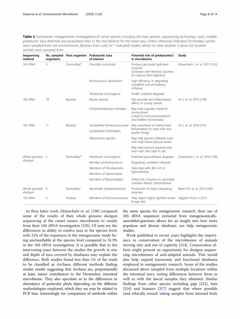
for intestinal microbiota which provide maximum re-source extraction from the harsh environment in whichthey live, with limited and typically static diets - a mutu-ally beneficial arrangement developing between thecamels and these microorganisms. It is interesting tonote the similarities across these studies, along with theolder Dromedary forestomach microbial investigation byBhatt et. al. [200] as regards the most abundant phyladetected. Salgado-Flores et. al. published the first meta-genomic study of the Muskoxen rumen in 2016, utilising16S rRNA techniques [201]. Bacteroidetes and Firmi-cutes were the dominant phyla, as with many othersequenced intestinal microbiomes, however they didnote that 53.7–59.3% of bacterial sequences couldn’t becharacterised. Also, the ratio between relative abundanceof Firmicutes and Bacteroidetes was greater as comparedto other ruminant intestinal microbiomes, 70.7–81.1%:16.8–25.3%. This may be an indication that the environ-ment, diet or a combination of the two in their aridhabitat favours members of Firmicutes more generallythan Bacteroidetes. However, reporting the most abun-dant phyla is not necessarily the most useful finding toexplore given that research has found the same phylapredominant in the intestinal microbiota of humans[165], pigs [155], baleen whales [166] and in fact (at dif-ferent relative positions) in the soil of the AtacamaDesert [202]. In their later work Gharechahi & Salekdehused shotgun sequencing methods to investigate func-tional traits of the camel intestinal microbiome. Theauthors determined that despite taxonomic similaritiesto a number of published rumen intestinal microbiomes
it was functionally distinct from them and more akin tothe Moose rumen microbiome in certain regards [198].Table 2. shows the Bactrian and Dromedary camel meta-genomic studies cited in this review including potentiallinks between relatively abundant or notable taxa andtheir roles. Both Camel species and Muskox share somenotable trends, with members of Lachnospiraceae andPrevotellaceae taking up a sizeable proportion of classi-fied bacterial reads. This is likely to be due to similaritiesin diet courtesy of similar traits in plants needed tosurvive in both cold and hot arid environments.
Considerations when studying arid microbiomesThe majority of studies discussed employ 16S rRNA se-quencing methods, although a few have also utilised shot-gun metagenomics. Many authors have used 16S rRNAapproaches due to cost and availability of accessible tools.However 16S rRNA only allows genus level resolution anddoes not give an indication of functional potential. This isa particular issue when studying highly adapted hosts orenvironments where it is expected that evolutionary adap-tation is at (or predicted to be at) the species/strain andfunctional level, rather than at genus level or higher. Thisreflects a trend in which many arid environments or arid-adapted animal microbiomes are sequenced with 16SrRNA and these results published with the caveat that anyfunctional data they present is by necessity derived fromthe taxonomies they have generated. Frequent use is madeof QIIME [203], mothur [204] and PICRUSt [205] - alongwith 16S rRNA databases such as Greengenes [206, 207]and Silva [208] to highlight what might be expected to bepresent in functional terms in the microbiome. Though16S rRNA sequencing currently has advantages in termsof cost and (comparative) ease of use, the limitation totaxonomy-derived functional predictions can be an issue iffunctional diversity differs significantly from taxonomicmeasures. Shotgun sequencing enables the direct assess-ment and investigation of functional diversity within themicrobial communities of arid environments, plants andanimals.Deeper sequencing power can create issues when
studying novel hosts, as there may be a high proportionof novel microbial taxa present i.e. a high proportion ofunidentified reads. This reflects the relative focus ofanimal-associated metagenomic studies on humans[209], mice [210] and others used as models for medicalresearch [211]. Unless attempting de novo classificationmethods, taxonomy in metagenomics depends on data-bases of known (or likely) classifications against whichreads from samples can be compared [212]. These arepopulated by researchers engaging in metagenomic andmicrobiological studies, thus trend towards easily culti-vated or human-associated; though published datasetscan be investigated for new genomes [213].
Osborne et al. Environmental Microbiome (2020) 15:20 Page 7 of 14
In their latter work, Gharechahi et. al. [198] comparedsome of the results of their whole genome shotgunsequencing of the camel rumen microbiome to resultsfrom their 16S rRNA investigation [195]. Of note are thedifferences in ability to resolve taxa to the species level,with 21% of the sequences in the metagenomic study be-ing unclassifiable at the species level compared to 51.9%in the 16S rRNA investigation. It is possible that in theintervening years between the studies the growth in sizeand depth of taxa covered by databases may explain thedifference. Both studies found less than 1% of the readsto be classified as Archaea, different methods findingsimilar results suggesting that Archaea are, proportionallyat least, minor contributors to the Dromedary intestinalmicrobiome. They also speculate as to the differences inabundance of particular phyla depending on the differentmethodologies employed, which they say may be related toPCR bias. Interestingly for comparison of methods within
the same species for metagenomic research, their use of16S rRNA sequences extracted from metagenomically-assembled-genomes allows for an insight into how morepopulous and diverse databases can help metagenomicstudies.Work published in recent years highlights the import-
ance in conservation of the microbiomes of animalsmoving into and out of captivity [214]. Conservation ef-forts might present an opportunity for shotgun sequen-cing microbiomes of arid-adapted animals. This wouldalso help expand taxonomic and functional databasesemployed in metagenomic research. Some of the studiesdiscussed above sampled from multiple locations withinthe intestinal tract, noting differences between them aswell as with the faecal samples they obtained. Similarfindings from other species including pigs [215], bats[216] and humans [217] suggest that where possible(and ethically sound) taking samples from internal body
Table 2 Summarises metagenomic investigations of camel species including the host species, sequencing technology used, notableprokaryotic taxa detected and postulated roles in the microbiome for the listed taxa. Unless otherwise indicated Dromedary camelswere sampled from hot environments, Bactrian from cold; for a indicated studies where no clear location is given for locationanimals were sampled from
Sequencingmethod
No. sampledorganisms
Host organism Prokaryotic taxaof interest
Potential role of prokaryote(s)in microbiome
Study
16S rRNA 3 Dromedarya Prevotella ruminicola Produce glycoside hydrolyseenzymesSynergise with fibrolytic bacteriato improve fibre digestion
Gharechahi J. et. al. 2015 [195]
Ruminococcus flavefaciens High efficiency in degradingcrystalline and amorphouscellulose
Fibrobacter succinogenes Prolific cellulose degrader
16S rRNA 18 Bactrian Blautia species May provide anti-inflammatoryeffects in young camels
He J. et. al. 2019 [199]
Christensenellaceae members May help regulate intestinalenvironmentLinked to immunomodulationand healthy homeostasis
16S rRNA 11 Bactrian Unclassified Ruminococcaceae May contribute to further feedfermentation to cope with lowquality forage
He J. et. al. 2018 [197]
Unclassified Clostridiales
Akkermansia species May help prevent diabetes evenwith high blood glucose levels
May help prevent hypertensioneven with diet high in salt
Whole genomeshotgun
3 Dromedarya Fibrobacter succinogenes Potential lignocellulose degrader Gharechahi J. et. al. 2018 [198]
Member of Ruminococcus Degrading crystalline cellulose
Members of Fibrobacteres Help deal with diet rich inlignocellulose
Members of Spirochaetes
Members of Bacteroidetes Utilise PUL enzymes to assimilatecomplex dietary carbohydrates
Whole genomeshotgun
6 Dromedary Bacteroides thetaiotaomicron Production of starch degradingenzymes
Bhatt V.D. et. al. 2013 [200]
16S rRNA 3 Muskox Members of Ruminococcaceae Help digest highly lignified winterforage diet
Salgado-Flores A [201].
Osborne et al. Environmental Microbiome (2020) 15:20 Page 8 of 14
sites is necessary to fully understand arid-adapted animalmicrobiomes. Meng et. al. investigated intestinal micro-biota of hot desert-dwelling weevils feeding on plantroots underground, finding that all of the 66 core weevilOTUs could be detected in the soil around the weevils;albeit at lower abundances [218]. This suggests that en-vironmental sampling around any animals investigatedmight help us understand the origins and developmentof the microbiome. Where feasible it would be beneficialto obtain samples of the environmental microbiome tosee what, if any relationship it has with the animal-associated microbial community. This would allow us todistinguish between transient microorganisms in the or-ganism, environmental contamination or true residents.Ideally comparison across closely related species shouldbe employed, as by Campbell et. al. (though not witharid-adapted organisms) [219], which will allow forbetter understanding of the interactions between hostgenome, environment and microbiome in studies ofadaptation.
Perspective and conclusionInstead of lifeless wastes, arid environments are home toorganisms from microscopic to enormous [220, 221].Their living conditions are harsh, but still life is able tosurvive and thrive. Though diversity drops off as theirhome becomes more hostile, organisms have been dis-covered in the depths of Antarctica [222], the Atacama[28] and even in artificially desiccated environments[223]. Adaptations to aridity have been noted in environ-mental microbiomes as conferring survival advantagesagainst other stresses which co-occur in those environ-ments. As such it is likely that similarities exist betweenarid microbiomes and those found in hyper-saline,extremely cold, UV irradiated and hot microbiomes;though as moisture levels increase these similaritieslikely diminish. Within this context it is worth consider-ing the extent to which animals in arid environmentsprovide more hospitable refuges for environmental mi-croorganisms, if potentially they allow for organismspresent in small fractions externally to colonise, be fruit-ful and multiply. Future investigations comparing arid-animal microbiomes to those of their surroundings willneed to take account of the impact of faeces and otherexcretions from the animals which may alter the micro-bial community in the environment around them [224].Going forward it is worth considering whether seeminglydivergent environmental conditions may in fact containa similar stress which could impact microbial communi-ties. When investigating environmental microbiomes itcould be of benefit (if possible) to take detailed measure-ments of abiotic stressors and assess whether these maybe directly - or through interactions with each other -responsible for community compositions. This may lead
to discoveries of shared taxonomic or functional trendsfrom distant and apparently dissimilar locations. It maybe useful in addition to sample harsh environmentswhich are not the most extreme, sampling a range ofwarm pools rather than the hottest spring water for in-stance; or taking transects across a harsh environmentas opposed to multiple samplings from the saltiest ormost irradiated sites.If conducting research on arid animal microbiomes in
the future it may be helpful to take environmentalsamples from sites where the animals feed, rest and other-wise spend their time. This could allow the determinationof the extent to which composition of animal-associatedmicrobiomes is purely a consequence of allowing minormembers of the wider environmental microbiome tothrive. It may also be interesting to observe in captive ani-mals if differences in diet and microbiome come from thealtered diet they consume in captivity or from the differentenvironmental microbiome in which they and their foodare kept. Both traditional and novel methods of culturingwill have a place in future studies of arid animal and envir-onmental microbiomes, as harsh conditions can be moreeasily and accurately replicated in the laboratory; enablingthe conversion of detected genotypes into observablephenotypes. Functional understanding of arid animal-associated microbiomes may require the use of in vivomodels to be completely elucidated; possibly leading tofindings which can be translated into industrial applica-tions. In their recent discussion of the ‘Eco-holobiont’Singh et. al. highlight how environmental factors, environ-mental microbiomes, animal genomes and animal-associated microbiota may interact to paint a completepicture of the micro-macroscopic relationships shapingthe world [225]. Ribeiro et. al. demonstrate that metage-nomic investigation can be a crucial component ofwell-rounded research into the life and adaptation of anarid-adapted animal, along with environmental, metabolicand genomic investigations [32]. Arid environments couldoffer a very useful proof of concept for this philosophicalapproach, with clear environmental influences and com-paratively simple living communities. As climate changethreatens arid environments and their inhabitants aroundthe globe it is increasingly important that we fully under-stand the functional and taxonomic composition of themicrobial communities they host so we can best protectand harness them [226].
AbbreviationsUV: Ultra Violet Light; DNA: Deoxyribonucleic Acid; CAZy: Carbohydrate-Active enZYmes; QIIME: Quantitative Insights Into Microbial Ecology;PICRUSt: Phylogenetic Investigation of Communities by Reconstruction ofUnobserved States; PCR: Polymerase Chain Reaction; OTU: OperationalTaxonomic Unit
AcknowledgementsNot applicable.
Osborne et al. Environmental Microbiome (2020) 15:20 Page 9 of 14
Authors’ contributionsPO conducted the review, prepared and wrote the manuscript. LH, WH andDT were responsible for the conception of the review, along with PO. NK-S,LH and WH provided considerable revisions to the work during writing. Allauthors read and approved the final manuscript.
FundingLH is funded by a Wellcome Trust Investigator Award (no. 100/974/C/13/Z)and an Institute Strategic Programme Gut Microbes and Health grant no. BB/R012490/1 and its constituent projects BBS/E/F/000PR10353 and BBS/E/F/000PR10356. DT and WH were supported by the BBSRC, Institute StrategicProgramme Grant [BB/J004669/1], BBSRC Core Strategic Programme Grant[BB/P016774/1]. PO is funded by the BBSRC as part of the Doctoral TrainingPartnership programme, code: CT471J03B.
Availability of data and materialsData sharing not applicable to this article as no datasets were generated oranalysed during the current study.
Ethics approval and consent to participateNot applicable.
Consent for publicationNot applicable.
Competing interestsThe authors declare that they have no competing interests.
Author details1Earlham Institute, Norwich Research Park Innovation Centre, Colney Lane,Norwich NR4 7UZ, UK. 2Gut Microbes & Health, Quadram Institute Bioscience,Norwich Research Park, Norwich NR4 7UQ, UK. 3Chair of IntestinalMicrobiome, School of Life Sciences, ZIEL – Institute for Food & Health,Technical University of Munich, 85354 Freising, Germany. 4School of Zoology,Tel Aviv University, Tel Aviv-Yafo, Israel. 5EMBL-EBI, Wellcome GenomeCampus, Hinxton, Cambridgeshire CB10 1SD, UK.
Received: 7 June 2020 Accepted: 12 November 2020
References1. Häussinger D. The role of cellular hydration in the regulation of cell
function. Biochem J. 1996;313:697–710.2. Barrow CJ. World atlas of desertification. Land Degradation Dev. 1992;3:249.3. Salem BB. Arid zone forestry: a guide for field technicians. In: FAO
Conservation Guide. Rome, Italy: FAO; 1989.4. Boetius A, Anesio AM, Deming JW, Mikucki JA, Rapp JZ. Microbial ecology
of the cryosphere: sea ice and glacial habitats. Nat Rev Microbiol. 2015;13:677–90.
5. Buyanovsky G, Dicke M, Berwick P. Soil environment and activity of soilmicroflora in the Negev desert. J Arid Environ. 1982;5:13–28.
6. Blix AS. Adaptations to polar life in mammals and birds. J Exp Biol. 2016;219:1093–105.
7. Ishida Y, Van Coeverden de Groot PJ, Leggett KEA, Putnam AS, Fox VE, Lai J,et al. Genetic connectivity across marginal habitats: the elephants of theNamib Desert. Ecol Evol. 2016;6:6189–201.
8. Taylor NP, Zappi DC. An alternative view of generic delimitation andrelationships in tribe Cereeae (Cactaceae). brad. 1989;1989:13–40.
9. Levy O, Dayan T, Porter WP, Kronfeld-Schor N. Time and ecologicalresilience: can diurnal animals compensate for climate change by shifting tonocturnal activity? Ecol Monogr. 2019;89:e01334.
10. Kronfeld N, Shkolnik A. Adaptation to Life in the Desert in the Brown Hare(Lepus capensis). J Mammal. 1996;77:171–8.
11. Silanikove N. The physiological basis of adaptation in goats to harshenvironments. Small Rumin Res. 2000;35:181–93.
12. Levy O, Dayan T, Porter WP, Kronfeld-Schor N. Foraging Activity Pattern IsShaped by Water Loss Rates in a Diurnal Desert Rodent. Am Nat. 2016;188:205–18.
13. Schmidt-Nielsen K. Desert Rodents: Physiological Problems of Desert Life. In:Prakash I, Ghosh PK, editors. Rodents in Desert Environments. Dordrecht:Springer Netherlands; 1975. p. 379–88.
14. Wen X, Wang S, Duman JG, Arifin JF, Juwita V, Goddard WA, et al. Antifreezeproteins govern the precipitation of trehalose in a freezing-avoiding insectat low temperature. PNAS. 2016;113:6683–8.
15. Hazard LC, Lechuga C, Zilinskis S. Secretion by the nasal salt glands of twoinsectivorous lizard species is initiated by an ecologically relevant dietaryion, chloride. J Exp Zool A Ecol Genet Physiol. 2010;313A:442–51.
16. Leinaas HP. UV Tolerance, Pigmentation and Life Forms in High ArcticCollembola. In: Hessen DO, editor. UV Radiation and Arctic Ecosystems.Berlin, Heidelberg: Springer; 2002. p. 123–34.
17. Osakabe Y, Osakabe K, Shinozaki K, Tran L-SP. Response of plants to waterstress. Front Plant Sci. 2014;5. https://doi.org/10.3389/fpls.2014.00086.
18. Nadeem M, Li J, Yahya M, Sher A, Ma C, Wang X, et al. Research Progressand Perspective on Drought Stress in Legumes: A Review. Int J Mol Sci.2019;20:2541.
19. Kay RNB. Responses of African livestock and wild herbivores to drought. JArid Environ. 1997;37:683–94.
20. Schnorr SL, Sankaranarayanan K, Lewis CM, Warinner C. Insights into humanevolution from ancient and contemporary microbiome studies. Curr OpinGenet Dev. 2016;41:14–26.
21. McDonald RC, Watts JEM, Schreier HJ. Effect of Diet on the EntericMicrobiome of the Wood-Eating Catfish Panaque nigrolineatus. FrontMicrobiol. 2019;10. https://doi.org/10.3389/fmicb.2019.02687.
22. Lu J, Synowiec S, Lu L, Yu YY, Bretherick T, Takada S, et al. Microbiotainfluence the development of the brain and behaviors in C57BL/6 J mice.PLoS One. 2018;13:29.
23. Kassas M. Desertification: a general review. J Arid Environ. 1995;30:115–28.24. Spinoni J, Vogt J, Naumann G, Carrao H, Barbosa P. Towards identifying
areas at climatological risk of desertification using the Köppen–Geigerclassification and FAO aridity index. Int J Climatol. 2015;35:2210–22.
25. Spinoni J, Micale F, Carrao H, Naumann G, Barbosa P, Vogt J. Global andcontinental changes of arid areas using the FAO Aridity Index over theperiods 1951–1980 and 1981–2010. Geophysical Research Abstracts; 2013.
26. Lal R. 4.10 - Vulnerability of Agroecosystems to Environmental Factors.In: Pielke RA (1st ed). Climate Vulnerability. Academic Press: Oxford,2013:109–116.
27. Pretty JN, Morison JIL, Hine RE. Reducing food poverty by increasingagricultural sustainability in developing countries. Agric Ecosyst Environ.2003;95:217–34.
28. Crits-Christoph A, Robinson CK, Barnum T, Fricke WF, Davila AF, Jedynak B,et al. Colonization patterns of soil microbial communities in the AtacamaDesert. Microbiome. 2013;1:28.
29. Neilson JW, Califf K, Cardona C, Copeland A, van Treuren W, Josephson KL,et al. Significant Impacts of Increasing Aridity on the Arid Soil Microbiome.mSystems. 2017;2:e00195–16 /msys/2/3/e00195–16.atom.
30. Mohammadipanah F, Wink J. Actinobacteria from Arid and Desert Habitats:Diversity and Biological Activity. Front Microbiol. 2016;6. https://doi.org/10.3389/fmicb.2015.01541.
31. Schmidt SK, Gendron EMS, Vincent K, Solon AJ, Sommers P, Schubert ZR,et al. Life at extreme elevations on Atacama volcanoes: the closest thing toMars on Earth? Antonie Van Leeuwenhoek. 2018;111:1389–401.
32. Ribeiro ÂM, Puetz L, Pattinson NB, Dalén L, Deng Y, Zhang G, et al. 31°South: The physiology of adaptation to arid conditions in a passerine bird.Mol Ecol. 2019;28:3709–21.
33. Bryan NC, Christner BC, Guzik TG, Granger DJ, Stewart MF. Abundance andsurvival of microbial aerosols in the troposphere and stratosphere. ISME J.2019;13:2789–99.
34. Ogwu MC, Srinivasan S, Dong K, Ramasamy D, Waldman B, Adams JM.Community Ecology of Deinococcus in Irradiated Soil. Microb Ecol. 2019;78:855–72.
35. Gorbushina AA. Life on the rocks. Environ Microbiol. 2007;9:1613–31.36. Coller E, Cestaro A, Zanzotti R, Bertoldi D, Pindo M, Larger S, et al.
Microbiome of vineyard soils is shaped by geography and management.Microbiome. 2019;7:140.
37. Lauber CL, Ramirez KS, Aanderud Z, Lennon J, Fierer N. Temporal variabilityin soil microbial communities across land-use types. Isme J. 2013;7:1641–50.
38. Overmann J, Abt B, Sikorski J. Present and Future of Culturing Bacteria.Annu Rev Microbiol. 2017;71:711–30.
39. Kaeberlein T, Lewis K, Epstein SS. Isolating ‘Uncultivable’ Microorganisms inPure Culture in a Simulated Natural Environment. Science. 2002;296:1127–9.
40. Chaudhary DK, Khulan A, Kim J. Development of a novel cultivationtechnique for uncultured soil bacteria. Sci Rep. 2019;9:1–11.
Osborne et al. Environmental Microbiome (2020) 15:20 Page 10 of 14
41. Browne HP, Forster SC, Anonye BO, Kumar N, Neville BA, Stares MD, et al.Culturing of ‘unculturable’ human microbiota reveals novel taxa andextensive sporulation. Nature. 2016;533:543–6.
42. Böttger EC. Rapid determination of bacterial ribosomal RNA sequences bydirect sequencing of enzymatically amplified DNA. FEMS Microbiol Lett.1989;53:171–6.
43. Venter JC, Remington K, Heidelberg JF, Halpern AL, Rusch D, Eisen JA, et al.Environmental Genome Shotgun Sequencing of the Sargasso Sea. Science.2004;304:66–74.
44. Murgia M, Fiamma M, Barac A, Deligios M, Mazzarello V, Paglietti B, et al.Biodiversity of fungi in hot desert sands. MicrobiologyOpen. 2019;8:e00595.
45. Ji P, Rhoads WJ, Edwards MA, Pruden A. Impact of water heatertemperature setting and water use frequency on the building plumbingmicrobiome. ISME J. 2017;11:1318–30.
46. Cole JK, Peacock JP, Dodsworth JA, Williams AJ, Thompson DB, Dong H,et al. Sediment microbial communities in Great Boiling Spring arecontrolled by temperature and distinct from water communities. ISME J.2013;7:718–29.
47. Barns SM, Fundyga RE, Jeffries MW, Pace NR. Remarkable archaeal diversitydetected in a Yellowstone National Park hot spring environment. Proc NatlAcad Sci U S A. 1994;91:1609–13.
48. Hugenholtz P, Pitulle C, Hershberger KL, Pace NR. Novel division levelbacterial diversity in a Yellowstone hot spring. J Bacteriol. 1998;180:366–76.
49. Ballaré CL, Caldwell MM, Flint SD, Robinson SA, Bornman JF. Effects of solarultraviolet radiation on terrestrial ecosystems. Patterns, mechanisms, andinteractions with climate change. Photochem Photobiol Sci. 2011;10:226–41.
50. Zaller JG, Caldwell MM, Flint SD, Scopel AL, Salo OE, Ballaré CL. Solar UV-Bradiation affects below-ground parameters in a fen ecosystem in Tierra delFuego, Argentina: implications of stratospheric ozone depletion. GlobChang Biol. 2002;8:867–71.
51. Mukhtar S, Mirza BS, Mehnaz S, Mirza MS, Mclean J, Malik KA. Impact of soilsalinity on the microbial structure of halophyte rhizosphere microbiome.World J Microbiol Biotechnol. 2018;34:136.
52. Zhang Y, Cao C, Guo L, Wu Q, Cui Z. Soil properties, bacterialcommunity composition, and metabolic diversity responses to soilsalinization of a semiarid grassland in northeast China. J Soil WaterConserv. 2015;70:110–20.
53. Weinmaier T, Probst AJ, La Duc MT, Ciobanu D, Cheng J-F, Ivanova N, et al.A viability-linked metagenomic analysis of cleanroom environments:eukarya, prokaryotes, and viruses. Microbiome. 2015;3:62.
54. Jiménez DJ, Andreote FD, Chaves D, Montaña JS, Osorio-Forero C, Junca H,et al. Structural and Functional Insights from the Metagenome of an AcidicHot Spring Microbial Planktonic Community in the Colombian Andes. PLoSOne. 2012;7. https://doi.org/10.1371/journal.pone.0052069.
55. Ogwu MC, Kerfahi D, Song H, Dong K, Seo H, Lim S, et al. Changes in soiltaxonomic and functional diversity resulting from gamma irradiation. SciRep. 2019;9:1–13.
56. Kato C, Li L, Nogi Y, Nakamura Y, Tamaoka J, Horikoshi K. ExtremelyBarophilic Bacteria Isolated from the Mariana Trench, Challenger Deep, at aDepth of 11,000 Meters. Appl Environ Microbiol. 1998;64:1510–3.
57. Li Y, Zheng L, Zhang Y, Liu H, Jing H. Comparative metagenomics studyreveals pollution induced changes of microbial genes in mangrovesediments. Sci Rep. 2019;9:1–11.
58. Noll M, Matthies D, Frenzel P, Derakshani M, Liesack W. Succession ofbacterial community structure and diversity in a paddy soil oxygengradient. Environ Microbiol. 2005;7:382–95.
59. Packer E, Scher S, Sagan C. Biological contamination of Mars II. Cold andaridity as constraints on the survival of terrestial microorganisms insimulated Martian environments. Icarus. 1963;2:293–316.
60. Song H-K, Shi Y, Yang T, Chu H, He J-S, Kim H, et al. Environmentalfiltering of bacterial functional diversity along an aridity gradient. SciRep. 2019;9:1–10.
61. Maestre FT, Delgado-Baquerizo M, Jeffries TC, Eldridge DJ, Ochoa V, GozaloB, et al. Increasing aridity reduces soil microbial diversity and abundance inglobal drylands. PNAS. 2015;112:15684–15,689.
62. Zeng Q, An S, Liu Y, Wang H, Wang Y. Biogeography and the drivingfactors affecting forest soil bacteria in an arid area. Sci Total Environ.2019;680:124–31.
63. Feng W, Zhang Y, Yan R, Lai Z, Qin S, Sun Y, et al. Dominant soil bacteriaand their ecological attributes across the deserts in northern China. Eur JSoil Sci. 2020;71:524–35.
64. Chen D, Saleem M, Cheng J, Mi J, Chu P, Tuvshintogtokh I, et al. Effects ofaridity on soil microbial communities and functions across soil depths onthe Mongolian Plateau. Funct Ecol. 2019;33:1561–71.
65. Fierer N, Leff JW, Adams BJ, Nielsen UN, Bates ST, Lauber CL, et al. Cross-biome metagenomic analyses of soil microbial communities and theirfunctional attributes. PNAS. 2012;109:21390–21,395.
66. Le PT, Makhalanyane TP, Guerrero LD, Vikram S, Van de Peer Y, Cowan DA.Comparative Metagenomic Analysis Reveals Mechanisms for StressResponse in Hypoliths from Extreme Hyperarid Deserts. Genome Biol Evol.2016;8:2737–47.
67. Afshinnekoo E, Meydan C, Chowdhury S, Jaroudi D, Boyer C, Bernstein N,et al. Geospatial Resolution of Human and Bacterial Diversity with City-ScaleMetagenomics. Cell Systems. 2015;1:72–87.
68. Anderson KL, Apolinario EE, Sowers KR. Desiccation as a Long-Term SurvivalMechanism for the Archaeon Methanosarcina barkeri. Appl EnvironMicrobiol. 2012;78:1473–9.
69. Antonelli F, Esposito A, Calvo L, Licursi V, Tisseyre P, Ricci S, et al.Characterization of black patina from the Tiber River embankments usingNext-Generation Sequencing. PLoS One. 2020;15:e0227639.
70. De Leo F, Antonelli F, Pietrini AM, Ricci S, Urzì C. Study of the euendolithicactivity of blackmeristematic fungi isolated from a marble statue in theQuirinale Palace’s Gardens inRome, Italy. Facies. 2019;65:18.
71. Goberna M, Pascual JA, García C, Sánchez J. Do plant clumps constitutemicrobial hotspots in semiarid Mediterranean patchy landscapes? Soil BiolBiochem. 2007;39:1047–54.
72. Aguilera LE, Armas C, Cea AP, Gutiérrez JR, Meserve PL, Kelt DA. Rainfall,microhabitat, and small mammals influence the abundance and distributionof soil microorganisms in a Chilean semi-arid shrubland. J Arid Environ.2016;126:37–46.
73. Grinberg M, Orevi T, Steinberg S, Kashtan N. Bacterial survival in microscopicsurface wetness. eLife. 8. https://doi.org/10.7554/eLife.48508.
74. Placella SA, Brodie EL, Firestone MK. Rainfall-induced carbon dioxide pulsesresult from sequential resuscitation of phylogenetically clustered microbialgroups. PNAS. 2012;109:10931–10,936.
75. Scambos TA, Campbell GG, Pope A, Haran T, Muto A, Lazzara M, et al.Ultralow Surface Temperatures in East Antarctica From Satellite ThermalInfrared Mapping: The Coldest Places on Earth. Geophys Res Lett. 2018;45:6124–33.
76. Kanokratana P, Chanapan S, Pootanakit K, Eurwilaichitr L. Diversity andabundance of Bacteria and Archaea in the Bor Khlueng Hot Spring inThailand. J Basic Microbiol. 2004;44:430–44.
77. Wilkins LGE, Ettinger CL, Jospin G, Eisen JA. Metagenome-assembledgenomes provide new insight into the microbial diversity of two thermalpools in Kamchatka, Russia. Sci Rep. 2019;9:15.
78. Inskeep WP, Rusch DB, Jay ZJ, Herrgard MJ, Kozubal MA, Richardson TH,et al. Metagenomes from High-Temperature Chemotrophic Systems RevealGeochemical Controls on Microbial Community Structure and Function.PLoS One. 2010;5:e9773.
79. Dai D, Rhoads WJ, Edwards MA, Pruden A. Shotgun Metagenomics RevealsTaxonomic and Functional Shifts in Hot Water Microbiome Due toTemperature Setting and Stagnation. Front Microbiol. 2018;9. https://doi.org/10.3389/fmicb.2018.02695.
80. Chan CS, Chan K-G, Tay Y-L, Chua Y-H, Goh KM. Diversity of thermophiles ina Malaysian hot spring determined using 16S rRNA and shotgunmetagenome sequencing. Front Microbiol. 2015;6. https://doi.org/10.3389/fmicb.2015.00177.
81. Sharp CE, Brady AL, Sharp GH, Grasby SE, Stott MB, Dunfield PF. Humboldt’sspa: microbial diversity is controlled by temperature in geothermalenvironments. ISME J. 2014;8:1166–74.
82. Armstrong A, Valverde A, Ramond J-B, Makhalanyane TP, Jansson JK,Hopkins DW, et al. Temporal dynamics of hot desert microbialcommunities reveal structural and functional responses to water input.Sci Rep. 2016;6:1–8.
83. Noronha MF, Lacerda Júnior GV, Gilbert JA, de Oliveira VM. Taxonomic andfunctional patterns across soil microbial communities of global biomes. SciTotal Environ. 2017;609:1064–74.
84. Soo RM, Wood SA, Grzymski JJ, McDonald IR, Cary SC. Microbial biodiversityof thermophilic communities in hot mineral soils of Tramway Ridge, MountErebus, Antarctica. Environ Microbiol. 2009;11:715–28.
85. Cockell CS, Harrison JP, Stevens AH, Payler SJ, Hughes SS, Kobs NawotniakSE, et al. A Low-Diversity Microbiota Inhabits Extreme Terrestrial Basaltic
Osborne et al. Environmental Microbiome (2020) 15:20 Page 11 of 14
Terrains and Their Fumaroles: Implications for the Exploration of Mars.Astrobiology. 2019;19:284–99.
86. Filippidou S, Wunderlin T, Junier T, Jeanneret N, Dorador C, Molina V, et al.A Combination of Extreme Environmental Conditions Favor the Prevalenceof Endospore-Forming Firmicutes. Front Microbiol. 2016;7. https://doi.org/10.3389/fmicb.2016.01707.
87. Savage AM, Hills J, Driscoll K, Fergus DJ, Grunden AM, Dunn RR. Microbialdiversity of extreme habitats in human homes. PeerJ. 2016;4. https://doi.org/10.7717/peerj.2376.
88. Varin T, Lovejoy C, Jungblut AD, Vincent WF, Corbeil J. MetagenomicAnalysis of Stress Genes in Microbial Mat Communities from Antarctica andthe High Arctic. Appl Environ Microbiol. 2012;78:549–59.
89. Muñoz PA, Márquez SL, González-Nilo FD, Márquez-Miranda V, BlameyJM. Structure and application of antifreeze proteins from Antarcticbacteria. Microb Cell Factories. 2017;16. https://doi.org/10.1186/s12934-017-0737-2.
90. Liljeqvist M, Ossandon FJ, González C, Rajan S, Stell A, Valdes J, et al.Metagenomic analysis reveals adaptations to a cold-adapted lifestyle in alow-temperature acid mine drainage stream. FEMS Microbiol Ecol. 2015;91.https://doi.org/10.1093/femsec/fiv011.
91. Hayes MA. The Geomyces Fungi: Ecology and Distribution. BioScience. 2012;62:819–23.
92. Su Y, Jiang X, Wu W, Wang M, Hamid MI, Xiang M, et al. Genomic,Transcriptomic, and Proteomic Analysis Provide Insights Into the ColdAdaptation Mechanism of the Obligate Psychrophilic Fungus Mrakiapsychrophila. G3: Genes, Genomes. Genetics. 2016;6:3603–13.
93. Castenholz RW, Garcia-Pichel F. Cyanobacterial Responses to UV Radiation.In: Whitton BA, editor. Ecology of Cyanobacteria II: Their Diversity in Spaceand Time. Dordrecht: Springer Netherlands; 2012. p. 481–99.
94. Worrest RC, Háder D-P. Overview of the Effects of Increased Solar UV onAquatic Microorganisms. Photochem Photobiol. 1997;65:257–9.
95. van de Water JAJM, Courtial L, Houlbrèque F, Jacquet S, Ferrier-Pagès C.Ultra-Violet Radiation Has a Limited Impact on Seasonal Differences in theAcropora Muricata Holobiont. Front Mar Sci. 2018;5. https://doi.org/10.3389/fmars.2018.00275.
96. Hansen AA, Merrison J, Nørnberg P, Lomstein BA, Finster K. Activity andstability of a complex bacterial soil community under simulated Martianconditions. Int J Astrobiol. 2005;4:135–44.
97. Fahimipour AK, Hartmann EM, Siemens A, Kline J, Levin DA, Wilson H, et al.Daylight exposure modulates bacterial communities associated withhousehold dust. Microbiome. 2018;6:175.
98. Sedláček I, Pantůček R, Králová S, Mašlaňová I, Holochová P, Staňková E,et al. Hymenobacter amundsenii sp. nov. resistant to ultraviolet radiation,isolated from regoliths in Antarctica. Syst Appl Microbiol. 2019;42:284–90.
99. Dai J, Wang Y, Zhang L, Tang Y, Luo X, An H, et al. Hymenobacter tibetensissp. nov., a UV-resistant bacterium isolated from Qinghai–Tibet plateau. SystAppl Microbiol. 2009;32:543–8.
100. Dorado-Morales P, Vilanova C, Peretó J, Codoñer FM, Ramón D, Porcar M. Ahighly diverse, desert-like microbial biocenosis on solar panels in aMediterranean city. Sci Rep. 2016;6:1–9.
101. Porcar M, Louie KB, Kosina SM, Van Goethem MW, Bowen BP, Tanner K,et al. Microbial Ecology on Solar Panels in Berkeley, CA, United States. FrontMicrobiol. 2018;9. https://doi.org/10.3389/fmicb.2018.03043.
102. Tanner K, Martí JM, Belliure J, Fernández-Méndez M, Molina-Menor E, PeretóJ, et al. Polar solar panels: Arctic and Antarctic microbiomes display similartaxonomic profiles. Environ Microbiol Rep. 2018;10:75–9.
103. Portero LR, Alonso-Reyes DG, Zannier F, Vazquez MP, Farías ME, Gärtner W,et al. Photolyases and Cryptochromes in UV-resistant Bacteria from High-altitude Andean Lakes. Photochem Photobiol. 2019;95:315–30.
104. Albarracín VH, Pathak GP, Douki T, Cadet J, Borsarelli CD, Gärtner W, et al.Extremophilic Acinetobacter Strains from High-Altitude Lakes inArgentinean Puna: Remarkable UV-B Resistance and Efficient DNA DamageRepair. Orig Life Evol Biosph. 2012;42:201–21.
105. Kurth D, Belfiore C, Gorriti MF, Cortez N, Farias ME, Albarracín VH. Genomicand proteomic evidences unravel the UV-resistome of the poly-extremophile Acinetobacter sp. Ver3. Front Microbiol. 2015;6. https://doi.org/10.3389/fmicb.2015.00328.
106. Pacelli C, Bryan RA, Onofri S, Selbmann L, Zucconi L, Shuryak I, et al. Survivaland redox activity of Friedmanniomyces endolithicus, an Antarctic endemicblack meristematic fungus, after gamma rays exposure. Fungal Biol. 2018;122:1222–7.
107. Selbmann L, Isola D, Zucconi L, Onofri S. Resistance to UV-B induced DNAdamage in extreme-tolerant cryptoendolithic Antarctic fungi: detection byPCR assays. Fungal Biol. 2011;115:937–44.
108. Jones DL, Baxter BK. DNA Repair and Photoprotection: Mechanisms ofOvercoming Environmental Ultraviolet Radiation Exposure in HalophilicArchaea. Front Microbiol. 2017;8. https://doi.org/10.3389/fmicb.2017.01882.
109. Raheja PC. Aridity and Salinity. In: Boyko H, editor. Salinity and Aridity:New Approaches to Old Problems. Dordrecht: Springer Netherlands;1966. p. 43–127.
110. Molina-Menor E, Tanner K, Vidal-Verdú À, Peretó J, Porcar M. Microbialcommunities of the Mediterranean rocky shore: ecology andbiotechnological potential of the sea-land transition. Microb Biotechnol.2019;12:1359–70.
111. Khomutova TE, Borisov AV. Estimation of microbial diversity in the desertsteppe surface soil and buried palaeosol (IV mil. BC) using the TRFLPmethod. J Arid Environ. 2019;171:104004.
112. Zajc J, Liu Y, Dai W, Yang Z, Hu J, Gostinčar C, et al. Genome andtranscriptome sequencing of the halophilic fungus Wallemia ichthyophaga:haloadaptations present and absent. BMC Genomics. 2013;14:617.
113. Pérez-Llano Y, Rodríguez-Pupo EC, Druzhinina IS, Chenthamara K, Cai F,Gunde-Cimerman N, et al. Stress Reshapes the Physiological Response ofHalophile Fungi to Salinity. Cells. 2020;9:525.
114. Degen AA. Energy requirements of the fat sand rat (Psammomys obesus)when consuming the saltbush, a triplex halimus: a review. J Basic ClinPhysiol Pharmacol. 1993;4:13–28.
115. Sato H, Ichino S, Hanya G. Dietary modification by common brown lemurs(Eulemur fulvus) during seasonal drought conditions in western Madagascar.Primates. 2014;55:219–30.
116. Randall JA. Behavioural adaptations of desert rodents (Heteromyidae). AnimBehav. 1993;45:263–87.
117. Schwimmer H, Haim A. Physiological adaptations of small mammals todesert ecosystems. Integrative Zool. 2009;4:357–66.
118. Kronfeld-Schor N, Dayan T. Thermal Ecology, Environments, Communities,and Global Change: Energy Intake and Expenditure in Endotherms. AnnuRev Ecol Evol Syst. 2013;44:461–80.
119. Macfarlane WV, Morris RJH, Howard B, McDonald J, Budtz-Olsen OE. Waterand electrolyte changes in tropical Merino sheep exposed to dehydrationduring summer. Aust J Agric Res. 1961;12:889–912.
120. Levy A, Perelman B, Grevenbroek MV, Creveld CV, Agbaria R, Yagil R. Effectof water restriction on renal function in ostriches (Struthio camelus). AvianPathol. 1990;19:385–93.
121. Shoemaker VH. The stimulus for the water-balance response to dehydrationin toads. Comp Biochem Physiol. 1965;15:81–8.
122. Coleman JC, Downs CT. Variation in urine concentrating ability and waterbalance of the Black-tailed Tree Rat Thallomys nigricauda, along an ariditygradient. Comp Biochem Physiol A Mol Integr Physiol. 2009;154:508–13.
123. Bassett JE. Habitat aridity and urine concentrating ability of nearctic,insectivorous bats. Comp Biochem Physiol A Comp Physiol. 1986;83:125–31.
124. Korine C, Vatnick I, van TIG, Pinshow B. New observations on urine contentsin water-deprived Negev Desert rodents. Can J Zool. 2003;81:941–5.
125. Sands JM, Layton HE. Advances in Understanding the Urine-ConcentratingMechanism. Annu Rev Physiol. 2014;76:387–409.
126. Diaz GB, Ojeda RA. Kidney structure and allometry of Argentine desertrodents. J Arid Environ. 1999;41:453–61.
127. Urity VB, Issaian T, Braun EJ, Dantzler WH, Pannabecker TL.Architecture of kangaroo rat inner medulla: segmentation ofdescending thin limb of Henle’s loop. Am J Phys Regul Integr CompPhys. 2011;302:R720–6.
128. Bozinovic F, Gallardo PA, Visser GH, Cortés A. Seasonal acclimatization inwater flux rate, urine osmolality and kidney water channels in free-livingdegus: molecular mechanisms, physiological processes and ecologicalimplications. J Exp Biol. 2003;206:2959–66.
129. Hoppe P, Kay RN, Maloiy GM. Proceedings: The rumen as a reservoir duringdehydration and rehydration in the camel. J Physiol Lond. 1976;254:76P–7P.
130. Letnic M, Webb JK, Jessop TS, Florance D, Dempster T. Artificial water pointsfacilitate the spread of an invasive vertebrate in arid Australia. J Appl Ecol.2014;51:795–803.
131. Dawson TJ, McTavish KJ, Munn AJ, Holloway J. Water use and thethermoregulatory behaviour of kangaroos in arid regions: insights into thecolonisation of arid rangelands in Australia by the Eastern Grey Kangaroo(Macropus giganteus). J Comp Physiol B. 2005;176:45.
Osborne et al. Environmental Microbiome (2020) 15:20 Page 12 of 14
132. Schmidt-Nielsen B, Schmidt-Nielsen K. A complete account of the watermetabolism in kangaroo rats and an experimental verification. J Cell CompPhysiol. 1951;38:165–81.
133. Lockard RB. Seasonal Change in the Activity Pattern of Dipodomysspectabilis. J Mammal. 1978;59:563–8.
134. Tieleman BI, Williams JB, Bloomer P. Adaptation of metabolism andevaporative water loss along an aridity gradient. Proc R Soc Lond Ser B BiolSci. 2003;270:207–14.
135. Schmidt-Nielsen B, Schmidt-Nielsen K. Pulmonary water loss in desertrodents. Am J Physiol Legacy Content. 1950;162:31–6.
136. Geiser F. The role of torpor in the life of Australian arid zone mammals.Austr Mammal. 2004:125–34.
137. Cooper CE, McAllan BM, Geiser F. Effect of torpor on the water economy ofan arid-zone marsupial, the stripe-faced dunnart (Sminthopsis macroura). JComp Physiol B. 2005;175:323–8.
138. Nowack J, Stawski C, Geiser F. More functions of torpor and their roles in achanging world. J Comp Physiol B. 2017;187:889–97.
139. Fouet C, Gray E, Besansky NJ, Costantini C. Adaptation to Aridity in theMalaria Mosquito Anopheles gambiae: Chromosomal InversionPolymorphism and Body Size Influence Resistance to Desiccation. PLoS One.2012;7:e34841.
140. Schmidt-Nielsen B, Schmidt-Nielsen K, Houpt TR, Jarnum SA. Water Balanceof the Camel. Am J Physiol Legacy Content. 1956;185:185–94.
141. Schmidt-Nielsen K. The physiology of the camel. Sci Am. 1959;201(6):140–51.142. Watanabe M. Anhydrobiosis in invertebrates. Appl Entomol Zool. 2006;41:
15–31.143. Somme L. Invertebrates in Hot and Cold Arid Environments. Berlin
Heidelberg: Springer-Verlag; 1995. https://doi.org/10.1007/978-3-642-79,583-1..
144. Davenport CF, Tümmler B. Advances in computational analysis ofmetagenome sequences. Environ Microbiol. 2013;15:1–5.
145. Kawalia A, Motameny S, Wonczak S, Thiele H, Nieroda L, Jabbari K, et al.Leveraging the Power of High Performance Computing for Next GenerationSequencing Data Analysis: Tricks and Twists from a High ThroughputExome Workflow. PLoS One. 2015;10:e0126321.
146. Heijtza RD, Wang SG, Anuar F, Qian Y, Bjorkholm B, Samuelsson A, et al.Normal gut microbiota modulates brain development and behavior. ProcNatl Acad Sci U S A. 2011;108:3047–52.
147. Stewart RD, Auffret MD, Warr A, Wiser AH, Press MO, Langford KW, et al.Assembly of 913 microbial genomes from metagenomic sequencing of thecow rumen. Nat Commun. 2018;9:11.
148. Falentin H, Rault L, Nicolas A, Bouchard DS, Lassalas J, Lamberton P, et al.Bovine Teat Microbiome Analysis Revealed Reduced Alpha Diversity andSignificant Changes in Taxonomic Profiles in Quarters with a History ofMastitis. Front Microbiol. 2016;7:14.
149. Ma C, Sun Z, Zeng B, Huang S, Zhao J, Zhang Y, et al. Cow-to-mouse fecaltransplantations suggest intestinal microbiome as one cause of mastitis.Microbiome. 2018;6. https://doi.org/10.1186/s40168-018-0578-1.
150. Wilkinson TJ, Huws SA, Edwards JE, Kingston-Smith AH, Siu-Ting K, HughesM, et al. CowPI: A Rumen Microbiome Focussed Version of the PICRUStFunctional Inference Software. Front Microbiol. 2018;9:10.
151. Dill-McFarland KA, Weimer PJ, Breaker JD, Suen G. Diet Influences EarlyMicrobiota Development in Dairy Calves without Long-Term Impacts onMilk Production. Appl Environ Microbiol. 2019;85. https://doi.org/10.1128/aem.02141-18.
152. Chen SY, Wang J, Peng DD, Li G, Chen J, Gu XH. Exposure to heat-stressenvironment affects the physiology, circulation levels of cytokines, andmicrobiome in dairy COWS. Sci Rep. 2018;8:11.
153. Hess M, Sczyrba A, Egan R, Kim TW, Chokhawala H, Schroth G, et al.Metagenomic Discovery of Biomass-Degrading Genes and Genomes fromCow Rumen. Science. 2011;331:463–7.
154. Jin W, Li Y, Cheng YF, Mao SY, Zhu WY. The bacterial and archaealcommunity structures and methanogenic potential of the cecal microbiotaof goats fed with hay and high-grain diets. Antonie Van Leeuwenhoek.2018;111:2037–49.
155. Lamendella R, Santo Domingo JW, Ghosh S, Martinson J, Oerther DB.Comparative fecal metagenomics unveils unique functional capacity of theswine gut. BMC Microbiol. 2011;11:103.
156. Rudi K, Angell IL, Pope PB, Vik JO, Sandve SR, Snipen LG. Stable Core GutMicrobiota across the Freshwater-to-Saltwater Transition for Farmed AtlanticSalmon. Appl Environ Microbiol. 2018;84:9.
157. Alessandri G, Milani C, Mancabelli L, Longhi G, Anzalone R, Lugli GA, et al.Deciphering the Bifidobacterial Populations within the Canine and FelineGut Microbiota. Appl Environ Microbiol. 2020;86. https://doi.org/10.1128/AEM.02875-19.
158. Antwis RE, Lea JMD, Unwin B, Shultz S. Gut microbiome composition isassociated with spatial structuring and social interactions in semi-feralWelsh Mountain ponies. Microbiome. 2018;6:11.
159. Compant S, Samad A, Faist H, Sessitsch A. A review on the plantmicrobiome: Ecology, functions, and emerging trends in microbialapplication. J Adv Res. 2019;19:29–37.
160. Cheng YT, Zhang L, He SY. Plant-Microbe Interactions Facing EnvironmentalChallenge. Cell Host Microbe. 2019;26:183–92.
161. Thakur D, Chawla A. Functional diversity along elevational gradients in thehigh altitude vegetation of the western Himalaya. Biodivers Conserv. 2019;28:1977–96.
162. Arroyo MTK, Squeo FA, Armesto JJ, Villagran C. Effects of Aridity on PlantDiversity in the Northern Chilean Andes: Results of a Natural Experiment.Ann Mo Bot Gard. 1988;75:55–78.
163. Arnan X, Arcoverde GB, Pie MR, Ribeiro-Neto JD, Leal IR. Increasedanthropogenic disturbance and aridity reduce phylogenetic and functionaldiversity of ant communities in Caatinga dry forest. Sci Total Environ. 2018;631–632:429–38.
164. Liu B, Sun J, Liu M, Zeng T, Zhu J. The aridity index governs the variation ofvegetation characteristics in alpine grassland, Northern Tibet Plateau. PeerJ.2019;7. https://doi.org/10.7717/peerj.7272.
165. Huttenhower C, Gevers D, Knight R, Abubucker S, Badger JH, Chinwalla AT,et al. Structure, function and diversity of the healthy human microbiome.Nature. 2012;486:207–14.
166. Sanders JG, Beichman AC, Roman J, Scott JJ, Emerson D, McCarthy JJ, et al.Baleen whales host a unique gut microbiome with similarities to bothcarnivores and herbivores. Nat Commun. 2015;6:8.
167. Singh RK, Chang H-W, Yan D, Lee KM, Ucmak D, Wong K, et al. Influence ofdiet on the gut microbiome and implications for human health. J TranslMed. 2017;15. https://doi.org/10.1186/s12967-017-1175-y.
168. King CH, Desai H, Sylvetsky AC, LoTempio J, Ayanyan S, Carrie J, et al.Baseline human gut microbiota profile in healthy people and standardreporting template. PLoS One. 2019;14:e0206484.
169. Grice EA, Kong HH, Renaud G, Young AC, Bouffard GG, Blakesley RW, et al. Adiversity profile of the human skin microbiota. Genome Res. 2008;18:1043–50.
170. Costello EK, Lauber CL, Hamady M, Fierer N, Gordon JI, Knight R. BacterialCommunity Variation in Human Body Habitats Across Space and Time.Science. 2009;326:1694–7.
171. Martínez-Mota R, Kohl KD, Orr TJ, Denise Dearing M. Natural diets promoteretention of the native gut microbiota in captive rodents. ISME J. 2019;14(1):1–12.
172. Chiarello M, Auguet J-C, Bettarel Y, Bouvier C, Claverie T, Graham NAJ, et al.Skin microbiome of coral reef fish is highly variable and driven by hostphylogeny and diet. Microbiome. 2018;6:147.
173. Antwis RE, Haworth RL, Engelmoer DJP, Ogilvy V, Fidgett AL, Preziosi RF. Exsitu Diet Influences the Bacterial Community Associated with the Skin ofRed-Eyed Tree Frogs (Agalychnis callidryas). PLoS One. 2014;9:8.
174. Couch CE, Arnold HK, Crowhurst RS, Jolles AE, Sharpton TJ, Witczak MF,et al. Bighorn sheep gut microbiomes associate with genetic and spatialstructure across a metapopulation. Sci Rep. 2020;10:6582.
175. Bierlich KC, Miller C, DeForce E, Friedlaender AS, Johnston DW, TemporalAA. Regional Variability in the Skin Microbiome of Humpback Whales alongthe Western Antarctic Peninsula. Appl Environ Microbiol. 2018;84:15.
176. Moghadam NN, Thorshauge PM, Kristensen TN, de Jonge N, Bahrndorff S,Kjeldal H, et al. Strong responses of Drosophila melanogaster microbiota todevelopmental temperature. Fly (Austin). 2017;12:1–12.
177. Sun Z, Kumar D, Cao G, Zhu L, Liu B, Zhu M, et al. Effects of transient hightemperature treatment on the intestinal flora of the silkworm Bombyx mori.Sci Rep. 2017;7:1–15.
178. Kokou F, Sasson G, Nitzan T, Doron-Faigenboim A, Harpaz S, Cnaani A, et al.Host genetic selection for cold tolerance shapes microbiome compositionand modulates its response to temperature. eLife. 2018;7:e36398.
179. Li J, Rui J, Li Y, Tang N, Zhan S, Jiang J, et al. Ambient temperature altersbody size and gut microbiota of Xenopus tropicalis. Sci China Life Sci. 2019.https://doi.org/10.1007/s11427-019-9540-y.
180. Fontaine SS, Novarro AJ, Kohl KD. Environmental temperature alters thedigestive performance and gut microbiota of a terrestrial amphibian. J ExpBiol. 2018;221:7.
Osborne et al. Environmental Microbiome (2020) 15:20 Page 13 of 14
181. Givens CE. A Fish Tale: Comparison of the Gut Microbiome of 15 FishSpecies and the Influence of Diet and Temperature on its Composition.Athens; 2012:227. https://getd.libs.uga.edu/pdfs/givens_carrie_e_201212_phd.pdf.
182. Sullam KE, Essinger SD, Lozupone CA, O’Connor MP, Rosen GL, Knight R,et al. Environmental and ecological factors that shape the gut bacterialcommunities of fish: a meta-analysis. Mol Ecol. 2012;21:3363–78.
183. Dehler CE, Secombes CJ, Martin SAM. Seawater transfer alters the intestinalmicrobiota profiles of Atlantic salmon (Salmo salar L.). Sci Rep. 2017;7:1–11.
184. Mendoza MLZ, Roggenbuck M, Vargas KM, Hansen LH, Brunak S, GilbertMTP, et al. Protective role of the vulture facial skin and gut microbiomes aidadaptation to scavenging. Acta Vet Scand. 2018;60:19.
185. Lavrinienko A, Tukalenko E, Mappes T, Watts PC. Skin and gut microbiomesof a wild mammal respond to different environmental cues. Microbiome.2018;6:209.
186. de FRA, Milanelo L, Bondan EF, Bentubo HDL. Filamentous fungi isolatedfrom the fur microbiota of callitrichids kept in captivity in Brazil. zamd. 2015;46:350–4.
187. Walker DM, Leys JE, Grisnik M, Grajal-Puche A, Murray CM, Allender MC.Variability in snake skin microbial assemblages across spatial scales anddisease states. ISME J. 2019;13:2209–22.
188. Ghaly S, Kaakoush NO, Lloyd F, Gordon L, Forest C, Lawrance IC, et al.Ultraviolet Irradiation of Skin Alters the Faecal Microbiome Independently ofVitamin D in Mice. Nutrients. 2018;10:1069.
189. Graves GR, Matterson KO, Milensky CM, Schmidt BK, O’Mahoney MJV,Drovetski SV. Does solar irradiation drive community assembly of vultureplumage microbiotas? Anim Microbiome. 2020;2:24.
190. Saalfeld WK, Edwards GP. Distribution and abundance of the feral camel(Camelus dromedarius) in Australia. Rangel J. 2010;32:1–9.
191. Tulgat R, Schaller GB. Status and distribution of wild Bactrian camelsCamelus bactrianus ferus. Biol Conserv. 1992;62:11–9.
192. Bergmann GT, Bates ST, Eilers KG, Lauber CL, Caporaso JG, Walters WA, et al.The under-recognized dominance of Verrucomicrobia in soil bacterialcommunities. Soil Biol Biochem. 2011;43:1450–5.
193. He J, Li GW, Hai L, Ming L, Yi L, Guo FC, et al. An analysis of theforestomach bacterial microbiota in the bactrian camel. J Camel Pract Res.2019;26:71–9.
194. Qi M, Wang P, O’Toole N, Barboza PS, Ungerfeld E, Leigh MB, et al. Snapshotof the Eukaryotic Gene Expression in Muskoxen Rumen—AMetatranscriptomic Approach. PLoS One. 2011;6:e20521.
195. Gharechahi J, Zahiri HS, Noghabi KA, Salekdeh GH. In-depth diversityanalysis of the bacterial community resident in the camel rumen. Syst ApplMicrobiol. 2015;38:67–76.
196. Bird S, Prewer E, Kutz S, Leclerc L-M, Vilaça ST, Kyle CJ. Geography,seasonality, and host-associated population structure influence the fecalmicrobiome of a genetically depauparate Arctic mammal. Ecol Evol. 2019;9:13202–13,217.
197. He J, Yi L, Hai L, Ming L, Gao W, Ji R. Characterizing the bacterial microbiotain different gastrointestinal tract segments of the Bactrian camel. Sci Rep.2018;8:654.
198. Gharechahi J, Salekdeh GH. A metagenomic analysis of the camel rumen’smicrobiome identifies the major microbes responsible for lignocellulosedegradation and fermentation. Biotechnol Biofuels. 2018;11:216.
199. He J, Hai L, Orgoldol K, Yi L, Ming L, Guo F, et al. High-ThroughputSequencing Reveals the Gut Microbiome of the Bactrian Camel in DifferentAges. Curr Microbiol. 2019;76:810–7.
200. Bhatt VD, Dande SS, Patil NV, Joshi CG. Molecular analysis of the bacterialmicrobiome in the forestomach fluid from the dromedary camel (Camelusdromedarius). Mol Biol Rep. 2013;40:3363–71.
201. Salgado-Flores A, Bockwoldt M, Hagen LH, Pope PB, Sundset MA. Firstinsight into the faecal microbiota of the high Arctic muskoxen (Ovibosmoschatus). Microbial Genomics. 2016;2:e000066.
202. Maza F, Maldonado J, Vásquez-Dean J, Mandakovic D, Gaete A, Cambiazo V,et al. Soil Bacterial Communities From the Chilean Andean Highlands:Taxonomic Composition and Culturability. Front Bioeng Biotechnol. 2019;7.https://doi.org/10.3389/fbioe.2019.00010.
203. Caporaso JG, Kuczynski J, Stombaugh J, Bittinger K, Bushman FD, CostelloEK, et al. QIIME allows analysis of high-throughput community sequencingdata. Nat Methods. 2010;7:335–6.
204. Schloss PD, Westcott SL, Ryabin T, Hall JR, Hartmann M, Hollister EB, et al.Introducing mothur: Open-Source, Platform-Independent, Community-
Supported Software for Describing and Comparing Microbial Communities.Appl Environ Microbiol. 2009;75:7537–41.
205. Langille MGI, Zaneveld J, Caporaso JG, McDonald D, Knights D, Reyes JA,et al. Predictive functional profiling of microbial communities using 16SrRNA marker gene sequences. Nat Biotechnol. 2013;31:814–21.
206. DeSantis TZ, Hugenholtz P, Larsen N, Rojas M, Brodie EL, Keller K, et al.Greengenes, a chimera-checked 16S rRNA gene database and workbenchcompatible with ARB. Appl Environ Microbiol. 2006;72:5069–72.
207. McDonald D, Price MN, Goodrich J, Nawrocki EP, DeSantis TZ, Probst A,et al. An improved Greengenes taxonomy with explicit ranks forecological and evolutionary analyses of bacteria and archaea. ISME J.2012;6:610–8.
208. Quast C, Pruesse E, Yilmaz P, Gerken J, Schweer T, Yarza P, et al. The SILVAribosomal RNA gene database project: improved data processing and web-based tools. Nucleic Acids Res. 2013;41:D590–6.
209. Consortium THMJRS. A Catalog of Reference Genomes from the HumanMicrobiome. Science. 2010;328:994–9.
210. Xiao L, Feng Q, Liang S, Sonne SB, Xia Z, Qiu X, et al. A catalog of themouse gut metagenome. Nat Biotechnol. 2015;33:1103–8.
211. Thomas AM, Segata N. Multiple levels of the unknown in microbiomeresearch. BMC Biol. 2019;17:48.
212. Walsh AM, Crispie F, O’Sullivan O, Finnegan L, Claesson MJ, Cotter PD.Species classifier choice is a key consideration when analysing low-complexity food microbiome data. Microbiome. 2018;6:50.
213. Parks DH, Rinke C, Chuvochina M, Chaumeil P-A, Woodcroft BJ, Evans PN,et al. Recovery of nearly 8000 metagenome-assembled genomessubstantially expands the tree of life. Nat Microbiol. 2017;2:1533–42.
214. Wei F, Wu Q, Hu Y, Huang G, Nie Y, Yan L. Conservation metagenomics: anew branch of conservation biology. Sci China Life Sci. 2019;62:168–78.
215. Liu Y, Zheng Z, Yu L, Wu S, Sun L, Wu S, et al. Examination of thetemporal and spatial dynamics of the gut microbiome in newbornpiglets reveals distinct microbial communities in six intestinal segments.Sci Rep. 2019;9:1–8.
216. Ingala MR, Simmons NB, Wultsch C, Krampis K, Speer KA, Perkins SL.Comparing Microbiome Sampling Methods in a Wild Mammal: Fecal andIntestinal Samples Record Different Signals of Host Ecology, Evolution. FrontMicrobiol. 2018;9. https://doi.org/10.3389/fmicb.2018.00803.
217. Araújo-Pérez F, McCoy AN, Okechukwu C, Carroll IM, Smith KM, Jeremiah K,et al. Differences in microbial signatures between rectal mucosal biopsiesand rectal swabs. Gut Microbes. 2012;3:530–5.
218. Meng F, Bar-Shmuel N, Shavit R, Behar A, Segoli M. Gut bacteria of weevilsdeveloping on plant roots under extreme desert conditions. BMC Microbiol.2019;19:311.
219. Campbell TP, Sun X, Patel VH, Sanz C, Morgan D, Dantas G. The microbiomeand resistome of chimpanzees, gorillas, and humans across host lifestyleand geography. ISME J. 2020;14(6):1–16.
220. Cowan DA, Hopkins DW, Jones BE, Maggs-Kölling G, Majewska R, Ramond J-B. Microbiomics of Namib Desert habitats. Extremophiles. 2019. https://doi.org/10.1007/s00792-019-01122-7.
221. Leggett KEA. Home range and seasonal movement of elephants in theKunene Region, northwestern Namibia. Afr Zool. 2006;41:17–36.
222. Cary SC, McDonald IR, Barrett JE, Cowan DA. On the rocks: the microbiologyof Antarctic Dry Valley soils. Nat Rev Microbiol. 2010;8:129–38.
223. Duc MTL, Dekas A, Osman S, Moissl C, Newcombe D, Venkateswaran K.Isolation and Characterization of Bacteria Capable of Tolerating the ExtremeConditions of Clean Room Environments. Appl Environ Microbiol. 2007;73:2600–11.
224. Kuznetsova TA, Kam M, Khokhlova IS, Kostina NV, Dobrovolskaya TG,Umarov MM, et al. Desert Gerbils Affect Bacterial Composition of Soil.Microb Ecol. 2013;66:940–9.
225. Singh BK, Liu H, Trivedi P. Eco-holobiont: a new concept to identify driversof host-associated microorganisms. Environ Microbiol. https://doi.org/10.1111/1462-2920.14900 n/a.
226. Cavicchioli R, Ripple WJ, Timmis KN, Azam F, Bakken LR, Baylis M, et al.Scientists’ warning to humanity: microorganisms and climate change. NatRev Microbiol. 2019;17:569–86.
Publisher’s NoteSpringer Nature remains neutral with regard to jurisdictional claims inpublished maps and institutional affiliations.
Osborne et al. Environmental Microbiome (2020) 15:20 Page 14 of 14