Embed Size (px)
Citation preview
www.wjpr.net Vol 6, Issue 11, 2017.
266
Bahuguna et al. World Journal of Pharmaceutical Research
A REVIEW ON TREATMENT OF RENAL ANEMIA IN PATIENTS
WITH CHRONIC KIDNEY DISEASE AND HEMODIALYSIS
Shalini Bahuguna* and Devesh Joshi
India.
ABSTRACT
Anemia is one of the most common and morbid complications of
chronic kidney disease, causing unpleasant symptoms and reducing the
quality of life. It is a very common complication of CKD and result in
the interference of erythropoietin production. The rate of release of
erythropoietin from the kidney is greatly enhanced by various forms of
hypoxia, anemia, and carbon monoxide poisoning. Anemia affects 60-
80% of patients with renal impairment and is common in both pre
-dialysis and on dialysis leading to reduced quality of life and additional risk factor for early
death. Treatment with erythropoietin with intravenous therapy in patients receiving
haemodialysis, showed that there was a dose dependent rate of response to the hormone. The
treatment with recombinant Epo has resulted in a remarkable improvement in global well
being and quality of life.
KEYWORDS: Anemia, Hypoxia, Chronic Kidney Disease, Erythropoietin, Hemodialysis,
Quality of Life.
INTRODUCTION
Anemia is one of the most common and morbid complications of chronic kidney disease,
causing unpleasant symptoms and reducing the quality of life.[1]
According to The World
Health Organization (WHO), the definition of anemia in general is a hemoglobin value below
130 g/L for males and 120 g/L for non-pregnant women.[2,3]
Chronic kidney disease (CKD) is
a major public health problem across the world and end-stage renal disease (ESRD) is
conventionally considered the most serious outcome of CKD.[4]
Prevalence of CKD ranges
from 0.79% to 1.4%. Kidneys disease is ranked 3rd amongst life threatening diseases in
India, after cancer and heart disease.[5]
Different characteristics can be effective to distinguish
chronic from acute renal failure. The primary differences between acute and chronic renal
World Journal of Pharmaceutical Research SJIF Impact Factor 7.523
Volume 6, Issue 11, 266-279. Review Article ISSN 2277– 7105
*Corresponding Author
Shalini Bahuguna
India.
Article Received on
25 July 2017,
Revised on 16 August 2017,
Accepted on 07 Sept. 2017
DOI: 10.20959/wjpr201711-9583
www.wjpr.net Vol 6, Issue 11, 2017.
267
Bahuguna et al. World Journal of Pharmaceutical Research
failure are the cause and duration of them.[6]
Patients with end-stage renal disease (ESRD) are
usually anemic and fail to show an appropriate compensatory erythropoietic response to the
anemia.[7,8]
Anemia is a very common complication of CKD and is the result of interference
of erythropoietin production. However, iron and vitamin deficiencies, blood loss, reduced
erythrocyte life span, chronic inflammation, and uremic milieu are also the contributing
factors for anemia in CKD patients.[4]
Chronic renal failure is also associated with variety of
hematological abnormalities. Anemia is the most common, consistent and severe of the
various hematological abnormalities. In addition to anemia, patients with chronic renal failure
are prone to develop infections and hemorrhagic diathesis. Abnormal haemostasis in chronic
renal failure is characterized by tendency to abnormal bleeding and bruising. Decreased
factor III activity, abnormal platelet aggregation and adhesiveness and impaired prothrombin
consumption contribute to the clotting defect in uremia.[9]
The association of anemia with the
kidney is important because this organ is responsible both for sensing oxygen availability to
tissues and for releasing erythropoietin (Ep) into the circulation. Under normal
circumstances, a low level of circulating Ep (10 to 30 milliunits/mi plasma) is capable of
maintaining a stable red cell mass.[7]
A reduction in haemoglobin concentrations in these
patients has been shown to be associated with impairment in quality of life, reduced energy,
neurocognitive decline, decreased exercise capacity, and increased mortality. The cause of
anaemia in such individuals is mainly related to a deficiency in the synthesis of endogenous
erythropoietin.[10]
With the advent of recombinant erythropoietin in the late 1980s, it became
possible to treat anemia without transfusion, ushering in a new era. It soon became clear that
additional considerations were important, such as ensuring adequate iron stores, providing
sufficient folate and vitamin B12, and identifying other conditions affecting the hemoglobin
level.[11]
The kidney releases a hormone, erythropoietin, that stimulates erythrocyte formation
in the bone marrow . The rate of release of erythropoietin from the kidney is greatly enhanced
by various forms of hypoxia, such as hypoxic hypoxia, anemia, and carbon monoxide
poisoning.[12]
The anemia of CKD increases morbidity and mortality from cardiovascular
complications which may lead to further deterioration of renal function.[13]
Red cell
production due to the Erythropoietin deficiency is too low in CRF and causes development of
anemia in this situation. Anemia is also seen in patients with acute renal failure but the exact
relationship between them remained unclear.[6]
www.wjpr.net Vol 6, Issue 11, 2017.
268
Bahuguna et al. World Journal of Pharmaceutical Research
Erythropoietin in normal erythropoiesis
In 1957 Jacobson and his colleagues reported that in response to anemia the kidneys produce
the then recently described eiythroid growth factor, Epo.[14]
Erythropoietin (EPO) is a
circulating glycoprotein with a molecular weight of approximately 34,000 daltons which after
neonatal life is produced by peritubular endothelial or interstitial cells in the cortex and outer
medulla of the kidney.[15]
The rate of production of Epo by the kidneys is inversely
proportional to the oxygen carrying capacity of blood, and it appears that the unique vascular
structure of the kidneys render them extremely sensitive to changes in oxygen supply. This
renal sensitivity to oxygen and the capacity of Epo to stimulate the production of oxygen
carrying red cells provide an efficient feedback mechanism which maintains an optimal
supply of oxygen to the tissues. The gene coding for Epo is located on chromosome 7 and
consists of five exons and four introns. Its upstream promoter is apparently not directly
responsive to hypoxia but this responsiveness is found in an enhancer located immediately
downstream from the gene. Hypoxia initially causes the production of a protein named HIF-1
for hypoxia inducible factor. This factor binds to the oxygen sensitive enhancer and acts as a
transcription factor. HIF-1 is produced in response to hypoxia by many different cell types
and apparently acts as a general transcription factor for a number of hypoxia inducible genes,
such as those coding for platelet derived growth factor and vascular endothelial growth factor
as well as for many glycolytic enzymes. Epo indistinguishable from that produced by the
kidneys can also be produced by hepatocytes and macrophages and possibly even by
erythroblasts. Although the rate of production of Epo is clearly related to the degree of
anemia and in turn to the supply of oxygen to the tissues, this relationship is quite broad,
suggesting that a number of other factors play a role. It would seem likely that toxic
metabolites retained in patients with chronic renal disease may also impair activation of the
Epo gene hut relevant observations are not available.[14]
Assessing kidney function and renal risk factors
Three aspects of renal function are pivotal to assess kidney dysfunction: (1) an estimate of the
filtration of the kidney; (2) quantification of proteinuria; and (3) urinalysis. The glomerular
function rate (GFR) can be assessed in a number of ways, which are all based on the unique
properties of creatinine. One way is to estimate the creatinine clearance by a 24-hour urine
collection, and plasma and urine determinations of creatinine. There are inherent limitations
with this estimation are urine collection errors and, at very low clearances, overestimation of
GFR by creatinine secreted into the tubules. Frequently used is the Modified Diet of Renal
www.wjpr.net Vol 6, Issue 11, 2017.
269
Bahuguna et al. World Journal of Pharmaceutical Research
Disease (MDRD) formula to calculate the GFR of a patient; it uses the age, sex, ethnicity and
an equation based on epidemiological data. This method is currently being further refined and
developed, since it was derived from CKD patients and has a limited accuracy in people with
a GFR >60 mL/min/1.73m2. The gold standard for the measurement of GFR is the inulin
infusion technique. Inulin lacks one of the limitations of creatinine, as it is solely filtered.[16]
Iron deficiency in patients with renal failure
The anemia of renal failure is caused by the lack of sufficient quantities of endogenous
erythropoietin. With the availability of recombinant human erythropoietin (rHuEPO)
however, it has become apparent that to achieve a given target, hematocrit requires proper
management of iron replacement, as well as the administration of rHuEPO. Iron deficiency,
either absolute or functional will occur in most, if not all, patients on hemodialysis receiving
rHuEPO because of the increased demand for iron in a group of hemodialysis patients
receiving rHuEPO driven by the accelerated erythropoiesis that occurs with exogenous
rHuEPO administration, coupled with ongoing blood losses from dialyzer and tubing, blood
sampling, gastrointestinal blood loss, and blood losses at the time of dialysis needle
placement and removal.[17]
Recombinant Human Erythropoietin (rHuEPO)
Structural and biological characteristics
Erythropoietin in blood is mainly of renal origin, with a small amount derived from the liver.
The human erythropoietin gene is situated at chromosome 7q11-22, consisting of five exons
and four introns, which produces a post-transcriptional single polypeptide containing 193
amino acids. During the post-translational modification, glycosylation occurs with the
addition of three N-linked (atAsn-24,Asn-38andAsn-83) and one O-linked (at Ser-126) acidic
oligosaccharides, the formation of two disulphide bonds at Cys-7 to Cys-161 and at Cys-29 to
Cys-33, concomitant with the removal of the 27 amino acid hydrophobic secretory sequence.
The Arg-166 at the COOH terminal is believed to be cleaved before the release of
erythropoietin into the circulation, with the primary structure of a mature erythropoietin
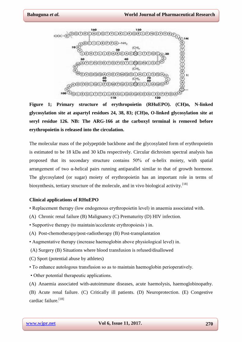
(andhence rHuEPO) containing165 aminoacids.(fig 1).
www.wjpr.net Vol 6, Issue 11, 2017.
270
Bahuguna et al. World Journal of Pharmaceutical Research
Figure 1; Primary structure of erythropoietin (RHuEPO). (CH)n, N-linked
glycosylation site at aspartyl residues 24, 38, 83; (CH)o, O-linked glycosylation site at
seryl residue 126. NB: The ARG-166 at the carboxyl terminal is removed before
erythropoietin is released into the circulation.
The molecular mass of the polypeptide backbone and the glycosylated form of erythropoietin
is estimated to be 18 kDa and 30 kDa respectively. Circular dichroism spectral analysis has
proposed that its secondary structure contains 50% of α-helix moiety, with spatial
arrangement of two α-helical pairs running antiparallel similar to that of growth hormone.
The glycosylated (or sugar) moiety of erythropoietin has an important role in terms of
biosynthesis, tertiary structure of the molecule, and in vivo biological activity.[18]
Clinical applications of RHuEPO
• Replacement therapy (low endogenous erythropoietin level) in anaemia associated with.
(A) Chronic renal failure (B) Malignancy (C) Prematurity (D) HIV infection.
• Supportive therapy (to maintain/accelerate erythropoiesis ) in.
(A) Post-chemotherapy/post-radiotherapy (B) Post-transplantation
• Augmentative therapy (increase haemoglobin above physiological level) in.
(A) Surgery (B) Situations where blood transfusion is refused/disallowed
(C) Sport (potential abuse by athletes)
• To enhance autologous transfusion so as to maintain haemoglobin perioperatively.
• Other potential therapeutic applications.
(A) Anaemia associated with-autoimmune diseases, acute haemolysis, haemoglobinopathy.
(B) Acute renal failure. (C) Critically ill patients. (D) Neuroprotection. (E) Congestive
cardiac failure.[18]
www.wjpr.net Vol 6, Issue 11, 2017.
271
Bahuguna et al. World Journal of Pharmaceutical Research
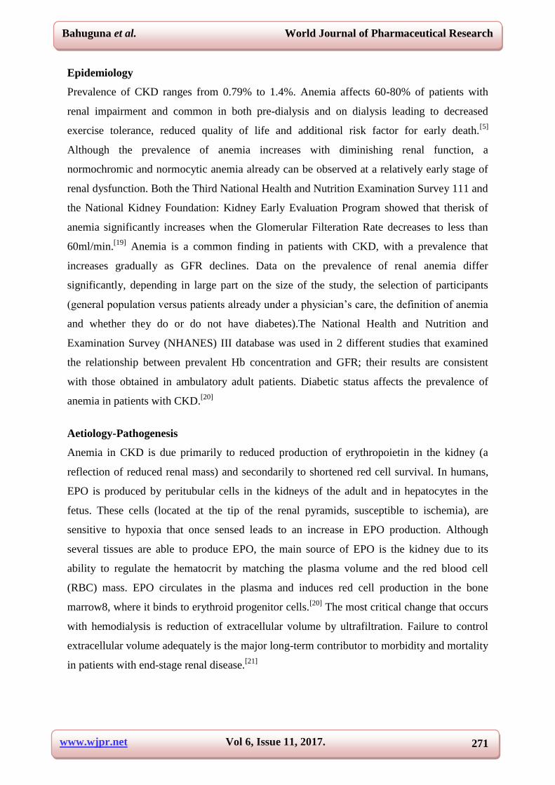
Epidemiology
Prevalence of CKD ranges from 0.79% to 1.4%. Anemia affects 60-80% of patients with
renal impairment and common in both pre-dialysis and on dialysis leading to decreased
exercise tolerance, reduced quality of life and additional risk factor for early death.[5]
Although the prevalence of anemia increases with diminishing renal function, a
normochromic and normocytic anemia already can be observed at a relatively early stage of
renal dysfunction. Both the Third National Health and Nutrition Examination Survey 111 and
the National Kidney Foundation: Kidney Early Evaluation Program showed that therisk of
anemia significantly increases when the Glomerular Filteration Rate decreases to less than
60ml/min.[19]
Anemia is a common finding in patients with CKD, with a prevalence that
increases gradually as GFR declines. Data on the prevalence of renal anemia differ
significantly, depending in large part on the size of the study, the selection of participants
(general population versus patients already under a physician’s care, the definition of anemia
and whether they do or do not have diabetes).The National Health and Nutrition and
Examination Survey (NHANES) III database was used in 2 different studies that examined
the relationship between prevalent Hb concentration and GFR; their results are consistent
with those obtained in ambulatory adult patients. Diabetic status affects the prevalence of
anemia in patients with CKD.[20]
Aetiology-Pathogenesis
Anemia in CKD is due primarily to reduced production of erythropoietin in the kidney (a
reflection of reduced renal mass) and secondarily to shortened red cell survival. In humans,
EPO is produced by peritubular cells in the kidneys of the adult and in hepatocytes in the
fetus. These cells (located at the tip of the renal pyramids, susceptible to ischemia), are
sensitive to hypoxia that once sensed leads to an increase in EPO production. Although
several tissues are able to produce EPO, the main source of EPO is the kidney due to its
ability to regulate the hematocrit by matching the plasma volume and the red blood cell
(RBC) mass. EPO circulates in the plasma and induces red cell production in the bone
marrow8, where it binds to erythroid progenitor cells.[20]
The most critical change that occurs
with hemodialysis is reduction of extracellular volume by ultrafiltration. Failure to control
extracellular volume adequately is the major long-term contributor to morbidity and mortality
in patients with end-stage renal disease.[21]
www.wjpr.net Vol 6, Issue 11, 2017.
272
Bahuguna et al. World Journal of Pharmaceutical Research
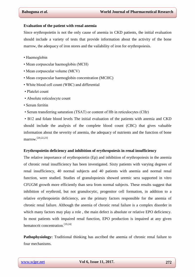
Evaluation of the patient with renal anemia
Since erythropoietin is not the only cause of anemia in CKD patients, the initial evaluation
should include a variety of tests that provide information about the activity of the bone
marrow, the adequacy of iron stores and the vailability of iron for erythropoiesis.
• Haemoglobin
• Mean corpuscular haemoglobin (MCH)
• Mean corpuscular volume (MCV)
• Mean corpuscular haemoglobin concentration (MCHC)
• White blood cell count (WBC) and differential
• Platelet count
• Absolute reticulocyte count
• Serum ferritin
• Serum transferring saturation (TSAT) or content of Hb in reticulocytes (CHr)
• B12 and folate blood levels The initial evaluation of the patients with anemia and CKD
should include the analysis of the complete blood count (CBC) that gives valuable
information about the severity of anemia, the adequacy of nutrients and the function of bone
marrow.[20,22,23]
Erythropoietin deficiency and inhibition of erythropoiesis in renal insufficiency
The relative importance of erythropoietin (Ep) and inhibition of erythropoiesis in the anemia
of chronic renal insufficiency has been investigated. Sixty patients with varying degrees of
renal insufficiency, 40 normal subjects and 40 patients with anemia and normal renal
function, were studied. Studies of granulopoiesis showed uremic sera supported in vitro
CFUGM growth more efficiently than sera from normal subjects. These results suggest that
inhibition of erythroid, but not granulocytic, progenitor cell formation, in addition to a
relative erythropoietin deficiency, are the primary factors responsible for the anemia of
chronic renal failure. Although the anemia of chronic renal failure is a complex disorder in
which many factors may play a role , the main defect is absolute or relative EPO deficiency.
In most patients with impaired renal function, EPO production is impaired at any given
hematocrit concentration.[19,24]
Pathophysiology: Traditional thinking has ascribed the anemia of chronic renal failure to
four mechanisms.
www.wjpr.net Vol 6, Issue 11, 2017.
273
Bahuguna et al. World Journal of Pharmaceutical Research
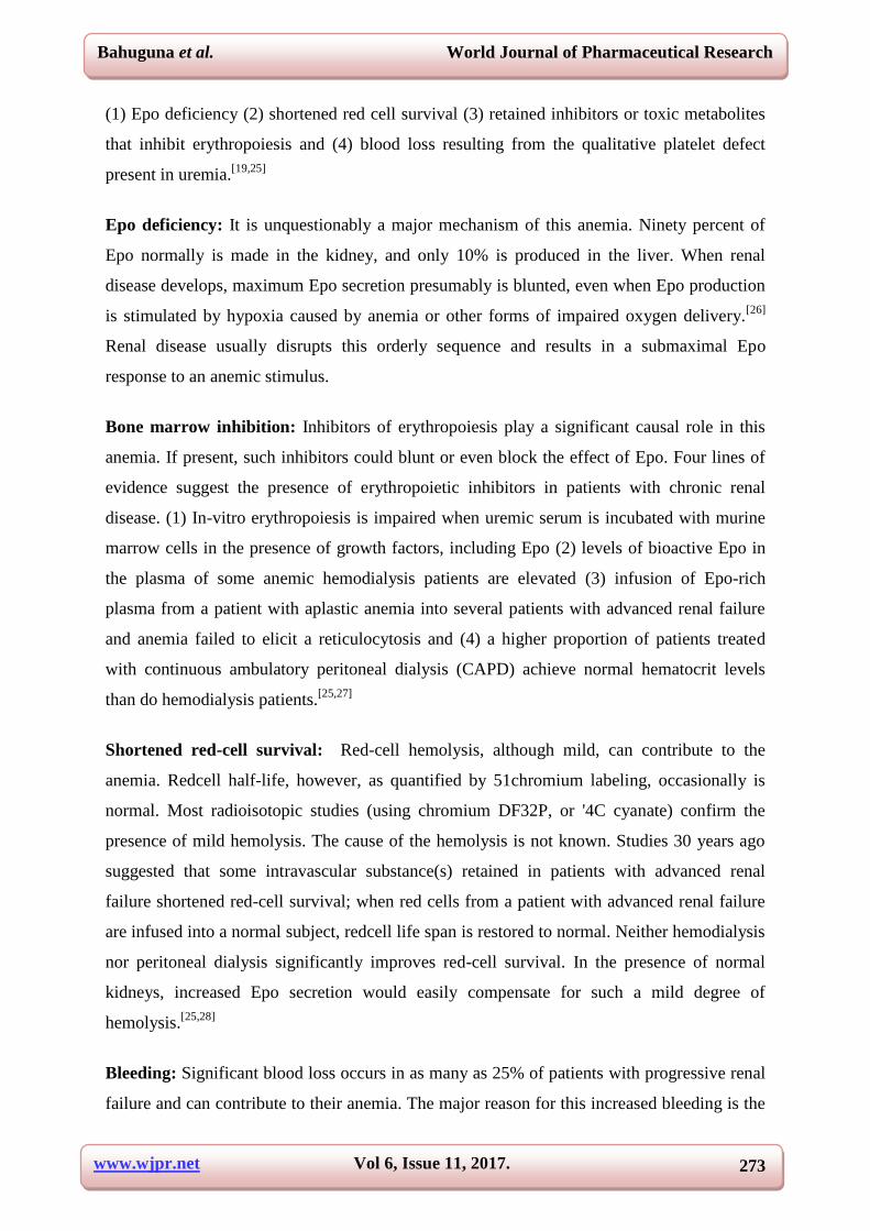
(1) Epo deficiency (2) shortened red cell survival (3) retained inhibitors or toxic metabolites
that inhibit erythropoiesis and (4) blood loss resulting from the qualitative platelet defect
present in uremia.[19,25]
Epo deficiency: It is unquestionably a major mechanism of this anemia. Ninety percent of
Epo normally is made in the kidney, and only 10% is produced in the liver. When renal
disease develops, maximum Epo secretion presumably is blunted, even when Epo production
is stimulated by hypoxia caused by anemia or other forms of impaired oxygen delivery.[26]
Renal disease usually disrupts this orderly sequence and results in a submaximal Epo
response to an anemic stimulus.
Bone marrow inhibition: Inhibitors of erythropoiesis play a significant causal role in this
anemia. If present, such inhibitors could blunt or even block the effect of Epo. Four lines of
evidence suggest the presence of erythropoietic inhibitors in patients with chronic renal
disease. (1) In-vitro erythropoiesis is impaired when uremic serum is incubated with murine
marrow cells in the presence of growth factors, including Epo (2) levels of bioactive Epo in
the plasma of some anemic hemodialysis patients are elevated (3) infusion of Epo-rich
plasma from a patient with aplastic anemia into several patients with advanced renal failure
and anemia failed to elicit a reticulocytosis and (4) a higher proportion of patients treated
with continuous ambulatory peritoneal dialysis (CAPD) achieve normal hematocrit levels
than do hemodialysis patients.[25,27]
Shortened red-cell survival: Red-cell hemolysis, although mild, can contribute to the
anemia. Redcell half-life, however, as quantified by 51chromium labeling, occasionally is
normal. Most radioisotopic studies (using chromium DF32P, or '4C cyanate) confirm the
presence of mild hemolysis. The cause of the hemolysis is not known. Studies 30 years ago
suggested that some intravascular substance(s) retained in patients with advanced renal
failure shortened red-cell survival; when red cells from a patient with advanced renal failure
are infused into a normal subject, redcell life span is restored to normal. Neither hemodialysis
nor peritoneal dialysis significantly improves red-cell survival. In the presence of normal
kidneys, increased Epo secretion would easily compensate for such a mild degree of
hemolysis.[25,28]
Bleeding: Significant blood loss occurs in as many as 25% of patients with progressive renal
failure and can contribute to their anemia. The major reason for this increased bleeding is the
www.wjpr.net Vol 6, Issue 11, 2017.
274
Bahuguna et al. World Journal of Pharmaceutical Research
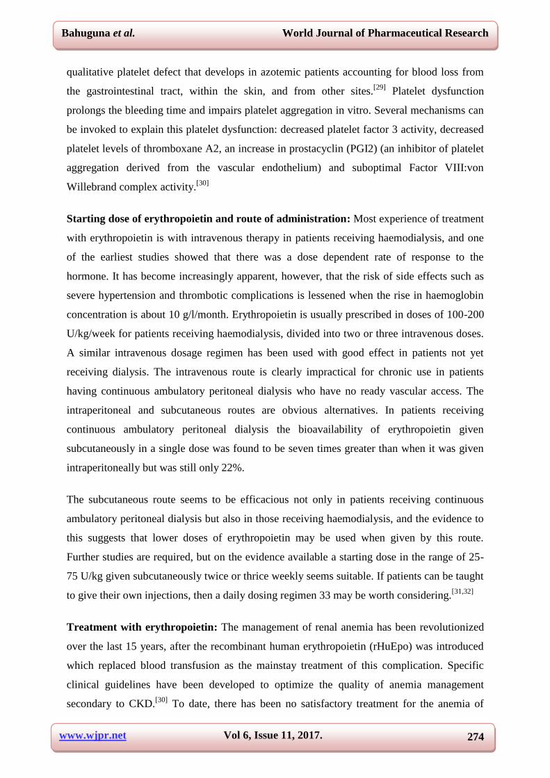
qualitative platelet defect that develops in azotemic patients accounting for blood loss from
the gastrointestinal tract, within the skin, and from other sites.[29]
Platelet dysfunction
prolongs the bleeding time and impairs platelet aggregation in vitro. Several mechanisms can
be invoked to explain this platelet dysfunction: decreased platelet factor 3 activity, decreased
platelet levels of thromboxane A2, an increase in prostacyclin (PGI2) (an inhibitor of platelet
aggregation derived from the vascular endothelium) and suboptimal Factor VIII:von
Willebrand complex activity.[30]
Starting dose of erythropoietin and route of administration: Most experience of treatment
with erythropoietin is with intravenous therapy in patients receiving haemodialysis, and one
of the earliest studies showed that there was a dose dependent rate of response to the
hormone. It has become increasingly apparent, however, that the risk of side effects such as
severe hypertension and thrombotic complications is lessened when the rise in haemoglobin
concentration is about 10 g/l/month. Erythropoietin is usually prescribed in doses of 100-200
U/kg/week for patients receiving haemodialysis, divided into two or three intravenous doses.
A similar intravenous dosage regimen has been used with good effect in patients not yet
receiving dialysis. The intravenous route is clearly impractical for chronic use in patients
having continuous ambulatory peritoneal dialysis who have no ready vascular access. The
intraperitoneal and subcutaneous routes are obvious alternatives. In patients receiving
continuous ambulatory peritoneal dialysis the bioavailability of erythropoietin given
subcutaneously in a single dose was found to be seven times greater than when it was given
intraperitoneally but was still only 22%.
The subcutaneous route seems to be efficacious not only in patients receiving continuous
ambulatory peritoneal dialysis but also in those receiving haemodialysis, and the evidence to
this suggests that lower doses of erythropoietin may be used when given by this route.
Further studies are required, but on the evidence available a starting dose in the range of 25-
75 U/kg given subcutaneously twice or thrice weekly seems suitable. If patients can be taught
to give their own injections, then a daily dosing regimen 33 may be worth considering.[31,32]
Treatment with erythropoietin: The management of renal anemia has been revolutionized
over the last 15 years, after the recombinant human erythropoietin (rHuEpo) was introduced
which replaced blood transfusion as the mainstay treatment of this complication. Specific
clinical guidelines have been developed to optimize the quality of anemia management
secondary to CKD.[30]
To date, there has been no satisfactory treatment for the anemia of
www.wjpr.net Vol 6, Issue 11, 2017.
275
Bahuguna et al. World Journal of Pharmaceutical Research
ESRD. Red cell transfusions have been the only sure way to correct the symptoms of tissue
hypoxia, but such therapy is only transiently effective and transfusions increase the risk of
exposure to hepatitis or other infectious agents.[33]
In the past, a few attempts were made to
treat patients with Epo isolated from the urine of anemic individuals however, it was in the
middle 80's with the availability of recombinant human Epo that clinical trials became
possible. Since then replacement therapy with recombinant human Epo has become the most
rational therapy for the anemia of chronic renal failure worldwide. The treatment with
recombinant Epo has resulted in a remarkable improvement in global well being and quality
of life. Erythroid marrow suppression may be another effect of transfusion therapy, especially
if multiple units of red cells are infused at once.[34]
Furthermore, the elimination of anemia
has been beneficial for a number of co-morbid conditions introduced by the aging and
prevalence of diabetes among patients initiating dialysis. This global success in dialysis
patients has motivated the subsequent use of recombinant human Epo in pre-dialysis patients
showing progressive renal failure. The core treatment of renal anemia is to fuel erythropoiesis
by regular injections with erythropoiesis stimulating agents (ESAs) and to secure sufficient
iron availability for proper erythropoiesis.[35]
Numerous studies have indicated that ESA
therapy results in an improvement in the patient’s quality of life. As a result, ESA treatment
is a routine management component for hemodialysis patients, and is frequently used in
nondialysis CKD as well.[36]
Initial concerns of accelerating renal function deterioration in
patients with progressive renal failure have not been substantiated. No significant alteration
in the progression of renal disease has been noted. However, avoidance of renal function
deterioration requires careful control of blood pressure before or soon after starting
recombinant human Epo through the aggressive use of antihypertensive agents. Slow
correction of anemia by using lower starting doses is advocated by some since such an
approach, particularly in children, tends to improve the glomerular filtration rate as assessed
indirectly. The mechanisms producing hypertension with recombinant human Epo therapy
are likely to be multifactorial: loss of hypoxic vasodilation, changes in blood viscosity, in
activation of renin-angiotensin system, in blood volume, or through a direct vascular effect.
This last mechanism may involve increased synthesis of endothelin-1, increased vascular
calcium uptake, and platelet-dependent mitogenic action. The role of recombinant human
Epo therapy in raising plasma endothelin-1 to levels, which directly can increase pressure
remains controversial. Nevertheless, if body wt and interdialytic fluid gains are controlled in
dialysis patients, systolic and diastolic blood pressure remain virtually unchanged despite
significant increases in hematocrit. Similar observations have been reported for predialysis
www.wjpr.net Vol 6, Issue 11, 2017.
276
Bahuguna et al. World Journal of Pharmaceutical Research
patients. Many of these predialysis patients need aggressive diuretic therapy during
recombinant human Epo treatment to maintain constant blood volume and thus avoid
hypertension. Regular hemodialysis therapy (RDT) and continuous ambulatory peritoneal
dialysis (CAPD) have both been reported to lead to an improvement in the anemia associated
with endstage renal disease.[37,38]
Regression of left ventricular hypertrophy (LVH), reduction
of left ventricular volume and improvement in exercise induced ST-segment depression may
occur following partial correction of anemia.[38]
CONCLUSION
After reviewing several articles it was seen that anemia is one of the most common and
morbid complications of chronic kidney disease, causing unpleasant symptoms and reducing
the quality of life of patients. In this review, we have conveyed concerns about treatment of
Renal Anemia in Patients with chronic Kidney Disease and Patients undergoing
Hemodialysis and what anemia management should be about: optimizing clinical outcomes,
including quality of life, and minimizing risks, while maintaining public trust by achieving
our clinical goals in the most cost-effective way possible. Appropriate management of
anemia will not only have a positive impact on quality of life but also reduce hospitalizations
of CKD patients due to cardiovascular events.
REFERENCES
1. Fishbane S, Nissenson AR. Anemia management in chronic kidney disease. Kidney
International, 2010; 78: 3-9.
2. Stefansson BV. Studies on treatment of renal anemia in patients on chronic hemodialysis,
2011; 4-40.
3. Salman M, Khan AH, Adnan AS, et al. Prevalence and management of anemia in pre-
dialysis Malaysian patients: A hospital-based study. Revista da Associacao Medica
Brasileira, 2016; 62(8): 742-7.
4. Mudiyammanavara RN, Dhanajaya PE, Agarwal R, et al. Cross sectional study of
anaemia in chronic kidney disease. Indian Journal of Basic and Applied Medical
Research, 2015; 4: 414-9.
5. Dorgalaleh A, Mahmudi M, Tabibian S, et al. Anemia and thrombocytopenia in acute and
chronic renal failure. International Journal of Hematology-oncology and stem cell
Research, 2013; 7(4): 34-39.
www.wjpr.net Vol 6, Issue 11, 2017.
277
Bahuguna et al. World Journal of Pharmaceutical Research
6. Rathod SG, Ade AK, Shekokar PP. A Study of Haematological Changes in Chronic
Renal Failure. Scholars Journal of Applied Medical Sciences, 2014; 2(4): 1232-34.
7. Eschbach JW, Adamson JW. Anemia of End- Stage Renal Disease. Kidney International,
1985; 28: 1-5.
8. Phrommintikul A, Haas SJ, Elsik M, et al. Mortality and target haemoglobin
concentrations in anaemic patients with chronic kidney disease treated with
erythropoietin: a meta-analysis. The Lancet, 2007; 369(9559): 381-8.
9. Remuzzi G, Ingelfinger JR. Correction of anemia-payoffs and problems. New England
Journal of Medicine, 2006; 355(20): 2144-46.
10. Bauer C, Kurtz A. Oxygen sensing in the kidney and its relation to erythropoietin
production. Annual Review of Physiology, 1989; 51(1): 845-56.
11. Thomas R, Kanso A, Sedor JR. Chronic kidney disease and its complications. Primary
care: Clinics In Office Practice, 2008; 35(2): 329-44.
12. Erslev AJ, Besarab A. Erythropoietin in the pathogenesis and treatment of the anemia of
chronic renal failure. Kidney international, 1997; 51(3): 622-30.
13. Chapman D, Moore R, Klarenbach S, et al. Residual renal function after partial or radical
nephrectomy for renal cell carcinoma. Can Urol Assoc J., 2010; 4(5): 337-43.
14. Nissenson AR, Strobos JU. Iron deficiency in patients with renal failure. Kidney
International, 1999; 55: 18-21.
15. Ng T, Marx G, Littlewood T, et al. Recombinant erythropoietin in clinical practice.
Postgraduate Medical Journal, 2003; 79(933): 367-76.
16. Tsagalis G. Renal anemia: a nephrologist's view. Hippokratia, 2011; 15: 39-43.
17. Segal GM, Eschbach JW, Egrie JC, et al. The anemia of end-stage renal disease:
Hematopoietic progenitor cell response. Kidney International, 1988; 33: 983-88.
18. Urena P, Eckardt K, Sarfati E, et al. Serum Erythropoietin and Erythropoiesis in Primary
and Secondary Hyperparathyroidism: Effect of Parathyroidectomy. Nephron, 1991; 59:
384-93.
19. Nangaku M, Eckardt K. Pathogenesis of Renal Anemia. Seminars In Nephrology, 2006;
26: 261-68.
20. Lazarus M. Complications in hemodialysis: An overview. Kidney International, 1980; 18:
783-96.
21. Andrews NC. Disorders of iron metabolism. New England Journal of Medicine, 1999;
341: 1986–95.
www.wjpr.net Vol 6, Issue 11, 2017.
278
Bahuguna et al. World Journal of Pharmaceutical Research
22. Siah CW, Ombiga J, Adams LA, et al. Normal Iron metabolism and the Pathophysiology
of Iron Overload Disorders. Clinical Biochemist Reviews, 2006; 27: 5–16.
23. Fehr T, Ammann P, Garzoni D, et al. Interpretation of erythropoietin levels in patients
with various degrees of renal insufficiency and anemia. Kidney International, 2004; 66:
1206-11.
24. Eschbach JW. The anemia of chronic renal failure: Pathophysiology and the effects of
recombinant erythropoietin. Kidney International, 1989; 35: 134-48.
25. Fried W. The liver as a source of extrarenal erythropoietin production. Blood, 1973; 40:
671-77.
26. Zappacosta AR, Caro J, Erslev A. Normalization of hematocrit in patients with end-stage
renal disease on continuous ambulatory peritoneal dialysis. The American Journal of
Medicine, 1982; 72: 53-7.
27. Eschbach JW, Funk D, Adamson JW, et al. Erythropoiesis in patients with renal failure
undergoing chronic dialysis. The New England Journal of Medicine, 1976; 276: 653-58.
28. Loge JP, Lange RD, Moore CV. Characterization of the anemia associated with chronic
renal insufficiency. The American Journal of Medicine, 1958; 24: 4-18.
29. Lewis JH, Zucker MB, Ferguson JH. Bleeding tendency in uremia. Blood, 1956; 11(12):
1073-6.
30. MacDougall IC, Hutton RD, Cavill I, et al. Treating renal anaemia with recombinant
human erythropoietin: practical guidelines and a clinical algorithm. British Medical
Journal, 1990; 300: 655-9.
31. Mayer G, Thum J, Cada EM, et al.Working capacity is increased following recombinant
human erythropoietin treatment. Kidney International, 1988; 34: 525-8.
32. Editorial. Androgens in the anaemia of chronic renal failure. British Medical Journal,
1977; 2: 417-8.
33. Escbach JW, Adamson JW, Cook JD. Disorders of red blood cell production in uremia.
Archives of Internal Medicine, 1970; 126(5): 812-15.
34. National KF. KDOQI Clinical Practice Guidelines and Clinical Practice
Recommendations for Anemia in Chronic Kidney Disease. American Journal of Kidney
Diseases, 2006; 47: 11-145.
35. Lundin AP, Delano BG, Quinn-Cefaro R. Perspectives on the improvement of quality of
life with epoetin alfa therapy. Pharmacotherapy: The Journal of Human Pharmacology
and Drug Therapy, 1990; 10(2P2): 22-6.
www.wjpr.net Vol 6, Issue 11, 2017.
279
Bahuguna et al. World Journal of Pharmaceutical Research
36. Gokal R, Mchugh M, Fryer R, et al. Continuous ambulatory peritoneal dialysis: one year's
experience in a UK dialysis unit. British Medical Journal, 1980; 281: 474-77.
37. Escbach JW, Funk P, Adamson JW, et al. Erythropoiesis in patients with renal failure
undergoing chronic dialysis. New England Journal of Medicine, 1967; 276: 653-58.
38. McGonigle RJS, Husserl F, Wallin JD, et al. Hemodialysis and continuous ambulatory
peritoneal dialysis effects on erythropoiesis in renal failure. Kidney International, 1984;
25: 430-36.