Embed Size (px)
Citation preview
2647
□ CASE REPORT □
A Serious Mediastinum Abscess Induced by EndobronchialUltrasound-guided Transbronchial Needle Aspiration
(EBUS-TBNA): A Case Reportand Review of the Literature
Hiroshi Ishimoto 1, Kazuhiro Yatera 1, Keigo Uchimura 1, Keishi Oda 1, Masaru Takenaka 2,
Toshinori Kawanami 1, Fumihiro Tanaka 2 and Hiroshi Mukae 1
Abstract
A 75-year-old man with interstitial pneumonia and enlarged mediastinal lymph nodes underwent endobron-
chial ultrasound-guided transbronchial needle aspiration (EBUS-TBNA). He developed a high-grade fever
seven days after EBUS-TBNA was performed; laboratory and radiologic findings showed intense inflamma-
tory reactions, with swelling of the mediastinal lymph nodes on chest computed tomography. Mediastinal
lymph node abscess was diagnosed, and it worsened in spite of systemic antibacterial treatment. Surgical
treatment using a median sternotomy was performed, and the cultivation of surgically obtained mediastinal
lymph node abscess fluid revealed Streptococcus intermedius. Combined treatment with antibiotics and surgi-
cal treatment was effective, leading to remission.
Key words: endobronchial ultrasound-guided transbronchial needle aspiration (EBUS-TBNA), mediastinal
lymphatic abscess, Streptococcus intermedius
(Intern Med 54: 2647-2650, 2015)(DOI: 10.2169/internalmedicine.54.4465)
Introduction
This case report describes a serious mediastinum lym-
phatic abscess caused by endobronchial ultrasound-guided
transbronchial needle aspiration (EBUS-TBNA).
Case Report
A 75-year-old Japanese man had experienced dyspnea
during exertion for approximately six months. He had un-
controlled diabetes mellitus [HbA1c, National Glycohemo-
globin Standardization Program (NGSP) = 9.1%], and had a
history of hepatic abscess. His vital signs were normal on
admission. Bilateral fine crackles were heard with ausculta-
tion of the chest. Chest X-rays revealed a reticular shadow
around both sides of the lower lung field (Fig. 1a). Chest
computed tomography (CT) showed honeycomb lung forma-
tions in the dorsal side of the bilateral lower lobes as well
as traction bronchiectasis (Fig. 1b). Slight mediastinal lymph
node hyperplasia was observed (Fig. 1c). Although his se-
rum angiotensin converting enzyme levels were within nor-
mal limits, slightly high serum calcium levels indicated sar-
coidosis as a possible cause of interstitial pneumonia; there-
fore, we performed EBUS-TBNA in order to carry out a
pathological examination of the mediastinal lymph nodes.
Under mild sedation with midazolam, EBUS-TBNA was
performed using the UC260FW (Olympus, Tokyo, Japan).
The blood flow within the mediastinum lymph node was
confirmed using color Doppler. A 22-G needle was used for
the puncture. The puncture was made using “the style of
stylet push out” method. Puncturing was performed twice in
each right tracheobronchial lymph node (#4R) and subcari-
nal lymph node (#7). This was followed by bronchoalveolar
lavage (BAL) in the right S5 and trans-bronchial lung biop-
sies (TBLB) in the right S9 using the 1T260 (Olympus).
1Department of Respiratory Medicine, University of Occupational and Environmental Health, Japan and 2Second Department of Surgery, Univer-
sity of Occupational and Environmental Health, Japan
Received for publication November 7, 2014; Accepted for publication February 22, 2015
Correspondence to Dr. Hiroshi Ishimoto, [email protected]

Intern Med 54: 2647-2650, 2015 DOI: 10.2169/internalmedicine.54.4465
2648
Figure 1. Chest X-ray (a) and computed tomography (b, c) images obtained at the first admission. Chest X-rays and computed tomography revealed a reticular shadow around both sides of the lower lung field (a, b). Slight mediastinal lymph node hyperplasia was observed at the #4R lymph node (c).
a) b) C)
Bar = 5cm
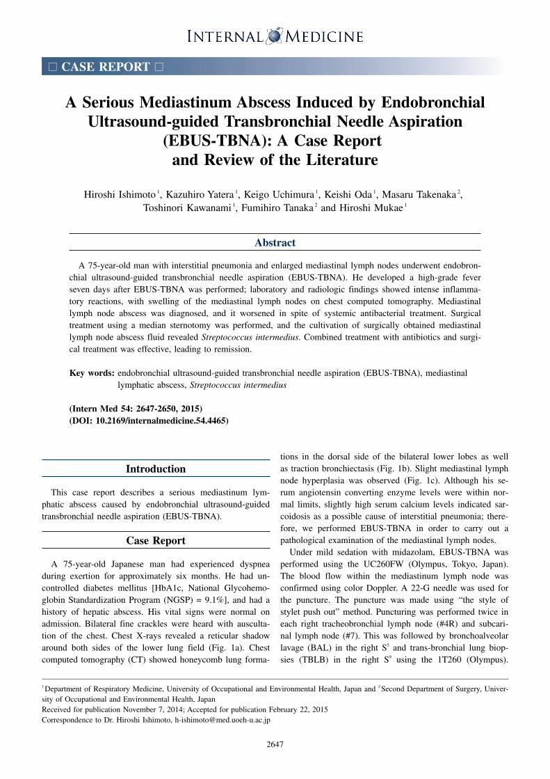
Figure 2. Chest X-ray (a) and computed tomography (b) images obtained at the second admission. Superior mediastinum expansion was recognized (a). The lymph node of #4R exhibited swelling, and a low-density area was recognized inside this lymph node (b).
a) b)
Bar = 5cm
Prophylactic antibiotics were not prescribed and the patient
was discharged from the hospital two days after the diagnos-
tic examination. Pathological examination of both the TBNA
and TBLB specimens showed nonspecific inflammatory
changes and no malignant or granulomatous findings.
The patient developed a persistent high fever seven days
after the diagnostic examination, and he returned to our hos-
pital nine days after the diagnostic examination. An ad-
vanced inflammatory reaction (white blood cell count=
18,100/μL; CRP=18.51 mg/dL) was observed, and expan-
sion of the superior mediastinum was revealed by chest X-
ray (Fig. 2a). Therefore, he was re-admitted to the hospital.
Chest CT revealed swelling of lymph node #4R, with a low-
density area inside the lymph node. This indicated that me-
diastinum lymphadenitis had occurred, with a portion be-
coming abscessed (Fig. 2b). Although meropenem (3 g/day)
and linezolid (1,200 mg/day) were initiated, the high fever
remained along with an irregularity of the circulatory move-
ment. Therefore, we decided to perform mediastinum lymph
node dissection the third day after admission. An acute ex-
acerbation of chronic respiratory failure in our patient did
not permit one-lung ventilation during surgery. Furthermore,
en bloc removal of the infected and swollen lymph nodes
was important; therefore, median sternotomy was performed
under general anesthesia. Following release of the superior
vena cava and the ascending aorta, swelling of the #4 lymph
node was exposed. The lymph node was punctured asepti-
cally, and the pus inside the lymph node was collected. The
#4 lymph node and the paratrachial lymph node (#2) were
removed en block. The inflammation had spread to some of
the fat tissues surrounding the lymph nodes.
A culture of the pus from the mediastinal lymph node
yielded growth of Streptococcus intermedius. Therefore, ce-
fazolin (4.5 g/day) and clindamycin (1,200 mg/day) were
administered for two weeks. Thereafter, doripenem (3 g/day)
was administered for two weeks for concomitant aspiration
pneumonia.
Discussion
Compared with mediastinoscopy, EBUS-TBNA is a mini-
mally invasive method for the investigation of mediastinum
lymph node swelling and thus has recently become increas-
ingly more popular. Furthermore, EBUS-TBNA characteris-
tically has few complications. Only a few reports have de-
scribed infectious complications of the mediastinum (1-6)

Intern Med 54: 2647-2650, 2015 DOI: 10.2169/internalmedicine.54.4465
2649
Table. The Case List of the Infectious Complication Caused by Endbronchial Ultrasound Guided Transbronchial Needle Aspiration.
age sex Lapsed days The kind of infectios disease Bacteria of a cause Surgical procedure Reference
50 M 19 pericardiocentesis Actinomyces odontolytiusStreptococcus mutans drainage 1
58 M 9 abscess in the lung mass N.D. - 1
89 F 14 mediastinal abscess alpha streptococcusDiphtheroids operation 2
68 M 35 mediastinal abscess Candida albicans-hemolytic Streptococcus - 3
66 M 5 mediastinal abscessPropionibacterium acnes
BacteroidesEubacterium species
operation 3
68 M 60 mediastinal abscess Streptococcus viridans operation 4
59 M 7 mediastinal abscess N.D. - 5
55 F 6 pericardiocentesis Streptococcus viridans operation 6
59 M 10 pericarditis and pneumonia Group C beta-haemolytic Streptococcus species - 6
75 M 9 mediastinal abscess Streptococcus intermidius operation Our Case
N.D.: no data
(Table).
Investigations to confirm bacteremia have been carried out
to examine the occurrence of infectious disease due to endo-
scopic maneuvers utilizing this technique (7). For EBUS-
TBNA, a previous investigation confirmed the presence of
bacteremia in a blood culture obtained during the diagnostic
examination (8). The frequency of the occurrence of bac-
teremia does not differ greatly between EBUS-TBNA and
conventional bronchoscopy. Therefore, bacterial invasion
from the mucous membrane is thought to be responsible for
bacteremia (9). However, these studies have limitations, and
the method for verifying bacteremia is not the same as that
for verifying the occurrence of mediastinum lymphadenitis
due to EBUS-TBNA. Because the frequency of the occur-
rence of infectious complications, including mediastinum
lymphadenitis, is very low (0.19%) (10), verifying mediasti-
num lymphadenitis is difficult. However, mediastinum lym-
phadenitis should be carefully considered in the differential
diagnosis because it is a serious complication with occasion-
ally fatal outcomes (6).
The bronchoscope is placed into the trachea through the
upper airway; therefore, maintaining aseptic conditions dur-
ing this procedure is difficult. Moreover, because an ultra-
sonic endoscope is a heteroscope, insertion is difficult in
comparison with usual bronchoscopy. One factor that influ-
ences contamination of the tip of the fiberscope is the ap-
proach after the tracheal intubation. However, past research
has shown that the occurrence of bacteremia due to tracheal
intubation is beyond 10% (10). Furthermore, deep anesthesia
is unnecessary when a diagnostic examination is performed
with EBUS-TBNA. The absorption of any saliva or fluid in
the intraoral region before bronchoscope insertion is a fun-
damental maneuver, and the elevation of the lower jaw of
the patient by the assistant is important for this step. After
encountering serious complications associated with stylet
handling, we discontinued the use of “the style of stylet
push out” and assigned stylet handling to an independent as-
sistant wearing sterile gloves to prevent contamination of the
mediastinal lymph nodes with the needle and stylet at our
hospital.
The existence of uncontrolled diabetes mellitus would in-
fluence this critical illness. Antibacterial medication is rec-
ommended as a prophylaxis for infectious endocarditis, cor-
responding to the patient’s risk factors (11). For the bron-
choscope, a previous investigation showed no validity for
using an antibacterial agent for prophylactic medica-
tion (12). However, a satisfactory evaluation cannot pres-
ently be carried out for EBUS-TBNA. In high-risk patients,
such as our patient who had uncontrolled diabetes mellitus,
we believe that prophylactic antibiotics should be adminis-
tered to avoid serious complications such as mediastinitis, as
observed in our patient. In addition, in recent report of peri-
carditis as a fatal complication of EBUS-TBNA, antibiotic
prophylaxis was recommended to avoid the possible occur-
rence of serious acute pericarditis in patients who underwent
EBUS-TBNA with necrotic lymph nodes located in the mid-
dle mediastinum (6). The intraoral resident florae become
the origin bacteria in many cases (Table). Therefore, cefa-
zolin and a ceftriaxone should be recommended according
to the guidelines of the American Heart Association (7).
The authors state that they have no Conflict of Interest (COI).

Intern Med 54: 2647-2650, 2015 DOI: 10.2169/internalmedicine.54.4465
2650
References
1. Haas AR. Infectious complications from full extension endobron-
chial ultrasound transbronchial needle aspiration. Eur Respir J 33:
935-938, 2009.
2. Moffatt-Bruce SD, Ross P Jr. Mediastinal abscess after endobron-
chial ultrasound with transbronchial needle aspiration: a case re-
port. J Cardiothorac Surg 5: 33, 2010.
3. Kouskov OS, Almeida FA, Eapen GA, Uzbeck M, Deffebach M.
Mediastinal infection after ultrasound-guided needle aspiration. J
Bronchology Interv Pulmonal 17: 338-341, 2010.
4. Huang CT, Chen CY, Ho CC, Yu CJ. A rare constellation of em-
pyema, lung abscess, and mediastinal abscess as a complication of
endobronchial ultrasound-guided transbronchial needle aspiration.
Eur J Cardiothorac Surg 40: 264-265, 2011.
5. Gochi F, Chen F, Aoyama A, Date H. Mediastinal infectious com-
plication after endobronchial ultrasound-guided transbronchial nee-
dle aspiration. Interact Cardiovasc Thorac Surg 17: 751-752, 2013.
6. Lee HY, Kim J, Jo YS, Park YS. Bacterial pericarditis as a fatal
complication after endobronchial ultrasound-guided transbronchial
needle aspiration. Eur J Cardiothorac Surg. (in press).
7. Yigla M, Oren I, Bentur L, et al. Incidence of bacteraemia follow-
ing fibreoptic bronchoscopy. Eur Respir J 14: 789-791, 1999.
8. Steinfort DP, Johnson DF, Irving LB. Incidence of bacteraemia
following endobronchial ultrasound-guided transbronchial needle
aspiration. Eur Respir J 36: 28-32, 2010.
9. Asano F, Aoe M, Ohsaki Y, et al. Complications associated with
endobronchial ultrasound-guided transbronchial needle aspiration:
a nationwide survey by the Japan Society for Respiratory Endos-
copy. Respir Res 14: 50, 2013.
10. Valdes C, Tomas I, Alvarez M, Limeres J, Medina J, Diz P. The
incidence of bacteraemia associated with tracheal intubation. An-
aesthesia 63: 588-592, 2008.
11. Wilson W, Taubert KA, Gewitz M, et al. Prevention of infective
endocarditis: guidelines from the American Heart Association: a
guideline from the American Heart Association Rheumatic Fever,
Endocarditis, and Kawasaki Disease Committee, Council on Car-
diovascular Disease in the Young, and the Council on Clinical
Cardiology, Council on Cardiovascular Surgery and Anesthesia,
and the Quality of Care and Outcomes Research Interdisciplinary
Working Group. Circulation 116: 1736-1754, 2007.
12. Park JS, Lee CH, Yim JJ, et al. Impact of antibiotic prophylaxis
on postbronchoscopy fever: a randomised controlled study. Int J
Tuberc Lung Dis 15: 528-535, 2011.
Ⓒ 2015 The Japanese Society of Internal Medicine
http://www.naika.or.jp/imonline/index.html
![Combined endobronchial and esophageal …patients who present with an abnormal mediastinum [9–11], but also in those with a normal mediastinum but increased risk of mediastinal involvement](https://img.pdfslide.net/doc/110x75/5e3858f26bdec60bd21ce0e3/combined-endobronchial-and-esophageal-patients-who-present-with-an-abnormal-mediastinum.jpg)