Embed Size (px)
Citation preview
A Statistical Model of Right Ventricle inTetralogy of Fallot for Prediction of Remodelling
and Therapy Planning
Abstract. Tetralogy of Fallot (ToF) is a severe congenital heart diseasethat mainly affects the right ventricle (RV). It requires surgical repairearly in infancy. Chronic regurgitations may appear due to damagedpulmonary valves, resulting in extreme RV dilation. To reduce risk factorslate after repair, new pulmonary valves must be re-implanted. However,establishing the best timing for re-intervention is a clinical challengebecause of the large variability in RV shape and in pathology evolution.The purpose of this study is to quantify the regional impacts of growthand regurgitations on the end-diastolic RV anatomy. The ultimate goalis to determine, among clinical variables, predictors for the shape inorder to build a statistical model that predicts RV remodelling. Theproposed approach relies on a forward model based on currents andLDDMM algorithm to estimate an unbiased template of 18 patients andthe deformations towards each individual shape. Then, cross-sectionalmultivariate analyses are employed to assess the effects of body surfacearea, tricuspid and transpulmonary valve regurgitations upon the RVshape. The significant deformation modes were found clinically relevant.Canonical correlation analysis was applied to derive a generative modelof RV remodelling, which was successfully tested on two new patients.
1 Introduction
Tetralogy of Fallot is a severe congenital heart defect that requires surgical re-pair early in infancy. Yet, pulmonary valves may be damaged by the surgery,causing chronic regurgitations. As a result, the right ventricle (RV) dilates ex-tremely, its shape is altered and the cardiac function is impaired: new valvesmust be implanted in adulthood to reduce the risk factors late after repair [1].Understanding and quantifying RV remodelling in repaired Tetralogy of Fallot(ToF) patients is crucial for patient management and therapy planning. How-ever, the high variability in pathology course and in RV anatomy make difficultthe decision of optimal timing for re-intervention [1].
Contrary to the left ventricle, whose shape and deformations under patho-logical conditions are well documented, RV anatomy is complex and can varytremendously among ToF patients. Several studies have been published aboutpossible correlations between clinical parameters in ToF [1]. However, few workshave quantified the anatomical alterations of the RV and their evolution dueto the disease [2, 3]. In [2], the authors measure the most striking differencesin RV shape with respect to normals, quantifying the complex RV remodelling
observed in ToF. However, only one-dimensional indices are considered despitethe availability of 3D segmentations. In [3], the authors present a 4D ActiveAppearance Model of the beating heart to segment RV in MRI. They proposeindices based on the shape modes and successfully classify patients from normal.Yet, they do not correlate their model with functional features of ToF.
The clinical challenges about ToF encourage applying image-based shapeanalysis techniques to model the RV anatomical alterations due to pathologicalfactors. These techniques generate a representative template of the population ofinterest and assess how it deforms within this population [4–7]. Yet, correlatingshape with clinical variables require a rigorous framework: Biases may appearif the template is not defined in a consistent way, which may yield drastic dif-ferences in the statistical conclusions. Two strategies are available to create thetemplate. The backward approach consists in modelling the template as the av-erage of the deformed observations plus some residuals [4, 5]. Such a templatecan be computed efficiently but the model parameters, especially the residuals,are more difficult to identify. The forward approach consists in modelling the ob-servations as deformations of the template plus some residuals [6, 7]. Computingthe template is more complex but model parameters can be faithfully estimatedfrom images and clinical data.
In view of assisting the cardiologists in establishing the best time for re-intervention, we aim at statistically predict the RV remodelling in ToF. As afirst step, we propose in this work to quantify the regional impacts of growth andregurgitations on the end-diastolic RV anatomy in a cohort of 18 ToF patients.We rely on a forward approach to estimate the main deformation modes. Then,cross-sectional multivariate analyses are employed to assess the effects of growthand regurgitations upon the RV shape. This yields a generative model of RVremodelling, model that is then tested on two new patients.
2 Methods
The segmentation of the Right Ventricle (RV) of multiple patients from cine-MRIis described in Sec. 3.1. To perform the analysis on this population of shapes,an unbiased template is first built. This template serves as the reference atlasto determine the deformations towards each individual shape, deformations thatare then analysed using a Principal Component Analysis (PCA) to extract themain deformations modes. The importance of each mode is statistically assessedwith respect to child growth and valvar regurgitation severity. In particular, weinvestigate how our generative model can predict the evolution of shape withrespect to body surface area.
2.1 Unbiased Template of the Right Ventricle in Tetralogy of Fallot
We apply the forward strategy proposed by [7] to generate the RV template. Thisapproach is particularly suited for our purposes as 1) it is non-parametric, shapesbeing represented by currents; 2) model parameters are well-defined and can be
estimated from clinical data, thus enabling statistical analyses; 3) template anddeformations are computed simultaneously and consistently and 4) new patientscan be integrated in the study seamlessly, re-estimating the template is notrequired. More precisely, in the forward framework, the RV surfaces, or shapes,are modelled as the sum of a diffeomorphic deformation φi of the template Tand a residual term εi that models the shape features that cannot be representedby the template (topology changes, acquisition artifacts, etc.): T i = φi.T + εi.
Currents are used to represent the shapes, the residuals and the deformationsin the same common framework. Currents form a vector space, thus enabling theusual operations (mean, variance...) on shapes. Intuitively, they can be seen asthe flux of any vector field ω ∈ W across the shapes. W is a vector spaceof infinite dimension (a reproducible kernel Hilbert space or RKHS) generatedby a Gaussian kernel KW (x,y) = exp(−‖x − y‖2/λ2
W ), which defines an innerproduct in W that can be easily computed. In this framework, a triangle centredat x with normal α is represented by the Dirac delta current δα
x . Thus, a discrete
mesh is encoded by the sum of the currents of its triangles T i =∑
k δαi
k
xik
. In such
a model, the residuals εi are naturally modelled as a Gaussian distribution on theαi
k. The deformation φi registering the template T to the current T i is estimatedusing the Large Deformation Diffeomorphic Mappings (LDDMM) framework [8].The key feature is that this deformation can be parametrised by its smoothinitial vector speed vi
0, which also belongs to a Gaussian RKHS with varianceλ2
V . Moreover, this initial speed vector field is completely defined by the momentvectors βi centred at the same point location as the template moments: vi
0(x) =∑k KV (xk,x)βi
0[k]. A two-step strategy is employed to estimate alternativelythe template T and the deformations φi towards each patient, until convergence.The algorithm is initialised from the mean current of the population.
2.2 Characterising Deformation Modes of RV Shapes in ToF
To assess the shape variabilities we consider the deformations φi only as we arefocused on the regional alterations of the RV anatomy due to ToF. PrincipalComponent Analysis (PCA) is performed directly on the moments βi to extractthe main deformation modes. The elements of the covariance matrix Σ are givenby Σij =
∑xk,xl
(βi(xk) − β(xk))KV (xk,xl)(βj(xl) − β(xl)) =< vi0 − v0,v
j0 −
v0 >V , xk being the positions of the kth Dirac delta currents of T . Then, themoment vector γm of the initial speed vector um
0 that is related to the mth
deformation mode is obtained through the reconstruction formula γm = β +∑i V
m[i](βi − β), where Vm is the mth eigenvector of Σ when the eigenvaluesare sorted in decreasing order. Finally, if the M first deformation modes areconsidered, the RV shape of each patient i is characterised by the shape vectorsi defined by:
si = {sim}m=1..M sm =< vi
0,um0 >V =
∑xk,xl
βi(xk)KV (xk,xl)γm(xl) (1)
2.3 Can We Predict the Shape from Clinical Parameters?
First, cross-sectional analysis of the impact of growth on RV shape was per-formed. Multiple linear regression between the shape vectors si and body surfacearea (BSA) was carried out to exhibit the effects of BSA on each deformationmode. To refine the model, non-related deformation modes were removed us-ing step-wise variable reduction until the overall significance of the regressionstopped decreasing, thus yielding an optimal set of deformation modes. Canon-ical Correlation Analysis (CCA) was then applied to quantify the amount ofvariation of each deformation mode when BSA varies. If R is the overall correla-tion coefficient between BSA and shape vectors, and ρ is the correlation vectorrelating each deformation mode with BSA, then the moments µ of the generativedeformation Φ are µ = R
∑k ρ[k]γk. Deforming the template T with Φ enables
quantifying the average RV remodelling observed in our population.Second, we assessed the impact of tricuspid and transpulmonary regurgita-
tions on each deformation mode. Two independent analyses were carried outbecause of the small number of subjects (18). As regurgitations were quantifiedthrough a five-level index, rank-based Kruskal-Wallis analysis of variance was ap-plied. If an effect was found for some deformation modes, post-hoc two-samplerank-based Wilconxon tests were used to determine which levels differed.
All the statistical tests were carried out using the shape vectors si (Equa-tion 1). The level of significance was set at p < 0.1 and multiple comparisonswere corrected using Bonferroni adjustment.
3 Experiments and Results
3.1 Data Collection
Subjects and Image Preparation We selected 18 patients (8 males, meanage ± SD = 15± 3) with repaired Tetralogy of Fallot (ToF). Body-surface area(BSA) was reported for each patient (Dubois formula, mean ± SD = 1.53±0.3).Steady-State Free Precession cine MRI of the heart were acquired during a singlebreath-hold with a 1.5T MR scanner (Avanto, Siemens). Images were acquiredin the short-axis view covering entirely both ventricles (10-15 slices; isotropic in-plane resolution: 1.1x1.1mm to 1.7x1.7mm; slice thickness: 6-10mm; temporalresolution: 25-40 phases). Tricubic resampling was performed to obtain isotropicvoxel sizes defined by the in-plane resolution.
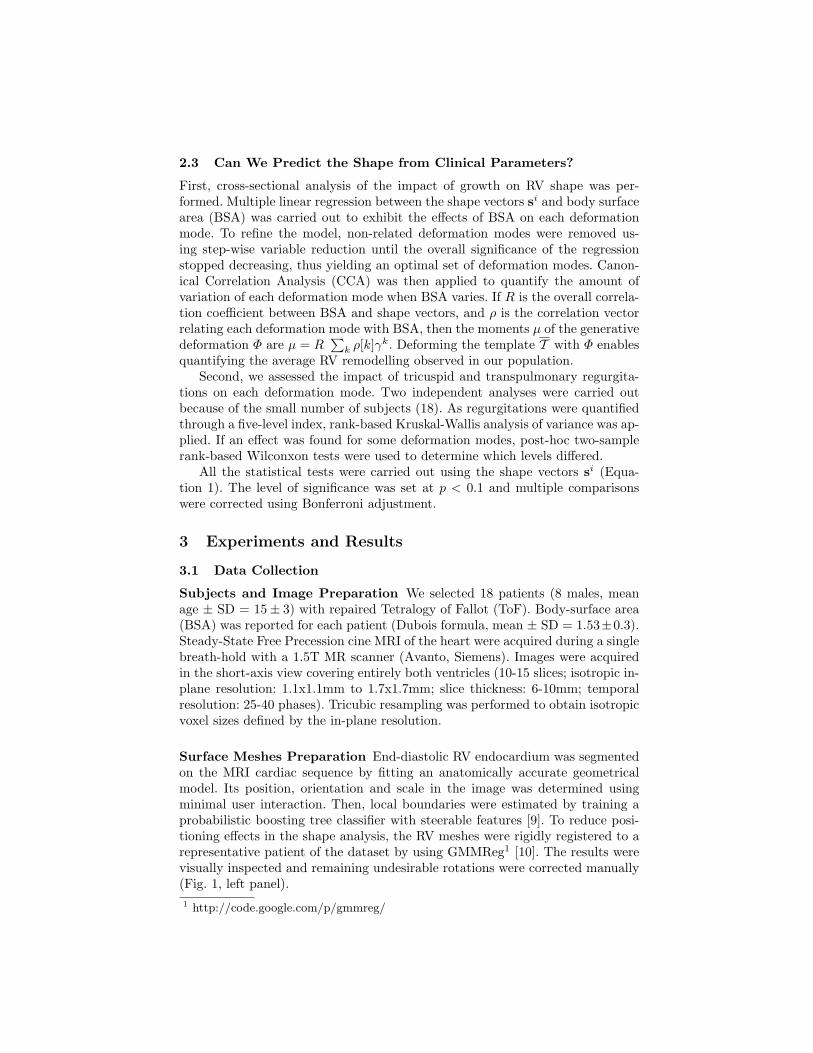
Surface Meshes Preparation End-diastolic RV endocardium was segmentedon the MRI cardiac sequence by fitting an anatomically accurate geometricalmodel. Its position, orientation and scale in the image was determined usingminimal user interaction. Then, local boundaries were estimated by training aprobabilistic boosting tree classifier with steerable features [9]. To reduce posi-tioning effects in the shape analysis, the RV meshes were rigidly registered to arepresentative patient of the dataset by using GMMReg1 [10]. The results werevisually inspected and remaining undesirable rotations were corrected manually(Fig. 1, left panel).1 http://code.google.com/p/gmmreg/
Rigid alignment Non-linear registration to the template
Fig. 1. 3D RV mesh of 18 ToF patients. Left panel: The meshes were rigidly registeredto a representative patient of the dataset. Observe the extreme variability in shape (seecompanion video). Right panel: The same meshes registered to the template using thenon-linear deformations estimated during the template creation.
3.2 Statistical Shape Model of the Right Ventricles
Building the template T required setting two parameters (Sec. 2): λV , whichdefines the “stiffness“ of the non-linear deformations (the higher is λV , the moreglobal is the transformation); and λW , which characterises the resolution of thecurrents representation (low λW values enable analysing subtle shape features).As we were mainly interested in the regional ToF alterations (dilation, valveenlargement, regional bulging), these parameters were set to λW = λV = 15mm,about the diameter of the RV outflow tract. Lower values would have beeninappropriate as the slice thickness of the images was approximately 10mm.
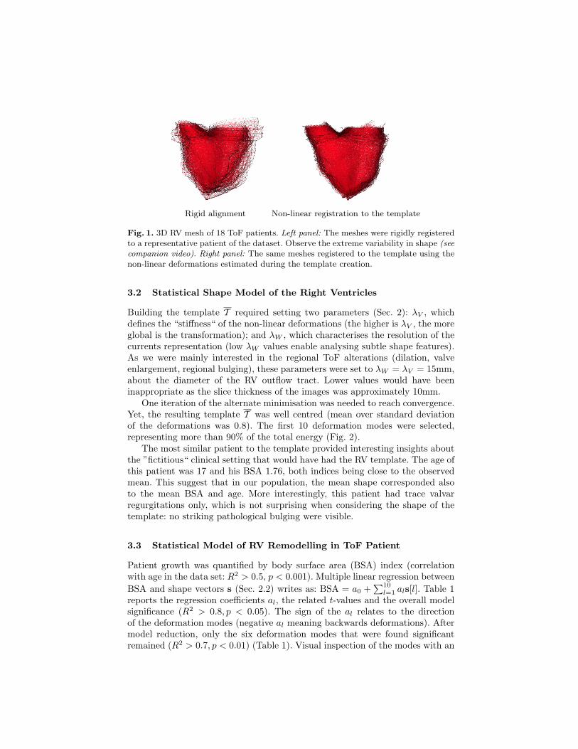
One iteration of the alternate minimisation was needed to reach convergence.Yet, the resulting template T was well centred (mean over standard deviationof the deformations was 0.8). The first 10 deformation modes were selected,representing more than 90% of the total energy (Fig. 2).
The most similar patient to the template provided interesting insights aboutthe ”fictitious“ clinical setting that would have had the RV template. The age ofthis patient was 17 and his BSA 1.76, both indices being close to the observedmean. This suggest that in our population, the mean shape corresponded alsoto the mean BSA and age. More interestingly, this patient had trace valvarregurgitations only, which is not surprising when considering the shape of thetemplate: no striking pathological bulging were visible.
3.3 Statistical Model of RV Remodelling in ToF Patient
Patient growth was quantified by body surface area (BSA) index (correlationwith age in the data set: R2 > 0.5, p < 0.001). Multiple linear regression betweenBSA and shape vectors s (Sec. 2.2) writes as: BSA = a0 +
∑10l=1 als[l]. Table 1
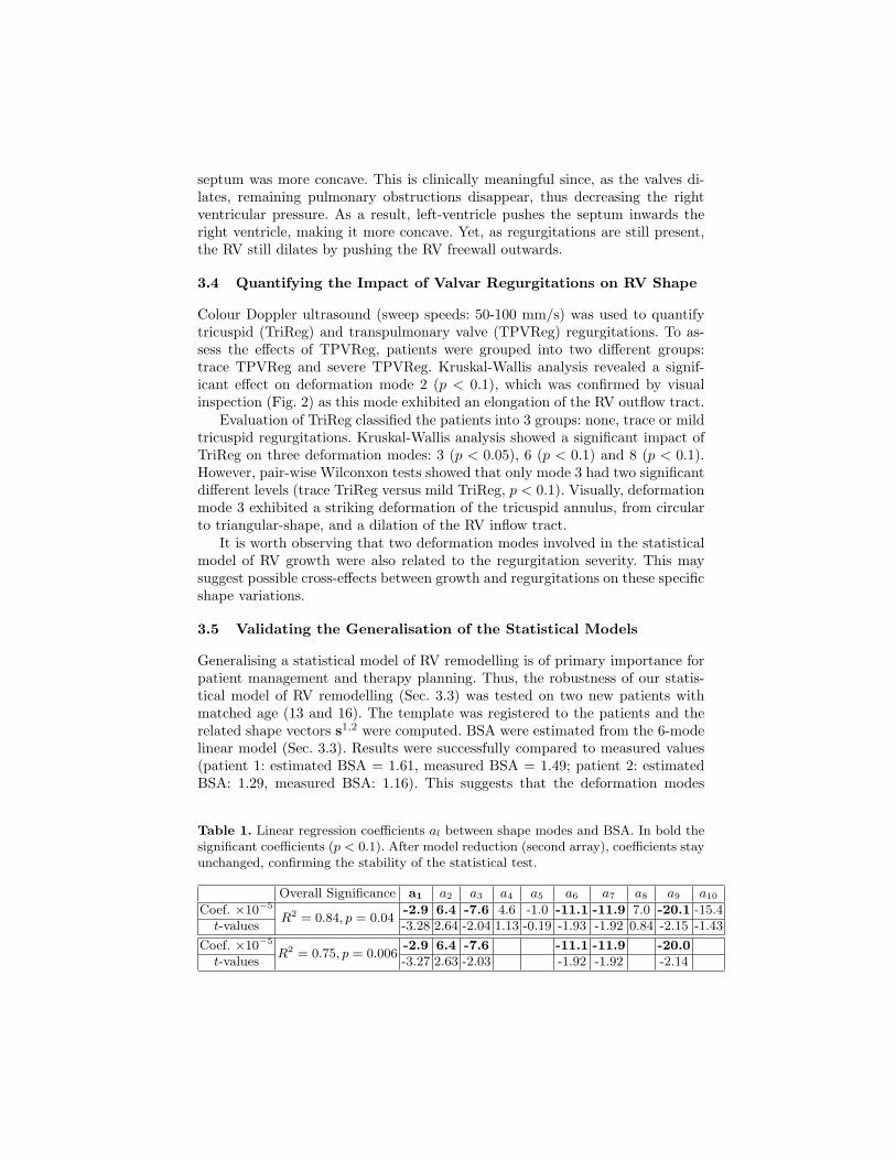
reports the regression coefficients al, the related t-values and the overall modelsignificance (R2 > 0.8, p < 0.05). The sign of the al relates to the directionof the deformation modes (negative al meaning backwards deformations). Aftermodel reduction, only the six deformation modes that were found significantremained (R2 > 0.7, p < 0.01) (Table 1). Visual inspection of the modes with an
Template
Mode 1 Mode 2 Mode 3 Mode 4 Mode 5
Mode 6 Mode 7 Mode 8 Mode 9 Mode 10
-
+
-
+
Fig. 2. 10 first deformation modes extracted by PCA a population of 18 patientssuffering from repaired Tetralogy of Fallot.
expert demonstrated their clinical relevance (Fig. 2). Mode 1 clearly representedthe overall RV dilation. Mode 2 seemed to model the dilation of the tricuspidannulus and of the inflow tract. Mode 3, 6, 7 and 9 exhibited a dilation of aspecific RV region: apex (mode 3), basal area under the tricuspid valve (mode6), apical area of the outflow tract (mode 7) and outflow tract (mode 9), reflectingpossible direct impact of regurgitations on the neighbouring tissues.
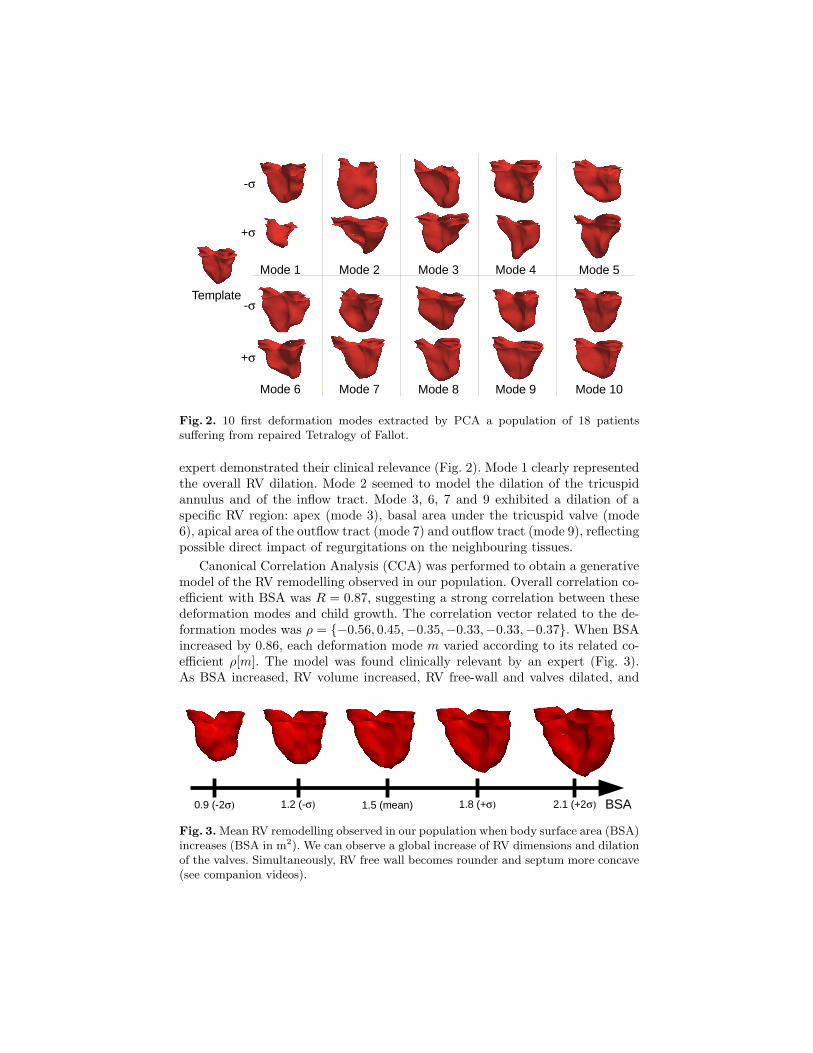
Canonical Correlation Analysis (CCA) was performed to obtain a generativemodel of the RV remodelling observed in our population. Overall correlation co-efficient with BSA was R = 0.87, suggesting a strong correlation between thesedeformation modes and child growth. The correlation vector related to the de-formation modes was ρ = {−0.56, 0.45,−0.35,−0.33,−0.33,−0.37}. When BSAincreased by 0.86, each deformation mode m varied according to its related co-efficient ρ[m]. The model was found clinically relevant by an expert (Fig. 3).As BSA increased, RV volume increased, RV free-wall and valves dilated, and
BSA1.5 (mean)0.9 (-2 1.2 (- 1.8 (+ 2.1 (+2
Fig. 3. Mean RV remodelling observed in our population when body surface area (BSA)increases (BSA in m2). We can observe a global increase of RV dimensions and dilationof the valves. Simultaneously, RV free wall becomes rounder and septum more concave(see companion videos).
septum was more concave. This is clinically meaningful since, as the valves di-lates, remaining pulmonary obstructions disappear, thus decreasing the rightventricular pressure. As a result, left-ventricle pushes the septum inwards theright ventricle, making it more concave. Yet, as regurgitations are still present,the RV still dilates by pushing the RV freewall outwards.
3.4 Quantifying the Impact of Valvar Regurgitations on RV Shape
Colour Doppler ultrasound (sweep speeds: 50-100 mm/s) was used to quantifytricuspid (TriReg) and transpulmonary valve (TPVReg) regurgitations. To as-sess the effects of TPVReg, patients were grouped into two different groups:trace TPVReg and severe TPVReg. Kruskal-Wallis analysis revealed a signif-icant effect on deformation mode 2 (p < 0.1), which was confirmed by visualinspection (Fig. 2) as this mode exhibited an elongation of the RV outflow tract.
Evaluation of TriReg classified the patients into 3 groups: none, trace or mildtricuspid regurgitations. Kruskal-Wallis analysis showed a significant impact ofTriReg on three deformation modes: 3 (p < 0.05), 6 (p < 0.1) and 8 (p < 0.1).However, pair-wise Wilconxon tests showed that only mode 3 had two significantdifferent levels (trace TriReg versus mild TriReg, p < 0.1). Visually, deformationmode 3 exhibited a striking deformation of the tricuspid annulus, from circularto triangular-shape, and a dilation of the RV inflow tract.
It is worth observing that two deformation modes involved in the statisticalmodel of RV growth were also related to the regurgitation severity. This maysuggest possible cross-effects between growth and regurgitations on these specificshape variations.
3.5 Validating the Generalisation of the Statistical Models
Generalising a statistical model of RV remodelling is of primary importance forpatient management and therapy planning. Thus, the robustness of our statis-tical model of RV remodelling (Sec. 3.3) was tested on two new patients withmatched age (13 and 16). The template was registered to the patients and therelated shape vectors s1,2 were computed. BSA were estimated from the 6-modelinear model (Sec. 3.3). Results were successfully compared to measured values(patient 1: estimated BSA = 1.61, measured BSA = 1.49; patient 2: estimatedBSA: 1.29, measured BSA: 1.16). This suggests that the deformation modes
Table 1. Linear regression coefficients al between shape modes and BSA. In bold thesignificant coefficients (p < 0.1). After model reduction (second array), coefficients stayunchanged, confirming the stability of the statistical test.
Overall Significance a1 a2 a3 a4 a5 a6 a7 a8 a9 a10
Coef. ×10−5
R2 = 0.84, p = 0.04-2.9 6.4 -7.6 4.6 -1.0 -11.1 -11.9 7.0 -20.1 -15.4
t-values -3.28 2.64 -2.04 1.13 -0.19 -1.93 -1.92 0.84 -2.15 -1.43
Coef. ×10−5
R2 = 0.75, p = 0.006-2.9 6.4 -7.6 -11.1 -11.9 -20.0
t-values -3.27 2.63 -2.03 -1.92 -1.92 -2.14
involved in the linear model of RV remodelling could be generalised, thus con-stituting potential quantitative parameters of RV remodelling in ToF.
4 Discussion and Future Works
In this study we investigated the impact of child growth and valvar regurgita-tions on RV anatomy at end diastole in patients suffering from repaired ToF.End-diastolic time point was chosen as it is the time when the effects of thepathology are the most evident [1, 2]. From multivariate statistical analyses, wederived a generative model of the observed RV remodelling. This model, as wellas the deformation modes that were found significantly related to growth and re-gurgitations, were clinically relevant as they exhibited realistic alterations in RVanatomy. Incorporating more patients is now required to confirm these findings.Adding more patients would enable identifying different groups of RV remod-elling (with aneurysm, with stiff myocardium, etc.), which might be of crucialinterest when deciding for valve replacement. Future works also include analysingthe 4D cardiac motion. To the best of our knowledge, this study constitutes afirst attempt in correlating 3D shape parameters to clinical measurements inToF. These analyses may yield quantitative image-based predictors about RVanatomy and remodelling in ToF.
References
1. Geva, T.: Indications and timing of pulmonary valve replacement after tetralogy ofFallot repair. In: Sem. Thor. and Card. Surg.: Ped. Card. Surg. Annual. Volume 9.,(2006) 11–22
2. Sheehan, F., Ge, S., Vick III, G., Urnes, K., Kerwin, W., Bolson, E., Chung,T., Kovalchin, J., Sahn, D., Jerosch-Herold, M., et al.: Three-Dimensional ShapeAnalysis of Right Ventricular Remodeling in Repaired Tetralogy of Fallot. TheAmerican Journal of Cardiology (2007)
3. Zhang, H., Walker, N., Mitchell, S., Thomas, M., Wahle, A., Scholz, T., Sonka, M.:Analysis of four-dimensional cardiac ventricular magnetic resonance images usingstatistical models of ventricular shape and cardiac motion. In: Proc. SPIE 2006.
4. Guimond, A., Meunier, J., Thirion, J.P.: Average brain models: A convergencestudy. Computer Vision and Image Understanding 77(2) (2000) 192–210
5. Joshi, S., Davis, B., Jomier, M., Gerig, G.: Unbiased diffeomorphic atlas construc-tion for computational anatomy. NeuroImage 23 (2004) 151–160
6. Allassonniere, S., Amit, Y., Trouve, A.: Towards a coherent statistical frameworkfor dense deformable template estimation. Journal of the Royal Statistical Society:Series B (Statistical Methodology) 69(1) (2007) 3–29
7. Durrleman, S., Pennec, X., Trouve, A., Ayache, N.: A forward model to buildunbiased atlases from curves and surfaces. In: Proc. MFCA 2008.
8. Vaillant, M., Glaunes, J.: Surface matching via currents. In: Proc. IPMI 2005. 3819. Zheng, Y., Barbu, A., Georgescu, B., Scheuering, M., Comaniciu, D.: Fast auto-
matic heart chamber segmentation from 3D CT data using marginal space learningand steerable features. In: Proc. ICCV 2007. 1–8
10. Jian, B., Vemuri, B.: A robust algorithm for point set registration using mixtureof Gaussians. In: Proc. ICCV 2005. 1246–1251