Embed Size (px)
Citation preview

University of Arkansas, Fayetteville University of Arkansas, Fayetteville
ScholarWorks@UARK ScholarWorks@UARK
Graduate Theses and Dissertations
12-2017
A Study of the Prevalence of Gastrointestinal Nematodes in Goats A Study of the Prevalence of Gastrointestinal Nematodes in Goats
Obtained from Northwest Arkansas Obtained from Northwest Arkansas
Christine Rose Weingartz University of Arkansas, Fayetteville
Follow this and additional works at: https://scholarworks.uark.edu/etd
Part of the Animal Studies Commons, and the Sheep and Goat Science Commons
Citation Citation Weingartz, C. R. (2017). A Study of the Prevalence of Gastrointestinal Nematodes in Goats Obtained from Northwest Arkansas. Graduate Theses and Dissertations Retrieved from https://scholarworks.uark.edu/etd/2587
This Thesis is brought to you for free and open access by ScholarWorks@UARK. It has been accepted for inclusion in Graduate Theses and Dissertations by an authorized administrator of ScholarWorks@UARK. For more information, please contact [email protected].

A Study of the Prevalence of Gastrointestinal Nematodes in Goats
Obtained from Northwest Arkansas
A thesis submitted in partial fulfillment
of the requirements for the degree of
Master of Science in Animal Science
by
Christine Rose Weingartz
Michigan State University
Bachelor of Science in Animal Science, 2011
December 2017
University of Arkansas
This thesis is approved for recommendation to the Graduate Council.
_________________________
Thomas Yazwinski, Ph.D.
Thesis Director
__________________________ ____________________________
Michael Looper, Ph.D. Jeremy Powell, D.V.M., Ph.D.
Committee Member Committee Member

Abstract
Parasitic helminths have always been an issue in small ruminant production; pathogens
that pose great negative impact on goat health and productivity. Insufficient work has been done
to document the prevalence of parasitic helminths in the United States, especially in the south
and southeast, where the largest goat populations are found.
The aim of this study was to survey the prevalence of infections by gastrointestinal
nematodes in goats in Northwestern Arkansas. Gastrointestinal tracts were examined from 41
goats of various locations around Northwest Arkansas. Worm species were identified and
population burdens were determined. Coprology was correlated with the actual worm
populations.
In descending order, the most prevalent adult nematodes were Trichostrongylus
colubriformis, Haemonchus contortus, Teladorsagia spp., Oesophagostomum spp., Trichuris
spp., Nematodirus spathiger and Cooperia curticei. Goats commonly harbored more than one
species of nematode. Nematode burdens varied greatly between animals, and respective of
management factors.

Acknowledgements
The author wishes to express her sincere appreciation and gratitude to Dr. T. A.
Yazwinski for his devoted support, guidance, and patience throughout the course of this study.
Another thank you goes to the faculty and staff at the University of Arkansas for their support
throughout my project and M.S. degree and the committee members, Dr. J. Powell, and Dr. M.
Looper for their editorial assistance and suggestions throughout this process.
Very special thanks and respect are extended to Dr. C. A. Tucker and Eva Wray for their
constant support, advice, assistance, and valued friendship throughout the course of this study.
And lastly, the author would like to send love and appreciation to her husband, family, friends
(including Esperanza) for their never-ending support and patience in this pursuit for an
advanced degree.

Table of Contents
Page
1.0 INTRODUCTION………………………………………………………………………………. 1
1.1 Nematodes…………………………………………………………………………….. 4
1.2 Abomasum Worms…………………………………………………………………….. 5
1.3 Small Intestine Worms……………………………………………………………….... 6
1.4 Large Intestine Worms………………………………………………………………… 8
1.5 Cestodes………………………………………………………………………………… 8
1.6 Trematodes…………………………………………………………………………….. 9
1.7 Anthelmintics……………………………………………………………………………. 10
2.0 MATERIALS AND METHODS………………………………………………………………… 12
3.0 RESULTS…………………………………………………………………………………….….. 15
4.0 DISCUSSION…………………………………………………………………………….……… 28
5.0 CONCLUSION……………………………………………………………………….………….. 29
6.0 REFERENCES ……..………………………………………………………….………………. 32
7.0 APPENDICES……………………………………………………………….…………………… 36
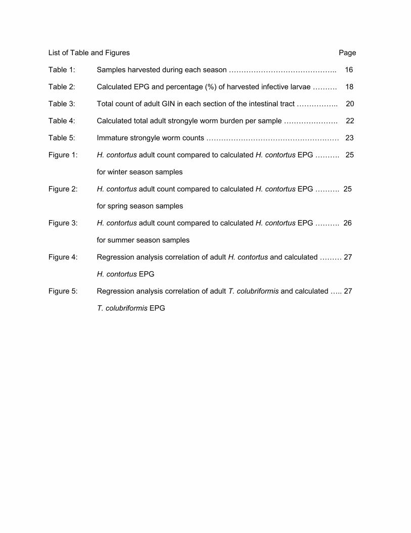
List of Table and Figures Page
Table 1: Samples harvested during each season …………………………………….. 16
Table 2: Calculated EPG and percentage (%) of harvested infective larvae ………. 18
Table 3: Total count of adult GIN in each section of the intestinal tract …………….. 20
Table 4: Calculated total adult strongyle worm burden per sample …………………. 22
Table 5: Immature strongyle worm counts ……………………………………………… 23
Figure 1: H. contortus adult count compared to calculated H. contortus EPG ………. 25
for winter season samples
Figure 2: H. contortus adult count compared to calculated H. contortus EPG ………. 25
for spring season samples
Figure 3: H. contortus adult count compared to calculated H. contortus EPG ………. 26
for summer season samples
Figure 4: Regression analysis correlation of adult H. contortus and calculated ……… 27
H. contortus EPG
Figure 5: Regression analysis correlation of adult T. colubriformis and calculated ….. 27
T. colubriformis EPG
1
1.0 Introduction
Since 1992, the meat goat industry has been a rapidly growing division of livestock
production. The primary reason is due to the goat’s popularity as livestock and the demand for
their products (“Marketing of Meat Goats”, Goat 2009). Goat production in the United States has
increased rapidly due to the economic value of the goat’s ability to convert low quality,
undesirable forages into high quality meat, milk, and fiber (Barkley et al., 2012). Demand for
goat production in the United States has not only increased due to the high quality of meat but
also in response to the ethnic populations in the United States that prefer goat meat and other
goat products (“Meat Goats”, “Goats”, “Meat Goat Ops-USDA APHIS” 2012). As of January
2015, goat and kid inventory in the United States totaled 2.68 million head (“Sheep and Goats”
2015). Eighty percent of the goats in the United States are classified as meat goats, 10% as
dairy goats and the remaining 10% as fiber goats (Solaiman, 2007). In 2015, Arkansas’ meat
goat inventory totaled 38,000 head, unchanged from 2014 (‘Arkansas Cattle, Goat and Sheep
Report”, 2015). Most goats, regardless of their initial use, eventually end up in the meat market
(“Marketing of Meat Goats”).
There are many important diseases of small ruminants, but none are as pervasive or as
direct a threat to the overall health of goats than internal parasites (Kaplan, 2010). This makes
control of intestinal parasites the most important health issue for goats of all ages (Barkley et al.,
2012; Nye et al., 2004; SARE, 2011; Waller, 2006, Várady et al., 2011; Hoste et al., 2005 and
Schoenian, 2009 a). Goats evolved as browsers. They consume a higher percentage of their
diet as brush, forbs, leaves, etc. (less desirable plants) than do other ruminants. The majority of
forage consumed by goats is located away from the ground, and this helps reduce the ingestion
of internal parasites (Barkley et al., 2012 and Fleming et al., 2006). If animals are allowed to
browse, their chances of acquiring parasitic larvae diminishes as the grazing distance from the
ground increases (Fleming et al., 2006). Goats are generally more susceptible to internal

2
parasites than sheep because goats have a lower capacity to develop an immune response
specific to helminths. The lower innate capacity for an “anti-worm” immune response is most
likely the result of their evolution (Hoste et al., 2008; McKenna and Watson, 1987; Lloyd, 1987;
Jambre, 1984; and Pomroy et al., 1986). Several studies have illustrated that both the
acquisition and the expression of immune responses against nematode species are less
efficient in goats than in sheep (Huntley et al., 1995; Pomroy et al.,1986; and Hoste et al., 2008)
In today’s goat production, a large percentage of goats are raised as grazers or intermediary
browsers. When goats are forced to graze on the same pastures as sheep, the shared
helminths may devastate the goat population while sheep are less affected (Pomroy et al.,
1986).
Prominent nematodes that infect goats and sheep include: Haemonchus contortus,
Trichostrongylus colubriformis, Teladorsagia circumcincta, Cooperia spp., Nematodirus
spathiger, Oesophagostomum spp, Trichuris spp., Dictyocaulus filaria, and Strongyloides
papillosus. These nematodes represent a major group of pathogenic agents which contribute to
the losses incurred by the goat industry. The proportions of each of these nematodes in small
ruminant populations vary according to host, geographic location, production management, etc.;
factors that dictate the overall extent of gastrointestinal parasitisms. Control of internal parasites
is of primary concern in any small ruminant health management program and is critical to
operational profitability. Naturally infected ruminants usually have mixed infections of different
species of nematodes. Goats and sheep share the same species of helminth parasites,
however, insufficient work has been conducted to determine the prevalence of parasitic
infections of goats in the United States; an initial step in constructing control strategies.
A major problem that the goat industry faces today is that resistance has developed to
all the classes of compounds used for worm control in small ruminants. Intestinal parasites have
become harder to manage in small ruminants because of the parasites’ increasing resistance to
all available chemical dewormers (SARE, 2011). Nematodes negatively impact the animal's

3
health, reduce productivity, reduce weight gain, reduce performance and increase costs due to
poor health (SARE, 2011). Depending on the balance between the parasite populations and the
host, parasitic infections can provoke clinical signs and mortality. Pathological importance is
primarily related to major production losses in quantity and quality; all induced by the direct
effect of worms.
Very few studies have been conducted in the United States, and no studies in Arkansas,
to survey the prevalence of internal parasites in goats. According to the Proceeding of the
International Symposium in 2006, research with goats is minimal because of the low economic
impact that goat products provide and the lack of organization among goat farms. In 2013,
Arkansas was ranked number 15 (out of 50) in goat production with 42,000 head (Pinkerton et
al., 2013). The majority of the goats in the US are raised in the southern and southeastern
states. The southeastern states, including Arkansas, have the most conducive climatic
conditions for the growth and establishment of large nematode parasite populations in resident
herbivores. The first report of complete failure of all classes of anthelmintics used in small
ruminants was made by Kaplan in 2005 at a meat goat farm in Arkansas (Fleming et al.,2006
and Kaplan et al., 2005). Resistance to all three drug classes of anthelmintics is now displayed
by all major nematode parasites of sheep and goats throughout the world (Waller, 2006 and
Mortensen et al., 2003).
This project was conducted to determine the incidence and prevalence of
gastrointestinal nematodes in goats residing throughout northwest Arkansas via coprologic and
necropsy examinations. Specific aims of this research were to: a) identify the worm species and
population burdens in the goats’ intestinal content, and b) to conduct fecal egg counts and larval
identifications for correlation with the actual worm populations.

4
1.1 Nematodes: “Round Worms”
Most nematodes of goats are dioecious and follow the typical Trichostrongylus, direct life
cycle. The direct life cycle is completed with one host and consists of one egg, four larval stages
and mature, reproductive adults. At all stages of development, the nematodes are cylindrical
and elongate in appearance. Extreme variations in length are seen with genus, species, sex,
and stage of development. Adult females are typically larger than adult males of the same
species. The nematodes are very site specific within the gastrointestinal tract and maintain their
position primarily by constant motility. Some nematodes attach to the mucosa by oral fixation or
wrap themselves around intestinal villi. Some “inactive” larvae embed in the tissue (or crypts) for
a varied length of time. All nematodes of small ruminants vary in their activities resulting in
pathology and also have varied means by which they are successful in the environment and
host.
Reproduction occurs in the gastrointestinal tract. The oviparous female produces eggs
that are voided from the host via the feces into the environment. Embryonation occurs
immediately if environmental conditions are suitable (temperature, moisture, oxygen). The first
stage larvae (L1) hatch out of the egg in approximately 1-2 days. The L1 feeds on bacteria and
organic material in the feces. After a few days, the L1 develops and molts into a second stage
larvae (L2). The L2 continues to live off the bacteria and organic matter in the feces (Barger,
1999, Smart drenching and FAMACHA integrated training, 2008). After approximately three
more days (one week after the egg is passed via feces) the L2 molts but does not ecdysis
(cuticle detaches from the larva but the sheath is not shed). The larva is now an en-sheathed,
infective third stage larvae (L3). The L3 stage migrates from the feces and migrates onto the
forage (negative geotropism). A grazing goat then ingests the L3 on the forage, beginning
prepatency.
Once inside the rumen of a host animal the L3 sheds its protective sheath. The L3 is
carried to its predilection location in the GI tract (abomasum, small intestine or large intestine)

5
and starts subsequent development. Within 7 days post infection, the L3 undergo the third
ecdysis and develop into a parasitic fourth stage larvae (L4). Approximately 7-20 days post
infection, the fourth and final ecdysis occurs as the L4 develops into an early adult (parasitic fifth
stage larvae (L5)). After about 7 more days, the nematode is mature. The prepatent period
(infection to egg production) is typically around 21 days, but can range from 15-40 days, post
infection. Natural death of nematodes typically occur 1 to 10 months after the adult stage is
reached (Yazwinski and Tucker, 2006). Infection is replenished by ingestion of the infective L3
by a grazing goat on a daily basis.
1.2 Abomasum Nematodes
Teladorsagia circumcincta (brown stomach worm), Figures 6a, 6b and Ostertagia
trifurcata, Figure 7, have males measuring 7.5-8.5 mm long and females measuring 9.5-12 mm
long. These worms thrive in cool, wet ambient environments. These worms follow the general
trichostrongyle life cycle. T. circumcincta, are “grazers” as they feed on the nutrients in the
mucus. The primary symptom of infection is diarrhea, due to the damage done to the stomach
lining (interfering with protein digestion and the host’s appetite). An infection with T. circimcincta
is commonly considered a production disease because the animals do not grow very well. This
worm enjoys its greatest populations in the northern tier of the US.
Haemonchus contortus (barberpole worm), Figure 8a and 8b, is the most important and
problematic nematode found in small ruminants and is mostly found in significant numbers in
the southern states (Kaplan, 2010). This large, voracious hematophagic worm measures 18-30
mm long and is readily visible on the surface of an opened abomasum. It is known as the
barberpole worm due to the appearance of the female’s white ovaries that twist around the red,
blood filled intestine. Females are very prolific egg producers (~3,000 eggs/day/female), making
them the most fecund nematode in ruminants. H. contortus is found primarily in tropical and
subtropical regions. They thrive under hot environmental conditions; being very successful in
the southeast US. Due to global warming, H. contortus is being found more and more north in

6
the US. These worms follow the general trichostrongyle life cycle, with developmental inhibition
occurring during the winter season (arrestment during the L4 stage). Transmission is the lowest
during the winter, increases in the spring (spring and post-parturient rise) with the warmer
temperature and moisture and peaks during the summer followed by a decrease in the fall.
Animals with a H. contortus infection show symptoms associated with anemia (pale mucous
membranes, bottle jaw, and hydrothorax). Blood loss can lead to death of the animal.
1.3 Small Intestine Nematodes
Trichostrongylus colubriformis (the bankrupt worm), Figures 9a and 9b, is the
predominant small intestine worm of sheep and goats. These small, thread-like worms measure
approximately 4.3-8.6 mm long and are found throughout the US. The males have a large bursa
with unequal, dark brown spicules and the females have a slit shaped vulva without distinctive
exterior lips. Both sexes have an excretory pore on the neck. These nematodes thrive under
cool and wet conditions. In small ruminants, this worm is generally the next most common and
important after H. contortus. T. colubriformis follows the general trichostrongyle life cycle. Once
in the small intestine, T. colubriformis feeds on nutrients in the mucosa, thereby causing
irritation to the mucosa and interference with digestion. Diarrhea, swelling of the intestinal wall
and edema, are common with large infections. The worm is called the bankrupt worm because
death of an animal is uncommon but the animal develops poor condition, leading to production
and income loss.
Cooperia curticei, Figures 10, is rare and relatively unimportant in small ruminants in the
US. These worms follow the general trichostrongyle life cycle and are mildly pathogenic, with no
extensive tissue invasion. These true ‘grazers’ live in the small intestine and suck on the
mucosa and villi. They will wind around the intestinal villi, causing villar constriction and rejection
(thigmokinetic effect).
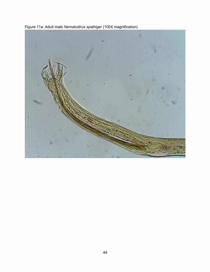
Nematodirus spathiger (the thread necked worm), Figure 11, is a large worm found in
the small intestine and is found throughout the US, usually in small numbers. These worms

7
follow the general trichostrongyle life cycle, with some unique variations. The L1, L2, and L3
stages develop and stay inside the egg, conferring high environmental resistance. Within the
egg, the larvae have the ability to exist on contaminated pastures for 2 years (Yazwinski,
unpublished 2012). On pasture the L3s hatch out of the egg due to proper environmental
conditions (time, temperature, moisture, etc.). In the small intestine, adults strangle and atrophy
the villi, triggering the thigmokinetic effect (villus rejection); this can cause diarrhea that leads to
production loss. Nematodirus spp. infections are limited to younger animals; a condition
primarily due to the animal’s age and not induced immunity.
Strongyloides papillosus (intestinal thread worm), Figure 12, is a unique worm found in
the small intestine of sheep, goats and cattle around the US. This worm has the ability to adjust
to its environment by alternating free-living and parasitic life cycles (heterogonic and homogonic
cycles, respectively), with only females being parasitic. The cycle executed is dependent on the
environment that the infective larvae encounter. If the free-living environment is good (wet) the
heterogonic cycle will predominate. If the free-living environment is bad (dry) the homogonic
cycle will predominate. Parthenogenetic females in the small intestine produce small, light
colored embryonated eggs and the eggs pass out in the feces. In the homogonic cycle;
environmental stages transverse to the filariform. Following the heterogonic cycle;
environmental stages include heterogonic males and females (free-living adult males and
females). Progeny of these adults are larvae that develop into infective L3s with the filariform
esophagus. No matter the cycle, the infective L3s penetrate through the skin and migrate
directly into the host’s blood stream. Transmammary infection has also been demonstrated but
is probably rare. Larvae in the blood stream break into the mammary glands of the lactating
animal and infect the offspring. Larvae are carried to the lungs from the blood, coughed up,
swallowed and are passed to the small intestine. Pathogenesis of these worms is small intestine
enteritis and diarrhea.

8
1.4 Large Intestine/Cecum Nematodes
Oesophagostomum spp. (the nodular worm), Figure 12, are “large” worms and are found
throughout the US in relatively low numbers. They follow the general trichostrongyle life cycle,
with a few variations. The infective L3 will infect per os or penetrate through the skin. Those that
infect transcutaneously go through a tracheal migration and end up in the small intestine. The
L3 larvae penetrate deep into the mucosa of the small intestine and nodules form around the
L4s. Animals will not develop nodules the first time they are infected with Oesophogostomum
spp., only upon a challenge infection. L4s in the nodules will either die or break out of the
nodules to migrate and reside in the large intestine as adults. Adults and L4 feed on the host
blood and tissue which contributes to the overall anemia of the host. Females are 13-24 mm
long and males are 11-16 mm long, both with cephalic vesicles. Conditions associated with an
active Oesophogostomum spp. infection include bloody, tarry diarrhea (caused by the L4s
leaving the nodules) emaciation and weakness.
Trichuris spp. (the whipworm), are usually found in relatively low numbers in the cecum.
Males measuring 50-80 mm long have a coiled body and females measuring 30-70 mm long
have a banana-shaped body. The anterior end of the whipworm is thread-like and is used to
thread the worm into the mucosa; making them hard to clear from an animal with most
anthelmintics. Trichuris spp. are haematophagic and a large population can contribute to the
overall anemia of the host. Usually, however they are relatively non-pathogenic. They follow the
general trichostrongyle life cycle, except that the infective larva is the second stage (L2) and it
stays inside the egg until eaten, and hence very resistant to the environment.
1.5 Cestodes: “Tapeworms”
Moniezia expansa is the primary tapeworm that infects the small intestine of small
ruminants in the United States. These tapeworms vary in length due to immunity, cestocial
treatment, “worm pressure” and age of the worm. The appearance of the strobilar (adult) form is
completely different from the metacestode (larval) form. This hermaphroditic worm completes an

9
indirect life cycle. The intermediate host (orbatid mite) ingests expelled tapeworm eggs and
eventually harbors the cysticercoid stage. The infection is transmitted when the definitive host
consumes the infected mite. Once inside the definitive host intestinal tract, the protoscolexes
are released from the cysticercoids and they attach to the intestinal wall. The scolex (head) of
the tapeworm actually attaches the worm to the wall of the small intestine via four suckers. The
neck of the scolex grows the proglottids of the worm. The adult tapeworm maintains its position
via the suckers, adhesion of the flat strobilus to the mucosa, and winding with the curves of the
GI tract. The strobilus “feeds” via diffusion through its microtriche tegument (cuticle), thereby
absorbing nutrients from the host’s digested feed. Many producers are alarmed by tapeworm
infections in their animals because the white segments (proglottids) are visible on the feces of
an infected host. In truth however, very little damage is caused by normal (small) tapeworm
infections. Heavy infections may reduce growth rates in kids and may cause intestinal blockage
but these conditions are rarely seen.
1.6 Trematodes “Flukes”
Fasciola hepatica is the liver fluke and causes fascioliasis in ruminants. According to
Martinez-Moreno et. al goat fascioliasis is less frequent and less important than infections in
sheep and cattle (Martinez-Moreno et. al., 1999). F. hepatica, infection is not a concern for small
ruminant producers in the Northwest region of Arkansas due to fluke life cycle requirements.
The pastureland in which animals are grazing must be partially aquatic for a good portion of the
year; a circumstance more of a concern in the southeastern states of the US. Liver flukes vary
in size due to immunity and age of the worm. This hermaphroditic worm completes an indirect
life cycle by using an active, semi-aquatic snail as an intermediate host. Leaf shaped adults
maintain their position in bile ducts via suckers, cuticular hooks, molding to the shape of the
surrounding environment, and becoming larger in size than their current location. These
parasites reside in and cause damage in the liver. Pre-adults have a continuum of growth until
they mature and wedge into the collecting bile ducts of the liver. Infection by F. hepatica in goats
10
usually develops into a chronic disease. This was confirmed in a study by Martinez-Moreno et.
al who showed that an immune response occurs in goats but the goats never develop complete
resistance, resulting in unthriftiness of the host, weight loss and sometimes death.
1.7 Anthelmintics: Classes of Anthelmintics
A. Benzimidazoles:
Fenbendazole (Safeguard and Panacur) and albendazole (Valbazen) are the two
most commonly used Benzimidazoles in goats. Fenbendazole has a wide margin
of safety but albendazole can be embryo-toxic (teratogenic). The benzimidazoles
are known as the “white wormers”, due to their white appearance.
Benzimidazoles kill helminths by disrupting microtubule formation. Currently in
the United States, there are high levels of resistance to the benzimidazoles by
both H. contortus and T. colubriformis populations (Howell et al., 2008).
Producers should use benzimidazoles to control gastrointestinal nematodes only
if their worm burdens have been shown to be drug responsive/susceptible
(FECRT).
B. Imidazothiazole/tetrahydropyrimidine:
Levamisole (Levasol, Tramisol, and Prohibit) kills gastrointestinal nematodes by
depolarizing nicotinic neuromuscular junctions. It also acts as a cholinergic
agonist in mammals, which is the reason for its narrow therapeutic index
(Williamson, 2013). It is very important that animals be properly weighed and
dosed when using levamisole (as well as any other anthelmintic). Animals should
not be fasted prior to administering levamisole because toxicity is a concern.
There are some populations of H. contortus in the U.S. that are still susceptible to
levamisole (Howell et al., 2008 and Williamson, 2013).
Morantel tartrate (Rumatel) is a tetrahydropyrimidine drug. It also acts as a
cholinergic agonist, but at a less potent level and has a larger margin of safety.
11
C. Macrocyclic Lactones (ML)
This group is composed of 2 groups; avermectins (ivermectin, doramectin,
eprinomectin) and milbemycins (moxidectin). The primary activity of the MLs is
directed at the glutamate-gated ion exchange gates in the cellular membrane of
the nerves and muscles of the nematodes. MLs cause flaccid paralysis of the
nematode by interfering with neurotransmission and muscle cell junction. The
antiparasitic effect is mediated through selective binding to glutamate-gated
chloride ion channels. MLs are lipophilic and do not cross the blood brain barrier
in most mammals. MLs have a wide safety margin in mammals. According to S
Howell, H. contortus in populations are already resistant to ivermectin and in the
process of becoming resistant to Moxidectin (Howell et al., 2008).
The number of FDA approved drugs for goats is very limited; morantel (rumatel),
thiabendazole (omnizole, no longer marketed), fenbendazole (Safeguard and Panacur) and
phenothiazine (feno-drench suspension) which is no longer available in the USA (Kaplan, 2010).
Effective control of gastrointestinal nematodes in goats can usually only be accomplished by
using drugs in an extra label manner and with the assistance of a licensed veterinarian.
Unapproved drugs that can be effective for the treatment of gastrointestinal nematodes in goats
include ivermectin, doramectin, moxidectin (Cydectin), albendazole and levamisole.
Goats metabolize (detoxify) drugs much more rapidly than other livestock, thereby
requiring high dosing (Kaplan, 2010). Depending on the anthelmintic being used, goats require
1.5-2 times the dose recommended for effectiveness on the label for sheep (SARE, 2011).
Reasons for resistance to develop against anthelmintics include under dosing the
animal, rotating drugs too rapidly, dosing animals too frequently, non-strategic dosing, etc.
Nematode resistance is genetically conferred. The use of chemical anthelmintics selects for
resistance in the nematode population over time. There is a need to balance chemical
intervention with proper management. Anthelmintics should only be administered to animals that

12
need treatment. The animals in the herd that remain untreated harbor gastrointestinal
nematodes that will stay more susceptible to anthelmintics (refugia), thereby helping prolong
chemical effectiveness.
2.0 Materials and methods
The following materials and methods were used throughout the entirety of the survey for
each study animal used.
2.1 Necropsy and intestinal helminth collection
Forty-one gastrointestinal tracts from goats were collected between October 2013 and
March 2015. All tracts were collected immediately post slaughter at local processing plants or
farms and transported to the University for immediate processing. Processing of all intestinal
tracts were conducted according to the W.A.A.V.P. guidelines (Wood et al., 2010).
Immediately after animal demise the omasal and pyloric ends of the abomasum, in addition to
the ileocecal junction, were ligated using heavy cotton string; thereby preventing the movement
of contents (and nematodes) from their proper locations within the gastrointestinal tract. The
three relevant sections of the GI tract (abomasum, small intestine and large intestine/cecum)
were separated and placed into separate basins. If available, a fecal sample was collected
directly from the rectum for coprology (fecal egg counts, coproculture, larval harvest and
identification).
The abomasum was opened longitudinally and the contents collected into a graduated
bucket. The opened abomasum was thoroughly rinsed and washed between each fold by hand.
The rinse water and contents were combined in the bucket and brought up to 2L using tap water
for aliquot retrieval. The cleaned abomasum was then covered with water and placed in a
refrigerator to soak overnight.
The mesentery around the small intestine and large intestine was removed. The small
intestine was opened along its entire length and the contents collected into a graduated bucket.
The small intestine was then rinsed and “stripped” by hand. The rinse water and contents were

13
combined in a bucket and brought to 4L by added water for aliquot retrieval. The cecum and
one-third of the length of the large intestine (from the illeocecal junction) was processed the
same way as the small intestine. The content and rinse water were brought to 2L by adding
water for aliquot retrieval. Visible adult worms (Trichuris spp.) were detached from the cecum
and added to the collected contents. No gall bladders or livers were collected to search for
Fasciola hepatica infections. Lungs were not collected for Dictyocaulus filaria.
2.2 Intestinal content preservation
Five percent aliquots of the abomasum, small intestine and large intestine/cecum
contents were removed during vigorous, constant stirring. The separate aliquots were
formalized using a sufficient amount of 10% formalin and placed at room temperature until
nematode isolation and identification. The abomasum (after the overnight soak) was thoroughly
stripped by hand to ensure all the mucus had been dislodged and made part of the soak
collection. One hundred percent of the soak collection was formalized and placed at room
temperature until nematode isolation, identification, and quantification.
2.3 Intestinal helminth isolation,identification and quantification
The aliquots were washed over appropriate mesh sieves; abomasum content, No. 100
(aperture of 150 μm), abomasum soak, No. 400 (aperture of 38 μm), small intestine content, No.
120 (aperture of 125 μm), and large intestine content, No. 60 (aperture of 250 μm).
Subsamples of suspended (appropriately stirred) sieved residues were examined under
a stereoscopic microscope at 10-60X for parasite isolation and counting (approximately 20 mL
subsamples from a measured amount). Most of the adult and L4 parasites were identified using
the stereoscopic microscope. Adults and L4s that could not be accurately identified were
mounted in lacto-phenol for identification and counting using a compound microscope at 40-
200X. All parasites were identified to genus, species (if possible), sex and stage of
development. Adult and larval identifications were based on Van Wyk and Mayhew, 2013.
2.4 Fecal egg per gram count: Direct fecal flotation

14
One gram of feces was homogenized in 10 mL of saturated magnesium sulfate
(MgSO4), and poured over a wet sieve (1mm aperture). The filtrate was poured into a 15 mL
plastic centrifuge tube, and additional MgSO4 was added until a slight meniscus was visible
over the rim of the tube. One glass coverslip was gently placed on the test tube. The tube was
centrifuged for three minutes. The coverslip was then placed on a glass microscope slide and
examined at 40-100X for the adhered egg counts. Using a compound microscope, eggs were
identified and counted as strongyle, Trichuris spp., or Nematodirus spp. Eggs (Figure 14). The
presence of Strongyloides and Moniezia eggs were noted.
2.5 Fecal coprocultures and harvesting of infective larvae
Samples of feces with an EPG greater than 20 were soaked in water, until softened, and
thoroughly homogenized with vermiculite to yield a moist, standardized mixture. The fecal-
vermiculite mixture (coproculture) was formed into a concave depression inside a 16 oz plastic
cup. Multiple vertical ridges were pressed into the mixture to increase the available surface
area. The culture was then covered with foil and allowed to stand at room temperature for 12-14
days before L3 harvest.
For larval harvest a one-inch section of the solo cup rim was scratched. Water was then
added to the cup until a slight meniscus protruded over the rim of the cup. A Petri plate was
inverted over the cup to form a seal. Using proper technique, the cup and Petri dish were
inverted and left at an incline, with the scratched area facing the lowest point (for L3 to escape
into the petri dish). Water was added to the Petri dish until the scratched area of the rim was
fully covered. The culture was left undisturbed for over three hours. The water in the Petri dish
was collected using a pipette and transferred to a glass centrifuge tube. Using a water squeeze
bottle, the empty Petri dish was rinsed and the water was collected and placed into the same
glass centrifuge tube. The L3 collection test tube was placed, uncovered, in a refrigerator for
one day (to allow for L3 settling).

15
2.6 Larva preparation for identification
Once the L3s had settled overnight, the supernate was carefully discarded using a
pipette, leaving about 4mls of water in the bottom of the tube. To kill the precipitated larvae, an
equal amount of formalin was added to the larval precipitation and agitated by hand. To
straighten out the L3, the suspension was heated over a flame until a transient boil. The killed
and straightened L3 were centrifuged for 15 minutes. The top fluid was discarded via pipetting
down to the L3 pellet. Using a pipette, the larvae were suspended, in the remaining liquid, and a
drop of the larval suspension was placed on a glass microscope slide and covered with a
coverslip.
2.7 Larva identification
Using a compound microscope, the genus-specific identification of the first 100 L3 per
sample was accomplished using the length of the tail of the sheath (STE), the head shape, and
overall L3 characteristics (Figure 15). Larvae were identified based on the published, detailed
features. (VanWyk et al., 2013).
3.0 Results
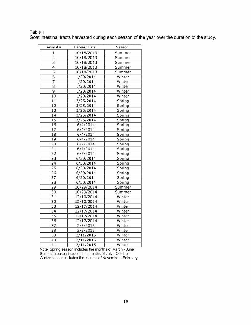
The month from which a goat intestinal tract was harvested and categorized into a
specific season is represented in Table 1. Spring season; March through June, includes 18
intestinal tracts, animal numbers 11-28. Summer season; July through October, includes seven
intestinal tracts, animal number 1-5 and 29-30. Winter season; November through February,
includes 16 intestinal tracts, animal number 6-10, 31-41.
16
Table 1 Goat intestinal tracts harvested during each season of the year over the duration of the study. _________________________________________ Animal # Harvest Date Season
1 10/18/2013 Summer
2 10/18/2013 Summer
3 10/18/2013 Summer
4 10/18/2013 Summer
5 10/18/2013 Summer
6 1/20/2014 Winter
7 1/20/2014 Winter
8 1/20/2014 Winter
9 1/20/2014 Winter
10 1/20/2014 Winter
11 3/25/2014 Spring
12 3/25/2014 Spring
13 3/25/2014 Spring
14 3/25/2014 Spring
15 3/25/2014 Spring
16 6/4/2014 Spring
17 6/4/2014 Spring
18 6/4/2014 Spring
19 6/4/2014 Spring
20 6/7/2014 Spring
21 6/7/2014 Spring
22 6/7/2014 Spring
23 6/30/2014 Spring
24 6/30/2014 Spring
25 6/30/2014 Spring
26 6/30/2014 Spring
27 6/30/2014 Spring
28 6/30/2014 Spring
29 10/29/2014 Summer
30 10/29/2014 Summer
31 12/10/2014 Winter
32 12/10/2014 Winter
33 12/17/2014 Winter
34 12/17/2014 Winter
35 12/17/2014 Winter
36 12/17/2014 Winter
37 2/5/2015 Winter
38 2/5/2015 Winter
39 2/11/2015 Winter
40 2/11/2015 Winter
41 2/11/2015 Winter
Note: Spring season includes the months of March - June Summer season includes the months of July - October Winter season includes the months of November - February

17
Of the 41 acquired intestinal tracts, fecal egg counts (FEC) were determined from 39
individual intestinal tracts. L3 larvae was harvested and quantified from 35 of those 39 fecal
samples, with results reported in Table 2. All 39 fecal samples examined were positive for
strongyle eggs. Strongyle egg per gram (EPG) counts ranged from 5 to 16,650 EPG. Strongyle
egg counts in the summer season ranged from 9-271 EPG, spring season samples ranged from
20-16,650 and winter season samples ranged from 5-2,769 EPG. Nematodirus spp. and
Trichuris spp. eggs were present in a small portion of the fecal samples; 10% and 23%,
respectively. H. contortus was the highest percentage of harvested L3 followed closely by T.
colubriformis. Oesophagostomum spp. L3 was present at a low number in 7 out of the 35
coproculture samples.

18
Table 2
Egg per gram (EPG) counts and genus specific percentages (%) of harvested coproculture infective
larvae (L3).
______________________________________________________________________ EPG as: % of L3 as:
___________________________ _________________________________________ Animal # Strongyle Nematodirus spp. Trichuris spp. H. contortus T. colubriformis Oesophagostomum spp
1 271 1 0 68 32 0
2 - - - - - -
3 209 0 0 65 35 0
4 54 0 0 42 58 0
5 104 0 0 50 50 0
6 44 0 0 - - -
7 5 0 0 0 100 0
8 47 0 0 62 38 0
9 68 0 0 4 96 0
10 91 0 0 88 12 0
11 160 0 4 63 37 0
12 206 0 2 89 11 0
13 1248 0 0 50 50 0
14 138 0 0 96 4 0
15 20 0 2 F.L. F.L. F.L
16 176 0 0 99 1 0
17 195 0 0 98 2 0
18 800 0 0 97 3 0
19 170 0 0 93 7 0
20 113 6 3 88 22 0
21 48 3 60 3 15 0
22 445 18 51 71 19 0
23 2600 0 0 50 49 1
24 5850 0 0 61 35 4
25 16650 0 0 91 9 0
26 12650 0 0 86 14 0
27 8350 0 0 81 17 2
28 1550 0 0 8 88 4
29 27 0 3 - - -
30 9 0 0 - - -
31 2769 0 0 50 50 0
32 437 0 0 27 73 0
33 1185 0 0 10 90 0
34 117 0 0 3 97 0
35 1500 0 15 19 81 0
36 212 0 1 14 33 53
37 29 0 0 16 56 28
38 67 0 0 22 78 0
39 1504 0 0 0 100 0
40 272 0 0 56 36 8
41 - - - - - -
Note: Fecal egg counts were quantified using direct flotation with MgSO4 of fecal filtrate from one gram of feces
collected directly from the rectum of each study sample.
FL represents free living larvae and - represents no sample available
19
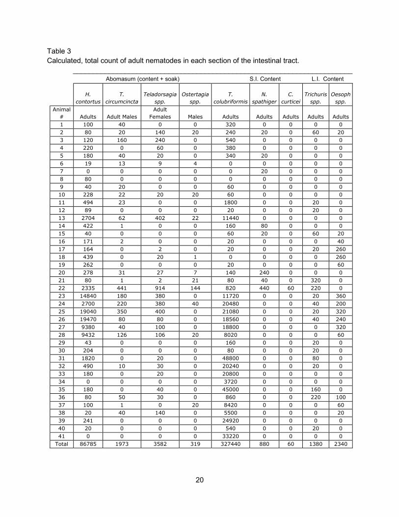
One hundred percent of the goat intestinal tracts surveyed were positive for at least one
species of adult nematode. The predominant adult nematodes found throughout the survey
were T. colubriformis and H. contortus. T. colubriformis had large populations in samples
obtained from the spring and winter seasons. Samples 22 through 28, obtained during the
spring season, had the highest number of H. contortus adults and immatures. During the
summer season is when T. colubriformis was at its lowest population. C. curticei was found in
one small intestine sample during the spring season. In the large intestine, small populations of
both Trichuris spp. and Oesophagostomum spp. were present throughout all seasons.
20
Table 3
Calculated, total count of adult nematodes in each section of the intestinal tract.
______________________________________________________________________
Abomasum (content + soak) S.I. Content L.I. Content
H.
contortus
T.
circumcincta
Teladorsagia
spp.
Ostertagia
spp.
T.
colubriformis
N.
spathiger
C.
curticei
Trichuris
spp.
Oesoph
spp.
Animal
# Adults Adult Males
Adult
Females Males Adults Adults Adults Adults Adults
1 100 40 0 0 320 0 0 0 0
2 80 20 140 20 240 20 0 60 20
3 120 160 240 0 540 0 0 0 0
4 220 0 60 0 380 0 0 0 0
5 180 40 20 0 340 20 0 0 0
6 19 13 9 4 0 0 0 0 0
7 0 0 0 0 0 20 0 0 0
8 80 0 0 0 0 0 0 0 0
9 40 20 0 0 60 0 0 0 0
10 228 22 20 20 60 0 0 0 0
11 494 23 0 0 1800 0 0 20 0
12 89 0 0 0 20 0 0 20 0
13 2704 62 402 22 11440 0 0 0 0
14 422 1 0 0 160 80 0 0 0
15 40 0 0 0 60 20 0 60 20
16 171 2 0 0 20 0 0 0 40
17 164 0 2 0 20 0 0 20 260
18 439 0 20 1 0 0 0 0 260
19 262 0 0 0 20 0 0 0 60
20 278 31 27 7 140 240 0 0 0
21 80 1 2 21 80 40 0 320 0
22 2335 441 914 144 820 440 60 220 0
23 14840 180 380 0 11720 0 0 20 360
24 2700 220 380 40 20480 0 0 40 200
25 19040 350 400 0 21080 0 0 20 320
26 19470 80 80 0 18560 0 0 40 240
27 9380 40 100 0 18800 0 0 0 320
28 9432 126 106 20 8020 0 0 0 60
29 43 0 0 0 160 0 0 20 0
30 204 0 0 0 80 0 0 20 0
31 1820 0 20 0 48800 0 0 80 0
32 490 10 30 0 20240 0 0 20 0
33 180 0 20 0 20800 0 0 0 0
34 0 0 0 0 3720 0 0 0 0
35 180 0 40 0 45000 0 0 160 0
36 80 50 30 0 860 0 0 220 100
37 100 1 0 20 8420 0 0 0 60
38 20 40 140 0 5500 0 0 0 20
39 241 0 0 0 24920 0 0 0 0
40 20 0 0 0 540 0 0 20 0
41 0 0 0 0 33220 0 0 0 0
Total 86785 1973 3582 319 327440 880 60 1380 2340

21
As shown in Table 4, goat intestinal tract samples obtained during the winter months of
November through February showed the highest adult strongyle worm burdens. The summer
months of July through October showed the lowest adult strongyle worm burdens. The total
combined adult strongyle worm burden for the individual animals varied, ranging from 0 to
50,640 adults. All samples with high strongyle EPG counts had either a higher number of H.
contortus and/or T. colubriformis adults in the intestinal tract. Immature strongyle worms were
the most prevalent in the spring season followed by the winter season (Table 5). H. contortus
immature worms were the most prevalent followed by T. colubriformis.

22
Table 4
Calculated total adult strongyle worm burden per study sample.
Season Animal #
H. contortus
Adult
T. colubriformis
Adult
Oesophagostomum spp.
Adult
Summer 1 100 320 0
Summer 2 80 240 20
Summer 3 120 540 0
Summer 4 220 380 0
Summer 5 180 340 0
Winter 6 19 0 0
Winter 7 0 0 0
Winter 8 80 0 0
Winter 9 40 60 0
Winter 10 228 60 0
Spring 11 494 1800 0
Spring 12 89 20 0
Spring 13 2704 11440 0
Spring 14 422 160 0
Spring 15 40 60 20
Spring 16 171 20 40
Spring 17 164 20 260
Spring 18 439 0 260
Spring 19 262 20 60
Spring 20 278 140 0
Spring 21 80 80 0
Spring 22 2335 820 0
Spring 23 14840 11720 360
Spring 24 2700 20480 200
Spring 25 19040 21080 320
Spring 26 19470 18560 240
Spring 27 9380 18800 320
Spring 28 9432 8020 60
Summer 29 43 160 0
Summer 30 204 80 0
Winter 31 1820 48800 0
Winter 32 490 20240 0
Winter 33 180 20800 0
Winter 34 0 3720 0
Winter 35 180 45000 0
Winter 36 80 860 100
Winter 37 100 8420 60
Winter 38 20 5500 20
Winter 39 241 24920 0
Winter 40 20 540 0
Winter 41 0 33220 0

23
Table 5
Immature nematode worm counts.
Animal
# H. contortus Teladorsagia spp.
T.
colubriformis Cooperia spp. Oesophagostomum spp.
1 0 0 0 0 0
2 0 0 0 0 0
3 0 0 0 0 0
4 0 0 0 0 0
5 0 0 0 0 0
6 7 0 0 0 0
7 0 0 0 0 0
8 0 0 0 0 0
9 2 0 0 0 0
10 45 0 0 0 0
11 6 1 20 0 0
12 2 0 0 0 340
13 1187 4 560 0 0
14 1363 22 0 0 0
15 1 0 0 0 0
16 0 0 0 0 880
17 0 0 0 0 500
18 2 0 0 0 540
19 0 0 0 0 120
20 476 31 60 0 0
21 206 2 20 0 80
22 1836 305 280 40 80
23 4180 230 460 0 60
24 2060 380 760 0 40
25 14560 880 540 0 40
26 19190 420 400 0 60
27 20980 280 340 0 0
28 3400 123 80 0 0
29 35 0 80 0 0
30 20 0 0 0 0
31 400 0 2600 0 0
32 2660 30 200 0 60
33 25 20 200 0 0
34 724 0 20 0 0
35 290 0 300 0 0
36 440 0 0 0 120
37 3230 209 120 0 100
38 980 50 40 0 40
39 87 0 640 0 0
40 0 0 0 0 0
41 0 0 980 0 0

24
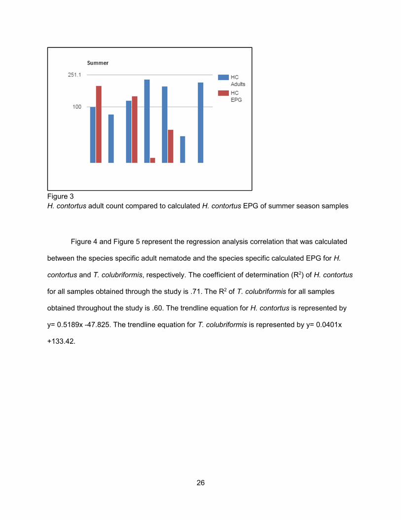
Figures 1 through 3 depict the populations of adult H. contortus in the intestinal tracts of
samples obtained in the winter, spring, and summer seasons, respectively, compared to the
calculated H. contortus EPG of the fecal samples determined from those same samples.The
greatest populations of H. contortus were found in the samples obtained during the spring
season. All 18 samples were positive for adult H. contortus, ranging from 40-19,470 adults. Of
the 16 fecal samples collected all were above 99 EPG. In the winter season samples 13 out of
16 samples were positive for H. contortus adults, ranging from 0 to 1820 adults. Out of the 14
winter fecal samples only 6 had a calculated H. contortus EPG of over 30. For the summer
season samples an EPG was conducted from 4 out of the 7 samples. All summer samples had
an H. contortus adult count ranging from 43-220.

25
Figure 1
H. contortus adult count compared to calculated H. contortus EPG of winter season samples.
Figure 2
H. contortus adult count compared to calculated H. contortus EPG of spring season samples.
26
Figure 3
H. contortus adult count compared to calculated H. contortus EPG of summer season samples
Figure 4 and Figure 5 represent the regression analysis correlation that was calculated
between the species specific adult nematode and the species specific calculated EPG for H.
contortus and T. colubriformis, respectively. The coefficient of determination (R2) of H. contortus
for all samples obtained through the study is .71. The R2 of T. colubriformis for all samples
obtained throughout the study is .60. The trendline equation for H. contortus is represented by
y= 0.5189x -47.825. The trendline equation for T. colubriformis is represented by y= 0.0401x
+133.42.

27
Figure 4
Regression analysis correlation of adult H. contortus and calculated H. contortus EPG. Note: equation y=0.5189x-47.825 is the Linear Regression Equation
R2=0.7141 is the coefficient of determination
Figure 5
Regression analysis correlation of adult T. colubriformis and calculated T. colubriformis EPG. Note: equation y=0.0401x + 133.42 is the Linear Regression Equation
R2=0.5949 is the coefficient of determination
28
4.0 Discussion
Under natural environmental conditions, goats commonly harbor more than one species
of nematode. The nematode burden of mature and immature worms varies greatly among
animals in the same season, as shown in this study. The degree of nematode infection acquired
by goats is determined by natural and management factors, seasonal and environmental
conditions, grazing behavior, previous exposure to nematodes, physiological state of the goat,
stocking rate, nutrition, age of the goat, and previous anthelmintic treatment.
As seen in this survey, worm burdens are not evenly distributed within the animal
population. It is a rule of thumb that 20-30% of the animals in a population harbor about 80% of
the parasites. These 20-30% of the animals with higher parasite burden are primarily
responsible for contaminating the environment with infective larvae for all other animals (Kaplan,
2010, Fleming et al., 2006). Forty out of the 41 of the goats sampled in this survey were positive
for at least one species of adult nematodes. Ninety-two percent and 90% of sampled goats were
positive for H. contortus and T. colubriformis, respectively. The total adult nematode worm
burden ranged from 0 to 50,640 adults.
Fecal egg counts are only relatively crude indicators of worm burdens. The number of
eggs in the feces may not always correlate with the number of parasites present in the intestinal
tract. Differences in fecundity may mask the number of nematodes and low EPG or negative
counts occur due to a large number of immature or non-fecund worms (Merck Manual,
McKenna and Watson, 1987, Hoste et al., 2001). The 39 fecal samples examined for this survey
were all positive for strongyle eggs. The lowest strongyle count was 5 EPG, found in sample #7,
obtained in the winter season. The highest strongyle count was 16,650 EPG, found in sample
#25, obtained in the spring season. A large EPG range for each season of this survey was
observed. Overall, the summer season observed the lowest EPG range while the spring season
had the highest EPG values. All samples with high strongyle EPG were correlated either a
29
higher number of H. contortus or T. colubriformis adults in the intestinal tract. As demonstrated
in figures 4 and 5, the calculated EPG of H. contortus and T. colubriformis both show positive
linear correlation to the species specific adults obtained from the goat samples during the
survey.
In the southern USA, the inhibited state of parasitic nematodes occurs during the heat of
the summer and the cold of the winter, and is dependent upon the nematode species.
Hypobiosis results in an extended time for an immature nematode to develop into an adult.
During hypobiosis, few eggs are deposited into the environment. In this survey, the summer
months of July through October showed the lowest adult worm burdens, and coincide with
decreased egg counts and pasture infectivity.
More adult nematodes were found in the winter samples, as shown by the highest adult
nematode worm burdens in samples from November through February. A more accurate picture
of nematode species seasonal prevalence would have been possible if a consistent and
representative number of intestinal tracts were inspected for each season. For example, the
summer season for this study contained only a few samples and those samples were all
collected during the month of October. In addition to more samples, information on each animal
in the study would allow for a better understanding of the various factors that dictate
parasitisms. Information such as age, exact farm location, herd size, grazing method, worming
schedule and healthcare history.
5.0 Conclusion
Gastrointestinal nematode infections of grazing livestock are almost always a mixture of
species and within each species there is a mixture of developmental stages. Each species of
nematode confers deleterious effects and collectively lead to illness or decreased performance
in the host animal (Waller, 2006). Effects of parasitisms are determined by the interactions
between the type of parasites present in the geographical area, parasitic life cycles, the

30
environment (including weather patterns), type of farm management, and a number of host
factors. According to Craig (1986), parasites cannot be eradicated but they can be limited in
their ability to cause economic loss to the producer. In order to achieve this goal there must be a
combination of proper treatment and strategic management. A major factor that contributes to
the fact that goats are more susceptible to gastrointestinal nematodes is that the goat’s
immunity to the nematodes is slow to develop and incomplete, even in mature goats (Kaplan,
2010).
The main challenge associated with limiting the gastrointestinal nematodes is the fact
that H. contortus and T. colubriformis have developed a high degree of anthelmintic resistance.
To exacerbate the situation, goats metabolize anthelmintic drugs much more rapidly than do
other livestock and require a higher dosage to receive “effective” chemical intervention (Kaplan,
2010). Depending on the anthelmintic used, goats need 1.5 -2 times the dose recommended for
sheep (SARE, Zajac et al., 2000; Mckenna and Watson, 1987; Varady et al., 2011). As
demonstrated in multiple studies, resistance to drugs can develop due to overuse and improper
dosing (e.g. giving goats the doses specific to sheep). Anthelmintics should only be used in
goats that actually need treatment. Untreated animals will “supply” unselected worms that will
stay more vulnerable to anthelmintics, prolonging the anthelmintics effectiveness.
Managing a goat herd to minimize the loss associated with gastrointestinal nematode
infections starts with selecting a good breed of goat that is acclimated or native to the farms’
climate. It is highly important to know which parasites are in the goat herd through larval
identification. By performing fecal egg count reduction tests (FECRT) it is possible to determine
which anthelmintics, if any, are effective against those species of parasites in the goat herd. All
farms should practice smart drenching, wherein treatment is confirmed only to those animals
that are shown to need it. That can be shown through a combination of fecal samples,
FAMACHA scores and body condition scores.

31
In order to maintain or work towards a resilient and resistant goat herd it is highly
important to review and improve your herd. Cull those animal in poor condition or those that
have to be treated with anthelmintics multiple times. Pasture management can also help with
decreasing the infective larvae available for consumption by the goats. Implementing rotational
grazing, having access to browse, resting the pastures, not allowing goats to graze forage
shorter than 6 inches, multispecies grazing, etc. Internal parasites continue to be a major
concern for small ruminant producers. Historically, producers were able to use anthelmintics to
manage the intestinal nematodes in their herds and flocks. However, the constant use of
anthelmintics is now known to be unsustainable and the cause of the high levels of anthelmintic
resistance in the gastrointestinal nematodes.

32
6.0 References
"Arkansas Cattle, Goat and Sheep Report." 1 Jan. 2015. United States Department of
Agriculture, National Agricultural Statistics Service.
Barger, I.A., 1999, "The role of epidemiological knowledge and grazing management for
helminth control in small ruminants." International Journal for Parasitology 29.1: 41-47.
Barkley, M.E., Knoll, K., Kime, L.F., and Harper, J.K., 2012, "Meat Goat Production (Ag
Alternatives)." Ag Alternatives (Penn State Extension).
Charles, T.P., Pompeu, J., and Miranda, D.B., 1989, "Efficacy of three broad-spectrum
anthelmintics against gastrointestinal nematode infections of goats." Veterinary Parasitology
34.1-2: 71-75.
Fleming, S.A., Craig, T.M., Kaplan, R.M., Miller, J.E., Navarre, C., Rings, M., 2006,
"Anthelmintic Resistance of Gastrointestinal Parasites in Small Ruminants." Journal of
Veterinary Internal Medicine 20.2: 435-444.
Fox, M.T., 2014, "Gastrointestinal Parasites of Sheep and Goats - Digestive System." The
Merck Veterinary Manual.
Fox, M.T., 2014, "Overview of Gastrointestinal Parasites of Ruminants - Digestive System." The
Merck Veterinary Manual.
Gaba, S., Chadoeuf, J., Monestiez, P., Sauve, C., Cortet, J., Cabaret, J., 2006, ‘Estimation of
abomasum strongyle nematode infections in sheep at necropsy: Tentative proposals for a
simplified technique’. Veterinary Parasitology 140:105-113.
Geisler, M., Apr 2013, "Meat Goats." Meat Goats | Agricultural Marketing Resource Center. Ag
MRC.
"Goats." Goats | Agricultural Marketing Resource Center. Agricultural Marketing Resource
Center.
Hoste, H., Leveque, H., and Dorchies, P.H., 2001, "Comparison of nematode infections of the
gastrointestinal tract in Angora and dairy goats in a rangeland environment: relations with the
feeding behaviour." Veterinary Parasitology 101.2: 127-35.
Hoste, H., Sotiraki, S., Landau, S.Y., Jackson, F., and Beveridge, I., 2010, "Goat–Nematode
interactions: think differently." Trends in Parasitology 26.8: 376-81.
Hoste, H., Torres-Acosta, J., Paolini, V., Aguilar-Caballero, A., Etter, E., Lefrileux, Y., Broqua,
C., 2005. “Interactions between nutrition and gastrointestinal infections with parasitic nematodes
in goats.” Small Ruminant Research, 60(1-2): 141-151.

33
Hoste, H., Torres-Acosta, J. F., & Aguilar-Caballero, A. J., 2008. “Nutrition–parasite interactions
in goats: is immunoregulation involved in the control of gastrointestinal nematodes?” Parasite
Immunology 30.2:79-88.
Howell, S.B., Burke, J.M., Miller, J.E., Terrill, T.H., Valencia, E., Williams, M.J., Williamson, L.H.,
Zajac, A.M., and Kaplan, R.M., 2008, "Prevalence of anthelmintic resistance on sheep and goat
farms in the southeastern United States." Journal of the American Veterinary Medical
Association 233.12: 1913-919.
Huntley, J.F., Patterson, M., Mackellar, A., Jackson, F., Stevenson, L.M., and Coop, R.I., 1995,
"A comparison of the mast cell and eosinophil responses of sheep and goats to gastrointestinal
nematode infections." Research in Veterinary Science 58.1: 5-10.
Jambre, L. F. 1984, "Stocking rate effects on the worm burdens of Angora goats and Merino
sheep." Australian Veterinary Journal 61.9: 280-82.
Jones, S.M., McCarter, M., and Cheney, S., "Marketing of Meat Goats - FSA3094 - University of
Arkansas ..." Agriculture and Natural Resources. University of Arkansas Division of Agriculture
Research and Extension.
Kaplan, R.M., 2010, “Small ruminant recommendations for control of parasites”, NAVC
Conference 2010: 348-356.
Kaplan, R.M., Burke, J.M., Rocconi, J.R., and Howell, S.B., 2005, "Total Anthelmintic Failure on
a Meat Goat Farm in Arkansas, USA." American Association of Veterinary Parasitologists.
Millennium Hotel, Minneapolis. Lecture.
Lloyd, S. 1987. Endoparasitic diseases in goats. Goat
Veterinary Society Journal 8: 32-39.
Martinez-Moreno, A., Jimenez-Luque, V., Moreno, T., Redondo, E., Mulas, J. D., and Perez, J.,
1999. “Liver pathology and immune response in experimental Fasciola hepatica infections of
goats.” Veterinary Parasitology, 82.1: 19-33.
Mckenna, P.B., and Watson, T.G., 1987, "The comparative efficacy of four broad spectrum
anthelmintics against some experimentally induced trichostrongylid infections in sheep and
goats." New Zealand Veterinary Journal 35.11: 192-95.
"Meat Goat Ops - USDA APHIS." United States Department of Agriculture. Animal and Plant
Health Inspection Services, Mar. 2012.
Mortensen, L. L., Williamson, L.H., Terrill, T.H., Kircher, R.A., Larsen, M., and Kaplan, R.M.,
2003, "Evaluation of prevalence and clinical implications of anthelmintic resistance in
gastrointestinal nematodes in goats." Journal of the American Veterinary Medical Association
223.4: 495-500.

34
Nationwide, SARE. Apr. 2011, "Sustainable Control of Internal Parasites in Small Ruminant
Production." Sustainable Control of Internal Parasites in Small Ruminant Production.
Nye, T.L.,, and Moore, R., 9 Mar. 2004, "Ohioline." Meat Goat Production and Budgeting |
Ohioline. Ohio State University.
Pinkerton, F.,, and McMillin K., 15 June 2013, "U.S. Meat Goat Situation Report." Tennessee
State University Annual Field Day.
Pomroy, W.E., Lambert, M.G., and Betteridge, K., 1986, "Comparison of faecal strongylate egg
counts of goats and sheep on the same pasture." New Zealand Veterinary Journal 34.3: 36-37.
Schoenian, S., 21 Oct. 2009, a. "Integrated Parasite Management (IPM) in Small Ruminants."
Small Ruminant Info Series. University of Maryland Extension.
Schoenian, S., 21 Dec. 2009, b. "Conflicting information about worm control." Maryland Small
Ruminant Page. University of Maryland Extension.
"Sheep and Goats." Sheep and Goats. National Agriculture Statistics Services, Agriculture
Statistics Board, United States Division of Agriculture, 30 Jan. 2015.
Smart drenching and FAMACHA integrated training for sustainable control of gastrointestinal
nematodes in small ruminants. Fort Valley: Fort Valley State University, 2008.
Solaiman, S. G., 2007, “Assessment of the meat goat industry and future outlook for U.S. small
farms.” Tuskegee University.
Thomas, C.M., 1986, "Epidemiology and Control of Gastrointestinal Nematodes and Cestodes
in Small Ruminants." Veterinary Clinics of North America: Food Animal Practice 2.2: 367-72.
"United States Goat 2009 Agriculture Animal and Part I: Reference of Goat Management
Practices in the United States, 2009." http://nahms.aphis.usda.gov. Unites States Department of
Agriculture, Dec. 2010.
Van Wyk, J.A. and Mayhew, E., 2013, “Morphological identification of parasitic nematode
infective larvae of small ruminants and cattle: A practical lab guide”, Onderstepoort Journal of
Veterinary Research 80(1).
Várady, M., Papadopoulos, E., Dolinská, M., and Königová, A., 2011, "Anthelmintic resistance in
parasites of small ruminants: sheep versus goats." Helminthologia 48.3: 137-44.
Waller, P.J., 2006 “Sustainable nematode parasite control strategies for ruminant livestock by
grazing management and biology control”, Animal Feed Science and Technology. 126:277-289.
Williamson, L.H., 20 May 2013, "Extending the Efficacy of Anthelmintics." American Consortium
of Small Ruminant Parasite Control. Fort Valley State University.

35
Wood, I.B., Amaral, N.K., Bairden, K., Duncan, J.I., Kassai, T., Malone, J.B., Pankavich, J.A.,
Reinecke, R.K., Slocombe, O., Taylor, S.M., and Vercruysse, J., 1995, "World Association for
the Advancement of Veterinary Parasitology (W.A.A.V.P.) second edition of guidelines for
evaluating the efficacy of anthelmintics in ruminants (bovine, ovine, caprine)." Veterinary
Parasitology 58.3: 181-213.
Yazwinski, T.A., Tucker, C.A., 2006. “A sampling of factors relative to epidemiology of
gastrointestinal nematode parasites of cattle in the United States”, Vet Clinic Food Animal
22:501-527.
Yazwinski, T.A., Tucker, C.A., Powell, J., Reynolds, J., Hornsby, P., Johnson, Z., 2009. “Fecal
egg count reduction and control trial determinations of anthelmintic efficacies for several
parasiticides utilizing a single set of naturally infected calves”, Veterinary Parasitology 164:232-
241.
Yazwinski, T. A., 2012. Helminths of herbivores. Lecture presented in University of Arkansas,
Fayetteville.
Zajac, A.M., and Gipson, T.A., 2000, "Multiple anthelmintic resistance in a goat herd."
Veterinary Parasitology 87.2-3: 163-72.

36
7.0 Appendices
Figure 6a: Adult male Teladorsagia circumcinta (100X magnification)

37
Figure 6b: Adult female Teladorsagia circumcinta (40X magnification)

38
Figure 7: Adult male Ostertagia trifurcata. (200X magnification)

39
Figure 8a: Adult male Haemonchus contortus (100X magnification)

40
Figure 8b: Adult female Haemonchus contortus (40X magnification)

41
Figure 9a: Adult male Trichostrongylus colubriformis (100X magnification)

42
Figure 9b: Adult female Trichostrongylus colubriformis (100X magnification)

43
Figure 10: Adult male Cooperia curticei.(100X magnification)
44
Figure 11a: Adult male Nematodirus spathiger (100X magnification)

45
Figure 12: Adult female Strongyloides papillosus (40X magnification)

46
Figure 13: Adult Oesophogostomum spp. head (100X magnification)

47
Figure 14: Small ruminant eggs (200X magnification)

48
Figure 15: Small ruminant infective larvae (100X magnification)