Embed Size (px)
Citation preview
BIOLOGICAL CONTROL OF GASTROINTESTINAL NEMATODES OF SMALL
RUMINANTS, USING BACILLUS THURINGIENSIS (BERLINER) AND
CLONOSTACHYS ROSEA (SCHROERS)
By
Mahlatse Annabella Baloyi
Submitted in fulfillment
Of the requirement of the degree of
Master of Science
In the
Discipline of Plant Pathology
School of Agricultural Sciences and Agribusiness
Faculty of Science and Agriculture
University of KwaZulu-Natal
Pietermaritzburg
Republic of South Africa
MARCH 2011

i
ABSTRACT
Gastrointestinal nematode parasites cause great losses in the production of small ruminants
through reduced productivity and the cost of preventive and curative treatments. Because of the
threat of anthelmintic resistance, biological control of sheep nematodes has been identified as an
alternative to anthelmintic drugs. Bacillus thuringiensis (Bt) (Berliner) and Clonostachys rosea
(Schroers) have been widely studied as biocontrol agents. B. thuringiensis has been used for the
biocontrol of insects and C. rosea has been successfully used as biocontrol agent of Botrytis
cinera (De Bary) in plants.
B. thuringiensis and C. rosea strains were isolated from soil collected from the Livestock Section
at Ukulinga Research Farm, University of KwaZulu Natal, Pietermaritzburg. Twenty-five strains
of Bt and 10 strains of C. rosea were successfully isolated. The Bt colonies were identified by
their circular, white, flat and undulate character, and the gram-positive and rod-shaped
endospores. C. rosea was identified by white colonies on Potato-dextose agar and the
characteristic conidiophores, which were branched and showed phialides at the tips.
In vitro screening of the isolates was undertaken to select the best isolates. The isolates that
caused significantly greater mortality were Bt isolate B2, B10 and B12 and C. rosea isolates P1,
P3 and P8. These isolates caused substantial nematode mortality in both faeces and water
bioassay. Nematode counts were reduced by 28.5% to 62% and 44% to 69.9% in faecal bioassay
for Bt and C. rosea, respectively. In the water bioassay, nematode counts were reduced by 62%
to 85% for Bt and by 62.7% to 89.3% for C. rosea.
The best inoculum level at which the best isolates were most effective, and the optimum
frequency of application were determined. The trial was conducted using bioassays with faeces
and water. Inoculum levels of 106, 108, 1010, 1012 spores ml-1 for Bt and 106, 108 and 1010
conidia ml-1 for C. rosea was used in the faecal bioassay. The inoculum levels tested in water
bioassay were 106, 108, 1010 and 1012 spores ml-1 for Bt and 109, 1010, 1011, 1012 conidia ml-1 for
C. rosea. In the faecal bioassay, B2 was the most effective Bt isolate at an inoculum level of 1010

ii
spores ml-1. Isolate P3 was the best C. rosea isolate at 108 conidia ml-1. In the water bioassay,
Isolate P3 caused a mortality of 85% at inoculum levels of 109, 1010 and 1011 conidia ml-1.
The performance of biological control agents in the field is sometimes inconsistent. Combining
different biocontrol agents may be a method of improving their reliability and performance.
However, the combination of most of the isolates was antagonistic, with efficacy less than that of
either individual biocontrol agent. In particular, Isolate P3 was more effective when used alone
than when combined with any other isolates. Therefore, the combination of biocontrol agents
does not always result in synergistic interaction. There were some additive interactions between
two bacterial isolates, and with one bacterial and fungal combination.
The effect of feeding the best of the biocontrol agents, or diatomaceous earth (DE), was
evaluated in sheep. Two doses of Bt (1g and 2g kg-1BW) and C. rosea (1g kg-1BW) reduced the
numbers of L3 nematode larvae in sheep faeces. The DE product (at 15% of feed) also
reduced L3 numbers but it was less effective than either the Bt or the C. rosea products.
Nematode counts were reduced by 74.6%, 75.1%, 84.6%, 68.5% and 27.5% for Bt 1g kg-1BW,
Bt 2g kg-1BW, C. rosea (1g kg-1 BW), DE and control, respectively.
iii
PREFACE
DECLARATION
I, Mahlatse A. Baloyi, declare that
i. The research reported in this thesis, except where otherwise indicated, is my original work. ii. This thesis has not been submitted for any degree or examination at any other university.
iii. This thesis does not contain other persons’ data, pictures, graphs or other information, unless specifically acknowledged as being sourced from other persons.
iv. This thesis does not contain other persons’ writing, unless specifically acknowledged as being sourced from other researchers. Where other written sources have been quoted, then:
a) their words have been re-written but the general information attributed to them has been referenced; b) where their exact words have been used, their writing has been placed inside quotation marks, and referenced.
v. Where I have reproduced a publication of which I am an author, co-author or editor, I have indicated in detail which part of the publication was actually written by myself alone and have fully referenced such publications.
vi. This thesis does not contain text, graphics or tables copied and pasted from the Internet, unless specifically acknowledged, and the source being detailed in the thesis and in the References sections
Signed:……………………………. Baloyi M.A. (Student) Signed:……………………………. Prof. Laing M.D. (Supervisor)
Signed:……………………………..
Dr. Yobo K.S. (Co-supervisor)

iv
ACKNOWLEDGEMENTS
First and foremost, I offer my sincerest gratitude to my supervisor, Prof. M.D. Laing, who has
supported me throughout my study. I attribute the level of my master’s degree to his
encouragement and effort. One could not wish for a better and friendlier supervisor.
I owe my deepest gratitude to Dr. K.S. Yobo who has made endless support available in many
ways as a co-supervisor.
I am grateful for the help and advice of Dr. M. Morris, and for providing formulations of
Bacillus thuringiensis and Clonostachys rosea that were used in the field trial.
I thank Ms. Marion Young, Department of Animal Science, for assisting with the field trial
design and the staff at Ukulinga Research Farm for providing sheep, the feedlot and technical
assistance to run the trial.
I would like to express my gratitude to the students and staff in the Plant Pathology Department
who supported me during the study.
I thank my mother, Mrs. Constance Baloyi, and my siblings, Tebogo, Nthabeleng and Allan, for
their moral support and practical assistance in looking after my daughter, Marumo.
I am heartily thankful to my sponsors, the Ford Foundation International Fellowship Programme
(Ford Foundation IFP) and the National Research Foundation (NRF), for the provision of
financial support that made this study possible.
v
TABLE OF CONTENTS ABSTRACT............................................................................................................................... i
PREFACE ............................................................................................................................... iii
ACKNOWLEDGEMENTS .................................................................................................... iv
TABLE OF CONTENTS ..........................................................................................................v
LIST OF FIGURES ............................................................................................................................ ix
LIST OF TABLES .............................................................................................................................. x
TO MY WONDERFUL DAUGHTER ..................................................................................... xii
CHAPTER ONE .......................................................................................................................1
LITERATURE REVIEW .........................................................................................................1
1.1 Introduction ................................................................................................................................... 1
1.1.1 Diagnosis and immune response of livestock nematodes .......................................................... 2
1.1.2 Climatic distribution and epidemiology ................................................................................... 3
1.1.3 Economic importance of livestock nematodes.......................................................................... 4
1.1.4 Life cycle of livestock nematodes ............................................................................................ 4
1.2 Control of livestock nematodes ...................................................................................................... 6
1.2.1 Chemical control of livestock nematodes ................................................................................. 6
1.2.3 Problems associated with chemical control ............................................................................ 10
1.2.4 Alternative control methods .................................................................................................. 12
1.6 References ................................................................................................................................... 25
CHAPTER TWO .................................................................................................................... 37
ISOLATION OF BACILLUS THURINGIENSIS (BERLINER) AND CLONOSTACHYS .. 37
ROSEA (SCHROERS) FROM GRAZING PASTURES AND ANIMAL FAECES ............. 37
2.1 Introduction ................................................................................................................................. 38
2.2 Materials and Methods ................................................................................................................. 39
2.2.1 Sample collection .................................................................................................................. 39
2.2.2 Isolation of Bacillus thuringiensis ......................................................................................... 39
2.2.3 Isolation of Clonostachys rosea ............................................................................................. 40
2.2.4 Microscopy ........................................................................................................................... 40
2.3 Results ......................................................................................................................................... 41
2.3.1 Isolation and identification of B. thuringiensis ....................................................................... 41
vi
2.4 Discussion ................................................................................................................................... 44
2.5 References ................................................................................................................................... 46
CHAPTER THREE ................................................................................................................ 49
IN VITRO SCREENING OF BACILLUS THURINGIENSIS (BERLINER) AND CLONOSTACHYS ROSEA (SCHROERS) AGAINST LIVESTOCK PARASITIC NEMATODES ......................................................................................................................... 49
3.1 Introduction ................................................................................................................................. 50
3.2 Materials and Methods ................................................................................................................. 51
3.2.1 Isolation of biocontrol agents ................................................................................................ 51
3.2.2 Pathogenicity bioassays ......................................................................................................... 52
3.2.3 Statistical analysis ................................................................................................................. 53
3.3 Results ......................................................................................................................................... 54
3.3.1 Pathogenicity of B. thuringiensis and C. rosea in faecal samples ........................................... 54
3.3.2 Pathogenicity of isolates in a water bioassay .......................................................................... 57
3.4 Discussion ................................................................................................................................... 59
3.5 References ................................................................................................................................... 61
THE EFFECT OF DOSE AND EXPOSURE PERIOD OF SELECTED ISOLATES OF BACILLUS THURINGIENSIS (BERLINER) AND CLONOSTACHYS ROSEA (SCHROERS) ON THE CONTROL OF GASTROINTESTINAL NEMATODES OF SHEEP ..................................................................................................................................... 65
4.1 Introduction ................................................................................................................................. 66
4.2 Materials and Methods ................................................................................................................. 68
4.2.1 Spore preparation .................................................................................................................. 68
4.2.2 Bioassay in faeces ................................................................................................................. 69
4.2.3 Bioassay in water .................................................................................................................. 69
4.2.4 Statistical analysis ................................................................................................................. 69
4.3 Results ......................................................................................................................................... 70
4.3.1 Faeces Bioassay .................................................................................................................... 70
4.3.2 Water bioassay ...................................................................................................................... 72
4.4 Discussion ................................................................................................................................... 79
4.5 References ................................................................................................................................... 81
CHAPTER FIVE..................................................................................................................... 85
vii
USE OF MIXED CULTURES OF BIOCONTROL AGENTS TO CONTROL SHEEP NEMATODES ......................................................................................................................... 85
5.1 Introduction ................................................................................................................................. 86
5.2 Materials and Methods ................................................................................................................. 88
5.2.1 Preparation of biocontrol agents ............................................................................................ 88
5.2.2 Faecal Bioassay of mixed culture of biocontrol agents ........................................................... 88
5.2.3 Statistical Analysis ................................................................................................................ 89
5.3 Results ......................................................................................................................................... 90
5.3.1 Effect of individual and mixed culture application of biocontrol agents in faeces ................... 90
5.4 Discussion ................................................................................................................................... 93
5.5 References ................................................................................................................................... 95
CHAPTER SIX ....................................................................................................................... 98
EFFECT OF BACILLUS THURINGIENSIS (BERLINER), CLONOSTACHYS ROSEA (SCHROERS) AND DIATOMACEOUS EARTH ON SHEEP NEMATODES: FEEDING TRIAL ..................................................................................................................................... 98
6.1 Introduction ................................................................................................................................. 99
6.2 Materials and Methods ............................................................................................................... 100
6.2.1 Formulation of biocontrol agents ......................................................................................... 100
6.2.2 Animal grouping ................................................................................................................. 101
6.2.3 Feed preparation.................................................................................................................. 101
6.2.4 Experimental procedure ...................................................................................................... 101
6.2.5 Parasitology and animal performance .................................................................................. 102
6.2.6 Statistical analysis ............................................................................................................... 103
6.3 Results ....................................................................................................................................... 104
6.3.1 Effect of treatments on body weight .................................................................................... 104
6.3.2 Effect of treatments on egg per gram (EPG) ........................................................................ 104
6.3.3 Effect of treatments on larvae per gram (LPG)..................................................................... 105
6.3.4 Effect of treatments on corrected mortality (Abbott’s Correction) ........................................ 106
6.3.5 Effect of treatments on Larval Development (LD) ............................................................... 107
6.4 Discussion ................................................................................................................................. 108
6.5 References ................................................................................................................................. 111

viii
THESIS OVERVIEW ........................................................................................................... 115
APPENDIX ............................................................................................................................ 118
ix
LIST OF FIGURES
Fig. 2.1: Light microscope images of C. rosea (x400) stained with lactophenol cotton blue stain. Fig. 2A shows the conidia produced at the tip of the conidiophores. Fig. 2B shows the branching of the fungus. Fig. 2C shows the septate hyphae................................................................................................................43
Fig. 2.2: Scanning electron microscopy of colonies of C. rosea were velvety in texture. Fig. 3A shows the septate hyphae, Fig. 3B shows the branching patterns of the fungus, Fig. 3C and 3D shows the conidia...........................................................................................................................44
Fig. 4.1: Mortality (%) of nematodes larvae in a faeces bioassay, when treated with three isolates of Bacillus thuringiensis at four inoculum levels. Inoculum at a level of 6 = 106 spores ml-1, 8 = 108 spores ml-1, 10 = 1010 spores ml-1 and 12= 1012 spores ml-1………………………….............................71
Fig. 4.2: Mortality (%) of nematodes larvae in faeces bioassays treated with three isolates of Clonostachys
rosea conidia at three inoculum levels. Inoculum at a level of 6 = 106 conidia ml-1, 8 = 108 conidia ml-1
and 10 = 1010 conidia ml-1………………………………………………………………............................72
Fig. 4.3: Mortality (%) of nematode larvae in a water bioassay when treated with three Bacillus
thuringiensis isolates at four inoculum levels. Inoculum at a level of 6 = 106 spores ml-1, 8 = 108 spores
ml-1 10 = 1010 spores ml-1 and 12 = 1012 spores ml-1……………………………………………………….76
Fig. 4.4: Mortality (%) of nematode larvae in a water bioassay when treated with three Clonostachys
rosea isolates at four inoculum levels. Inoculum at a level of 9 = 109 conidia ml-1, 10 = 1010 conidia ml-1
11 = 1011 conidia ml-1 and 12 = 1012 conidia ml-1………………………………………………………...77

x
LIST OF TABLES
Table 1.1 Cost of livestock intestinal parasites chemical control…………………………………… …….2
Table 1.2 Summary of livestock nematodes derived from Oslon (1974); and Swartson and Nsahlai (2006)
………………………………………………………………………………………………………………5
Table 1.3: Worldwide levels of anthelmintic resistance among livestock hosts (as outlined by Drudge et al., 1964, and subsequently by Van Wyk et al. (1999) and Coles et al. (2006)
……………………………………………………………………………………………………………..12
Table 1.4: Some botanicals used to control livestock nematodes
……………………………………………………………………………………………………………..13
Table: 1.5 Alternative strategies for controlling gastrointestinal nematodes of livestock
……………………………………………………………………………………………………………..14
Table 1.6: Isolation of Duddingtonia flagrans, Clonostachys rosea, and Bacillus thuringiensis
……………………………………………………………………………………………………………..24
Table 2.1: Distribution of Bacillus thuringiensis isolates sampled from soil
……………………………………………………………………………………………………………..41
Table 2.2: Source of Clonostachys rosea isolates
……………………………………………………………………………………………………………..42
Table 3.1: Habitat of isolates of Bacillus thuringiensis and Clonostachys rosea ……………………………………………………………………………………………………………..55 Table 3.2: Mean mortality (%) of gastrointestinal nematodes of sheep that reached L3 after 7 d, following exposure to treatment with Bacillus thuringiensis at a concentration of 108 spores ml-1 in a sheep faeces bioassay……………………………………………………………………………………………………56 Table 3.3: Mean mortality (%) of gastrointestinal nematodes (L3 stage) 7 d, after exposure to treatment with Clonostachys rosea isolates at a concentration 106 conidia ml-1 in a sheep faeces bioassay ……………………………………………………………………………………………………..............57 Table 3.4: Mortality (%) of nematodes larvae treated with a Bacillus thuringiensis spore suspensions at a concentration of 108 spores ml-1 for 7 d at 250C in a water bioassay...........................................................58
xi
Table 3.5: Mortality of nematodes larvae treated with Clonostachys rosea conidial suspensions at a concentration of 106 conidia ml-1 for 7d at 250C in a water bioassay...........................................................58
Table 4.1: Mortality (%) of nematode larvae in a water bioassay, showing a three way interaction between Bacillus thuringiensis Isolates x Period of Exposure x Bt Inoculum Levels...............................................73
Table 4.2: Factorial ANOVA Table of Main and Interaction Effects of Bacillus thuringiensis Isolates x Period of Exposure x Inoculum Levels, from a water bioassay…………………………………………...73
Table 4.3: Mortality (%) of nematode larvae in a water bioassay, reflecting a three way interaction between Clonostachys rosea Isolates x Period of Exposure x Conidial Inoculum Levels .........................74
Table 4.4: Factorial ANOVA Table of Main and Interaction Effects of Clonostachys rosea Isolates x Period of Exposure x Inoculum Levels, from a water bioassay…………………………………………...75
Table 5.1: Performance of individual and mixed biocontrol agents in controlling livestock nematodes……..……………………………………………………………………………………….......91
Table 5.2: Effect of combining Bacillus thuringiensis and Clonostachys rosea in controlling livestock nematodes in faeces…………………………………………………………………………………….....92
Table 6.1: Effect of treatments on the body weight of sheep when treated with Clonostachys rosea, two levels of Bacillus thuringiensis (Bt), and diatomaceous earth (DE) as feed additives for 1 week……………………………...............................................................................................................104
Table 6.2: Effect of treatments on the faecal egg counts of nematodes of sheep when treated with Clonostachys rosea, two levels of Bacillus thuringiensis (Bt), and diatomaceous earth (DE) as feed additives for 1 week...…………………………………………………………………………………....105
Table 6.3: Effect of treatments on the larvae per gram (LPG) of nematodes of sheep when treated with Clonostachys rosea, two levels of Bacillus thuringiensis (Bt), and diatomaceous earth (DE) as feed additives for 1 week..…………………………………………………………………………………….106
Table 6.4: Effect of treatments on mortality (%) of nematodes of sheep when treated with Clonostachys rosea, two levels of Bacillus thuringiensis (Bt), and diatomaceous earth (DE) as feed additives for 1 week…………………………………………………………………………………………………….107
Table 6.5: Effect of treatments on the larval development of nematodes of sheep when treated with Clonostachys rosea, two levels of Bacillus thuringiensis (Bt), and diatomaceous earth (DE) as feed additives for 1 week..…………………………………………………………………………………….108

xii
TO MY WONDERFUL DAUGHTER
FOR ALL THE TIME I SPENT AWAY FROM YOU WHILE STUDYING
xiii
THESIS INTRODUCTION
Gastrointestinal nematodes of livestock have detrimental effects on animal health (Coles, 2005),
causing financial losses due to the cost of treatments, reduced weight gain, and animal deaths
(De and Sanyal, 2009). Consequently, farmers routinely use agrochemical anthelmintics.
However, misuse and an over-reliance on these chemicals have resulted in the development of
drug resistance to most classes of anthelmintics around the world (Condor et al., 1995; Waller,
1997; Van Wyk, 2001; Kaplan, 2004; De and Sanyal, 2009).
Several methods have been suggested to supplement or alternate with the use of these chemical
anthelmintics. These include selective breeding for animals with greater nematode resistance
(O’Grady, 2007), improved grazing management strategies (Barger, 1999), the use of condensed
tannins (Min et al., 2004), use of the FAMACHA chart (FAfa MAlan CHArt, which is the
acronym for the originator of the idea, Dr. Faffa Malan) for the detection of anaemia levels (Van
Wyk and Bath, 2002), vaccination, and biological control (Smith, 1997).
Biological control can be defined as the control of parasitic organisms by the use of naturally
occurring antagonists to reduce the parasite populations to sub-economic levels. Research on
Duddingtonia flagrans (Cooke) has shown that this fungus can be used successfully to control
livestock nematodes (Larsen et al., 1995; De and Sanyal, 2009).
The research undertaken in this thesis followed a sequential series of trials following a logical
progression from isolation, in vitro screening for efficacy, in vitro determination of dose levels,
in vitro determination of frequency of application and speed of action, in vitro testing of
combinations of the best biocontrol agents, and finally, in vivo field evaluation of the best agents.
The overall goal was therefore to isolate several biocontrol agents from local soils and to identify
the best isolates, doses, frequency of applications and to determine their performance in the field
as biocontrol agents against livestock nematodes of small ruminants.
xiv
Thesis Structure
Chapter One is a review of the scientific literature covering the biology, epidemiology, economic
importance and control options for livestock nematodes.
Chapter Two outlines the isolation of Bt and C. rosea from different habitats of the livestock
section of Ukulinga Research Farm, University of KwaZulu-Natal, Pietermaritzburg.
Chapter Three covers the screening of different Bt and C. rosea isolates for biological control of
nematodes.
Chapter Four reports on trials to identify the most effective concentrations of Bt and C. rosea and
their interaction with time, for the control of sheep nematodes.
Chapter Five reports on trials using mixtures of Bt and C. rosea isolates against nematodes in
vitro.
Chapter Six evaluates the effect of feeding of Bt, C. rosea and diatomaceous earth (DE) for
control of sheep nematodes in an in vivo trial.
The Thesis Overview reviews the experimental results in terms of the original objectives and
recommendations for future research.
xv
References
Barger, I.A. 1999. The role of epidemiological knowledge and grazing management for helminth
control in small ruminants. Int. J. Parasitol. 29, 41-48.
Coles, G.C. 2005. Anthelmintic resistance - looking to the future: a UK perspective. Res.Vet.
Sci. 78, 99-108.
Condor, G.A., Campbell, W.C. 1995. Chemotherapy of nematode infections of veterinary
importance, with special reference to drug resistance. Adv. Parasitol. 35, 1-84.
De, S., Sanyal, P.K. 2009. Biological control of helminth parasites by predatory fungi. Vetscan
4, 1-8.
Kaplan, R.M. 2004. Drug resistance in nematodes of veterinary importance: A status report.
Trends Parasitol. 20, 477-581.
Larsen, M., Nansen, P., Wolstrup, J., Henriksen, S.A., Zom, A. 1995. Biological control of
trichostrongyles in calves by hte fungus Duddingtonia flagrans fed to animal under
natural grazing conditions. Vet. Parasitol. 60, 321-330.
Min, B.R., Pomroy, W.E., Hart, S.P., Sahlu, T. 2004. The effect of short-term consumption of a
forage containing condensed tannins on gastrointestinal nematode parasite infections in
grazing weather goats. Small Rumin. Res. 51, 279–283.
O’Grady, J., Akhurst, R.J., Kotze, A.C. 2007. The requirement for early exposure of
Haemonchus contortus larvae to Bacillus thuringiensis for effective inhibition of larval
development. Vet. Parasitol. 150, 97-103.
Smith, W.D. 1997. Prospects for vaccines against gastrointestinal helminth parasites of
ruminants. In: Managing Anthelmintic Resistance in Endoparasites. A Workshop held at
the 16th International Congress of the World Association for the Advancement of
Veterinary Parasitology. (Eds.) Van Wyk, J.A., Van Schalkwyk, P.C. Sun City, Mafikeng,
South Africa, pp. 6-12.
xvi
Van Wyk, J.A. 2001. Refugia- overlooked as perhaps the most potent factor concerning the
development of anthelmintic resistance. Ond. J. Vet. Res. 68, 55-67.
Van Wyk, J.A., Bath, G.F. 2002. The FAMACHA system for managing haemonchosis in sheep
and goats by clinically identifying individual animals for treatment. Vet. Res. 33, 509-
529.
Waller, P.J. 1997. Sustainable helminth control of ruminants in developing countries. Vet.
Parasitol. 71, 195-207.
1
CHAPTER ONE
LITERATURE REVIEW
1.1 Introduction
Many parasitic protozoa and helminths inhabiting the alimentary tract of grazing ruminants and
horses worldwide are of veterinary importance because their parasitism causes structural and
functional changes in the digestive physiology of the host (Hoste, 2001). Gastrointestinal
nematodes (roundworms) (GIN) are the most damaging internal parasites (Waller, 1997;
Vercruysse and Clarebout, 2001). In a survey by Perry et al. (2002), investigating the priorities
for health of livestock among smallholder livestock keepers in Africa and Asia, GIN infections
were identified as being the most serious problem. Vatta et al. (2001) identified Haemonchus
contortus (Rudolphi) as the single most important constraint to the production of small ruminants
on both commercial farms and in resource-poor farming systems in South Africa. Gray (1993)
classified the principal livestock nematodes according to their location in the host, which
included those that attack the abomasum: Haemonchus spp. [H. contortus (sheep, goat, and
young calves), H. placei (Place) (cattle)], Ostertagia spp., [O. ostertagi (Stiles) (cattle), O.
circumcincta (Stadelmann) (sheep, goat)], Trichostrongylus axei (Cobbold) (ruminants and
horses); those that attack the small intestine; (Trichostrongylus spp., Cooperia spp., Nematodirus
spp.); and those that attack the lung; Dictyocaulus spp., [D. viviparous (Bloch) (cattle) and D.
filarial (Rudolphi) (sheep, goat)]. Host susceptibility varies as a function of age, vigour, genetic
constitution, presence or absence of an already established infection, and in some instances,
acquired immunity (Georgi, 1969).
Mitreva et al. (2007) associated the infections by these pathogens with extensive suffering in
veterinary animals, resulting in major losses in livestock production, due to disease and the cost
of implementing control programs. In Australia McLeon (1995) estimated a loss of about $222M
per annum (Table 1.1), as a result of control of livestock internal parasites via chemicals and
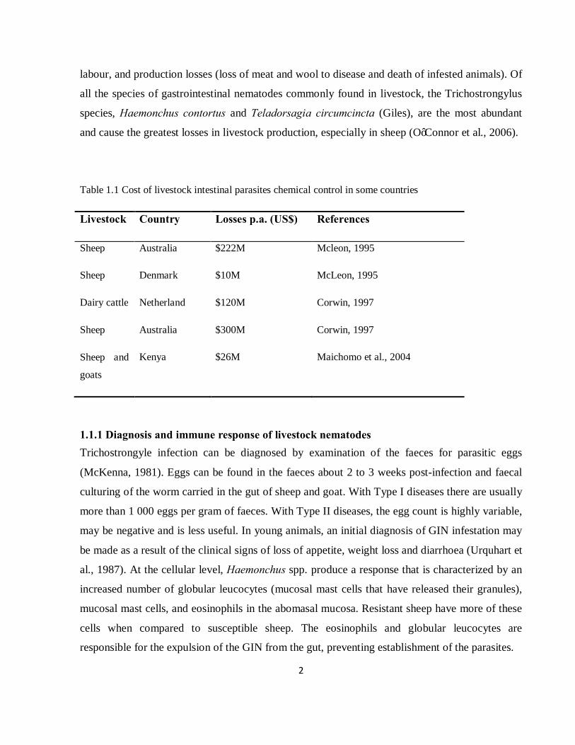
2
labour, and production losses (loss of meat and wool to disease and death of infested animals). Of
all the species of gastrointestinal nematodes commonly found in livestock, the Trichostrongylus
species, Haemonchus contortus and Teladorsagia circumcincta (Giles), are the most abundant
and cause the greatest losses in livestock production, especially in sheep (O’Connor et al., 2006).
Table 1.1 Cost of livestock intestinal parasites chemical control in some countries
Livestock Country Losses p.a. (US$) References
Sheep
Sheep
Australia
Denmark
$222M
$10M
Mcleon, 1995
McLeon, 1995
Dairy cattle Netherland $120M Corwin, 1997
Sheep Australia $300M Corwin, 1997
Sheep and
goats
Kenya $26M Maichomo et al., 2004
1.1.1 Diagnosis and immune response of livestock nematodes Trichostrongyle infection can be diagnosed by examination of the faeces for parasitic eggs
(McKenna, 1981). Eggs can be found in the faeces about 2 to 3 weeks post-infection and faecal
culturing of the worm carried in the gut of sheep and goat. With Type I diseases there are usually
more than 1 000 eggs per gram of faeces. With Type II diseases, the egg count is highly variable,
may be negative and is less useful. In young animals, an initial diagnosis of GIN infestation may
be made as a result of the clinical signs of loss of appetite, weight loss and diarrhoea (Urquhart et
al., 1987). At the cellular level, Haemonchus spp. produce a response that is characterized by an
increased number of globular leucocytes (mucosal mast cells that have released their granules),
mucosal mast cells, and eosinophils in the abomasal mucosa. Resistant sheep have more of these
cells when compared to susceptible sheep. The eosinophils and globular leucocytes are
responsible for the expulsion of the GIN from the gut, preventing establishment of the parasites.
3
1.1.2 Climatic distribution and epidemiology Development and survival of the infective stage depends on the prevailing conditions of
temperature and moisture. Optimum requirements vary distinctly among GIN species (Georgi,
1969). In South Africa, O. circumcincta is the dominant parasite in winter and in uniform rainfall
areas, with their main survival advantage being a greater resistance to drought and the ability to
develop at lower temperatures than H. contortus (O’Connor et al., 2006). Haemonchus contortus
is the most important nematode species of sheep in tropical and subtropical areas, or regions with
summer-dominant rainfall. H. contortus becomes less significant as the climate tends towards
winter (O’Connor et al., 2006). The success rate and speed of development of gastrointestinal
nematodes are determined mostly by temperature and moisture, both of which have major
influences on the epidemiology of the nematodes (Chatterjee et al., 2007). According to
Stromberg (1997), temperature controls the rate of development, but without moisture, there will
be no change in the growth rate of the nematodes. Furthermore, temperature may affect viability
and/or infectivity of larvae. The movement of nematode larvae is facilitated by a continuous film
of moisture on herbage, as a result of irrigation, rainfall or dew. When moisture is not a limiting
factor, temperature asserts a greater influence over migration (Stromberg, 1997). Trichostrongylus
circumcincta and T. colubriformis develop at lower temperatures than H. contortus. Hsu and
Levine, (1977) observed that at 200C and 250C, eggs of H. contortus and T. colubriformis took a
minimum of 4 and 3 days respectively to reach the L3 larval stage. O’Connor et al. (2006) stated
that once development to the L3 stages is complete, all three major species are less susceptible to
unfavourable climatic conditions. They further suggested that this could be attributed in some
part to the larval ability to migrate to more favourable micro-environments. Hiding in the sheath
of the previous larval stage prevents feeding of infective larvae, and makes them susceptible to
higher temperatures which increase their metabolic rate through depletion of energy reserves.
Research in Ethiopia indicated that there was a distinct seasonal increase in the availability of
larvae on pastures in response to climatic changes (Tembely et al., 1997). In Tanzania, faecal egg
count (FEC) increased during the rainy season (December-April) (Keyyu et al., 2005).

4
1.1.3 Economic importance of livestock nematodes There is a worldwide awareness that GIN infections are one of the major causes of loss of
productivity in grazing livestock (Perry et al., 1999). Infestation of livestock with high levels of
GIN detrimentally affects the profitability of invested capital in terms of animal health and
performance (Corwin, 1997). This includes weight losses due to reduced feed intake, reduced
carcass quality and reduced wool production and quality (Gray, 1993). To measure the effects of
subclinical or economic thresholds of GIN parasitism, measurement of performance parameters
such as weight gain, feed conversion, forage utilization, conception rate, calving-breeding
interval, milk production and disease resistance are employed (Bumgarner et al., 1986; Williams
et al., 1986; Wohlgemuth et al., 1990; Gibbs, 1992; Toombs and Craig, 1994 cited by Corwin,
1997). Economic losses are primarily due to mortality, but can also result from subclinical effects
such as inadequate weight gain, feed consumption, and reproductive potential, as well as reduced
lactation, wool and meat production due to infections by Oesophagostomum spp. Reports indicate
that mortality in sheep flocks may exceed 40%, while weight losses of 6-12 kg/year/animal may
occur (IEMVT, 1980 cited by Krattiger, 1997). However, decreased efficiency in feed utilization
associated with clinical or chronic conditions is often the main cause of economic losses
(Holmes, 1987). About US$ 800 million p.a. were estimated to be spent on chemical treatments
to control helminths (Krattiger, 1997). Because nematodes such as H. contortus are blood
feeders, heavy infestations in animals can cause severe anaemia and death. Effects of other
nematodes, such as those from the genus Cooperia and Trichuris spp., are in addition to those of
infestation with other nematodes.
1.1.4 Life cycle of livestock nematodes Most livestock nematodes undergo the following lifecycle, according to Urquhart et al. (1987).
The cycle begins when the mature female lays eggs which are passed with animal faeces to the
ground. Under favourable conditions, these eggs hatch into the first larval stage (L1). The larvae
feed, grow and moult in the faeces. After a first moult, the larvae develops into the second larval
stage (L2) which grows, feeds and eventually undergoes the second moult. In most Strongylida

5
nematode species, the sheath of the second moult is retained, thus providing an enclosed space
for the development of the third larval stage (L3), which does not feed. This is the infective stage,
and the L3 stage is typically eaten by livestock, along with pasture grass on which the L3 stage
sits. After entering the animal host, the larvae removes the sheath of the second moult and is then
referred to as the third parasitic stage (P3). At this stage the nematode feeds on the animal host
and grows until it moults for the third time, after which the development of the sexual organs
starts. In the fourth stage, the sexual organs continue to develop, and males and females are easily
identified for the first time. The parasites feed and grow considerably before undergoing the
fourth moult. Growth and sexual development continues up to the fifth stage. True adults are only
present after mating.
Table 1.2 Summary of livestock nematodes from Oslon (1974); and Swartson and Nsahlai (2006)
Nematode
Genus
Morphology Life cycle Symptoms Common name
Haemonchus contortus
Males are 10-20 mm and females 18-30 mm long. White uteri and ovaries; wind around the blood-filled intestine in a barber-pole appearance
Direct (does not require intermediate host).
Acute anaemia, severe blood loss, bottle jaw, diarrhoea, pale gums and inside of the eyelids (conjunctiva).
Barber-pole
Nematodirus spp. 10-30 mm long, with thin anterior and swollen appearance of the head.
Direct. 15-28 days.
Loss of appetite, diarrhoea, weight loss and reduced wool production.
Thread necked strongyle
6
Nematode
Genus
Morphology Life cycle Symptoms Common name
Trichostongylus spp.
Direct. Prepatient period of 20- 25 days.
Severe weight loss, poor growth, wart-like swellings, diarrhoea and reduced appetite
Bankrupt worm or stomach hair worm
Cooperia spp. Brownish-red, 4-6 mm in length with swelled anterior
Direct. Prepatient period of 15-20 days
Loss of appetite, diarrhoea and weight loss.
Small intestine worm
Oesophagostomum spp.
Up to 20 mm long, with a narrowed front.
Direct. 6-7 days. Diarrhoea, thickening of large intestinal wall, mucus production in faeces,
Nodular worm
Trichuris spp. Have slender neck and a thick hind-end, males are 50-80 mm with a curved tail. Females are 35-70 mm.
Prepatient period of 1-3 months.
Thickening of the caecal wall and diarrhoea.
Whipworm
1.2 Control of livestock nematodes Extensive experience exists in the control of parasitic nematodes that infect agricultural and
companion animals. Methods of nematode control are diverse and have focused on control rather
than eradication (Waller and Thambsborg, 2004; Sayers Sweeney, 2005; Stear et al., 2006). This
is because of continuous to long-lived parasites on pastures and incomplete resistance responses
of the host.
1.2.1 Chemical control of livestock nematodes According to Mitreva et al. (2007), the use of anthelmintics is still the core strategy for nematode
control. Their success has been cyclical and directly related to the timely introduction of new
drugs as resistance to older drugs has surfaced. Anthelmintics are grouped into four main
chemical classes, according to their modes of action (Stepek et al., 2004).
7
1.2.1.1 The benzimidazoles class
The benzimidazoles (e.g. albendazole) are a large chemical family used to treat nematode and
trematode infections in domestic animals. This group disrupts β-tubulin synthesis (Stepek, et al.,
2004). However, with the widespread development of resistance and the availability of more
efficient compounds which are easier to administer, their use has rapidly decreased. In ruminants,
treatment with the benzimidazoles removes most of the major adult gastrointestinal (GI) parasites
and many of the larval stages. Metabolism and excretion of benzimidazole compounds is more
extensive in cattle than in sheep. Consequently, the systemic anthelmintic activity of most
benzimidazoles is greater in sheep than in cattle, and dose rates in cattle are often higher than
those in sheep. Albendazole, fenbendazole, oxfendazole, and febantel are active against inhibited
fourth-stage larvae of Ostertagia spp.; however, inconsistent efficacy has been reported. Efficacy
against D. viviparus has also been noted for these insoluble benzimidazoles. Oxfendazole,
albendazole, and febantel are minimally teratogenic in sheep, whereas fenbendazole,
mebendazole, and oxibendazole are not. An oxfendazole pulse-release bolus for intraruminal use
has been developed for cattle whereby 5 therapeutic doses of oxfendazole (750 or 1,250 mg /
tablet) are released approximately every three weeks in the rumen. An albendazole slow-release
capsule containing 3.85 g albendazole and delivering a daily dose of 36.7g for 107 days has been
marketed for small ruminants (Merck and Co., Inc., 2008).
Albendazole and netobimin at 20 mg kg-1 are active against mature F. hepatica. The other
benzimidazoles and probenzimidazoles used for nematode control have only a marginal efficacy
against liver flukes (Merck and Co., Inc., 2008). Because of the lack of efficacy against the
immature stages, most benzimidazoles are not indicated for treatment of acute fascioliosis (Merck
and Co., Inc., 2008).
The anthelmintic activity of tetramisole, a racemic mixture, lies in the l-isomer, levamisole. It is
commonly used in cattle, sheep, pigs, goats and poultry to treat nematode infections; however it
has no activity against flukes and tapeworms. The efficacy is generally considered equivalent
with either product (Merck and Co., Inc., 2008).
8
1.2.1.2 Imidazothiazole class
Levamisole acts on the roundworm nervous system and is not ovicidal. Its broad spectrum of
activity, ease-of-use (being water soluble), reasonable safety margin, and lack of teratogenic
effects have allowed it to be used successfully. Because of its mechanism of action, the peak
blood concentration is more relevant to its antiparasitic activity than the duration of
concentration. Levamisole resistance appears to be associated with a loss of cholinergic
receptors. Levamisole has immunostimulant effects at dosage rates higher than those used for
anthelmintic activity, and it has been used in humans and, to a limited extent, in other animals
against several diseases (Merck and Co., Inc., 2008).
In ruminants, levamisole is highly effective against the common adult GI nematodes and
lungworms, and many larval stages. It lacks efficacy against arrested larvae, such as those of
Ostertagia ostertagi. Slow-release boluses of levamisole are available in some countries and
contain 22.05 mg levamisole. They release 2.5 mg during the first 24 hours and the remainder
over a 90-day period (Merck and Co., Inc., 2008).
Pyrantel was first introduced as a broad-spectrum anthelmintic against GIN of sheep and has also
been used in cattle, horses, dogs and pigs. It is available as a citrate, tartrate, embonate, or
pamoate salt. Aqueous solutions are subject to isomerization on exposure to light, with a resultant
loss in potency; therefore, suspensions should be kept out of direct sunlight. It is not
recommended for use in severely debilitated animals because of its levamisole-type
pharmacological action.
Pyrantel is used as a suspension, paste, drench, or tablets. Both pyrantel and morantel are
effective against adult gut worms and larval stages that dwell in the lumen or on the mucosal
surface. Pyrantel tartrate is effective as a broad-spectrum anthelmintic in ruminants; however, its
activity is mainly limited to the adult GI nematodes (Merck and Co., Inc., 2008).
1.2.1.3 Macrocyclic lactones class
The macrocyclic lactones (avermectins and milbemycins) are chemical by-products from soil
microorganisms of the genus Streptomyces. The macrocyclic lactones are highly lipophilic
anthelmintics which are known to bind to ligand-gated ion channels, opening them (Prichard and

9
Roulet, 2007). As a result, they have a potent, broad-spectrum antiparasitic activity at low dose
levels (Merck and Co., Inc., 2008). They are active against many immature nematodes and
arthropods. However, the commercially available products are primarily excreted in the faeces
and kill non-target dung insects, therefore affecting dung dispersal. Decomposition of
invermectin in faeces or soil is slow, especially in winter (Merck and Co., Inc., 2008).
1.2.1.4 Amino-acetonitrile derivatives class (AAD)
This is a new class of chemicals with a novel mode of action involving a unique, nematode-
specific clade of acetylcholine receptor subunits. They are well tolerated and of low toxicity to
mammals, and overcome existing resistance to the currently available anthelmintics. The AAD
are low molecular mass compounds that are easily accessible by alkylation of phenols with
chloroacetone. When tested in ruminants, all AADs were able to eliminate fourth larval (L4)
stages of H. contortus in sheep and Cooperia oncophora (Raiilet) in cattle with a single oral dose
of 20 mg (Malan et al., 2001).
1.2.2 Mechanism of action
Anthelmintics inhibit metabolic processes that are vital to the parasite but not vital to, or absent
in, the host. Alternatively, they have inherent pharmacokinetic properties that cause the parasite
to be exposed to higher concentrations of the anthelmintic than the host cells. While the exact
mode of action of many anthelmintics is not fully documented, the sites of action and
biochemical mechanisms of many of them are known. Parasitic helminths must preserve an
appropriate feeding site, and nematodes must actively ingest and move food through their
digestive tracts to maintain an appropriate energy state. Feeding and reproductive processes
require proper neuromuscular coordination. Parasites must also maintain homeostasis despite host
immune reactions. The pharmacological basis of treatments against helminths generally involves
interference with the integrity of parasite cells, neuromuscular coordination, or protective
mechanisms against host immunity, which leads to starvation, paralysis, and expulsion of the
parasite (Merck and Co., Inc., 2008).
10
1.2.3 Problems associated with chemical control -Multi-drug resistance
Using only a chemical control strategy has numerous problems. Among these is the overuse or
under-dosing of chemotherapeutic drugs, which results in resistance to these drugs (Geerts and
Greyseels, 2000; De and Sanyal, 2009). Occurrences of internal parasite populations resistant to
available anthelmintics have increased. Resistance has been reported to drugs within three major
classes of anthelmintics (Terrill et al., 2001). Anthelmintic resistance of gastrointestinal
nematodes of livestock is most prevalent in Australia, New Zealand, South America and South
Africa (Van Wyk, 1990; Coles et al., 1994; Van Wyk et al., 1999). The initial reports of
anthelmintic resistance were to phenothiazine, reported in the late 1950s and early 1960s. Drudge
(1957) reported the first incidence of drug resistance with H. contortus in sheep. This was
followed by reports of resistance by cyathostomins (small strongyles) of horses (Poynter and
Hughes, 1958; Gibson, 1960) (Table 1.3). In 1961, thiabendazole was introduced as the first
anthelmintic that provided a broad-spectrum nematicidal activity coupled with low mammalian
toxicity. The rapid acceptance and widespread use of thiabendazole and other benzimidazole
anthelmintics resulted in resistance to this class evolving within a few years, first by the sheep
nematode, H. contortus (Conway, 1964; Drudge et al., 1964), followed by equine cyathostomins.
Benzimidazole resistance was later reported in other major gastrointestinal trichostrongylid
nematodes of sheep, namely Teladorsagia (Ostertagia) circumcincta (brown stomach worm) and
Trichostrongylus colubriformis (black scour worm). By the mid-1970s, studies showed common
and widespread incidences of resistance to the benzimidazoles by a wide range of nematodes.
Similarly, resistance to the imidazothiazole–tetrahydropyrimidine and avermectin–milbemycin
classes of anthelmintics was observed after their introduction. Most alarmingly, by the early
1980s, reports of multiple-drug resistant (MDR) worms appeared for the first time (Coles, 1986).
11
-Other related problems of anthelmintics
In addition to anthelmintic resistance, increasing public awareness regarding contamination of
food with chemicals has resulted in consumer pressure to reduce drug residues in meat and meat
products. This influences the use of anthelmintics (Larsen, 2000). Levamisole and the
benzimidazoles pose little cause for concern, but there is greater worry regarding the avemectins
(De and Sanyal, 2009). Ivermectin is excreted in faeces in sufficient quantity to have a
detrimental effect on invertebrates that usually degrade dung heaps, and hence on organisms
higher up the food chain (Cox, 1999; De and Sanyal, 2009). Anthelmintics are relatively
expensive for smallholder farmers in relation to available resources, reducing the usefulness of
these drugs in developing countries (Knox, 2000; Dalton et al., 2001).

12
Table 1.3: Worldwide levels of anthelmintic resistance among livestock hosts (as outlined by
Drudge and Elem, 1964, and subsequently by Van Wyk et al. (1999) and Coles et al. (1994))
Drug class Hosts with high levels of resistant GIN
Hosts with emerging resistance in GIN
Major livestock-producing areas where drug is still highly effective in sheep, goats and horses
Benzimidazoles Sheep, goats, horses
Cattle None
Imidothiazoles
Levamisole (ruminants)
Sheep, goats Cattle None
Pyrantel (horses) Horses (USA only)
Horses Unknown – few recent studies outside USA
Avermectin
Ivermectin Sheep, goats, cattle
Cattle, horses Horses – worldwide
Sheep, goats – Europe, Canada
Moxidectin Goats Sheep, goats, cattle, horses
Horses – worldwide
Sheep – most regions
1.2.4 Alternative control methods 1.2.4.1 Botanicals
An alternative anthelmintic strategy with considerable potential is the use of extracts from
medicinal plants (Stepek et al., 2004). A wide range of plants and plant extracts has been used
traditionally for the treatment of helminth infections of humans (Waller et al., 2001). Many
attempts have been made to evaluate the plant extracts with anthelmintic potential for the control
of livestock parasitic nematodes (Table 1.4 and 1.5).
13
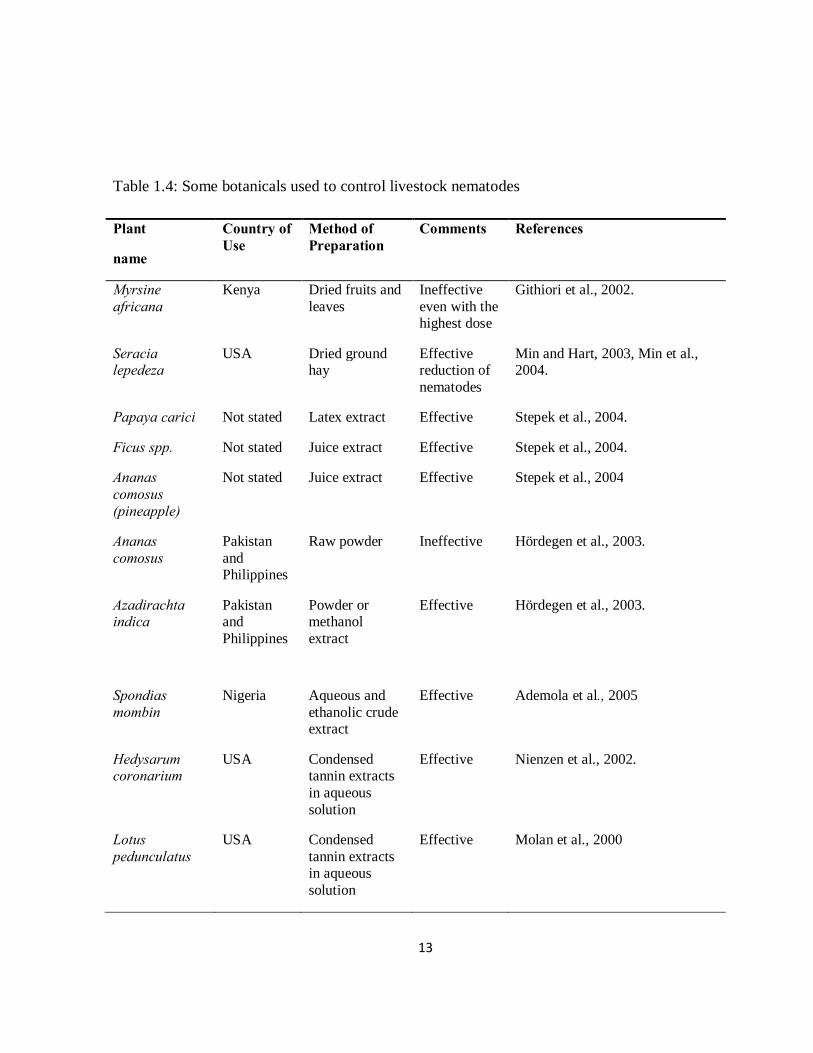
Table 1.4: Some botanicals used to control livestock nematodes
Plant
name
Country of Use
Method of Preparation
Comments References
Myrsine africana
Kenya Dried fruits and leaves
Ineffective even with the highest dose
Githiori et al., 2002.
Seracia lepedeza
USA Dried ground hay
Effective reduction of nematodes
Min and Hart, 2003, Min et al., 2004.
Papaya carici Not stated Latex extract Effective Stepek et al., 2004.
Ficus spp. Not stated Juice extract Effective Stepek et al., 2004.
Ananas comosus (pineapple)
Not stated Juice extract Effective Stepek et al., 2004
Ananas comosus
Pakistan and Philippines
Raw powder Ineffective Hördegen et al., 2003.
Azadirachta indica
Pakistan and Philippines
Powder or methanol extract
Effective Hördegen et al., 2003.
Spondias mombin
Nigeria Aqueous and ethanolic crude extract
Effective Ademola et al., 2005
Hedysarum coronarium
USA Condensed tannin extracts in aqueous solution
Effective Nienzen et al., 2002.
Lotus pedunculatus
USA Condensed tannin extracts in aqueous solution
Effective Molan et al., 2000
14
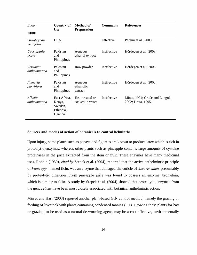
Plant
name
Country of Use
Method of Preparation
Comments References
Ornobrychis viciafolia
USA Effective Paolini et al., 2003
Caesalpinia crista
Pakistan and Philippines
Aqueous ethanol extract
Ineffective Hördegen et al., 2003.
Vernonia anthelmintica
Pakistan and Philippines
Raw powder Ineffective Hördegen et al., 2003.
Fumaria parviflora
Pakistan and Philippines
Aqueous ethanolic extract
Ineffective Hördegen et al., 2003.
Albizia anthelmintica
East Africa, Kenya, Sweden, Ethiopia, Uganda
Heat treated or soaked in water
Ineffective Minja, 1994; Grade and Longok, 2002; Desta, 1995.
Sources and modes of action of botanicals to control helminths
Upon injury, some plants such as papaya and fig trees are known to produce latex which is rich in
proteolytic enzymes, whereas other plants such as pineapple contains large amounts of cysteine
proteinases in the juice extracted from the stem or fruit. These enzymes have many medicinal
uses. Robbin (1930), cited by Stepek et al. (2004), reported that the active anthelmintic principle
of Ficus spp., named ficin, was an enzyme that damaged the cuticle of Ascaris suum, presumably
by proteolytic digestion. Fresh pineapple juice was found to possess an enzyme, bromelain,
which is similar to ficin. A study by Stepek et al. (2004) showed that proteolytic enzymes from
the genus Ficus have been most closely associated with botanical anthelmintic action.
Min et and Hart (2003) reported another plant-based GIN control method, namely the grazing or
feeding of livestock with plants containing condensed tannins (CT). Growing these plants for hay
or grazing, to be used as a natural de-worming agent, may be a cost-effective, environmentally
15
friendly alternative to the exclusive use of chemical anthelmintics by small ruminant producers
(Shaik et al., 2004).
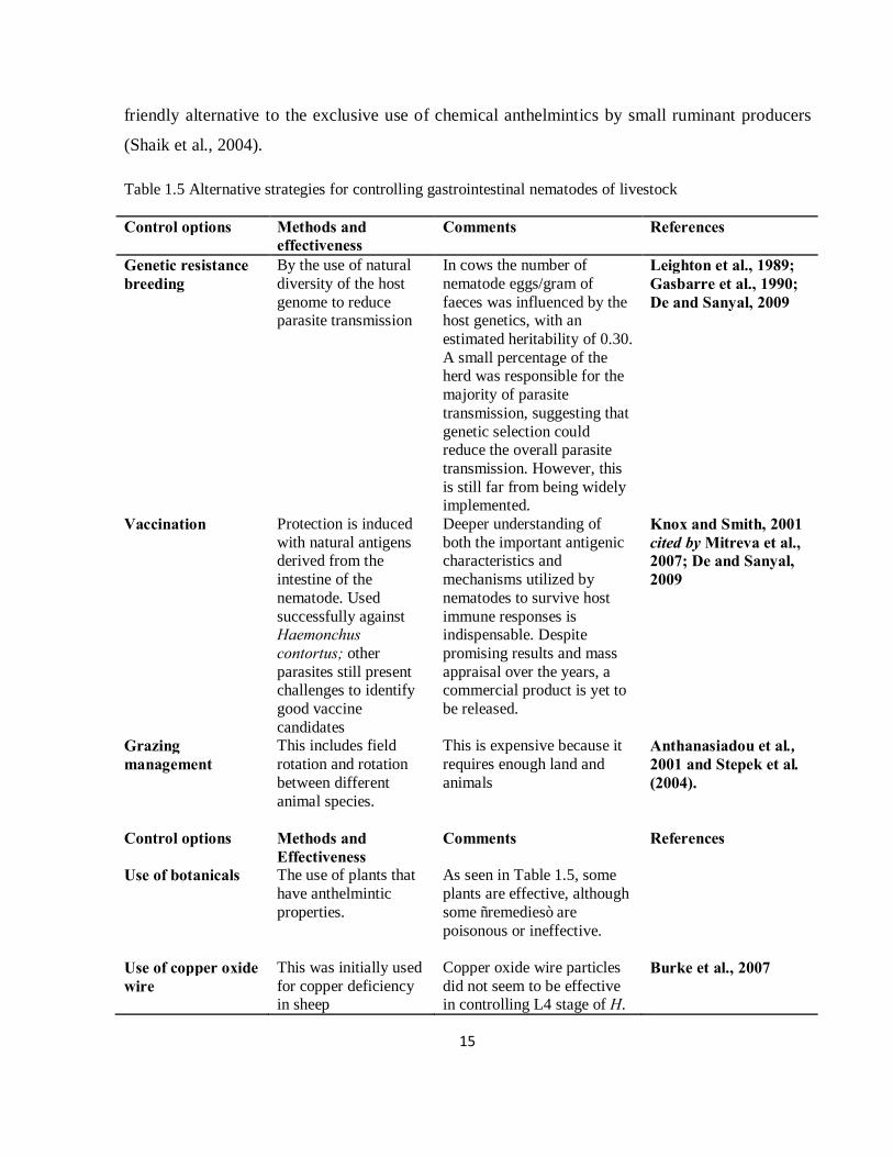
Table 1.5 Alternative strategies for controlling gastrointestinal nematodes of livestock
Control options Methods and effectiveness
Comments References
Genetic resistance breeding
By the use of natural diversity of the host genome to reduce parasite transmission
In cows the number of nematode eggs/gram of faeces was influenced by the host genetics, with an estimated heritability of 0.30. A small percentage of the herd was responsible for the majority of parasite transmission, suggesting that genetic selection could reduce the overall parasite transmission. However, this is still far from being widely implemented.
Leighton et al., 1989; Gasbarre et al., 1990; De and Sanyal, 2009
Vaccination Protection is induced with natural antigens derived from the intestine of the nematode. Used successfully against Haemonchus contortus; other parasites still present challenges to identify good vaccine candidates
Deeper understanding of both the important antigenic characteristics and mechanisms utilized by nematodes to survive host immune responses is indispensable. Despite promising results and mass appraisal over the years, a commercial product is yet to be released.
Knox and Smith, 2001 cited by Mitreva et al., 2007; De and Sanyal, 2009
Grazing management
This includes field rotation and rotation between different animal species.
This is expensive because it requires enough land and animals
Anthanasiadou et al., 2001 and Stepek et al. (2004).
Control options Methods and Effectiveness
Comments References
Use of botanicals The use of plants that have anthelmintic properties.
As seen in Table 1.5, some plants are effective, although some “remedies” are poisonous or ineffective.
Use of copper oxide wire
This was initially used for copper deficiency in sheep
Copper oxide wire particles did not seem to be effective in controlling L4 stage of H.
Burke et al., 2007

16
contortus which also feed on blood of livestock. Although results are promising at higher doses they may lead to copper toxicity.
FAMACHA©
A novel clinical test developed to assess the level of anaemia caused by haemonchosis in sheep and to determine which animals will receive anthelmintic treatments
The product has shown to minimise the incidence of chemical resistance by dosing sheep at the right time
Malan et al., 2001
Uses of biocontrol agents
Use of microorganisms to control gastrointestinal nematodes
Biological control on pasture includes the use of predatory fungus and bacteria to kill a variety of nematode species and substantially reduce the intensity of infection. Promising candidates are D. flagrans, C. rosea and Bt
Larsen, 1999 and O’Grady et al., 2007
1.2.4.2 Biological control
Over the last 10-15 years, biological control has been identified as a viable alternative to
chemical anthelmintics (De and Sanyal, 2009). Biological control may be defined as the use of
living microorganisms introduced into an environment to control a target microorganism and
thereby reduce the population growth of the target to a threshold below which it no longer causes
clinical problems and economic losses (Larsen, 2006).
Waller and Faedo (1996) explained that biological control agents may not only include the
classically exploited organisms, but also that these organisms may be genetically modified to
enhance their properties. Hence biological control may be divided into two major categories:
natural and applied. Natural biological control is influenced by native or co-evolved natural
enemies in the environment without human intervention. Applied biological control involves the
repeated application of cultured biological control agents (De and Sanyal, 2009)
17
Waller (1997) reported that biocontrol agents target the free-living larval stage of the nematode
rather than the parasitic stages in the host. This approach focuses on the faecal deposits in which
eggs, and the L1 and L2 larval stages live. According to De and Sanyal (2009), biocontrol agents
do not totally eliminate target organisms, but reduce their numbers to acceptable levels, and
maintain a population balance between the pathogen and the antagonist. Therefore, the goal of
using a biocontrol approach is to lower the numbers of parasitic populations to below the clinical
level, aiming to go below the economic threshold above which production losses occur.
According to Faedo and Krecek (2002), when considering the control of helminths in livestock, it
is important to abandon the unrealistic concept of eradication which has led to an over-reliance
on anthelmintic drugs, resulting in drug resistance within the target population.
Githiori et al. (2002) mentioned that although anthelmintic drugs have long been considered
effective, they are expensive and sometimes unavailable to smallholder farmers and pastoralists
in developing countries. Thus biological control has been considered as an alternative control
method for small-scale livestock farmers.
Biological control of parasitic nematodes in livestock therefore aims to establish a situation
where the grazing animals are exposed to a low level of infective larvae, at which naturally
acquired immunity will develop in the animals (Thamsborg et al., 1999).
One of the most important aspects in biocontrol programmes is the accurate identification of the
pest and any hyperparasitic organisms that might control that pest. This aspect has a direct impact
not only on determining the geographic range of the pest and its hyperparasites, but also on the
acquisition of permits necessary for the release of biocontrol agents (Schnepf et al., 1998).
Biological control has the capacity to reduce a range of nematode parasites, not only within but
also between species of livestock (Waller, 1997). Fontenot et al. (2003) reported an overall
reduction of a wide range of nematodes, as a result of the application of biocontrol agents.
Furthermore, biocontrol agents are chemical-free, thus producers can capitalise on the residue-
free, organic meat products that are in high demand from consumers and command a premium
price (Waller, 1997). However, because B. thuringiensis is increasingly used in agricultural pest
management, this has resulted in the occurrence of resistance in a cruciferous pest, diamond-
18
backed moth (Plutella xylostella) (Navon, 2000). This is a recognized problem if Bt strains with a
single Cry toxin are used, and reflects the need to manage resistance, even with biocontrol agents.
Several methods of incorporating this agent into the gut of target animals have been tested and, so
far, daily feeding of the spores or cells of the biocontrol agents, together with supplements, or in
the use of slow release devices such as mineral blocks, pellets or boluses, seem to be the most
successful (Waller, 1997; Fontenot et al., 2003). For biocontrol agents such as B. thuringiensis,
delivery of viable spores into the intestinal tract of the sheep is a challenge. Studies by Wu and
Papas (1997) showed that application of reverse enteric coating techniques allowed for the
passage of the formulated material through the rumen, followed by release into the abomasum
because it was stimulated by entry into an acidic environment. This emphasised the need to
protect the spores from the acidic conditions in the faeces. Shimoda et al. (2001) and Senel and
McClure (2004), discovered that the use of the adhesive properties of chitosan ensured that Bt
was localised at the site of the nematode.
The following requirements need to be met by a commercial biocontrol product for use in an
integrated control strategy: the product must be (i) cheap to produce, (ii) safe to handle, (iii)
survive passage through the gastrointestinal tract and (iv) be able to grow and subsequently
germinate in the voided dung, in order to kill the eggs and developing larvae (Larsen et al., 1991).
Duddingtonia flagrans as a biocontrol agent
Many fungi exhibit anti-nematode properties, characterized by their ability to capture and exploit
nematodes either as a primary source of nutrients or as a supplement to a saprophytic existence.
These fungi are divided into predacious, endo-parasitic and egg-destroying fungi (Faedo and
Krecek, 2002).
Duddingtonia flagrans (Cooke) has been shown to have considerable potential as a biocontrol
agent, and may be commercialized in the near future, given that the basic research on the fungus
has been completed. D. flagrans was selected based on its ability to survive passage through the
gastrointestinal tract of ruminants and subsequently to trap the developing parasite larvae in the
deposited faeces (Larsen et al., 1994). This species (formerly Trichothecium flagrans) belongs to
19
the Family Deuteromycetes, which are the members of the Fungi Imperfecti, of which a number
are known as nematode destroying fungi (Cooke et al., 1964; Barron, 1977). D. flagrans has a
particular characteristic of producing abundant intercalary chlamydospores (Cooke, 1969).
Previous studies in Denmark by Larsen and co-workers on the ability of nematophagous fungi to
survive the environmental rigours of gut passage in cattle, showed better survival of D. flagrans
than the other well-known nematophagous genus, Arthrobotrys, under both in vitro (Larsen et al.,
1991) and in vivo (Larsen et al., 1992) conditions.
According to Larsen et al. (1991), the special feature that ranks D. flagrans above other
nematophagous-fungi is its production of many resistant, thick-walled resting spores. These
survive the unfavourable conditions in the gut. This property allows chlamydospores to be
administered orally, resulting in their deposition in dung simultaneously with the eggs of internal
parasites. In warm dung the parasite eggs hatch to release free-living, bacterial-feeding, juvenile
stages, while the chlamydospores germinate to form mycelia that produce traps in response to
nematode activity.
Daily feeding of D. flagrans spores to grazing animals for 3-4 months prevents the build up of
dangerous levels of infective GIN larvae on pasture. Knox et al. (2001) found that sheep that
were fed a supplement containing D. flagrans chlamydospores had lower egg counts and
improved live weight gains compared to untreated animals. A study by Fontenot et al. (2003)
showed that daily feeding of 5 x 10⁵ chlamydospores per kg of body weight of D. flagrans
resulted in a reduction of GIN larvae in faeces, which subsequently resulted in lower pasture
infectivity and reduced nematode burdens in tracer animals. According to Larsen et al. (1998)
and Peña et al. (2002), this dosage of 5 x 10⁵ chlamydospores per kg of body weight is adequate
to produce substantial nematode trapping. Reports by Larsen et al. (1991), Gronvold et al. (1993),
Mendozade de Gives et al. (1998) and Peña et al. (2002), estimated that routine oral doses of D.
flagrans resulted in a reduction of GIN L3 larval stage counts in faecal culture of 30% to 90%,
with an 85% reduction being the average.
Challenges to the use of nematode trapping fungi in the control of nematodes have been the
requirement for daily administration of the fungus to the host, and achieving the required fungal
density inside the dung. However, an isolate of D. flagrans from New Zealand (Skipp et al.,
20
2002) demonstrated a trapping efficiency of 78% and activity for up to 90 days on pasture
(Waghorn et al., 2003).
The interest in studying the infection biology of these fungi comes from their potential use as
biological control agents against plant and animal parasitic nematodes (Tunlid et al., 1999).
Utilization of nematode-trapping fungi for agriculture could be a powerful tool for biological
control of plant-feeding nematodes on farms (Stirling et al., 1998).
Mechanism of action of D. flagrans
D. flagrans targets the free-living larval stages outside the host. These stages exist as larvae
within the faecal deposit, and as infective third-stage larvae on pastures. The best way to apply
the fungus to the faecal environment is via passage through the gastrointestinal tract of the host.
D. flagrans is unique in that it produces large numbers of chlamydospores which, when fed to
livestock, can survive passage through the gastrointestinal tract to be deposited in their faeces.
Once voided in the faeces, D. flagrans grows within the faecal deposit and traps emerging larvae.
While developing, the fungus produces three-dimensional mycelia “nets” that trap migrating
nematodes and thus greatly reduce parasitic larval development (Larsen, 2000). This prevents the
L3 larvae from developing (Faedo et al., 2002). However, nematophagous fungi such as D.
flagrans do not affect the established populations of nematodes within the host and are thus not
curative (Faedo et al., 2002).
Clonostachys rosea as a biological control agent
Clonostachys rosea (Schroers) was previously known as Gliocladium roseum (Bainier) classified
under the phylum Ascomycota and family Bionectriaceae (Schroers et al., 1999). This fungus is
an important biological control agent of plant fungi and nematodes (Huang et al., 2009). Dong et
al. (2004) reported that some toxin isolated from filtrate of C. rosea showed strong nematicidal
activities against Caenorhabditis elegans (Maupas). C. rosea is capable of withstanding harsh
environmental conditions (Morandi et al., 2003). It has mostly been isolated from cultivated
grassland and woodlands, forests, heathland, freshwater and coastal soil of neutral to alkaline pH
soils (Sutton et al., 1997). C. rosea is mostly found in temperate and tropic regions. C. rosea is a
21
motoscopic fungi used as phytopathogenic fungi (Li et al., 2002), and a facultative parasite of
pathogenic nematodes (Yang et al., 2000). When C. rosea was applied to strawberry on a weekly
basis, numbers of rotten fruits were reduced by 73% and were similar to the control provided by
application of fungicide (Valdeberito-Sanhueza et al., 1997).
Mode of action of C. rosea
According to Zhang et al. (2008), pathogenesis occurs when the conidia adhere to nematode
cuticle and germinates, then produces a germ tube that penetrate the host body and kill it. The
mycoparasitic activity has been attributed to the secretion of its cell-wall degrading enzymes
capable of degrading chitin. Zhao et al. (2005) and Li et al. (2006) reported two extracellular
serine proteases (PrC and Lmzl) from C. rosea which were both found to play a role in nematode
infection. C. rosea Isolate 87 from soil sample showed capability of parasitizing and digesting
nematodes (Zhang and Zhao et al., 2004).
Bacillus thuringiensis as a biocontrol agent
Bacillus thuringiensis (Bt) (Berliner) is a gram-positive, spore-forming bacterium which has the
unusual property of producing a parasporal protein crystal (delta-endotoxin) which is toxic (Cry
proteins) to some insect pests (Lecadet et al., 1999, cited by Quesada-Moraga et al., 2004).
Swadener (1994) described B. thuringiensis as a species of bacterium that has insecticidal
properties affecting a selective range of insect orders. Swadener (1994) further explained that
there are at least 34 subspecies of B. thuringiensis (also called serotypes or varieties). There are
already more than 800 strain isolates (De Barjac and Franchon, 1990). B. thuringiensis products
have captured 90-95% per cent of the biopesticide market because of their unique properties and
advantages as biopesticides (Entwistle et al., 1993). One of Bt’s most desirable characteristics is
its selectivity, due to the selectivity of the protein crystals. However, this is complemented by the
many strains available that attack a wide range of specific target pests.
22
B. thuringiensis is fermentation-friendly and therefore it is relatively easy to exploit
commercially. Furthermore, Bt is a prokaryote, meaning it does not have dominant or recessive
alleles, thus making it relatively easy to manipulate genetically. The gene for the crystal protein
toxins are each coded for by a single gene (monocistronic). According to Federici, (1993) cited
by Chatterjee et al., (2007), these advantages have favoured the commercialization of Bt products
internationally.
In addition to strains that kill pests, some strains also kill nematodes. It has been shown that
several Bt isolates are highly toxic to larvae and adults of H. contortus, Trichostrongylus
colubriformis and O. circumcincta in vitro, thereby indicating that these strains of this bacterium
may hold some promise as anthelmintics (Kotze et al., 2005). Given that Bt is toxic to both larval
and adult nematodes (Kotze et al., 2005), it can be utilised as an anthelmintic targeting adult
nematodes of the gastrointestinal tract, and/or the free-living larval stages that develop in faeces
or on pastures. B. thuringiensis would be an ideal biocontrol candidate due to its low mammalian
toxicity (Siegel, 2001), and the species specificity shown by particular endotoxin groups
(Schnepf et al., 1998). While current chemotherapeutic approaches to animal-parasitic nematode
control are aimed almost exclusively at the adult stages, the potential contribution of non-
chemical strategies aimed at reducing the levels of larvae developing on pastures has been
recognised (Larsen et al., 1998; Larsen 2000).
Kotze et al. (2005) discovered that several uncharacterised Bt strains have significant activity
against the eggs and larvae of the parasitic nematode Trichostrongylus colubriformis. However,
O’Grady et al. (2007) found that a significant time constraint exists where Bt is fed to livestock to
control GIN. A time delay occurs during which Bt spores germinate, followed by vegetative cell
growth and the development of crystal toxins. However, freshly hatched larvae are more
susceptible to Bt than later stages, and this has resulted in a significant reduction in the ability of
the Bt isolates to inhibit larval development.
23
Mechanism of action of B. thuringiensis
The crystal toxin proteins from B. thuringiensis are widely used for biological control (Schnepf et
al., 1998; Lacey et al., 2001). When conditions for bacterial growth are not optimal, Bt forms
spores. Spores are the dormant stage of the bacterial life cycle, when the organism waits for
better growing conditions. Unlike many other bacteria, when Bt develops into spores, it also
produces a crystal protein protoxin. This crystal protein is the insect-toxic component of Bt. After
the crystal is ingested, it dissolves in the alkaline gut, and then the digestive enzymes break down
the protoxin into the Bt insecticidal components called the delta-endotoxins. The delta-endotoxin
binds to the cells lining the midgut membrane, creating pores in the membrane. The haemolymph
flows into the gut through the pores, where the gut bacteria proliferate and kill the pest with
“peritonitis”. The spore germinates after the gut membrane is broken; it then reproduces and
makes more spores (Swadener, 1994). Bt uses the same mechanism to kill nematodes (Wei et al.,
2003). However, Kotze et al. (2005) have shown that the toxicity of individual Cry proteins vary
towards nematodes. Wei et al. (2003) showed that Cry5B, Cry6A, Cry14A and Cry21A are toxic
to larvae of a number of free-living nematodes, and that Cry5B, 14A, 21A were also highly toxic
to the free-living larval stages of the rodent-parasitic nematode, Nippostrongylus brasiliensis
(Travvasos). Both studies found that Cry proteins cause damage to the intestine of nematodes as
with insects.
Kotze et al. (2005) identified two Bt strains that showed activity against three economically
important nematode parasites of livestock. This study further extended findings of activity of Bt
against larval stages of nematodes by showing that Bt is also toxic towards the adult stages of
nematode parasites. This indicates that the nematode adult life stages, which impact directly on
animal health, possess the necessary components (proteases, receptors) for activation of Bt
isolates, and are susceptible to their toxic effects. The isolation of these biocontrol agents is
outlined in Table 1.6.
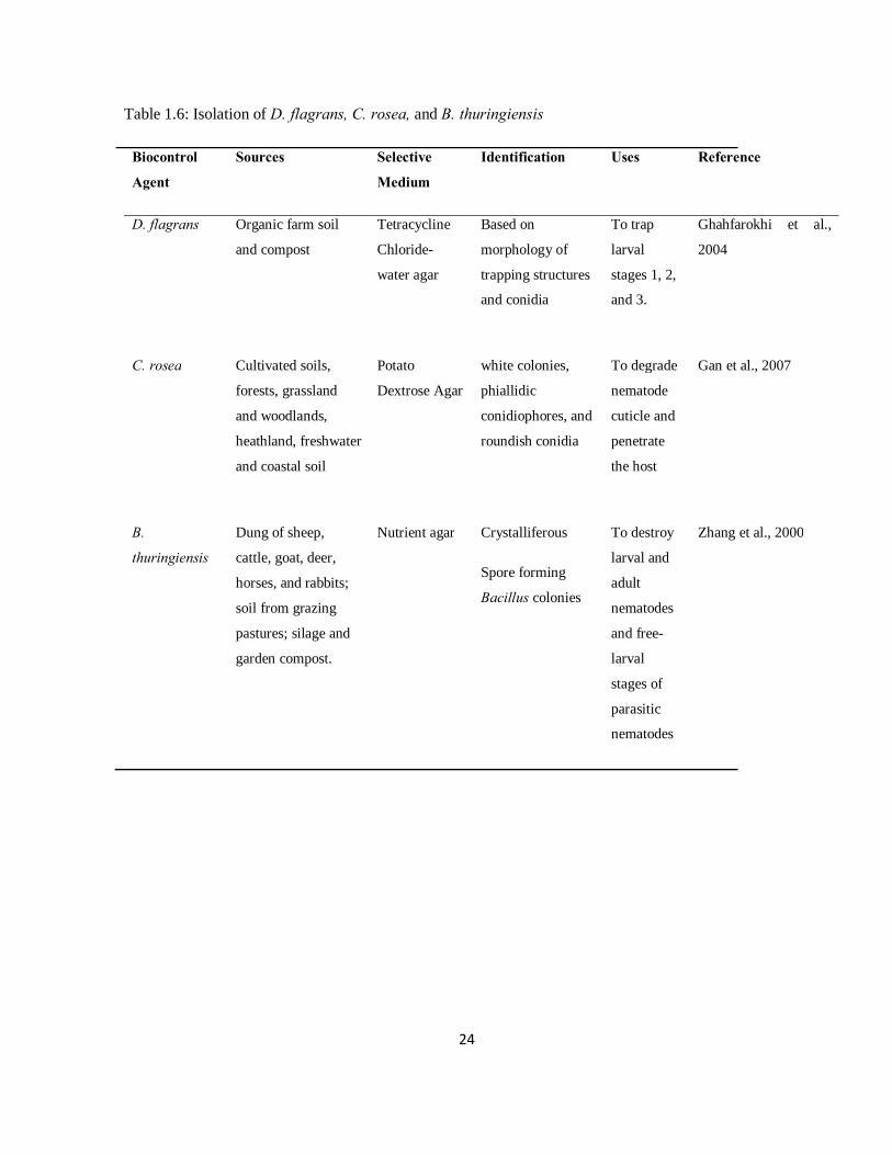
24
Table 1.6: Isolation of D. flagrans, C. rosea, and B. thuringiensis
Biocontrol
Agent
Sources Selective
Medium
Identification Uses Reference
D. flagrans Organic farm soil
and compost
Tetracycline
Chloride-
water agar
Based on
morphology of
trapping structures
and conidia
To trap
larval
stages 1, 2,
and 3.
Ghahfarokhi et al.,
2004
C. rosea Cultivated soils,
forests, grassland
and woodlands,
heathland, freshwater
and coastal soil
Potato
Dextrose Agar
white colonies,
phiallidic
conidiophores, and
roundish conidia
To degrade
nematode
cuticle and
penetrate
the host
Gan et al., 2007
B.
thuringiensis
Dung of sheep,
cattle, goat, deer,
horses, and rabbits;
soil from grazing
pastures; silage and
garden compost.
Nutrient agar Crystalliferous
Spore forming
Bacillus colonies
To destroy
larval and
adult
nematodes
and free-
larval
stages of
parasitic
nematodes
Zhang et al., 2000
25
1.6 References
Ademola, T.O., Fagbemi, B.O., Idowi, S.O. 2005. Anthelmintic activity of extracts of Spondias
mombin against gastrointestinal nematodes of sheep: Studies in In vitro and In vivo. Trop.
An. Health Prod. 37, 233-235.
Athanasiadou, S., Kyriazakis, I., Jackson, F., Coop, R.L. 2001. Direct anthelmintic effects of
condensed tannins towards different gastrointestinal nematodes of sheep: in vitro and in
vivo studies. Vet. Parasitol. 99, 199-204.
Barron, G.L. 1977. Observations on predatory fungi. Can. J. Bot. 57, 187-193.
Burke, J.M., Terrill, T.H., Kallu, R.R., Miller, J.E., Mosjidis, J.A. 2007. Use of copper oxide wire
particles to control gastrointestinal nematodes in goats. J. An. Sci. 85, 2753-2761.
Chatterjee, S.N., Bhattacharya, T., Danger, T.K., Chandra, G. 2007. Ecology and diversity of
Bacillus thuringiensis in soil environment. Af. J. Biotech. 6, 1587-1591.
Coles, G.C. 1986. Anthelmintic resistance in sheep. Vet. Clin. North Am. Food An. Pract. 2,
423–432.
Coles, G.C., Borgsteede, F.H.M., Geerts, S. 1994. Anthelmintic resistance in nematodes of farm
animals. European Commission, Brussels. Pp191.
Conway, D.P. 1964. Variance in effectiveness of thiabendazole against Haemonchus contortus in
sheep. Am. J. Vet. Res. 25, 844–845.
Cooke, R.C. 1969. Two nematode-trapping Hyphomycetes, Dudddingtonia flagrans gen. et
comb. nov. and Monacrosporium mutabilis nov. Trans. Br. Mycol. Soc. 53, 315-319.
Cooke, R.C., Godfrey, B.E.S. 1964. A key to the nematode-destroying fungi. Trans. Br. Mycol.
Soc. 47, 61-74.
Corwin, R.M. 1997. Economic gastrointestinal parasitism of cattle. Vet. Parasitol. 72, 451-460.
26
Cox, J. 1999. The nature conservation of dung. Br. Wildlife 11, 28-36.
Dalton, J.P., Mulcachy, G. 2001. Parasite vaccine-a reality. Vet. Parasitol. 98, 149-167.
De Barjac, H. Franchon, E. 1990. Classification of Bacillus thuringiensis strains. J. Entomophoga
35, 233-240.
De, S., Sanyal, P.K. 2009. Biological control of helminth parasites by predatory fungi. Vetscan 4,
1-8.
Desta, B., 1995. Ethiopian traditional herbal drugs. Part I. Studies on the toxicity and therapeutic
activity of local taenicidal medications. J. Ethnopharm. 45, 27–33.
Dong, J.Y., Zhao, Z.X., Cai, L., Liu, S.Q., Zhang, H.R., Duan, M. 2004. Nematicidal effect of
freshwater fungal cultures against the pine-wood nematode Bursaphelenchus xylophilus.
Fungal Divers, 15, 125–135.
Drudge, J.H. Elem, G. 1964. Field studies on parasite control in sheep: comparison of
thiabendazole, ruelene, and phenothiazine, Am. J. Vet. Res. 25, 1512–1518.
Drudge, J.H., 1957. Strain variation in the response of sheep nematodes to the action of
phenothiazine: II. Studies on pure infections of Haemonchus contortus. Am. J. Vet. Res. 18,
317–325.
Entwistle, P.F., Cory, J.S., Bailey, M.J., Higgs, S. (Eds.), 1993. Bacillus thuringiensis, an
Environmental Biopesticide: Teory and Practice. John Wiley, Chichester.
Faedo, M., Krecek, R.C. 2002. Possible application of a nematophagous fungus as a biological
control agent of parasitic nematodes on commercial sheep farms in South Africa. J. S. Af.
Vet. Assoc. 73, 31–35.
Fontenot, M.E., Miller, J.E., Peña, M.T., Larsen, M., Gillespie, A. 2003. Efficiency of feeding
Duddingtonia flagrans chlamydospores to grazing ewes on reducing availability of parasitic
nematode larvae on pasture. Vet. Parasitol. 118, 203-213.
27
Gan, Z., Yang, J., Tao, N., Yu, Z., Zhang, K. 2007. Cloning and expression analysis of a
chitinase gene Crchi1 from the mycoparasitic fungus Clonostachys rosea (syn.
Gliocladium roseum). J. of Micro. 45, 422-430.
Gasbarre, L.C., Leighton, E.A., Davies, C.J. 1990. Genetic control of immunity to
gastrointestinal nematodes of cattle. Vet. Parasitol. 37, 257–272.
Geerts, S., Gryseels, B. 2000. Drug resistance in human helminths: current situation and lessons
from livestock. Clin. Micro. Rev. 13, 207-222.
Georgi, J.R. 1969. Parasitology for Veterinarians. W.B. Saunders Company. London.
Ghahfarokhi, M.S., Abyaneh, M.R., Bahadori, S.R., Eslami, A., Zare, R., Ebrahimi, M. 2004.
Screening of soil and sheep faecal samples for predacious fungi: Isolation and
characterization of the nematode-trapping fungus Arthrobotrys oligospora. Iran. Biomed.
J. 8, 135-142.
Gibson, T.E. 1960. Some experiences with small daily doses of phenothiazine as a means of
control of strongylid worms in the horse. Vet. Rec. 72, 37–41.
Githiori, J.B., Höglund, J., Waller, P.J., Baker, R.L. 2002. Anthelmintic activity of preparations
derived from Myrsine africana and Rapanea melanophloeos against the nematode parasite,
Haemonchus contortus, of sheep. J. Ethnopharm. 80, 187-191.
Grade, J.T., Longok, A. 2002. Karamajong scientists: participatory field trial of a local
dewormer. In: Proceedings of the Uganda Veterinary Association Scientific Conference,
September 28–29, 2000.
Gray, A. 1993. Management of vertisols in Sub-Saharan Africa. Preseedings of a conference held
at ILCA, Addis Ababa, Ethiopia. FAO Corporate Document Repository.
Gronvold, J., Wolstrup, P., Larsen, M., Henriksen, S.A., Narsen, P. 1993. Biological control of
Ostertagia ostertagi by feeding selected nematode-trapping fungi to calves. J. Helminthol.
67, 31-36.
Holmes, P.H. 1987. Pathophysiology of nematode infections. Int. J. Parasitol. 17, 443-451.
28
Hördegen, P., Hertzberg, H., Heilmann, J., Langhans, W., Maurer, V. 2003. The anthelmintic
efficacy of five plant products against gastrointestinal trichostrongylids in artificially
infected lambs. Vet. Parasitol. 117, 51- 60.
Hoste, H. 2001. Adaptive physiological processes in the host during gastrointestinal parasitism.
Int. Parasitol. 31, 231-244.
Hsu, C., Levine, N.D. 1977. Degree-day concept in development of infective larvae of
Haemonchus contortus and Trichostrongylus colubriformis under constant and cyclic
condition. Am. J. Vet. Res. 38, 1115-1119.
Huang, X., Li, Y., Su, Y., Ding, J., Zhang, K. 2009. The influences on conidiophores
pleomorphosm in Clonostachys rosea and RAPD analysis to the mutant producing only
verticillate conidiophores.
Keyyu, J.D., Kyvsgaard, N.C., Monrad, J., Kassuku, A.A. 2005. Epidemiology of gastrointestinal
nematodes in cattle on traditional small-scale dairy and large-scale dairy farms in Iringa
district Tanzania. Vet. Parasitol. 127, 285-294.
Knox, D.P. 2000. Development of vaccines against gastrointestinal nematodes. Parasitology 120,
53-61.
Knox, M.R., Faedo, M. 2001. Biological control of field infections of nematodes parasites of
young sheep with Duddingtonia flagrans and effects of spore intake on efficacy. Vet.
Parasitol. 101, 155-160.
Kotze, A.C., O’Grady, J., Gough, J.M., Pearson, R., Bagnall, N.H., Kemp, D.H. Akhurst, R.J.
2005. Toxicity of Bacillus thuringiensis to parasitic and free-living life-stages of
economically important nematode parasites of livestock. Int. J. Parasitol. 35, 1013–1022.
Krattiger, F. 1997. Insect resistant crops: A case study of Bacillus thuringiensis and its transfer to
developing countries. ISAAA briefs, no. 2:42.
Lacey, L.A., Frutos, R., Kaya, H.K., Vale, P. 2001. Insect pathogens as biological control agents:
do they have a future? Biol. Control 21, 230–248.
29
Larsen, M. 1999. Biological control of helminths. Int. J. Parasitol. 29, 139-146.
Larsen, M. 2000. Prospects for controlling animal parasitic nematodes by predacious micro fungi.
Parasitology 120, 121–131.
Larsen, M. 2006. Biological control of nematode parasites in sheep. J. An. Sci. 84, E133-E139.
Larsen, M., Faedo, M., Waller P.J., Hennessy, D.R. 1998. The potential of nematophagous fungi
to control the free-living stages of nematode parasites of sheep: Studies with Duddingtonia
flagrans. Vet. Parasitol. 76, 121-128.
Larsen, M., Faedo, M., Waller, P.J. 1994. The potential of nematophagous fungi to control the
free-living stages of nematode parasites of sheep: survey for the presence of fungi in fresh
faeces of grazing livestock in Australia. Vet. Parasitol. 53, 275–281.
Larsen, M., Nansen, P., Gronvold, J., Wolstrup, J., Henriksen, A. 1992. In vivo passage of
nematophagous fungi selected for biocontrol of parasitic nematodes in ruminants. J.
Helminth. 66, 137-141.
Larsen, M., Nansen, P., Wolstrup, J., Gronvold, J., Henriksen, S.A., Zorn, A. 1995. Biological
control of trichostrongylus in calves by the fungus Duddingtonia flagrans fed to animals
under natural grazing conditions. Vet. Parasitol. 60, 321-330.
Larsen, M., Wolsrup, J., Henriken, S.A., Dackman, C., Grovold, J., Nansen, P. 1991. In vitro
stress selection of nematophagous fungi for biocontrol of parasitic nematodes in ruminants.
J. Helminth. 65, 193-200.
Leighton, E.A., Murrell, K.D., Gasbarre, L.C. 1989. Evidence for genetic control of nematode
egg-shedding rates in calves. J. Parasitol. 75, 498-504.
Li, G.Q., Huang, H.C., Kokko, E.G., Acharya, S.N. 2002. Ultra structural study of
mycoparasitism of Gliocladium roseum on Botrytis cinera. Bot. Bul. of Aca. Sinica. 43,
211-218.
Li, J., Yang, J.K., Huang, X.W., Zhang, K.Q. 2006. Purification and characterization of an
extracellular serine protease from Clonostachys rosea and its potential as a pathogenic
factor. Process Biochem. 41, 925-929.

30
Maichomo, M.W., Kagira, J.M., Walker, T. 2004. The point prevailence of gastrointestinal
parasites in calves, sheep, and goat in Magadi division, South-West Kenya. Ond. J. Vet. Res.
71, 257-261.
Malan, F.S., Van Wyk, J.A., Wessels, C. 2001. Clinical evaluation of anaemia in sheep: early
trials. Onderstepoort J. Vet. Res. 68, 165–174.
McKenna, P.B. 1981. The diagnostic value and interpretation of faecal egg count in sheep. N.Z.
Vet. J. 29, 129- 132.
McLeon, R.S. 1995. Cost of major parasites to the Australian livestock industry. Int. J. Parasitol.
25, 1363-1367.
Mendoza de Gives, P., Crepso, J.F., Rodriquez, D.H., Prats, V.V., Hernandez, E.L., Fernandez,
G.E.O. 1998. Biological control of Haemonchus contortus infective larvae in ovine faeces
by administering an oral suspension of Duddingtonia flagrans chlamydospores to sheep. J.
Helminth. 72, 343–347.
Merck and Co., Inc. 2008. The Meck Veterinary Manual. USA.
http://www.merckvetmanual.com/mvm/index.jsp?cfile=htm/bc/191512.htm.
Min, B.R., Hart, S.P. 2003. Tannins for suppression of internal parasites. J. An. Sci. 81, 102–109.
Min, B.R., Pomroy, W.E., Hart, S.P., Sahlu, T. 2004. The effect of short-term consumption of a
forage containing condensed tannins on gastrointestinal nematode parasite infections in
grazing weather goats. Small Rumin. Res. 51, 279–283.
Minja, M.M. 1994. Medicinal plants used in the promotion of animal health in Tanzania. Rev.
Sci. Tech. 13, 905–925.
Mitreva, M.D.S., Zarlenga, J.P., McCoorter, D.P.J. 2007. Parasitic nematodes from genomes to
control. Vet. Parasitol. 143, 31-42.
Molan, A.L., Waghorn, G.C., Min, B.R., McNabb, W.C. 2000. The effect of condensed tannins
from seven herbages on Trichostrongylus colubriformis larval migration in vitro Folia
Parasitologica 47, 39–44.

31
Morandi, M.A.B., Maya, L.A., Mizubuti, E.S.G., Alfenas, A.C., Barbosa, J.G. 2003. Suppression
of Botrytis cinera sporulation by Clonostachys rosea on rose debris: a valuable component
in Botrytis blight management in commercial greenhouse. Biol. Control 26, 311–317.
Navon, A. 2000. Bioassays of Bacillus thuringiensis products used against agricultural pests. In,
Bioassay of Entomopathogenic Microbes and Nematodes. (Ed), Navon, A., Ascher, K.R.S.
CABI Publishing, New York.
Nienzen, J.H., Charleston, W.A.G., Robertson, H.A., Shelton, D., Waghorn, G.C., Green, R.
2002. The effect of feeding sulla (Hedysarum coronarium) or Lucerne (Medicago sativa) on
lamb parasite burdens and development of immunity to gastrointestinal nematodes. Vet.
Parasitol. 105, 229–245.
O`Connor, L., W. Stephen and W.W. Brown, 2006. Ecology of the freeliving stages of major
Trichostrongyloid parasites of sheep. Vet.Parasitol. 142, 1–15
O’Grady, J.O., Akurst, R.J., Kotze, A.C. 2007. The requirement for early exposure of
Haemonchus contortus larvae to Bacillus thuringiensis for effective inhibition of larval
development. Vet. Parasitol. 150, 97-103.
Oslon, O.W. 1974. Animal Parasites: Their Life Cycles and Ecology. Dover Publications Inc.,
New York.
Paolini, V., Dorchies, P., Hoste, H. 2003. Effects of sainfoin hay on gastrointestinal with
nematodes in goats. Vet. Rec. 152, 600–601.
Peña, M.T., Miller, J.E., Fontenot, M.E., Gillespie, A., Larsen, M., 2002. Evaluation of
Duddingtonia flagrans in reducing infective larvae of Haemonchus contortus in faeces of
sheep. Vet. Parasitol. 103, 259–265.
Perry, B.D., McDermott, J.J., Randolph, T.F., Sones, K R., Thornton, P.K. 2002. Investing in
Animal Health Research to Alleviate Poverty. International Livestock Research Institute,
Nairobi, Kenya.
32
Perry, B.D., Radolph, T.F. 1999. Improving assessment of the economic impact of parasitic
diseases in production animals. Vet. Parasitol. 84, 143- 166.
Poynter, D., Hughes, D.L. 1958. Phenothiazine and piperazine, an efficient anthelmintic mixture
for horses. Vet. Rec. 70, 183–1188.
Prichard, R.K., Roulet, A. 2007. ABC transporters and β-tubulin in macrocyclic lactone
resistance: prospects for marker development. Parasitology 134, 1123-1132.
Quesada-Moraga, E., Garcia-Tovar, E., Vaverde-Garcia, P., Santiago-Alvarenz, C. 2004.
Isolation, geographical diversity and insecticidal activity of Bacillus thuringiensis from soils
in Spain. Microbiol. Res. 159, 59-71.
Sayers, G., Sweeney, T. 2005. Gastrointestinal nematode infection in sheep—a review of the
alternatives to anthelmintics in parasite controls. An. Health Res. Rev. 6, 159–171.
Schnepf, E., Crickmore, N., Van Rie, J., Lereclus, D., Baum, J., Feitelson, J., Zeigler, Dean, D.R.
1998. Bacillus thuringiensis and its pesticidal crystal proteins. Micro. Mol. Biol. Rev. 62,
775–806.
Schroers, H.J., Samuels, G.J., Seifert, K.A., Gams, W. 1999. Classification of the mycoparasite
Clonostachys rosea in Clonostachys as C. rosea, its relationship to Bionectria ochroleuca,
and notes on other Gliocladium-like fungi. Mycologia 91, 365–385.
Senel, S., McClure, S.J. 2004. Potential applications of chitosan in veterinary medicine. Adv.
Drug Deliv. Rev. 56, 1467–1480.
Shaik, S.A., Terrill, T.H., Miller, J.E., Kouakou, B., Kannan, G., Kallu, R.K. Mosjidis, J.A. 2004.
Effects of feeding Sericea lespedeza hay to goats infected with Haemonchus contortus, S.A.
J. An. Sci. 34, 234–236.
Shimoda, J., Onishi, H., Machida, Y. 2001. Bioadhesive characteristics of chitosan microspheres
to the mucosa of rat small intestine. Drug Dev. Ind. Pharm. 27, 567–576.
Siddiqui, Z.A., Mahmood, I. 1996. Biological control of plant parasitic nematodes by fungi: a
review. Bior. Tech. 58, 229- 239.
33
Siegel, J.P. 2001. The mammalian safety of Bacillus thuringiensis-based insecticides. J. Invert.
Path. 77, 13–21.
Skipp, R.A., Chen, L.Y., Yeates, G.W., Glare, R. 2002. Occurrence, morphological
characteristics and ribotyping of New Zealand isolates of Duddingtonia flagrans, a candidate
for biocontrol of animal parasitic nematodes. N.Z. J. Agric. Res. 45, 187–196.
Stear, M.J., Doligalska, M., Donskow-Schmelter, K. 2006. Alternatives to anthelmintics for the
control of nematodes in livestock. Parasitology 134, 1–13.
Stepek, G., Behnke, J.M., Buttle, D.J., Duce, I.R. 2004. Natural plant cysteine proteinases as
anthelmintics. Trends Parasitol. 20, 323-327.
Stirling, G.R., Smith, L.J., Licastro, K.A., Eden, L.M. 1998. Development control of root-knot
nematode with formulations of the nematode-trapping fungus Arthrobotrys dactyloides. Biol.
Control 11, 224–230.
Stromberg, B.E. 1997. Environmental factors influencing transmission. Vet. Parasitol. 72, 247-
264.
Sutton, J.C., Li, D.W. Gang, P. Hai Y., Zhang. P. 1997. Gliocladium roseum a versatile adversary
of a Botrytis cinera in crops. Plant Dis. 81, 316-328.
Swadener, C. 1994. Bacillus thuringiensis (Bt). J. Pest Reform 14, 13-20.
Swartson, H., Nsahlai, I. 2006. Animal Production. MacMillan South Africa Ltd (Pty),
Pietermaritzburg.
Tembely, S., Lahlou-Kassie, A., Rege, J.E.O., Sovani, S., Diedhiou, M.L., Baker, R.L. 1997. The
epidemiology of nematode infections in sheep in a cool tropical environment. Vet.
Parasitol. 70, 129-141.
Terrill, T.H., Kaplan, R.M., Larsen, M., Samples, O.M., Miller, J.E., Gelaye, S. 2001.
Anthelmintic resistance on goat farms in Georgia: efficacy of anthelmintics against
gastrointestinal nematodes in two selected goat herds. Vet. Parasitol. 97, 261–268.

34
Thamsborg, S.M., Roepstorff, A., Larsen, M. 1999. Integrated and biological control of parasites
in organic and conventional production systems. Vet. Parasitol. 84, 169-186.
Tunlid, A., Ahman, J., Oliver, R.P. 1999. Transformation of the nematode-trapping fungus
Arthrobotrys oligospora, FEMS. Micro. Lett. 173, 111–116.
Urquhart, G.M., Armour, J., Duncan, J.L., Dunn, A.M., Jennings, F.W. 1987. Veterinary
Parasitology. Longman Scientific and Technical. New York, U.S.A.
Valdebenito-Sanhueza, R.M., Sutton, J.C., Perazzolo, I., Czermainski, A.B.C., 1997. Controle
biológico de Botrytis cinera em morangueiros cultivados em estufa. Fitopatol. Bras. 22, 69–
73.
Van Wyk, J.A. 1990. Occurrence and dissemination of anthelmintic resistance in South Africa,
and management of resistant worm strains. In: Boray, J.C., Martin, P.J., Roush, R.T., (Eds.)
Resistance of parasites to antiparasitic drugs. Round Table Conference held at the VIIth
International Congress of Parasitology, Paris. MSD. Agvet, Division of Merck, Rahway,
New Jersey, 103-113.
Van Wyk, J.A., Stenson, M.O., Van der Merwe, J.S., Vorster, R.J., Viljoen, P.G. 1999.
Anthelmintic resistance in South Africa: surveys indicate an extremely serious situation in
sheep and goat farming. Ond. J. Vet. Res. 66, 273–284.
Vatta, A.F., Letty, B.A., Van der Linde, M.J., Van Wijk, E.F., Hansen, J.W., Krecek, R.C. 2001.
Testing for clinical anaemia caused by Haemonchus spp. In goats farmed under resource-
poor conditions in South Africa using an eye colour chart develop for sheep. Vet. Parasitol.
99, 1-14.
Vercruysse, I., Clarebout, E. 2001. Treatment vs non-treatment of helminth infections in cattle:
defining the thresholds. Vet. Parasitol. 98, 195-214.
Waghorn, T.S., Leathwick, D.M., Chen, L.Y., Skipp, R.A. 2003. Efficacy of the nematode-
trapping fungus Duddingtonia flagrans against three species of gastro-intestinal nematodes
in laboratory faecal cultures from sheep and goats. Vet. Parasitol. 118, 227–234.157.
35
Waller, P.J. 1997. Nematode parasite control of livestock in the tropics/subtropics: the need for
novel approaches. Int. J. Parasitol. 27, 1193–1201.
Waller, P.J., Bernes, G., Thamsborg, S.M., Sukura, A., Richter, S.H., Ingebrigtsen, K., Hoglund,
J. 2001. Plants as de-worming agents of livestock in the Nordic countries: historical
perspective, popular beliefs and prospects for the future. Acta Vet. Scand. 42, 41-44.
Waller, P.J., Echevarria, F., Eddi, C., Maciel, S., Nari, A., Hansen, J.W. 1996. The prevalence of
anthelmintic resistance in nematode parasites of sheep in southern Latin America: general
overview. Vet. Parasitol. 62, 181-187.
Waller, P.J., Faedo, M. 1996. The prospects for biological control of the free-living stages of
nematode parasites of livestock. Int. J. Parasitol. 26, 915–925.
Waller, P.J., Thamsborg, S.M. 2004. Nematode control in ‘green’ ruminant production systems.
Trends Parasitol. 20, 493–497.
Wei, J.Z., Hale, K., Carta, L., Platzer, E., Wong, C., Fang, S.C., Aroian, R.V. 2003. Bacillus
thuringiensis crystal proteins that target nematodes. Nat. Acad. Sci., USA 100, 2760-2765.
Wu, S.H.W., Papas, A., 1997. Rumen-stable delivery systems. Adv. Drug Deliv. Rev. 28, 323-
334.
Yang, X.J., He, Y.X., Chen, F.R., Zheng, L. 2000. Separating and selection of egg-mass fungi of
Meloidogyne sp. in Fujian province. Fujian J. of Agric. Sci. 15, 12-15.
Zhang, H.Y., Yu, Z.N., Deng, W.X. 2000. Isolation, distribution and toxicity of Bacillus
thuringiensis from warehouses in China. Crop Prot. 19,449-454.
Zhang, K.Q., Zhao, M.L. 2004. Recent advance of nematophagous fungi from China and with a pecial
case on Clonostachys rosea (Gliocladium roseum). J. of Zhejiang University (Agriculture & Life
Sciences). 30, 389-389.
Zhang, L., Yang, J., Nia, Q., Zhao, X., Ye, F., Liang, L., Zhang, K.Q. 2008. Investigation on the infection
mechanism of the fungi Clonostachys rosea against nematodes using green fluorescent protein.
Appl. Microbiol. Biotech. 78, 983-990.

36
Zhao, M.L., Huang, J.S., Mo, M.H., Zhang, K.Q. 2005. A potential virulence factor involved in
fungal pathogenecity serine-like protease activity of nematophagous fungus Clonostachys
rosea. Fungal Divers. 19, 217-234.
37
CHAPTER TWO
ISOLATION OF BACILLUS THURINGIENSIS (BERLINER) AND CLONOSTACHYS
ROSEA (SCHROERS) FROM GRAZING PASTURES AND ANIMAL FAECES
M.A. Baloyi, M.D. Laing and K.S. Yobo
Discipline of Plant Pathology, School of Agricultural Sciences and Agribusiness,
University of KwaZulu-Natal, Private Bag X01, Scottsville 3201, Pietermaritzburg,
South Africa
38
Abstract
Biological control agents may be an alternative to agrochemicals for the control of livestock
nematodes. Strains of Bacillus thuringiensis (Bt) and Clonostachys rosea were isolated from
grazing pastures and from faeces collected from pens of livestock. On nutrient agar, Bt colonies
are circular, white, flat and undulate. B. thuringiensis isolates are gram-positive, rod-shaped and
form endospores. About 51% of endospore-forming bacteria isolated were B. thuringiensis. C.
rosea were identified by white colonies and the characteristic conidiophores which were
branched and showed phialides at the tips. A total of 25 isolates of B. thuringiensis and 10
isolates of C. rosea were successfully isolated.
2.1 Introduction
Gastrointestinal parasite infection is the most important limiting factor to small ruminant
production worldwide, resulting in serious economic losses (Perry et al., 1999; Jackson and
Coop, 2000; Perry et al., 2002; Kaplan, 2004). Traditionally, anthelmintics have been used to
control these parasites (Borgsteede, 1998). The frequent use of anthelmintics drugs has resulted
in resistance, which is currently a major problem in all sheep producing countries, including
South Africa (Van Wyk et al., 1999; Vatta et al., 2001). Currently, biological control is
considered to be one of the most promising alternatives that might be employed for the control of
these parasitic nematodes (Larsen, 1999; Chandrawathani et al., 2003; Waller, 2006; De and
Sanyal, 2009). Consequently, the number of studies on how to isolate, screen and characterize
biocontrol agents of livestock nematodes is growing.
Some parasporal crystalline inclusions of Bacillus thuringiensis (Bt) (Berliner) have shown
strong activity against both insects and nematodes (Lee and Pankhurst, 1992). The latter was
discovered by Waller (2003) and confirmed by Kotze et al., (2005), who reported that several
isolates of Bt were highly toxic to larvae and adults of livestock nematodes, Haemonchus
contortus (Rudolphi), Trichostrongylus colubriformis (Giles) and Ostertagia circumcincta
(Stadelmann) in in vitro studies. This suggest that Bt strains could be used as biological control
39
agents against adult nematodes in livestock, and the free-living larval stages that develop in
pastures, primarily in manure (O’Grady et al., 2007).
The fungus Clonostachys rosea (Schroers) has mostly been used as a biological control agent on
plant parasitic nematodes. This fungus is commonly found in soil. According to Zhao et al.
(2005) and Li et al. (2006), the ability of this fungus to destroy nematodes lies in its ability to
penetrate the cuticle, through the production of a germ tube which penetrates the host body and
kills it.
Consequently, the aim of this study was to isolate these two biocontrol agents, Bt and C. rosea,
from soil of goat and sheep pens and grazing pastures.
2.2 Materials and Methods
2.2.1 Sample collection Samples were collected from different sites in the livestock section at the Ukulinga Research
Farm, University of KwaZulu-Natal, Pietermaritzburg. The samples comprised of faeces from
pens of cattle, sheep and goats, and soil from the grazing pastures of horses, sheep, cattle and
goats. Surface soil (15 mm depth) was removed with a spatula and approximately 20g soil and
faecal samples were each collected into separate plastic bags. Soil samples were stored in a fridge
at 40C until used.
2.2.2 Isolation of Bacillus thuringiensis Bacillus thuringiensis was isolated from soil and faecal samples as described by Kotze et al.
(2005). Each one gram sample of soil and faecal matter was suspended in 9ml of sterile distilled
water and heat treated for 15 min at 800C in a rotary water bath. The suspensions were serially
diluted, 0.1ml of 10-4 dilution was plated onto nutrient agar and kept at room temperature for 4d
to sporulate. Spores were harvested into tryptone soy broth (15ml), shaken at 150 rpm for 1wk at
280C, then centrifuged (10,000 rpm, 10 min, 40C). The pellet was washed once in 0.01M NaCl,
and twice in cold sterile distilled water. The final pellet was re-suspended in 4ml of sterile
distilled water and stored at -800C (as described by Kotze et al., 2005).

40
2.2.3 Isolation of Clonostachys rosea A basal medium containing MgS04(0.1g), K2HPO4(0.4g), KCl(0.075g), NH4NO3(0.5g), glucose
(1.5g) and agar power (10g) were mixed and added to 475 ml distilled water and autoclaved.
Propamocarb (Previcur) fungicide (0.6g) and crystallized chloramphenicol (0.25g) was added to
50ml of distilled water and separately added to the basal medium.
A one gram sample of soil was suspended in 9ml of sterile distilled water and plated on the
medium. The plates were incubated at 250C and monitored for the development of lilac colonies.
These colonies were subcultured onto Potato Dextose Agar (PDA) and incubated at 250C, for
1wk.
2.2.4 Microscopy To look for the presence of parasporal bodies of Bt, an aliquot of a sporulated colony was
transferred onto a microscope slide. The slide was heat fixed by passing the slide three times over
a Bunsen burner flame, then stained with Coomasie Blue stain (0.133% Coomassie Blue stain in
50% acetic acid), rinsed with distilled water, air-dried and observed under a light microscope
(1000x) under oil immersion (Ammons and Ramphersad, 2002).
For C. rosea, mycelium was taken from 2wk old cultures grown on PDA and mounted on a slide
and stained with lactophenol cotton blue. The specimens were viewed at a 400x magnification.
For electron microscopy, the fungal specimens were fixed overnight in 3% glutaraldehyde in
0.05M cacodylate buffer. The specimens were washed in 0.05M cacodylate buffer twice for 30
min in each wash, then washed in 2% osmium tetroxide for 1 to 4 hours. The specimens were
then dehydrated in an ethanol series (30, 50, 70, 80, 90 and 100%) for 10 min each, with 3
changes in the 100% ethanol, with each wash lasting 10min. After dehydration, the specimens
were transferred to a critical point drying (CPD) basket submerged in 100% alcohol. The
specimen were then placed in a pressure bomb for critical point drying. The specimens were then
transferred to scanning electron microscopy (SEM) stubs and examined at 3500x magnification
(Kurztman et al., 1974).
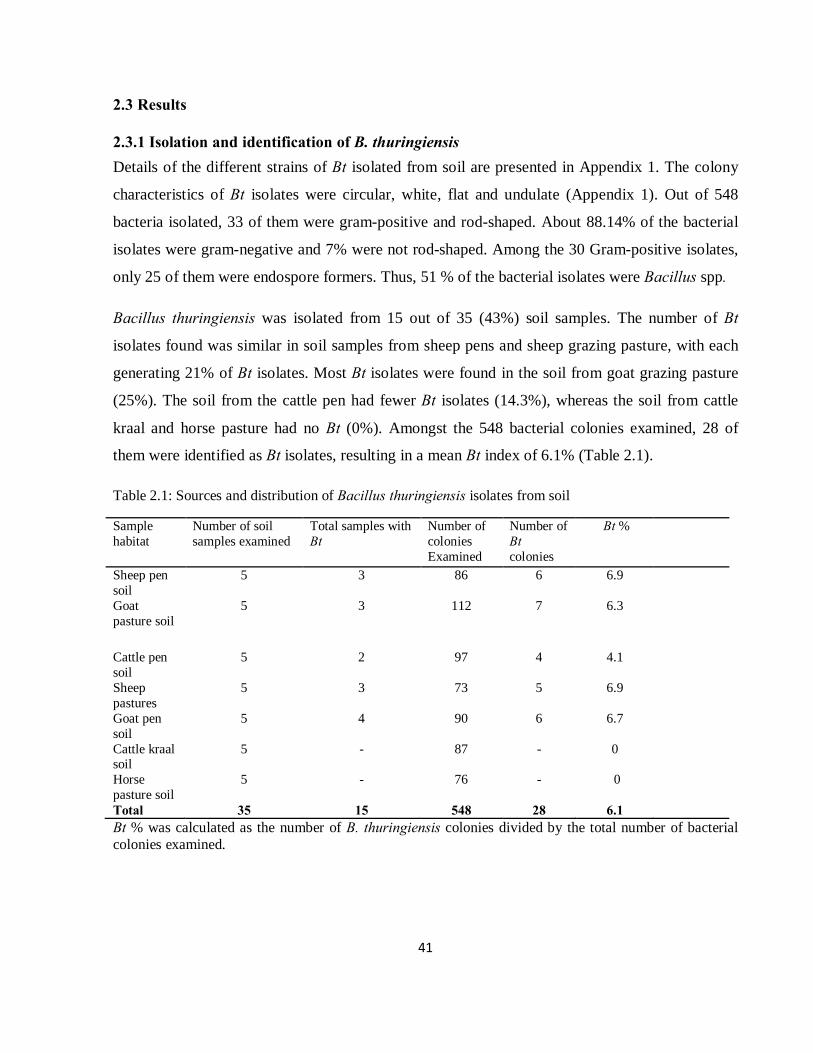
41
2.3 Results
2.3.1 Isolation and identification of B. thuringiensis Details of the different strains of Bt isolated from soil are presented in Appendix 1. The colony
characteristics of Bt isolates were circular, white, flat and undulate (Appendix 1). Out of 548
bacteria isolated, 33 of them were gram-positive and rod-shaped. About 88.14% of the bacterial
isolates were gram-negative and 7% were not rod-shaped. Among the 30 Gram-positive isolates,
only 25 of them were endospore formers. Thus, 51 % of the bacterial isolates were Bacillus spp.
Bacillus thuringiensis was isolated from 15 out of 35 (43%) soil samples. The number of Bt
isolates found was similar in soil samples from sheep pens and sheep grazing pasture, with each
generating 21% of Bt isolates. Most Bt isolates were found in the soil from goat grazing pasture
(25%). The soil from the cattle pen had fewer Bt isolates (14.3%), whereas the soil from cattle
kraal and horse pasture had no Bt (0%). Amongst the 548 bacterial colonies examined, 28 of
them were identified as Bt isolates, resulting in a mean Bt index of 6.1% (Table 2.1).
Table 2.1: Sources and distribution of Bacillus thuringiensis isolates from soil
Sample habitat
Number of soil samples examined
Total samples with Bt
Number of colonies Examined
Number of Bt colonies
Bt %
Sheep pen soil
5 3 86 6 6.9
Goat pasture soil
5 3 112 7 6.3
Cattle pen soil
5 2 97 4 4.1
Sheep pastures
5 3 73 5 6.9
Goat pen soil
5 4 90 6 6.7
Cattle kraal soil
5 - 87 - 0
Horse pasture soil
5 - 76 - 0
Total 35 15 548 28 6.1 Bt % was calculated as the number of B. thuringiensis colonies divided by the total number of bacterial colonies examined.
42
2.3.2 Isolation and identification of C. rosea
Colonies of C. rosea were velvety in texture. The colours of colonies were initially white, and
then became yellow in some isolates. The white colonies had a hyaline septate hyphae and
defined branching patterns were observed for the isolates. Conidiophores were straight, with
conidia produced at the tip, which were rod-shaped.
Eight isolates of C. rosea were isolated from sheep grazing pasture and goat grazing pasture, with six of
the isolates being isolated from the goat grazing pasture (Table 2.2).
Table 2.2: Source of Clonostachys rosea isolates
Source of soil Fungal Isolate
Sheep grazing pasture Paec1M
Sheep grazing pasture Paec2M
Goat grazing pasture Paec3M
Goat grazing pasture Paec4M
Goat grazing pasture Paec5M
Goat grazing pasture Paec6M
Goat grazing pasture Paec7M
Goat grazing pasture Paec8M
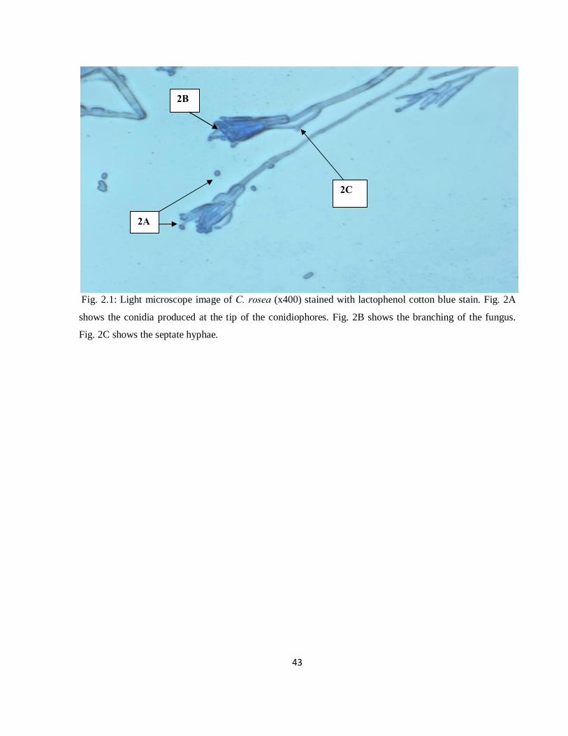
43
Fig. 2.1: Light microscope image of C. rosea (x400) stained with lactophenol cotton blue stain. Fig. 2A
shows the conidia produced at the tip of the conidiophores. Fig. 2B shows the branching of the fungus.
Fig. 2C shows the septate hyphae.
2A
2B
2C
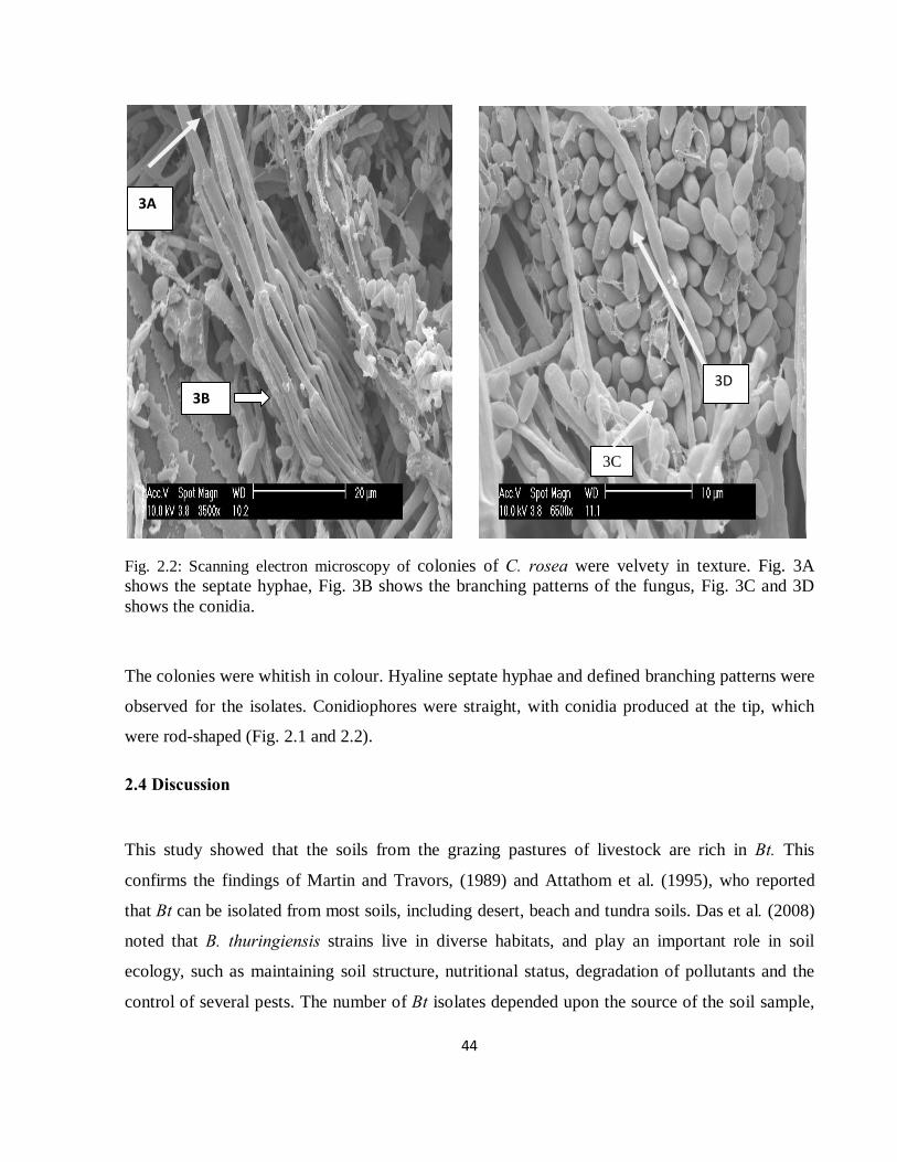
44
Fig. 2.2: Scanning electron microscopy of colonies of C. rosea were velvety in texture. Fig. 3A shows the septate hyphae, Fig. 3B shows the branching patterns of the fungus, Fig. 3C and 3D shows the conidia.
The colonies were whitish in colour. Hyaline septate hyphae and defined branching patterns were
observed for the isolates. Conidiophores were straight, with conidia produced at the tip, which
were rod-shaped (Fig. 2.1 and 2.2).
2.4 Discussion
This study showed that the soils from the grazing pastures of livestock are rich in Bt. This
confirms the findings of Martin and Travors, (1989) and Attathom et al. (1995), who reported
that Bt can be isolated from most soils, including desert, beach and tundra soils. Das et al. (2008)
noted that B. thuringiensis strains live in diverse habitats, and play an important role in soil
ecology, such as maintaining soil structure, nutritional status, degradation of pollutants and the
control of several pests. The number of Bt isolates depended upon the source of the soil sample,
3B
3A
3C
3D
45
which might be due to different nutrient availability of the soils, as suggested by Glare and O-
Callaghan (2000). Goat pen soil and sheep pen soil gave similar numbers of Bt isolates. Soil from
cattle grazing pasture produced only four isolates, while there were no Bt isolates found from
soils of the cattle kraal and horse grazing pasture. The morphological characteristics of the Bt
isolates are similar to those described by Chatterjee et al. (2007).
The presence of the parasporal bodies was revealed by the presence of numerous dark-blue
staining objects. Ammons and Ramphersad, (2002) reported the importance of using stained
specimens and light microscopy instead of phase-contrast microscopy, and described dark-blue,
rod-shaped staining parasporal bodies such as those observed.
Clonostachys rosea has mostly been isolated from soil (Sutton, 1997). This fungus was present in
grazing pasture soils collected from the livestock section at Ukulinga Research Farm. The goat
grazing pasture soil produced 63% of the C. rosea isolates, more than the sheep and the cattle
grazing pastures. All of these isolates had oval shaped conidia with septate hyphae. The
characteristic of the colony development was as described by Schroers (1997), Gan et al. (2007)
and St. German, (1996). Hyaline septate hyphae with defined branching patterns resembling that
of C. rosea were observed.
46
2.5 References
Ammons, D., Ramphersad, J. 2002. Usefulness of staining of the parasporal bodies when
screening for Bacillus thuringiensis. J. Invert. Parasitol. 79, 203-204.
Attathom, T., Chongrattanameteekul, W., Chanpaisang, J., Sirijan, R. 1995. Morphological
diversity and toxicity of delta-endotoxin produced by various strains of Bacillus
thuringiensis. Bull. Ento. Res. 85, 167-173.
Borgsteede, F.H.M. 1998. Gastrointestinal helminthiasis: Anthelmintic resistance and how to
prevent and control. Symp. Parasitol. Int. 47, 23-48.
Chandrawathani, P., Jamnah, O., Waller, P.J, Larsen, M., Gillespie, A.T., Zahari, W.M. 2003. Biological control of nematode parasites of small ruminants in Malaysia using the
nematophagous fungus Duddingtonia flagrans. Vet. Parasitol. 117, 173-183.
Chatterjee, S.N., Bhattacharya, T., Danger, T.K., Chandra, G. 2007. Ecology and diversity of
Bacillus thuringiensis in soil environment. Af. J. Biotech. 6, 1587-1591.
Das, J., Das, B., Dangar, T.K. 2008. Microbial populations and Bacillus thuringiensis diversity in
saline rice field soils of coastal Orissa, India. Af. J. Micro. Res. 2, 326-331.
De, S., Sanyal, P.K. 2009. Biological control of helminth parasites by predatory fungi. Vetscan 4,
1-8.
Glare, T.R., O-Callaghan, M. 2000. Bacillus thuringiensis: Biology, Ecology and Safety. Wiley,
Chichester.
Gan, Z., Yang, J., Tao, N., Yu, Z., Zhang, K. 2007. Cloning and Expression Analysis of a
Chitinase Gene Crchi1 from the Mycoparasitic Fungus Clonostachys rosea (syn.
Gliocladium roseum). The J. of Micro. 45, 422-430.
47
Jackson, F., Coop, R.L. 2000. The development of anthelmintic resistance in sheep nematodes.
Parasitology 120, 95–107.
Kaplan, R.M. 2004. Drug resistance in nematodes of veterinary importance: A status report.
Trends Parasitol. 20, 477-481.
Kotze, A.C., O’Grady, J., Gough, J.M., Pearson, R., Bagnall, D.H., Akhurst, R.J. 2005. Toxicity
of Bacillus thuringiensis to parasitic and free-living life stages of nematode parasitic of
livestock. Int. Parasitol. 35, 1013-1022.
Kurtzman, C.P., Baker, F.L. Smiley, M.J. 1974. Specimen holder to critical point dry
microorganisms for scanning electron microscopy. Appl. Micro. 28, 708-712.
Larsen, M. 1999. Biological control of helminths. Int. J. Parasitol. 29, 139-146.
Lee, K.E., Pankhurst, C.E. 1992. Soil organisms and sustainable productivity. Austr. J. Soil Res.
30, 855-892.
Li, J., Yang, J.K., Huang, X.W., Zhang, K.Q. 2006. Purification and characterization of an
extracellular serine protease from Clonostachys rosea and its potential as a pathogenic
factor. Process Biochem. 41, 925-929.
Martin, P.A.W., Travers, R.S. 1989. Worldwide abundance and distribution of Bacillus
thuringiensis isolates. Appl. Env. Micro. 55, 2437-2442.
O’Grady, J., Akhurst, R.J., Kotze, A.C. 2007. The requirement for early exposure of
Haemonchus contortus larvae to Bacillus thuringiensis for effective inhibition of larval
development. Vet. Parasitol. 150, 97-103.
Perry, B.D., Randolph, T.F. 1999. Improving the assessment of the economic impact of parasitic
diseases and of their control in production animals. Vet. Parasitol. 84, 145–168.
Perry, B.D., Randolph, T.F., McDermott, J.J., Sones, K.R., Thornton, P.K. 2002. Investing in
Animal Health Research to Alleviate Poverty. International Livestock Research Institute
(ILRI), Nairobi, Kenya, p. 148.
Schroers, H.J., Samuels, G.J., Seifert, K.A., Gams, W. 1999. Classification of the mycoparasite
Clonostachys rosea in Clonostachys as C. rosea, its relationship to Bionectria ochroleuca,
and notes on other Gliocladium-like fungi. Mycologia 91, 365–385.
48
St. Germain, G., Summerbell, R. 1996. Identifying Filamentous Fungi – A Clinical Laboratory
Handbook, 1st ed. Star Publishing Company. Carlifornia, U.S.A.
Sutton, J.C., Li, D.W. Gang, P. Hai Y., Zhang. P. 1997. Gliocladium roseum a versatile adversary
of a Botrytis cinera in crops. Plant Dis. 81, 316-328.
Van Wyk, J.A., Stenson, M.O., Van der Merwe, J.S., Vorster, R.J., Viljoen, P.G. 1999.
Anthelmintic resistance in South Africa: surveys indicate an extremely serious situation in
sheep and goat farming. Ond. J. Vet. Res. 66, 273-284.
Vatta, A.F., Letty, B.A., Van der Linde, M.J., Van Wijk, E.F., Hansen, J.W., Krecek, R.C. 2001.
Testing for clinical anaemia caused by Haemonchus spp. in goats farmed under resource –
poor conditions in South Africa using an eye colour chart developed for sheep. Vet.
Parasitol. 99, 1-9.
Waller, P.J. 2003. Global perspectives on nematode parasite control in ruminant livestock: the
need to adopt alternatives to chemotherapy, with emphasis on biological control. An. Health
Res. Rev. 4, 35-43.
Waller, P.J. 2006. Sustainable nematode parasite control strategies for ruminant livestock by
grazing management and biological control. An. Feed Sci. 126, 277-289.
Zhao, M.L., Huang, J.S., Mo, M.H., Zhang, K.Q. 2005. A potential virulence factor involved in
fungal pathogenicity serine-like protease activity of nematophagous fungus Clonostachys
rosea. Fungal Divers. 19, 217-234.
49
CHAPTER THREE
IN VITRO SCREENING OF BACILLUS THURINGIENSIS (BERLINER) AND CLONOSTACHYS ROSEA (SCHROERS) AGAINST LIVESTOCK PARASITIC NEMATODES
M.A. Baloyi, M.D. Laing and K.S. Yobo
Discipline of Plant Pathology, School of Agricultural Sciences and Agribusiness,
University of KwaZulu-Natal, Private Bag X01, Scottsville 3201, Pietermaritzburg,
South Africa
50
Abstract
In vitro studies were conducted to determine the efficacy of Bacillus thuringiensis (Bt) and
Clonostachys rosea isolates against livestock nematodes in sheep faeces. Suspensions of B.
thuringiensis spores and C. rosea conidia were tested on sheep nematodes in faeces and in water
in the laboratory. Following incubation at 250C for 7d, treated faecal samples were Baermanized
overnight and larvae counted. Twenty-five isolates of Bt and 10 of C. rosea reduced nematode
counts considerably. Isolates of Bt and C. rosea reduced nematode counts by 28.5% to 62% and
44% to 69.9%, respectively, in the faecal bioassay. In the water bioassay, nematode count
reductions of 62% to 85% and 62.7% to 89.3% by Bt and C. rosea, respectively, were observed.
Most of the best nematode-killing isolates were from a goat grazing pasture.
3.1 Introduction
A number of microorganisms have been found to kill nematodes. This includes fungi, bacteria,
viruses, insects, mites and some invertebrates (Stirling, 1991; Siddiqui et al., 1996). Sheep
farming enterprises in South Africa maintain a flock of approximately 29.2 million sheep
(Faedo and Krecek, 2002). The industry faces serious production constraints due to parasitic
gastrointestinal nematodes (GIN). These nematodes are known to significantly limit livestock
productivity for both commercial and small scale farmers. Parasitism by these nematodes may
have direct effects, resulting in acute illness and death, forcing premature slaughter, and rejection
of parts of the carcass during meat inspection at abattoirs. They also cause indirect losses due to
sub-clinical levels of parasitism (Larsen, 2002). For years, chemical control has been the sole
solution applied to the problem. However, the widespread development of anthelmintic resistance
(Waller, 2003; Kaplan, 2004; Waller 2006) has made the search for alternative control measures,
such as biological control, of utmost importance to the global livestock industry.
Bacillus thuringiensis (Bt) (Berliner) is a heterogeneous species of rod-shaped, gram-positive
bacterium, which produces Cry toxin proteins during sporulation. These are used in insect pest
control in agriculture (Schnepf, 1998). However, some Bt strains also show activity against
51
livestock nematodes (Kotze et al., 2005). Kotze et al. (2005) also demonstrated that isolates of
this bacterium were able to consistently reduce larval counts and adult populations of these
nematodes. Kotze et al. (2005) also demonstrated the capability of Bt isolates to kill free-living
stages of parasitic nematodes in vitro. This toxicity was believed to be due to one or more
parasporal inclusions produced during sporulation. The use of Bt in control strategies may be
better than the traditional use of chemical approaches because they are host specific and do not
leave chemical residues. The target species specificity of particular Cry proteins prevents off-
target effects on beneficial arthropods, and has a low mammalian toxicity (Schnepf, et al., 1998;
Siegel, 2001). Lee and Pakhurst (1992) first discovered B. thuringiensis in the faeces of
herbivorous animals in Japan. Several authors have long found soil to be a natural habitat of Bt
(Ohba and Aizawa, 1986; Martin and Travers, 1989; Hastowo et al., 1992).
Clonostachys rosea (Schroers) is a soil saprophytic fungus that has been isolated mostly from the
soil. This fungus has shown to be parasitic to plant nematodes and other fungi (Zhao et al., 2005;
Li et al., 2006). Many researchers have shown the potential of another nematophagous fungus,
Duddingtonia flagrans (Cooke), to reduce livestock nematode populations in both laboratory and
pasture trials (Waller et al., 2001; Peña et al., 2002; Terrill et al., 2004).
The aim of this study was to evaluate the pathogenicity of Bt and C. rosea isolates on the larvae
of a mixed culture of sheep nematodes in in vitro trials, conducted in both sheep faeces and in
water.
3.2 Materials and Methods
3.2.1 Isolation of biocontrol agents Pathogenic strains of Bt and C. rosea were isolated from pasture soils of the livestock section of
Ukulinga Research Farm at University of KwaZulu-Natal, Pietermaritzburg, as described in
Chapter 2.

52
3.2.2 Pathogenicity bioassays
3.2.2.1 Preparation of biocontrol agents
Spores of B. thuringiensis
Spores of Bt were obtained by scraping a frozen culture that was stored at -800C. These were
streaked onto Tryptone Soy Agar (TSA) plates and allowed to grow for 4 d at room temperature.
The spores were harvested into 250ml Erlenmeyer flask containing 50 ml of Tryptone Soy Broth.
The spores were grown by shaking on an orbital shaker at 150 rpm at 280C for 2 wks. After
incubation, the cultures were centrifuged (10 000 rpm at 40C for 15 min), washed once in 0.1
mole (M) NaCl and twice in sterile distilled cold water (as described by Kotze et al., 2005). The
pellets were suspended in sterile distilled water (40 ml) and the number of spores was determined
by a dilution plating technique and adjusted to a concentration of 108 spores ml-1.
Conidia of C. rosea
Conidia were produced by growing the fungal cultures on 2% water agar for 3 wk in petri dishes.
Conidia were recovered by washing the surface of the agar covered with mycelia, with sterile
distilled water into a beaker. The conidial suspension was filtered through a double layer of
cheese cloth into a sterilized beaker to remove mycelia. The concentration of spores was
determined by a haemocytometer and adjusted to a concentration of 106 conidia ml-1.
53
3.2.2.2 Bioassay in sheep faeces
To conduct the bioassay, a modified method of Ghahfarokhi et al. (2004) was used. Faecal
samples were collected directly from the rectum of naturally infested sheep that fed on grass
pastures. The sheep had a mixed infection of Haemochus contortus and Trichostrogylus spp.
nematodes. The faecal pellets were crushed and mixed thoroughly to homogenize the sample, and
then a subsample of 2 g of the faeces was placed in each well of a 24 multiwell microtitre plate,
then treated with a Bt cell suspension or a C. rosea conidial suspension, at a ratio of 1:1 (w/v).
The plates were covered with parafilm to prevent evaporation. Small holes were punched in the
parafilm above each well to allow in air. Plates were incubated at 250C for 7 d and nematode
larvae were recovered using the Baermann technique (Ichhpujani and Bhatia, 1998).
3.2.2.3 Bioassay in water
Rearing of nematodes
Faeces were collected directly from the rectum of sheep infected with a mixed culture of
nematodes. The faeces were incubated for 4 d at 250C. After incubation, the nematodes were
extracted using the Baermann technique. Approximately 20 ml of sediment containing larvae was
drawn off into McCartney bottles and left to settle at 40C for a minimum of 8 h. The nematodes
were surface sterilized in 0.525% sodium hypochlorite (v/v) for 5 min, then washed a further
three times in sterile distilled water through a 25 µm sieve (Fischer-Le Saux et al., 1998). The
number of larvae present in each 1 ml sample was adjusted to 55-65 L2 larvae. One milliliter
containing nematodes was poured into a sterilized test tube and 667 µl of biocontrol agent
suspension was added, with the Control receiving 667 µl of sterile distilled water. Luria-Bertani
(LB) broth (10 µl) was added to the tubes (as described by O’Grady et al., 2007) and the samples
were incubated at 250C. After 7 d incubation, the numbers of nematode larvae were counted
under a dissecting microscope. The living nematodes formed a C-shape while the dead
nematodes were just straight.
3.2.3 Statistical analysis Treatments were arranged in a Randomized Complete Block Design with three replications. The
experiment was repeated three times for the faecal culture bioassay and two times for the water
54
bioassay. The nematode count values were pooled. The nematode counts of the faeces and water
bioassays were angular transformed to normalize the data. Data was analysed using ANOVA
Genstat® Release 11.1. If the F-test was significant, then the means of nematode counts were
separated using Duncan’s Multiple Range Test for Bt isolates in the faecal bioassay or Fisher’s
least significant difference test (LSD) for C. rosea in both water and faecal bioassay and Bt in the
water bioassay.
The following formula was used to determine the degree of control of nematodes by the isolates
tested in faeces:
Percentage reduction = [C1 – C2] x 100
C1
Where, C1 = mean number of larvae in untreated manure;
C2 = number of larvae in treated manure
Henderson-Tilton's formula (Henderson and Tilton, 1955) was used to determine the mortality
(%) in water bioassay:
Corrected mortality (%) = (1 - n in Ctl before treatment x n in T after treatment) x 100
n in Ctl after treatment x n in T before treatment
Where n = number of nematodes, Ctl = control, T = treatment
3.3 Results
3.3.1 Pathogenicity of B. thuringiensis and C. rosea in faecal samples The Bt isolates were significantly different from each other in their activity against livestock
nematodes. Similarly, isolates of C. rosea were significantly different in their ability to reduce the
number of infective larvae (P < 0.001).
Isolates of Bt caused nematode mortalities that ranged from 28.5% to 64%. Fourteen isolates
caused a reduction of nematode counts of more than 50% (Table 3.1). Three isolates, Bt2(4)M,
Bt10(1)M, Bt10(2)M, caused a nematode mortality of more than 60% in faeces. Eleven Bt
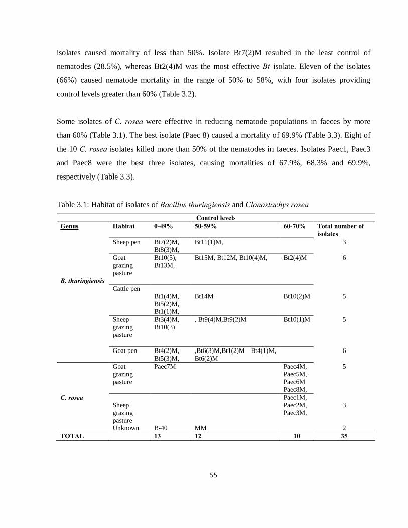
55
isolates caused mortality of less than 50%. Isolate Bt7(2)M resulted in the least control of
nematodes (28.5%), whereas Bt2(4)M was the most effective Bt isolate. Eleven of the isolates
(66%) caused nematode mortality in the range of 50% to 58%, with four isolates providing
control levels greater than 60% (Table 3.2).
Some isolates of C. rosea were effective in reducing nematode populations in faeces by more
than 60% (Table 3.1). The best isolate (Paec 8) caused a mortality of 69.9% (Table 3.3). Eight of
the 10 C. rosea isolates killed more than 50% of the nematodes in faeces. Isolates Paec1, Paec3
and Paec8 were the best three isolates, causing mortalities of 67.9%, 68.3% and 69.9%,
respectively (Table 3.3).
Table 3.1: Habitat of isolates of Bacillus thuringiensis and Clonostachys rosea Control levels
Genus Habitat 0-49% 50-59% 60-70% Total number of isolates
Sheep pen Bt7(2)M, Bt8(3)M,
Bt11(1)M, 3
Goat grazing pasture
Bt10(5), Bt13M,
Bt15M, Bt12M, Bt10(4)M, Bt2(4)M 6
B. thuringiensis Cattle pen Bt1(4)M,
Bt5(2)M, Bt1(1)M,
Bt14M Bt10(2)M 5
Sheep grazing pasture
Bt3(4)M, Bt10(3)
, Bt9(4)M,Bt9(2)M
Bt10(1)M 5
Goat pen Bt4(2)M, Bt5(3)M,
,Bt6(3)M,Bt1(2)M Bt4(1)M, Bt6(2)M
6
Goat grazing pasture
Paec7M Paec4M, Paec5M, Paec6M Paec8M,
5
C. rosea Sheep grazing pasture
Paec1M, Paec2M, Paec3M,
3
Unknown B-40 MM 2 TOTAL 13 12 10 35
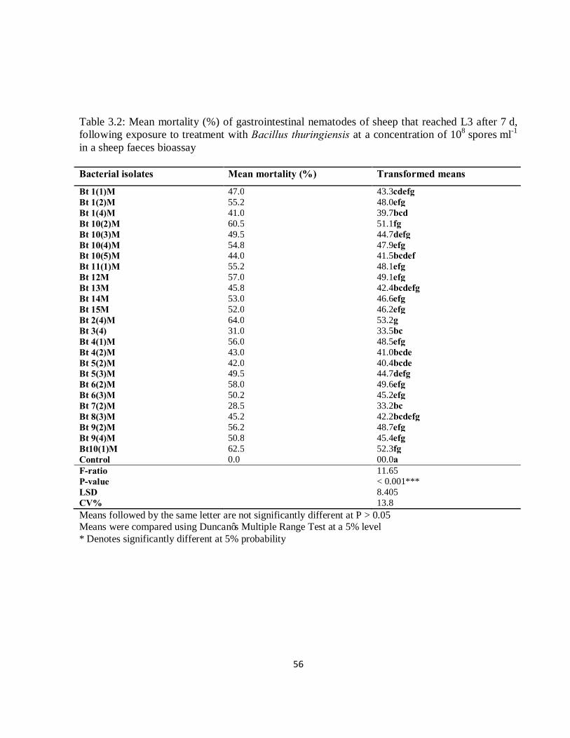
56
Table 3.2: Mean mortality (%) of gastrointestinal nematodes of sheep that reached L3 after 7 d, following exposure to treatment with Bacillus thuringiensis at a concentration of 108 spores ml-1 in a sheep faeces bioassay Bacterial isolates Mean mortality (%) Transformed means
Bt 1(1)M 47.0 43.3cdefg Bt 1(2)M 55.2 48.0efg Bt 1(4)M 41.0 39.7bcd Bt 10(2)M 60.5 51.1fg Bt 10(3)M 49.5 44.7defg Bt 10(4)M 54.8 47.9efg Bt 10(5)M 44.0 41.5bcdef Bt 11(1)M 55.2 48.1efg Bt 12M 57.0 49.1efg Bt 13M 45.8 42.4bcdefg Bt 14M 53.0 46.6efg Bt 15M 52.0 46.2efg Bt 2(4)M 64.0 53.2g Bt 3(4) 31.0 33.5bc Bt 4(1)M 56.0 48.5efg Bt 4(2)M 43.0 41.0bcde Bt 5(2)M 42.0 40.4bcde Bt 5(3)M 49.5 44.7defg Bt 6(2)M 58.0 49.6efg Bt 6(3)M 50.2 45.2efg Bt 7(2)M 28.5 33.2bc Bt 8(3)M 45.2 42.2bcdefg Bt 9(2)M 56.2 48.7efg Bt 9(4)M 50.8 45.4efg Bt10(1)M 62.5 52.3fg Control 0.0 00.0a F-ratio 11.65 P-value < 0.001*** LSD 8.405 CV% 13.8 Means followed by the same letter are not significantly different at P > 0.05 Means were compared using Duncan’s Multiple Range Test at a 5% level * Denotes significantly different at 5% probability
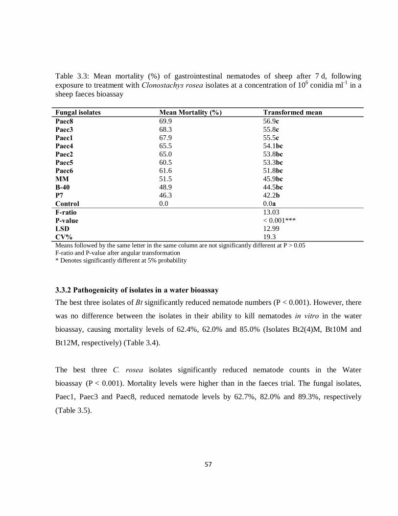
57
Table 3.3: Mean mortality (%) of gastrointestinal nematodes of sheep after 7 d, following exposure to treatment with Clonostachys rosea isolates at a concentration of 106 conidia ml-1 in a sheep faeces bioassay Fungal isolates Mean Mortality (%) Transformed mean Paec8 69.9 56.9c Paec3 68.3 55.8c Paec1 67.9 55.5c Paec4 65.5 54.1bc Paec2 65.0 53.8bc Paec5 60.5 53.3bc Paec6 61.6 51.8bc MM 51.5 45.9bc B-40 48.9 44.5bc P7 46.3 42.2b Control 0.0 0.0a F-ratio 13.03 P-value < 0.001*** LSD 12.99 CV% 19.3 Means followed by the same letter in the same column are not significantly different at P > 0.05 F-ratio and P-value after angular transformation * Denotes significantly different at 5% probability
3.3.2 Pathogenicity of isolates in a water bioassay The best three isolates of Bt significantly reduced nematode numbers (P < 0.001). However, there
was no difference between the isolates in their ability to kill nematodes in vitro in the water
bioassay, causing mortality levels of 62.4%, 62.0% and 85.0% (Isolates Bt2(4)M, Bt10M and
Bt12M, respectively) (Table 3.4).
The best three C. rosea isolates significantly reduced nematode counts in the Water
bioassay (P < 0.001). Mortality levels were higher than in the faeces trial. The fungal isolates,
Paec1, Paec3 and Paec8, reduced nematode levels by 62.7%, 82.0% and 89.3%, respectively
(Table 3.5).
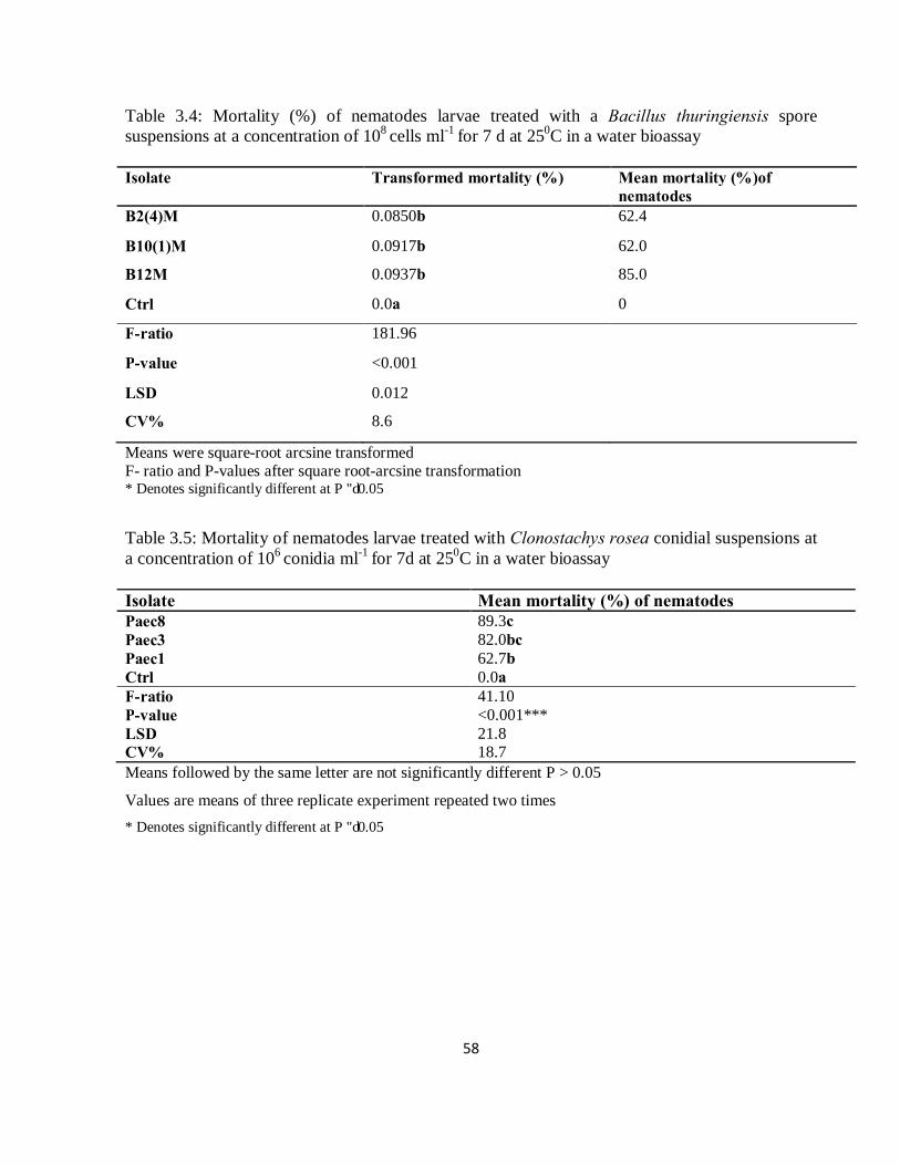
58
Table 3.4: Mortality (%) of nematodes larvae treated with a Bacillus thuringiensis spore suspensions at a concentration of 108 cells ml-1 for 7 d at 250C in a water bioassay Isolate Transformed mortality (%) Mean mortality (%)of
nematodes B2(4)M 0.0850b 62.4
B10(1)M 0.0917b 62.0
B12M 0.0937b 85.0
Ctrl 0.0a 0
F-ratio 181.96
P-value <0.001
LSD 0.012
CV% 8.6
Means were square-root arcsine transformed F- ratio and P-values after square root-arcsine transformation * Denotes significantly different at P ≤ 0.05
Table 3.5: Mortality of nematodes larvae treated with Clonostachys rosea conidial suspensions at a concentration of 106 conidia ml-1 for 7d at 250C in a water bioassay Isolate Mean mortality (%) of nematodes Paec8 89.3c Paec3 82.0bc Paec1 62.7b Ctrl 0.0a F-ratio 41.10 P-value <0.001*** LSD 21.8 CV% 18.7 Means followed by the same letter are not significantly different P > 0.05
Values are means of three replicate experiment repeated two times * Denotes significantly different at P ≤ 0.05
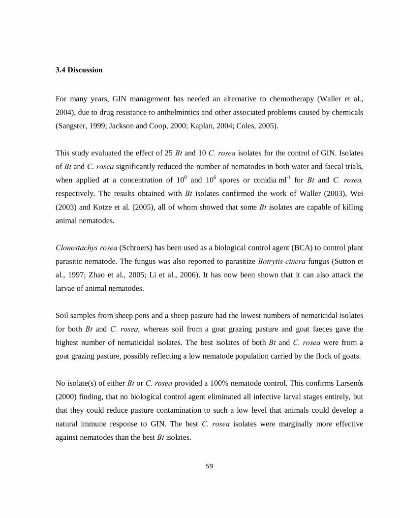
59
3.4 Discussion
For many years, GIN management has needed an alternative to chemotherapy (Waller et al.,
2004), due to drug resistance to anthelmintics and other associated problems caused by chemicals
(Sangster, 1999; Jackson and Coop, 2000; Kaplan, 2004; Coles, 2005).
This study evaluated the effect of 25 Bt and 10 C. rosea isolates for the control of GIN. Isolates
of Bt and C. rosea significantly reduced the number of nematodes in both water and faecal trials,
when applied at a concentration of 108 and 106 spores or conidia ml-1 for Bt and C. rosea,
respectively. The results obtained with Bt isolates confirmed the work of Waller (2003), Wei
(2003) and Kotze et al. (2005), all of whom showed that some Bt isolates are capable of killing
animal nematodes.
Clonostachys rosea (Schroers) has been used as a biological control agent (BCA) to control plant
parasitic nematode. The fungus was also reported to parasitize Botrytis cinera fungus (Sutton et
al., 1997; Zhao et al., 2005; Li et al., 2006). It has now been shown that it can also attack the
larvae of animal nematodes.
Soil samples from sheep pens and a sheep pasture had the lowest numbers of nematicidal isolates
for both Bt and C. rosea, whereas soil from a goat grazing pasture and goat faeces gave the
highest number of nematicidal isolates. The best isolates of both Bt and C. rosea were from a
goat grazing pasture, possibly reflecting a low nematode population carried by the flock of goats.
No isolate(s) of either Bt or C. rosea provided a 100% nematode control. This confirms Larsen’s
(2000) finding, that no biological control agent eliminated all infective larval stages entirely, but
that they could reduce pasture contamination to such a low level that animals could develop a
natural immune response to GIN. The best C. rosea isolates were marginally more effective
against nematodes than the best Bt isolates.
60
Most isolates of the two biocontrol agents killed nematodes in both the faecal bioassay and in the
water bioassay. Sheep are mostly infected with a mixed culture of nematodes, thus an effective
biocontrol agent should be able to work against a variety of nematode species (Waghorn et
al., 2003). In this study, the faeces were collected from sheep naturally infected with a mixed
culture of nematodes. Fortunately, the two groups of biocontrol agents used in these bioassays
both reduced the total numbers of the mixed populations of nematodes. Most of the successful
BCA research on livestock nematodes has centred on the nematophagous fungus D. flagrans
(Larsen et al., 1994; Larsen et al., 1998; Waller et al., 2001; Paraud et al., 2006). However, Bt
and C. rosea are competitive BCAs that can be used to reduce the population of livestock
nematodes in pastures.
In conclusion, the study showed that locally isolated strains of Bt and C. rosea had nematicidal
effects and could be incorporated in a GIN control programme, as an alternative to chemical
anthelmintics. Further studies need to be undertaken to determine the most effective way of
administering these BCAs via animal feeds.
61
3.5 References
Coles, G.C. 2005. Anthelmintic resistance-looking to the future: a UK perspective. Res. Vet. Sci.
78, 99–108.
Faedo, M., Krecek, R.C. 2002. Possible application of a nematophagous fungus as a biological
control agent of parasitic nematodes on commercial sheep farms in South Africa. J. S.A. Vet.
Assoc. 73, 31-35.
Fischer-Le Saux, M., Mauleon, H., Constant, P., Brunel, B., Boermare, N. 1998. PCR-ribotyping
of Xenorhabdus and Photorhabdus isolates from the Caribbean region in relation to the
taxonomy and geographic distribution of their nematode hosts. Appl. Env. Micro. 64, 4246–
4254.
Ghahfarokhi, M.S., Abyaneh, M.R., Bahadori, S.R., Eslami, A., Zare, R., Ebrahimi, M. 2004.
Screening of soil and sheep faecal samples for predacious fungi: Isolation and
characterization of the nematode-trapping fungus Arthrobotrys oligospora. Iran. Biomed.
J. 8, 135-142.
Hastowo, S., Lay, B.W., Ohba, M. 1992. Naturally occuring Bacillus thuringiensis in Indonesia.
J. Appl. Bact. 73, 108-113.
Henderson, C.F., Tilton, E.W. 1955. Tests with acaricides against the brown wheat mite. J. Econ.
Ento. 48, 157-161.
Ichhpujani, R.L., Bhatia, R. 1998. Medical Parasitology, Second Ed. Jaypee Brothers Medical
Publishers, Ltd., New Delhi, India.
Jackson, F., Coop, R.L. 2000. The development of anthelmintic resistance in sheep nematodes.
Parasitology 120, S95–S107.
Kaplan, M. 2004. Drug resistance in nematodes of veterinary importance: a status report. Trends
Parasitol. 20, 477-481.
Kotze, A.C., O'O’Grady, J., Gough, J.M., Pearson, R., Bagnall, D.H., Akhurst, R.J. 2005.
Toxicity of Bacillus thuringiensis to parasitic and free-living life stages of nematode
parasitic of livestock. Int. Parasitol. 35, 1013-1022.
Larsen, M. 2000. Prospects for controlling animal parasitic nematodes by predacious microfungi.
Parasitology 120, 121-131.

62
Larsen, M. 2002. Biological control in a global perspective- A review with emphasis on
Duddingtonia flagrans. In, Biological Control of Nematode Parasite Control of Small
Ruminants. FAO, Malaysia, Asia. pp. 19-37.
Larsen, M., Faedo, M., Waller, P.J. 1994. The potential of nematophagous fungi to control the
free-living stages of nematode parasites of sheep: survey for the presence of fungi in fresh
faeces of grazing livestock. Austr. J. Vet. Parasitol 53, 275-281.
Larsen, M., Faedo, M., Waller, P.J., Hennessy, D.R. 1998. The potential of nematophagous fungi
to control the free-living stages of nematode parasites of sheep: Studies with Duddingtonia
flagrans. Vet. Parasitol. 76, 121-128.
Lee, K.E., Pankhurst, C.E. 1992. Soil organisms and sustainable productivity. Austr. J. Soil Res.
30, 855-892.
Li, J., Yang, J.K., Huang, X.W., Zhang, K.Q. 2006. Purification and characterization of an
extracellular serine protease from Clonostachys rosea and its potential as a pathogenic
factor. Process Biochem. 41, 925-929.
Martin, P.A.W., Travers, R.S. 1989. Worldwide abundance and distribution of Bacillus
thuringiensis isolates. Appl. Env. Micro. 55, 2437-2442.
O’Grady, J., Akhurst, R.J., Kotze, A.C. 2007. The requirement for early exposure of
Haemonchus contortus larvae to Bacillus thuringiensis for effective inhibition of larval
development. Vet. Parasitol. 150, 97-103.
Ohba, M., Aizawa, K. 1986. Distribution of Bacillus thuringiensis in soils of Japan. J. Invert.
Path. 47, 277-282.
Paraud, C., Pors, I., Chicard, C., Chartier, C. 2006. Comparative efficacy of the nematode-
trapping fungus Duddingtonia flagrans against Haemonchus contortus, Teladorsagia
circumcincta and Trichostrongylus colubriformis in goat faeces: influence of the duration
and of the temperature of coproculture. Parasitol. Res. 98, 207-213.
Peña, M.T., Miller, J.E., Fontenot, M.E., Gillespie, A., Larsen, M. 2002. Evaluation of
Duddingtonia flagrans in reducing infective larvae of Haemonchus contortus in faeces of
sheep. Vet. Parasitol. 103, 259–265.
Sangster, N.C. 1999. Anthelmintic resistance: past, present and future. Int. J. Parasitol. 29, 115–
124.

63
Schnepf, E., Crickmore, N., Van Rie, J., Lereclus, D., Baum, J., Feitelson, J., Zeigler, D.R.,
Dean, D.H. 1998. Bacillus thuringiensis and its pesticidal crystal proteins. Micro. Mol. Biol.
Rev. 62, 775-806.
Siddiqui, Z.A., Mahmood, I. 1996. Biological control of plant parasitic nematodes by fungi: a
review. Bior. Technol. 58, 229-239.
Siegel, J.P. 2001. The mammalian safety of Bacillus thuringiensis-based insecticides. J. Invert.
Path. 77, 13-21.
Stirling, G.R. 1991. Biological Control of Plant Parasitic Nematodes: Progress, Problems and
Prospects. CAB International, Wallingford, UK.
Sutton, J.C., Li, D.W., Gang, P., Hai, Y., Zhang, P. 1997. Gliocladium roseum a versatile
adversary of a Botrytis cinera in crops. Plant Dis. 81, 316-328.
Terrill, T.H., Larsen, M., Samples, O., Husted, S., Miller, J.E., Kaplan, R.M., Gelaye, S. 2004.
Capability of the nematode-trapping fungus Duddingtonia flagrans to reduce infective larvae
of gastrointestinal nematodes in goat faeces in the southeastern United States: dose titration
and dose time interval studies. Vet. Parasitol. 120, 285–296.
Waghorn, T.S., Leathwick, D.M., Chen, L.Y., Skipp, R.A. 2003. Efficacy of the nematode-
trapping fungus Duddingtonia flagrans against three species of gastrointestinal nematodes in
laboratory faecal cultures from sheep and goats. Vet. Parasitol. 118, 227-234.
Waller, P.J. 2003. Global perspectives on nematode parasite control in ruminant livestock: the
need to adopt alternatives to chemotherapy with emphasis on biological control. An. Health
Res. Rev. 4, 35-43.
Waller, P.J. 2006. Sustainable nematode parasite control strategies for ruminant livestock by
grazing management and biological control. An. Feed Sci. 126, 277-289.
Waller, P.J., Knox, M.R., Faedo, M. 2001. The potential of nematophagous fungi to control the
free-living stages of nematode parasites of sheep: feeding and block studies with
Duddingtonia flagrans. Vet. Parasitol. 102, 321-330.
Waller, P.J., Thamsborg, S.M., Larsen, M., Knox, M.R., Peter, R., Miller, J.E., Molento, M.B.,
Beriajaya, Hood, G.M. 2004. In: Miller, J.E., Waller, P.J. (Eds.), Novel Approaches to
Control of Parasites-A Workshop. Vet. Parasitol. 125, 59–68.

64
Wei, J.Z., Hale, K., Carta, L., Platzer, E., Wong, C., Fang, S.C., Aroian, R.V. 2003. Bacillus
thuringiensis crystal proteins that target nematodes. Natl. Acad. Sci., USA100, 2700-2765.
Zhao, M.L., Huang, J.S., Mo, M.H., Zhang, K.Q. 2005. A potential virulence factor involved in
fungal pathogenicity serine-like protease activity of nematophagous fungus Clonostachys
rosea. Fungal Divers. 19, 217-234.
65
CHAPTER FOUR
THE EFFECT OF DOSE AND EXPOSURE PERIOD OF SELECTED ISOLATES OF BACILLUS THURINGIENSIS (BERLINER) AND CLONOSTACHYS ROSEA (SCHROERS) ON THE CONTROL OF GASTROINTESTINAL NEMATODES OF SHEEP
M.A. Baloyi and M.D. Laing
Discipline of Plant Pathology, School of Agricultural Sciences and Agribusiness,
University of KwaZulu-Natal, Private Bag X01, Scottsville 3201, Pietermaritzburg,
South Africa
66
Abstract
The interaction between inoculum level and time of exposure of selected biocontrol agents
(BCAs) on livestock nematodes was investigated in this study. The effect of dosage and time of
exposure of Bacillus thuringiensis (Bt) and Clonostachys rosea (Schroers) were studied
separately in bioassays conducted in faeces and in water. In a faecal trial different inoculum
levels (106, 108, 1010 and 1012 spores ml-1 for Bt and 106, 108 and 1010 conidia ml-1 for C. rosea)
were used while 106, 108, 1010 and 1012 spores ml-1 for Bt and 109, 1010, 1011, 1012 conidia ml-1 for
C. rosea were used for the water bioassay. In the faecal bioassay, there was no significant
interaction between Isolate and Inoculum levels. In the water bioassay, significantly different
numbers of surviving nematodes were observed, due to different biological control agents
inoculum levels (P < 0.001) and Period of Exposure (P < 0.001 and P < 0.012, for Bt and C.
rosea, respectively), and interactions between inoculum levels and time of exposure (P < 0.001
and P < 0.012 for Bt and C. rosea, respectively).
In the faecal bioassay, Bt isolate B2 at 1010 spores ml-1 provided the best control of all the
bacterial isolates at all inoculum levels. Isolate P3 was the best fungal isolate at 108 conidia ml-1
with a control level of 66.6%. In the water bioassay, the C. rosea isolate P3 provided 85% control
when applied at inoculum levels of 109, 1010 and 1011 conidia ml-1. The importance of time was
shown by the increased control of nematodes with time of exposure to the biocontrol agents.
Inoculum levels also played a significant part in the activity of the biocontrol agents, with control
levels generally increasing with increased spore or conidial doses.
4.1 Introduction
Gastrointestinal nematodes are a serious concern in the small ruminant industry worldwide
(Miller and Horohove, 2006). These nematodes cause economic losses due to decreased
production, weight loss, reproductive inefficiency, and the cost of treatment and prevention
(Barger et al., 1989; McLeod, 1995; Perry and Randolph, 1999; Perry et al., 2002). Anthelmintic
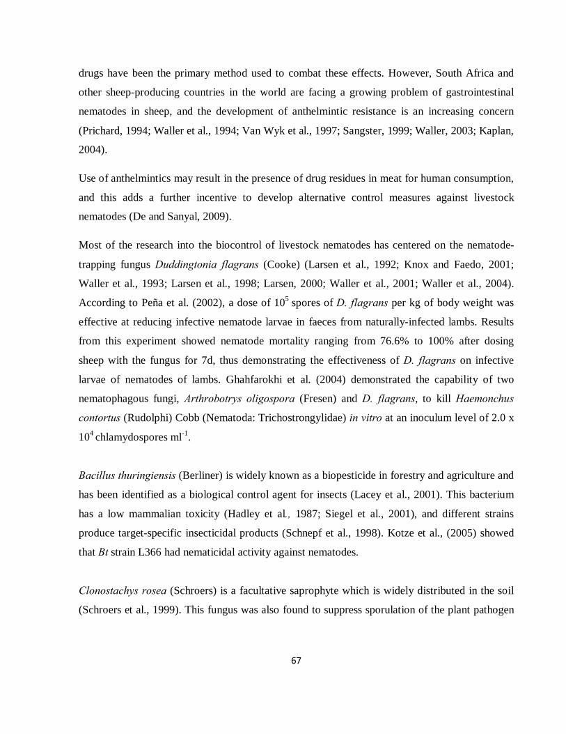
67
drugs have been the primary method used to combat these effects. However, South Africa and
other sheep-producing countries in the world are facing a growing problem of gastrointestinal
nematodes in sheep, and the development of anthelmintic resistance is an increasing concern
(Prichard, 1994; Waller et al., 1994; Van Wyk et al., 1997; Sangster, 1999; Waller, 2003; Kaplan,
2004).
Use of anthelmintics may result in the presence of drug residues in meat for human consumption,
and this adds a further incentive to develop alternative control measures against livestock
nematodes (De and Sanyal, 2009).
Most of the research into the biocontrol of livestock nematodes has centered on the nematode-
trapping fungus Duddingtonia flagrans (Cooke) (Larsen et al., 1992; Knox and Faedo, 2001;
Waller et al., 1993; Larsen et al., 1998; Larsen, 2000; Waller et al., 2001; Waller et al., 2004).
According to Peña et al. (2002), a dose of 105 spores of D. flagrans per kg of body weight was
effective at reducing infective nematode larvae in faeces from naturally-infected lambs. Results
from this experiment showed nematode mortality ranging from 76.6% to 100% after dosing
sheep with the fungus for 7d, thus demonstrating the effectiveness of D. flagrans on infective
larvae of nematodes of lambs. Ghahfarokhi et al. (2004) demonstrated the capability of two
nematophagous fungi, Arthrobotrys oligospora (Fresen) and D. flagrans, to kill Haemonchus
contortus (Rudolphi) Cobb (Nematoda: Trichostrongylidae) in vitro at an inoculum level of 2.0 x
104 chlamydospores ml-1.
Bacillus thuringiensis (Berliner) is widely known as a biopesticide in forestry and agriculture and
has been identified as a biological control agent for insects (Lacey et al., 2001). This bacterium
has a low mammalian toxicity (Hadley et al., 1987; Siegel et al., 2001), and different strains
produce target-specific insecticidal products (Schnepf et al., 1998). Kotze et al., (2005) showed
that Bt strain L366 had nematicidal activity against nematodes.
Clonostachys rosea (Schroers) is a facultative saprophyte which is widely distributed in the soil
(Schroers et al., 1999). This fungus was also found to suppress sporulation of the plant pathogen
68
fungus Botrytis cinera (De Bary) and also used for the control of botrytis blight. C. rosea showed
strong nematicidal activity against Caenorhabditis elegans (Maupas).
The primary aim of this study was to find an effective dose and time of exposure of selected
bacterial and fungal biocontrol isolates to determine the lowest inoculum level that will give a
consistent and high rate of kill the infective stages of gastrointestinal nematodes of sheep.
4.2 Materials and Methods
4.2.1 Spore preparation B. thuringiensis spores
Spores of three Bt isolates, namely, B2(4)M, B10(1)M and B12M were obtained by scraping a
frozen culture of bacterial cells that had been stored at -800C, streaking the cells onto Tryptone
Soy Agar, and allowing them to grow for 4d at 250C. Bacterial colonies were then harvested into
an Erlenmeyer flask with 50ml of Tryptone Soy Broth (TSB). These cultures were grown by
shaking the flasks in an orbital shaker at 150 rpm at 280C for 2wks. The cultures were then
centrifuged (10 000 rpm at 40C for 15min) and washed once in 0.1M NaCl and twice in sterile,
distilled, ice cold water. The pellets were diluted into sterile, distilled water (40ml) and a standard
dilution plate technique was used for calculating the inoculum level of B. thuringiensis (as
described by Kotze et al., 2005).
Fungal conidia
Conidia of three fungal isolates, namely, Paec1, Paec 3 and Paec 8 were harvested from 3 wk old
cultures grown on PDA by scraping the agar surface with a sterile scalpel after addition of 10 ml
sterile distilled water. The conidial suspension was filtered through a double layer of cheese cloth
into a sterilized 25ml McCartney bottle. The number of conidia in the suspension was determined
using a Neubauer haemocytometer under x400 magnification and subsequently adjusted to four
inoculum levels (106, 108, 1010 and 1012 conidia ml-1) for faecal trial and 109, 1010, 1011, and 1012
conidia ml-1 for water bioassay.
69
4.2.2 Bioassay in faeces
Faecal samples were collected directly from the rectum of naturally infested sheep that fed on
grazing pastures. The sheep were infested with a mixed infection of nematodes, dominated by
Haemonchus contortus (Rudolphi). The faecal pellets were crushed and mixed thoroughly, and
2 g subsamples were placed in each well of a 24 multiwell microtitre plate. The faeces were
sprayed with 2 ml of spore and conidia suspension at different inoculum level (106, 108, 1010 and
1012 and 106, 108 and 1010for Bt and C. rosea, respectively) of each of the selected bacterial and
fungal isolates (as described by Ghahfarokhi et al., 2004).
4.2.3 Bioassay in water This technique examined the effect of the biocontrol agents on L3 nematodes extracted into
water. The faeces were mixed thoroughly with vermiculite in a 1:1.5 ratio. The mixture was put
into a plastic container (100 x 120 x 12.5mm deep), then moistened with sterile distilled water
until it was damp but not saturated. The cultures were covered with a lid with holes punched on
the top to allow aeration, and incubated at 250C for 7d (as described by Graminha et al., 2005).
Cultures were kept moist for the entire period by spraying with distilled water when necessary.
Following incubation, L3 larval stages were extracted using the Baermann technique
(Chandrawathani et al., 1998) and transferred into a 500 ml sterile beaker. The nematodes were
surface sterilized with 0.525% sodium hypochlorite solution for 5 min and washed twice with 1L
sterile distilled water. One milliliter was pipetted into a counting dish and counted under
dissecting microscope. The final inoculum level was adjusted to approximately 35 nematodes ml-
1. The nematode suspensions were poured into test tubes and inoculated with spores and conidia
of Bt and C. rosea at levels 106, 108, 1010, 1012, and 109, 1010, 1011,1012 for Bt and C. rosea,
respectively. Nematodes were counted for 2 d for Bt and over 3 d for C. rosea.
4.2.4 Statistical analysis Treatments were arranged in a randomized complete block design, with three replications for
each treatment. Both the faecal and the water bioassay were repeated three times.

70
The following formula was used to determine the nematode mortality (%) by the BCA isolates
tested in faeces:
Nematode Mortality (%) = [C1 – C2] x 100
C1
where C1 = mean number of larvae in untreated manure;
C2 = number of larvae in treated manure
Data was subjected to factorial ANOVA using GenStat® 11.1 Edition to determine the
significance levels of interaction of the main factors (isolate x inoculum level x time). Means of
nematode counts were compared using Fisher’s Least Significant Difference (LSD) test at a 5%
significance level.
Henderson-Tilton's formula (Henderson and Tilton, 1955) was used to determine the corrected
percent mortality in test tubes:
Corrected nematode mortality (%) = (1 - n in Ctrl before treatment x n in T after treatment) x 100
n in Ctrl after treatment x n in T before treatment
Where n = number of nematodes, Ctrl = control, T = treatment
4.3 Results
4.3.1 Faeces Bioassay In the faeces bioassay, none of the Interaction Effects were significant between inoculums level
and nematode counts (P = 0.43 and P = 0.53 for B. thuringiensis and C. rosea, respectively). The
data is therefore presented as a series of regressions, reflecting the patterns of responses to
inoculum levels for the different biocontrol agents, in terms of nematode mortality.
71
Fig. 4.1: Mortality (%) of nematodes larvae in a faeces bioassay, when treated with three isolates of B. thuringiensis
at four inoculum levels. Inoculum at a level of 6 = 106 spores ml-1, 8 = 108 spores ml-1, 10 = 1010 spores ml-1 and 12 =
1012 spores ml-1
The inoculum level of 106 spore ml-1 was the least effective level at killing nematodes for the
three B. thuringiensis isolates, with B12 being less effective than B10 and B2. All the Bt isolates
killed less than 60% of the nematodes when used at the inoculum level of 106 spore ml-1. In
contrast, at the inoculum levels of 108 and 1012 spores ml-1, the three Bt isolates increased the
nematode mortality by 60.3% to 66.1% (Fig. 4.1).
B. thuringiensis Isolate B2 caused the greatest mortality of 70.7% when the inoculum level of
1010 spores ml-1was used (Fig. 4.1). This isolate was also the best when used at the lowest
inoculum level, with a nematode mortality of 57.7%. This isolate consistently maintained a
control level of 62%, 70.7% and 64.4% in faeces when applied at inoculum levels of 108, 1010
and 1012 spores ml-1. Isolate B12 was also effective at inoculum levels of 108 and 1012 spore ml-1,
outperforming B2 at these inoculum levels, causing a mortality of 66.5% and 66.1% at 108 and
1012 spores ml-1 respectively.
y = -2.65x2 + 16.13x + 43.25; R² = 0.787
y = -2.325x2 + 15.15x + 41.27; R² = 0.680
y = -5.325x2 + 32.37x + 15.57; R² = 0.987
0
20
40
60
80
100
6 8 10 12
Nem
atod
e co
unt r
educ
tion
(%)
B2
B10
B12
Inoculum Level (spore ml-1)
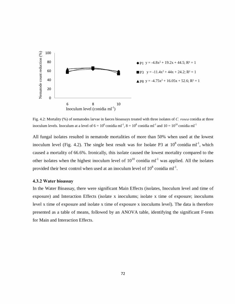
72
Fig. 4.2: Mortality (%) of nematodes larvae in faeces bioassays treated with three isolates of C. rosea conidia at three
inoculum levels. Inoculum at a level of 6 = 106 conidia ml-1, 8 = 108 conidia ml-1 and 10 = 1010 conidia ml-1
All fungal isolates resulted in nematode mortalities of more than 50% when used at the lowest
inoculum level (Fig. 4.2). The single best result was for Isolate P3 at 108 conidia ml-1, which
caused a mortality of 66.6%. Ironically, this isolate caused the lowest mortality compared to the
other isolates when the highest inoculum level of 1010 conidia ml-1 was applied. All the isolates
provided their best control when used at an inoculum level of 108 conidia ml-1.
4.3.2 Water bioassay In the Water Bioassay, there were significant Main Effects (isolates, Inoculum level and time of
exposure) and Interaction Effects (isolate x inoculums; isolate x time of exposure; inoculums
level x time of exposure and isolate x time of exposure x inoculums level). The data is therefore
presented as a table of means, followed by an ANOVA table, identifying the significant F-tests
for Main and Interaction Effects.
y = -4.8x2 + 19.2x + 44.5; R² = 1
y = -11.4x2 + 44x + 24.2; R² = 1
y = -4.75x2 + 16.05x + 52.6; R² = 1
0
20
40
60
80
100
6 8 10
Nem
atod
e co
unt r
educ
tion
(%)
P1
P3
P8
Inoculum level (conidia ml-1)
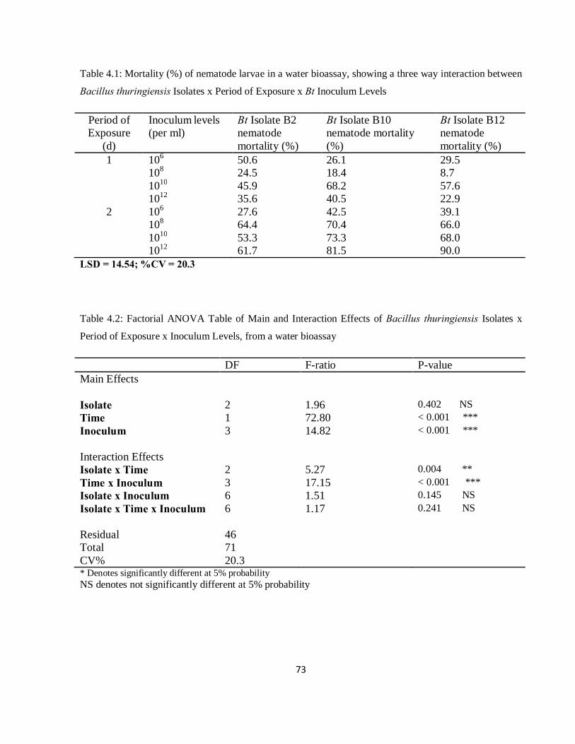
73
Table 4.1: Mortality (%) of nematode larvae in a water bioassay, showing a three way interaction between
Bacillus thuringiensis Isolates x Period of Exposure x Bt Inoculum Levels
Period of Exposure
(d)
Inoculum levels (per ml)
Bt Isolate B2 nematode mortality (%)
Bt Isolate B10 nematode mortality (%)
Bt Isolate B12 nematode mortality (%)
1 106 50.6 26.1 29.5 108 24.5 18.4 8.7 1010 45.9 68.2 57.6 1012 35.6 40.5 22.9 2 106 27.6 42.5 39.1
108 64.4 70.4 66.0 1010 53.3 73.3 68.0 1012 61.7 81.5 90.0 LSD = 14.54; %CV = 20.3
Table 4.2: Factorial ANOVA Table of Main and Interaction Effects of Bacillus thuringiensis Isolates x
Period of Exposure x Inoculum Levels, from a water bioassay
DF F-ratio P-value Main Effects
Isolate 2 1.96 0.402 NS Time 1 72.80 < 0.001 *** Inoculum 3 14.82 < 0.001 *** Interaction Effects
Isolate x Time 2 5.27 0.004 ** Time x Inoculum 3 17.15 < 0.001 *** Isolate x Inoculum 6 1.51 0.145 NS Isolate x Time x Inoculum
6 1.17 0.241 NS
Residual 46 Total 71 CV% 20.3 * Denotes significantly different at 5% probability NS denotes not significantly different at 5% probability
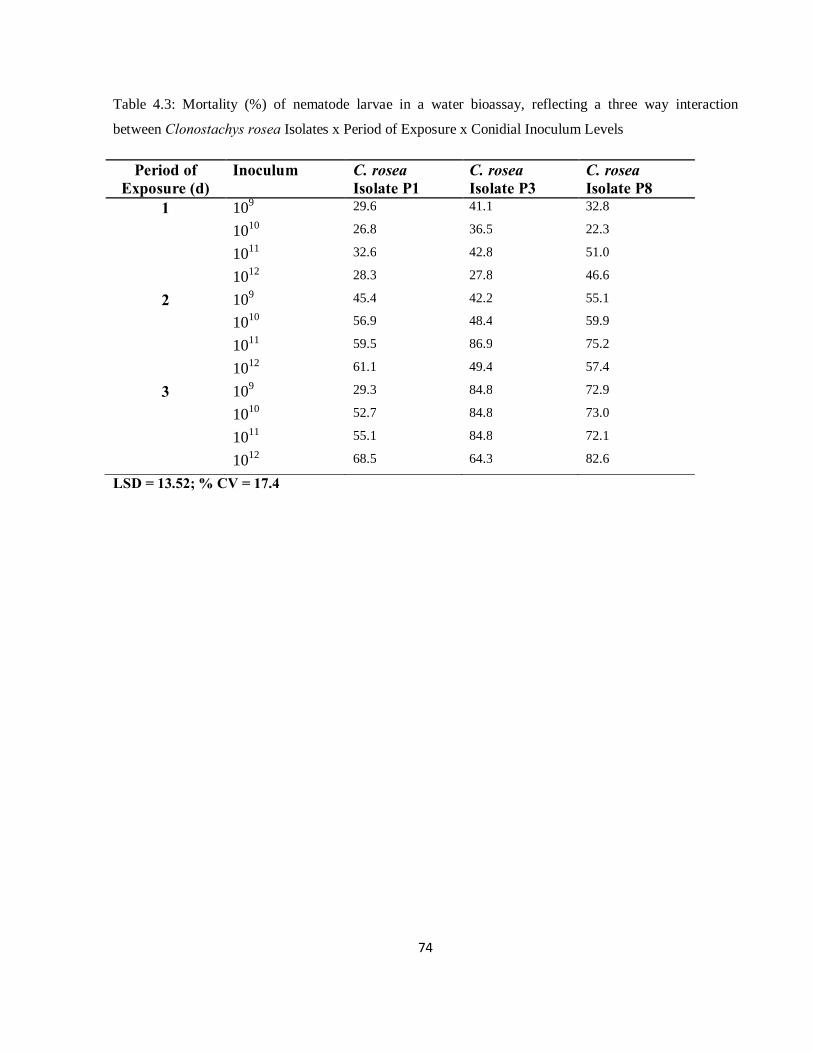
74
Table 4.3: Mortality (%) of nematode larvae in a water bioassay, reflecting a three way interaction
between Clonostachys rosea Isolates x Period of Exposure x Conidial Inoculum Levels
Period of Exposure (d)
Inoculum C. rosea Isolate P1
C. rosea Isolate P3
C. rosea Isolate P8
1 109 29.6 41.1 32.8
1010 26.8 36.5 22.3
1011 32.6 42.8 51.0
1012 28.3 27.8 46.6
2 109 45.4 42.2 55.1
1010 56.9 48.4 59.9
1011 59.5 86.9 75.2
1012 61.1 49.4 57.4
3 109 29.3 84.8 72.9
1010 52.7 84.8 73.0
1011 55.1 84.8 72.1
1012 68.5 64.3 82.6
LSD = 13.52; % CV = 17.4
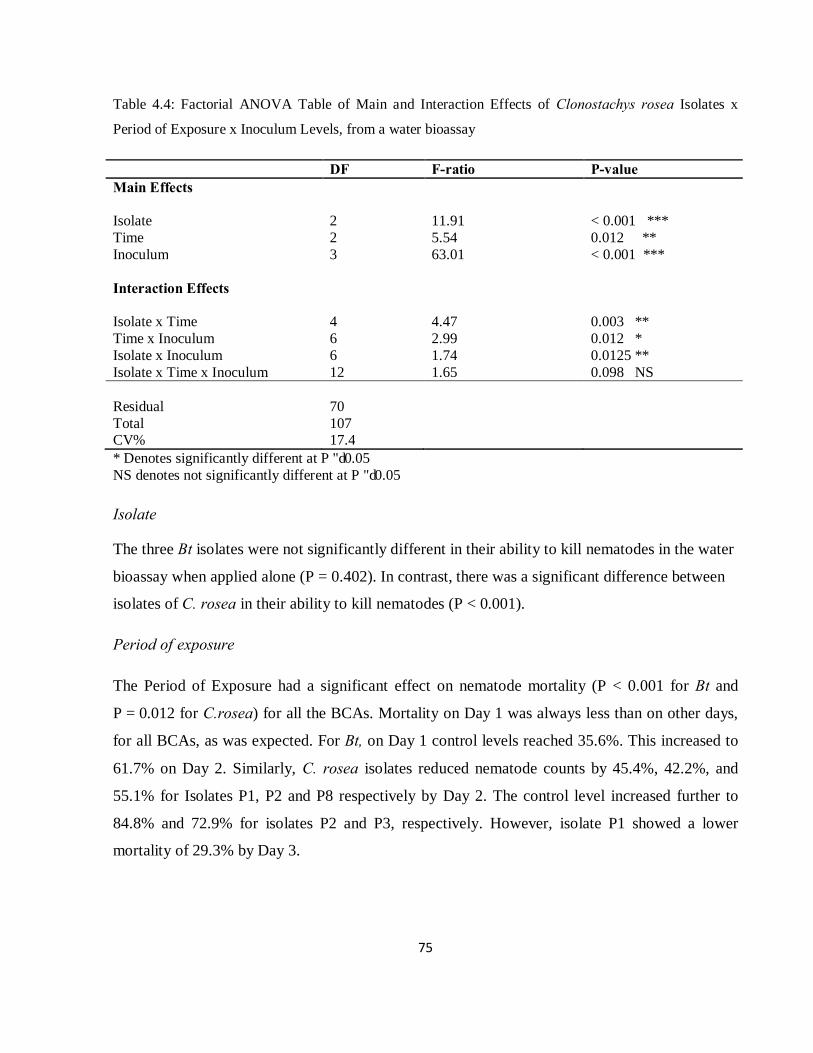
75
Table 4.4: Factorial ANOVA Table of Main and Interaction Effects of Clonostachys rosea Isolates x
Period of Exposure x Inoculum Levels, from a water bioassay
DF F-ratio P-value Main Effects
Isolate 2 11.91 < 0.001 *** Time 2 5.54 0.012 ** Inoculum 3 63.01 < 0.001 *** Interaction Effects
Isolate x Time 4 4.47 0.003 ** Time x Inoculum 6 2.99 0.012 * Isolate x Inoculum 6 1.74 0.0125 ** Isolate x Time x Inoculum 12 1.65 0.098 NS Residual
70
Total 107 CV% 17.4 * Denotes significantly different at P ≤ 0.05 NS denotes not significantly different at P ≤ 0.05
Isolate
The three Bt isolates were not significantly different in their ability to kill nematodes in the water
bioassay when applied alone (P = 0.402). In contrast, there was a significant difference between
isolates of C. rosea in their ability to kill nematodes (P < 0.001).
Period of exposure
The Period of Exposure had a significant effect on nematode mortality (P < 0.001 for Bt and
P = 0.012 for C.rosea) for all the BCAs. Mortality on Day 1 was always less than on other days,
for all BCAs, as was expected. For Bt, on Day 1 control levels reached 35.6%. This increased to
61.7% on Day 2. Similarly, C. rosea isolates reduced nematode counts by 45.4%, 42.2%, and
55.1% for Isolates P1, P2 and P8 respectively by Day 2. The control level increased further to
84.8% and 72.9% for isolates P2 and P3, respectively. However, isolate P1 showed a lower
mortality of 29.3% by Day 3.
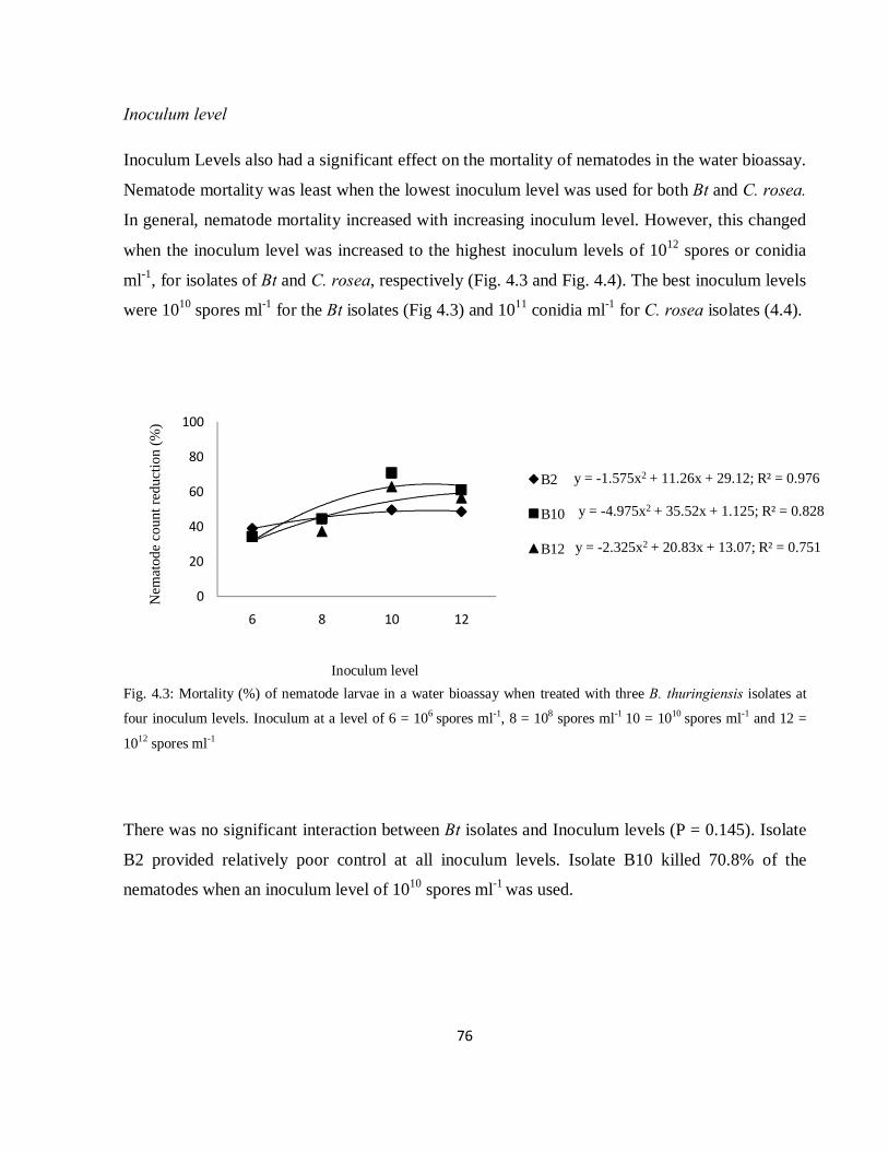
76
Inoculum level
Inoculum Levels also had a significant effect on the mortality of nematodes in the water bioassay.
Nematode mortality was least when the lowest inoculum level was used for both Bt and C. rosea.
In general, nematode mortality increased with increasing inoculum level. However, this changed
when the inoculum level was increased to the highest inoculum levels of 1012 spores or conidia
ml-1, for isolates of Bt and C. rosea, respectively (Fig. 4.3 and Fig. 4.4). The best inoculum levels
were 1010 spores ml-1 for the Bt isolates (Fig 4.3) and 1011 conidia ml-1 for C. rosea isolates (4.4).
Fig. 4.3: Mortality (%) of nematode larvae in a water bioassay when treated with three B. thuringiensis isolates at
four inoculum levels. Inoculum at a level of 6 = 106 spores ml-1, 8 = 108 spores ml-1 10 = 1010 spores ml-1 and 12 =
1012 spores ml-1
There was no significant interaction between Bt isolates and Inoculum levels (P = 0.145). Isolate
B2 provided relatively poor control at all inoculum levels. Isolate B10 killed 70.8% of the
nematodes when an inoculum level of 1010 spores ml-1 was used.
y = -1.575x2 + 11.26x + 29.12; R² = 0.976
y = -4.975x2 + 35.52x + 1.125; R² = 0.828
y = -2.325x2 + 20.83x + 13.07; R² = 0.751
0
20
40
60
80
100
6 8 10 12
Nem
atod
e co
unt r
educ
tion
(%)
B2
B10
B12
Inoculum level
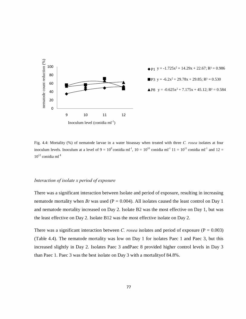
77
Fig. 4.4: Mortality (%) of nematode larvae in a water bioassay when treated with three C. rosea isolates at four
inoculum levels. Inoculum at a level of 9 = 109 conidia ml-1, 10 = 1010 conidia ml-1 11 = 1011 conidia ml-1 and 12 =
1012 conidia ml-1
Interaction of isolate x period of exposure
There was a significant interaction between Isolate and period of exposure, resulting in increasing
nematode mortality when Bt was used (P = 0.004). All isolates caused the least control on Day 1
and nematode mortality increased on Day 2. Isolate B2 was the most effective on Day 1, but was
the least effective on Day 2. Isolate B12 was the most effective isolate on Day 2.
There was a significant interaction between C. rosea isolates and period of exposure (P = 0.003)
(Table 4.4). The nematode mortality was low on Day 1 for isolates Paec 1 and Paec 3, but this
increased slightly in Day 2. Isolates Paec 3 andPaec 8 provided higher control levels in Day 3
than Paec 1. Paec 3 was the best isolate on Day 3 with a mortalityof 84.8%.
y = -1.725x2 + 14.29x + 22.67; R² = 0.986
y = -6.2x2 + 29.78x + 29.85; R² = 0.530
y = -0.625x2 + 7.175x + 45.12; R² = 0.584
0
20
40
60
80
100
9 10 11 12
nem
atod
e co
unt r
educ
tion
(%)
P1
P3
P8
Inoculum level (conidia ml-1)
78
Interaction of period of exposure x inoculum level
There was a significant interaction between Period of Exposure and Inoculum Level (P < 0.001)
for the Bt isolates. Control of nematodes was similar in both Day 1 and Day 2 when the lowest
inoculum level was used. The difference in nematode mortality was only 1% between the two
days. As inoculum levels increased to 108 spores ml-1, the difference in control levels between the
two days increased by 49.8%. At the inoculum level of 108 spores ml-1, nematode mortality
increased to a gap of 17.2% to 67.0% for Day 1 and Day 2, respectively. When 1010 spores ml-1
was used, control also differed between the days from 57.0% on Day 1 to 64.9% on Day 2.
Moreover, this inoculum level resulted in the best level of control on Day 1. At the highest
inoculum level of 1012 spores ml-1 a lower level of mortality occurred on Day 1, and then
increased to 77% on Day 2.
For C. rosea there was also a significant interaction between period of exposure and inoculum
level (P = 0.012). Inoculum levels of 109, 1010 and 1012 conidia ml-1 killed more nematodes as the
period of exposure increased. Inoculum levels of 1010 and 1012 conidia ml-1 cause a similar
nematode mortality on Day 3. At an inoculum level of 1011 conidia ml-1 the highest level of
mortality occurred on Day 2. On Day 3, nematode mortality was similar for inoculum levels of
1010, 1011 and 1012 conidia ml-1.
Three way interaction of BCAs
There were no significant three ways interactions for either the Bt or the C. rosea isolates.
However, the single most effective treatment with Bt was with Isolate B12 at the highest
inoculum level (1012 spores ml-1) on Day 2 and Isolate P8 at 1012 conidia ml-1 on Day 3.
.

79
4.4 Discussion
Mortality of nematodes caused by B. thuringiensis isolates varied as a function of the inoculum
level used, with a very strong relationship between Bt inoculum levels and mortality reflected in
both bioassays. Although the highest inoculum level of 1012 spores ml-1 was the most effective for
Isolates B10 and B12, it may not be the most cost-effective because the cost of production may
be higher and will increase the costs of application for livestock farmers. Interestingly, Isolates
B10 and B12 used at 108 spore ml-1 reduced the number of nematodes by 70.4% and 66.0% on
Day 2, in the water bioassay, whereas at 1012 spore ml-1 the mortalities were 81.5% and 90.0%,
respectively. In other words, for an increase of 11.1% and 24.0% in nematode mortality, 10,000
times as many spores of Bt had to be applied.
C. rosea did not seem to respond as much to inoculum level in order to achieve much higher
mortality rates. At an inoculum level of 108 conidia ml-1, P3 was able to kill more nematodes than
it did at a higher inoculum level of 1012 conidia ml-1. All isolates resulted in a nematode mortality
of more than 50% at the lowest inoculum level.
Control of nematodes was highly significant at different inoculum levels (P < 0.001) and on
different days (P < 0.001) for both Bt and C. rosea. Further more, C. rosea isolates also showed
significantly different effect in controlling nematodes (P < 0.001). This means that the BCAs
activity against nematodes was influenced by exposure period, inoculum level and isolates in the
case of C. rosea.
Exposure period has an important role in the control of nematodes. Generally, low activity
against nematodes was observed on the first day. However, this increased on the second day. This
suggests that the activity of the biocontrol agent is not immediate. Peña et al. (2002) also reported
similar results of larval development being significant between doses from Day 2. However,
Mendoza de Gives et al. (1998) showed that D. flagrans killed nematodes within 14 h. The low
activity of BCAs on Day 1 reflects the need for nematodes to consume Bt spores before being
killed, and that may take some time. The importance of time was also observed by O’Grady et al.
(2007) who suggest that it would be best to use B. thuringiensis L366 to control livestock
nematodes by administering it early to allow it to germinate and grow through a vegetative phase,

80
followed by sporulation and production of Cry proteins, before any anthelmintic effect could be
observed.
Inoculum level is also of great importance in controlling nematodes. This is shown by the
positive response of isolates to increasing inoculum levels. Isolates of Bt achieved satisfactory
results when 1010 spores ml-1 were used and further increase in control resulted from increases in
inoculum levels for isolates B10 and B12.
Use of lower inoculum levels of 109 and 1010 conidia ml-1 for C. rosea resulted in satisfactory
control of nematodes. However, an inoculum of 1011 conidia ml-1 provided better level of
nematode control. A higher inoculum level of 1012 conidia ml-1 did not result in increased control.
Isolate B10 killed nematodes faster than the other isolates. Although Isolate B2 killed higher
number of nematodes on Day 1 than the other isolates, its activity did not increase much on the
second day. Better control was provided by the fungal isolates on Day 3, with P3 and P8
responding strongly to an increase in period of exposure. Isolate P1 did not increase its activity
with increasing periods of exposure.
When Bt spores were used, the inoculum level influenced the speed of kill. The highest inoculum
level resulted in the highest level of control of nematodes within 2 days. Inoculum level of fungal
conidia did not influence the speed of kill. This study agrees with Maddox et al. (2000) who
found that many species of fungi that produced microsporidia did not immediately kill their
insect or nematode hosts, but often caused death in later developmental stages.
In conclusion, for both biocontrol agents, Bt and C. rosea, the inoculum level and period of
exposure are important parameters that affect the performance of the biocontrol agents. An
inoculum level of 108 spore ml-1 for the three Bt isolates was enough to effectively reduce the
number of nematodes in faeces. In contrast, high levels of inoculum were not as important for the
C. rosea isolates, especially Isolate P3, which should make its application economically
attractive. For example, at a level of 109 conidia ml-1 Isolate P3 killed 85% of the nematodes
within three days.

81
4.5 References
Barger, I.A. 1989. Helminth parasites and animal production. In, Biology and Control of
Endoparasites. (Ed.) Symonds, L.E.A. Donald, A.D., Dineen, J.K. Academic Press, New
York.
Chandrawathani, P., Omar, J., Waller, P.J. 1998 The control of the free-living stages of
Strongyloides papillosus by the nematophagous fungus, Arthrobotrys oligospora. Vet.
Parasitol. 76, 321–325.
De, S., Sanyal, P.K. 2009. Biological control of helminth parasites by predatory fungi. Vetscan 4,
1-9.
Ghafarokhi, M.S., Abyaneh, M.R., bahadori, S.R., Eslami, A., Zara, R., Ebrahimi, M. 2004.
Screening of soil and sheep faecal samples for predacious fungi: Isolation and
characterization of the nematode-trapping fungus Arthrobotrys oligospora. Iran. Biomed.
J. 8, 135-142.
Graminha, E.B.N., Costa, A.J., Oliveira, G.P., Monteiro, A.C., Palmeira, S.B.S. 2005. Biological
control of sheep parasite nematodes by nematode-trapping fungi: in vitro activity and
after passage through gastrointestinal tract. World J. Micro. Biotech. 21, 717-722.
Hadley, W.M., Scott, W.B., McDowell, T.D., Thilsted, J.P., Hibbs, C.M., Whorton, J.A., Day,
P.W., Friedmann, M.B., Stoll, R.E. 1987. Five months oral (Diet) toxicity/infectivity
study of Bacillus thuringiensis insecticides in sheep. Fund. Appl. Toxicol. 8, 236-242.
Henderson, C.F., Tilton, E.W. 1955. Tests with acaricides against the brown wheat mite. J. Econ.
Ento. 48, 157-161.
Kaplan, R.M. 2004. Drug resistance in nematodes of veterinary importance: A status report.
Trends Parasitol. 20, 477-581.
Knox, M.R., Faedo, M. 2001. Biological control of field infections of nematode parasites of
young sheep with Duddingtonia flagrans and effects of spore intake on efficacy. Vet.
Parasitol. 101, 155-160.
82
Kotze, A.C., O’Grady, J., Gough, J.M., Pearson, R., Bagnall, N.H., Kemp, D.H. Akhurst, R.J.
2005. Toxicity of Bacillus thuringiensis to parasitic and free-living life-stages of
economically important nematode parasites of livestock. Int. J. Parasitol. 35, 1013–1022.
Lacey, L.A., Frutos, R., Kaya, H.K., Vale, P. 2001. Insect pathogens as biological control agents:
do they have future? Biol. Control 21, 230-248.
Larsen, M. 2000. Prospects for controlling animal parasitic nematodes by micro fungi.
Parasitology 120, S120-S131.
Larsen, M., Faedo, M., Waller, P.J., Hennessy, D.R. 1998. The potential of nematophagous fungi
to control the free-living stages of nematode parasites of sheep: Studies Duddingtonia
flagrans. Vet. Parasitol. 76, 121-128.
Larsen, M., Nansen, P., Gronvold, J., Wolstrup, J., Henriksen, S.A. 1992. In vivo passage of
nematophagous fungi selected for biocontrol of parasitic nematodes in ruminants. J.
Helminth. 66, 137-141.
Maddox, J.V., Brooks, W.M., Solter, L.F. 2000. Bioassay of microsporidia. In, Bioassays of
Entomopathogenic Microbes and Nematodes, (Ed.) Navon, A., Ascher, K.R.S. CABI
Publishing, New York.
McLeod, R.S. 1995. Costs of major parasites of the Australian livestock industries. Int. J.
Parasitol. 25, 1363-1367.
Mendoza de Gives, P., Flores Crespo, J., Herrera Rodriguez, D., Vazquez Prats, V., Liebano
Hernandez, G.E. 1998. Biological control of Haemonchus contortus larvae in ovine faeces
by administering an oral suspension of Duddingtonia flagrans chlamydospores to sheep.
J. Helminth. 72, 343-347.
Miller, J.E., Horohove, D.W. 2006. Immunological aspects of nematode parasite in sheep. J. An.
Sci. 84, E124-E132.
O’Grady, J., Akhurst, R.J., Kotze, A.C. 2007. The requirement for early exposure of
Haemonchus contortus larvae to Bacillus thuringiensis for effective inhibition of larval
development. Vet. Parasitol. 150, 97-103.
Peña, M.T., Miller, J.E., Fontenot, M.E., Gillipie, A., Larsen, M. 2002. Evaluation of
Duddingtonia flagrans in reducing infective larvae of Haemonchus contortus in faeces of
sheep. Vet. Parasitol. 103, 259-265.
83
Perry, B.D., Randolph, T.F. 1999. Improving the assessment of the economic impact of parasitic
diseases and of their control in production animals. Vet. Parasitol. 84, 145-168.
Perry, B.D., Randolph, T.F., McDermott, J.J., Sones, K.R., Thornton, P.K. 2002. Investing in
Animal Health Research to Alleviate Poverty. ILRI (International Livestock Research
Institute), Nairobi, Kenya, 148pg.
Prichard, R.K. 1994. Anthelmintic resistance. Int. J. Parasitol. 54, 259-268.
Sangster, N.C. 1999. Anthelmintic resistance: Past, present and future. Int. J. Parasitol. 29, 115-
124.
Schnepf, E., Crickmore, N., Van Rie, J., Lereclus, D., Baum, J., Feitelson, J., Zigler, D.R., Dean,
D.H. 1998. Bacillus thuringiensis and its pesticidal crystal proteins. Micro. Mol. Biol.
Rev. 62, 775-806.
Schroers, H.J., Samuels, G.J., Seifert, K.A., Gams, W. 1999. Classification of the mycoparasite
Clonostachys rosea in Clonostachys as C. rosea, its relationship to Bionectria
ochroleuca, and notes on other Gliocladium-like fungi. Mycologia 91, 365–385.
Siegel, J.P. 2001. The mammalian safety of Bacillus thuringiensis-based insecticides. J. Invert.
Path. 77, 13-21.
Van Wyk, J.A., Malan, F.S., Randles, J.L. 1997. How long before resistance makes it impossible
to control some field strains of Haemonchus contortus in South Africa with any of the
anthelmintics? Vet. Parasitol. 70, 111-122.
Waller, P.J. 1994. The development of anthelmintic resistance in ruminants livestock. Acta Trop.
56, 233-243.
Waller, P.J. 2003. Global perspectives on nematode parasite control in ruminant livestock: the
need to adopt alternatives to chemotherapy, with emphasis on biological control. An.
Health Res. Rev. 4, 35-43.
Waller, P.J., Faedo, M. 1993. The potential of nematophagous fungi to control the free-living
stages of nematode parasites: In vitro and in vivo studies. Vet. Parasitol. 51, 289-299.
Waller, P.J., Knox, M.R., Faedo, M. 2001. The potential of nematophagous fungi to control the
free-living stages of sheep: Feeding and block studies with Duddingtonia flagrans. Vet.
Parasitol. 102, 321-330.

84
Waller, P.J., Schwan, O., Ljungstrom, B.L., Rydzik, A., Yeates, G.W. 2004. Evaluation of
biological control of sheep parasites using Duddingtonia flagrans under commercial
farming conditions on the island of Gotland, Sweden. Vet. Parasitol. 126, 299-315.
85
CHAPTER FIVE
USE OF MIXED CULTURES OF BIOCONTROL AGENTS TO CONTROL SHEEP NEMATODES
M.A. Baloyi and M.D. Laing
Discipline of Plant Pathology, School of Agricultural Sciences and Agribusiness,
University of KwaZulu-Natal, Private Bag X01, Scottsville 3201, Pietermaritzburg,
South Africa
86
Abstract
Biological control is a promising non-chemical approach for the control of gastrointestinal
nematodes of sheep. Use of combinations of biocontrol agents have been reported to be an
effective method to increase the efficacy of biological control. In this study, combinations of
either two Bt or C. rosea isolates and Bt+C. rosea isolates were evaluated in vitro in microtitre
plates for their biocontrol activity on sheep nematodes. Results indicate that there was a
significant reduction of nematode counts due to a combination of biocontrol agents (P < 0.001).
Combinations of Bt isolates reduced nematodes counts by 72.8%, 64% and 29.8% for
combinations B2+B12, B2+B10 and B10+B12. The results revealed a control level of 57% when
C. rosea isolates P3+P8 were combined. Combinations of Bt and C. rosea isolates B10+P8
caused the greatest mortality of 76.7%. Most combinations were antagonistic (B10+B12,
B12+P8, B2+P8, B10+P3, B2+P3, B12+P3 and P3+P8), with only a few combinations showing
an additive effect (B2+B10, B2+B12 and B10+P8). None were synergistic.
5.1 Introduction
Parasitic nematode infections are generally regarded as the most prevalent health problem of
small ruminants worldwide, causing significant production losses (Perry and Radondolph, 1999).
Although anthelminthic drugs have been a reliable control method, resistance by nematodes to
these drugs has become a serious threat to sheep production (Prichard, 1994; Waller, 1994; Van
Wyk et al., 1997; Sangster, 1999; Waller, 2003; Kaplan, 2004).
A number of microorganisms have been found to feed on nematodes, reducing the number of
free-living larvae in the soil ecosystem (Stirling, 1991; Chen and Dichinson, 2004) in a process
known as biological control. Amongst these microorganisms, the genera Duddingtonia,
Arthrobotrys, Nematoctonus and Monacrosporium have shown their capability to kill and digest
animal parasitic nematodes, which reflects their potential as biocontrol agents (Dimander et al.,
2003; Campos et al., 2008).
87
Kotze et al. (2005) showed that several isolates of the bacterium Bacillus thuringiensis (Bt)
(Berliner) are nematicidal to larvae of Haemonchus contortus (Rudolphi), Trichostrongylus
colubriformis (Giles) and Ostertagia circumcincta (Stadelmann), which are nematodes of
livestock. Isolates of this bacterium produce highly specific insecticidal crystal proteins, which
are widely used for the control of pests in agriculture.
McPartland (2000) discovered that Clonostachys rosea (Schroers) was compatible when
combined with Trichoderma harzianum (Rifai). Many isolates of C. rosea are highly efficient
antagonists against several plant pathogenic fungi and soil fungi (Gan et al., 2007). This fungus is
known to produce serine protease and chitinase as its key mode of action against fungi and
nematodes of plants (Zhao et al., 2005; Li et al., 2006).
Both Bt and C. rosea have shown their ability to reduce nematode populations. However, most
biocontrol agents have been inconsistent in the field. Inconsistency of performance of the
biocontrol agents is influenced by both biotic and abiotic factors. Biotic factors include
interactions with non-target organisms, varying rhizosphere or soil colonization by the biocontrol
agent, varying initial population levels and the genetic diversity of target pathogens (Stirling,
1991; Pierson and Waller, 1994; Meyer and Roberts, 2002).
In this regard, combinations of biocontrol agents may have more potential to suppress pathogens
due to their different traits responsible for parasitism (Pierson and Waller, 1994; Crump, 1998).
Literature on the effect of combining biocontrol agents on crops is well documented. However,
there are no references on combining biocontrol agents for the control of livestock nematodes.
The aim of this study was to evaluate the effect of combining Bt and C. rosea for the control of
livestock nematode populations when mixed in a culture of sheep faeces.
88
5.2 Materials and Methods Biological control agents
The isolates of Bt and C. rosea were isolated from pasture soils of the livestock section at
Ukulinga Research Farm of the University of KwaZulu-Natal, Pietermaritzburg, as described in
Chapter 2.
5.2.1 Preparation of biocontrol agents Spores of B. thuringiensis
Spores of Bt were obtained by scraping a frozen culture that was stored at -800C and streaking
cells onto Tryptone Soy Agar plates and kept for 4d at room temperature. The cells were then
cultured in an Erlenmeyer flask containing 50ml of Tryptone Soy Broth, incubated and shaken at
150 rpm at 280C for 2wks. After incubation, the cultures were centrifuged (10 000 rpm at 40C for
15min), washed once in 0.1 mole (M) NaCl and twice in sterile distilled ice cold water. The
pellets were diluted into 9ml of sterile distilled water. The number of bacterial spores was
determined using a dilution plate technique and adjusted to 108 spores m1-1.
Conidia of C. rosea
Conidia were produced by growing the fungal cultures on PDA for 3wks. Spores were recovered
from the fungal culture by washing the surface of the agar with sterile distilled water. The
conidial suspension was filtered through a double layer of cheese cloth into a sterilized beaker.
The suspension of conidia was counted with a haemocytometer and adjusted to 106 conidia ml-l.
5.2.2 Faecal Bioassay of mixed culture of biocontrol agents To conduct the bioassay, faecal samples were collected directly from the rectum of naturally
infested sheep that feed on grazing pastures. The sheep had a mixed infection of nematodes. The
faecal pellets were broken up and mixed thoroughly, and then a subsample of 2g of the faeces
were placed in each well of a 24 multiwell microtitre plate and treated with a spore or conidial
suspension at a ratio of 1:1 (w/v) as described by Ghahfarokhi et al. (2004) with minor
modifications. The isolates were used individually and in combinations that consisted of the same
genus (i.e. mixture of two Bt or C. rosea isolates) and different genera (i.e. mixture of Bt and C.

89
rosea). The treated samples were incubated at 250C for 7d. The surviving nematode larvae were
recovered using the Baermann technique.
5.2.3 Statistical Analysis Experiments were repeated twice, with treatments arranged in a randomized complete block
design (RCBD). Data were subjected to analysis of variance (ANOVA) using the Genstat® 11th
edition statistical package. Fisher’s Least Significant Difference (LSD) was used to determine
differences between treatment means. The following formula was used to determine the mortality
of nematodes (Abbott, 1925).
Nematode mortality (%) = [C1 – C2] x 100
C1
Where C1 = mean number of larvae in untreated manure;
C2 = number of larvae in treated manure
In order to determine the combined effects of Bt and C. rosea isolates, the observed mortality
levels were compared to the expected mortality rate under the assumption of an independent
effect, as described by Jazzar and Hammad (2004).
For example, the expected mortality of the combination B2+P8 for the Bt: Isolate B2 and C.
rosea Isolate P8 was calculated using the following formula:
B2+P8 (Expected) = B2 + P8 (1-B2)
Where B2 and P8 are the observed proportion mortality caused by Bt and C. rosea isolates alone.
The same calculation was used for all the other combinations.
90
After calculating the χ2 the results were compared to the table (df = 1, P ≤ 0.05)
χ2 = (O – E)2 / E
Where O is the observed mortality for either of the isolates and E is the expected mortality for
either of the isolates. Additivity would be indicated by the χ2 < 3.84, whereas synergistic or
antagonistic interactions would be indicated by the χ2 > 3.84. If O < E, the interaction would be
considered antagonist, whereas if O > E then the interaction was synergistic (Finny, 1964).
5.3 Results
5.3.1 Effect of individual and mixed culture application of biocontrol agents in faeces Individual application of biocontrol agents caused expected levels of mortality of nematodes. The
best isolate was the fungal Isolate P3, which caused a control level of 66.3% of nematodes,
followed by P8, which caused a reduction of 54.0% (Table 5.1). The Bt isolates caused control
levels of 52.0%, 48.7% and 47.0%, for Isolates B12, B10 and B2, respectively.
Mixing of biocontrol agents caused a significant interaction effect (P < 0.001). The combination
of isolates from the same genus affected nematode mortality in various ways. Combinations of
bacterial isolates caused mortality of 64% and 29.8% for Isolates B2+B10 and Isolates B10+B12,
respectively. The combination of fungal Isolates P3 and P8 caused a control level of 57%. The
results revealed a difference in mortality of 15% when comparing the effects of mixing two
fungal isolates and two bacterial isolates.
The best combination of Bt and C. rosea isolates was B10+P8 which caused a nematode
mortality of 76.7%. This combination caused the greatest mortality of nematodes when compared
to all the other combinations, and had a great effect than when they were applied individually.
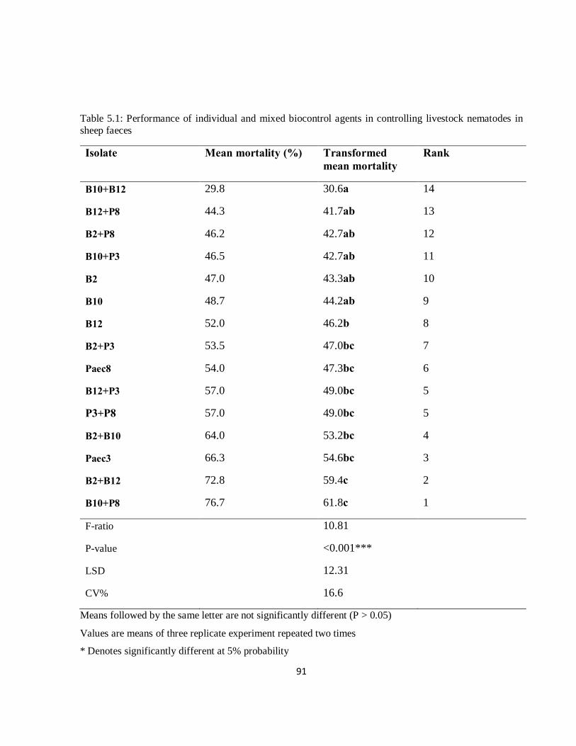
91
Table 5.1: Performance of individual and mixed biocontrol agents in controlling livestock nematodes in sheep faeces
Isolate Mean mortality (%) Transformed mean mortality
Rank
B10+B12 29.8 30.6a 14
B12+P8 44.3 41.7ab 13
B2+P8 46.2 42.7ab 12
B10+P3 46.5 42.7ab 11
B2 47.0 43.3ab 10
B10 48.7 44.2ab 9
B12 52.0 46.2b 8
B2+P3 53.5 47.0bc 7
Paec8 54.0 47.3bc 6
B12+P3 57.0 49.0bc 5
P3+P8 57.0 49.0bc 5
B2+B10 64.0 53.2bc 4
Paec3 66.3 54.6bc 3
B2+B12 72.8 59.4c 2
B10+P8 76.7 61.8c 1
F-ratio 10.81
P-value <0.001***
LSD 12.31
CV% 16.6
Means followed by the same letter are not significantly different (P > 0.05)
Values are means of three replicate experiment repeated two times
* Denotes significantly different at 5% probability
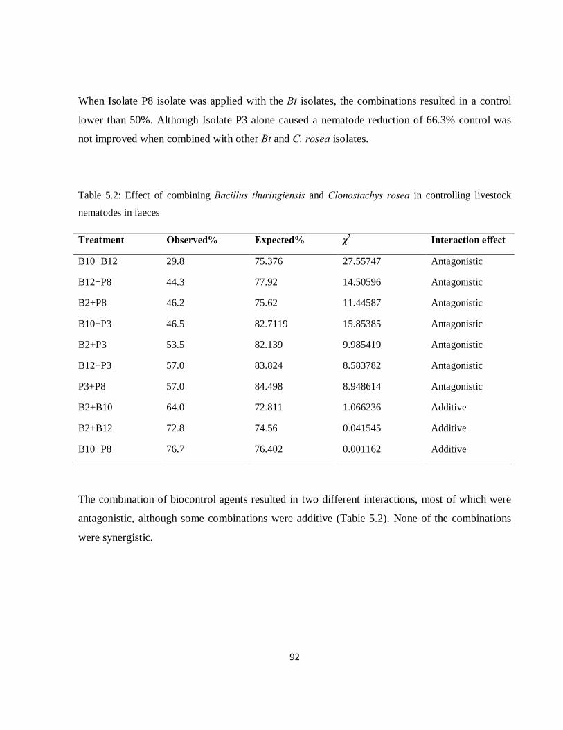
92
When Isolate P8 isolate was applied with the Bt isolates, the combinations resulted in a control
lower than 50%. Although Isolate P3 alone caused a nematode reduction of 66.3% control was
not improved when combined with other Bt and C. rosea isolates.
Table 5.2: Effect of combining Bacillus thuringiensis and Clonostachys rosea in controlling livestock
nematodes in faeces
Treatment Observed% Expected% χ2 Interaction effect
B10+B12 29.8 75.376 27.55747 Antagonistic
B12+P8 44.3 77.92 14.50596 Antagonistic
B2+P8 46.2 75.62 11.44587 Antagonistic
B10+P3 46.5 82.7119 15.85385 Antagonistic
B2+P3 53.5 82.139 9.985419 Antagonistic
B12+P3 57.0 83.824 8.583782 Antagonistic
P3+P8 57.0 84.498 8.948614 Antagonistic
B2+B10 64.0 72.811 1.066236 Additive
B2+B12 72.8 74.56 0.041545 Additive
B10+P8 76.7 76.402 0.001162 Additive
The combination of biocontrol agents resulted in two different interactions, most of which were
antagonistic, although some combinations were additive (Table 5.2). None of the combinations
were synergistic.
93
5.4 Discussion
All the isolates controlled nematodes effectively when applied alone. Individual application of C.
rosea isolates caused greater mortality of nematodes than the bacterial isolates. Isolate P3 was the
best isolate and ranked third, compared to both individual and combination applications of other
C. rosea isolates (Table 5.1).
The performance of Isolate P3 did not improve when combined with other bacterial and fungal
isolates. The interaction effect of combining P3 with all the bacterial isolates and the fungal
Isolate P8 was antagonistic. Combination of bacterial Isolates B10+B12 also had a negative
interaction. Other researchers have reported similar cases of negative interaction between
biocontrol agents (Meyer and Roberts, 2002). This might be due to the competition between
biological control agents (McVay et al., 1977), or biochemical substances produced that may
interfere with the action of the other biocontrol agents (Fravel, 1988). This indicates that,
although they are species in the same genus, they may exhibit a certain degree of defensiveness
against members of their own genus or species (Alabouvette and Lemanceau, 1998). Meyer and
Roberts (2002) identified the possibility of biocontrol agents mechanisms being also directed to
the companion biocontrol agent within the combination. Guetsky et al. (2002) noted the
possibility of one mechanism to compensate for the other mechanism when multiple mechanisms
are involved under a certain set of conditions.
The combination of the bacterial Isolate B10 and the fungal Isolate P8 was additive in its control
of nematodes. This may be due to the different modes of action involved because C. rosea is
known to target nematodes by producing protease and chitinase enzymes (Zhao et al., 2005). On
the other hand, Bt is known to produce endotoxins that directly target free-living and adult larval
stages after ingestion (Kotze et al., 2005). Moreover, combining isolates B2 with B10 improved
their efficacy in controlling nematodes more than each of the isolates used alone.
94
In this study, no combinations resulted in a synergistic interaction. To produce a synergistic
effect, the combined effect of biological control agents must be greater than their additive
individual effects (Ansari et al., 2008), which was not the case in this study. Some authors have
reported the synergistic effect of combining biological agents, reflecting the interactive advantage
of combining the biocontrol agents (Alabouvette et al., 1996). Hence, selection of biocontrol
agents that have different modes of action can enhance biocontrol activity.
A combination of biological control agents may be considered in cases where the isolate
combination has the ability to kill more nematodes than when applied alone. In this study, only
three combinations may be recommended, two of them bacterial and one of bacterial and fungi
(B2+B10; B2+B12 and B10+P8).
In conclusion, combining bacterial isolates of the same genus from the same ecology may have
an advantage in controlling nematode counts. Also, biological control agents from different
genera with different modes of action that target different stages in the lifecycle of the target
organism may increase the control of nematodes. However, in this study most isolates were
antagonistic towards other isolates, irrespective of the genera. Thus, a generalized claim that
combining biological control agents will be beneficial is not always true.
95
5.5 References
Abbott, W.S. 1925. A method of computing the effectiveness of an insecticide. J. Econ. Ento. 18,
265–267.
Alabouvette, C., Lemanceau, P. 1998. Joint action of microbials for disease control. In:
Biopesticides: Use and Delivery. Hall, F.R., Menn, J.J. (Eds.), Humana Press, Ootowa,
Canada.
Alabouvette, C., Lemanceau, P., Steinberg, C. 1996. Biological control of Fusarium wilts:
opportunities for developing a commercial product. In: Hall, R. (Ed.) Principles and Practice
of Managing Soilborne Plant Pathogens, APS Press, St Paul, United States. pp. 192–212.
Ansari, M.A., Shah, F.A., Butt, T.M. 2008. Combined use of entomopathogenic nematodes and
Metarhizuim anisopliae as a new approach for black vine weevil, Otiorhynchus sulcatus,
control. Ent. Exp. Appl. Vol. 129, 340-347.
Campos, A.K., Arau´jo, J.V., Guimara˜es, M.P. 2008. Interaction between the nematophagous
fungus Duddingtonia flagrans and infective larvae of Haemonchus contortus (Nematoda:
Trichostrongyloidea). J. Helminthol. 82, 337–341.
Chen, S., Dickinson, D.W. 2004. Biological control of nematodes by fungal antagonists. In:
Chen, Z., Chen, S., Dickinson, D.W. (Eds.), Nematology-Advances and Perspectives, Vol.
II. Nematode Management and Utilization. Tsinghua University Press and CABI
Publishing, Beijing, China and Wallingford, UK.
Crump, D.H. 1998. Biological control of potato and beet cyst nematodes. Ann. Appl. Biol. 53,
383–386.
Dimander, S.O., Hoglund, J., Uggla, A., Sporndly, E., Waller, P.J. 2003. Evaluation of gastro-
intestinal nematode parasite control strategies for first-season grazing cattle in Sweden. Vet.
Parasitol. 111, 192–209.
Finney, D.J. 1964. Probit Analysis. Cambridge University Press, London, UK.
96
Fravel, D.R. 1988. Role of antibiosis in the biocontrol of plant diseases. Ann. Rev. Phytopath. 26,
75-91.
Gan, Z., Yang, J., Tao, N., Yu, Z., Zhang, K. 2007. Cloning and expression analysis of a
chitinase gene Crchi1 from the mycoparasitic fungus Clonostachys rosea (syn.
Gliocladium roseum). J. of Micro. 45, 422-430.
Ghahfarokhi, M.S., Abyaneh, M.R., Bahadori, S.R., Eslami, A., Zare, R., Ebrahimi, M.
2004. Screening of soil and sheep faecal samples for predacious fungi: Isolation and
characterization of the nematode-trapping fungus Arthrobotrys oligospora. Iran. Biomed.
J. 8, 135-142.
Guetsky, R., Shtienberg, D., Elad, Y., Fischer, E., Dinoor, A. 2002. Improving biological control
by combining biocontrol agents each with several mechanisms of disease suppression.
Phytopathology 91, 621-627.
Jazzar, C., Hammad, A.F. 2004. Efficacy of multiple biocontrol agents against the sweet potato
whitefly Bemisia tabaci (Gennadius) (Homoptera: Aleyrodidae) on tomato. J. Appl. Ento.
128, 188-194.
Kaplan, R.M. 2004. Drug resistance in nematodes of veterinary importance: A status report.
Trends Parasitol. 20, 477-581.
Kotze, A.C., O'O’Grady, J., Gough, J.M., Pearson, R., Bagnall, D.H., Akhurst, R.J. 2005.
Toxicity of Bacillus thuringiensis to parasitic and free-living life stages of nematode
parasitic of livestock. Int. Parasitol. 35, 1013-1022.
Li, J., Yang, J.K., Huang, X.W., Zhang, K.Q. 2006. Purification and characterization of an
extracellular serine protease from Clonostachys rosea and its potential as a pathogenic
factor. Process Biochem. 41, 925-929.
McPartland, J. 2000. Biological control of hemp. Third In. Symposium: Bioresource hemp.
(Wolfsburg, Germany).
McVay, J.R., Gudauskas, R.T., Harper, J.D. 1977. Effects of Bacillus thuringiensis nuclear-
polyhydrosis virus mixtures on Trichoplusiani larvae. J. Invert. Path. 29, 367-372.
97
Meyer, S.L.F., Roberts, D.P. 2002. Combinations of biocontrol agents for management of plant-
parasitic nematodes and soilborne plant-pathogenic fungi. J. Nematol. 34, 1–8.
Perry, B.D., Randolph, T.F. 1999. Improving the assessment of the economic impact of parasitic
diseases and of their control in production animals. Vet. Parasitol. 84, 145-168.
Pierson, E.A., Waller, D.M. 1994. Use of mixtures of fluorescent pseudomonads to suppress
take-all and improve the growth of wheat. Phytopathology 84, 940–947.
Prichard, R.K. 1994. Anthelminthic resistance. Int. J. Parasitol. 54, 259-268.
Raupach, G.S., Kloepper, J.W. 1998. Mixtures of plant growth-promoting rhizobacteria enhance
biological control of multiple cucumber pathogens. Phytopathology 88, 1158–1164.
Sangster, N.C. 1999. Anthelminthic resistance: Past, present and future. Int. J. Parasitol. 29, 115-
124.
Stirling, G.R. 1991. Biological Control of Plant Parasitic Nematodes: Progress, Problems and
Prospects. CAB International, Wallingford, UK.
Van Wyk, J.A., Malan, F.S., Randles, J.L. 1997. How long before resistance makes it possible to
control some field strains of Haemonchus contortus in South Africa with any of the
anthelminthics? Vet. Parasitol. 70, 11-122.
Waller, P.J. 1994. The development of anthelmintic resistance in ruminant livestock. Acta Trop.
56, 233-243.
Waller, P.J. 2003. Global perspectives on nematode parasite control in ruminant livestock: the
need to adopt alternatives to chemotherapy, with emphasis on biological control. An.
Health Res. Rev. 4, 35-43.
Zhao, M.L., Huang, J.S., Mo, M.H., Zhang, K.Q. 2005. A potential virulence factor involved in
fungal pathogenicity serine-like protease activity of nematophagous fungus Clonostachys
rosea. Fungal Divers. 19, 217-234.
98
CHAPTER SIX
EFFECT OF BACILLUS THURINGIENSIS (BERLINER), CLONOSTACHYS ROSEA (SCHROERS) AND DIATOMACEOUS EARTH ON SHEEP NEMATODES: FEEDING TRIAL
M.A. Baloyi and M.D. Laing
Discipline of Plant Pathology, School of Agricultural Sciences and Agribusiness,
University of KwaZulu-Natal, Private Bag X01, Scottsville 3201, Pietermaritzburg,
South Africa
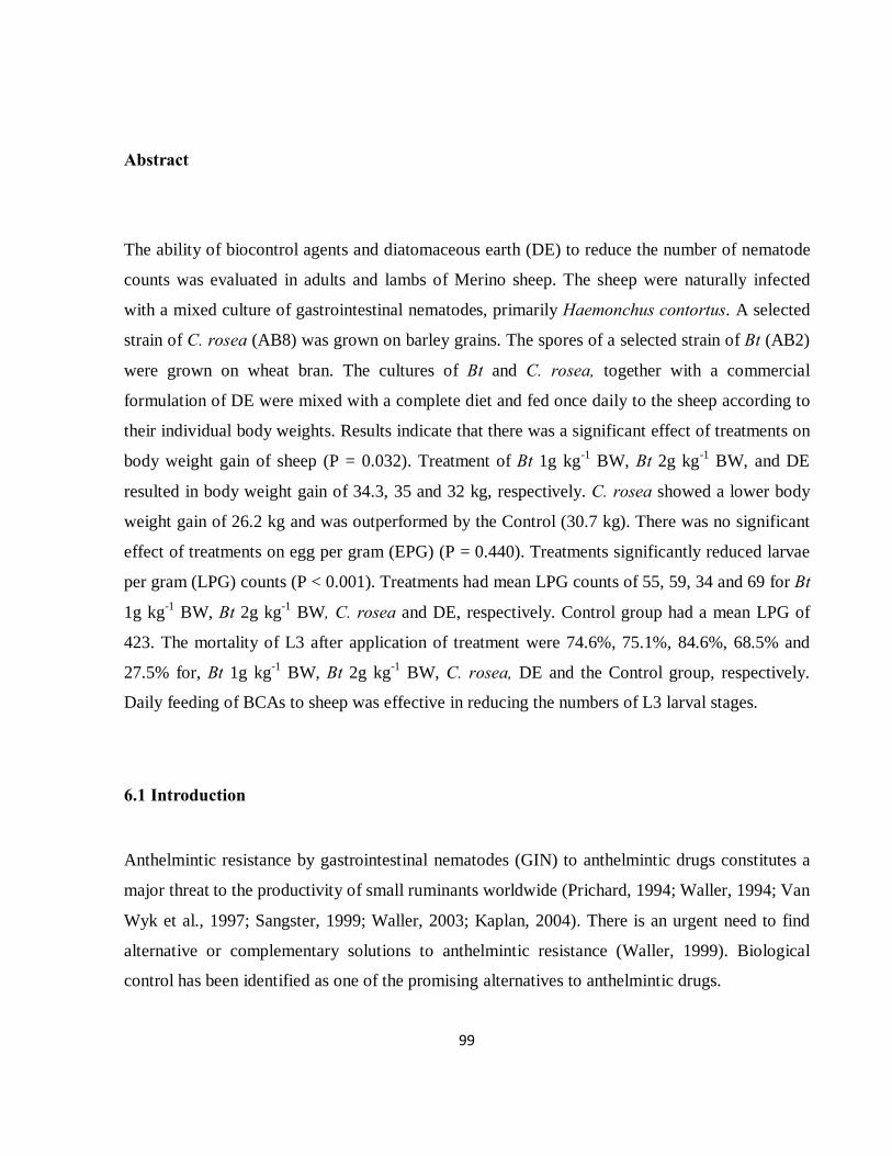
99
Abstract
The ability of biocontrol agents and diatomaceous earth (DE) to reduce the number of nematode
counts was evaluated in adults and lambs of Merino sheep. The sheep were naturally infected
with a mixed culture of gastrointestinal nematodes, primarily Haemonchus contortus. A selected
strain of C. rosea (AB8) was grown on barley grains. The spores of a selected strain of Bt (AB2)
were grown on wheat bran. The cultures of Bt and C. rosea, together with a commercial
formulation of DE were mixed with a complete diet and fed once daily to the sheep according to
their individual body weights. Results indicate that there was a significant effect of treatments on
body weight gain of sheep (P = 0.032). Treatment of Bt 1g kg-1 BW, Bt 2g kg-1 BW, and DE
resulted in body weight gain of 34.3, 35 and 32 kg, respectively. C. rosea showed a lower body
weight gain of 26.2 kg and was outperformed by the Control (30.7 kg). There was no significant
effect of treatments on egg per gram (EPG) (P = 0.440). Treatments significantly reduced larvae
per gram (LPG) counts (P < 0.001). Treatments had mean LPG counts of 55, 59, 34 and 69 for Bt
1g kg-1 BW, Bt 2g kg-1 BW, C. rosea and DE, respectively. Control group had a mean LPG of
423. The mortality of L3 after application of treatment were 74.6%, 75.1%, 84.6%, 68.5% and
27.5% for, Bt 1g kg-1 BW, Bt 2g kg-1 BW, C. rosea, DE and the Control group, respectively.
Daily feeding of BCAs to sheep was effective in reducing the numbers of L3 larval stages.
6.1 Introduction
Anthelmintic resistance by gastrointestinal nematodes (GIN) to anthelmintic drugs constitutes a
major threat to the productivity of small ruminants worldwide (Prichard, 1994; Waller, 1994; Van
Wyk et al., 1997; Sangster, 1999; Waller, 2003; Kaplan, 2004). There is an urgent need to find
alternative or complementary solutions to anthelmintic resistance (Waller, 1999). Biological
control has been identified as one of the promising alternatives to anthelmintic drugs.
100
Biological control involves the use of natural enemies of nematodes to reduce the number of
nematodes developing on pastures to a level which is sub-clinical (Larsen, 2006). Duddingtonia
flagrans (Cooke) has been the most researched biocontrol agent (BCA) that targets free-living
stages of nematodes in livestock faeces (Larsen et al., 1992; Waller and Faedo, 1993; Larsen et
al., 1998; Larsen, 2000; Waller et al., 2001b; Waller et al., 2004).
There are other microorganisms that target nematodes, including fungi, bacteria, viruses, insects,
mites and some invertebrates (Stirling, 1991; Siddiqui, et al., 1996). Bacillus thuringiensis
(Berliner) has also shown the ability to destroy free-living and adult stages of sheep nematodes
(Kotze et al., 2005). The mode of action has been shown to be the same as that involved in
insects (Marroquin et al., 2000). Clonostachys rosea (Schroers) is a facultative parasite of
pathogenic plant nematodes (Yang et al., 2000). The hydrolytic enzymes (protease and chitinase)
produced by C. rosea play a key role in its ability to penetrate and kill a nematode host (Zhao et
al., 2004).
Diatomaceous earth (DE) is a naturally occurring siliceous sedimentary compound derived from
the microscopic remains of unicellular algae-like plants called diatoms. It contains elements such
as silicon oxide (SiO2), aluminium oxide (Al2O3), iron oxide (Fe2O3), calcium oxide (CaO) and
magnesium oxide (MgO), to name a few. The mode of action of DE is mechanical. The diatom
particles have sharp edges that pierce the skin of nematodes when in contact, causing the parasite
to dehydrate and die.
Other researchers have demonstrated the potential of biocontrol agents to control livestock
nematodes. However, much of this research has been from in vitro studies. Therefore, this study
aimed to determine the effect of the biocontrol agents in the field (in vivo)by testing the effects of
B. thuringiensis, C. rosea and DE on larvae of sheep nematodes.
6.2 Materials and Methods
6.2.1 Formulation of biocontrol agents Isolates of Bt (B2(4)M and C. rosea (Paec 8) were previously isolated from soil of the Livestock
Section of the University of KwaZulu-Natal, Pietermaritzburg.
101
These were grown on wheat bran and barley grain, respectively, by1 Plant Health Products (PHP)
(Pty) Ltd.
6.2.2 Animal grouping Fourty sheep per treatment, naturally infected with mixed cultures of GIN were used in this
experiment. The predominant species was H. contortus (2Allerton Veterinary Laboratory). The
sheep were grouped according to age (young and adults) and by sex (ewes and rams). Sheep were
kept in feeding crates in the sheep facility at Ukulinga Research Farm. The feeding crate size
was 0.90 × 1.50m. These were each provided with an individual feeder and drinker. The flooring
was of slatted wood to ensure that urine and faeces did not accumulate near the sheep. The
facility was temperature regulated with large fans.
6.2.3 Feed preparation The standard sheep feed consisted of the following ingredients: cottonseed cake (37.5kg),
hormony chop (33.3kg), liquid molasses (8.6kg) and Vitamin Premix (1.1 kg). The Bt product
was added to the standard feed at a rate of either 1 or 2g per kg body weight (BW) for each
sheep. The C. rosea product was added to the standard feed at a rate of 1g per kg BW for each
sheep. The DE product was added to the standard feed at a rate of 15% by weight of the BW of
each sheep. The products were thoroughly mixed with the standard feed before being given to the
sheep. The sheep were fed 1.5% of the mixed feed per kg of their body weight daily between
07:00-09:00am and then given hay ad libitum.
6.2.4 Experimental procedure Pre-treatment period
During a pre-treatment period, the sheep were randomly allocated to individual sheep feeding
stalls to acclimatise them to the handling facilities for 2 wk. Sheep were given feed supplement in
the morning and hay ad libitum. At the end of this 2 wk period, initial values of response
variables namely, body weight, EPG, LPG (larva per gram) and LD (larval development) were
determined.
1Plant Health Products (Pty) Ltd., P.O. Box 207, Nottingham Road, South Africa
102
During treatment period
Eight sheep were assigned to one of the five treatments, (i) two levels of Bt application (1 and 2g
kg-1 BW), (ii) C. rosea, (iii) 15% DE and (iv) a Control group at 1g kg-1 BW per day of the
standard ration formulation. All the treatments were included in the total daily mixed ration
formulation for 1 wk.
Post-treatment period
Treatments were stopped and sheep were fed the standard feed for a period of 1 wk.
6.2.5 Parasitology and animal performance Sheep were weighed weekly. Rectal grab samples were collected weekly and sent to the Allerton
Veterinary Laboratories (No. 458 Town Bush Road, Montrose, Pietermaritzburg, South Africa)
for larval identification (LID), while faecal egg count (FEC) were calculated using the McMaster
Method (Whitlock, 1949). Two grams of sheep faeces were weighed and mixed with 52ml of a
saturated table salt solution. The aliquot was stirred in a N, W, S, E direction and a sample
pippetted onto a McMaster slide for the counting of eggs under a light microscope (x100).
Samples were counted within 15 min to avoid the eggs being distorted due to the hypertonic salt
solution. Larval mortality was determined by weighing 2g samples of faeces from each sheep into
a microtitre plates. The faeces were well aerated, incubated for 7d and moistened when
necessary. The infective larvae were collected using the Baermann technique. Counting of larvae
was performed using three aliquots of 2ml from each culture under a dissecting microscope.
Allerton Veterinary Laboratories, No. 458 Town Bush Road, Montrose, Pietermaritzburg, South Africa
103
6.2.6 Statistical analysis Percent mortality of the nematodes was determined using Abbott’s Formula (Abbott, 1925),
which is as follows:
Mortality = [C1 – C2] x 100
C1
where: C1 = mean number of larvae in untreated manure; C2 = number of larvae in treated
manure
Means were compared by Fisher’s LSD at a significance level of 5%.
Larvae per gram from each sample was obtained by dividing the total number of larvae counted
in each sample of 2g of faeces used, by 2 to obtain nematode counts in 1g, as described by Ojeda-
Robertos et al., (2008).
Larval development (LD) was calculated by the formula of Paraud et al. (2005), which is as
follows:
LD = (EPG/LPG) x 100
Where: EPG is the egg per gram, LPG is the larvae per gram.
ANOVA was applied to the values of BW, EPG, LPG, mortality and LD. The Genstat® statistical
package was used, after transforming the EPG and mortality by log transformation to normalize
homogeneity of variance of the data. The mortality of L3 nematodes was analysed using
ANOVA, as a percentage of the Control L3 nematode counts, after the data was log transformed.
These data are presented in the tables together with the untransformed means. The transformed
data was used to calculate LSD values to separate treatment means at 5% significance level.
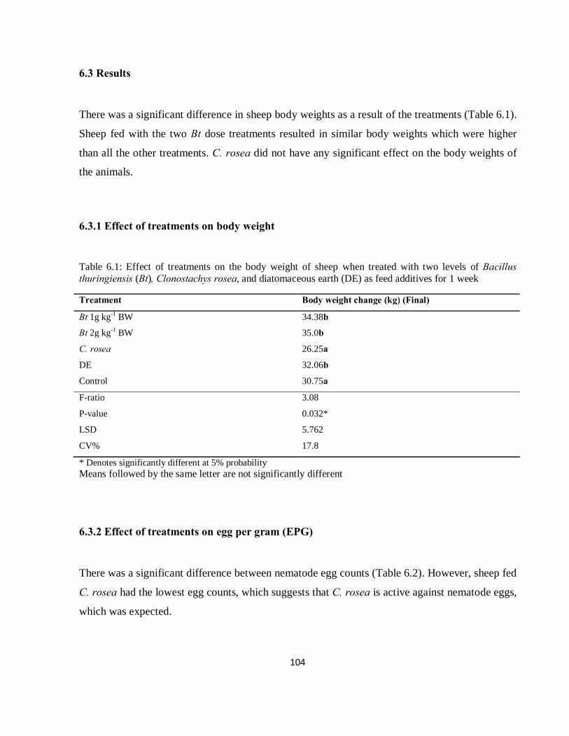
104
6.3 Results
There was a significant difference in sheep body weights as a result of the treatments (Table 6.1).
Sheep fed with the two Bt dose treatments resulted in similar body weights which were higher
than all the other treatments. C. rosea did not have any significant effect on the body weights of
the animals.
6.3.1 Effect of treatments on body weight
Table 6.1: Effect of treatments on the body weight of sheep when treated with two levels of Bacillus thuringiensis (Bt), Clonostachys rosea, and diatomaceous earth (DE) as feed additives for 1 week
Treatment Body weight change (kg) (Final)
Bt 1g kg-1 BW 34.38b
Bt 2g kg-1 BW 35.0b
C. rosea 26.25a
DE 32.06b
Control 30.75a
F-ratio 3.08
P-value 0.032*
LSD 5.762
CV% 17.8
* Denotes significantly different at 5% probability Means followed by the same letter are not significantly different
6.3.2 Effect of treatments on egg per gram (EPG)
There was a significant difference between nematode egg counts (Table 6.2). However, sheep fed
C. rosea had the lowest egg counts, which suggests that C. rosea is active against nematode eggs,
which was expected.
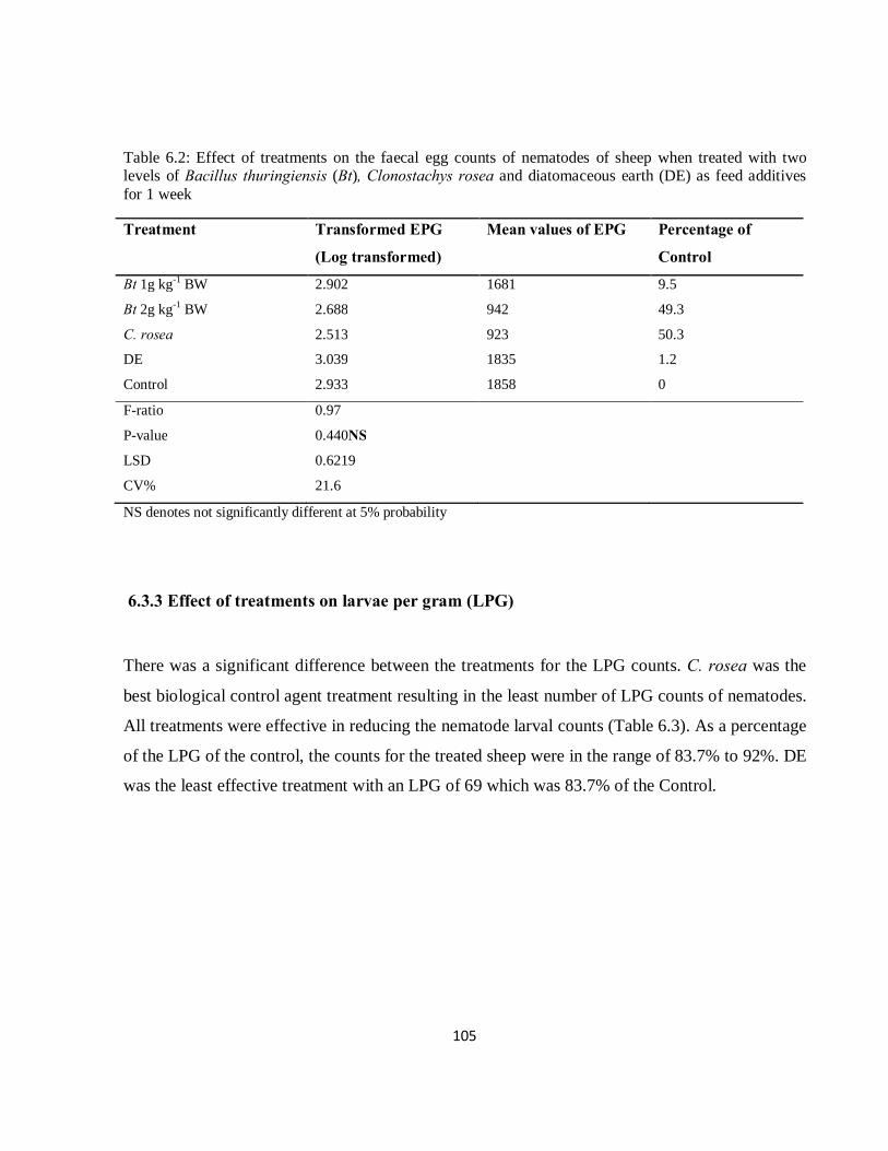
105
Table 6.2: Effect of treatments on the faecal egg counts of nematodes of sheep when treated with two levels of Bacillus thuringiensis (Bt), Clonostachys rosea and diatomaceous earth (DE) as feed additives for 1 week
Treatment Transformed EPG
(Log transformed)
Mean values of EPG Percentage of
Control
Bt 1g kg-1 BW 2.902 1681 9.5
Bt 2g kg-1 BW 2.688 942 49.3
C. rosea 2.513 923 50.3
DE 3.039 1835 1.2
Control 2.933 1858 0
F-ratio 0.97
P-value 0.440NS
LSD 0.6219
CV% 21.6
NS denotes not significantly different at 5% probability
6.3.3 Effect of treatments on larvae per gram (LPG)
There was a significant difference between the treatments for the LPG counts. C. rosea was the
best biological control agent treatment resulting in the least number of LPG counts of nematodes.
All treatments were effective in reducing the nematode larval counts (Table 6.3). As a percentage
of the LPG of the control, the counts for the treated sheep were in the range of 83.7% to 92%. DE
was the least effective treatment with an LPG of 69 which was 83.7% of the Control.

106
Table 6.3: Effect of treatments on the larvae per gram of nematodes of sheep when treated with two levels of Bacillus thuringiensis (Bt), Clonostachys rosea and diatomaceous earth (DE) as feed additives for 1 week
Treatments Transformed LPG
values
(Log transformed)
Mean LPG
(final)
Percentage
reduction in
relation to Control
Bt 1g kg-1 BW 1.787a 55 87
Bt 2g kg-1 BW 1.787a 59 86.1
C. rosea 1.622a 34 92
DE 1.875a 69 83.7
Control 2.405b 423 0
F-ratio 9.57
P-value < 0.001***
LSD 0.2803
CV% 14.4
* Denotes significantly different at 5% probability Means followed by the same letter in the same column are not significantly different
6.3.4 Effect of treatments on corrected mortality (Abbott’s Correction)
The mortality of nematodes was significantly different for the four treatments applied (Table 6.4).
The Bt doses were also very effective and caused nematode mortalities of 74.6% and 75.1% (1g
kg-1 BW and 2g kg-1 BW, respectively). C. rosea was the best biocontrol agent resulting in a
nematode mortality of 84.6%. DE ranked fourth, but was also effective, causing a nematode
mortality of 68.5%.
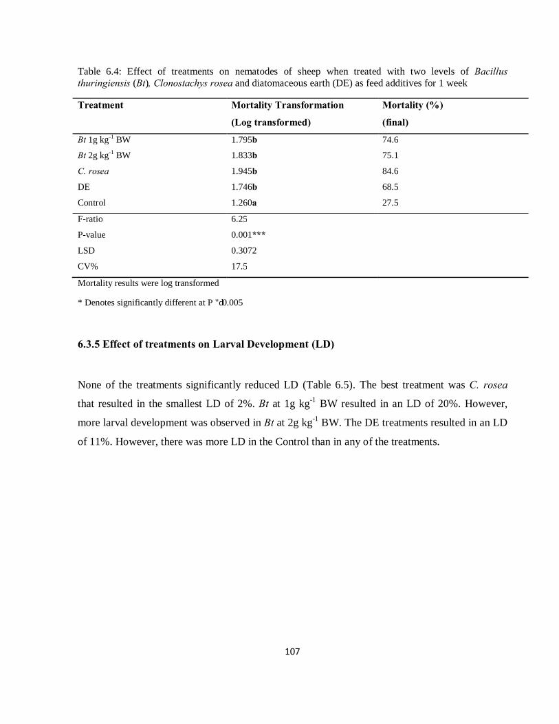
107
Table 6.4: Effect of treatments on nematodes of sheep when treated with two levels of Bacillus thuringiensis (Bt), Clonostachys rosea and diatomaceous earth (DE) as feed additives for 1 week
Treatment Mortality Transformation
(Log transformed)
Mortality (%)
(final)
Bt 1g kg-1 BW 1.795b 74.6
Bt 2g kg-1 BW 1.833b 75.1
C. rosea 1.945b 84.6
DE 1.746b 68.5
Control 1.260a 27.5
F-ratio 6.25
P-value 0.001***
LSD 0.3072
CV% 17.5
Mortality results were log transformed
* Denotes significantly different at P ≤ 0.005
6.3.5 Effect of treatments on Larval Development (LD)
None of the treatments significantly reduced LD (Table 6.5). The best treatment was C. rosea
that resulted in the smallest LD of 2%. Bt at 1g kg-1 BW resulted in an LD of 20%. However,
more larval development was observed in Bt at 2g kg-1 BW. The DE treatments resulted in an LD
of 11%. However, there was more LD in the Control than in any of the treatments.
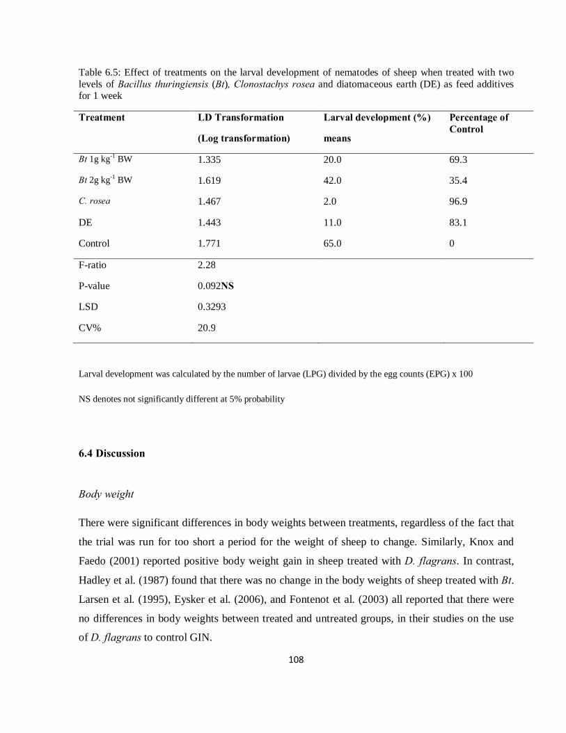
108
Table 6.5: Effect of treatments on the larval development of nematodes of sheep when treated with two levels of Bacillus thuringiensis (Bt), Clonostachys rosea and diatomaceous earth (DE) as feed additives for 1 week
Treatment LD Transformation
(Log transformation)
Larval development (%)
means
Percentage of Control
Bt 1g kg-1 BW 1.335 20.0 69.3
Bt 2g kg-1 BW 1.619 42.0 35.4
C. rosea 1.467 2.0 96.9
DE 1.443 11.0 83.1
Control 1.771 65.0 0
F-ratio 2.28
P-value 0.092NS
LSD 0.3293
CV% 20.9
Larval development was calculated by the number of larvae (LPG) divided by the egg counts (EPG) x 100
NS denotes not significantly different at 5% probability
6.4 Discussion
Body weight
There were significant differences in body weights between treatments, regardless of the fact that
the trial was run for too short a period for the weight of sheep to change. Similarly, Knox and
Faedo (2001) reported positive body weight gain in sheep treated with D. flagrans. In contrast,
Hadley et al. (1987) found that there was no change in the body weights of sheep treated with Bt.
Larsen et al. (1995), Eysker et al. (2006), and Fontenot et al. (2003) all reported that there were
no differences in body weights between treated and untreated groups, in their studies on the use
of D. flagrans to control GIN.
109
The sheep always consumed their feed, irrespective of the formulation of the treatment. Previous
attempts of formulating biocontrol agents with D. flagrans included mixing spores in daily
supplement, feed blocks (Waller et al., 2001b; Chandrawathani et al., 2002) and slow release
devices (Waller et al., 2001a). Hence, this aspect is unlikely to have affected body weights.
Application of control agents in feed is important because they will be excreted together with the
eggs in faeces. They will then germinate to parasitize the eggs and developing larvae. In this
manner, pasture contamination would be reduced (Waller, 2006). Lee et al. (2002) succeeded in
recovering ingested spores of Bt from the faeces of sheep. According to Larsen et al. (1991), for
BCAs to be effective against GIN, they should withstand the passage through the gut of the
alimentary tract and colonize the dung pat to prey on the free-living stages of the parasitic
nematodes before these reach the L3 stages and become infective to livestock. Studies on D.
flagrans have shown that its efficacy was proportional to its spore density (Sanyal, 2004; Sanyal
et al., 2008).
Faecal egg counts (EPG)
There was no significant result because none of the agents affected adult GIN in the gut of the
treated sheep. Therefore, egg laying continued unchanged. An environmentally friendly treatment
that parasitizes the nematode eggs such as Paecilomyces lilacinus (Samson) is needed.
Meadows et al. (1989) found that toxins of Bt kurstaki were lethal towards eggs, and the L1, L2
and L3 larval stages of Trichostrongylus colubriformis (Giles). Bottjer et al. (1985) also reported
the lethal effect of the toxin of Bt israelensis on eggs of T. colubriformis in the laboratory. Kotze
et al. (2005) showed the ability of Bt Strain L366 to kill adult livestock nematodes in the
laboratory. He suggested that the use of Bt would require the encapsulation of the Bt to protect it
from the acidity of the abomasal lumen. Our results show that this is not necessary. Shimoda et
al. (2001) and Senel and McClure (2004) proposed the use of the mucosal adhesive properties of
chitosan to ensure a localisation of the Bt at the site of nematode infection within the mucosa or
intestinal epithelium, if Bt is to be used as a deworming agent. According to Kotze et al. (2005),
this approach would ensure a degree of protection in the neutral pH environment of secreted
mucous in the microenvironment inhabited by the nematodes.
110
Larvae per gram (LPG)
Counts of nematode larvae were significantly different. All treatments reduced the LPG counts
(P = 0.001). C. rosea caused lower larval counts than all the other treatments. The results parallel
the mortality data.
Corrected mortality
A mortality of GIN in the livestock faeces was achieved after one week of feeding BCAs to
sheep. Given that it can produce chlamydospores in culture, this fungus has considerable
potential to be commercialized as a BCA against GIN. This is the first report of C. rosea to be
used as a biocontrol agent against sheep nematodes.
There was no difference in the levels of mortality of GIN caused by the two doses of Bt (1g and
2g Kg-1 BW). B. thuringiensis is a widely used biocontrol agent for the control of insect pests of
plants and animals. Two other sets of experiments, using Bt to control sheep GIN, were
undertaken in the laboratory (Kotze et al., 2005; O’Grady et al., 2007). It would seem there are
no other in vivo trials with Bt involving the feeding of a Bt product to sheep. DE also proved to be
effective on nematodes, and therefore provides another treatment option.
These results accord with the successful results reported by Larsen et al., (1995) and
Chandrawathani et al., (2002), on the use of the biocontrol agent D. flagrans to reduce sheep
nematode counts in the field.
Larval development
The recovery of larvae from the faeces of treated sheep was reduced. C. rosea resulted in the
lowest LD value of 2%. Bt dose of 1g kg-1 BW also resulted in lower counts of nematodes that
developed, its LD was 20%.
In conclusion, all treatments were effective and ensured that counts of the L3 larval stages of GIN
were reduced. Other researchers have shown that if the L3 population is reduced, then infection
decreases and GIN count in the gut of livestock decreases and subsequently EPG is lower
(Larsen, 1995; Van Wyk et al., 1999).

111
6.5 References
Abbott, W.S. 1925. A method of computing the effectiveness of an insecticide. J. Econ. Ento. 18,
265-267.
Bottjer, K.P., Bone, L.W., Gill, S.S. 1985. Nematoda: susceptibility of the egg to Bacillus
thuringiensis toxins. Exp. Parasitol. 60, 239-244.
Chandrawathani, P., Jamnah, O., Waller, P.J., Höglund, J., Larsen, M., Zahari, W.M. 2002.
Nematophagous fungi as a biological control agent for nematode parasites of small
ruminants in Malaysia: A special emphasis on Duddingtonia flagrans. Vet. Res. 33, 235-
396.
Eysker, M., Bakker, N., Kooyman, F.N.J., Olde-Olthuis, S., Ploeger, H.W. 2006. Effect of
biological control through the daily application of spores of Duddingtonia flagrans in
lambs kept under an evasive grazing system in the Netherlands. Vet. Parasitol. 140, 312-
320.
Fontenot, M.E., Miller, J.E., Peña, M.T., Larsen, M., Gillespie, A. 2003. Efficiency of feeding
Duddingtonia flagrans chlamydospores to grazing ewes on reducing availability of
parasitic nematode larvae on pasture. Vet. Parasitol. 118, 203-213.
Hadley, W.M., Burchiel, S.W., McDowell, T.D., Thilsted, J.P., Hibbs, C.M., Whorston, J.A.,
Day, P.W., Friedman, M.B., Stoll, R.E. 1987. Five month oral (Diet) toxicity/infectivity
study of Bacillus thuringiensis insecticides in sheep. Fund. Appl. Toxicol. 8, 236-242.
Kaplan, R.M. 2004. Drug resistance in nematodes of veterinary importance: A status report.
Trends Parasitol. 20, 477-581.
Knox, M.R., Faedo, M. 2001. Biological control of field infections of nematode parasite of sheep
with Duddingtonia flagrans and effects of spore intake on efficacy. Vet. Parasitol. 101, 155-
160.
112
Kotze, A.C., O’Grady, J., Gough, J.M., Pearson, R., Bagnall, D.H., Akhurst, R.J. 2005. Toxicity
of Bacillus thuringiensis to parasitic and free-living life stages of nematode parasitic of
livestock. Int. Parasitol. 35, 1013-1022.
Larsen, M. 2000. Prospects for controlling animal parasitic nematodes by micro fungi.
Parasitology 120, S120-S131.
Larsen, M. 2006. Biological control of nematode parasites in sheep. J. An. Sci. 84, E133-E139.
Larsen, M., Faedo, M., Waller, P.J., Hennessy, D.R. 1998. The potential of nematophagous fungi
to control the free-living stages of nematode parasites of sheep: Studies on Duddingtonia
flagrans. Vet. Parasitol. 76, 121-128.
Larsen, M., Nansen, P., Gronvold, J., Wolstrup, J., Henriksen, S.A. 1992. In vivo passage of
nematophagous fungi selected for biocontrol of parasitic nematodes in ruminants. J.
Helminthol. 66, 137-141.
Larsen, M., Nansen, P., Wolstrup, J., Gronvold, J., Henriksen, S.A., Zorn, A. 1995. Biological
control of trichostrongylus in calves by the fungus Duddingtonia flagrans fed to animals
under natural grazing conditions. Vet. Parasitol. 60, 321-330.
Larsen, M., Wolstrup, J., Henriksen, S.A., Dackman, C., Gronvold, J., Nansen, P. 1991. In vitro
stress selection of nematophagous fungi for biological control of parasitic nematodes in
ruminants. Vet. Parasitol. 65, 141-193.
Lee, D.H., Machii, J., Ohba, M. 2002. High frequency of Bacillus thuringiensis in faeces of
herbivorous animals maintained in a zoological garden in Japan. Appl. Ento. Zoo. 37,
509-516.
McPartland, J. 2000. Biological control of hemp. Third In. Symposium: Bioresource hemp.
(Wolfsburg, Germany).
Meadows, J., Gill, S.S., Bone, L.W. 1989. Factors influencing lethality of Bacillus thuringiensis
kurstaki toxin for eggs and larvae of Trichostrongylus colubriformis (Nematoda). J.
Parasitol. 75, 191-194.
O’Grady, J., Akhurst, R.J., Kotze, A.C. 2007. The requirement for early exposure of
Haemonchus contortus larvae to Bacillus thuringiensis for effective inhibition of larval
development. Vet. Parasitol. 150, 97-103.
113
Ojeda-Robertos, N.F., Torres-Acosta, J.F., Aguilar-Caballero, A.J., Ayala-Burgos, A., Cob-Galera,
L.A., Sandoval-Castro, C.A., Barrientos-Medina, R.C., Mendozade Gives, P. 2008.
Assessing the efficacy of Duddingtonia flagrans chlamydospores per gram of faeces to
control Haemonchus contortus larvae. Vet. Parasitol. 158, 329–335.
Paraud, C., Hoste, H., Lefrileux, Y., Pommaret, A., Paolini, V., Pors, I., Chartier, C. 2005.
Administration of Duddingtonia flagrans chlamydospores to goats to control
gastrointestinal nematodes: dose trials. Vet. Res. 36, 157-166.
Prichard, R.K. 1994. Anthelmintic resistance. Int. J. Parasitol. 54, 259-268.
Sangster, N.C. 1999. Anthelmintic resistance: Past, present and future. Int. J. Parasitol. 29, 115-
124.
Sanyal, P.K. 2004. Density dependent nematophagous behaviour of Duddingtonia flagrans.
Indian Vet. J. 81, 16-19.
Sanyal, P.K., Patel, N.K., Mandal, S.C., Pal, S. 2008. Formulation of a strategy for the
application Duddingtonia flagrans to control caprine parasitic gastroenteritis. J. Helminthol.
82, 169-174.
Senel, S., McClure, S.J. 2004. Potential applications of chitosan in veterinary medicine. Adv.
Drug Deliv. Rev. 56, 1467–1480.
Shimoda, J., Onishi, H., Machida, Y. 2001. Bioadhesive characteristics of chitosan microspheres
to the mucosa of rat small intestine. Drug Dev. Ind. Pharm. 27, 567–576.
Siddiqui, Z.A., Qureshi, S.A., Sultana, V., Ehteshamul-Haque, S., Ghaffar, A. 2000. Biological
control of root-knot nematode disease complex of tomato. Plant Soil 227, 163-169.
Stirling, G.R. 1991. Biological Control of Plant Parasitic Nematodes: Progress, Problems and
Prospects. CABI, Wallingford, UK.
Van Wyk, J.A., Malan, F.S., Randles, J.L. 1997. How long before resistance makes it impossible
to control some field strains of Haemonchus contortus in South Africa with any of the
anthelmintics? Vet. Parasitol. 70, 11-122.
Waller, P.J. 1994. The development of anthelmintic resistance in ruminants livestock. Acta Trop.
56, 233-243.
114
Waller, P.J. 1999. International approaches to the concept of integrated control of nematode
parasites of livestock. Int. J. Parasitol. 29, 155–164.
Waller, P.J. 2003. Global perspectives on nematode parasite control in ruminant livestock: the
need to adopt alternatives to chemotherapy, with emphasis on biological control. An.
Health Res. Rev. 4, 35-43.
Waller, P.J. 2006. Sustainable nematode parasite control strategies for ruminant livestock by
grazing management and biological control. An. Feed Sci. 126, 277-289.
Waller, P.J., Faedo, M. 1993. The potential of nematophagous fungi to control the free-living
stages of nematode parasites: In vitro and in vivo studies. Vet. Parasitol. 51, 289-299.
Waller, P.J., Faedo, M., Ellis, K. 2001a. The potential of nematophagous fungi to control the
free-living stages of nematode parasites of sheep: Towards the development of a fungal
controlled release device. Vet. Parasitol. 102, 299-308.
Waller, P.J., Knox, M.R., Faedo, M. 2001b. The potential of nematophagous fungi to control the
free-living stages of sheep: Feeding and block studies with Duddingtonia flagrans. Vet.
Parasitol. 102, 321-330.
Waller, P.J., Schwan, O., Ljungstrom, B.L., Rydzik, A., Yeates, G.W. 2004. Evaluation of
biological control of sheep parasites using Duddingtonia flagrans under commercial
farming conditions on the island of Gotland, Sweden. Vet. Parasitol. 126, 299–315.
Whitlock, H.R. 1948. Some modifications of the Modified McMaster helminth egg counting
technique apparatus. J. Council Sci. Indus. Res. 21, 177-180.
Yang, X.J., He, Y.X., Chen, F.R., Zheng, L. 2000. Separating and selection of egg-mass fungi of
Meloidogyne sp. in Fujian province. Fujian J. of Agric. Sci. 15, 12-15.
Zhao, M.L., Mo, M.H., Zhang, K.Q. 2004. Characterization of a neutral serine protease and its
full-length cDNA from the nematode-trapping fungus Arthrobotrys oligospora. Mycologia
96, 16-22.
115
THESIS OVERVIEW
The bacterium Bacillus thuringiensis (Bt) (Berliner) and the fungus Clonostachys rosea
(Schroers) have been widely studied as biocontrol agents in pest control of plants. To date, there
is still no commercialized Bt or C. rosea formulation for the control of livestock pests.
The problem of anthelmintic resistance has been the driving force behind the search for
alternative control measures. Moreover, chemical residues in foods and the demand to reduce
chemical inputs in agriculture are another major concern (De and Sanyal, 2009).
Biological control is becoming an important non-chemical option because biocontrol agents have
low mammalian toxicity, high efficacy and are naturally occurring (Moore et al., 2000).
In the research reported in this thesis, 25 Bt and 10 C. rosea isolates were obtained from grazing
pastures and pens of sheep at the Livestock Section, Ukulinga Research Farm, UKZN,
Pietermaritzburg. The isolates were characterized according to their morphology. The Bt colonies
were white in colour, had a rough texture with flat elevation, undulate margins and irregular
shape. The spores were rod-shaped, stained gram positive and clear. C. rosea were identified by
whitish colonies with the characteristic conidiophores which were branched and showed phialides
at the tips. The goat grazing pasture soil was a good source for the best and most effective
isolates used as biological control agents during this study.
The isolates were screened in the laboratory, and some were highly pathogenic to livestock
nematode larvae. Fifteen Bt isolates and eight C. rosea isolates were effective in reducing the L3
larval stage in faeces and water bioassays.
The most effective concentration of each isolate was also determined, and indeed lower
concentrations of C. rosea were effective in reducing larvae counts under laboratory conditions.
The best concentrations were 1010 spores ml-1 and 109 conidia ml-1 for Bt and C. rosea,
respectively. C. rosea efficacy did not increase with the increase in concentrations tested and was
the most effective biocontrol agent.
Theoretically, combinations of biocontrol agents might be more effective than a single biocontrol
agent. Some Bt isolates improved their efficacy when combined with others and showed an
116
additive interaction effect. Isolate P3 of C. rosea was antagonistic when mixed with other
bacterial isolates (B2, B10 and B12) and fungal Isolate P8. However, the level of control by this
isolate was highest when used alone.
The incorporation of biocontrol agents in animal feed caused a mortality of L3 larval stages of
nematodes. There was no dose response of nematode mortality to Bt treatment. C. rosea caused
the greatest mortality of nematodes. Diatomaceous earth was effective against nematodes larvae.
The results showed that these biocontrol agents were effective in controlling animal nematodes
and should be adopted into integrated pest management programmes.
Future studies
As biocontrol agents have proven to be effective in the laboratory, field trials that extend for
longer periods need to be undertaken. Further studies are needed to evaluate the efficacy in
pasture, to determine whether pasture contamination is reduced.
The speed of kill of BCAs in the field under extreme environmental conditions needs to be
determined. In addition, the effect of these BCAs on eggs needs to be determined.
The mechanisms of C. rosea involved in killing livestock nematode needs to be determined, to
find out as to whether it uses the same mechanism as that used on plant nematodes.
The relative cost of daily feeding of biocontrol agents needs to be addressed relative to the cost of
anthelmintics used locally.
The effects of anthelmintics such as benzimidazoles on biocontrol agents need to be studied as to
whether they inhibit C. rosea and other nematophagous fungi. Also, common antibiotics and
other medicines fed to livestock need to be tested for compatibility.
The best biocontrol agent should be tested on various livestock endoparasites to determine if they
can be used as broad-spectrum control agents.
117
References
De, S., Sanyal, P.K. 2009. Biological control of helminth parasites by predatory fungi. Vetscan 4,
1-9.
Moore, D., Lord, J.C. Smith, S.M. 2000. Pathogens. In: Subramanyam, B., Hagstrum, D.W.,
(Eds.), Alternatives to Pesticides in Stored-Product IPM, Kluwer Academic Publishers,
Dordreecht, pp. 193–227.

118
APPENDIX
Appendix 1: Characterization of Bacillus thuringiensis isolates
Morphological characteristics of colony Spores
Isolate colour Texture Elevation Margin Shape Shape Gram
stain
Shape colour Position
B1(1) white Rough Flat Undulate Irregular Rod + Oval Clear Centre
B1(2) white Rough Flat Undulate Irregular Rod + Oval Clear Centre B1(3) white Rough Flat Undulate Irregular Rod + Oval Clear Centre B1(4) white Rough Flat Undulate Irregular Rod + Oval Clear Centre B2(1) white Rough Flat Undulate Irregular Rod + Oval Clear Centre B2(2) white Rough Flat Undulate Irregular Rod + Oval Clear Centre B3(3) white Rough Flat Undulate Irregular Rod + Oval Clear Centre B4(1) white Rough Flat Undulate Irregular Rod + Oval Clear Centre B4(2) white Rough Flat Undulate Irregular Rod + Oval Clear Centre B3(4) white Rough Flat Undulate Irregular Rod + Oval Clear Centre B1(4) white Rough Flat Undulate Irregular Rod + Oval Clear Centre B3(6) white Rough Flat Undulate Irregular Rod + Oval Clear Centre B5(3) white Rough Flat Undulate Irregular Rod + Oval Clear Centre B6(3) white Rough Flat Undulate Irregular Rod + Oval Clear Centre B6(2) white Rough Flat Undulate Irregular Rod + Oval Clear Centre B5(1) white Rough Flat Undulate Irregular Rod + Oval Clear Centre B7(2) white Rough Flat Undulate Irregular Rod + Oval Clear Centre B8(3) white Rough Flat Undulate Irregular Rod + Oval Clear Centre B10(4) white Rough Flat Undulate Irregular Rod + Oval Clear Centre B10(2) white Rough Flat Undulate Irregular Rod + Oval Clear Centre B10(3) white Rough Flat Undulate Irregular Rod + Oval Clear Centre B9(2) white Rough Flat Undulate Irregular Rod + Oval Clear Centre B9(4) white Rough Flat Undulate Irregular Rod + Oval Clear Centre B10(1) white Rough Flat Undulate Irregular Rod + Oval Clear Centre B9(3) white Rough Flat Undulate Irregular Rod + Oval Clear Centre B8(2) white Rough Flat Undulate Irregular Rod + Oval Clear Centre
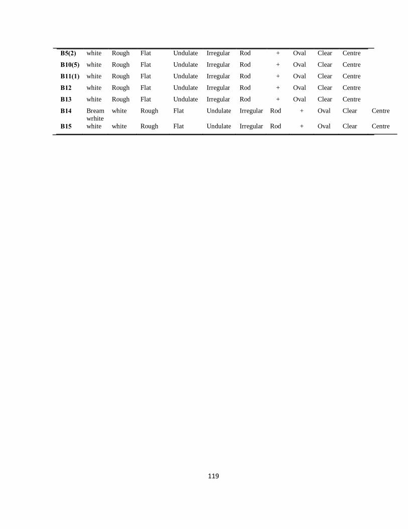
119
B5(2) white Rough Flat Undulate Irregular Rod + Oval Clear Centre B10(5) white Rough Flat Undulate Irregular Rod + Oval Clear Centre B11(1) white Rough Flat Undulate Irregular Rod + Oval Clear Centre B12 white Rough Flat Undulate Irregular Rod + Oval Clear Centre B13 white Rough Flat Undulate Irregular Rod + Oval Clear Centre B14 Bream
wrhite white Rough Flat Undulate Irregular Rod + Oval Clear Centre
B15 white white Rough Flat Undulate Irregular Rod + Oval Clear Centre