Embed Size (px)
Citation preview
Report
A Subset of Serotonergic N
eurons Evokes Hunger inAdult DrosophilaHighlights
d Activation of a small set of neurons induces a hunger
response in sated flies
d The behaviors promoted include feeding and appetitive
memory performance
d These serotonergic brain neurons project broadly and
mediate the hunger sensation
Albin et al., 2015, Current Biology 25, 1–6October 5, 2015 ª2015 Elsevier Ltd All rights reservedhttp://dx.doi.org/10.1016/j.cub.2015.08.005
Authors
Stephanie D. Albin, Karla R. Kaun,
Jon-Michael Knapp, Phuong Chung,
Ulrike Heberlein, Julie H. Simpson
[email protected] (S.D.A.),[email protected] (J.H.S.)
In Brief
Albin et al. have identified a small set of
neurons that can induce sated flies to
feed as though starved, as well as provide
the hunger signal required for appetitive
memory performance. The serotonergic
subset of these neurons is responsible for
conveying the sensation of hunger.
Please cite this article in press as: Albin et al., A Subset of Serotonergic Neurons Evokes Hunger in Adult Drosophila, Current Biology (2015), http://dx.doi.org/10.1016/j.cub.2015.08.005
Current Biology
Report
A Subset of Serotonergic NeuronsEvokes Hunger in Adult DrosophilaStephanie D. Albin,1,* Karla R. Kaun,1,2 Jon-Michael Knapp,1 Phuong Chung,1 Ulrike Heberlein,1 and Julie H. Simpson1,3,*1Janelia Research Campus, Howard Hughes Medical Institute, 19700 Helix Drive, Ashburn, VA 20147, USA2Present address: Department of Neuroscience, Brown University, Providence, RI 02912, USA3Present address: Department of Molecular, Cellular and Developmental Biology, University of California Santa Barbara, Santa Barbara,CA 93106, USA
*Correspondence: [email protected] (S.D.A.), [email protected] (J.H.S.)
http://dx.doi.org/10.1016/j.cub.2015.08.005
SUMMARY
Hunger is a complex motivational state that drivesmultiple behaviors. The sensation of hunger iscaused by an imbalance between energy intakeand expenditure. One immediate response to hungeris increased food consumption. Hunger also modu-lates behaviors related to food seeking such asincreased locomotion and enhanced sensory sensi-tivity in both insects [1–5] and vertebrates [6, 7]. Inaddition, hunger can promote the expression offood-associated memory [8, 9]. Although progressis being made [10], how hunger is represented inthe brain and how it coordinates these behavioral re-sponses is not fully understood in any system. Here,we use Drosophila melanogaster to identify neuronsencoding hunger. We found a small group of neuronsthat, when activated, induced a fed fly to eat asthough it were starved, suggesting that these neu-rons are downstream of the metabolic regulation ofhunger. Artificially activating these neurons also pro-motes appetitive memory performance in sated flies,indicating that these neurons are not simply feedingcommand neurons but likely play a more generalrole in encoding hunger. We determined that theneurons relevant for the feeding effect are seroto-nergic and project broadly within the brain, suggest-ing a possible mechanism for how various responsesto hunger are coordinated. These findings extendour understanding of the neural circuitry that drivesfeeding and enable future exploration of how state in-fluences neural activity within this circuit.
RESULTS AND DISCUSSION
Animals require nourishment for survival, growth, and repro-
duction. Depletion of an animal’s nutrient stores leads to physio-
logical changes that result in the sensation of hunger. Most prior
studies of feeding in Drosophila have used chronic manipula-
tions, such as genetic mutation [11] or neuropeptide overexpres-
sion [12], but these can lead to compensatory metabolic and
behavioral effects, confounding the study of hunger. Here, we
Current Biolog
use acute neuronal activitymanipulations and short-term feeding
assays to perform a behavioral screen to identify neurons whose
acute activation evokes feeding in sated flies. First, we modified
existing feeding assays to better differentiate between sated and
starved animals [13, 14]: flies were exposed to blue-colored food
for 15 min and the amount ingested was assessed qualitatively
by visual inspection of the abdomen or quantitatively from
whole-fly extracts by spectrophotometry. By several metrics,
sated control flies consumed significantly less food than their
siblings that were starved for 24 hr (Figures 1A, S1A, and S1B).
We expressed the temperature-sensitive cation channel
dTrpA1 (UAS-dTrpA1) [15] in different populations of neurons
using the Gal4/UAS system [16] and tested food consumption
at 32�C, a temperature at which dTrpA1 activates neurons.
Our Gal4 collection included lines expressed in several neuro-
peptidergic systems reported to regulate aspects of feeding in
Drosophila, including sNPF-, NPF-, hugin-, and insulin-express-
ing neurons [12, 17–19], but activation of these neurons was not
sufficient to induce feeding, and thus they do not meet our
criteria for encoding the hunger state. From �2,760 Gal4 lines
with distinct expression patterns [20], we identified 20 Gal4 lines
that demonstrated increased feeding (data not shown). The line
with the strongest phenotype was R50H05-Gal4.
R50H05-Gal4 Expresses in Neurons that Induce Feedingin Sated FliesActivating R50H05 neurons triggered starvation-like levels of
feeding in sated flies (Figures 1A, 1B, S1A, S1B, and S1D). More-
over, activating R50H05 neurons did not result in indiscriminate
feeding: these flies do not ingest abnormally large amounts of
either water or bitter foods (Figure S1E), suggesting that their
sense of taste is unimpaired.
Next, we asked whether activating R50H05 neurons could
lead to changes in feeding behavior that persist beyond the dura-
tion of activation. Following the experimental design shown in
Figure 1C, we activated R50H05 neurons in sated flies in empty
vials. Flies that were transferred to colored food at the activation
temperature ate as if starved, but those that were transferred to
room temperature food did not, indicating that R50H05 neuron
activation did not induce a persistent state change (Figure 1C).
To visualize the neurons labeled by R50H05-Gal4, we ex-
pressed the membrane-bound fluorescent reporter mCD8-GFP
detected by an antibody to GFP, which revealed a small number
of central brain neurons (40 per brain hemisphere), many with
broadly projecting arbors (Figure 1D). There was no expression
y 25, 1–6, October 5, 2015 ª2015 Elsevier Ltd All rights reserved 1
ED
mCD8GFP nc82
Fee
ding
Sco
re
0.00.51.01.52.02.53.0
32°C22°CActivatedB
Sated Sated Starved
***
C
Sated
**
ns
Fee
ding
Sco
re
0.00.51.01.52.02.53.0
32° 22°
ASated
ControlStarvedControl
Sated +Activation dTrpA1 Control
R50H05 Control
R50H05>dTrpA1
Genotype:
32° 32°
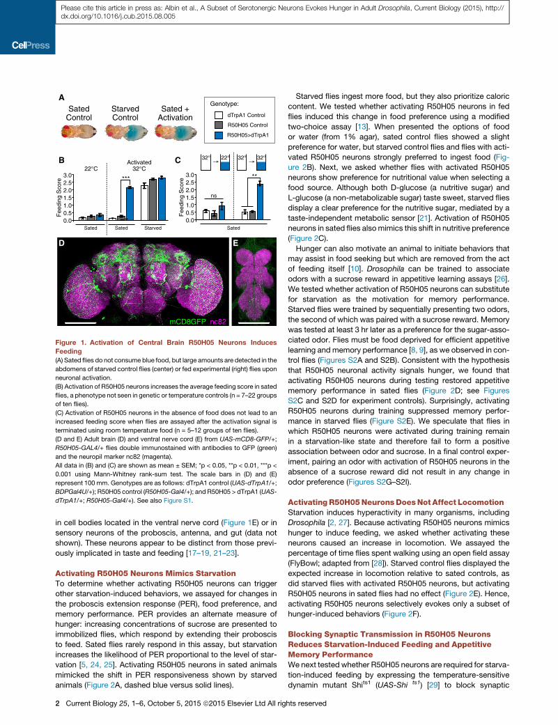
Figure 1. Activation of Central Brain R50H05 Neurons Induces
Feeding
(A) Sated flies do not consume blue food, but large amounts are detected in the
abdomens of starved control flies (center) or fed experimental (right) flies upon
neuronal activation.
(B) Activation of R50H05 neurons increases the average feeding score in sated
flies, a phenotype not seen in genetic or temperature controls (n = 7–22 groups
of ten flies).
(C) Activation of R50H05 neurons in the absence of food does not lead to an
increased feeding score when flies are assayed after the activation signal is
terminated using room temperature food (n = 5–12 groups of ten flies).
(D and E) Adult brain (D) and ventral nerve cord (E) from UAS-mCD8-GFP/+;
R50H05-GAL4/+ flies double immunostained with antibodies to GFP (green)
and the neuropil marker nc82 (magenta).
All data in (B) and (C) are shown as mean ± SEM; *p < 0.05, **p < 0.01, ***p <
0.001 using Mann-Whitney rank-sum test. The scale bars in (D) and (E)
represent 100 mm. Genotypes are as follows: dTrpA1 control (UAS-dTrpA1/+;
BDPGal4U/+); R50H05 control (R50H05-Gal4/+); and R50H05 > dTrpA1 (UAS-
dTrpA1/+; R50H05-Gal4/+). See also Figure S1.
Please cite this article in press as: Albin et al., A Subset of Serotonergic Neurons Evokes Hunger in Adult Drosophila, Current Biology (2015), http://dx.doi.org/10.1016/j.cub.2015.08.005
in cell bodies located in the ventral nerve cord (Figure 1E) or in
sensory neurons of the proboscis, antenna, and gut (data not
shown). These neurons appear to be distinct from those previ-
ously implicated in taste and feeding [17–19, 21–23].
Activating R50H05 Neurons Mimics StarvationTo determine whether activating R50H05 neurons can trigger
other starvation-induced behaviors, we assayed for changes in
the proboscis extension response (PER), food preference, and
memory performance. PER provides an alternate measure of
hunger: increasing concentrations of sucrose are presented to
immobilized flies, which respond by extending their proboscis
to feed. Sated flies rarely respond in this assay, but starvation
increases the likelihood of PER proportional to the level of star-
vation [5, 24, 25]. Activating R50H05 neurons in sated animals
mimicked the shift in PER responsiveness shown by starved
animals (Figure 2A, dashed blue versus solid lines).
2 Current Biology 25, 1–6, October 5, 2015 ª2015 Elsevier Ltd All rig
Starved flies ingest more food, but they also prioritize caloric
content. We tested whether activating R50H05 neurons in fed
flies induced this change in food preference using a modified
two-choice assay [13]. When presented the options of food
or water (from 1% agar), sated control flies showed a slight
preference for water, but starved control flies and flies with acti-
vated R50H05 neurons strongly preferred to ingest food (Fig-
ure 2B). Next, we asked whether flies with activated R50H05
neurons show preference for nutritional value when selecting a
food source. Although both D-glucose (a nutritive sugar) and
L-glucose (a non-metabolizable sugar) taste sweet, starved flies
display a clear preference for the nutritive sugar, mediated by a
taste-independent metabolic sensor [21]. Activation of R50H05
neurons in sated flies alsomimics this shift in nutritive preference
(Figure 2C).
Hunger can also motivate an animal to initiate behaviors that
may assist in food seeking but which are removed from the act
of feeding itself [10]. Drosophila can be trained to associate
odors with a sucrose reward in appetitive learning assays [26].
We tested whether activation of R50H05 neurons can substitute
for starvation as the motivation for memory performance.
Starved flies were trained by sequentially presenting two odors,
the second of which was paired with a sucrose reward. Memory
was tested at least 3 hr later as a preference for the sugar-asso-
ciated odor. Flies must be food deprived for efficient appetitive
learning and memory performance [8, 9], as we observed in con-
trol flies (Figures S2A and S2B). Consistent with the hypothesis
that R50H05 neuronal activity signals hunger, we found that
activating R50H05 neurons during testing restored appetitive
memory performance in sated flies (Figure 2D; see Figures
S2C and S2D for experiment controls). Surprisingly, activating
R50H05 neurons during training suppressed memory perfor-
mance in starved flies (Figure S2E). We speculate that flies in
which R50H05 neurons were activated during training remain
in a starvation-like state and therefore fail to form a positive
association between odor and sucrose. In a final control exper-
iment, pairing an odor with activation of R50H05 neurons in the
absence of a sucrose reward did not result in any change in
odor preference (Figures S2G–S2I).
ActivatingR50H05NeuronsDoesNot Affect LocomotionStarvation induces hyperactivity in many organisms, including
Drosophila [2, 27]. Because activating R50H05 neurons mimics
hunger to induce feeding, we asked whether activating these
neurons caused an increase in locomotion. We assayed the
percentage of time flies spent walking using an open field assay
(FlyBowl; adapted from [28]). Starved control flies displayed the
expected increase in locomotion relative to sated controls, as
did starved flies with activated R50H05 neurons, but activating
R50H05 neurons in sated flies had no effect (Figure 2E). Hence,
activating R50H05 neurons selectively evokes only a subset of
hunger-induced behaviors (Figure 2F).
Blocking Synaptic Transmission in R50H05 NeuronsReduces Starvation-Induced Feeding and AppetitiveMemory PerformanceWe next tested whether R50H05 neurons are required for starva-
tion-induced feeding by expressing the temperature-sensitive
dynamin mutant Shits1 (UAS-Shi ts1) [29] to block synaptic
hts reserved
B 22°C Activated32°C22°C ActivatedC
nsP
refe
renc
e In
dex
1.00.80.60.4
0.00.2
-0.8-0.6-0.4-0.2
StarvedSated StarvedSated
*
L-glucose
D-glucose
ns
***
32°C
Pre
fere
nce
Inde
x
1.00.80.60.4
0.00.2
-0.6-0.4-0.2
StarvedSated StarvedSated
Food
Agar-0.8
ns
ns* ***
Sated Starved
20
30
40
10
0Per
cent
age
time
wal
king
(%
)
E 32°CActivated
***
*
ns
32°CActivated
Sated
****
0
0.1
0.2
0.3
Mem
ory
Inde
x
0.4
-0.1
D F Starvation
R50H05 neurons
non-R50H05neurons
Locomotion FeedingProboscisExtension
AppetitiveMemory
A
Pro
bosc
is E
xten
sion
(%
)
0
25
50
75
100
0 50 100 200Sucrose Concentration (mM)
ns
***
dTrpA1 Control
R50H05 Control
R50H05>dTrpA1
Genotype:Sated Starved
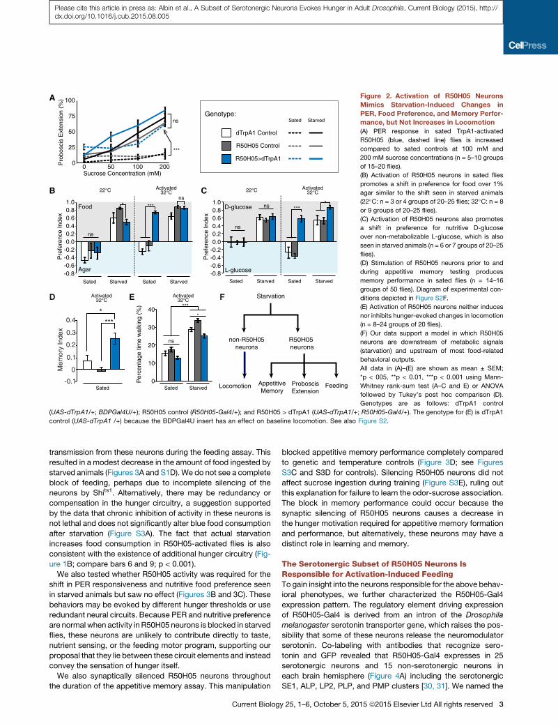
Figure 2. Activation of R50H05 Neurons
Mimics Starvation-Induced Changes in
PER, Food Preference, and Memory Perfor-
mance, but Not Increases in Locomotion
(A) PER response in sated TrpA1-activated
R50H05 (blue, dashed line) flies is increased
compared to sated controls at 100 mM and
200 mM sucrose concentrations (n = 5–10 groups
of 15–20 flies).
(B) Activation of R50H05 neurons in sated flies
promotes a shift in preference for food over 1%
agar similar to the shift seen in starved animals
(22�C: n = 3 or 4 groups of 20–25 flies; 32�C: n = 8
or 9 groups of 20–25 flies).
(C) Activation of R50H05 neurons also promotes
a shift in preference for nutritive D-glucose
over non-metabolizable L-glucose, which is also
seen in starved animals (n = 6 or 7 groups of 20–25
flies).
(D) Stimulation of R50H05 neurons prior to and
during appetitive memory testing produces
memory performance in sated flies (n = 14–16
groups of 50 flies). Diagram of experimental con-
ditions depicted in Figure S2F.
(E) Activation of R50H05 neurons neither induces
nor inhibits hunger-evoked changes in locomotion
(n = 8–24 groups of 20 flies).
(F) Our data support a model in which R50H05
neurons are downstream of metabolic signals
(starvation) and upstream of most food-related
behavioral outputs.
All data in (A)–(E) are shown as mean ± SEM;
*p < 005, **p < 0.01, ***p < 0.001 using Mann-
Whitney rank-sum test (A–C and E) or ANOVA
followed by Tukey’s post hoc comparison (D).
Genotypes are as follows: dTrpA1 control
(UAS-dTrpA1/+; BDPGal4U/+); R50H05 control (R50H05-Gal4/+); and R50H05 > dTrpA1 (UAS-dTrpA1/+; R50H05-Gal4/+). The genotype for (E) is dTrpA1
control (UAS-dTrpA1 /+) because the BDPGal4U insert has an effect on baseline locomotion. See also Figure S2.
Please cite this article in press as: Albin et al., A Subset of Serotonergic Neurons Evokes Hunger in Adult Drosophila, Current Biology (2015), http://dx.doi.org/10.1016/j.cub.2015.08.005
transmission from these neurons during the feeding assay. This
resulted in a modest decrease in the amount of food ingested by
starved animals (Figures 3A and S1D). We do not see a complete
block of feeding, perhaps due to incomplete silencing of the
neurons by Shits1. Alternatively, there may be redundancy or
compensation in the hunger circuitry, a suggestion supported
by the data that chronic inhibition of activity in these neurons is
not lethal and does not significantly alter blue food consumption
after starvation (Figure S3A). The fact that actual starvation
increases food consumption in R50H05-activated flies is also
consistent with the existence of additional hunger circuitry (Fig-
ure 1B; compare bars 6 and 9; p < 0.001).
We also tested whether R50H05 activity was required for the
shift in PER responsiveness and nutritive food preference seen
in starved animals but saw no effect (Figures 3B and 3C). These
behaviors may be evoked by different hunger thresholds or use
redundant neural circuits. Because PER and nutritive preference
are normal when activity in R50H05 neurons is blocked in starved
flies, these neurons are unlikely to contribute directly to taste,
nutrient sensing, or the feeding motor program, supporting our
proposal that they lie between these circuit elements and instead
convey the sensation of hunger itself.
We also synaptically silenced R50H05 neurons throughout
the duration of the appetitive memory assay. This manipulation
Current Biolog
blocked appetitive memory performance completely compared
to genetic and temperature controls (Figure 3D; see Figures
S3C and S3D for controls). Silencing R50H05 neurons did not
affect sucrose ingestion during training (Figure S3E), ruling out
this explanation for failure to learn the odor-sucrose association.
The block in memory performance could occur because the
synaptic silencing of R50H05 neurons causes a decrease in
the hunger motivation required for appetitive memory formation
and performance, but alternatively, these neurons may have a
distinct role in learning and memory.
The Serotonergic Subset of R50H05 Neurons IsResponsible for Activation-Induced FeedingTo gain insight into the neurons responsible for the above behav-
ioral phenotypes, we further characterized the R50H05-Gal4
expression pattern. The regulatory element driving expression
of R50H05-Gal4 is derived from an intron of the Drosophila
melanogaster serotonin transporter gene, which raises the pos-
sibility that some of these neurons release the neuromodulator
serotonin. Co-labeling with antibodies that recognize sero-
tonin and GFP revealed that R50H05-Gal4 expresses in 25
serotonergic neurons and 15 non-serotonergic neurons in
each brain hemisphere (Figure 4A) including the serotonergic
SE1, ALP, LP2, PLP, and PMP clusters [30, 31]. We named the
y 25, 1–6, October 5, 2015 ª2015 Elsevier Ltd All rights reserved 3
A
B
C
D
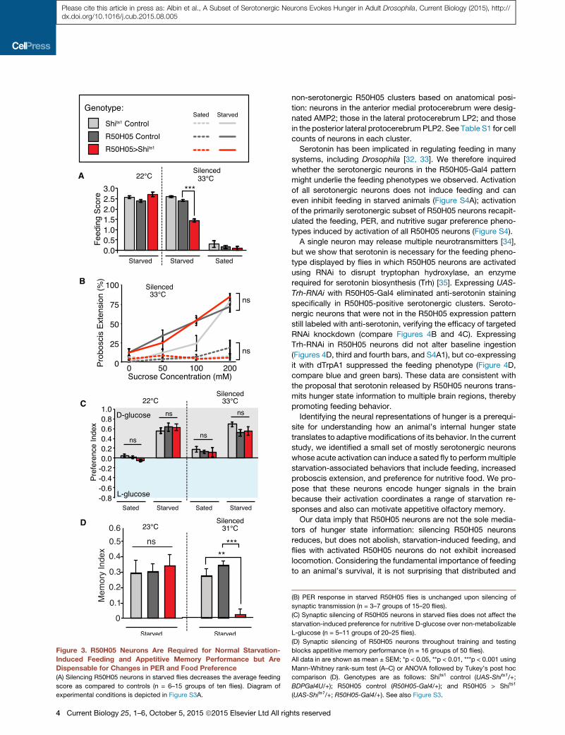
Figure 3. R50H05 Neurons Are Required for Normal Starvation-
Induced Feeding and Appetitive Memory Performance but Are
Dispensable for Changes in PER and Food Preference
(A) Silencing R50H05 neurons in starved flies decreases the average feeding
score as compared to controls (n = 6–15 groups of ten flies). Diagram of
experimental conditions is depicted in Figure S3A.
4 Current Biology 25, 1–6, October 5, 2015 ª2015 Elsevier Ltd All rig
Please cite this article in press as: Albin et al., A Subset of Serotonergic Neurons Evokes Hunger in Adult Drosophila, Current Biology (2015), http://dx.doi.org/10.1016/j.cub.2015.08.005
non-serotonergic R50H05 clusters based on anatomical posi-
tion: neurons in the anterior medial protocerebrum were desig-
nated AMP2; those in the lateral protocerebrum LP2; and those
in the posterior lateral protocerebrumPLP2. See Table S1 for cell
counts of neurons in each cluster.
Serotonin has been implicated in regulating feeding in many
systems, including Drosophila [32, 33]. We therefore inquired
whether the serotonergic neurons in the R50H05-Gal4 pattern
might underlie the feeding phenotypes we observed. Activation
of all serotonergic neurons does not induce feeding and can
even inhibit feeding in starved animals (Figure S4A); activation
of the primarily serotonergic subset of R50H05 neurons recapit-
ulated the feeding, PER, and nutritive sugar preference pheno-
types induced by activation of all R50H05 neurons (Figure S4).
A single neuron may release multiple neurotransmitters [34],
but we show that serotonin is necessary for the feeding pheno-
type displayed by flies in which R50H05 neurons are activated
using RNAi to disrupt tryptophan hydroxylase, an enzyme
required for serotonin biosynthesis (Trh) [35]. Expressing UAS-
Trh-RNAi with R50H05-Gal4 eliminated anti-serotonin staining
specifically in R50H05-positive serotonergic clusters. Seroto-
nergic neurons that were not in the R50H05 expression pattern
still labeled with anti-serotonin, verifying the efficacy of targeted
RNAi knockdown (compare Figures 4B and 4C). Expressing
Trh-RNAi in R50H05 neurons did not alter baseline ingestion
(Figures 4D, third and fourth bars, and S4A1), but co-expressing
it with dTrpA1 suppressed the feeding phenotype (Figure 4D,
compare blue and green bars). These data are consistent with
the proposal that serotonin released by R50H05 neurons trans-
mits hunger state information to multiple brain regions, thereby
promoting feeding behavior.
Identifying the neural representations of hunger is a prerequi-
site for understanding how an animal’s internal hunger state
translates to adaptivemodifications of its behavior. In the current
study, we identified a small set of mostly serotonergic neurons
whose acute activation can induce a sated fly to performmultiple
starvation-associated behaviors that include feeding, increased
proboscis extension, and preference for nutritive food. We pro-
pose that these neurons encode hunger signals in the brain
because their activation coordinates a range of starvation re-
sponses and also can motivate appetitive olfactory memory.
Our data imply that R50H05 neurons are not the sole media-
tors of hunger state information: silencing R50H05 neurons
reduces, but does not abolish, starvation-induced feeding, and
flies with activated R50H05 neurons do not exhibit increased
locomotion. Considering the fundamental importance of feeding
to an animal’s survival, it is not surprising that distributed and
(B) PER response in starved R50H05 flies is unchanged upon silencing of
synaptic transmission (n = 3–7 groups of 15–20 flies).
(C) Synaptic silencing of R50H05 neurons in starved flies does not affect the
starvation-induced preference for nutritive D-glucose over non-metabolizable
L-glucose (n = 5–11 groups of 20–25 flies).
(D) Synaptic silencing of R50H05 neurons throughout training and testing
blocks appetitive memory performance (n = 16 groups of 50 flies).
All data in are shown as mean ± SEM; *p < 0.05, **p < 0.01, ***p < 0.001 using
Mann-Whitney rank-sum test (A–C) or ANOVA followed by Tukey’s post hoc
comparison (D). Genotypes are as follows: Shits1 control (UAS-Shits1/+;
BDPGal4U/+); R50H05 control (R50H05-Gal4/+); and R50H05 > Shits1
(UAS-Shits1/+; R50H05-Gal4/+). See also Figure S3.
hts reserved
A1 B D
CA2
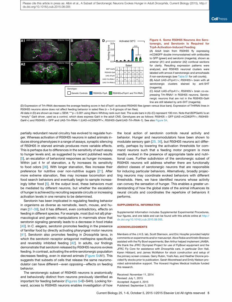
Figure 4. Some R50H05 Neurons Are Sero-
tonergic, and Serotonin Is Required for
TrpA-Activation-Induced Feeding
(A) Adult brain from R50H05 fly expressing
mCD8GFP double immunostained with antibodies
to GFP (green) and serotonin (magenta), shown as
anterior (A1) and posterior (A2) confocal sections
for clarity. Resulting expression patterns were
analyzed, and R50H05 neuronal clusters were
labeled with arrows if serotonergic and arrowheads
if non-serotonergic (see Table S1 for cell counts).
(B) Adult UAS-dTrpA1/+; R50H05/+ brain with all
serotonergic clusters stained by anti-5HT
(magenta).
(C) Adult UAS-dTrpA1/+; R50H05/+ brain co-ex-
pressing Trh-RNAi1 in R50H05 neurons. Seroto-
nergic neurons that are not in the R50H05-Gal4
line are still labeled by anti-5HT (magenta).
(D) Expression of Trh-RNAi decreases the average feeding score in fed dTrpA1-activated R50H05 flies (green versus blue bars). Expression of TrhRNAi lines in
R50H05 neurons alone does not affect feeding behavior in sated flies (n = 5–9 groups of ten flies).
All data in (D) are shown as mean ± SEM; ***p < 0.001 using Mann-Whitney rank-sum test. The scale bars in (A)–(C) represent 100 mm. Note that BDPGal4U is an
‘‘empty’’ Gal4 driver, used as a control, which does express Gal4 in the adult CNS. Genotypes are as follows: R50H05 > GFP (UAS-mCD8GFP/+; R50H05-
Gal4/+) and R50H05 > GFP and UAS-Trh-RNAi-1 (UAS-mCD8GFP/+; R50H05-Gal4/UAS-Trh-RNAi-1). See also Figure S4.
Please cite this article in press as: Albin et al., A Subset of Serotonergic Neurons Evokes Hunger in Adult Drosophila, Current Biology (2015), http://dx.doi.org/10.1016/j.cub.2015.08.005
partially redundant neural circuitry has evolved to regulate hun-
ger. Whereas activation of R50H05 neurons in sated animals in-
duces strong phenotypes in a range of assays, synaptic silencing
of R50H05 in starved animals produces more variable effects.
This is perhaps due to differences in the sensitivity of each assay
to hunger levels and, as suggested by recent published results
[5], an escalation of behavioral responses as hunger increases.
Within just 4 hr of starvation, a fly increases its sensitivity
to food odors [36]. With longer starvation, flies increase their
preference for nutritive over non-nutritive sugars [21]. After
more extreme starvation, flies may increase locomotion and
food search behaviors and eventually begin to sample increas-
ingly bitter food [5]. At the output level, these behaviors must
be mediated by different neurons, but whether the escalation
of hunger is achieved by recruiting separate circuits or increasing
activation levels in one remains to be determined.
Serotonin has been implicated in regulating feeding behavior
in organisms as diverse as nematode, leech, mouse, and hu-
man [37–39], but it has different, even contradictory, effects on
feeding in different species. For example, most (but not all) phar-
macological and genetic manipulations in mammals show that
serotonin signaling generally leads to a decrease in food intake
[40]. In C. elegans, serotonin promotes feeding in the presence
of familiar food by directly activating pharyngeal motor neurons
[41]. Serotonin also promotes feeding in Drosophila larva, in
which the serotonin receptor antagonist metitepine specifically
and reversibly inhibited feeding [42]. In adults, our findings
demonstrate that serotonin released by R50H05 neurons evokes
feeding. In contrast, activating the entire serotonergic population
decreases feeding, even in starved animals (Figure S4A1). This
suggests that subsets of cells that release the same neuromo-
dulator can have different—even opposing—effects on feeding
behavior.
The serotonergic subset of R50H05 neurons is anatomically
and behaviorally distinct from neurons previously identified as
important for feeding behavior (Figures S4D–S4H). Looking for-
ward, access to R50H05 neurons enables investigation of how
Current Biolog
the local action of serotonin controls neural activity and
behavior. Hunger and neuromodulators have been shown to
modulate sensory gain [25, 36], but R50H05 neurons act differ-
ently, perhaps by lowering the activation thresholds for com-
mand neurons such that a feeding motor program is more
readily evoked in the presence of appropriate taste and nutri-
tional cues. Further subdivision of the serotonergic subset of
R50H05 neurons will address whether there are functionally
distinct classes of serotonergic neurons that are responsible
for inducing particular behaviors. Alternatively, broadly projec-
ting neurons may coordinate evoked behaviors with different
thresholds. Here, we have identified neurons whose activity
can convey the sensation of hunger. This enables a greater un-
derstanding of how the global state of the animal influences its
neural circuits and coordinates the repertoire of behaviors it
performs.
SUPPLEMENTAL INFORMATION
Supplemental Information includes Supplemental Experimental Procedures,
four figures, and one table and can be found with this article online at http://
dx.doi.org/10.1016/j.cub.2015.08.005.
ACKNOWLEDGMENTS
Members of the J.H.S. lab, Scott Sternson, and Eric Hoopfer provided helpful
comments on experiments and/ormanuscript. Alice Robie andKristin Branson
assisted with the Fly Bowl experiments; Ben Arthur helped implement JAABA.
We thank the JFRC Olympiad Project for use of FlyBowl equipment and the
JFRC Fly Core for assistance with Drosophila care, in particular Don Hall,
Karen Hibbard, and James McMahon for stock construction and setup of
the primary screen crosses. Gerry Rubin, Yoshi Aso, and Heather Dionne pro-
vided fly stocks prior to publication. Sarah Moorehead and Emily Nielson pro-
vided administrative support. The Howard Hughes Medical Institute funded
this research.
Received: November 11, 2014
Revised: July 1, 2015
Accepted: August 4, 2015
Published: September 3, 2015
y 25, 1–6, October 5, 2015 ª2015 Elsevier Ltd All rights reserved 5
Please cite this article in press as: Albin et al., A Subset of Serotonergic Neurons Evokes Hunger in Adult Drosophila, Current Biology (2015), http://dx.doi.org/10.1016/j.cub.2015.08.005
REFERENCES
1. Dethier, V.G. (1976). The Hungry Fly: A Physiological Study of the Behavior
Associated with Feeding (Harvard University Press).
2. Lee, G., and Park, J.H. (2004). Hemolymph sugar homeostasis and
starvation-induced hyperactivity affected by genetic manipulations of
the adipokinetic hormone-encoding gene in Drosophila melanogaster.
Genetics 167, 311–323.
3. Sengupta, P. (2013). The belly rules the nose: feeding state-depen-
dent modulation of peripheral chemosensory responses. Curr. Opin.
Neurobiol. 23, 68–75.
4. Su, C.Y., and Wang, J.W. (2014). Modulation of neural circuits: how stim-
ulus context shapes innate behavior in Drosophila. Curr. Opin. Neurobiol.
29, 9–16.
5. Inagaki, H.K., Panse, K.M., and Anderson, D.J. (2014). Independent, recip-
rocal neuromodulatory control of sweet and bitter taste sensitivity during
starvation in Drosophila. Neuron 84, 806–820.
6. Pager, J., Giachetti, I., Holley, A., and Le Magnen, J. (1972). A selective
control of olfactory bulb electrical activity in relation to food deprivation
and satiety in rats. Physiol. Behav. 9, 573–579.
7. Pirke, K.M., Broocks, A., Wilckens, T., Marquard, R., and Schweiger, U.
(1993). Starvation-induced hyperactivity in the rat: the role of endocrine
and neurotransmitter changes. Neurosci. Biobehav. Rev. 17, 287–294.
8. Krashes, M.J., andWaddell, S. (2008). Rapid consolidation to a radish and
protein synthesis-dependent long-term memory after single-session
appetitive olfactory conditioning in Drosophila. J. Neurosci. 28, 3103–
3113.
9. Krashes, M.J., DasGupta, S., Vreede, A., White, B., Armstrong, J.D., and
Waddell, S. (2009). A neural circuit mechanism integrating motivational
state with memory expression in Drosophila. Cell 139, 416–427.
10. Sternson, S.M., Nicholas Betley, J., and Cao, Z.F. (2013). Neural circuits
and motivational processes for hunger. Curr. Opin. Neurobiol. 23,
353–360.
11. Al-Anzi, B., Armand, E., Nagamei, P., Olszewski, M., Sapin, V., Waters, C.,
Zinn, K., Wyman, R.J., and Benzer, S. (2010). The leucokinin pathway and
its neurons regulate meal size in Drosophila. Curr. Biol. 20, 969–978.
12. Lee, K.S., You, K.H., Choo, J.K., Han, Y.M., and Yu, K. (2004). Drosophila
short neuropeptide F regulates food intake and body size. J. Biol. Chem.
279, 50781–50789.
13. Tanimura, T., Isono, K., Takamura, T., and Shimada, I. (1982). Genetic
dimorphism in the taste sensitivity to trehalose inDrosophila mela-
nogaster. J. Comp. Physiol. 147, 433–437.
14. Edgecomb, R.S., Harth, C.E., and Schneiderman, A.M. (1994). Regulation
of feeding behavior in adult Drosophila melanogaster varies with feeding
regime and nutritional state. J. Exp. Biol. 197, 215–235.
15. Hamada, F.N., Rosenzweig, M., Kang, K., Pulver, S.R., Ghezzi, A., Jegla,
T.J., and Garrity, P.A. (2008). An internal thermal sensor controlling
temperature preference in Drosophila. Nature 454, 217–220.
16. Brand, A.H., and Perrimon, N. (1993). Targeted gene expression as a
means of altering cell fates and generating dominant phenotypes.
Development 118, 401–415.
17. Melcher, C., and Pankratz, M.J. (2005). Candidate gustatory interneurons
modulating feeding behavior in the Drosophila brain. PLoS Biol. 3, e305.
18. Wu, Q., Zhao, Z., and Shen, P. (2005). Regulation of aversion to noxious
food by Drosophila neuropeptide Y- and insulin-like systems. Nat.
Neurosci. 8, 1350–1355.
19. Zhao, X.L., and Campos, A.R. (2012). Insulin signalling in mushroom body
neurons regulates feeding behaviour in Drosophila larvae. J. Exp. Biol.
215, 2696–2702.
20. Jenett, A., Rubin, G.M., Ngo, T.T., Shepherd, D., Murphy, C., Dionne, H.,
Pfeiffer, B.D., Cavallaro, A., Hall, D., Jeter, J., et al. (2012). A GAL4-driver
line resource for Drosophila neurobiology. Cell Rep. 2, 991–1001.
6 Current Biology 25, 1–6, October 5, 2015 ª2015 Elsevier Ltd All rig
21. Dus,M., Ai, M., and Suh, G.S. (2013). Taste-independent nutrient selection
is mediated by a brain-specific Na+ /solute co-transporter in Drosophila.
Nat. Neurosci. 16, 526–528.
22. Flood, T.F., Iguchi, S., Gorczyca, M., White, B., Ito, K., and Yoshihara, M.
(2013). A single pair of interneurons commands the Drosophila feeding
motor program. Nature 499, 83–87.
23. Cobb, M., Scott, K., and Pankratz, M. (2009). Gustation in Drosophila
melanogaster. SEB Exp. Biol. Ser. 63, 1–38.
24. Scheiner, R., Sokolowski, M.B., and Erber, J. (2004). Activity of cGMP-
dependent protein kinase (PKG) affects sucrose responsiveness and
habituation in Drosophila melanogaster. Learn. Mem. 11, 303–311.
25. Inagaki, H.K., Ben-Tabou de-Leon, S., Wong, A.M., Jagadish, S.,
Ishimoto, H., Barnea, G., Kitamoto, T., Axel, R., and Anderson, D.J.
(2012). Visualizing neuromodulation in vivo: TANGO-mapping of dopa-
mine signaling reveals appetite control of sugar sensing. Cell 148,
583–595.
26. Tempel, B.L., Bonini, N., Dawson, D.R., and Quinn, W.G. (1983). Reward
learning in normal and mutant Drosophila. Proc. Natl. Acad. Sci. USA
80, 1482–1486.
27. Knoppien, P., van der Pers, J.N.C., and van Delden, W. (2000).
Quantification of locomotion and the effect of food deprivation on locomo-
tor activity in Drosophila. J. Insect Behav. 13, 27–43.
28. Simon, J.C., and Dickinson, M.H. (2010). A new chamber for studying the
behavior of Drosophila. PLoS ONE 5, e8793.
29. Kitamoto, T. (2001). Conditional modification of behavior in Drosophila by
targeted expression of a temperature-sensitive shibire allele in defined
neurons. J. Neurobiol. 47, 81–92.
30. Alekseyenko, O.V., Lee, C., and Kravitz, E.A. (2010). Targeted manipula-
tion of serotonergic neurotransmission affects the escalation of aggres-
sion in adult male Drosophila melanogaster. PLoS ONE 5, e10806.
31. Valles, A.M., and White, K. (1986). Development of serotonin-containing
neurons in Drosophila mutants unable to synthesize serotonin.
J. Neurosci. 6, 1482–1491.
32. Vargas, M.A., Luo, N., Yamaguchi, A., and Kapahi, P. (2010). A role for S6
kinase and serotonin in postmating dietary switch and balance of nutrients
in D. melanogaster. Curr. Biol. 20, 1006–1011.
33. Luo, J., Becnel, J., Nichols, C.D., and Nassel, D.R. (2012). Insulin-produc-
ing cells in the brain of adult Drosophila are regulated by the serotonin
5-HT1A receptor. Cell. Mol. Life Sci. 69, 471–484.
34. Gutierrez, R. (2009). Co-existence and Co-release of Classical
Neurotransmitters: Ex Uno Plures (Springer-Verlag), pp. 15–22.
35. Coleman, C.M., and Neckameyer, W.S. (2005). Serotonin synthesis by two
distinct enzymes in Drosophila melanogaster. Arch. Insect Biochem.
Physiol. 59, 12–31.
36. Root, C.M., Ko, K.I., Jafari, A., and Wang, J.W. (2011). Presynaptic facili-
tation by neuropeptide signaling mediates odor-driven food search. Cell
145, 133–144.
37. Song, B.M., and Avery, L. (2012). Serotonin activates overall feeding by
activating two separate neural pathways in Caenorhabditis elegans.
J. Neurosci. 32, 1920–1931.
38. Gaudry, Q., and Kristan, W.B., Jr. (2012). Decision points: the factors influ-
encing the decision to feed in the medicinal leech. Front. Neurosci. 6, 101.
39. Donovan, M.H., and Tecott, L.H. (2013). Serotonin and the regulation of
mammalian energy balance. Front. Neurosci. 7, 36.
40. Lam, D.D., Garfield, A.S., Marston, O.J., Shaw, J., and Heisler, L.K. (2010).
Brain serotonin system in the coordination of food intake and body weight.
Pharmacol. Biochem. Behav. 97, 84–91.
41. Song, B.M., Faumont, S., Lockery, S., and Avery, L. (2013). Recognition of
familiar food activates feeding via an endocrine serotonin signal in
Caenorhabditis elegans. eLife 2, e00329.
42. Gasque, G., Conway, S., Huang, J., Rao, Y., and Vosshall, L.B. (2013).
Small molecule drug screening inDrosophila identifies the 5HT2A receptor
as a feeding modulation target. Sci. Rep. 3, srep02120.
hts reserved