Embed Size (px)
Citation preview

PEABODY MUSEUM OF NATURAL HISTORY YALE UNIVERSITY
BULLETIN 20
A Systematic Study of the Demospongiae
of Port Royal, Jamaica
BY
GEORGE JOHN HECHTEL
Department of Biology State University of New York
Stony Brook, L. I., N. Y.
NEW HAVEN, CONNECTICUT 1965

Bulletins published by the Peabody Museum of Natural History at Yale University embody research carried out under the auspices of the Museum. The issues are numbered consecutively as independent monographs and appear at irregular intervals. Shorter papers are published at frequent intervals in the Peabody Museum Postilla Series.
EDITORIAL BOARD: Elwyn L. Simons, Chairman Charles L. Remington N. Philip Ashmole
EDITOR: Ellen T. Drake
Communications concerning purchase or exchange of publications should be addressed to the Editor, Peabody Museum of Natural History, Yale University, New Haven, Connecticut 06520, U.S.A.
Issued March 1, 1965

A SYSTEMATIC STUDY OF THE DEMOSPONGIAE OF PORT ROYAL, JAMAICA

Printed in the United States of America

CONTENTS
LIST OF ILLUSTRATIONS
ABSTRACT
INTRODUCTION
ACKNOWLEDGMENTS
METHODS
HABITATS
PREVIOUS JAMAICAN RECORDS
CLASSIFICATION
TAXONOMIC CRITERIA
SYSTEMATICS
FAMILY Spongiidae Dysideidae Aplysillidae Haliclonidae Desmacidonidae Adociidae Callyspongiidae Tedaniidae Microcionidae Mycalidae Halichondriidae Spirastrellidae Suberitidae Clionidae Placospongiidae Tethyidae Geodiidae Chondrillidae Plakinidae
DISTRIBUTION BY HABITAT
FIELD KEY
ZOOGEOGRAPHICAL NOTES
SUMMARY
LITERATURE CITED
INDEX
PLATES I-VIII
vi
1
2
2
2
3
4
4
5
5 8
14 17 18 21 26 31 37 41 45 52 54 59 60 62 65 68 74 76 77 80 83 84
85
90
95
V

ILLUSTRATIONS
TEXT-FIGURES
1. Spicules of Haliclona hogarthi, p. 20. 2. Spicules of Adocia implexiformis, p. 27. 3. Spicules of Adocia albifragilis, p. 28. 4. Spicules of Pellina coeliformis, p. 29. 5. Spicules of Sigmadocia caerulea, p. 30. 6. Spicules of Callyspongia pallida, p. 36. 7. Spicules of Microciona microchela, p. 41. 8. Spicules of Microciona rarispinosa, p. 43. 9. Spicules of Thalyseurypon conulosa, p. 44.
10. Spicules of Ulosa hispida, p. 51. 11. Spicules of Halichondria magniconulosa, p. 53. 12. Spicules of Diplastrella megastellata, p. 58. 13. Spicules of Geodia papyracea, p. 71. 14. Spicules of Erylus ministrongylus, p. 73. 15. Spicules of Corticium tetralophum, p. 77.
PLATES
I.
II.
III .
IV.
V.
VI.
VII.
VIII.
Fig. 1. Figs. 2,3. Fig. 4, Fig. 5 Fig. Fig. 2 Fig. 3, Fig. 4 Fig. 1 Figs. 2,3 Fig. 4, Fig. 5 Fig. Fig. 2 Fig. 1 Fig. 2, Fig. 3 Fig. 4 Fig. Fig. 2, Fig. 1 Fig. 2 Fig. 3 Figs. 4,5. Figs. 1,2. Fig. 3.
Verongia fistularis (Pallas). Haliclona erina de Laubenfels. Haliclona doria de Laubenfels. Oligoceras hemorrhages de Laubenfels. Haliclona hogarthi n. sp. Adocia implexiformis n. sp. Gelliodes areolata (Wilson). Desmapsamma anchorata (Carter). Pellina coeliformis n. sp. Adocia carbonaria (Lamarck). Sigmadocia caerulea n. sp. Callyspongia pallida n. sp. Callyspongia plicifera (Lamarck). Callyspongia fallax Duchassaing and Michelotti. Halichondria magniconulosa n. sp. Halichondria melanadocia de Laubenfels. Zygomycale parishii (Bowerbank). Tedania ignis (Duchassaing and Michelotti). Thalyseurypon conulosa n. sp. Mycale laevis (Carter). Placospongia carinata (Bowerbank). Diplastrella megastellata n. sp. Terpios zeteki (de Laubenfels). Anthosigmella varians (Duchassaing and Michelotti). Geodia papyracea n. sp. Erylus ministrongylus n. sp.
VI

YALE UNIVERSITY PEABODY MUSEUM OF NATURAL HISTORY BULLETIN NO. 20, 104 p., 8 PLS., 15 TEXT-FIGS., 1965.
A SYSTEMATIC STUDY OF T H E DEMOSPONGIAE OF PORT ROYAL, JAMAICA
By GEORGE JOHN HECHTEL
ABSTRACT
A systematic study of the shallow water sponges of Port Royal has added 54 species to the Jamaican faunal list, of which 16 are new. One new genus, Neofibularia, is proposed for the preoccupied Fibularia Carter. The new species are Darwinella rosacea, Haliclona hogarthi, Adocia implexiformis, Adocia albifragilis, Pellina coeliformis, Sigmadocia caerulea, Callyspongia pallida, Halichondria magniconulosa, Microciona microchela, Microciona rarispinosa, Thalyseurypon conulosa, Ulosa hispida, Diplastrella megastellata, Geodia (Cydonium) papyracea, Erylus minis-trongylus, and Corticium tetralophum.
The use of field characteristics on the species level is discussed. The classification of de Laubenfels (1936a) is utilized, with several modifications. The Adociidae are transferred to the Haplosclerida, and Lundbeck's family Mycalidae is used in place of de Laubenfels' Amphilectidae. A review of the taxonomy and distribution of each species is presented. The zoogeographical affinities of the Jamaican sponge fauna are noted.
1

A SYSTEMATIC STUDY OF THE DEMOSPONGIAE OF PORT ROYAL, JAMAICA*
GEORGE JOHN HECHTEL
INTRODUCTION
The West Indian sponge fauna has not been intensively studied. Detailed regional surveys were not begun until the twentieth century, when Wilson (1902a) described a collection of Puerto Rican sponges. Arndt (1927) has studied the fauna of Curacao. In a series of papers (1936a, 1936b, 1949, 1950a, 1953a) de Laubenfels discussed the shallow water sponges of localities on the periphery of the West Indies (the Dry Tortugas, in the Florida Keys; Panama; Bimini in the Bahamas; Bermuda; and the west coast of Florida, respectively.) The fauna of the Antilles is still poorly known.
Jamaica was chosen for study as one of the Greater Antilles. The University College of the West Indies maintains a marine station on the Jamaican south coast. The present report is a systematic study of the Demospongiae growing in less than twenty feet of water near the station.
This study was submitted in 1962 to the Faculty of the Graduate School of Yale University as a dissertation for the degree of Doctor of Philosophy.
ACKNOWLEDGMENTS
I wish to express my sincere appreciation to Dr. Willard D. Hartman, who introduced me to the study of sponges. His encouragement, advice and criticisms have been invaluable. An initial trip to Jamaica in the summer of 1959 was made possible by the Department of Zoology and the Peabody Museum of Yale University. Extensive field work was made possible by an assistantship at the University College of the West Indies during the academic year 1959-60 and 1960-61. Professor David M. Steven of the Zoology Department at the University College of the West Indies generously provided laboratory facilities at the University and its marine station. Dr. Ivan Goodbody and Dr. Thomas Goreau, also of the University, contributed valuable specimens. Mr. John Howard and Dr. Patricia R. Bergquist at the Peabody Museum of Yale University gave invaluable aid in the preparation of the plates. Finally, I am deeply grateful to my wife, Florence M. Hechtel, for her aid and encouragement.
METHODS
Most specimens were collected by hand, after wading or diving with snorkel and face mask. Several specimens were collected in deeper water by Dr. T. F. Goreau, using an aqua-lung. Field characteristics, such as color, consistency and oscular size, were noted at the time of collection. The specimens were preserved in two changes of 75 per cent alcohol.
Spicule preparations were obtained by boiling small samples in concentrated nitric acid, within a small Erlenmeyer flask. The acid was removed by a process
* Published with the aid of a National Science Foundation Publication Grant, No. GN257.
2

INTRODUCTION 3
of washing, centrifuging, and decanting. Water and then alcohol were used in washing. The spicules were then suspended in alcohol and poured onto slides for drying and mounting.
Dermal structure was studied from thin strips of the surface, removed by a razor blade or forceps. Internal morphology was examined in hand sections cut from paraffin-embedded pieces of the specimens. Sections were taken tangentially and at right angles to the surface. Longitudinal sections were made of elongate specimens. Most preparations were stained with Poinceau red or light green. Spicules are conspicuous in such sections, and spongin becomes somewhat more deeply stained than the surrounding flesh.
HABITATS
Port Royal is a fishing village at the tip of the Palisadoes Peninsula. The latter forms the southern shore of Kingston Harbor. The northern edge of the peninsula is fringed by mangrove swamps which begin about one mile northeast of the marine station. A chain of reefs and islands (the Kingston Cays) lies offshore from Port Royal. The geography of the area is detailed in British Admiralty maps 454 and 456. A variety of habitats could easily be reached by small boat. The principal collecting areas were:
Habitat A—Port Royal Docks: Sponges were a conspicuous element of the dock and seawall community. The water was 8-10 feet deep at the base of the seawall. Many specimens were collected from the dock of the old university marine station, located just beyond the northeastern wall of Port Royal. A second collecting point was a seawall near the tide guage of the Port Royal police post on Port Royal Point.
Habitat B—Port Royal Harbor: Specimens were collected in 15-20 feet of water, just offshore from Port Royal, by Dr. T. F. Goreau using an aqua-lung. They were growing on coral masses.
Habitat C—Rasta's wreck: A shallow, mangrove-fringed inlet opens into Port Royal harbor between the naval yard and Gallow's Point. The inlet is located at 17° 56'30" N. Lat., 76° 50' W. Long. A wooden hulk, known locally as Rasta's wreck, was submerged in 4-8 feet of water at the eastern end of the inlet. Sponges covered much of its surface.
Habitat D—Port Royal turtle grass bed: Turtle grass grew over much of the muddy bottom of the inlet near Rasta's wreck. The water was 3-6 feet in depth. A few species were growing on or among the turtle grass.
Habitat E—Mangroves bordering inlet: The well-illuminated water was 1-2 feet deep. Many mangrove roots were covered with sponges.
Habitat F—Main boat channel: A rowboat channel, averaging 10 feet in width and 2-3 feet in depth, running from the northeastern corner of the inlet toward Kingston harbor. It was shaded by the tops of mangroves and had a steady current. Sponges were abundant on mangrove roots along the channel.
Habitat G—Pigeon House Pile: Several species were dredged from a mussel bank near Pigeon House Pile, in Kingston harbor, at 17° 57'30" N. Lat., 76° 49'30" W. Long. The sponges were growing in 10-15 feet of water.
Habitat H—Shallow water at the cays: Sponges were collected under rocks and ledges in 1-6 feet of water. Study was concentrated on Drunkenman's Cay, and to a lesser extent, Maiden Cay. Drunkenman's Cay is located at 17° 54' N. Lat., 76° 30'36" W. Long. Maiden Cay is at 17° 54'30" N. Lat., 76° 4 8 W W. Long.

4 A SYSTEMATIC STUDY OF THE DEMOSPONGIAE OF JAMAICA
Habitat I—Deeper water near the cays: Sponges were common on coral masses and gorgonian colonies in 10-15 feet of water. Most specimens were collected in an area between Lime Cay (17° 55' N. Lat., 76° 49' W. Long) and Maiden Cay.
Habitat J—Offshore turtle grass beds: Several areas of sand and turtle grass were located near Port Royal. Study was concentrated on a small patch in several feet of water on the leeward side of Drunkenman's Cay.
PREVIOUS JAMAICAN RECORDS
Twelve published reports of Jamaican sponges, referring to ten species, have been noted in the course of the present study. The earliest record is that of Higgin (1877). His Halichondria birotulata (transferred to Iotrochota by Ridley, 1884) was described from Venezuela, the Bahamas, and Kingston Bay, Jamaica. De Laubenfels (1932a, p. 40) examined Jamaican specimens (those of Higgin?) in the British Museum (Natural History) collections. The species is abundant at Port Royal.
Suberites angulospiculatus was described from Jamaica by Carter (1879a, p. 346). Its peculiar spicules are somewhat similar in form to those of Plakortis, but are much larger. De Laubenfels (1936a, p. 162) attributed Dry Tortugas specimens to Carter's species, which he transferred to Epipolasis. De Laubenfels' sponges, with oxeas and strongyloxeas, are not even congeneric with Suberites angulospiculatus. Carter's species was not found in the present study, and its taxonomic position remains obscure.
Suberites coronarius Carter (1882, p. 352) was described from Honduras, the Bahamas, and Jamaica. The species was transferred to Anthosigmella by Topsent (1918, p. 557) and placed into synonymy with A. varians by de Laubenfels (1936a, p. 13). Several specimens were collected in the present study.
Spinosella maxima Dendy (1887d, p. 506; 1890, p. 365) was described from Nassau in the Bahamas. A Jamaican variety was characterized by the presence of vestigial spicules. Dendy's species, with its variety, is almost certainly con-specific with Callyspongia plicvfera, which is common near the Kingston Cays.
Lendenfeld included three Jamaican records in his 1889 monograph of the Keratosa. The specimen attributed to Hircinia variabilis Schmidt was probably an example of Ircinia fasciculata. His record of Aplysina flagelliformis Carter (1886) probably refers to Verongia longissima. Both species occur at Port Royal. His Aplysina spengelii, reported from Jamaica and Ceylon, is probably a synonym of Verongia lacunosa, as suggested by Topsent (1932). It was not found at Port Royal.
Spirastrella andrewsii George and Wilson (1919, p. 135) was described from North Carolina and Jamaica. De Laubenfels (1932b, p. 50) has shown that the species is conspecific with Spheciospongia vesparia. A specimen was collected in the present survey.
Uliczka (1929, p. 38) described Cinachyra rhizophyta from Kingston, Jamaica. No further specimens have been found.
Geodia media Bowerbank was reported by Uliczka (1929, p. 38) from Barbados, Tortugas, and Kingston. G. media is a probable synonym of the common Geodia gibberosa.
CLASSIFICATION
The classification of de Laubenfels (1936a) has been adopted in large part. It requires considerable revision but remains the most complete and widely-used system. Several alterations do seem necessary, however. The keratose sponges
SYSTEMATICS 5
are divided into the suborders Dictyoceratina and Dendroceratina of Minchin (1900). The Adociidae are transferred to the order Haplosclerida, as discussed in the systematic section. The family Mycalidae Lundbeck (1905) is used in place of the similarly defined family Amphilectidae of de Laubenfels. The order Homosclerophorida, reconstituted by Levi (1953, 1956b), has been utilized in preference to de Laubenfels* treatment.
TAXONOMIC CRITERIA
In de Laubenfels* classification, genera are usually based on spicule complement or fiber composition. Species are characterized by their shape, color, consistency, spicule size ranges and details of architecture.
Burton (1932, p. 377) has questioned the taxonomic value of microscleres, even on a species level. In the present study only two species were found to vary in spicule complement. The first, Sigmadocia caerulea, is a new species established for blue, sigma-rich, inshore sponges. Blue, algae-infested cay sponges with extremely rare sigmas are only provisionally considered to be conspecific with it. Gelliodes areolata provides a case of variation in the presence of microscleres between specimens which are certainly conspecific. It seems probable that most sponge species have a stable spicule complement. Spicule size ranges, considered as variable by Burton, have been relatively consistent within a species in my experience.
The field characteristics of color and consistency have been invaluable in the recognition of species. The color is unfortunately lost from almost all species after preservation. The consistency of preserved sponges is usually not unlike that of the living ones. The uniformity of coloration in a large majority of species is very striking. The observed specimens of common species such as Haliclona rub ens, Iotrochota birotulata and Tedania ignis did not differ appreciably in color. The intensity of coloration does vary in many species. Algae occasionally impart a greenish tinge to a sponge. Terpios zeteki does exhibit a wide range of colors, however, but can be recognized by its characteristic shape and consistency. Levi (1952, p. 37) has also emphasized the taxonomic value of color in tropical sponges.
Sponges have often been described as extremely plastic in form, (cf., for example, Bowerbank, 1866, p. 212). Burton (1932, p. 376) and Wells et al. (1960, p. 202) have noted that most species do have a characteristic shape or range of shapes. The shape can, however, be described only in general terms, such as incrusting, massive and ramose. An occasional sponge may depart from the normal. A single massive example of the usually ramose Iotrochota birotulata and a single semi-ramose example of the incrusting Microciona microchela were found.
SYSTEMATICA
A nomenclatorial summary is included with the description of each previously described species. The localities of collection are included within the summaries. The zoogeographical range of each species is presented under the heading of distribution, using the terminology of Ekman (1953). The term spicule category refers to spicules having the same morphological form, such as sigmas. In spicule analyses, the number of measured spicules is indicated in parentheses after the range. The spicule ranges of holotypes are starred (*).
Fifty-four species are discussed in the present report. One species cannot be completely classified at this time. Of the remaining 53, 16 are new. One new

6 A SYSTEMATIC STUDY OF T H E DEMOSPONGIAE OF JAMAICA
genus is established. T h e known Jamaican sponge fauna now consists of 57 species, including three (Verongia lacunosa, Suberites angulospiculatus, and Cinachyra rhizophyta) not found in the present study. T h e collection is deposited in the Peabody Museum of Na tu ra l History, Yale University (YPM), New Haven, Connecticut. I n the text, the Uni ted States Nat ional Museum is abbreviated as USNM.
The fauna of Port Royal is summarized in the following outline:
CLASS DEMOSPONGIAE
ORDER KERATOSA
SUBORDER Dictyoceratina
FAMILY SPONGIIDAE
Ircinia fasciculata (Pallas), p. 8 Ircinia strobilina (Lamarck), p. 10 Oligoceras hemorrhages de Laubenfels, p. 11 Verongia fistularis (Pallas), p. 12 Verongia longissima (Carter), p. 13
FAMILY DYSIDEIDAE
Dysidea fragilis (Montagu), p. 14 lanthella ardis de Laubenfels, p. 16
SUBORDER Dendroceratina
FAMILY APLYSILLIDAE
Darwinella rosacea n. sp., p. 17
ORDER HAPLOSCLERIDA
FAMILY HALICLONIDAE
Haliclona doria de Laubenfels, p. 18 Haliclona rub ens (Pallas), p. 18 Haliclona erina de Laubenfels, p. 19 Haliclona hogarthi n. sp., p. 20
FAMILY DESMACIDONIDAE
Desmapsamma anchorata (Carter), p. 21 Neofibularia n. gen., p. 22 Neofibularia massa (Carter), p. 23 Iotrochota birotulata (Higgin), p. 24 Gelliodes areolata (Wilson), p. 25
FAMILY ADOCIIDAE
Adocia carbonaria (Lamarck), p. 26 Adocia implexiformis n. sp., p. 27 Adocia albifragilis n. sp., p. 28 Pellina coeliformis n. sp., p. 29 Sigmadocia caerulea n. sp., p. 30
FAMILY CALLYSPONGIIDAE
Callyspongia fallax Duchassaing and Michelotti, p. 31 Callyspongia vaginalis (Lamarck), p. 32 Callyspongia plicifera (Lamarck), p. 34 Callyspongia pallida n. sp., p. 36
SYSTEMATICS 7
ORDER POECILOSCLERIDA
FAMILY TEDANIIDAE
Tedania ignis (Duchassaing and Michelotti), p. 37 Lissodendoryx isodictyalis (Carter), p. 38 Acanthacarnus souriei LeVi, p. 40
FAMILY MICROCIONIDAE
Microciona microchela n. sp., p. 41 Microciona rarispinosa n. sp., p. 42 Thalyseurypon conulosa n. sp., p. 44
FAMILY MYCALIDAE
Mycale laevis (Carter), p. 46 Mycale microsigmatosa Arndt, p. 47 Zygomycale parishii (Bowerbank), p. 48 Ulosa hispida n. sp., p. 51
ORDER HALICHONDRIDA
FAMILY HALICHONDRIIDAE
Halichondria melanadocia de Laubenfels, p. 52 Halichondria magniconulosa n. sp., p. 53
ORDER HADROMERIDA
FAMILY SPIRASTRELLIDAE
Spirastrella coccinea (Duchassaing and Michelotti), p. 54 Anthosigmella varians (Duchassaing and Michelotti), p. 55 Spheciospongia vesparia (Lamarck), p. 57 Diplastrella megastellata n. sp., p. 58
FAMILY SUBERITIDAE
Terpios zeteki de Laubenfels, p. 59
FAMILY CLIONIDAE
Cliona vermifera Hancock, p. 60 Cliona viridis (Schmidt), p. 61
FAMILY PLACOSPONGIIDAE
Placospongia carinata (Bowerbank), p. 62
ORDER EPIPOLASIDA
FAMILY TETHYIDAE
Tethya seychellensis (Wright), p. 65 Tethya actinia de Laubenfels, p. 66 Tethya sp., cf. maza Selenka, p. 67
ORDER CHORISTIDA
FAMILY GEODIIDAE
Geodia (Geodia) gibberosa (Lamarck), p. 68 Geodia (Cydonium) papyracea n. sp., p. 71 Erylus ministrongylus n. sp., p. 72
FAMILY CHONDRILLIDAE
Chondrilla nucula Schmidt, p. 74
8 A SYSTEMATIC STUDY OF T H E DEMOSPONGIAE OF JAMAICA
ORDER HOMOSCLEROPHORIDA
FAMILY PLAKINIDAE
Plakortis zyggompha (de Laubenfels), p. 76 Corticium tetralophum n. sp., p. 77
CLASS DEMOSPONGIAE Sollas, 1888
ORDER KERATOSA Grant, 1861
SUBORDER Dictyoceratina Minchin, 1900
FAMILY SPONGIIDAE Gray, 1867, sensu de Laubenfels, 1948
GENUS IRC INI A Nardo
Ircinia fasciculata (Pallas) de Laubenfels
Spongia fasciculata Pallas, 1766, p. 381 [neotype: Dry Tortugas, Florida (not from type locality); USNM no. 22503]; Lamarck, 1813, p. 372.
[non] Spongia fasciculata Esper, 1794, p. 253 (fide Topsent, 1930.) Hircinia fasciculata, Schmidt, 1862, p. 34; Lendenfeld, 1889, p. 587; Topsent, 1920b, p.
320; idem, 1930, p. 16. Ircinia fasciculata, de Laubenfels, 1948, p. 66; idem, 1949, p. 5; idem, 1950a, p. 9; idem,
1953a, p. 514; idem, 1956, p. 2; Hartman, 1955, p. 165; Wells et al., 1960, p. 206; Little, 1963, p. 34.
[non] Hircinia fasciculata, Row, 1911, p. 373. Hircinia variabilis, Lendenfeld (not Schmidt, 1862), 1889, p. 557 [partim]; Wilson, 1902a,
p. 405; de Laubenfels, 1936a, p. 19; idem, 1936b, p. 457; idem, 1947, p. 35. [non] Ircinia variabilis, de Laubenfels, 1950a, p. 14. Hircinia ectofibrosa George and Wilson, 1919, p. 166 (fide de Laubenfels, 1948.)
HABITAT. Common on pilings at Port Royal and on rocks near the cays. SHAPE. Lobate or branching. Many specimens consist of a cluster of laterally fused
cylindrical lobes. The sponges are often fist-sized, but may reach 15 cm in height. Branches and projections are typically several cm in diameter. Their distal ends are somewhat rounded in outline.
COLOR. In life, the upper surfaces may be yellowish-brown, reddish-brown, or reddish-purple. The interior and basal parts of the sponge are drab to cream. The oscules are dark brown to black. Preserved specimens retain some pigment, particularly around the oscular rims. The upper surfaces become olive green to gray in alcohol.
CONSISTENCY. Tough, slightly compressible, difficult to cut or tear. ODOR. Fetid. SURFACE. Finely conulose. The conules on the side and upper surfaces are 0.5 to 3,
often 1 to 2 mm, in height, and 1 to 2, or occasionally 3 mm, apart. They are less numerous near the base of the sponge. Many conules are connected to adjacent ones by ridges. A network of lines, visible to the unaided eye, radiates outward from the conules.
The dark-rimmed oscules are scattered apically, sometimes in ill-defined groups. In life, they are several millimeters to nearly a centimeter in diameter. Most of them are closed in preserved specimens, with their positions indicated by darkened areas. The oscules of one branching specimen are located terminally on thin processes which are about 5-6 mm in height and a few millimeters in diameter.
ECTOSOME. The ectosome is a tough skin difficult to detach from the underlying flesh. A few openings, perhaps ostia, 80-100 /x in diameter, were found in surface strips. The surface contains foreign material, knobbed filaments, and spotted, knobbed filaments. Some of the debris is condensed into a network of loose tracts. The tracts are 30-150 fi

SYSTEMATICS 9 in diameter; the meshes about 150 p. in width. Filaments and spotted filaments may be grouped into dense bands. The branching specimen with projecting oscules has a very course and irregular dermal network of spongin fibers which are 20-60 p in diameter. George and Wilson (1919) have reported dermal fibers in their North Carolina specimens.
ENDOSOME. The skeleton is a coarse fibroreticulation. The primary fibers are 20-75 JM in diameter and contain an abundance of foreign material. No fibrillar pith is present. The primary fibers are grouped into loose or condensed fascicles. The fascicles ascend to the conules at intervals of 0.5-1 mm. The connective fibers are similar in size to those of the fascicles. They contain less debris than the primaries, and indeed are often composed entirely of spongin. Often simple in mid-length, they divide into a root-like network of narrow fibers near the fascicles. The roots may join a fascicle or run parallel to it. In many cases, the debris-filled primaries form an axial tract within a column of clear secondary fibers. Such compound fascicles have been noted in Hircinia ectofibrosa George and Wilson, here considered as a synonym of Ircinia fasciculata. Adjacent connectives may be nearly a millimeter apart.
The flesh contains some scattered debris and an abundance of filaments. The latter have a strand diameter of 3-5 p. They terminate in spherical knobs, 7-10 fi in diameter. Many strands have a similar form but a slightly larger diameter. They also differ from typical Ircinia filaments through the presence of small refractile bodies on the strands and terminal swellings. George and Wilson (1919, p. 167) noted the presence of such "spotted fibers" in Hircinia ectofibrosa. Many strands are only partially spotted, thus providing a clear link between the spotted and typical filaments. Spotted filaments are also present in Jamaican specimens of Ircinia strobilina. De Laubenfels (1954a, p. 22) has suggested that the spotted filaments are algae which become digested and modified by the sponge.
DISTRIBUTION. Tropical Atlantic America: North Carolina—George and Wilson, 1919, p. 166 (as Hircinia ectofibrosa); de Laubenfels, 1947, p. 35 (as Hircinia variabilis)', Wells et al., 1960, p. 206. Florida—Hartman, 1955, p. 165. Dry Tortugas, Florida—de Laubenfels, 1936a, p. 19 (as Hircinia variabilis). West coast, Florida—de Laubenfels, 1953a, p. 514; Little, 1963, p. 34. Atlantic coast: Panama—de Laubenfels, 1936b, p. 457 (as Hircinia variabilis). Brazil—de Laubenfels, 1956, p. 2. Bermuda—de Laubenfels, 1950a, p. 9; Hartman, 1955, p. 165. Bahamas—de Laubenfels, 1949, p. 5; Hartman, 1955, p. 165. Jamaica—Lendenfeld, 1889, p. 557, partim (as Hircinia variabilis). Puerto Rico—Wilson, 1902a, p. 405 (as Hircinia variabilis).
Mediterranean—-Pallas, 1766, p. 381 (as Spongia fasciculata); Lamarck, 1813, p. 372 (as Spongia fasciculata) (?); Adriatic—Schmidt, 1862, p. 34 (as Hircinia fasciculata) (?).
DISCUSSION. The branched specimen with oscular projections and surface spongin fibers is provisionally considered to be conspecific with the other specimens. Its large, dark-rimmed oscules are of the usual Ircinia fasciculata type. The specimen resembles others in conule pattern, surface reticulation and internal architecture.
The Jamaican specimens are considered to be conspecific with the fine-grained West Indian sponges attributed to Ircinia fasciculata by de Laubenfels. A few Jamaican specimens have the elongate, blunt-tipped form of / . ramosa (Keller). They differ from Keller's species in having large, dark-rimmed, fasciculata-type oscules. In addition, bundles of thick filaments and tracts of foreign material are present in the ectosome. Hartman (1955) found such a pattern in specimens of / . fasciculata but not in / . ramosa.
Ircinia fasciculata was originally described by Pallas for Mediterranean specimens which have apparently been lost. The Mediterranean specimens of Esper and Lamarck have been restudied by Topsent (1920b, 1930). Topsent (1930) concluded that the two sponges were not conspecific. Lamarck's specimens, with thick filaments and debris-containing fibers, is certainly similar in skeletal structure to the West Indian species. Schmidt (1862) used the name fasciculata for an unrecognizable, macerated, Adriatic sponge. Row's Red Sea sponge, with widely scattered conules, is certainly not conspecific with the West Indian species. The relationship of the common fine-grained West Indian Ircinia to similar sponges in other parts of the world remains obscure. Many specimens of Ircinia with low conules have been recorded as Ircinia variabilis (Schmidt), which was

10 A SYSTEMATIC STUDY OF T H E DEMOSPONGIAE OF JAMAICA
considered to be a circumtropical species. Lendenfeld's Jamaican specimen is very probably an Ircinia fasciculata. De Laubenfels (1948, 1950a) concluded that most records of / . variabilis Schmidt were not conspecific with the common West Indian fine-grained Ircinia. He ignored the extant Mediterranean specimens of Esper and Lamarck and unwisely chose an Ircinia from the Dry Tortugas, Florida, as the neotype of / . fasciculata (Pallas). He also listed an extensive synonymy for the species. It is by no means certain that the West Indian species under consideration has a wider distribution. De Laubenfels' synonymy certainly requires verification in every case. Vacelet (1959) has already conserved / . oros and / . dendroides Schmidt as distinct species.
Levi (1952, 1959, 1960), Vacelet (1959, 1961) Sara and Siribelli (1960) and Sara (1958b, 1960a, 1961a, 1961b) have used / . fasciculata for Mediterranean and West African sponges which formerly would have been called / . variabilis. The specimens of Vacelet (1959) and Sara (1961a) may be conspecific with the West Indian species. LeVi's Gulf of Guinea sponge, however, differs from the West Indian in conule pattern and color.
Ircinia strobilina (Lamarck) de Laubenfels
Spongia strobilina Lamarck, 1816, p. 383 [type: locality uncertain; Mus. nat. Hist. nat. Paris]; idem, 1836, p. 573.
Hircinia strobilina, de Laubenfels, 1936a, p. 18. Ircinia strobilina, de Laubenfels, 1948, p. 71; idem, 1948, p. 71; idem, 1949, p. 6; idem,
1950a, p. 14; idem, 1953a, p. 514; Sara, 1958a, p. 240 (?); Little, 1963, p. 35. [non] Ircinia strobilina irregularis, de Laubenfels, 1954a, p. 21. Spongia linteiformis var. ft Lamarck, 1813, p. 457 (fide Topsent, 1933a.) Hircinia gigantea, Topsent (not Lendenfeld, 1889), 1933a, p. 15.
HABITAT. Common on pilings at Port Royal and on corals offshore from the cays. SHAPE. Massive, often lobate or cake-shaped. Many specimens attain a height of 8-10
cm, and a maximum diameter of 4-6 cm. Sponges as large as 2 or 3 feet in diameter were seen in the field.
COLOR. The upper and lateral surfaces are gray to black in life and when preserved in alcohol. The basal surfaces and the interior are a dull yellowish-orange. The oscular rims are always dark gray or black.
CONSISTENCY. Tough, compressible. ODOR. Unpleasant, strong. SURFACE. Coarsely conulose. The thick, blunt, often bifid conules are mostly 3-7 mm
high, and 3-8 mm apart. Many of them are joined by high connecting ridges. The conules become lower and less frequent near the base. Fine tracts, visible to the unaided eye, radiate outward from the conules. They are joined by fine connective tracts. The oscules are 2-3 mm, occasionally 5 mm, in diameter. They occur in groups on the apices of the lobes.
ECTOSOME. A tough detachable skin contains an abundance of filaments, spicule fragments and sand. Much of the debris is condensed into spongin-free tracts which are 50-150 fi in diameter. They form the above-mentioned network, with meshes frequently 150-200 /M in width. A few dermal pores, 70-150 p in diameter, were noted in the interstices.
ENDOSOME. Numerous macroscopic canals traverse the interior. The skeleton consists of ascending fascicular columns of spongin fibers. The individual fibers, 30-250 /x in diameter, are heavily charged with foreign material. Short intervals of debris-free spongin occur sporadically in the skeleton. In one specimen considerable lengths of fiber have little or no foreign matter. The spongin in such intervals often appears laminated. The fibers never have the fibrillar pith characteristic of the subgenus Sarcotragus as redefined by Vacelet (1959). Adjacent fascicles may be more than a millimeter apart. Connective fibers are rarely present. The flesh contains scattered debris and, occasionally, large clumps of foreign material. It is permeated by a thick feltwork of Ircinia filaments which may even form thick bands. The filament strands are 3-5 /x, occasionally 7 /x, in diameter. The terminal knobs are 7-10 /x in diameter. In addition to typical filaments, the skin

SYSTEMATICS 11
and interior contain slightly thicker knobbed filaments over which small refractile bodies are sparsely to thickly scattered.
DISTRIBUTION. Tropical Atlantic America: Dry Tortugas, Florida—de Laubenfels, 1936a, p. 18 (as Hircinia strobilina). West coast, Florida—de Laubenfels, 1953a, p. 514; Little, 1963, p. 35. Bermuda—de Laubenfels, 1950a, p. 14. Bahamas—de Laubenfels, 1949, p. 6. Mediterranean: Ligurean Sea—Sara, 1958a, p. 240 (?).
DISCUSSION. The Jamaican specimens are similar in appearance to Ircinia strobilina as described by de Laubenfels and photographed by Topsent. Foreign material has been recorded for fibers of I. strobilina by de Laubenfels (1950a). Topsent did not mention foreign matter in his redescription of the type. He considered Lamarck's sponge to be conspecific with Hircinia gigantea (Lendenfeld), which lacks foreign material. The Jamaican specimens are certainly conspecific with those of de Laubenfels who also studied the type. It should be noted that the type locality is unknown. Lamarck listed it as doubtfully Mediterranean. Sara (1958a) has reported the species from the Mediterranean, but he may be referring to Ircinia muscarum (Schmidt). (Cf. Vacelet 1959).
De Laubenfels (1948) considered a number of Indo-Pacific species to be conspecific with 7. strobilina. New field studies and examination of the types will be needed to establish the synonymies. The Palau Islands specimen recorded as Ircinia strobilina, subspecies irregularis (Polejaeff) by de Laubenfels (1954a) differs from typical West Indian specimens by its brick red endosome and small conules.
Genus OLIGOCERAS Schulze
Oligoceras hemorrhages de Laubenfels (Plate I, fig. 5)
Oligoceras hemorrhages de Laubenfels, 1936a, p. 16 [type: Dry Tortugas, Florida; USNM No. 22484]; idem, 1949, p. 6; idem, 1953b, p. 16; Hartman, 1955, p. 162.
Oligoceras collectrix hemorrhages, de Laubenfels, 1948, p. 57.
HABITAT. Common in the turtle grass area near Drunkenman's Cay. One specimen was dredged from the Pigeon House mussel bank.
SHAPE. Small, lobate, spreading masses, usually less than two cm in height. COLOR. The exterior is basically brown, with red, green or purple tinges in some
specimens. The interior is drab. In alcohol, the exterior becomes brown, drab, or olive green. The sponge emits a vivid red exudate when handled some time after collection. De Laubenfels has stated that death must occur before the sponge emits the pigment, (1949, 1953b). The red pigment quickly leaches out in alcohol.
CONSISTENCY. Spongy, compressible. SURFACE. The surface is covered with fine conules which are less than a millimeter
in height, and only 0.5-3 mm apart. The scattered oscules are 2-4 mm in diameter. ECTOSOME. Little dermal specialization. The thin skin contains a small to considerable
quantity of debris. The ostia are 30-40 ^ in diameter. Both the ectosome and the endosome are heavily penetrated by blue-green filamentous algae, 5 p in diameter.
ENDOSOME. The skeleton is a very irregular network of spongin-cemented masses of debris. A specimen with much spicular debris has occasional debris-cored spongin fibers 30-70 fj, in diameter. Such fibers are rarely found in sand-rich specimens. The latter contain clumps of coarse sand which may be as much as 700 JU, in diameter. Ascending skeletal columns typically terminate in the conules. Connecting fibers, where evident, contain foreign material. The flagellated chambers are 30-40 ju, in diameter.
DISTRIBUTION. Tropical Atlantic America: Dry Tortugas, Florida—de Laubenfels, 1936a, p. 16. Yucatan—Hartman, 1955, p. 162. Bahamas—de Laubenfels, 1949, p. 6.
DISCUSSION. De Laubenfels (1948) somewhat doubtfully reduced Oligoceras hemorrhages to a subspecies of Oligoceras collectrix Schulze (1879). Schulze's Adriatic sponge, however, was black and had sparsely distributed conules. No exudate was reported. The skeleton had an antler-like (hirschgeweihahnlich) mode of branching. The differences warrant the retention of the West Indian sponge as a distinct species.
Wilson's (1902a) Cacospongia spongeliformis of Puerto Rico was transferred to Oligo-

12 A SYSTEMATIC STUDY OF T H E DEMOSPONGIAE OF JAMAICA
ceras by de Laubenfels (1948). It differs from O. hemorrhages in having an elongate, cylindrical shape. It also has less debris, particularly in the secondary fibers.
Genus VERONGIA Bowerbank
Verongia fistularis (Pallas) Bowerbank (Plate I, fig. 1)
Spongia fistularis Pallas, 1766, p. 385 [type: American seas; repository unknown]; Esper, 1794, p. 228; Lamarck, 1813, p. 435.
Fistularia fistularis, Bowerbank, 1841c, p. 32; idem, 1844a, p. 37. (Genus preoccupied, fide Bowerbank, 1845.)
Verongia fistularis, Bowerbank, 1845, p. 403; idem, 1864, p. 209; Hyatt, 1875, p. 402; Polejaeff, 1884a, p. 70; de Laubenfels, 1936a, p. 21; idem, 1948, p. 82; idem, 1949, p. 6; idem, 1950a, p. 17; idem, 1953a, p. 515; idem, 1956, p. 2.
Luff aria fistularis, Duchassaing and Michelotti, 1864, p. 60. Aplysina fistularis, Lendenfeld, 1889, p. 422; Verrill, 1907, p. 332; Topsent, 1932, p. 71, 72. Spongia tubaeformis Lamarck, 1813, p. 435 (fide Topsent, 1932.) Verongia tenuissima Hyatt, 1875, p. 403 (fide de Laubenfels, 1948); Polejaeff, 1884a, p. 71. Verongia hirsuta var. fistularoides Hyatt, 1875, p. 403 (fide de Laubenfels, 1948.) Aplysina hirsuta, Wilson, 1902a, p. 409.
HABITAT. Most of the observed specimens were growing on coral heads in 10-15 feet of water near the cays. A few specimens were found on pilings at Port Royal.
SHAPE. Thick-walled, nearly cylindrical tubes of rather uniform diameter. Colonies of several basally-united tubes occur. Many specimens are 10-30 cm in height. The walls may be considerably more than a centimeter in thickness, particularly near the base of the sponge.
COLOR. The living sponge is a bright yellow and sometimes has mustard or greenish tinges. When removed from the water the sponge soon becomes reddish and then progressively darker in color. It eventually becomes a very dark brown or black.
CONSISTENCY. Toughly spongy in life, becoming stiff after death. SURFACE. The outer surface is finely conulose. The conules are usually under a milli
meter in height and several millimeters apart. Many of the conules are connected to adjacent ones by low ridges. The resultant interconular depressions are shallow and inconspicuous in life and when preserved in alcohol. Dried specimens come to have a honeycombed surface with prominent saucer-shaped depressions.
The wide-mouthed cloacal openings are typically several centimeters in diameter. Immediately inside the rim is a very narrow, membranous ridge. The lining of the cloacal cavity ranges from smooth to wrinkled, or even finely conulose. The cavity narrows toward the base of the sponge. A few small openings, 1 mm in diameter or less, occur on the cloacal and also the outer surface.
ECTOSOME. A densely pigmented skin covers both the outer and cloacal surfaces of the sponge. No microscopic openings could be found in strips of the cloacal surface. The outer surface is pierced by numerous, scattered dermal pores. Typical ostial sizes are 72 X 48, 87 X 58 and 101 x 72 /x.
ENDOSOME. The skeleton is a coarse, irregular network of fibers, 70-175 JX in diameter. The fibers are amber, pithed and stratified. The pith typically occupies slightly more than I/3 of the fiber. The pith can undergo considerable variation in diameter along a fiber. Parallel fibers are 100 ^-1 mm apart.
DISTRIBUTION. Tropical Atlantic America—America: Lamarck, 1813, p. 435 (as Spongia fistularis); 1813, p. 435 (as Spongia tubaeformis). Florida—Hyatt, 1875, p. 402; 1875, p. 403 (as Verongia tenuissima). West coast, Florida—de Laubenfels, 1953a, p. 515. South America—Esper, 1794, p. 228 (as Spongia fistularis). Brazil—Polejaeff, 1884a, p. 71 (as Verongia tenuissima) (?); de Laubenfels, 1956, p. 2. Bermuda—Bowerbank, 1845, p. 403; Hyatt, 1875, p. 402; Verrill, 1907, p. 332 (as Aplysina fistularis); de Laubenfels, 1950a, p. 17. Bahamas—Hyatt, 1875, p. 402; de Laubenfels, 1949, p. 6. Cuba—Hyatt, 1875, p. 403 (as Verongia hirsuta fistularoides). Puerto Rico—Wilson, 1902a, p. 409 (as Aplysina hirsuta). Guadeloupe—Duchassaing and Michelotti, 1864, p. 60 (as Luff aria fistularis).
SYSTEMATICS 13 DISCUSSION. Verongia fistularis is a West Indian tubular species which undergoes a
marked color change with death. The large dimensions of the tubes suggest that the slender, solid West Indian specimens of Verongia are not conspecific with fistularis.
De Laubenfels (1948) has placed Luffaria sebae Duchassaing and Michelotti (1864, p. 59), and Aplysina praetexta Hyatt (1875, p. 405) into synonymy with Verongia fistularis. The first, to judge from the original description, is more likely to be V. lacunosa. The second was very inadequately described and apparently had a cup-like shape. Hyatt's Verongia hirsuta included both solid and tubular sponges with a rough, probably macerated surface. Aplysina fenestrata, also listed as a synonym of V. fistularis by de Laubenfels, is clearly congeneric with it, but not conspecific. As described by both Carter (1882) and Wilson (1902a), the internal skeleton has a peculiar honeycombed architecture.
Verongia longissima (Carter) de Laubenfels
Aplysina longissima Carter, 1882, p. 271 [type: Nassau, Bahamas; Brit. Mus. (Nat. Hist.)] Verongia longissima, de Laubenfels, 1936a, p. 21, 23; idem, 1948, p. 85; idem, 1953a, p.
515; idem, 1956, p. 2, 3; Little, 1963, p. 35. Aplysina flagelliformis, Lendenfeld (not Carter, 1886), 1889, p. 412 \partim]; Wilson,
1902a, p. 406 (including var. rugosa, p. 407.)
HABITAT. The first specimen was collected by Dr. T. F. Goreau of the University of the West Indies in 20 feet of water offshore from Port Royal. He found a second specimen in shallow water at a point eleven miles east of Kingston in St. Thomas parish.
SHAPE. The first (i.e. Port Royal) sponge is a solid cylinder 20 cm in length and about 1 cm in diameter. It arises from a basal crust which has an area of several square centimeters. The second specimen is a cluster of erect, anastomosing branches with a maximum height of 18 cm.
COLOR. When received, the first specimen was reddish in color; the second had a yellowish to reddish coloration. The Port Royal sponge is grayish-brown in alcohol. The second specimen, which is dry, is now a dark reddish-brown.
CONSISTENCY. Both sponges were tough and stiff when received. SURFACE. Finely conulose. The conules are seldom 1 mm and often only 100-300 //, in
height. Adjacent conules are usually 1 mm apart and are often connected by low ridges. The skeleton of the dried, partially macerated St. Thomas sponge projects above the flesh over much of the surface. The oscules are 1-2 mm in diameter. They are scattered irregularly on the first and partly arranged in linear rows in the second example. Some oscules are in depression in the latter sponge. The depressions often have conules on their walls and probably resulted from drying.
ECTOSOME. A thin outer skin is present in both examples. Dermal pores are scattered over the surface at intervals of about 75 p. Typical ostial sizes are 58 X 43, 87 X 65, 116 x 58, and 145 x 58 p. Spongin fibers occasionally run parallel to and just beneath the surface in the interconular ridges.
ENDOSOME. In both examples, the skeleton is a coarse-meshed reticulation of stratified, amber, pithed spongin fibers. Fiber diameter ranges between 40-115 //,. Parallel fibers are 300 /A to 1 mm apart. The finely granular pith usually occupies y3 to 14 of the diameter.
DISTRIBUTION. Tropical Atlantic America: Dry Tortugas, Florida—de Laubenfels, 1936a, p. 21, 23. West coast, Florida—de Laubenfels, 1953a, p. 515; Little, 1963, p. 35. Brazil—de Laubenfels, 1956, p. 2, 3. Bahamas—Carter, 1882, p. 271 (as Aplysina longissima). Jamaica—Lendenfeld, 1889, p. 412, partim (?). Puerto Rico—Wilson, 1902a, p. 406 (as Aplysina flagelliformis and A. flagelliformis var. rugosa).
DISCUSSION. The Jamaican specimens are similar in color and consistency to Aplysina longissima Carter. The peculiar star-like surface knots of Carter's sponge are not present. They may have been the result of maceration. My specimens are almost certainly conspecific with Verongia longissima as understood by de Laubenfels (1936a, 1948). He stressed particularly the relative lack of color change in the species. The sponge is described as yellow in life, carmine soon after collection, and dull gray in alcohol. My specimen when received may have already begun to alter in color.
Carter described three species, under as many generic names, which are closely related
14 A SYSTEMATIC STUDY OF T H E DEMOSPONGIAE OF JAMAICA
to, if not conspecific with, V. longissima. The first, Aplysina cauliformis Carter (1882, p. 270), of the Bahamas, is a solid cylindrical sponge which differs from V. longissima only in having "flaccid" spongin fibers and "subpenicillate" rather than star-like surface fiber knots. De Laubenfels (1948, p. 87) considered A. cauliformis to be a synonym of Verongia aurea. The pinkish-brown color reported by Carter is certainly inconsistent with de Laubenfels' statement (1948, p. 85) that aurea "has to an even greater extent than fistularis the property of changing color upon death." Aplysina cauliformis is more likely to be conspecific with V. longissima. It would, in that case, take precedence over the latter name. Until the types can be re-examined, it is certainly convenient to retain the name V. longissima for the sponges reported by de Laubenfels from several localities. We can provisionally characterize the Aplysina cauliformis of Carter by its flaccid skeleton.
Luff aria cauliformis Carter (1882, p. 268) of Antigua and Nassau was described as a typically black, stiff, cylindrical sponge, containing stiff, fragile fibers. Carter reported that Luff aria cauliformis and Aplysina cauliformis often grew in "conjunction" at Nassau. The pale varieties of L. cauliformis are very similar in external appearance to A. cauliformis. The distinction between Luffaria and Aplysina as understood by Carter was based largely on the presence of a continuous pith in the former and a supposedly discontinuous pith in the latter genus (cf. Carter, 1875). We can distinguish Verongia longissima from Luffaria cauliformis on the basis of the black color reported for the dried typical variety of the latter species. A dried, black Verongia had very probably undergone considerable color change after death.
Hircinia flagelliformis Carter (1886a, p. 372) of southern Australia is also similar in form to V. longissima. Carter himself considered it "just possible that the two are the same." (p. 373.) Lendenfeld, who examined the type of flagelliformis, concluded that the Australian sponge was indeed conspecific with Carter's earlier species. He retained the later-published name in violation of the rules of priority. Lendenfeld also assigned a Jamaican sponge in the collections of the British Museum to Aplysina flagelliformis (Carter). De Laubenfels (1948, p. 87) considered Hircinia flagelliformis Carter to be unrecognizable. He (p. 86) curiously included Lendenfeld's Australian record, based on Carter's sponge, in the distribution of V. longissima. Two factors weigh against the acceptance of the conspecificity of V. longissima and the Australian species. The geographical gap is wide, and the fibers may differ in structure. Carter described Hircinia flagelliformis as containing sand-cored primary and transparent lateral fibers. The Hirciniosa of his classification (1875, p. 136) were characterized by just such a distinction between the fibers. Lendenfeld neither mentioned nor denied the existence of two types of fibers.
FAMILY DYSIDEIDAE Gray, sensu de Laubenfels, 1948 GENUS DYSIDEA Johnston
Dysidea fragilis (Montagu) Johnston
Spongia fragilis Montagu, 1818, p. 114 [type: Devon, England; Brit. Mus. (Nat. Hist.) (?)]. Duseideia fragilis, Johnston, 1842, p. 187 (corrected to Dysidea on p. 251.); Burton, 1932,
p. 341. Dysidea fragilis, Johnston, 1842, p. 251; Bowerbank, 1864, p. 211; idem, 1866, p. 381;
idem, 1874, p. 175; idem, 1882, p. 188; Hyatt, 1875, p. 545; Carter, 1876, p. 232; Burton, 1934, p. 583; idem, 1936a, p. 26; idem, 1937, p. 41; idem, 1956, p 137; idem, 1959b, p. 51; de Laubenfels, 1936a, p. 27; idem, 1948, p. 137; idem, 1950a, p. 22; idem, 1950b, p. 9; idem, 1951b, p. 213; idem, 1953a, p. 515; idem, 1954a, p. 35; idem, 1955, p. 138; Rao, 1941, p. 463; Alander, 1942, p. 17; Arndt, 1943, p. 343; Sara, 1958a, p. 241; idem, 1958b, p. 274; idem, 1960a, p. 468; idem, 1960b, p. 263; Sara and Siribelli, 1960, p. 87; Sara, 1961b, p. 15; LeVi, 1959, p. 138; Vacelet, 1959, p. 67; idem, 1960, p. 270; idem, 1961, p. 43; Wells et al., 1960, p. 206; Bergquist, 1961, p. 211.
Spongelia fragilis, Schmidt, 1870, p. 27; Lendenfeld, 1889, p. 660 [partim]; Topsent, 1891, p. 533; idem, 1891c, p. 127; idem, 1892, p. 134; idem, 1894, p. 43; idem, 1896, p. 123;

SYSTEMATICS 15 idem, 1897, p. 482; idem, 1902b, p. 329; idem, 1925b, p. 452; Topsent and Olivier, 1943, p. 6; Dendy, 1905, p. 208; idem, 1916a, p. 139; Verrill, 1907, p. 332; Stephens, 1912, p. 39; idem, 1917, p. 14; Hentschel, 1912, p. 447; idem, 1929, p. 994; Hernandez, 1916, p. 38; idem, 1917, p. 32; Wilson, 1925, p. 476 (var. fasciculata); Burton, 1933, p. 242; Br0ndsted, 1934, p. 25.
Halichondria areolata Johnston, 1842, p. 121 (fide Bowerbank, 1866.) Spongelia incrustans Schmidt, 1862, p. 29 (fide Schmidt, 1864, and Burton, 1934.) Spongelia pallescens Schmidt, 1862, p. 30 {fide Burton, 1934); idem, 1864, p. 28; idem,.
1870, p. 27; Carter, 1876, p. 232; Schulze, 1878a, p. 150, 154; Polejaeff, 1884, p. 42; Wilson, 1902a, p. 410; Topsent, 1891b, p. 13; idem, 1925a, p. 715.
[non] Spongelia pallescens, de Laubenfels, 1935b, p. 327 (fide de Laubenfels, 1950b.) Dysidea coriacea Bowerbank, 1874, p. 175 (fide Stephens, 1912, and Burton, 1934); idem,
1882, p. 188. Spongelia elastica var. massa and var. lobata Lendenfeld, 1889, p. 658. Spongelia elastica var. lobosa Dendy, 1905, p. 208.
HABITAT. Common on pilings and mangrove roots at Port Royal. Also found in offshore turtle grass beds.
SHAPE. Incrusting to massive or lobate. Many specimens are fist-sized. COLOR. Living specimens vary in color from a dull gray to a gray-blue or a pale pur
ple. In alcohol the sponges become drab, with purplish specimens retaining tinges of that color.
CONSISTENCY. Soft, compressible. SURFACE. Finely conulose. The conules are 0.5 to 1 mm in height and 1-2 mm apart.
A delicate, nearly transparent skin covers the surface. It is pierced by oscules which are usually 2-3 mm in diameter. In life, the oscules are often rimmed by transparent collars several millimeters in height.
ECTOSOME. A detachable skin contains a small amount of foreign matter. The dermal pores are 20-40 jx in diameter. Groups of 6-12 ostia are separated by dense bands of flesh which are 20-35 p in diameter.
ENDOSOME. The skeletal fibers form a very irregular reticulation. In the interior, parallel fibers may be as much as one millimeter apart. Considerable amounts of debris, including large clumps, may be present in the flesh. Larger fibers, particularly in a sand-rich specimen, are columns of debris cemented by spongin. Smaller fibers, 30-150 /*, in diameter, are composed of spongin which is lightly to heavily cored with foreign material. The quantity of sand or spicule fragments present in the sponge probably depends on the available material. The sandiest specimen was growing in a sandy turtle grass area at Drunkenman's Cay. Specimens collected from pilings and mangrove roots contain largely spicular debris. The interior contains numerous, narrow, elongate flagellated chambers. Typical sizes are 58 x 29, 65 x 50, and 87 X 72 /*,. Large embryos are present in specimens collected in September and December.
DISTRIBUTION. Tropical Atlantic America: North Carolina—Wells et al., 1960, p. 206. Florida—Schmidt, 1870, p. 27 (as Spongelia pallescens); Hyatt, 1875, p. 545. Dry Tortu-gas, Florida—de Laubenfels, 1936a, p. 27. West coast, Florida—de Laubenfels, 1953a, p. 515. Brazil—Polejaeff, 1884, p. 42 (as Spongelia pallescens); Lendenfeld, 1889, p. 660 (as Spongelia fragilis). Bermuda—Verrill, 1907, p. 332 (as Spongelia fragilis); de Laubenfels, 1950a, p. 22. Antilles—Schmidt, 1870, p. 27 (as Spongelia pallescens). Puerto Rico— Wilson, 1902a, p. 410 (as Spongelia pallescens).
European Boreal Atlantic—North Atlantic: Carter, 1876, p. 232; Topsent, 1892, p. 134 (as Spongelia fragilis). Iceland—Schmidt, 1870, p. 27 (as Spongelia pallescens); Carter, 1876, p. 232 (as Spongelia pallescens); Burton, 1959b, p. 51. Ireland—Stephens, 1912, p. 39 (as Spongelia fragilis); 1917, p. 14 (as Spongelia fragilis). Britain—Montagu, 1818, p. 114 (as Spongia fragilis); Johnston, 1842, p. 187 (as Duseideia) and p. 251 (as Dysidea); Johnston, 1842, p. 121 (as Halichondria areolata); Bowerbank, 1864, p. 211; 1866, p. 381; 1874, p. 175; 1882, p. 188; Bowerbank, 1874, p. 175, (as Dysidea coriacea); 1882, p. 188 (as Dysidea coriacea). Sweden—Alander, 1942, p. 17. Germany—Arndt, 1943, p. 343. Roscoff, France—Topsent, 1891, p. 533, (as Spongelia fragilis). Atlantic coast,
16 A SYSTEMATIC STUDY OF T H E DEMOSPONGIAE OF JAMAICA
France—Topsent, 1891c, p. 127 (as Spongelia fragilis). Atlantic coast, Spain—Hernandez, 1917, p. 32 (as Spongelia fragilis).
Mediterranean—Atlantic—Mediterranean coast: Spain—Hernandez, 1916, p. 38 (as Spongelia fragilis). Mediterranean coast, France—Topsent, 1896, p. 123 (as Spongelia fragilis); 1925b, p. 17 (as Spongelia fragilis); Topsent and Olivier, 1943, p. 6 (as Spongelia fragilis); Vacelet, 1959, p. 67; 1960, p. 270. Ligurean Sea, Italy—Sara, 1958a, p. 241. Naples, Italy—Topsent, 1925a, p. 715 (as Spongelia pallescens); Sara, 1958b, p. 274; 1960a, p. 468; 1960b, p. 263; 1961b, p. 15; Sara and Siribelli, 1960, p. 87. Adr i a t i c -Schmidt, 1862, p. 29 (as Spongelia incrustans); 1862, p. 30 (as Spongelia pallescens); Schulze, 1878a, p. 150, 154 (as Spongelia pallescens). Corsica—Vacelet, 1961, p. 43. Mediterranean coast, Egypt, Burton, 1936a, p. 26. Tunisia—Topsent, 1894a, p. 43 (as Spongelia fragilis). Algeria—Topsent, 1902b, p. 328 (as Spongelia fragilis). Black Sea— de Laubenfels, 1951b, p. 213.
Tropical West Africa—Topsent, 1891b, p. 13 (as Spongelia pallescens); Burton, 1956, p. 137; Levi, 1959, p. 138.
South Atlantic: Ascension Island—Burton, 1932, p. 341 (as Duseideia fragilis). South Africa—Burton, 1933, p. 242 (as Spongelia fragilis). Indian Ocean—Zanzibar,
East Africa—Hyatt, 1875, p. 545; Burton, 1959a, p. 272. South Arabian coast—Burton, 1959a, p. 272. Mauritius—Lendenfeld, 1889, p. 660 (as Spongelia fragilis). Okhamandal, India—Dendy, 1916a, p. 139 (as Spongelia fragilis). Madras, India—Burton, 1937, p. 41. Ceylon—Dendy, 1905, p. 208 (as Spongelia fragilis); 1905, p. 208 (as Spongelia elastica var. lobosa); Rao, 1941, p. 463. Indo-Malaya—Malaya—Rao, 1941, p. 463. Straits of Malacca—Lendenfeld, 1889, p. 658 (as Spongelia elastica varieties massa and lobata). East Indies—Topsent, 1897, p. 482 (as Spongelia fragilis); Hentschel, 1912, p. 447 (as Spongelia fragilis); Br0ndsted, 1934, p. 25 (as Spongelia fragilis).
Australia—Lendenfeld, 1889, p. 660 (as Spongelia fragilis); 1889, p. 658 (as Spongelia elastica, varieties massa and lobata). Great Barrier Reef—Burton, 1934, p. 583. Chatham Islands—Bergquist, 1961, p. 211.
West Central Pacific—de Laubenfels, 1954a, p. 35; 1955, p. 138. DISCUSSION. The circumtropical Dysidea fragilis extends into cold temperate and even
sub-Arctic waters in the northeastern Atlantic. Although it seems unlikely that all of the populations are conspecific, present evidence does not permit a division into species or subspecies. The extensive synonymies of Burton (1934) and de Laubenfels (1948) require verification. Bowerbank (1866) and Burton (1934) have discussed intraspecific variation in Dysidea fragilis.
De Laubenfels (1936a) described Dysidea etheria from the West Indies. It differs from the Jamaican sponge in having a brilliant sky-blue color and a coarser surface. Its primary and secondary fibers enclosed different types of foreign matter.
GENUS IANTHELLA Gray Ianthella ardis de Laubenfels
lanthella ardis de Laubenfels, 1950a, p. 31 [type: Bermuda; Brit. Mus. (Nat. Hist.) reg. no. 1950.5.23.1]; idem, 1953a, p. 516; Little, 1963, p. 36.
lanthella basta, de Laubenfels (not Pallas, 1766), 1936a, p. 31 (fide de Laubenfels, 1950a).
HABITAT. A single specimen was collected by Dr. T. F. Goreau of the University of the West Indies, in shallow water, eleven miles east of Kingston (in St. Thomas parish).
SHAPE. Massive, attaining a maximum thickness of 5 cm. COLOR. The sponge had a bluish-purple exterior, and a yellowish interior when re
ceived on shore. In alcohol, the entire specimen is purplish-black, the exterior being darkest in color.
CONSISTENCY. The sponge is stiff and exhibits little compressibility. When cut, it has the consistency of cheese.
SURFACE. Smooth to the touch, but uneven. Low, blunt conules, 1-3 mm in height, are scattered over the surface. They are joined to each other by flattened ridges and raised

SYSTEMATICS 17 plateaux. The resultant depressions may be areas up to 5 mm in diameter, or merely narrow furrows. The sparsely distributed oscules are 2-3 mm in diameter.
ECTOSOME. The detachable skin is strongly pigmented. It is pierced by dermal pores, that vary in size from 40 x 30 to 90 X 40 /A. Groups of 5-10 ostia are located in irregularly polygonal areas, 150-220 ^ in maximum width. They are separated by broad dense bands of flesh.
ENDOSOME. The interior is traversed by canals which often reach a centimeter in diameter. The skeleton is coarse-meshed and irregular. Adjacent fibers are usually several millimeters apart. They branch acutely and occasionally anastomose. The spongin fibers are yellow, laminated and pithed. Fiber diameter varies between 300-600 [i, with the pith accounting for 35-90 per cent of the total. The amount of pith is not constant even for fibers of similar diameter. The pith contains a granular material and also small cells, 7-10 n in diameter, of unknown function. Debris, largely in the form of coarse sand grains, is sparingly distributed in the flesh, but is absent from the fibers.
DISTRIBUTION. Tropical Atlantic America: Dry Tortugas, Florida—de Laubenfels, 1936a, p. 31 (as Ianthella basta). West coast, Florida—de Laubenfels, 1953a, p. 516; Little, 1963, p. 36. Bermuda—de Laubenfels, 1950a, p. 31.
DISCUSSION. De Laubenfels has recorded the color in life as yellow, yellow-green or grass-green. He has noted a change to blue and then to purple after death. My specimen had apparently begun such a transformation. Ianthella ianthella de Laubenfels, 1949, another West Indian representative of the genus, differs from the present species by its carmine red color in life, more dendritic skeleton, peculiarly knobbed conules and much more delicate fibers.
SUBORDER Dendroceratina Minchin, 1900 FAMILY APLYSILLIDAE Vosmaer, sensu de Laubenfels, 1948
GENUS DARWINELLA Muller Darwinella rosacea n. sp.
HOLOTYPE. YPM 5032. Mangrove boat channel, July 13, 1961. HABITAT. The sponge is common on mangrove roots and shells. SHAPE. A small thin crust, seldom exceeding 0.5 mm in thickness. Most specimens are
only 1-2 square cm in area. COLOR. The living sponge is pink. In alcohol, it becomes pinkish-brown or beige. CONSISTENCY. Soft. SURFACE. Conulose. The slender conules rise about 0.5 mm above the surrounding
surface. They are one to several millimeters apart. No oscular openings were seen. A network of bands, visible under a magnification of 20 X, radiates outward from the conules.
ECTOSOME. A delicate detachable dermal membrane is present. The dermal pores are 20-30 fj, in diameter. They are typically located in groups of 6 to 12. The partitions between adjacent pores are 7-15 p in diameter. The previously mentioned surface bands, 30 fx in diameter, enclose the pore groups.
ENDOSOME. The spongin fibers run obliquely from their basal expansions up to the conules. The fibers branch infrequently and never anastomose with adjacent parts of the skeleton. They are 40-100 ^ in diameter just above the base, and gradually diminish in size toward their distal end. The fibers consist of a longitudinally striated cortex and a central pith. The latter usually occupies somewhat more than one half of the total fiber diameter. At irregular intervals, the pith is crossed by transverse, distally convex lines.
The spongin spicules are triactinal, with sharp-pointed rays. They lie free in the flesh, near the base of the sponge. The rays are only 130-276 ^ in length. Their basal diameter ranges between 10-17 p. The rays may be nearly straight, with slightly irregular outlines, or bent near their proximal or distal ends.
Much of the interior is occupied by closely packed, sac-like flagellated chambers. Typical sizes include 87 X 43, 94 x 72, and 108 X 87 /x. Specimens collected in May, July and August contain embryos near the substratum.
18 A SYSTEMATIC STUDY OF T H E DEMOSPONGIAE OF JAMAICA
DISCUSSION. TWO species of Darwinella have been reported from Tropical Atlantic America. Darwinella joyeuxi Topsent, 1889, is a cylindrical sponge with long-rayed spicules. D. millleri (M. Schultze) Miiller is a yellow sponge with multi-rayed spicules. The Darwinella millleri recorded by de Laubenfels (1950a) from Bermuda is a large ramose sponge with long-rayed triactinal spicules. It is certainly conspecific with neither the Jamaican sponge nor the D. millleri of Miiller and Schulze.
The Jamaican sponge is most similar to D. intermedia Topsent (1894e) of France. Topsent's species differs in having a yellow color and spicules with markedly flexuous rays of a rather uniform small size (100-150 p). Taking the differences and wide geographical gap into consideration, the Jamaican sponge is unlikely to be conspecific with D. intermedia.
ORDER HAPLOSCLERIDA Topsent 1928 FAMILY HALICLONIDAE de Laubenfels 1932b
GENUS HALICLONA Grant Haliclona doria de Laubenfels
(Plate I, fig. 4)
Haliclona doria de Laubenfels, 1936b, p. 458 [type: Fort Randolph, Panama; USNM no. 22245.]
HABITAT. Several specimens were collected at Gun Cay. The species was very abundant in the shallow water turtle grass bed at the Port Royal end of the Port Royal-Kingston mangrove boat channel.
SHAPE. A typical specimen is a complex of repent, anastomosing, cylindrical to ovoid branches which are 1-1.5 cm in diameter. A colony may extend over several feet of turtle grass. Gun Cay specimens are thicker, with branches often several centimeters in diameter.
COLOR. In life, the upper surfaces are brownish-yellow to dark brown with a purplish tinge. Under-surfaces are light brown to cream. The interior is yellowish. In alcohol, the specimens are drab.
CONSISTENCY. Stiff, easily broken into pieces. SURFACE. Even. The scattered oscules, 1-4 mm in diameter, often have low rims. ECTOSOME. No dermal specialization. The vertical bundles of the interior merely come
to an end, forming a microhispid surface. ENDOSOME. The skeleton is a rather symmetrical framework of spicule tracts. The
meshes are square to rectangular, with a maximum width of 70-240 p. The bundles are loosely organized, 30-60 ^ in diameter, and occasionally bound at nodes by small amounts of spongin. In addition, many spicules are scattered in the flesh.
SPICULES. Oxeas, stout, straight to slightly curved, typically hastate, with the points beginning at about 1 1/2 diameters from the end, 140-188 X 3-12 /A. Individual analyses, lengths in microns, 50 spicules each: 140-176; 152-174; 140-176; 152-188; 140-176; 140-176.
DISTRIBUTION. Tropical Atlantic America; Atlantic coast of Panama—de Laubenfels, 1936b, p. 458.
DISCUSSION. The dark brown color and pasteboard consistency of Haliclona doria is unusual for the genus. H. longleyi de Laubenfels, 1932a, of the West Indies does resemble H. doria in shape and consistency. It differs in having a yellowish-green color in life. Its spicules are more fusiform and less robust than those of H. doria. The Gun Cay specimens are identical in structure and spiculation with the inshore specimens. They have, however, a coarsely porous surface and thicker, more irregularly shaped branches. They are therefore only provisionally considered as conspecific with Haliclona doria.
Haliclona rub ens (Pallas) de Laubenfels
Spongia rubens Pallas, 1766, p. 389 [type: American seas; repository unknown]; Duchassa-ing and Michelotti, 1864, p. 41.
Pachychalina rubens, Schmidt, 1870, p. 37; Wilson, 1902a, p. 392. Chalina rubens, Carter, 1882, p. 276 [partim.]
SYSTEMATICS 19
Haliclona rubens, de Laubenfels, 1932a, p. 59; idem, 1936a, p. 40; idem, 1949, p. 9; idem, 1953a, p. 519; Burton, 1954, p. 223; Hartman, 1955, p. 167; Little, 1963, p. 39.
Amphimedon arhorescens Duchassaing and Michelotti, 1864, p. 79 {fide Wilson, 1902a.)
HABITAT. The species is common on coral outcroppings near the cays. SHAPE. Some specimens are irregularly massive but most are ramose. In the latter
case, the erect branches may reach a height of more than one foot and a diameter of 1-2 cm.
COLOR. A very constant dark red which becomes somewhat darker in preserved and dried specimens.
CONSISTENCY. Spongy, compressible. SURFACE. Even, punctiform. The scattered oscules are 2-5 mm in diameter. ECTOSOME. No dermal specialization. ENDOSOME. The skeleton is an irregular network of fibers and tracts, typically with
rounded meshes. Numerous interstitial spicules are present. Spongin varies in amount within the skeleton of an individual. Many fibers of 30-45 /x diameter seem to lack it entirely. In adjacent parts of the skeleton spongin may form either a cementing layer or the greater part of the fiber. In the latter case, the fibers contain a multispicular core and may reach 70-90 ju, in total diameter.
SPICULES. Oxeas, blunt to hastate, mostly curved, 117-163 x 3-8 p. As noted by Hart-man (1955), many of the blunt spicules are actually styles or strongyles. Their frequency varies in different specimens. In one specimen, nearly 20 per cent of the spicules are strongyles. Some strongyles have irregular lumps in mid-shaft. Individual analyses, oxeas, lengths in microns, 50 spicules each: 117-146; 117-140; 123-163; 117-146.
DISTRIBUTION. Tropical Atlantic America: American Seas—Pallas, 1776, p. 389 (as Spongia rubens); Florida—Schmidt, 1870, p. 37 (as Pachychalina rubens); Dry Tortugas, Florida—de Laubenfels 1932a, p. 59 and 1936a, p. 40; West Coast, Florida—de Laubenfels, 1953a, p. 519; West Coast, Florida—Little, 1963, p. 39; Yucatan—Hartman, 1955, p. 167; Bahamas—Carter, 1882, p. 276 (as Chalina rubens), de Laubenfels, 1949, p. 9; Cuba—Duchassaing and Michelotti, 1864, p. 41 (as Spongia rubens); Puerto Rico— Wilson, 1902a, p. 392 (as Pachychalina rubens); Gorda Cay, Mosquito Bank, West Indies —Burton, 1954, p. 223. Lesser Antilles—Viecques, St. Thomas, Guadeloupe, Dominican Republic—Duchassaing and Michelotti, 1864, p. 41 (as Spongia rubens); St. Thomas, Tortole (?), St. Barthelemy—Duchassaing and Michelotti, 1864, p. 79 (as Amphimedon arborescens); Antilles—Schmidt, 1870, p. 37, (as Pachychalina rubens); Australia—South Australia—Carter, 1882, p. 276 (as Chalina rubens)}
DISCUSSION. The species is attributed to Pallas whose red ramose sponge came from American Seas. De Laubenfels (1936a) has verified Duchassaing and Michelotti's record of H. rubens from their specimens. The type of their red Amphimedon arbor escens had apparently been lost. Cacochalina rubiginosa Schmidt (1870, p. 73) may be another synonym of H. rubens. Carter's Australian record, based on a light yellow sponge, is almost certainly erroneous.
Haliclona erina de Laubenfels (Plate I, figs. 2, 3)
Haliclona erina de Laubenfels, 1936b, p. 457 [type: Fort Randolph, Panama; USNM no. 22245]; idem, 1956, p. 3.
[non] Haliclona erina, Burton, 1954, p. 223.
HABITAT. Common in the turtle grass bed near Port Royal. It also occurs in the mangrove boat channel. Some of the mangrove specimens were growing over specimens of Geodia.
SHAPE. A thickly encrusting to massive base, with volcano-shaped oscular projections. The sponge may also be somewhat ramose.
COLOR. A dull dark green in life. The color is similar in specimens growing under

20 A SYSTEMATIC STUDY OF T H E DEMOSPONGIAE OF JAMAICA
shaded and well-lit conditions. Preserved specimens become pale drab or dull yellowish-green.
CONSISTENCY. Firm. The sponge is very easily crumbled. SURFACE. Even, punctiform and smooth to the touch. The oscules are 3-4 mm in
diameter. Most of the vents are apically placed on volcanic projections, 1-6 cm in height. The thick-walled projections enclose axial cloacal cavities which may be several centimeters long.
ECTOSOME. No skeletal specialization. The scattered dermal pores are 30-60 ^ in maximum diameter. They pierce an aspiculous dermal membrane.
ENDOSOME. Microcavernous. The skeleton is a spicular network with some spongin at the nodes. The network is composed of single spicules and ill-defined tracts with several rows of spicules. The meshes are typically 3-4 sided, with parallel sides 70-150 //, apart. Radial and connective bundles are well developed near the periphery, becoming as much as 90 jx in diameter. The tracts often project slightly beyond the fleshy surface.
SPICULES. Robust oxeas, slightly curved to straight, 135-188 x 5-12 /x. A few long oxeas, apparently proper, are 200-270 ^ in length. The spicules narrow near the ends but are seldom strongly hastate. Mucronate spicules also occur. The developmental stages are thin and fusiform. A few styles, strongyles and centrotylote strongyles are present. Individual analyses, lengths in microns, 50 spicules each: 152-198; 163-188; 152-188; 141-176; 141-182; 135-182.
DISTRIBUTION. Tropical Atlantic America: Atlantic coast of Panama—de Laubenfels, 1936b, p. 457; Brazil—de Laubenfels, 1956, p. 3. American Atlantic Boreal: Newfoundland—Burton, 1954, p. 223?
DISCUSSION. TWO species of green haliclonids occur in the West Indies. Haliclona viridis (Duchassaing and Michelotti) de Laubenfels is said to have a limp, soft consistency in life. Most of the reported specimens have spicules which are much less robust than those of my specimens. The North Carolina example of H. viridis reported by Wells et al. does have robust spicules. It differs in color and consistency from the Jamaican sponge. A Spicule slide prepared from de Laubenfels' Bahaman specimen at the American Museum of Natural History contains only thin fusiform oxeas. A sample of 50 spicules varied in length between 130-152 fi. The diameter was only 2-3 /x. A Bermudan specimen in the Yale Peabody Museum, identified by de Laubenfels, has oxeas, 123-145 x 2-3 JJL (25 spicules).
De Laubenfels emphasized a brilliant green color and wide range of spicule size in his brief description of Haliclona erina. Oscular position was not described. Except for a less brilliant color, the Jamaican sponges are similar to H. erina and are provisionally considered to be conspecific with it.
Burton's Newfoundland record is questionable in view of the locality and the inadequacy of the original description.
Haliclona hogarthi n. sp. (Text-fig. 1; Plate II, fig. 1)
Fig. 1. —Spicules of Haliclona hogarthi. Oxeas. YPM 5033. Holotype.
SYSTEMATICS 21 HOLOTYPE. YPM 5033. Mangrove boat channel, July 13, 1961. HABITAT. Abundant on mangrove roots near Port Royal. SHAPE. The sponge consists of numerous, slender, anastomosing branches. The
branches are often only several millimeters and are seldom more than 1 cm in diameter. COLOR. The living sponge has a light reddish-purple periphery. It gradually becomes
drab toward the interior. In alcohol, the sponges are a pale to dark drab. CONSISTENCY. Limp, soft, compressible, somewhat resilient. SURFACE. Punctiform. Even but wrinkled. The surface is microtuberculate as seen
under a dissecting microscope at a magnification of 14 x . The numerous scattered oscules are 1-3 mm in diameter. They may be even with the surface or slightly raised.
ECTOSOME. The dermal pores are 12-35 p, in diameter. They are separated by narrow aspiculous bands of flesh. At the surface many spicules are tangential while others project from the surface.
ENDOSOME. An isodictyal, largely unispicular reticulation with 3- or 4-sided meshes. No spicule bundles are present. Spongin is present at nodes and occasionally envelops an entire spicule. The thin developmental stages of oxeas are scattered in the flesh. Aspiculous embryos are present in specimens collected in January, February and May.
SPICULES. Oxeas, straight to slightly curved, hastate, occasionally mucronate, 117-157 X 5-9 fi. A few styles occur.
Individual analyses, lengths in microns, 50 spicules each: 128-152*; 123-140; 123-145; 128-146; 117-152; 117-157.
DISCUSSION. The species is characterized by its anastomosing form, reddish-purple color, limp consistency and hastate spicules. It is named in honor of Mr. W. E. Hogarth, of Kingston, Jamaica, whose encouragement is deeply appreciated.
FAMILY DESMACIDONIDAE Gray, sensu de Laubenfels, 1936a GENUS DESMAPSAMMA Burton
Desmapsamma Burton, (1934) has the spiculation of Desmacidon Bowerbank and a similar structure, aside from the presence of debris. De Laubenfels (1936a, p. 98) placed Desmapsamma in his heterogeneous family Psammascidae, which only obscures its relationships.
Desmapsamma anchorata (Carter) Burton (Plate II, fig. 4)
Fibularia anchorata Carter, 1882, p. 283 [type: Antigua; Brit. Mus. (Nat. Hist.)(?)] Desmapsamma anchorata, Burton, 1934, p. 547; idem, 1956, p. 131; idem, 1959, p. 239;
Levi, 1959, p. 135. Desmacidon reptans Ridley and Dendy, 1886, p. 345 (fide Burton, 1934); idem, 1887, p.
105; Lindgren, 1897, p. 482; idem, 1898, p. 303. Desmacidon carterianum Arndt, 1927, p. 147.
HABITAT. Common on pilings at Port Royal and on coral heads in 10-12 feet of water near the cays.
SHAPE. In a typical example a number of solid cylindrical branches arise from a common base. The branches may be several millimeters to more than a centimeter in diameter and often attain a height of 12 cm. Small oscules are sparsely scattered over their surface. In many specimens the base also gives rise to thick-walled oscular tubes. The tubes have an axial cloaca and wide apical oscules. The projections are typically 1-2 cm in diameter and 1-3 cm in height. A few specimens have only the oscular type of projection.
COLOR. In life the exterior is a pale pink and the interior, a darker reddish-orange. In alcohol the sponges become beige to light gray but often retain a pinkish tinge.
CONSISTENCY. Compressible, quite resilient. SURFACE. The surface is smooth to the touch but somewhat wrinkled. A dermal mem
brane can be detached with some difficulty. The oscules are 1-2 mm in diameter along
22 A SYSTEMATIC STUDY OF T H E DEMOSPONGIAE OF JAMAICA
the branches and up to 5 mm in diameter on the oscular projections. All oscules have a membranous rim which is depressed with respect to the adjacent surface.
ECTOSOME. The dermal membrane is supported by the plumose, slightly projecting ends of the interior spicule fibers. The dermal membrane contains sigmas, very numerous isochelas and scattered oxeas. Considerable amounts of sand and spicule fragments are scattered in the skin of all the examined specimens. The proper megascleres and debris form a reticulate pattern when they are condensed in inter-ostial partitions. The dermal pores are 30-65 /x in diameter.
ENDOSOME. The interior contains scattered oxeas and an irregular reticulation of spicule tracts. The latter are often 10-90 p in diameter and 100 ^ apart. Longitudinal strands are well developed. They give off radial bundles which end in dermal spicule tufts. Microscleres of both types are common in the flesh. Very little foreign material is present in the interior.
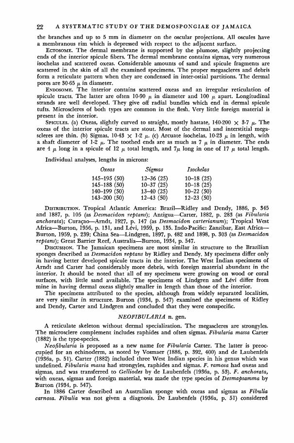
SPICULES, (a) Oxeas, slightly curved to straight, mostly hastate, 140-200 X 3-7 p. The oxeas of the interior spicule tracts are stout. Most of the dermal and interstitial megascleres are thin, (b) Sigmas, 10-43 X 1-2 fi. (c) Arcuate isochelas, 10-23 ^ in length, with a shaft diameter of 1-2 /x. The toothed ends are as much as 7 fi in diameter. The ends are 4 jn long in a spicule of 12 p total length, and 7/J, long in one of 17 fi total length.
Individual analyses, lengths in microns:
Oxeas Sigmas Isochelas
145-195 (50) 12-36 (25) 10-18 (25) 145-188 (50) 10-37 (25) 10-18 (25) 140-199 (50) 13-40 (25) 10-22 (50) 143-200 (50) 12-43 (50) 12-23 (50)
DISTRIBUTION. Tropical Atlantic America: Brazil—Ridley and Dendy, 1886, p. 345 and 1887, p. 105 (as Desmacidon reptans); Antigua—Carter, 1882, p. 283 (as Fibularia anchorata); Curasao—Arndt, 1927, p. 147 (as Desmacidon carterianum); Tropical West Africa—Burton, 1956, p. 131, and LeVi, 1959, p. 135. Indo-Pacific: Zanzibar, East Africa— Burton, 1959, p. 239; China Sea—Lindgren, 1897, p. 482 and 1898, p. 303 (as Desmacidon reptans); Great Barrier Reef, Australia—Burton, 1934, p. 547.
DISCUSSION. The Jamaican specimens are most similar in structure to the Brazilian sponges described as Desmacidon reptans by Ridley and Dendy. My specimens differ only in having better developed spicule tracts in the interior. The West Indian specimens of Arndt and Carter had considerably more debris, with foreign material abundant in the interior. It should be noted that all of my specimens were growing on wood or coral surfaces, with little sand available. The specimens of Lindgren and LeVi differ from mine in having dermal oxeas slightly smaller in length than those of the interior.
The specimens attributed to the species, although from widely separated localities, are very similar in structure. Burton (1934, p. 547) examined the specimens of Ridley and Dendy, Carter and Lindgren and concluded that they were conspecific.
NEOFIBULARIA n. gen.
A reticulate skeleton without dermal specialization. The megascleres are strongyles. The microsclere complement includes raphides and often sigmas. Fibularia massa Carter (1882) is the type-species.
Neofibularia is proposed as a new name for Fibularia Carter. The latter is preoccupied for an echinoderm, as noted by Vosmaer (1886, p. 392, 400) and de Laubenfels (1936a, p. 51). Carter (1882) included three West Indian species in his genus which was undefined. Fibularia massa had strongyles, raphides and sigmas. F. ramosa had oxeas and sigmas, and was transferred to Gelliodes by de Laubenfels (1936a, p. 53). F. anchorata, with oxeas, sigmas and foreign material, was made the type species of Desmapsamma by Burton (1934, p. 547).
In 1886 Carter described an Australian sponge with oxeas and sigmas as Fibulia carnosa. Fibulia was not given a diagnosis. De Laubenfels (1936a, p. 51) considered

SYSTEMATICS 23
Fibulia to be a direct substitution for the preoccupied Fibularia. Burton (1929, p. 142) had already established a new genus Plumocolumella for Fibulia carnosa on the assumption that Fibulia was a misprint for Fibularia. Burton later concluded (1936b, p. 142) that "no substantial evidence of a misprint" was available. He therefore considered Fibulia to be a valid genus with F. carnosa Carter as its type-species. Carter gave no indication that Fibulia was to be a substitute for Fibularia. A new generic name is therefore necessary for Fibularia massa Carter.
In addition to the type species, Neofibularia includes the following species: Fibulia nolitangere (Duchassaing and Michelotti, 1864, p. 82) de Laubenfels, 1936a, p. 51. Fibulia raphidifera (Topsent, 1889, p. 16) de Laubenfels, 1950a, p. 78. Fibulia bermuda de Laubenfels, 1950a, p. 52.
Neofibularia massa (Carter) new combination
Fibularia massa Carter, 1882, p. 282 [type: Bahamas; Brit. Mus. (Nat. Hist.) (?)] Gellius massa, Arndt, 1927, p. 151. Fibulia massa, de Laubenfels, 1950a, p. 53; idem, 1953a, p. 527.
HABITAT. A common species near the cays. It often grows on the underside of projecting ledges in shallow water. In deeper water (10-15 feet) it grows on coral masses.
SHAPE. The shallow water specimens are laterally spreading masses several centimeters in thickness. Their large cloacae lead to terminal openings which are flush with the surface. The offshore specimens may be vasiform or consist of several thick-walled hollow projections arising from a basal mass. Large vases often reach 12 cm in height. Their walls reach a thickness of 2-4 cm and their cloacal openings, a diameter of 2-4 cm.
COLOR. In life the peripheral 1-2 mm are a reddish or mustard brown. The color gradually fades to a dull yellow toward the interior. Preserved specimens are beige to drab with a greenish tinge and occasionally have purple tinges.
CONSISTENCY. Soft, very easily crumbled. SURFACE. The outer and cloacal surfaces are fibrohispid. The outer surface is rough
to the touch but even. It is pierced by a few oscular (?) openings which are less than 0.5 mm in diameter. Numerous exhalant canals, 0.5-5 mm in diameter, empty into the cloacal cavity. They are often continued upward as longitudinal grooves in the cloacal surface.
ECTOSOME. Fibrohispid. No dermal specialization. ENDOSOME. Cavernous. The skeleton is a coarse-meshed, symmetrical reticulation of
stout spicule tracts, which are 40-150 //, in diameter. The rectangular meshes are 300-1000 ji in maximum width. Raphides, sigmas and some megascleres are strewn in the flesh.
SPICULES, (a) Strongyles, slightly to strongly curved, 261-386 X 5-10 /x. (b) Raphides, 68-1 50JU, in length. They occasionally reach 3 p in diameter but are usually much thinner. Many are grouped into trichodragmata, 30-60 ^ in diameter, (c) Sigmas, 16-26 /x.
Individual analyses, length in microns:
Strongyles Raphides Sigmas
261-334 (50) 68-122 (25) 17-24 (25) 281-386 (50) 79-150 (25) 18-26 (25) 292-357 (50) 72-133 (25) 18-23 (25) 26^358 (50) 76-130 (25) 16-23 (25)
DISTRIBUTION. Tropical Atlantic America: Florida—de Laubenfels, 1950a, p. 53 (as Fibulia massa); West coast, Florida—de Laubenfels, 1953a, p. 527 (as Fibulia massa); Bahamas—Carter, 1882, p. 282 (as Fibularia massa); Curasao—Arndt, 1927, p. 151 (as Gellius massa).
DISCUSSION. A graded series was observed between vasiform and thickly incrusting specimens. Carter's sponge was of the latter type. The species is very irritating to the skin.

24 A SYSTEMATIC STUDY OF T H E DEMOSPONGIAE OF JAMAICA
GENUS IOTROCHOTA Ridley Iotrochota birotulata (Higgin) Ridley
Halichondria birotulata Higgin, 1877, p. 296 [type: Venezuela; Brit. Mus. (Nat. Hist.) (?)] [non] Halichondria birotulata, Carter, 1886, p. 52 (fide Dendy, 1896); idem, 1887, p. 72. Iotrochota birotulata, Ridley, 1884, p. 433; de Laubenfels, 1932a, p. 37; idem, 1936a, p.
49; idem, 1953a, p. 522.
HABITAT. Common on pilings at Port Royal and on coral masses offshore from the cays.
SHAPE. Typically ramose with branches 1-2 centimeters in diameter. One massive specimen was collected.
COLOR. Very dark purple, nearly black, with a greenish surface sheen in life. A purplish slime is emitted when the sponge is handled. Dried and preserved specimens are purplish black.
CONSISTENCY. Tough, rather stiff. SURFACE. Numerous conules and connecting ridges make the surface very uneven.
The narrow sharp-pointed conules are 1-3 mm in height and 1-8 mm apart. The shallow depressions between the elevated areas are covered by a thin skin. The oscules are small, scattered and infrequent. Orange zoanthids are present on the surface.
ECTOSOME. The skin contains sparsely scattered, long, thin styles and numerous birotules. It is pierced by numerous ostia which are 20-30 fi in diameter. In places, the surface is underlain by subdermal cavities which are often 200 /x in depth.
ENDOSOME. The skeleton includes a reticulation of spicule bundles and scattered interstitial megascleres. Birotules are most abundant near the canals and periphery The spicule tracts are 35-180 p in diameter and 170-650 ^ apart. Stout bundles extend into the conules. No localization of megasclere types could be detected in the endosome. A specimen collected during November, 1960, contains numerous densely pigmented aspiculous embryos, as large as 500 /x in longest diameter.
SPICULES, (a) Strongyles, mostly curved, 117-286 p, in length. The diameter varies between 3-8 /x, with the longest spicules typically thin and occasionally anisostrongylote. (b) Styles, less frequent, mostly curved, 129-253 x 5-10 /*. The longer spicules, such as those in the ectosome, are frequently thin, (c) Oxeas, hastate, rare or absent, 152-188 p. Their diameter is similar to that of the styles and strongyles. (d) Birotules, 10-18 /JL in length.
Individual analyses, lengths in microns:
Strongyles
117-217 (50) 140-246 (50) 117-258 (50) 152-286 (50)
present - present present
Styles
130-182 (50) 159-253 (30) 129-193 (30) 130-186 (30)
present present present
Oxeas
152-188 (25) 183 (1)
none 163 (2)
rare rare rare
Birotules
13-16 (25) 13-17 (25) 12-14 (25) 12-15 (25) 13-18 (25) 13-17 (25) 10-13 (25)
DISTRIBUTION. Tropical Atlantic America: Dry Tortugas, Florida—de Laubenfels, 1932a, p. 37, 1936a, p. 49; West coast, Florida—de Laubenfels, 1953a, p. 522; Venezuela— Higgin, 1877, p. 296 (as Halichondria birotulata); Bahamas—Higgin, 1877, p. 296 (as Halichondria birotulata); Jamaica—Higgin, 1877, p. 296 (as Halichondria birotulata), de Laubenfels, 1932a, p. 37; Indo-Pacific: Mergui Archipelago, off Burma—Carter, 1887, p. 72 (as Halichondria birotulata)(?).
DISCUSSION. Oxeas are known to occur in the genus, being common, for example, in I. acerata Dendy (1896). They have not been recorded previously for 7. birotulata. Although rare at best, their uniformity of shape indicates that they are proper to the sponge.
De Laubenfels reported that the birotules may be rare or absent. Higgin's sponge

SYSTEMATICS 25 from the Bahamas was described as having no microscleres. Numerous birotules were present in the Jamaican specimens examined by Higgin, de Laubenfels and myself. Spongin could not be detected in the present specimens. Higgin found it to be variable in quantity and least abundant in his Jamaican specimen.
Dendy (1896) re-examined Carter's 1886 Australian sponge and found it to be con-specific with Iotrochota coccinea (Carter, 1887) rather than with Higgin's species. Carter's 1887 record of an Indian Ocean / . birotulata, based on an undescribed reddish-purple specimen, must be questioned.
GENUS GELLIODES Ridley Gelliodes areolata (Wilson) new combination
(Plate II, fig. 3)
Pachychalina areolata Wilson, 1902a, p. 392 [type: off Vieques; USNM no. 7671.] Haliclona areolata, de Laubenfels, 1936a, p. 39; Wells et al., 1960, p. 207. Haliclona excelsa, de Laubenfels (not Schmidt, 1870), 1947, p. 36 (fide Wells et al., 1960.)
HABITAT. Abundant on pilings at Port Royal. SHAPE. Solid, cylindrical. The sponge may consist of a single flattened cylinder or a
cluster of cylindrical branches. The branches are typically 2-3 cm in diameter. Many specimens are 10-20 cm in maximum length.
COLOR. In life, the sponges are a dull reddish pink or a dull purple. They become beige in alcohol.
CONSISTENCY. Compressible, resilient. SURFACE. Rough to the touch but macroscopically even. The scattered oscules are
1.5-3 mm in diameter. They are often raised slightly above the surface. Numerous zoanthids are usually present on the surface.
ECTOSOME. NO dermal specialization. The internal fibers subdivide repeatedly near the surface where they end in non-plumose tufts.
ENDOSOME. A coarse fibroreticulation with rectangular meshes, 100-350 /x in width. The fibers are 25-235 ^ in diameter. They consist of numerous, closely packed rows of spicules cemented by a varying amount of spongin. Some megascleres are scattered in the flesh. Microscleres were absent in five analyzed specimens but abundant in the sixth.
SPICULES, (a) Robust oxeas, nearly straight or slightly curved, 182-276 x 5-18 p. The abruptly narrowed ends are sometimes rounded or irregular in outline. A varied number of megascleres are really styles or strongyles. The latter are often only 150 /n in length. A few abnormal spicules in all six specimens have a short, oblique spine or ray near one end. (b) Sigmas, in one specimen, 13-17 X 1 ft-
Individual analyses, length
Oxeas (50)
182-258 200-258 211-269 199-264 211-252 217-276
in microns:
Styles (25)
176-234 182-234 182-246 194-234 182-234 present
Strongyles (25)
188-217 152-264 146-211 164-176 176-234 present
Sigmas
none none none none none
13-17 (,25)
DISTRIBUTION. Tropical Atlantic America: North Carolina—de Laubenfels, 1947, p. 37 (as Haliclona excelsa), Wells et al., 1960, p. 207; Puerto Rico—Wilson, 1902a, p. 392 (as Pachychalina areolata).
DISCUSSION. In form, architecture and oxea size the Jamaican sponge is very similar to Pachychalina areolata Wilson (1902a, p. 392) of Puerto Rico. Wilson found no sigmas in his species, and I have found none in a slide prepared from his type. They were lacking in 5 or 6 Jamaican sponges. Puerto Rican sponges in the Yale Peabody Museum, which are certainly conspecific with the Jamaican ones, do have a varied number of thin sigmas. (W. D. Hartman, personal communication). Wells et al. (1960, p. 207) have

26 A SYSTEMATIC STUDY OF T H E DEMOSPONGIAE OF JAMAICA
attributed a North Carolina sponge, lacking microscleres, to Wilson's species. Their specimen is also similar to the Jamaican one in architecture and megasclere size. Wells et al. list Pachychalina millepora Verrill (1907, p. 336) as a synonym. Verrill's species and the Puerto Rican Pachychalina aurantiaca (Lendenfeld) of Wilson (1902a, p. 393) are certainly closely related to, if not conspecific with, the present species.
FAMILY ADOCIIDAE de Laubenfels, 1934 A simple megasclere complement, with diactinal spicules. Microscleres are often
present and may include sigmas, chelas, toxas and raphides. A distinct tangential dermal skeleton is present. The architecture is reticulate.
De Laubenfels (1936a) placed the Adociidae in the order Poecilosclerida despite their lack of spicule specialization. De Laubenfels himself had pointed out the close relationship of the adociids with the haplosclerid sponges. Many adociid genera can be distinguished from haliclonid or desmacidonid genera only by the greater development of a dermal skeleton. Species with an intermediate type of architecture, such as the Jamaican Sigmadocia caerulea, are known in many such cases. The Adociidae, indeed, are probably a heterogeneous group. They can be provisionally maintained as a family, at least for convenience, within the order Haplosclerida. Levi (1958) has also commented on the similarities between the adociids and haplosclerids of de Laubenfels* classification.
GENUS ADOCIA Gray
Adocia carbonaria (Lamarck) new combination (Plate III, figs. 2, 3)
Spongia carbonaria Lamarck, 1813, p. 375 (type: American seas; Mus. nat. Hist. nat. Paris.)
Thalysias carbonaria, Duchassaing and Michelotti, 1864, p. 83. Pachychalina carbonaria, Arndt, 1927, p. 152. Phloeodictyon carbonaria, Topsent, 1930, p. 26. [non] Pellina carbonaria, de Laubenfels, 1936a, p. 68; idem, 1954a, p. 102; idem, 1955,
p. 139. Thalysias proxima Duchassaing and Michelotti, 1864, p. 84 (fide Arndt, 1927.)
HABITAT. The species is common in the turtle grass bed at Drunkenman's Cay. It also occurs in a similar habitat at Dunn's River on the North Coast of Jamaica.
SHAPE. Thick incrustations or repent branches. The incrusting examples are 2-3 cm in thickness. The branches, always solid, are 0.5-1.5 cm in diameter.
COLOR. The living sponges are entirely black. When dried or preserved they become a very dark brown. Alcohol becomes blue-black when the sponge is placed in it.
CONSISTENCY. Brittle and easily crumbled even when alive. SURFACE. Smooth to the touch, but uneven. The oscules are 1-3 mm in diameter. They
are scattered over the surface in both incrusting and branching specimens. Many oscules are the apertures of exhalant canals which extend straight downwards into the sponge. The oscules may be flush with the surface, or raised on low volcano-shaped elevations. Although the skin is easily detachable, macroscopic subdermal spaces are not present.
ECTOSOME. The dermal membrane is supported by a three- to four-sided spicule network. The meshes are generally unispicular but occasionally have two or three spicules on a side. The interstices contain one to several dermal pores which are 30-75 p in diameter. Small quantities of spongin are present at the nodes* Spicules sometimes join a node at their mid-length. Numerous pigment cells, 7 p in diameter, are present in both the ectosome and interior.
ENDOSOME. The interior has a largely unispicular network, with some but not all of the spicules bound by spongin nodes. The skeleton also includes an irregular reticulation of spicule tracts, which are 30-45 p in diameter.
SPICULES. Oxeas, mostly curved, 176 x 246 X 3-9 p. The ends may be rounded, mucro-nate, or slightly stair-stepped. The stair-stepped spicules may have truncate ends. A few styles and strongyles are present. The developmental stages are curved, fusiform oxeas.

SYSTEMATICS 27 Individual analyses, lengths in microns, 50 spicules each: Oxeas, 199-229; 176-246;
188-234. DISTRIBUTION. Tropical Atlantic America: Lamarck, 1813, p. 375 (as Spongia car
bonaria); Lesser Antilles—Guadeloupe, St. Croix, St. Barthelemy, St. Thomas—Duchassaing and Michelotti, 1864, p. 83 (as Thalysias carbonaria); St. Thomas—Duchassaing and Michelotti, 1864, p. 84 (as Thalysias proximo); Curasao—Arndt, 1927, p. 152 (as Pachy-chalina carbonaria).
DISCUSSION. In spicule shape the present specimens are somewhat similar to the black Pellina carbonaria studied by de Laubenfels (1936a, p. 66) in the Dry Tortugas. That sponge often grew in sand, with only its long oscular projections protruding above the surface. He considered it to be conspecific with Spongia carbonaria Lamarck but later (1954a, p. 107) expressed some reservations about the identification. The spicules of the Dry Tortugas sponges are smaller than those of either the Jamaican sponge or of Lamarck's specimen. In addition the spicule was described as "fundamentally a strongyle," with the ends "sometimes provided with caps of successively smaller diameter."
De Laubenfels' sponge differs further from the present specimen in having a dermal skeleton of irregularly strewn spicules and subdermal spaces. It was described as being very dark green rather than black. The consistency was said to be delicately elastic and easily torn, rather than stiff. De Laubenfels, who paid close attention to color changes in alcohol, made no mention of any blue-black exudate. The oscules were clearly of the Pellina type. It could be that the delicate projections were suppressed in the Jamaican sponges, which were indeed subject to considerable surf. The presence of delicate, stiff, slender branches in the Jamaican specimens weighs against such a conclusion. The numerous differences strongly suggest that the Dry Tortugas sponge is not conspecific with the Jamaican.
Pellina carbonaria (Lamarck) as reported from the central Pacific by de Laubenfels (1954a, p. 102) does have large spicules but also has oscular chimneys and lacks a blue-black exudate. Its relationship to the sponges of the Dry Tortugas and Jamaican is uncertain.
The poorly preserved type of Spongia carbonaria Lamarck has been restudied by Topsent (1930). It bore no trace of oscular projections. Its curious system of cavities and lamellae may be the result of drying. The oxeas had abrupt but not stair-stepped points. They were similar in length to those of the Jamaican specimens. Duchassaing and Michelotti's Thalysias carbonaria resembles the Jamaican specimens in shape and color. No information is available on its spiculation. Arndt's sponges from Curasao are probably conspecific with the Jamaican ones. His specimens had a similar shape, color, dermal skeleton and spicule size range. The tracts of the main skeleton were somewhat better developed. The spicules often had wedge-shaped points but apparently were not stair-stepped.
The Jamaican specimens are somewhat doubtfully considered to be conspecific with those of Lamarck, Arndt, and Duchassaing and Michelotti. The lack of oscular tubes places the species in the genus Adocia.
Adocia implexiformis n. sp. (Text-fig. 2; Plate II, fig. 2)
Fig. 2. —Spicules of Adocia implexiformis. Oxeas. YPM 5034. Holotype.
HOLOTYPE. YPM 5034, Mangrove boat channel, Jan. 25, 1961. HABITAT. Abundant on mangrove roots.

28 A SYSTEMATIC STUDY OF T H E DEMOSPONGIAE OF JAMAICA
SHAPE. Thick-walled, cylindrical oscular projections arise from the base. They are typically 2-4 cm in height and 1-2 cm in diameter. Adjacent oscular lobes may be fused laterally. Slender, solid projections, 0.5-1 cm in height, and 2-3 mm in diameter, are often present on distal parts of the sponge.
COLOR. The living sponge is a dull purple, devoid of any reddish tinge. The sponges are drab in alcohol.
CONSISTENCY. The sponge retains its shape when removed from water. It is compressible but very easily crumbled.
SURFACE. Even. The oscules are 2-10 mm in diameter. They are the openings of elongate axial cavities in the oscular lobes.
ECTOSOME. A detachable dermal membrane is present. It is supported by a tangential, isodictyal, largely unispicular network of oxeas. Spongin is present at the nodes. The three- to four-sided meshes contain closely-spaced ostia which are 12-35 ^ in diameter. A few large subdermal spaces are present.
ENDOSOME. A spicular network similar to that of the ectosome. The meshes occasionally have 2 or 3 rows of spicules on a side but long tracts are absent.
SPICULES. Oxeas, straight to slightly curved, 129-182 X 5-10 /*. Thin, sharp-pointed developmental stages are present. The mature spicules have blunt ends, and a few are styles and strongyles. The ends of many oxeas are narrow, but rounded, rather than sharp-pointed. Individual analyses, lengths in microns, 50 spicules each: 145-174; 145-182*; 137-181; 129-179.
DISCUSSION. In form, the present species resembles the heterogeneous assemblage of sponges attributed to Haliclona implexa (Schmidt). The sponges assigned to the latter species lack a special dermal skeleton. A. chilensis (Thiele, 1905) of Chile also has oscular projections. It differs from the Jamaican species in having strongly strongylote spicules, spicule tracts and well-developed subdermal cavities. A. grossa (Schmidt), as recorded by Topsent (1925a) from Naples, is similar in form and spicule range to the present species. It differs in having an uneven surface and multispicular tracts. The Philippine projection-bearing A. baeri (Wilson, 1925) de Laubenfels, 1935b, has a tuberculate surface, dark color and small oxeas. A. tub if era (George and Wilson, 1919) Hartman, 1958, of North Carolina has small oscular projections on anastomosing branches. The skeleton includes spongin fibers.
Adocia albifragilis n. sp. (Text-fig. 3)
I 50//
Fig. 3. —Spicules of Adocia albifragilis. Oxeas. YPM 5035. Holotype.
HOLOTYPE. YPM 5035. Drunkenman's Cay, June 24, 1961. HABITAT. Under rocks at the cays, in a few feet of water. SHAPE. Thin crusts, reaching a maximum thickness of 2-3 mm. COLOR. The living sponge is white. It is pale, nearly white in alcohol. CONSISTENCY. Compressible, but easily torn. SURFACE. Even. Under a magnification of 14 x, the surface appears micro tuberculate
but not hispid. The oscules are about 2 mm in diameter and flush with the surface. ECTOSOME. The surface has a tangential network closely connected to the skeleton of
the interior. No large subdermal spaces are present. The sides of the skeletal network may consist of single spicules or several loosely arranged spicules. Compact tracts do not occur. Small amounts of spongin are present at the nodes. A node may include as many
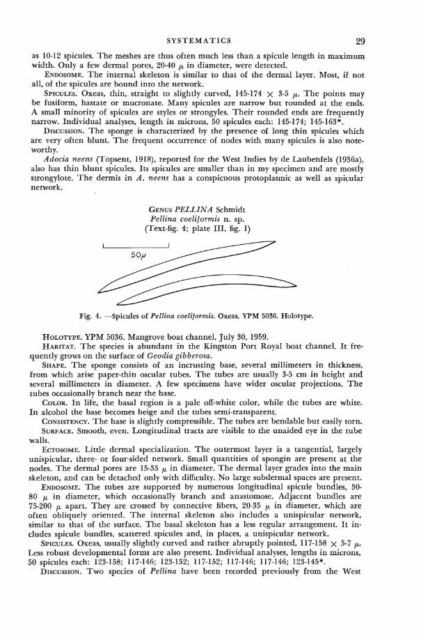
SYSTEMATICS 29 as 10-12 spicules. The meshes are thus often much less than a spicule length in maximum width. Only a few dermal pores, 20-40 ^ in diameter, were detected.
ENDOSOME. The internal skeleton is similar to that of the dermal layer. Most, if not all, of the spicules are bound into the network.
SPICULES. Oxeas, thin, straight to slightly curved, 145-174 x 3-5 p. The points may be fusiform, hastate or mucronate. Many spicules are narrow but rounded at the ends. A small minority of spicules are styles or strongyles. Their rounded ends are frequently narrow. Individual analyses, length in microns, 50 spicules each: 145-174; 145-163*.
DISCUSSION. The sponge is characterized by the presence of long thin spicules which are very often blunt. The frequent occurrence of nodes with many spicules is also noteworthy.
Adocia neens (Topsent, 1918), reported for the West Indies by de Laubenfels (1936a), also has thin blunt spicules. Its spicules are smaller than in my specimen and are mostly strongylote. The dermis in A. neens has a conspicuous protoplasmic as well as spicular network.
GENUS PELLINA Schmidt Pellina coeliformis n. sp.
(Text-fig. 4; plate III, fig. 1)
Fig. 4. —Spicules of Pellina coeliformis. Oxeas. YPM 5036. Holotype.
HOLOTYPE. YPM 5036. Mangrove boat channel, July 30, 1959. HABITAT. The species is abundant in the Kingston Port Royal boat channel. It fre
quently grows on the surface of Geodia gibberosa. SHAPE. The sponge consists of an incrusting base, several millimeters in thickness,
from which arise paper-thin oscular tubes. The tubes are usually 3-5 cm in height and several millimeters in diameter. A few specimens have wider oscular projections. The tubes occasionally branch near the base.
COLOR. In life, the basal region is a pale off-white color, while the tubes are white. In alcohol the base becomes beige and the tubes semi-transparent.
CONSISTENCY. The base is slightly compressible. The tubes are bendable but easily torn. SURFACE. Smooth, even. Longitudinal tracts are visible to the unaided eye in the tube
walls. ECTOSOME. Little dermal specialization. The outermost layer is a tangential, largely
unispicular, three- or four-sided network. Small quantities of spongin are present at the nodes. The dermal pores are 15-35 ^ in diameter. The dermal layer grades into the main skeleton, and can be detached only with difficulty. No large subdermal spaces are present.
ENDOSOME. The tubes are supported by numerous longitudinal spicule bundles, 30-80 n> in diameter, which occasionally branch and anastomose. Adjacent bundles are 75-200 JJL apart. They are crossed by connective fibers, 20-35 fx in diameter, which are often obliquely oriented. The internal skeleton also includes a unispicular network, similar to that of the surface. The basal skeleton has a less regular arrangement. It includes spicule bundles, scattered spicules and, in places, a unispicular network.
SPICULES. Oxeas, usually slightly curved and rather abruptly pointed, 117-158 X 5-7 /x. Less robust developmental forms are also present. Individual analyses, lengths in microns, 50 spicules each: 123-158; 117-146; 123-152; 117-152; 117-146; 117-146; 123-145*.
DISCUSSION. TWO species of Pellina have been recorded previously from the West
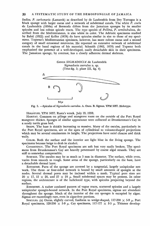
30 A SYSTEMATIC STUDY OF T H E DEMOSPONGIAE OF JAMAICA
Indies. P. carbonaria (Lamarck) as described by de Laubenfels from Dry Tortugas is a black sponge with larger oxeas and a network of subdermal canals. The white P. coela de Laubenfels (1950a) of Bermuda differs from the Jamaican sponges by its smaller spicules and less robust spicule tracts. The type species of Pellina, P. semitubulosa, described from the Mediterranean, is also white in color. The Adriatic specimens studied by Babic (1922) and Keller (1878) do have spicules similar in size to those of my specimens. Topsent's Mediterranean specimen, however, has more robust oxeas and a second category of small ectosomal microxeas. He reported an extensive network of subdermal canals in the basal regions of his material. Schmidt (1862, 1870) and Topsent both emphasized the presence of a well-developed, easily detachable skin in their specimens. The Jamaican sponge, by contrast, has a closely adherent dermal skeleton.
GENUS SIGMADOCIA de Laubenfels Sigmadocia caerulea n. sp.
(Text-fig. 5; plate III, fig. 4)
A
50// Fig. 5. —Spicules of Sigmadocia caerulea. A. Oxea. B. Sigmas. YPM 5037. Holotype.
HOLOTYPE. YPM 5037. Rasta's wreck. July 19, 1959. HABITAT. Common on pilings and mangrove roots on the outside of the Port Royal
mangrove thicket. Sponges of similar appearance were collected at Drunkenman's Cay in a sandy turtle grass bed.
SHAPE. The base is thickly incrusting to massive. Many of the oscules, particularly in the Port Royal specimens, are at the apex of cylindrical to volcano-shaped projections ivhich may be several centimeters in height. The projections have axial cloacae and thick -walls.
COLOR. Both the surface and the interior are light blue in the living sponge. The specimens become beige to drab in alcohol.
CONSISTENCY. The Port Royal specimens are soft but very easily broken. The specimens from Drunkenman's Cay are heavily penetrated by coarse algal strands. They are stiff to somewhat compressible.
SURFACE. The oscules may be as much as 5 mm in diameter. The surface, while even, varies from smooth to rough. Some areas of the sponge, particularly on the base, have a detachable dermal layer.
ECTOSOME. Parts of the sponge are covered by a tangential, largely unispicular network. The three- or four-sided network is bound by small amounts of spongin at the nodes. Several dermal pores may be inclosed within a mesh. Typical pore sizes are 20 X 17, 12 X 20, and 27 X 24 ^. Small subdermal spaces may be present. In other regions, the architecture is of the haliclonid type, with spicules projecting beyond the surface.
ENDOSOME. A rather confused pattern of vague tracts, scattered spicules and a largely unispicular spongin-bound network. In the Port Royal specimens, sigmas are abundant throughout the sponge. Much of the interior of the cay sponges is occupied by algae. Sigmas are exceedingly rare, even in algae-free portions.
SPICULES, (a) Oxeas, slightly curved, fusiform to wedge-shaped, 117-200 X 5-9 p. Port Royal specimens, 128-200 x 5-9 /A- Cay specimens, 117-177 x 3-5 /*,. Thinner develop-

SYSTEMATICS 31
mental forms are common. A few styles of similar size occur, (b) Sigmas, thin, often sharply curved in the middle, with the ends nearly in one plane, 13-28 JU,. Individual analyses, lengths in microns:
Port Royal Specimens
Oxeas
143-200 (50)* 164-199 (50) 128-182 (50) 140-182 (50) 140-188 (50) 137-188 (50) 145-188 (50)
Sigmas
17-23 (25)* 22-25 (25) 20-26 (25) 20-30 (25) 20-28 (25) 15-20 (25) 15-26 (25)
Cay Specimens
117-177 (50) 12-20 (20) 123-158 (50) 13-20 (25) 117-170 (50) 15-20 (25)
DISCUSSION. The varied development of a tangential dermal skeleton in this species indicates the close relationship of Sigmadocia and Gellius. The cay specimens are provisionally considered as conspecific with those of Port Royal. The type specimen is a sponge collected at Port Royal. The two lots of material agree in color and architecture. Oscular projections are, at best, weakly developed in the cay sponges which, however, grew in an area subject to considerable wave action. The cay sponges have thinner oxeas. The presence of rare sigmas similar in shape to those of the inshore specimens provides the best evidence of conspecificity.
The species is noteworthy for its blue color and sharply curved sigmas. In spicule size, it is similar to some of the sponges attributed to Gellius fibulatus (Schmidt, 1862). Schmidt's original description provides little information on his species. The sponge photographed by Topsent (1925a) has a coarsely porous surface quite unlike that of my specimens. The sponge reported from Israel by LeVi (1957) differs from mine in having curious sigmas with a series of bends. Babic's specimens (1922), recorded as Gelliodes fibulata (Schmidt), had well-developed spongin fibers.
FAMILY CALLYSPONGIIDAE de Laubenfels, 1936a GENUS CALLYSPONGIA Duchassaing and Michelotti
Callyspongia fallax Duchassaing and Michelotti (Plate IV, fig. 2)
Callyspongia fallax Duchassaing and Michelotti, 1864, p. 57 [type: St. Thomas; Brit. Mus. (Nat. Hist.) reg. no. 28.11.12.5]; Burton, 1934, p. 539; de Laubenfels, 1949, p. 13.
HABITAT. Several specimens were collected in 10-15 feet of water near Maiden Cay. One was growing on the side of a Verongia fistularis, while the others were on coral masses.
SHAPE. The sponge consists of a single, solid cylindrical branch, or an anastomosing group of such branches. The specimens range in length from 7-21 cm. The branches vary in diameter and also in outline along their length. They are usually 0.5-1.5 cm in width. A single specimen widens at one end to enclose a short vertical channel which is open at both ends. The cavity is lined by a smooth skin resembling that of the rest of the surface. It is occupied by a small crustacean and is certainly not a cloaca.
COLOR. In life, the sponges are a dull purple. In alcohol, they become grayish-brown with or without traces of the original purple color.
CONSISTENCY. Compressible. SURFACE. Smooth to the touch. The skin is even over wide areas of the surface. In

32 A SYSTEMATIC STUDY OF T H E DEMOSPONGIAE OF JAMAICA
places, particularly distally, it is lifted into thorn-like conules, 2-4 mm high. Adjacent conules may be several millimeters or several centimeters apart. The oscules are usually scattered along the branches but may be sub-linear. They are 2-3 mm in diameter. Several exhalant canals open into each.
ECTOSOME. An intergrading double network of spiculiferous spongin fibers. The coarse fibers have a diameter of 15-60 ju and contain 1-6 rows of spicules. They form an irregularly polygonal network, with parallel fibers 100-300 /x apart. The coarse mesh grades into a network of slender unispicular fibers which are 3-8 ^ in diameter. The fine meshes are usually less than a spicule length in width.
ENDOSOME. The skeleton consists of uni- to multispicular spongin fibers, which are 15-115 [A, but most frequently 30-60 JJL in diameter. The spicules may form a continuous core or may be spaced along the fiber. The nodes of fiber intersection are multispicular expansions, 70-115 ^ in diameter. Near the surface endosomal fibers run into the dermal mesh. The endosomal interstices are 100-300 fx in diameter and frequently rounded in outline. They contain scattered spicules similar in size and form to those of the fibers.
SPICULES. Oxeas, straight to very slightly curved, with hastate or eroded points, 58-130 X 3-7 /A. The erosion may extend as far back as 2 diameters from the tip. Some spicules have low irregular swellings in mid-shaft. Individual analyses, lengths in microns, 50 spicules each: 58-123; 70-130; 70-130.
DISTRIBUTION. Tropical Atlantic America: Bahamas—de Laubenfels, 1949, p. 13; St. Thomas—Duchassaing and Michelotti, 1864, p. 57.
DISCUSSION. The Jamaican solid, cylindrical, lavender Callyspongia is similar in appearance and architecture to the Bahaman sponges attributed by de Laubenfels to Callyspongia fallax. The spicules of my specimens, however, while small, are certainly not minute. Although C. fallax is the type-species of a large genus, little information is available on its structure. Duchassaing and Michelotti (1864) merely recorded specimens which, in their opinion, were conspecific with the unrecognizable Spongia papillaris of earlier authors. They established the name C. fallax, claiming that the name C. papillaris was preoccupied. Burton (1934) selected Duchassaing and Michelotti's specimen as the type of Callyspongia fallax. The type has been reexamined by Burton and probably by de Laubenfels as well. To my knowledge, neither author has published a detailed description of the sponge. Burton characterized the genus Callyspongia by its double dermal network, but curiously proceeded to emphasize the lack of such differentiation in the type-species, C. fallax.
Burton's taxonomic summary (1934) cannot be accepted on the basis of published information. He rejects body form and spicule complement as valid specific criteria in the genus but does not present detailed evidence in support of this viewpoint. Judging from de Laubenfels' 1949 discussion, the type of C. fallax is solid and cylindrical. Burton could find no toxas in it. He considers Callyspongia eschrichtii Duchassaing and Michelotti, and Patuloscula procumbens Carter as synonyms of C. fallax, although he found toxiform spicules in them. In addition, both supposed synonyms are tubular or partially tubular sponges. Whatever the status of Duchassaing and Michelotti's Tuba scrobiculata, the species was established by Lamarck, whose material has been re-examined by Topsent, (1932). Callyspongia scrobiculata (Lamarck), a synonym of C. plicifera (Lamarck), can be distinguished from C. fallax by its tubular form, internal architecture, and strongylote spicules. Ceraochalina vanderhorsti Arndt, 1927, also included in the synonymy, is again basically tubular. The Jamaican sponges are clearly not conspecific with Arndt's species, from which they differ in having larger oxeas, a solid form and an abundance of spicules in the dermal network.
Callyspongia vaginalis (Lamarck) de Laubenfels
Spongia vaginalis Lamarck, 1813, p. 436 [type: American seas; Mus. nat. Hist. nat. Paris.] Tuba vaginalis, Duchassaing and Michelotti, 1864, p. 52. Callyspongia vaginalis, de Laubenfels, 1936a, p. 56; idem, 1949, p. 14; idem, 1950a, p. 56;
idem, 1953a, p. 523; Wells et al., 1960, p. 210; Little, 1963, p. 41. [non] Callyspongia vaginalis, de Laubenfels, 1939, p. 2.

SYSTEMATICS 33
Tuba sororia Duchassaing and Michelotti, 1864, p. 46 (fide de Laubenfels, 1936a); Carter, 1884, p. 204.
Spinosella sororia, Vosmaer, 1886, p. 342; Dendy, 1887d, p. 503; idem, 1890, p. 360; Wilson, 1902a, p. 394; Verrill, 1907, p. 334; Arndt, 1927, p. 155; Topsent, 1931, p. 75.
[non] Spinosella sororia, Row, 1911, p. 326.
HABITAT. Common on corals in 10-15 feet of water offshore from the cays. SHAPE. The sponge may be a single, cylindrical tube or, more frequently, a group of
such tubes. The tubes have an even to jagged cloacal (oscular?) orifice, but never a papyraceous collar. The tubes are 1-3, occasionally 5 cm in diameter, and 5-15 cm in length. The width is rather uniform along the length of the tube. None of the specimens is a flaring vase. The walls are only a few millimeters in thickness. The cavities of adjacent tubes are often confluent at the base of the sponge.
COLOR. The sponge is dull purple in life. In alcohol it retains much of its original color.
CONSISTENCY. Compressible. SURFACE. The outer surface can vary between smooth and uneven along a tube. The
elevations involve considerable areas of the surface. The inner wall has numerous openings, 0.5-1 mm in diameter, and longitudinal ribs.
ECTOSOME. A well-developed callyspongiid double network of spongin fibers. The coarse fibers are mostly 20-30 /m in diameter and contain 2-4 rows of spicules. They outline irregularly polygonal spaces, 100-450 ^ in maximum width. The more symmetrical, finer network encloses apertures only a spicule or two in width. The fibers are 5-15 jn in diameter. They have a unispicular, usually spaced, core of oxeas. In the ectosome, the spicules are confined to the fibers. The cloacal surface has a coarse fiber network, which is condensed at the ribs into anastomosing, interwoven columns.
ENDOSOME. The skeleton is formed by a reticulation of spongin fibers 15-75 ̂ (mostly 30-45 ju,) in diameter. All fibers have abundant spongin and most have a continuous core of several rows of spicules. Even large fibers may have only single, separated spicules over part of their length. The meshes are often nearly rectangular, with parallel fibers 70-300 /x apart. A considerable number of spicules, mostly thin, are scattered in the flesh. The dermal network is supported by ascending fibers and often overlies subdermal cavities.
SPICULES. Oxeas, 76-131 X 2-5 p. The spicules may be elongate and slightly curved, or short, straight and conspicuously hastate. The points are sometimes eroded. Individual analyses, lengths in microns, 50 spicules each: 83-123; 80-131; 76-123.
DISTRIBUTION. Tropical Atlantic America: American Seas—Lamarck, 1813, p. 436 (as Spongia vaginalis); North Carolina—Wells et al., 1960, p. 210; Florida—Carter, 1884, p. 204 (as Tuba sororia); Dry Tortugas, Florida—de Laubenfels, 1936a, p. 56; West Coast, Florida—de Laubenfels, 1953a, p. 523; West Coast, Florida—Little, 1963, p. 41; Bermuda— Verrill, 1907, p. 334 (as Spinosella sororia) and de Laubenfels, 1950a, p. 56; Bahamas— Dendy, 1887d, p. 503; 1890, p. 360 (as Spinosella sororia), and de Laubenfels, 1949, p. 14; Puerto Rico—Wilson, 1902a, p. 324 (as Spinosella sororia); Lesser Antilles: Guadalupe, St. Thomas, St. Croix, Viecques, Tortola—Duchassaing and Michelotti, 1864, p. 52 (as Tuba vaginalis); St. Thomas, Viecques—Duchassaing and Michelotti, 1864, p. 46 (as Tuba sororia); Curasao—Arndt, 1927, p. 155 (as Spinosella sororia). Tropical Pacific America: Galapagos Is.—de Laubenfels, 1939, p. 2 (?).
DISCUSSION. Lamarck's specimen has been restudied by both Topsent (1932) and de Laubenfels (1936a). The spicules of Jamaican specimens are rather robust for the species. Published data indicate that there is considerable variation in the size and abundance of the spicules. The fiber size of the Jamaican specimens agrees closely with that given in previous descriptions.
The records of Duchassaing and Michelotti, Carter, and Verrill require verification. The two non-West Indian records are both very dubious. De Laubenfels' identification of the Galapagos Callyspongia was only tentative, since the specimen "does not agree well with ordinary examples of vaginalis." (1939, p. 2) Unfortunately, no other data were

34 A SYSTEMATIC STUDY OF T H E DEMOSPONGIAE OF JAMAICA
provided. The Red Sea sponge studied by Row certainly differs from typical specimens of the West Indian C. vaginalis in interior structure. It had strong multispicular primary fibers, containing 10-20 rows of spicules, and delicate, unispicular secondary fibers.
De Laubenfels (1950a, p. 57) placed Siphonochalina stolonifera Whitfield, 1901, of Bermuda into synonymy with C. vaginalis. Whitfield, however, described the spicules of his sponge as cylindrical, which may mean strongylote. Schmidt's Siphonchalina papyracea (1870), often considered as a synonym of C. vaginalis, was described as having strongylote spicules ("umspitzigen nadeln"). Since a strongylote vase-shaped species, Callyspongia plicifera, occurs in the West Indies, the two cannot safely be taken as synonyms of C. vaginalis.
Callyspongia plicifera (Lamarck) de Laubenfels (Plate IV, fig. 1)
Spongia plicifera Lamarck, 1813, p. 435 [type: American seas (?); Mus. nat. Hist. nat. Paris.]
Tuba plicifera, Duchassaing and Michelotti, 1864, p. 53; Schmidt, 1870, p. 29; Carter, 1882, p. 365.
Spinosella plicifera, Dendy, 1887d, p. 506; idem, 1890, p. 365; Topsent, 1932, p. 73. Callyspongia plicifera, de Laubenfels, 1950a, p. 61. [non] Patuloscula plicifera, de Laubenfels, 1936a, p. 57 (fide de Laubenfels, 1950a, p. 61.) Spongia scrobiculata Lamarck, 1813, p. 436. Tuba scrobiculata, Duchassaing and Michelotti, 1864, p. 53. Spinosella scrobiculata, Topsent, 1932, p. 74. Spinosella maxima Dendy, 1887d, p. 506; idem, 1890, p. 365.
HABITAT. Growing on coral masses in 10-15 feet of water offshore from the cays. SHAPE. The sponge may be a single tube or a cluster of tubes. The individual tubes
may be cylindrical or, more' frequently, vase-shaped. Small hollow, open buds are often present on the outside, and in one exceptional case, on the inside of the tube. The sponges are typically 10-30 cm in height. The cloacal (oscular?) orifice seldom has a diameter of less than 5 cm. In vase-shaped examples, the maximum diameter may exceed the length of the sponge. The walls are only a few millimeters in thickness.
COLOR. A dull purple in life. In alcohol, the sponge may be a purplish or a dull yellow, or a mixture of the two colors. Dried specimens are beige.
CONSISTENCY. Compressible. SURFACE. The surface becomes even toward the distal end. The terminus has a thin
margin, in which a fiber reticulation is visible to the unaided eye. In some specimens, ribs composed of longitudinal fibers project beyond the general level of the cloacal terminus. The margin may be only a few millimeters or as much as several centimeters in length. The remainder of the surface may vary between uneven and strongly uneven on a single specimen. Many specimens are covered with broad blunt elevations which often grade into longitudinal or horizontal ridges. The elevations are several millimeters in height. The inner surface is even and pierced by numerous small oscules.
ECTOSOME. An intergrading double network of spongin fibers which are mostly unispicular. The finer fibers are 15-30 ^ in diameter. They enclose meshes which are frequently 150-300 fx in maximum width. Coarse fibers are typically 40-150 ^ in diameter, with parallel fibers often more than 0.5 mm apart. The longitudinal fibers of the top margin and fibrous projections are stout (70-225 fi in diameter) and frequently multispicular. In the latter case, the spicules form an axial core. The primaries are joined by less robust connectives and also by thin fibers similar to those of the endosome.
ENDOSOME. The main skeleton is a wide-meshed, rounded to polygonal network of spongin fibers. The fibers are 40-90 ^ or occasionally as much as 300 ^ in diameter. They may be unispicular, or have a multispicular axial core. The spicules may be spaced, or they may form a continuous line. A very irregular finer mesh joins the main skeleton and is often interwoven with it. Its fibers are 15-30 JJL in diameter and contain spaced single spicules.
SYSTEMATICS 35 SPICULES. Strongyles, truncate, straight to somewhat curved or bent, 53-100 X 1-2 JJL.
Individual analyses, lengths in microns, 50 spicules each: 67-97; 63-100; 68-86; 53-91. DISTRIBUTION. Tropical Atlantic America: American Seas—Lamarck, 1813, p. 435 (as
Spongia plicifera); Florida—Schmidt, 1870, p. 29 (as Tuba plicifera); Bahamas—Dendy, 1887d, p. 506; 1890, p. 365 (as Spinosella plicifera), Dendy, 1887d, p. 506; 1890, p. 365 (as Spinosella maxima); Antilles—Duchassaing and Michelotti, 1864, p. 53 (as Tuba plicifera), Schmidt, 1870, p. 29 (as Tuba plicifera); Jamaica—Dendy, 1887d, p. 506, 1890, p. 365 (as Spinosella maxima); Lesser Antilles, St. Thomas, Tortole, Guadeloupe— Duchassaing and Michelotti, 1864, p. 53 (as Tuba scrobiculata), Grenada—Carter, 1882, p. 365 (as Tuba plicifera),
DISCUSSION. The Jamaican specimens are similar in architecture and spiculation to Spongia scrobiculata Lamarck, as redescribed by Topsent. Burton (1934, p. 539) attributed that species to Duchassaing and Michelotti and considered it as a synonym of Callyspongia fallax. Their specimen of scrobiculata was described as tubular, and thus is unlikely to be conspecific with C. fallax. Spongia scrobiculata and S. plicifera Lamarck have been treated as closely related species by Lamarck, Duchassaing and Michelotti, and Topsent. The latter species is characterized by well-developed, largely horizontal folds and grooves. According to Topsent (1932) spicules are absent in the type. S. scrobiculata also has surface convolutions which outline deep fossae in Lamarck's specimens. Topsent found thin strongylote spicules in the fibers of the fossa-bearing specimens. He also found strongyles in a specimen (photographed by Topsent, 1932, plate 2, figure 4) which Lamarck had assigned to Spongia plicifera. The specimen has low, irregular to horizontal folds, making it very similar to my material. Topsent considered the sponge to be an example of scrobiculata, using the presence or absence of spicules as a distinguishing characteristic.
A collection of Cuban specimens (CU-6, CU-6A-E) on loan to the Yale Peabody Museum from the U. S. National Museum strongly suggests that the two species are actually conspecific. The dried Cuban sponges are a pale beige in color. They have a thin distal margin as in the Jamaican sponges. On the basis of surface folds and depressions all of the Cuban specimens must be assigned to Callyspongia plicifera. CU-6C, for example, is identical in appearance with the specimens of plicifera figured by Dendy and by Duchassaing and Michelotti. The surface lamellae are well-developed in all and are almost entirely horizontal in CU-6C. The folds become irregular and meandriform over part or most of the surface in CU-6B and CU-6. Strongly convoluted Jamaican specimens certainly approach that condition.
Strongylote spicules of 2 p, diameter are present in all of the Cuban examples. They are similar in shape and size to those of the Jamaican sponges. For example, a sample of 50 spicules in CU-6 varied in length between 68 and 94 p. Topsent, Schmidt, and Dendy were unable to find spicules in their examples of C. plicifera. Carter, however, reported very thin acerate spicules, 72 /x in length, for his Grenada specimen. The Cuban specimens have a skeletal pattern quite comparable to that found in the Jamaican sponges. The surface is made up of a coarse double network. The main skeleton of the interior is constructed of stout spongin fibers (35-150 ^ in diameter). They may contain only single spicules or a multispicular axial core. Several of the specimens (CU-6A, CU-6E) have fewer spicules than the Jamaican sponges. The main skeleton is joined at intervals by a very irregular network of fine fibers. The latter are only about 15 ^ in diameter and contain very few spicules. Dendy (1890) reported a similar network in his specimens of plicifera. In summary, the Jamaican and Cuban specimens differ only in the extent to which the surface is thrown into horizontal convolutions.
Spinosella maxima Dendy, 1887d, is very probably conspecific with Callyspongia plicifera. The outer surface is uneven except for a thin distal margin. The interior skeleton had the same pattern as in plicifera, including an irregular network of delicate fibers. Spicules were said to be absent from the Bahaman examples, but thin vestigial oxeas were reported for a Jamaican specimen.
Siphonochalina papyracea Schmidt, 1870, often considered as a synonym of Callyspongia vaginalis, is more likely to prove to be conspecific with C. plicifera. The external

36 A SYSTEMATIC STUDY OF T H E DEMOSPONGIAE OF JAMAICA
appearance is certainly similar, and the spicules, described as "umspitzigen nadeln," are apparently strongylote.
Callyspongia pallida n. sp.
Fig. 6. —Spicules of Callyspongia pallida. Oxeas, style, strongyle. YPM 5038. Holotype.
HOLOTYPE. YPM 5038. Seawall of Police Post. November 30, 1960. HABITAT. The specimen was collected on the seawall of the Police Post at Port Royal
in several feet of water. SHAPE. The sponge is a horizontally spreading complex of lobes. The lobes are 1.5-2
cm in height and have apical oscules. Connecting stems are about 1 cm long and 0.5-1 cm in diameter.
COLOR. The sponge was dull yellow in life and is a pale beige in alcohol. CONSISTENCY. Compressible, soft, elastic. SURFACE. Even, smooth to the touch. ECTOSOME. The dermal layer can be detached only with difficulty. It is a callyspongiid
double network of spongin fibers. The two networks gradually intergrade with respect to fiber diameter. The small fibers are 7-30 LL in diameter. They are mostly unispicular, with the spicules in a continuous line, or less frequently, somewhat spaced. The meshes are 60-220 LL in width. They are often nearly square or rectangular, with rounded corners. The large fibers are 30-45 ^ in diameter and usually have several rows of spicules in a continuous axial core. The meshes are triangular to polygonal. Parallel fibers may be 200-580 LL apart. The dermal pores are 35-45 LL in diameter. A few interstitial spicules are present.
ENDOSOME. The interior fibers are 20-75 LL in diameter. Thinner fibers are mostly unispicular. Thicker fibers have a continuous axial core of 3-6 rows. The square to rectangular meshes are mostly 300-600 LL in maximum width. A few interstitial spicules occur in the flesh. A number of dense, nearly spherical embryos, 160-217 LL in diameter, are present in the specimen which was collected in November.
SPICULES. Mostly diactinal, straight to very slightly curved, 48-102 X 1-5 LL.* (125 spicules.) Most of the spicules are centrotylote, with a central irregularly swollen area about 7 LL long, and 3-5 LL in diameter. The ends are most frequently narrow but blunt. Others are acutely pointed, irregular in outline, or round. In the last case, the spicules are styles or strongyles. The modified spicules may be centrotylote. The spicules of the interior fibers are often very thin, with the shaft (aside from any central swelling) only 1-2 LL in diameter.

SYSTEMATICS 37
DISCUSSION. The most closely related species is Callyspongia aspinosa Levi, 1959, of West Africa. They are similar in appearance, color, and consistency. Levi's sponge is more tubular in shape. The spicules of the West African sponge are substrongyles which may reach 125 p in length. The dimensions of the interior skeleton are somewhat different.
ORDER POECILGSCLERIDA Topsent 1928 FAMILY TEDANIIDAE Ridley and Dendy, sensu de Laubenfels, 1936a
GENUS TEDANIA Gray Tedania ignis (Duchassaing and Michelotti) Verrill
(Plate V, fig. 4) Thalysias ignis Duchassaing and Michelotti, 1864, p. 83 [type: St. Thomas; Brit. Mus.
(Nat. Hist.) reg. no. 28.11.12.43.] Tedania ignis, Verrill, 1907, p. 339; de Laubenfels, 1936a, p. 89; idem, 1936b, p. 459;
idem, 1949, p. 16; idem, 1950a, p. 69; idem, 1950b, p. 21 (?); idem, 1951a, p. 260 (?); idem, 1956, p. 3; Little, 1963, p. 48.
Tedania ignis pacifica de Laubenfels, 1954a, p. 129 (?). Tedania digitata, Schmidt (not Schmidt, 1862), 1870, p. 43 [partim]; Wilson, 1902a, p.
395 (fide de Laubenfels, 1936a); Arndt, 1927, p. 146. Tedania digitata var. bermudensis Ridley and Dendy, 1887, p. 51. Reniera digitata, Carter, 1882, p. 287; idem, 1884, p. 205. Tedania brucei Wilson, 1894, p. 320 (fide de Laubenfels, 1936a.) Tedania nigrescens, Burton and Rao, 1932, p. 353 [partim] (not Schmidt, 1862.) Tedania anhelans, Burton, 1954, p. 229 (not Lieberkiihn, 1859.)
HABITAT. Common on mangrove roots, pilings at Port Royal and on coral rubble near the cays.
SHAPE. Thickly incrusting, and massive to lobate. Typical specimens are somewhat larger than fist-sized. Massive specimens have prominent volcano-like oscular projections.
COLOR. The exterior is a very uniform flame red. The interior is a pale red-orange. In alcohol the sponges become beige.
CONSISTENCY. Compressible. De Laubenfels often described the species as extremely soft and delicate, but that is certainly not the case in the Jamaican specimens.
SURFACE. The smooth dermal skin is made uneven by low meandering ridges and conules. The numerous oscules, often raised on projections, are several millimeters to 1 cm in diameter.
ECTOSOME. The skin contains thickly strewn tylotes and onychaetes. The tylotes often form broad but ill-defined tracts. The scattered ostia are 40-60 p in diameter.
ENDOSOME. The skeleton includes styles grouped into spicule tracts 40-70 //, in diameter. The tracts grade into loose bundles and scattered spicules. Onychaetes are thickly strewn in the flesh. Large gemmules, 300-500 ^ in longest diameter, are present in the interior. They are dense aspiculous masses, bright red in life, and white in alcohol. All of the dozens of specimens examined in the field at all times of the year had numerous gemmules. Their development into larvae has been followed by Wilson (1894).
SPICULES, (a) Dermal tylotes, straight, with narrow, elongate heads, 181-253 X 3-7 //,. The heads are terminally microspined. (b) Styles, smooth, straight to slightly curved, with narrowed heads and rather hastate points, 232-297 X 5-12 /x. (c) Onychaetes, microspined raphides, 47-264 X 1-3 p.. It is difficult to determine the lower range. Individual analyses, lengths in microns, 25 spicules each:
Tylotes 214-236 199-234 205-234 210-232 210-253 181-246
Styles
253-293 234-297 234-281 232-270 239-283 232-283
Raphides 47-258
159-253 203-264 87-232
137-253 101-239

38 A SYSTEMATIC STUDY OF T H E DEMOSPONGIAE OF JAMAICA
DISTRIBUTION. Tropical Atlantic America: Florida—Carter, 1884, p. 205 (as Reniera digitata); Dry Tortugas, Florida—de Laubenfels, 1936a, p. 89; West coast, Florida— Little, 1963, p. 48; Atlantic coast, Panama—de Laubenfels, 1936b, p. 459; Brazil—de Laubenfels, 1956, p. 3; Bermuda—Ridley and Dendy, 1887, p. 51 (as Tedania digitata bermudensis), Verrill, 1907, p. 339, de Laubenfels, 1950a, p. 69; Bahamas—Wilson, 1894, p. 320 (as Tedania brucei), de Laubenfels, 1949, p. 16; Puerto Rico—Wilson, 1902a, p . 395 (as Tedania digitata), Lesser Antilles—St. Thomas—Duchassaing and Michelotti, 1864, p. 83 (as Thalysias ignis), Schmidt, 1870, p. 43 (as Tedania digitata)) Antigua— Carter, 1882, p. 287 (as Reniera digitata); Curasao—Arndt, 1927, p. 146 (as Tedania digitata); Turneffe Is. (West Indies)—Burton, 1954, p. 229 (as Tedania anhelans); Central Pacific—de Laubenfels, 1954a, p. 129 (?); Hawaiian Is.—de Laubenfels, 1950b, p. 21, 1951a, p. 260 (?).
DISCUSSION. Specimens of Tedania with similar-sized spicules are widely distributed in warm seas. Burton and Rao (1932) concluded that all were conspecific with the Mediterranean sponge now known as Tedania anhelans (Lieberkiihn). De Laubenfels (1936a, 1950a, and 1950b) maintained that the West Indian population was specifically distinct from the European one, largely on the basis of differences in coloration and style thickness. A review of the literature casts doubt on the latter criterion. The uniformity of coloration in the West Indian examples of Tedania is most striking. For example, all of the many dozens of Tedania ignis seen in a variety of shallow water habitats over a two-year period in Jamaica had the same flame-red color. De Laubenfels noted only three variants in a population of "literally thousands" at the Dry Tortugas. (1936a, p. 90.) A study of eastern Caribbean faunas might provide information of value, since Schmidt recorded both black and red sponges from St. Thomas. By contrast, the Mediterranean and West African sponges are variously reported as green, orange, gray, brown and black. A branching growth form has been recorded for Tedania from Europe but has not been found in the West Indian sponges. Tedania ignis is said to irritate severely the skin of many people, although it did not affect me. Such irritation has not, to my knowledge, been associated with the Mediterranean sponge. Gemmules have been reported only for the West Indian sponge (cf. Wilson 1894, de Laubenfels 1950a) and may be a characteristic of T. ignis. The West Indian population, therefore, forms a rather homogeneous group, which is very probably not conspecific with the Mediterranean T. anhelans.
Specimens of Tedania from the Indo-Pacific also present a taxonomic problem. De Laubenfels only doubtfully assigned Hawaiian and Central Pacific sponges to the West Indian species. His Pacific specimens were always incrusting. He concluded that they were at least a distinct subspecies. Australasian specimens of Tedania, recorded under a number of species names, are described as orange or red in color. Their relationship to the European and West Indian species remains to be clarified.
GENUS LISSODENDORYX Topsent Lissodendoryx isodictyalis (Carter) Topsent
Halichondria. isodictyalis Carter, 1882, p. 285 [type: Puerto Cabello, Venezuela, and Acapulco, Mexico; Brit. Mus. (Nat. Hist.) (?)]; idem, 1886, p. 52.
Myxilla isodictyalis, Dendy, 1896, p. 30; Row, 1911, p. 343. Lissodendoryx isodictyalis, Topsent, 1897, p. 456; idem, 1925a, p. 701; idem, 1936, p. 18;
Burton, 1948, p. 755; de Laubenfels, 1936a, p. 93; idem, 1947, p. 35; idem, 1950a, p. 73; idem, 1956, p. 2, 3; Dickinson, 1945, p. 20; L£vi, 1952, p. 48; Hopkins, 1956, p. 24; Hartman, 1958, p. 41; Wells et al., 1960, p. 212; Sara and Siribelli, 1960, p. 53; Little, 1963, p. 48.
Lissodendoryx isodictyalis paucispinosa Topsent, 1928, p. 240 (?). [non] Lissodendoryx isodictyalis jacksoniana Burton, 1936b, p. 141. Tedania leptoderma Topsent, 1889, p. 49 (fide Topsent, 1897.) Lissodendoryx leptoderma, Topsent, 1894, p. 35. Lissodendoryx similis Thiele, 1899, p. 18 (fide Topsent, 1925a); Burton and Rao, 1932,
p. 331 (fide Topsent, 1936.)

SYSTEMATICS 39
Lissodendoryx carolinensis Wilson, 1911, p. 11; George and Wilson, 1919, p. 150; Mc-Dougall, 1943, p. 331.
HABITAT. The single specimen was collected at Drunkenman's Cay, at a turtle grass bed in three feet of water.
SHAPE. The sponge is a low cushion-shaped mass 4.5 X 3 cm in area. It attains a maximum thickness of two cm.
COLOR. In life, light purple. In alcohol the sponge is nearly cream. CONSISTENCY. Spongy, compressible. SURFACE. The dermis is a detachable skin. Its low, meandering convolutions are
similar to those of Tedania ignis. A few oscules, 1-5 mm in diameter, are scattered over the surface.
ECTOSOME. The dermal membrane contains tylotes and abundant microscleres. The tylotes are strewn singly and in loose bundles, 30-40 /m in diameter. Dermal pores pierce the skin singly and in groups of three to six. Typical ostial sizes are 50 X 35, 87 X 72, and 116 x 94 ^. Extensive subdermal cavities, 100-300 LL in depth, underlie the skin. The surface layer is supported by broad tufts of tylotes which begin as bundles below the subdermal spaces.
ENDOSOME. The styles form a confused to subisodictyal pattern in the interior. Ill-defined spicule tracts are present in places. A few tylotes are present in the floor of the subdermal spaces. Microscleres are extremely abundant. The chelas are often clustered into groups and lines. A few clumps of debris were noted in the flesh.
SPICULES, (a) Dermal tylotes, straight, with swollen elongate ends, 188-223 X 3-5 LL (50 spicules), (b) Endosomal styles, usually with the shaft curved near the basal end, 152-188 X 3-7 fi. The spicules are somewhat subtylostylote, with a slight neck constriction and narrow head. Their points are rather hastate, narrowing from within lV -̂2 diameters of the end (50 spicules), (c) Sigmas, 22-28 /* (30 spicules), (d) Tridentate (arcuate) isochelas, with a slightly curved shaft, 27-37 LL (30 spicules). The central teeth are each 10 [x long, in a spicule of 27 LL total length. The ends have narrow but definitely rounded apices.
DISTRIBUTION. North American temperate Atlantic: New England—Hartman, 1958, p. 41.
Tropical Atlantic America: North Carolina—Wilson, 1911, p. 11. (as Lissodendoryx carolinensis), George and Wilson, 1919, p. 150, (as Lissodendoryx carolinensis), Mc-Dougall, 1943, p. 331 (as Lissodendoryx carolinensis), de Laubenfels, 1947, p. 35, Wells et al., 1960, p. 212; South Carolina—Hopkins, 1956, p. 24; Dry Tortugas, Florida—de Laubenfels, 1936a, p. 93; West coast, Florida—Little, 1963, p. 48; Venezuela—Carter, 1882, p. 285 (as Halichondria isodictyalis); Brazil—de Laubenfels, 1956, p. 2, 3; Bermuda—de Laubenfels, 1950a, p. 73, Hartman, 1958, p. 41; Guadeloupe—Topsent, 1889, p. 49 (as Tedania leptoderma).
Mediterranean-Atlantic: Monaco—Topsent, 1925a, p. 701, Topsent, 1936, p. 18; Naples, Italy—Topsent, 1925a, p. 701, Sara and Siribelli, 1960, p. 53; Venice, Italy— Topsent, 1925a, p. 701.
Tropical West Africa: Cape Verde Is.—Topsent, 1928, p. 240 (?); Senegal, LeVi, 1952, p. 48; Congo—Burton, 1948, p. 755.
Indo-Pacific-Red Sea—Row, 1911, p. 343 (as Myxilla isodictyalis); Mergui Archipelago, off Burma—Burton and Rao, 1932, p. 311 (as Lissodendoryx similis); East Indies— Topsent, 1897, p. 456; Thiele, 1899, p. 18 (as Lissodendoryx similis); Australia—Carter, 1886, p. 52 (as Halichondria isodictyalis); Dendy, 1896, p. 30 (as Myxilla isodictyalis); Whitelegge, 1902, p. 25 (as Myxilla isodictyalis);
Tropical Pacific America: Acapulco, Mexico—Carter, 1882, p. 285 (as Halichondria isodictyalis); Gulf of California—Dickinson, 1945, p. 20.
DISCUSSION. A variety of colors has been reported for this species, but the present specimen is the first purple example. The unpleasant Ircinia-like odor reported by de Laubenfels (1950a), and apparently noticed by Hopkins, who called it the garlic sponge, was noted in the Jamaican sponge.

40 A SYSTEMATIC STUDY OF T H E DEMOSPONGIAE OF JAMAICA
In spiculation, my specimen agrees well with Topsent's Guadeloupe sponge, and, except for sigma size, with Carter's type. The various specimens attributed to the species exhibit a wide range of spicule sizes. The reported variations cannot be correlated with distribution. For example, wide extremes of microsclere size have been reported for both West Indian and Mediterranean specimens. Sponges from the East Coast of the United States do form a rather homogeneous group, characterized by small chelas and large sigmas (as compared with the known ranges for the species). They also have peculiar, contractile, pore-bearing dermal papillae. Hartman (1958) has referred to them as the carolinensis type, and they could be considered as a separate subspecies.
Lissodendoryx similis Thiele has spicules similar in size to those of L. isodictyalis. Thiele emphasized the presence of chelas with round rather than pointed ends in his species definition. Topsent (1925a, 1936) concluded that chela shape was a variable characteristic within L. isodictyalis.
Lissodendoryx jacksoniana (Lendenfeld, 1888), as redescribed by Hallmann (1914), differs sufficiently from L. isodictyalis to warrant its retention as a separate species. Its endosomal skeleton includes small strongyles in addition to the usual styles. Burton (1936b) has reported it from South Africa as L. isodictyalis jacksoniana.
The poorly known Lissodendoryx isodictyalis paucispinosa Topsent is similar in spicule sizes to Carter's species. It approaches the myxillid condition with varying degrees of spination on the base and even the shaft of the styles.
GENUS ACANTHACARNUS Levi Acanthacarnus souriei Levi
Acanthacarnus souriei Levi, 1952, p. 54 [type: Senegal; repository not specified]; idem, 1959, p. 132; Vacelet, 1961, p. 42.
Acanthacarnus levii Vacelet, 1960, p. 267 (fide Vacelet, 1961.)
HABITAT. The single example was collected at Drunkenman's Cay. SHAPE. A very thin film, in crevices of a coral rock. COLOR. Bright red-orange. CONSISTENCY. TOO thin to determine. SURFACE. Smooth to the touch, even. No oscules are apparent. ECTOSOME. The dermal tylotes are thickly strewn at the surface. Toxas and chelas are
abundant. ENDOSOME. The shape of the specimen made sectioning difficult. A few bundles of
styles, about 30 fx in diameter, could be seen. They are obliquely echinated by clado-tylotes and acanthostyles. The cladome end of the cladotylotes and the apices of the acanthostyles are directed outward from the spicule bundles.
SPICULES, (a) Dermal tylotes, straight, heads usually microspined, 119-357 X 3-7 /x (50 spicules), (b) Endosomal styles, often subtylostylote, slightly curved, 240-381 X 3-5 xi (30 spicules). Their bases are slightly swollen and roughened, or irregularly shaped. (c) Cladotylotes (rose-stem spicules), straight to slightly curved, 70-205 x 3-5 /x (60 spicules). One end has four well-developed recurved clads. The other end may have short irregularly oriented clads or lumpy projections. The stem bears numerous thorn-like spines, which usually point away from the cladome. (d) Echinating acanthostyles, thin, straight, finely and entirely spined, 68-104 x 3-5 ^ (30 spicules), (e) Palmate isochelas, 17-21 X 2 /a (50 spicules), (f) Toxas, varied in shape, 50 X 2 to 330 X less than 2 /x. The more elongate toxas are thin, with a short, shallow central arch. The smaller ones are thick and strongly arched.
DISTRIBUTION. Mediterranean-Atlantic: Corsica—Vacelet, 1960, p. 267 (as Acanthacarnus levii); Vacelet, 1961, p. 42.
Tropical West Africa: Senegal—Levi, 1952, p. 54; Gulf of Guinea—Levi, 1959, p. 132. DISCUSSION. The Jamaican specimen is the first West Indian record of the species. My
specimen agrees closely with the West African examples.

SYSTEMATICS 41
FAMILY MICROCIONIDAE Hentschel, sensu de Laubenfels, 1936a GENUS MICROCIONA Bowerbank
Microciona microchela n. sp.
n i 10//
c— -A 3H I 1
100//
Fig. 7. —Spicules of Microciona microchela. A. Tylostyle. B. Head of subtylostyle enlarged to show spination. C. Acanthostyle. D. Toxa of two sizes. E. Palmate isochelas of two sizes. C, D: scale I. B, E: scale II. A: scale III. YPM 5040. Holotype.
HOLOTYPE. YPM 5040. Rasta's Wreck, May 24, 1961. HABITAT. Common on shells and pilings at Port Royal. SHAPE. Most specimens are incrustations about one mm in thickness. They often cover
several square feet of the substratum. One specimen is an anastomosing complex of flattened, repent branches attached to the substratum at several places. Its branches are 1-5 mm in maximum diameter. It is identical to the incrusting specimens in spiculation.
COLOR. In life the sponges have a pale pink surface and a pinkish-orange interior. In alcohol they first become bluish-gray and finally beige to brown.
CONSISTENCY. Tough, very slightly compressible. SURFACE. Smooth to the touch; even or partly uneven. River-like systems of narrow,
efferent subdermal canals lead to small oscules. Neither canals nor events are evident in preserved specimens.
ECTOSOME. The dermal palisade is difficult to detach. It consists of radiating tufts of short, thin tylostyles. The spicules project slightly beyond the dermal membrane. The latter contains an abundance of minute isochelas, as well as smaller numbers of the other microsclere types. The dermal pores, clustered in groups of 4-8, are 20-45 //, in diameter.
ENDOSOME. Small acanthostyles and long, robust subtylostyles are embeddedd in a thin basal layer of spongin. Short spongin columns, with a diameter of 15-75 p, ascend from the basal plate in the incrusting specimens. The interior of the branching sponge is supported by an irregular fibroreticulation. The columns and fibers contain one to many rows of subtylostyles. They are echinated by acanthostyles and long, robust subtylostyles. The flesh contains all categories of microscleres and numerous long, thin subtylostyles.
SPICULES, (a) Subtylostyles, straight, gradually pointed, 106-410 x 2-10 /x. Many spicules of all lengths have finely microspined or roughened heads. The dermal spicules are 2-5 juL in diameter. The long, robust echinators are 5-10 fi in diameter. A few of the

42 A SYSTEMATIC STUDY OF T H E DEMOSPONGIAE OF JAMAICA
robust spicules are styles, (b) Acanthostyles, straight, with sharp points, 50-100 ^ in length. The heads are 5-8 /x in diameter. Conical spines, 1-3 LL in height, are irregularly strewn over the head and basal 2/3-4/5 of the shaft, (c) Large palmate isochelas, 10-18 LL in length, with a maximum head diameter of 3 LI. (d) Small palmate isochelas, 4-7 LL in length, with a maximum head diameter of 2 ju,. (e) Large toxas, 48-413 X 1-2 fx. The longer spicules have a short, shallow central arch and straight sides. A very few are actually raphides. The short spicules have a deeper arch and slightly recurved ends. Toxodragmata are present, (f) Small toxas, 5-13 X 1-2 /A, with straight sides. The spicules approach an inverted, flattened V in shape. Individual analyses, lengths in microns:
Subtylostyles
110-386 (70) 106-410 (70) 117-351 (25) 104-340 (50)*
Small isochelas
4-5 (25) 5-7 (25) 4-7 (25) 5-7 (25)*
Acanthostyles
50-100 (50) 53-73 (50) 50-78 (25) 50-80 (25)*
Large toxas
present 48-270 (15)
136-413 (13) 48-320 (15)*
Large isochelas
10-13 (25) 10-17 (25) 10-16 (25) 13-18 (25)*
Small toxas
present 7-10 (13) present
5-13 (25)*
DISCUSSION. The Jamaican sponge is characterized by the presence of two sizes of both toxas and chelas. Microciona parthena de Laubenfels of California (cf. de Laubenfels, 1932b) has small toxas which are similar in shape to those of the present specimens. The Californian species is otherwise quite different.
Only a few species of Microciona have minute isochelas in addition to the typical isochelas of the genus. Clathria favosa Whitelegge, 1906, of Australia has such spicules, but lacks minute toxas. It is a branching sponge with a honeycombed surface. Clathria copiosa Topsent, var. curacaoensis Arndt, 1927, also has minute isochelas. It is certainly not conspecific with Topsent's material. Arndt's Curasao sponge differs from the Jamaican in having styles rather than subtylostyles. Its acanthostyles are entirely spined and smaller in size. Small toxas are not present. Microciona maunaloa de Laubenfels (1951a) of Hawaii is similar in color, shape and structure to the Jamaican species. The long toxas are of similar form. M. maunaloa differs in lacking small toxas. Its subtylostyles are devoid of spines, and its acanthostyles are entirely spined. The differences, combined with the geographical gap, suggest that the Jamaican sponge is not conspecific with M. maunaloa. They are certainly closely related species.
Microciona rarispinosa n. sp. (Text-fig. 8)
HOLOTYPE. YPM 5041. Rasta's Wreck, June 24, 1961. HABITAT. Incrusting on pilings and mussel shells at Port Royal. SHAPE. All of the several dozen observed specimens were very thin films. Most were
less than 0.5 mm in thickness. COLOR. The living sponges are bright red. Preserved specimens are drab to gray. CONSISTENCY. Soft. SURFACE. Even, microhispid, punctiform. No oscules were visible. ECTOSOME. No specialization. Many of the tylostyles project beyond the protoplasmic
surface. ENDOSOME. The spongin skeleton consists of a basal plate and short ascending fibers
which are 30-45 fx in diameter. The plate and fibers lack coring spicules but are echinated by stout subtylostyles. The echinators are basally implanted in spongin, often for i/s of their length. The shorter echinators usually have roughened bases. The flesh contains both thick and thin subtylostyles of all sizes along with numerous microscleres.

SYSTEMATICS 43
SPICULES. The subtylostyles can be readily divided into two categories by their thickness. (a) The echinators and some of the scattered megascleres are stout, with dimensions of 72-702 X 5-20 /A. A slight but definite constriction separates the rounded head from the shaft. The longer spicules are usually curved toward the basal end. The shorter spicules are usually straight and frequently have roughened heads. A very few stout subtylostyles exhibit a slight tendency toward shaft spination. The spines, when present,
I L
Fig. 8. —Spicules of Microciona rarispinosa. A. Stout subtylostyles. B. Thin subtylostyle. C. Heads of stout tylostyles enlarged to show spination. D. Palmate isochelas. E. Toxa. A, B: scale I. C, D: scale II. E: scale III. YPM 5041. Holotype.
are only 1-3 in number and usually less than 1 p in height, (b) Thin, scattered subtylostyles, with narrow ovoid heads, 116-608 X 2-5 p. (c) Narrow palmate isochelas, 12-20 p long, with a maximum head width of 3 fi. (d) Toxas 16-23 X 2 fi or less. Most are tricurvate, with a wide deep arch, and recurved ends. The smaller toxas have a shallow arch and straight ends. Individual analyses, lengths in microns:
Thick Tylostyles
128-702 (50) 72-398 (50) 82-538 (75)*
Thin Tylostyles
153-375 (50) 116-348 (50) 146-608 (50)*
Isochelas
13-16 (50) 12-15 (25) 13-20 (25)*
Toxas
16-60 (50) 20-44 (25) 20-63 (50)*
DISCUSSION. The species is characterized by a lack of acanthostyles, and the near absence of shaft spination on the echinating megascleres. It is therefore intermediate between Microciona and the ophlitaspongiid genus Axociella. In spicule size ranges the present species is very similar to the Puerto Rican sponges described as Microciona spinosa by Wilson (1902a). Wilson's species differs by its complete lack of spination and by the presence of conules. De Laubenfels (1936a, p. 113) transferred M. spinosa to Axociella. De Laubenfels' Florida specimens differ from Wilson's in shape, spicule complement and megasclere distribution. They are therefore unlikely to be conspecific with either the Puerto Rican or Jamaican sponges.
Microciona similis Stephens (1915) of South Africa is also similar in structure to the Jamaican species but differs in having thicker fibers and larger toxas. The smallest styles have a considerable number of spines on the shaft. De Laubenfels (1936a) established a
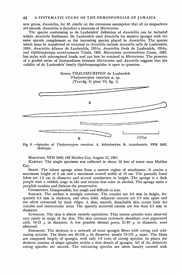
44 A SYSTEMATIC STUDY OF T H E DEMOSPONGIAE OF JAMAICA
new genus, Axocielita, for M. similis on the erroneous assumption that all its megascleres are smooth. Axocielita is therefore a synonym of Microciona.
The species conforming to de Laubenfels' definition of Axocielita can be included within Axociella Hallmann. De Laubenfels used Axociella for massive sponges with the same spicule complement as the incrusting species placed in Axocielita. The species which must be transferred or returned to Axociella include Axociella calla de Laubenfels, 1934; Axocielita kilauea de Laubenfels, 1951a; Axocielita linda de Laubenfels, 1954a; and Ophlitaspongia membranacea Thiele, 1905. Microciona aceratoobtusa Carter, 1887, has styles with microspined heads, and can best be retained in Microciona. The presence of a graded series of intermediates between Microciona and Axociella suggests that the validity of de Laubenfels* family Ophlitaspongiidae is open to question.
GENUS THALYSEURYPON de Laubenfels Thalyseurypon conulosa n. sp.
(Text-fig. 9; plate VI, fig. 1)
Fig. 9. —Spicules of Thalyseurypon conulosa. A. Subtylostyles. B. Acanthostyle. YPM 5042. Holotype.
HOLOTYPE. YPM 5042. Off Maiden Cay, August 12, 1961. HABITAT. The single specimen was collected in about 10 feet of water near Maiden
Cay. SHAPE. The lobate sponge arises from a narrow region of attachment. It attains a
maximum height of 8 cm and a maximum overall width of 10 cm. The partially fused lobes are 1-2 cm in diameter and several centimeters in height. The sponge is a dark purple with a reddish tinge in life and retains that color in alcohol. The sponge emits a purplish exudate and darkens the preservative.
CONSISTENCY. Compressible, but tough and difficult to tear. SURFACE. The surface is strongly conulose. The conules are 3-6 mm in height, fre
quently 0.5 mm in thickness, and often bifid. Adjacent conules are 2-5 mm apart and are often connected by thick ridges. A thin, smooth, detachable skin covers both the conules and interconular areas. The sparsely scattered oscules are less than 0.5 mm in diameter.
ECTOSOME. The skin is almost entirely aspiculous. Thin oxeote spicules were observed very rarely in strips of the skin. The skin contains extremely abundant oval pigmented cells, 10-13 ix in diameter. A few possible dermal pores, 21-35 ^ in diameter, were observed.
ENDOSOME. The skeleton is a network of stout spongin fibers with coring and echi-nating spicules. The fibers are 40-150 ^ in diameter, mostly 75-175 p apart. The fibers are composed largely of spongin, with only 1-3 rows of coring spicules. In places the skeleton consists of single spicules within a thin sheath of spongin. All of the definitely coring spicules are smooth. The echinating spicules are often basally covered with

SYSTEMATICS 45 spongin. Many echinators are entirely smooth. Others have a smooth shaft and finely spined head. Here and there, an entirely spined acanthostyle can be observed. A very few thin oxeote spicules are present in the flesh.
SPICULES, (a) Styles, slightly curved to straight, 203-449 x 7-10 ^ (125 spicules). The spicules are somewhat subtylostylote, with a slight subterminal expansion and a rounded end. For example, a spicule with a shaft diameter of 10 microns has a maximum head diameter of 12 p. Below the head, the spicules taper gradually to a sharp point. A few spicules are definitely styles. Tylostrongyles occur in small numbers. Some echinators have roughened or finely spined heads. Immature spicules range in form from thin lobate tylostyles to thin blunt-ended styles. A few spicules of typical shape are extremely long. Spicules of 807, 898, 1037 and 1334 ^ were recorded, (b) Echinating acanthostyles, 130-230 X 5-12 [JL (25 spicules). The very fine spines are scattered irregularly over the entire surface of the spicule. They are 1 /x or less in height, and 2-15 ^ apart, (c) Thin, fusiform oxeas, 341-652 X 2-3 p. (15 spicules). They are uncommon, but apparently proper to the sponge.
DISCUSSION. The large size, lack of microscleres, and presence of some spiny echinators, places the specimen in the genus Thalyseurypon. The presence of some entirely smooth echinators is noteworthy for the genus. The sponge is quite different from any of the West Indian members of the genus. T. vasiformis de Laubenfels (1953a) is a vase with very large tylostyles. T. foliacea (Topsent, 1889) has spinous coring spicules and leaf-like branches. T. carteri Topsent (1889) has thick, flat branches, and numerous interstitial spicules.
Thalyseurypon conulosa is similar in shape, conule size and color to Pandaros acanthi-folium Duchassaing and Michelotti (1864, p. 90). The latter species was described from St. Thomas and reported from the Dry Tortugas by de Laubenfels (1936a, p. 123). De Laubenfels reported "occasional acanthose modifications" in the spicules of the St. Thomas specimen (p. 124). T. conulosa differs from Pandaros acanthifolium in having definite echinators, including a distinctive category of small acanthostyles. De Laubenfels emphasized the fact that "The fibers are not at all echinated" in his redefinition of Pandaros (p. 123). T. conulosa also lacks the subdermal spaces and unusually spongy consistency of de Laubenfels* material.
FAMILY MYCALIDAE Lundbeck, 1905 (=Amphilectidae, de Laubenfels 1936a)
A reticulate skeleton. The fibers or spicule tracts are never echinated. The mega-scleres are monactinal. The microscleres, usually present, may include sigmas, chelas, toxas or raphides.
De Laubenfels (1936a) curiously assigned Mycale and Zygomycale to his family Ophlitaspongiidae, which is defined as having smooth, echinating spicules. The two genera lack echinators and thus key out to his family Amphilectidae.
Mycale (along with its synonym Esperella, and the preoccupied Esperia) has always been used for sponges with compact spicule tracts. Topsent (1924), revising the European species of Mycale, described the skeleton as consisting of polyspicular fibers. The writer has found no mention of echinators in any description of mycalid species, including those of de Laubenfels. Mycale has indeed been used by many authors as the type genus of a family characterized by the lack of echinators.
Gray (1867, p. 531) established a family Esperiadae to include Esperia and Mycale. (The two genera were distinguished on the basis of their microsclere complement). Vosmaer (1886, p. 353) noted that Esperia was a preoccupied name and replaced it by Esperella. Ridley and Dendy (1887), divided their family Desmacidonidae into two subfamilies, the Esperellinae and the Ectyoninae. The former subfamily was characterized by non-echinated, and the latter by echinated fibers. Thiele (1905, p. 949) placed Esperella into synonymy with Mycale, and Lundbeck (1905, p. 7) accordingly replaced the name Esperellinae with Mycalinae. The group was raised to a family in the classification of Hentschel (1923).
The Ophlitaspongiidae and Amphilectidae, as listed by de Laubenfels (1936a) probably require further revision, but many of the genera are unfamiliar to me.
46 A SYSTEMATIC STUDY OF T H E DEMOSPONGIAE OF JAMAICA
GENUS MYCALE Gray Mycale laevis (Carter) de Laubenfels
(Plate VI, fig. 2)
Esperia laevis Carter, 1882, p. 291 [type: Puerto Cabello, Venezuela; Brit. Mus. (Nat. Hist.) (?)]
Mycale laevis, de Laubenfels, 1936a, p. 116.
HABITAT. Very common on pilings at Port Royal. SHAPE. A typical specimen is fist-sized, massive and lobate. Cylindrical specimens also
occur. An example growing on a crab's back is a horizontally spreading mass, two mm to 2 cm in thickness.
COLOR. In life, the exterior is a light orange, while the interior is a dull yellow. A specimen dried after initial preservation in alcohol became white in color. In alcohol, the sponges are beige, with or without traces of the original orange color.
CONSISTENCY. Spongy, compressible. SURFACE. Smooth to the touch, but uneven in places. The oscules are 0.5-1.5 cm in
diameter. Each receives the openings of several large exhalant canals. In some cases, a deep cloaca is present. The oscules are usually apical on the lobes. They are bordered by a very characteristic membranous rim which is sometimes divided into filiform processes. The resultant collar may reach a centimeter in height.
ECTOSOME. The oscular rim contains stout, longitudinal, occasionally anastomosing spicule bundles, 100-600 fx in diameter. They are also joined by connective tracts. The rim membranes contain numerous microscleres, including rosettes of large anisochelas.
A detachable skin (which easily becomes macerated) covers the surface. The dermal skeleton includes an irregular reticulation of spicule bundles, 40-145 jx in diameter. The bundles grade into loosely organized groups of spicules. A considerable number of megascleres are strewn in the flesh. The interstices of the network are often 70-150 fx in width. They contain microscleres of all types, except for large anisochelas. The scattered ostia are 30-145 ju, in diameter. Their partition walls are often 30-40 ju. in width and contain relatively few microscleres (mostly raphides and small anisochelas.)
ENDOSOME. The interior contains scattered megascleres and a coarse irregular network of spicule bundles. The interstitial spicules are of varying thickness, while those of the tracts are robust. The reticulate skeleton becomes more prominent near the periphery. Numerous tracts ascend to the surface, sometimes branching in the process, to end below the dermis in slightly expanded tufts. Conspicuous rosettes of a dozen or more anisochelas are common near the ascending tracts. The periphery of the endosome also contains a great abundance of very compact trichodragmata, 50-70 /*, in length and about 10 fx in diameter. Such compact bundles are often clustered together. Compact trichodragmata, in smaller numbers, occur throughout the sponge along with single raphides and loose sheaves of raphides similar to those figured by Carter. Sigmas are present in great numbers in the flesh.
SPICULES, (a) Styles, sometimes slightly subtylostylote, straight to somewhat curved, with a narrowed base, and rather abrupt point, 377-596 x 7-18 p. (b) Large palmate anisochelas, with a straight, thick shaft, 57-100 JX total length x 7-10 [x maximum shaft diameter. The large (upper) end consists of a prominent wide central tooth flanked by attached palms, similar in length to the tooth. The lower end is expanded transversely, terminating at each side in a plate which is directed away from the shaft on the same side of the spicule as the central upper tooth. The upper end of the spicule is about as long as the tooth gap and is between two and three times the length of the lower end. The respective figures for a typical chela are 34, 30, and 13 [i. The maximum width of the upper end is usually about 25-40 LI. The lower end is 2/3 to % the width of the larger end. (c) Small anisochelas, with very long upper ends, and narrow, short, lower ends, 15-28 jx total length X 2 /x shaft diameter. As seen in profile, the upper tooth extends down nearly to the level of the lower end. The lower end is sometimes extended by a small apical projection about 2 LL in length, (d) Sigmas, 17-77 X 1-3 p. (e) Raphides,

SYSTEMATICS 47 27-102 X 2 /i. Many are grouped into the previously described trichodragmata and sheaves. Individual analyses, lengths in microns, 50 styles and 25 of each category of microsclere per sponge:
Styles
463-565 421-541 467-596 492-590 377-536
Large Anisochelas
68-87 65-86 70-96 57-100 63-82
Small Anisochelas
15-23 17-28 17-23 20-27 17-22
Sigmas
18-50 17-63 18-57 23-77 24-46
Raphides
50-87 48-102 51-85 27-93 50-87
DISTRIBUTION. Tropical Atlantic America: Venezuela—Carter, 1882, p. 291 (as Esperia laevis).
DISCUSSION. The Jamaican sponge resembles Carter's Venezuelan species in form, spicule assortment and spicule sizes. The shapes of the spicules are, for the most part, in close agreement. The small anisochelas of my specimens, however, have at best a slight lower projection which never attains the proportions shown in Carter's figures. Carter mentions neither membranous nor feathery rims for the oscules. They are certainly quite characteristic of the Jamaican sponge. Being delicate, they may have been destroyed on the specimens available to Carter.
Mycale microsigmatosa (Arndt) Burton
Mycale fistulata var. microsigmatosa Arndt, 1927, p. 144 [type: Curasao; repository not specified.]
Mycale microsigmatosa, Burton, 1956, p. 129. Mycale cecilia de Laubenfels, 1936b, p. 447; idem, 1950b, p. 24; Wells et al., 1960, p.
212 (?) Mycale senegalensis LeVi, 1952, p. 46; idem, 1959, p. 129.
HABITAT. Common on mangrove roots and on turtle grass. SHAPE. All of the dozens of specimens seen in the field were incrustations, 1-3 mm in
thickness. COLOR. In life, pale orange or pink, or most frequently a dull bluish-green. Bright
red-orange specks, presumably embryos, are present in most specimens at all times of the year. The sponges are predominantly beige in alcohol.
CONSISTENCY. Soft. SURFACE. Smooth to the touch, even. In life, the oscules have delicate, transparent,
raised collars. The oscules are supplied by river-like systems of subdermal exhalant canals. The canals are often several millimeters in diameter and extend outward for as much as 1 cm.
ECTOSOME. A thin, detachable dermal membrane is pierced by numerous ostia, 35-120 JU, in diameter. The partitions between adjacent pores are 2-5 p, in thickness and contain scattered microscleres. The pores are clustered into groups of 3-12 or more by dense bands of flesh, which are 10-30 //, in diameter. The skin is supported by the plumose ends of ascending tracts, but megascleres are not present in the dermal membrane.
ENDOSOME. The skeletal framework consists of spicule tracts 20-100 p in diameter. The tracts occasionally branch or anastomose in the interior. They often subdivide in ascending to the surface. Microscleres are abundant throughout the sponge. The anisochelas are not localized by size. The ovoid embryos are 175-350 ^ in longest diameter.
SPICULES, (a) Tylostyles, straight to curved, with narrow, somewhat ovoid heads and hastate points, 214-304 X 3-12 /A. The maximum diameter is at the mid-length of the shaft, (b) Sigmas, strongly curved, 30-50 x 1-3 /x. (c) Anisochelas, narrow and palmate, 15-27 p. The shaft is about 2 /x in diameter. The upper palm has a maximum width of 7 ft in larger spicules. It is usually equal to i/2 or more of the total length of the spicule.
48 A SYSTEMATIC STUDY OF T H E DEMOSPONGIAE OF JAMAICA
Some of the smaller spicules have very short shafts. Individual analyses, lengths in microns, 50 megascleres each, and 25 microscleres per category each:
Tylostyles
214-287 234-275 234-264 228-287 234-304
Sigmas
33-46 30-42 33-50 30-44 33-46
Anisochelas
16-25 16-23 17-27 15-26 15-23
DISTRIBUTION. Tropical Atlantic America: North Carolina—Wells et al., 1960, p. 212 (as Mycale cecilia) ?; Curasao, Arndt, 1927, p. 144 (as Mycale fistulata var. microsigma-tosa). Tropical West Africa: Senegal—LeVi, 1952, p. 46 (as Mycale senegalensis). Hawaiian Is.—de Laubenfels, 1950b, p. 24 (as Mycale cecilia). Tropical Pacific America: Pacific coast of Panama—de Laubenfels, 1936b, p. 447 (as Mycale cecilia).
DISCUSSION. The anisochelas were of a single size range in all of the Jamaican sponges. Continuous ranges have also been reported by Arndt (1927), de Laubenfels (1950b) and LeVi (1952). A spicule preparation obtained from one of de Laubenfels' Hawaiian specimens (U. S. National Museum number 22747) contains anisochelas 13-24 ^ in length. Wells et al., (1960) reported two distinct sizes of anisochelas. Their North Carolina material differs further from the Jamaican in having tangential dermal tylostyles and larger sigmas. The dermal membrane of USNM specimen 22747 contains only microscleres. De Laubenfels (1936b) reported two sizes of anisochelae in his original description of Mycale cecilia but noted the presence of intermediates in the legend for figure 41.
LeVi (1959) has already suggested that Mycale senegalensis L£vi, 1952 might be con-specific with Arndt's M. fistulata microsigmatosa. The Curacao sponge is certainly not conspecific with the tubular Australian Mycale fistulata Hentschel, 1911. M. cecilia de Laubenfels cannot be distinguished from Arndt's variety and must be regarded as a synonym of M. microsigmatosa.
Closely related species are known from Hawaii (M. maunakea de Laubenfels, 1951a) and Australia (M. phyllophila Hentschel, 1911). M. maunakea differs from M. microsigmatosa in having unusually narrow isochelas. Its spicule tracts neither branch nor anastomose. M. phyllophila, while very similar to the West Indian sponge, is geographically remote. M. microsigmatosa is therefore retained as a separate species.
GENUS ZYGOMYCALE Topsent Zygomycale parishii (Bowerbank) Topsent
(Plate V, fig. 3)
Raphiodesma parishii Bowerbank, 1875, p. 283 [type: Straits of Malacca; Brit. Mus. (Nat. Hist.) (?)]
Esperia parishii, Ridley, 1884, p. 436. Esperella parishii, Ridley and Dendy, 1887, p. 65; Dendy, 1905, p. 159. Zygomycale parishii, Topsent, 1929, p. 431; de Laubenfels, 1950b, p. 25; idem, 1956, p.
3; Levi, 1956a, p. 14. Mycale parishii, Burton and Rao, 1932, p. 328. Esperia plumosa Carter, 1882, p. 298 (fide Burton and Rao, 1932); idem, 1887, p. 72. Esperella plumosa, Dendy, 1905, p. 159; idem, 1916a, p. 121; Dendy and Frederick, 1924,
p. 503. Esperella ridleyi Lendenfeld, 1888, p. 211 (fide Burton and Rao, 1932); Hallmann, 1914,
p. 402. Mycale isochela Hentschel, 1911, p. 297 (fide Burton and Rao, 1932.) Mycale pectinicola Hentschel, 1911, p. 299 (fide Burton and Rao, 1932.)
HABITAT. Specimens were collected on pilings at Port Royal and also at Gunboat Beach on the peninsula between Port Royal and Kingston.
SYSTEMATICS 49 SHAPE. The sponge has a small, incrusting or irregularly massive base from which
arise long, subdividing, anastomosing branches. Many specimens reach 20-25 cm in length. The branches are usually only a few millimeters in diameter but occasionally reach a width of 1.5-2 cm.
COLOR. In life the peripheral layer may be tan, brown, grayish-brown, or purple. Toward the interior the color gradually becomes a dull yellow. In alcohol, the sponges may be pale yellow, beige, or partially purple.
CONSISTENCY. Somewhat compressible but tough. SURFACE. The sponge is covered by a skin which is smooth to the touch. The overall
appearance is uneven. In addition to numerous projections and branches, the skin is thrown into low elevations, ridges and conules. On the branches, the conules are sometimes several millimeters in height. The small scattered oscules are less than 1 mm in diameter.
ECTOSOME. A pore-bearing membrane is supported by a reticulation of loose to compact spicule tracts, which are 30-50 p in diameter. The meshes are triangular, irregularly polygonal, or roughly rectangular in outline. Parallel bundles are frequently 150-250 p apart. The interstices are crowded with dermal pores separated by narrow walls often only 2-3 JX in width. Typical ostial sizes are 40 x 30, 70 x 60, and 88 X 40 ju. The partition walls contain infrequent microscleres, with isochelas being the most numerous category. Rosettes of large anisochelas are frequently present near the bundles. Interstitial megascleres are infrequent.
ENDOSOME. The interior is microcavernous, particularly near the periphery. Subdermal spaces are occasionally present. The skeleton consists of a very irregular network of spicule tracts. The tracts are 40-200 p, in diameter. Spongin is sometimes present. At the periphery, tracts ascend to the surface, to end in slightly expanded tufts under the dermal membrane. Many megascleres are strewn in the flesh. Conspicuous rosettes of 6-10 large anisochelas are present in the periphery. The endosome contains an abundance of microscleres, with isochelas the most numerous, and large sigmas the least numerous category. Small sigmas may be very abundant or quite infrequent. Aspiculous ovoid embryos, about 300 X 400 p as seen in section, were found in specimens collected in February and June.
SPICULES, (a) Subtylostyles, occasionally becoming styles, with narrow elongate heads, curved to undulating shafts, and gradually narrowing apices, 217-334 x 3-12 /x. The maximum diameter is at the mid-length of the shaft. A few megascleres have abnormal swellings and projections, (b) Large palmate anisochelas, 37-67 X 2 p shaft diameter. They are similar in shape to the anisochelas of Mycale laevis. The width of the upper end is 15-20 p; that of the lower end 10-12 p. A typical spicule 48 microns long has an upper tooth of 24 p, a lower end of 9 p and a tooth gap of 15 p. The spicules occur singly or, more frequently, in rosettes near the periphery of the endosome. (c) Small palmate anisochelas, 13-28 tt long. The maximum width at the upper end is about 6 /*,. A spicule 17 p long has an upper tooth of 10 p, a lower end of 4 p, and tooth gap of 3 ju,. (d) Palmate isochelas, 10-13 p long. The cap-shaped ends are about 3/x in width. Very abundant, (e) Large sigmas, with sharply recurved ends, 54-106 x 2-7 p.. They are very abundant, (f) Small slender sigmas, 15-42 X 1-2 p. Varied in abundance, (g) Toxas, often in toxodragmata of several spicules, 22-107 X 2 p. They have a shallow, gradually curved arch, and flattened to slightly recurved ends, (h) Raphides, often in trichodragmata, 23-53 X 1-2 JU,. All categories are present in the seven analysed specimens. Individual analyses, lengths in microns, 50 megascleres per sponge, 25 microscleres of each category per sponge:
Large Small Styles Anisochelas Anisochelas Isochelas
236-326 43-67 13-28 10-13 234-334 46-54 16-28 10-13 217-314 45-52 17-22 10-13 239-290 37-52 17-20 10-12
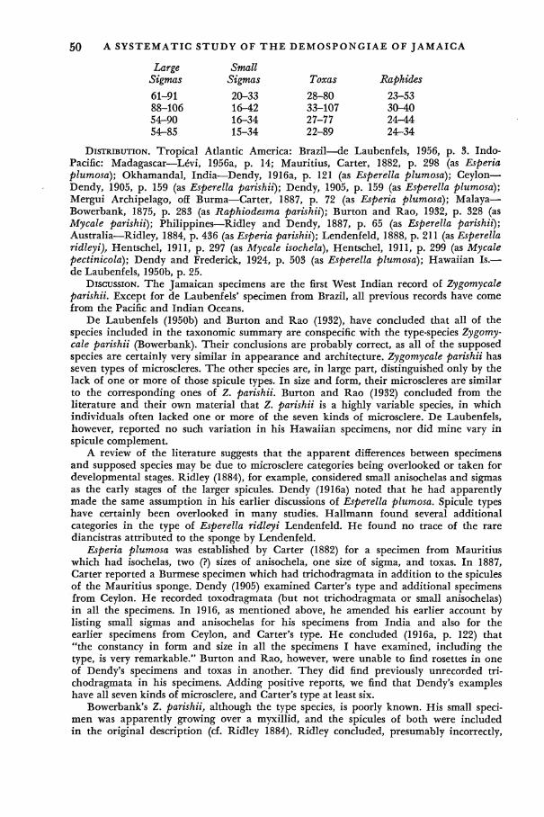
50 A SYSTEMATIC STUDY OF T H E DEMOSPONGIAE OF JAMAICA
Large Sigmas
61-91 88-106 54r-90 54-85
Small Sigmas
20-33 16-42 16-34 15-34
Toxas
28-80 33-107 27-77 22-89
Raphides
23-53 30-40 24-44 24-34
DISTRIBUTION. Tropical Atlantic America: Brazil—de Laubenfels, 1956, p. 3. Indo-Pacific: Madagascar—L£vi, 1956a, p. 14; Mauritius, Carter, 1882, p. 298 (as Esperia plumosa); Okhamandal, India—Dendy, 1916a, p. 121 (as Esperella plumosa); Ceylon— Dendy, 1905, p. 159 (as Esperella parishii); Dendy, 1905, p. 159 (as Esperella plumosa); Mergui Archipelago, off Burma—Carter, 1887, p. 72 (as Esperia plumosa); Malaya— Bowerbank, 1875, p. 283 (as Raphiodesma parishii); Burton and Rao, 1932, p. 328 (as Mycale parishii); Philippines—Ridley and Dendy, 1887, p. 65 (as Esperella parishii); Australia—Ridley, 1884, p. 436 (as Esperia parishii); Lendenfeld, 1888, p. 211 (as Esperella ridleyi), Hentschel, 1911, p. 297 (as Mycale isochela), Hentschel, 1911, p. 299 (as Mycale pectinicola); Dendy and Frederick, 1924, p. 503 (as Esperella plumosa); Hawaiian Is.— de Laubenfels, 1950b, p. 25.
DISCUSSION. The Jamaican specimens are the first West Indian record of Zygomycale parishii. Except for de Laubenfels* specimen from Brazil, all previous records have come from the Pacific and Indian Oceans.
De Laubenfels (1950b) and Burton and Rao (1932), have concluded that all of the species included in the taxonomic summary are conspecific with the type-species Zygomycale parishii (Bowerbank). Their conclusions are probably correct, as all of the supposed species are certainly very similar in appearance and architecture. Zygomycale parishii has seven types of microscleres. The other species are, in large part, distinguished only by the lack of one or more of those spicule types. In size and form, their microscleres are similar to the corresponding ones of Z. parishii. Burton and Rao (1932) concluded from the literature and their own material that Z. parishii is a highly variable species, in which individuals often lacked one or more of the seven kinds of microsclere. De Laubenfels, however, reported no such variation in his Hawaiian specimens, nor did mine vary in spicule complement.
A review of the literature suggests that the apparent differences between specimens and supposed species may be due to microsclere categories being overlooked or taken for developmental stages. Ridley (1884), for example, considered small anisochelas and sigmas as the early stages of the larger spicules. Dendy (1916a) noted that he had apparently made the same assumption in his earlier discussions of Esperella plumosa. Spicule types have certainly been overlooked in many studies. Hallmann found several additional categories in the type of Esperella ridleyi Lendenfeld. He found no trace of the rare diancistras attributed to the sponge by Lendenfeld.
Esperia plumosa was established by Carter (1882) for a specimen from Mauritius which had isochelas, two (?) sizes of anisochela, one size of sigma, and toxas. In 1887, Carter reported a Burmese specimen which had trichodragmata in addition to the spicules of the Mauritius sponge. Dendy (1905) examined Carter's type and additional specimens from Ceylon. He recorded toxodragmata (but not trichodragmata or small anisochelas) in all the specimens. In 1916, as mentioned above, he amended his earlier account by listing small sigmas and anisochelas for his specimens from India and also for the earlier specimens from Ceylon, and Carter's type. He concluded (1916a, p. 122) that "the constancy in form and size in all the specimens I have examined, including the type, is very remarkable." Burton and Rao, however, were unable to find rosettes in one of Dendy's specimens and toxas in another. They did find previously unrecorded trichodragmata in his specimens. Adding positive reports, we find that Dendy's examples have all seven kinds of microsclere, and Carter's type at least six.
Bowerbank's Z. parishii, although the type species, is poorly known. His small specimen was apparently growing over a myxillid, and the spicules of both were included in the original description (cf. Ridley 1884). Ridley concluded, presumably incorrectly,
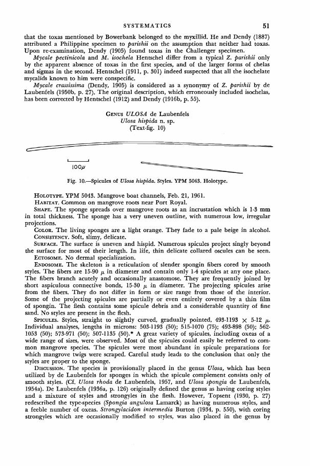
SYSTEMATICS 51 that the toxas mentioned by Bowerbank belonged to the myxillid. He and Dendy (1887) attributed a Philippine specimen to parishii on the assumption that neither had toxas. Upon re-examination, Dendy (1905) found toxas in the Challenger specimen.
Mycale pectinicola and M. isochela Hentschel differ from a typical Z. parishii only by the apparent absence of toxas in the first species, and of the larger forms of chelas and sigmas in the second. Hentschel (1911, p. 301) indeed suspected that all the isochelate mycalids known to him were conspecific.
Mycale crassissima (Dendy, 1905) is considered as a synonymy of Z. parishii by de Laubenfels (1950b, p. 27). The original description, which erroneously included isochelas, has been corrected by Hentschel (1912) and Dendy (1916b, p. 55).
GENUS ULOSA de Laubenfels Ulosa hispida n. sp.
(Text-fig. 10)
Fig. 10.—Spicules of Ulosa hispida. Styles. YPM 5043. Holotype.
HOLOTYPE. YPM 5043. Mangrove boat channels, Feb. 21, 1961. HABITAT. Common on mangrove roots near Port Royal. SHAPE. The sponge spreads over mangrove roots as an incrustation which is 1-3 mm
in total thickness. The sponge has a very uneven outline, with numerous low, irregular projections.
COLOR. The living sponges are a light orange. They fade to a pale beige in alcohol. CONSISTENCY. Soft, slimy, delicate. SURFACE. The surface is uneven and hispid. Numerous spicules project singly beyond
the surface for most of their length. In life, thin delicate collared oscules can be seen. ECTOSOME. No dermal specialization. ENDOSOME. The skeleton is a reticulation of slender spongin fibers cored by smooth
styles. The fibers are 15-90 /x in diameter and contain only 1-4 spicules at any one place. The fibers branch acutely and occasionally anastomose. They are frequently joined by short aspiculous connective bonds, 15-30 p in diameter. The projecting spicules arise from the fibers. They do not differ in form or size range from those of the interior. Some of the projecting spicules are partially or even entirely covered by a thin film of spongin. The flesh contains some spicule debris and a considerable quantity of fine sand. No styles are present in the flesh.
SPICULES. Styles, straight to slightly curved, gradually pointed, 493-1193 X 5-12 /*. Individual analyses, lengths in microns: 503-1193 (50); 515-1070 (75); 493-898 (50); 562-1053 (50); 573-971 (50); 507-1135 (50).* A great variety of spicules, including oxeas of a wide range of sizes, were observed. Most of the spicules could easily be referred to common mangrove species. The spicules were most abundant in spicule preparations for which mangrove twigs were scraped. Careful study leads to the conclusion that only the styles are proper to the sponge.
DISCUSSION. The species is provisionally placed in the genus Ulosa, which has been utilized by de Laubenfels for sponges in which the spicule complement consists only of smooth styles. (Cf. Ulosa rhoda de Laubenfels, 1957, and Ulosa spongia de Laubenfels, 1954a). De Laubenfels (1936a, p. 126) originally defined the genus as having coring styles and a mixture of styles and strongyles in the flesh. However, Topsent (1930, p. 27) redescribed the type-species (Spongia angulosa Lamarck) as having numerous styles, and a feeble number of oxeas. Strongylacidon intermedia Burton (1934, p. 550), with coring strongyles which are occasionally modified to styles, was also placed in the genus by

52 A SYSTEMATIC STUDY OF T H E DEMOSPONGIAE OF JAMAICA
de Laubenfels. Burton's species is probably not congeneric with any of the species now in Ulosa.
The present species differs sharply from the type-species in appearance and spicule size. It does seem congeneric with U. spongia and U. rhoda de Laubenfels. The distinctive characters of the Jamaican sponge include the large size of its spicules, the abundance of foreign matter, the spongin-rich fibers, and the hispidation of the surface by single spicules.
ORDER HALICHONDRIDA Vosmaer 1887, sensu de Laubenfels, 1936a FAMILY HALICHONDRIIDAE Gray, sensu de Laubenfels, 1936a
GENUS HALICHONDRIA Gray Halichondria melanadocia de Laubenfels
(Plate V, fig. 2)
Halichondria melanadocia de Laubenfels, 1936a, p. 133 [type: Dry Tortugas; USNM no. 22463]; Wells et al., 1960, p. 225; Little, 1963, p. 54.
HABITAT. Abundant on pilings and mangrove roots near Port Royal. SHAPE. Specimens on mangrove roots often spread horizontally along the roots, at
taining a thickness of one to several centimeters. Others are irregularly massive, lobate or even partly ramose.
COLOR. The exterior is grayish-black. The interior has a very characteristic yellowish-green tinge. In alcohol, the specimens are dark gray to nearly black with a drab interior.
CONSISTENCY. Compressible. SURFACE. Smooth to the touch. Projecting lobes and low oscular chimneys often make
the surface rather uneven. The oscules, which also may be flush with the surface, are 2-3 mm in diameter. They have a membranous, irregularly outlined rim.
ECTOSOME. The detachable dermal skin contains a tangential reticulation of compact spicule bundles, 35-130 p in diameter. The interstices are irregularly polygonal in shape, and 100-200 ^ in maximum width. They contain a few scattered spicules and one to several dermal pores. Typical ostial sizes are 40 x 30, 90 X 60, and 145 x 90 p. The subdermal channels are very extensive and often reach 1 mm in depth. They open widely into macroscopic inhalant canals which run toward the interior. The thick partitions between the subdermal spaces are fleshy extensions of the endosome, 300-700 fi in diameter. They contain scattered spicules and also spicule tracts which run obliquely towards the surface.
ENDOSOME. The interior is cavernous and confused. The thickly strewn spicules are occasionally grouped into loosely organized tracts.
SPICULES. Oxeas, straight to considerably curved, 134-802 X 3-20 p. The ends may be gradually pointed, somewhat hastate, irregular in outline, or even stair-stepped. The spicules are not localized in the sponge by size. Individual analyses, lengths in microns, 50 spicules each: 140-656; 164-686; 134-597; 134-691; 141-743; 152-802.
DISTRIBUTION. Tropical Atlantic America: North Carolina—Wells et al., 1960, p. 225 (?); Dry Tortugas, Florida—de Laubenfels, 1936a, p. 133; West Coast, Florida—Little, 1963, p. 54.
DISCUSSION. The dark Jamaican Halichondria agrees closely with H. melanadocia in structure and external appearance. De Laubenfels' brief description is at least not inconsistent with the dermal structure found in my specimens. It would be of great interest to know if the tangentially arranged dermal spicules of his sponge are merely strewn, or grouped into bundles. The fetid odor mentioned by de Laubenfels did not occur in my sponges. A spicule preparation obtained from the type contains oxeas 116-507 fi in length. Most of the spicules are straight and hastate. Their ends are occasionally irregular or stair-stepped. The Jamaican Halichondria is therefore provisionally considered as conspecific with that of de Laubenfels.
The North Carolina record listed by Wells et al. concerns a thin brown conulose incrustation. Further specimens will be needed to confirm the identification.

SYSTEMATIGS 58
Halichondria magniconulosa n. sp. (Text-fig. 11; plate V, fig. 1)
Fig. 11.—Spicules of Halichondria magniconulosa. Oxeas. YPM 5039. Holotype.
HOLOTYPE. YPM 5039. Rasta's Wreck, June 24, 1961. HABITAT. Several specimens were found on pilings at Port Royal. SHAPE. Massive to lobate, reaching a height of as much as 11 cm. COLOR. The surface and interior of the living sponge is yellow. In alcohol, exposed
surfaces become mottled grayish-brown to dark brown. The interior becomes a dull yellow.
CONSISTENCY. The sponge is tough and only slightly compressible. SURFACE. The skin is wrinkled, but smooth to the touch. It can be detached only
with difficulty. The surface is frequently thrown into thick, sharp-pointed conules. The conules are 1-7 mm in height, several millimeters in width at the base, and typically about 1 mm in thickness. They may be sparsely scattered or spaced only a few millimeters apart. In the two smaller specimens, the infrequent oscules are 1-3 mm in diameter. The largest specimen has two apical vents, each being 1 cm in diameter.
ECTOSOME. The dermal membrane receives the terminal tufts of radial spicule tracts. It also contains rather sparsely scattered tangential spicules of all sizes. Small spicules are particularly abundant. The dermal pores range in size from 40 x 30-100 X 60 /x. They are divided into ill-defined groups by bands of flesh which are 15-60 p in diameter. The spicules within the partitions may be scattered or organized into loose bundles. Some areas of the skin seem to lack pores.
ENDOSOME. The interior skeleton includes a feltwork of scattered spicules and also spicule bundles, 30-100 p in diameter. The well-developed radial tracts end in dermal tufts. Numerous spicule bundles enter each conule. Subdermal spaces are infrequent.
SPICULES. Oxeas, mostly straight, occasionally slightly curved, 108-369 x 3-7 p. They may be fusiform or somewhat hastate. Small spicules (123-166 p) and large spicules (290-340 fi) are particularly common but intermediates do occur. The spicules are not localized by size within the sponge. Individual analyses, lengths in microns, 50 spicules each: 116-363*; 108-348; 123-369.
DISCUSSION. The massive shape and thick conules of the Jamaican yellow Halichondria separate it from both H. panicea and H. bowerbanki. The dermis of the Jamaican sponge resembles that of H. bowerbanki, but is less well developed. Many of the spicules seen in strips of the skin are clearly the ends of radial bundles. Subdermal cavities are quite restricted in my specimens. The Jamaican sponges have a much lower limit of spicule size than either North Carolina or New England summer specimens of H. bowerbanki. In addition, the smaller spicules are very abundant in my material. The marked darkening in alcohol is a striking characteristic of the new species. H. bowerbanki, by contrast, becomes a pale yellowish-green in alcohol.

54 A SYSTEMATIC STUDY OF T H E DEMOSPONGIAE OF JAMAICA
ORDER HADROMERIDA Topsent, 1898b FAMILY SPIRASTRELLIDAE Ridley and Dendy, sensu de Laubenfels, 1936a
GENUS SPIRASTRELLA Schmidt Spirastrella coccinea (Duchassaing and Michelotti) de Laubenfels
Thalysias coccinea Duchassaing and Michelotti, 1864, p. 84 [type: St. Thomas; Brit. Mus. (Nat. Hist.) reg. no. 28.11.12.46.]
Spirastrella coccinea, de Laubenfels, 1936a, p. 143; idem, 1949, p. 19; idem, 1950a, p. 96; Dickinson, 1945, p. 36; Wells et al., 1960, p. 228; Little, 1963, p. 55.
Spirastrella cunctatrix Schmidt, 1868, p. 17; Carter, 1879a, p. 357; idem, 1881a, p. 384; idem, 1882, p. 351; idem, 1886, p. 112 (fide Dendy, 1897); idem, 1887, p. 188; Topsent, 1894a, p. 44; idem, 1918, p. 544; idem, 1925a, p. 630; idem, 1938, p. 21; Dendy, 1897, p. 202, 253; Arndt, 1927, p. 140; Burton, 1936a, p. 22; de Laubenfels, 1936b, p. 461; idem, 1956, p. 2; LeVi, 1957, p. 204; idem, 1959, p. 124; Sara, 1958a, p. 221; idem, 1960a, p. 451; Sara and Siribelli, 1960, p. 39; Sara, 1961a, p. 34.
Chondrilla phyllodes Schmidt, 1870, p. 26 (fide Topsent, 1918); Topsent, 1889, p. 32; Carter, 1890, p. 565.
Chondrillina phyllodes, de Laubenfels, 1936a, p. 182. Spirastrella decumbens, Topsent (not Ridley, 1884, p. 470), 1897, p. 440 (fide Topsent,
1918.) Spirastrella mollis Verrill, 1907, p. 344 (fide de Laubenfels, 1950a.) Spirastrella purpurea, Vosmaer, 1911a, p. 9, 11 [partim]; idem, 1933, p. 416 [partim];
(not Alcyonium purpureum Lamarck, 1815.)
HABITAT. Drunkenman's Cay; on rocks. SHAPE. Incrusting, about 1 mm thick. COLOR. One specimen was dull orange in life and is a pale grayish-brown in alcohol.
The second was brown in life and is a pale brown in preservative. CONSISTENCY. Tough, slightly compressible. SURFACE. Smooth to the touch; even to very slightly wrinkled. In the living sponge,
river-like systems of subdermal canals lead to small oscules. Neither vents nor canals are evident in preserved material.
ECTOSOME. No detachable skin. Spirasters, mostly of small size, form a cortical layer 40-100 ii in thickness. Bands of microscleres inclose dermal pores which are about 30 /*, in diameter.
ENDOSOME. Numerous tylostyles are strewn in confusion. A few spicule tracts are present, particularly in the originally brown specimen. The tracts are often obliquely oriented. They end in plumose tufts at the surface. Spirasters, mostly of large size, are abundant in the flesh.
SPICULES, (a) Tylostyles, pin-like, with long, straight, gradually pointed shafts and distinct heads, 281-480 x 3-12 fi (typically 5-9 ^t). The heads may be ovoid, lobate, or occasionally spherical. They are about 9 p in diameter, (b) Spirasters, prominently spined, 7-58 x 2-7 //,. The microscleres have a wide variety of forms, which are all connected by intermediates. The smaller spicules typically have a slender, slightly curved shaft which bears spines 2-3 /A in height. Some are amphiasters, with spines grouped at the ends of a straight shaft. Most of the larger spicules have a stout curved axis, with one or two bends. Their conical spines are 5-7 fx or even as much as 13 /x in height. Some have a rather short axis with strong spines and resemble the spicules of Diplastrella bistellata (Schmidt) Topsent. A few of the endosomal spirasters have a long, thin axis and low spines. They are 20-25 x 2 fi, and have spines 2 ^ in height. Individual analyses, lengths in microns, 50 spicules per category:
Tylostyles Spirasters 334-445 7-47 281-480 8-58
DISTRIBUTION. Tropical Atlantic America: North Carolina—Wells et al., 1960, p. 228; Dry Tortugas, Florida—de Laubenfels, 1936a, p. 143; West Coast, Florida—Little, 1963,

SYSTEMATICS 55 p. 55; Gulf of Campeche, Mexico—Topsent, 1889, p. 32 (as Chondrilla phyllodes); Atlantic coast of Panama—de Laubenfels, 1936b, p. 461 (as Spirastrella cunctatrix); Brazil— de Laubenfels, 1956, p. 2 (as Spirastrella cunctatrix); Bermuda—Verrill, 1907, p. 344 (as Spirastrella mollis), de Laubenfels, 1950a, p. 96; Bahamas—de Laubenfels, 1949, p. 19; Antilles—Schmidt, 1870, p. 26 (as Chondrilla phyllodes); St. Thomas, W. I.—Duchassa-ing and Michelotti, 1864, p. 84 (as Thalysias coccinea); Curasao—Arndt, 1927, p. 140 (as Spirastrella cunctatrix); Fernando Noronha, tropical South Atlantic—Carter, 1890, p. 565 (as Chondrilla phyllodes)}
Mediterranean—Atlantic—Ligurean Sea, Italy—Sara, 1958a, p. 221 (as Spirastrella cunctatrix); Ischia, Italy—Sara, 1960a, p. 451 (as Spirastrella cunctatrix); Naples, Italy— Sara and Siribelli, 1960, p. 39 (as Spirastrella cunctatrix), Topsent, 1925a, p. 630, (as Spirastrella cunctatrix); Adriatic Sea—Sara, 1961a, p. 34 (as Spirastrella cunctatrix); Israel—LeVi, 1957, p. 204 (as Spirastrella cunctatrix); Mediterranean off Egypt—Burton, 1936a, p. 22 (as Spirastrella cunctatrix), Tunisia—Topsent, 1894a, p. 44 (as Spirastrella cunctatrix); Algeria—Schmidt, 1868, p. 17, and Carter, 1882, p. 351 (both as Spirastrella cunctatrix); Cyprus—Schmidt, 1868, p. 17 (as Spirastrella cunctatrix); West Africa— Topsent, 1918, p. 544, and LeVi, 1959, p. 124 (both as Spirastrella cunctatrix).
Indo-Pacific: Indian Ocean—Mauritius—Carter, 1879a, p. 357, and 1882, p. 351 (both as Spirastrella cunctatrix); Mergui Archipelago, off Burma—Carter, 1887, p. 188 (as Spirastrella cunctatrix).
Pacific Ocean: East Indies—Topsent, 1897, p. 440 (as Spirastrella decumbens) and Vosmaer, 1911a, p. 9, 11 (as Spirastrella purpurea) partim; Australia—Carter, 1879a, p. 357, 1881a, p. 384, 1882, p. 351 (all as Spirastrella cunctatrix)}.
Tropical Pacific America: Gulf of California—Dickinson, 1945, p. 36. DISCUSSION. The spirasters of the Jamaican sponges are similar in range of size and
shape to the microscleres figured by Topsent (1918, p. 543, 545) for Spirastrella cunctatrix Schmidt. Topsent later verified his identification by comparison with Schmidt's type. (cf. Topsent, 1938). S. cunctatrix, along with its synonym Chondrilla phyllodes Schmidt, has been recorded from the West Indies by Topsent (1889, 1918) and Arndt (1927).
Thalysias coccinea Duchassaing and Michelotti, 1864, was originally described as a red incrusting sponge with short-rayed stelliform spicules. De Laubenfels (1936a), who had apparently examined the type, transferred the species to Spirastrella. He distinguished S. coccinea from S. cunctatrix largely by the smaller size of the spirasters in the former species, (cf. de Laubenfels, 1956, p. 2). De Laubenfels did, however, report rather large spirasters in a Bermudan sponge attributed to S. coccinea. (cf. de Laubenfels, 1950a).
An examination of material identified as 5. coccinea by de Laubenfels suggests that S. cunctatrix and S. coccinea are conspecific. The specimens include one from Bermuda in the Yale Peabody Museum (YPM 714). It contains spirasters 13-34 ^ in length. U. S. National Museum specimen 22498, collected by de Laubenfels in the Dry Tortugas, has spirasters 10-34 p in length. The two sponges are indistinguishable from Sv cunctatrix and the Jamaican sponges in architecture and range of spiraster form. Most of the spirasters in one Jamaican specimen (having an overall size range of 7-47 fx) are 35 ^ or less in length. The observed differences in maximum length are almost certainly individual variations. S. cunctatrix is therefore considered to be a synonym of the earlier-named S. coccinea.
Carter's Indo-Pacific records require verification. His material included massive specimens. Their spirasters, as figured by Vosmaer (1911a), are not dissimilar to those of S. cunctatrix. Dendy (1887) considered S. cunctatrix variety robusta Carter, 1886 to be specifically distinct from the Mediterranean S. cunctatrix. He transferred Carter's variety porcata (1886) to another species.
GENUS ANTHOSIGMELLA Topsent Anthosigmella varians (Duchassaing and Michelotti) de Laubenfels
(Plate VII, figs. 4, 5)
Thalysias varians Duchassaing and Michelotti, 1864, p. 86 [type: St. Thomas; Brit. Mus. (Nat. Hist.) reg. no. 28.11.12.48.]

56 A SYSTEMATIC STUDY OF T H E DEMOSPONGIAE OF JAMAICA
Anthosigmella varians, de Laubenfels, 1936a, p. 143; idem, 1949, p. 19; idem, 1953a, p. 539; idem, 1954a, p. 203; Little, 1963, p. 55.
[non] Anthosigmella varians, Wells et al., 1960, p. 228. Suberites tuberculosus Schmidt, 1870, p. 46 (fide Topsent, 1920.) Suberites coronarius Carter (not Carter, 1887, p. 74.), 1882, p. 352 (fide de Laubenfels,
1936a.) Spirastrella coronaria, Topsent, 1894, p. 26. Anthosigmella coronarius, Topsent, 1918, p. 557; idem, 1920a, p. 30. [non] Cliona coronaria, Dendy, 1916a, p. 132. Papillina arcuata Topsent, 1889, p. 35 (fide Topsent, 1894.)
HABITAT. Several specimens were dredged from mussel beds in about 10 feet of water near the Kingston side of the Port Royal mangrove thickets. One specimen was collected at a similar depth near the cays.
SHAPE. Irregularly massive. COLOR. The upper surface is brownish, the lower one cream to brown. The interior
is a drab yellow. In alcohol, the sponges are beige with yellow, green or rust-red tinges. CONSISTENCY. Somewhat rubbery, but rather stiff. SURFACE. The sponge may have occasional irregular ridges and projections. Most of
the surface is covered by small, flattened, welt-like tubercles, about 1 mm in diameter. At a magnification of 60 x > the surface is hispid with projecting spicules. Small oscules were detected in one specimen.
ECTOSOME. The deeply pigmented periphery is packed with tylostyles, mostly with points directed outward.
ENDOSOME. Microcavernous. The megascleres are thickly strewn around the canals. Occasional subdermal spaces, 175-500 p, deep, are present under the dermal palisade.
SPICULES, (a) Tylostyles with well-developed oblong heads, slightly constricted necks and somewhat curved shafts, tapering to a point, 194-467 X 3-18 /x. (b) Anthosigmas, with knob-like spines on the ends and convex side of a slightly to strongly curved shaft, 13-25 x 2 p. A few spicules are amphiasters, and a very few tend toward the spirastral form. The microscleres are abundant in all specimens. Individual analyses, lengths in microns:
Tylostyles Anthosigmas
269-451 (70) 13-25 (50) 217-433 (50) 15-25 (50) 19^-467 (50) 15-25 (50) 182-439 (50) 15-23 (50)
DISTRIBUTION. Tropical Atlantic America: Florida—Schmidt, 1870, p. 46 (as Suberites tuberculosus); de Laubenfels, 1949, p. 19; West Coast, Florida—de Laubenfels, 1953a, p. 539; West Coast, Florida—Little, 1963, p. 55; Campeche, Mexico—Topsent, 1889, p. 35 (as Papillina arcuata); Honduras—Carter, 1882, p. 352 (as Suberites coronarius)) Bahamas—Carter, 1882, p. 352 (as Suberites coronarius)) de Laubenfels, 1949, p. 19; Jamaica—Carter, 1882, p. 352, (as Suberites coronarius); Lesser Antilles—Guadeloupe, St. Barthelemy, St. Thomas, Tortole—Duchassaing and Michelotti, 1864, p. 86 (as Thalysias varians).
DISCUSSION. De Laubenfels (1953a, p. 539-540) has reported considerable variation in the presence or absence of microscleres and in the relative abundance of anthosigmas and typical spirasters. None of the other investigators of the species have reported such variation. If all of de Laubenfels' specimens are conspecific, the species presents us with an interesting intermediate between the spirastrellids and the suberitids. Wells et al. (1960) have assigned very thin incrustations devoid of microscleres to A. varians. It is far more likely that they are a species of Prosuberites.
Anthosigmella varians has had a complicated taxonomic history. It was first recognizably described by Carter. Topsent transferred it to Spirastrella in 1894 on the basis

SYSTEMATIGS 57 of the presence of microscleres. He concluded that his Papillina arcuata of 1889 was conspecific with Carter's species. In 1918 he established the genus Anthosigmella for spirastrellids with the peculiar microscleres designated as anthosigmas. De Laubenfels (1936a, p. 143) concluded, presumably after examining Duchassaing and Michelotti's type, that Carter's sponge was conspecific with the earlier Thalysias varians. The sponge figured on plate 17 of Duchassaing and Michelotti's paper could well be the present species. Finally, Topsent (1920a) discovered anthosigmas in the type of Schmidt's Suberites tuberculosus and placed it into synonymy with Carter's species.
Thiele (1902) and Annandale (1915) have shown that Carter's Burmese sponge (1887) is a Cliona. Dendy's Indian record is probably the same species, Cliona orientalis Thiele, 1903.
GENUS SPHECIOSPONGIA Marshall Spheciospongia vesparia (Lamarck) Marshall
Alcyonium vesparium Lamarck, 1815, p. 78 [type: Antilles; Mus. nat. Hist. nat. Paris.] Thalysias vesparia, Duchassaing and Michelotti, 1864, p. 85. Spheciospongia vesparia, Marshall, 1892, p. 32; de Laubenfels, 1932b, p. 50; idem, 1936a,
p. 140; idem, 1947, p. 34; idem, 1949, p. 18; idem, 1953a, p. 536; Topsent, 1933a, p. 30; Wells et al., 1960, p. 227; Little, 1963, p. 55.
Papillina cribrosa Schmidt, 1870, p. 48 (fide de Laubenfels, 1932b.) Hymeniacidon pulvinatus Bowerbank, 1872, p. 126 (fide de Laubenfels, 1932b.) Spirastrella pulvinata, Ridley, 1884a, p. 187. [non] Spirastrella pulvinata, Arndt, 1927, p. 139. Spongia dysoni Carter, 1879a, p. 348 (fide de Laubenfels, 1932b); idem, 1882, p. 350. Spirastrella purpurea, Vosmaer, 1911a, p. 6 [partim.] Spirastrella andrewsii George and Wilson, 1919, p. 135 (fide de Laubenfels, 1932b.) Poterion atlantica George and Wilson, 1919, p. 139 (fide de Laubenfels, 1932b.)
HABITAT. The single specimen was collected by Dr. T. F. Goreau of the University of the West Indies in 20 feet of water offshore from Port Royal. He has seen numerous specimens in deeper Jamaican waters.
SHAPE. A large, massive cake-shaped sponge, about one foot in diameter. Only a few small pieces of the sponge were successfully preserved.
COLOR. The living sponge is entirely black. In alcohol, the fragments become gray. CONSISTENCY. Almost woody when first collected. The fragments are rather tough and
incompressible. SURFACE. Uneven, with many low, gradually rising elevations. The large oscules (8
and 15 mm wide in the fragments) are not raised above the surface. Numerous openings, 1-1.5 mm in diameter, are scattered singly, and also arranged in sieve areas. Each sieve has about 20-25 openings. No microscopic pores could be detected.
ECTOSOME. The cortex is a thick feltwork of tylostyles and overlies extensive sub-dermal spaces. Spirasters are abundant.
ENDOSOME. Very cavernous, with the smooth-lined canals often reaching 1 mm in diameter. The partition walls contain a thick feltwork of tylostyles. Microscleres are common only in the canal linings.
SPICULES, (a) Tylostyles, with small, nearly circular heads, straight to considerably curved shafts, and gradually narrowing points, 156-472 X 5-17 ^. (75 spicules), (b) Spirasters, occasionally without bends, but usually zigzagged with 1-6 (typically 3-4) bends in the shaft, 12-30 ^ in length x 2-3 //, in total diameter. (50 spicules). The numerous low spines, no more than 2 /x in height, are arranged spirally in at least the larger spicules. The spines are most prominent at the apices of the bends.
DISTRIBUTION. Tropical Atlantic America: North Carolina—George and Wilson, 1919, p. 135 (as Spirastrella andrewsi) and p. 139 (as Poterion atlantica), de Laubenfels, 1947, p. 34, Wells et al., 1960, p. 227. Florida—Schmidt, 1870, p. 48 (as Papillina cribrosa), Dry Tortugas, Florida—de Laubenfels, 1936a, p. 140; West Coast, Florida—de Laubenfels, 1953a, p. 536 and Little, 1963, p. 55; British Honduras—Bowerbank, 1872, p. 126 (as

58 A SYSTEMATIC STUDY OF T H E DEMOSPONGIAE OF JAMAICA
Hymeniacidon pulvinatus), Carter, 1879a, p. 348 (as Spongia dysoni); Bahamas—de Laubenfels, 1949, p. 18; Jamaica—George and Wilson, 1919, p. 135 (as Spirastrella andrewsi); Guadeloupe, St. Croix—Duchassaing and Michelotti, 1864, p. 85 (as Thalysias vesparia).
DISCUSSION. The specimens of Lamarck and Schmidt have been reexamined by both Topsent (1933a) and de Laubenfels (1936a). The two species of George and Wilson have been examined by de Laubenfels and by Wells et al. These authors presumably have found microscleres in Poterion atlantica. Carter's Spongia dysoni Bowerbank apparently refers to the sponge which Bowerbank called Hymeniacidon pulvinatus. Carter took the name from a label in the British Museum, but Bowerbank never used it in his publications. The Curacao sponge assigned to Bowerbank's species by Arndt (1927) is a Spirastrella with two sizes of spirasters.
De Laubenfels considered the Bermudan Spheciospongia as a distinct species, S. othella. It has enormous raised oscules, patches of yellow both on the surface and in the interior, and somewhat smaller tylostyles than S. vesparia. De Laubenfels has, however, reported oscules of as much as 7 cm in diameter (raised?) in Bahaman examples of S. vesparia. In 1953, he reported an entirely yellow Spheciospongia with a spiculation "exactly that of 5. vesparia." (1953a, p. 537). More specimens will be needed to clarify the number of species of Spheciospongia in the West Indies.
GENUS DIPLASTRELLA Topsent Diplastrella megastellata n. sp.
Fig. 12.—Spicules of Diplastrella megastellata. A. Tylostyles. B. Two forms of spherasters. C. Small asters. B, C: scale I. A: scale II. YPM 5044. Holotype.
HOLOTYPE. YPM 5044. Maiden Cay, Aug. 12, 1961. HABITAT. The single specimen was collected at Maiden Cay in a few feet of water. SHAPE. A horizontally spreading crust with a maximum thickness of 1 mm. The sponge
covered approximately a square foot of the coral substratum.

SYSTEMATICS 59 COLOR. The sponge is a dull brownish olive and retains that color in preservative. CONSISTENCY. Slightly compressible. SURFACE. Even. Under a magnification of 14 x , the surface appears hispid and slightly
uneven. No oscules could be detected. ECTOSOME. Little specialization. The spirasters are densely crowded in a peripheral
layer of the sponge, 70-150 ^ in thickness. Many tylostyles project beyond the flesh. ENDOSOME. In many places, only a single layer of tylostyles is present in the sponge.
Most of the megascleres are erect. Spirasters and their derivatives are numerous throughout the sponge. They gradually become more abundant toward the surface. A thin layer, often a single row, of spherasters covers the substratum. The spherastral layer is present in all parts of the sponge. The flesh contains conspicuous spherical cells 7 LI in diameter.
SPICULES, (a) Tylostyles, robust, straight to very slightly curved, 253-841 x 9-26 /x (range of length of 100 spicules). The prominent rounded heads are wider than long, and their diameter exceeds that of the shaft, (b) Asters, of varied form, 9-20 LL in total diameter (50 spicules). The most common form has 4-5 branching rays arising from an unexpanded central region. Many spicules are spirasters with short, slightly curved, entirely spined shafts. Terminally spined amphiasters also occur. A few large spicules are nearly spherastral, and resemble the microscleres described for Diplastrella by Topsent (1918). The spines and rays are 1-7 /x in height, (c) Spherasters, 29-79 fx in diameter (50 spicules). Smaller spicules have short simple rays of equal length. Larger spherasters have rays of unequal length and varied form. Their shorter rays are usually blunt-tipped. Their longer rays have an irregular outline, and may be extended into several blunt, roughened knobs or projections. The rays sometimes resemble an arm with fingers. The spicules are limited to a basal crust.
DISCUSSION. The present species is closely related to Diplastrella ornata Rutzler and Sara (1962), described from the Mediterranean coast of Italy, and the Adriatic. Both species have large branching spherasters concentrated in a layer next to the substratum. The Jamaican sponge lacks the small oxeas found in the European species.
FAMILY SUBERITIDAE Schmidt, sensu de Laubenfels, 1936a GENUS TERPIOS Duchassaing and Michelotti Terpios zeteki (de Laubenfels) de Laubenfels
(Plate VII, fig. 3)
Laxosuberites zeteki de Laubenfels, 1936b, p. 450 [type: Balboa, Canal Zone; USNM no. 22212]; Dickinson, 1945, p. 37.
Terpios zeteki, de Laubenfels, 1950a, p. 106; idem, 1950b, p. 28; idem, 1951a, p. 265; idem, 1954b, p. 339.
HABITAT. The species is abundant on mangrove roots along the larger channels. SHAPE. Large, massive, with lobate or digitate projections. The branches are often
10 cm in height and 1 cm in diameter. Specimens are often considerably larger than hand-sized. Dickinson has aptly described the sponge as "a mass of giant fingers fused." (1945, p. 37).
COLOR. The interior and the basal surfaces are yellowish-orange. The remainder of the surface is one of a wide range of colors, including red, red-orange, orange, green, blue-green and lavender. The color is always uniform within a specimen. Reddish-brown and blue specimens have been recorded in the literature. The sponges are light gray in alcohol.
CONSISTENCY. Tough, slightly compressible. SURFACE. Basically smooth to the touch, and even. The sponge is, however, quite con
tractile. Preserved specimens, as well as many in the field, may thus appear wrinkled and tuberculate. At a magnification of 60 X, the surface is microhispid. The oscules are often as large as a centimeter in the living animal but are usually quite obscure, due to contraction, in preserved specimens.
ECTOSOME. The external color is confined to a thin peripheral layer. Large dermal tufts form a nearly continuous palisade at the surface. The spicules project beyond the

60 A SYSTEMATIC STUDY OF T H E DEMOSPONGIAE OF JAMAICA
surface for y3 to i/2 of their length, with their apices nearly on a level over considerable distances. The radiating tufts have broad bases. Tylostyles of all sizes are found in the tufts.
ENDOSOME. The tylostyles, again of all sizes, are strewn abundantly in the flesh. They are grouped into bundles near the periphery. While the overall pattern is one of confusion, the spicules tend to be oriented longitudinally in the axis of the surface projections.
SPICULES. Tylostyles, straight, fusiform, gradually pointed, 140-877 X 3-18 /x. The prominent heads are typically lobate but occasionally rounded. Individual analyses, lengths in microns, 50 spicules each. The color of each specimen is indicated. 164-819 (red-orange); 134-726 (orange); 152-796 (green); 170-749 (red); 140-877 (orange); 164-644 (lavender); 143-819 (orange); 140-842 (orange); 170-830 (orange).
DISTRIBUTION. Tropical Atlantic America: Atlantic coast of Panama—de Laubenfels, 1936b, p. 450 (as Laxosuberites zeteki). Indo-Pacific: Hawaiian Is.—de Laubenfels, 1950b, p. 28, 1951a, p. 265, 1954b, p. 339. Tropical Pacific America: Gulf of California—Dickinson, 1945, p. 37 (as Laxosuberites zeteki); Pacific coast of Panama—de Laubenfels, 1936b, p. 450 (as Laxosuberites zeteki).
DISCUSSION. The present record extends the distribution of Terpios zeteki into the Antilles. The color variants are identical in structure and spiculation. A variety of colors, a wide range of spicule length, and a large maximum spicule size, are all characteristics of the species.
FAMILY CLIONIDAE Gray, sensu de Laubenfels, 1936a GENUS CLIONA Gray
Cliona vermifera Hancock
Cliona vermifera Hancock, 1867, p. 239 [type: locality unknown; Brit. Mus. (Nat. Hist.) (?)]; Topsent, 1889, p. 35; idem, 1900, p. 8; idem, 1933b, p. 565; Vosmaer, 1933, p. 398; Volz, 1939, p. 18.
Vioa vermifera, Lendenfeld, 1897, p. 80.
HABITAT. The specimen was found in coral rubble at Maiden Cay in a few feet of water.
SHAPE. Scattered papillae, 0.5-1.5 mm in width, extend to the surface of the coral. The interior canals are 1-3 mm in diameter.
COLOR. In life, bright red. The preserved sponge is a dull yellow. STRUCTURE. The canal linings contain numerous tylostyles strewn in confusion. A con
siderable number of microscleres are present. Granular, spherical cells, 7-12 fi in diameter, are abundant and conspicuous.
SPICULES, (a) Tylostyles, straight to curved with prominent heads and gradually pointed shafts, 137-391 x 5-10 /x (100 spicules). The heads are 7-13 fi in maximum diameter. Most of them are round to lobate, but some are subterminal. Polytylote spicules are rare. The shaft exhibits little or no constriction below the head. The longer megascleres are mostly straight and rather thin. Their shaft diameter is no greater than, and may be less than, that of the smaller spicules, (b) Spirasters, smooth, curved to undulating, with rounded ends, 32-70 X 2-5 fi. (50 spicules). The diameter is uniform along the length of most spicules. A few spirasters have a swollen or lumpy region, which, when present, is near the midlength of the spicule. The two ends of a spicule may lie along a single axis or be considerably out of line. The curves of a spicule may vary in amplitude along its length. Most spicules have from two to five bends, but some are nearly straight, and others have as many as seven turns.
DISTRIBUTION. Tropical Atlantic America: Gulf of Campeche, Mexico—Topsent, 1889, p. 35. Mediterranean-Atlantic: Naples, Italy—Vosmaer, 1933, p. 565; Adriatic Sea— Lendenfeld, 1897, p. 80, (as Vioa vermifera), Volz, 1939, p. 18.
Indo-Pacific: Tuamotu and Gambier Archipelagos, Central Pacific—Topsent, 1933b, p. 565.
DISCUSSION. The species has been only infrequently reported, although its geographic

SYSTEMATICS 61
range is extensive. The spirasters of the Adriatic specimens are seldom, if ever, multiply bent.
Cliona viridis (Schmidt) Gray
Vioa viridis Schmidt, 1862, p. 77 [type: Zara Channel, Yugoslavia; repository unknown]; Lendenfeld, 1897, p. 59; Burton, 1936a, p. 21.
Cliona viridis, Gray, 1867, p. 545; Topsent, 1896, p. 123; idem, 1900, p. 84; idem, 1902b, p. 345; idem, 1906a, p. 564; idem, 1925a, p. 630; idem, 1925b, p. 18; idem, 1928, p. 146; idem, 1933b, p. 562; idem, 1934, p. 13; Annandale, 1915, p. 13; Kumar, 1925, p. 228; Burton, 1930, p. 496; idem, 1948, p. 755; Volz, 1939, p. 13; LeVi, 1957, p. 202; Sara and Siribelli, 1960, p. 39; Sara, 1961a, p. 38; Sara, 1961b, p. 14; Vacelet, 1960, p. 261; idem, 1961, p. 30.
Cliona viridis var. caribboea, Topsent, 1933b, p. 563. [non] Cliona viridis, Wells et al., 1960, p. 32; Little, 1963, p. 58. Papillina nigricans Schmidt, 1862, p. 69 (fide Topsent, 1900); idem, 1868, p. 15. Osculina polystomella Schmidt, 1868, p. 3 (fide Topsent, 1900). Hymeniacidon angulata Bowerbank, 1872, p. 632 (fide Topsent, 1902b). Cliona subulata Sollas, 1878, p. 65 (fide Topsent, 1900); Topsent, 1889, p. 35. Cribrella labiata Keller, 1880, p. 275 (fide Topsent, 1900). Cliona caribboea Carter, 1882, p. 346 (fide Topsent, 1900); Topsent, 1889, p. 49. Cliona celata, Vosmaer (not Grant, 1826), 1933, p. 349 [partim.]
HABITAT. TWO specimens were collected at Maiden Cay, boring in coral rubble. SHAPE. The low surface papillae are 1-1.5 mm in diameter. The galleries are 1-3 mm
in diameter. COLOR. The papillae are yellow in life and dull yellow-green in alcohol. STRUCTURE. The canal linings contain thickly strewn tylostyles and numerous spirasters.
Oval refractile cells, 7-10 p diameter, are also abundant. The papillae contain a single rank of parallel tylostyles. The infrequent spirasters of the papillae are similar in size and shape to those of the galleries.
SPICULES, (a) Tylostyles, mostly straight, sometimes slightly curved, 195-384 x 5-12 ju,. The maximum shaft diameter is exceeded by that of the head. The spherical to ovoid heads are usually 13-15 p, in diameter. The maximum head diameter is almost always near the terminus. A low second swelling may be present just below the head. The shaft gradually diminishes to a sharp point, (b) Spirasters, thin, covered with spines, typically multiangular, 20-48 p in length, with a shaft diameter of 1-2 p. The numerous spines are thin, conical and 2-7 p in length. They are most prominent on the convex parts of the shaft. Most spicules have 3-4 bends, but the range is 1-7. A very few spicules are straight or nearly so. Individual analyses, lengths in microns:
Tylostyles Spirasters
239-384 (125) 24-48 (75) 195-398 (50) 20-42 (50)
DISTRIBUTION. Tropical Atlantic America: Gulf of Campeche, Mexico—Topsent, 1889, p. 35 (as Cliona subulata); Lesser Antilles: Guadeloupe—Topsent, 1889, p. 49 (as Cliona caribboea); St. Vincent—Carter, 1882, p. 346 (as Cliona caribboea); European Boreal Atlantic: Norway—Burton, 1930, p. 496. Mediterranean-Atlantic: French Mediterranean —Topsent, 1896, p. 123; 1900, p. 84; 1925b, p. 18; Monaco—Topsent, 1934, p. 13; Naples, Italy—Topsent, 1925a, p. 630; Sara and Siribelli, 1960, p. 39; Sara, 1961b, p. 14; Capri, Italy—Keller, 1880, p. 275 (as Cribrella labiata); Adriatic—Schmidt, 1862, p. 77 (as Vioa viridis); Schmidt, 1862, p. 69 (as Papillina nigricans); Lendenfeld, 1897, p. 59 (as Vioa viridis); Volz, 1939, p. 13; Sara, 1961a, p. 38; Israel—LeVi, 1957, p. 202; Mediterranean coast of Egypt—Burton, 1936a, p. 21 (as Vioa viridis); Algeria—Schmidt, 1868, p. 15 (as Papillina nigricans); 1868, p. 3 (as Osculina polystomella); Topsent, 1902b, p. 345; Corsica—Vacelet, 1960, p. 261 and 1961, p. 30; Madeira Is.—Bowerbank, 1872, p. 632, (as
62 A SYSTEMATIC STUDY OF T H E DEMOSPONGIAE OF JAMAICA
Hymeniacidon angulata); Topsent, 1928, p. 146; Cape Verde Is.—Topsent, 1928, p. 146; Congo coast—Burton, 1948, p. 755.
Indo-Pacific: Red Sea—Topsent, 1906a, p. 564; India—Kumar, 1925, p. 228; Margui Archipelago, off Burma—Annandale, 1915, p. 13; Polynesia—Topsent, 1933b, p. 562 (including Cliona viridis caribboea).
DISCUSSION. Clionids with robust, large-headed tylostyles and long, multiangular, prominently spined spirasters have been recorded from many warm water areas. Spiraster thickness varies in specimens from the same locality, (cf. Topsent 1900, 1933b). Several species may be involved, but the Cliona viridis complex cannot be subdivided on present evidence. European specimens do tend to have a larger maximum tylostyle size than West Indian specimens. The Jamaican sponges have smaller papillae than most but not all European specimens, (cf. Topsent 1900, Volz 1939). Mediterranean sponges exhibit a strong tendency toward the fusion of papillae and for development into the massive form. (Topsent, 1900; Volz 1939; Hartman, personal communication). Studies on growth may provide a basis for distinguishing the West Indian sponges from C. viridis of Mediterranean waters.
Sponges of the C. viridis type have been recorded from the West Indies as C. caribboea Carter (by Carter, 1882, and Topsent, 1889) and as C. subulata Sollas (by Topsent, 1889). The spicule sizes of the Jamaican sponges are in close agreement with the descriptions of Carter and Topsent. (cf. Topsent, 1900, for spicule data on his 1889 material). Wells et al. (1960) listed Suberites undulatus George and Wilson, 1919, as a synonym of C. viridis. Dr. W. D. Hartman has informed me that this species is not a clionid and that George and Wilson's name is valid. Sponges without microscleres have been attributed to C. caribboea Carter by Verrill (1907), de Laubenfels (1936a, 1936b, 1950a, 1953a), and Wells et al. (1960). The relationship of their sponges to the spiraster-containing West Indian clionids is uncertain.
FAMILY PLACOSPONGIIDAE Gray, 1867 GENUS PLACOSPONGIA Gray
Placospongia carinata (Bowerbank) Carter (Plate VII, fig. 1)
Geodia carinata Bowerbank, 1858a, p. 308, 314 (manuscript name); idem, 1864, p. 254, pi. X (manuscript name); idem, 1874a, p. 298 [type: South Seas; Brit. Mus. (Nat Hist.) (?)]; idem, 1875b, p. 295.
Placospongia carinata, Carter, 1879a, p. 148; Ridley, 1884, p. 481; Sollas, 1888, p. 272; Lindgren, 1897, p. 485; idem, 1898, p. 45; Vosmaer and Vernhout, 1902a, p. 3; Dendy, 1905, p. 126; idem, 1916a, p. 132; idem, 1921, p. 144; Hentschel, 1912, p. 324; Levi, 1956a, p. 10; Burton, 1959a, p. 213; Little, 1963, p. 56.
Placospongia sp., Carter, 1880, p. 53. Placospongia intermedia Sollas, 1888, p. 272 (fide Vosmaer and Vernhout, 1902a); de
Laubenfels, 1936b, p. 454. Placospongia mixta Thiele, 1900, p. 72 (fide Vosmaer and Vernhout, 1902a.)
HABITAT. The sponge grows over hard objects near Port Royal and at the cays. SHAPE. Incrusting. Branched specimens are known for the species, but none were seen
in the present study. Specimens several square feet in area were observed on the sea wall of the Police Depot at Port Royal. None of them were more than a few millimeters in thickness.
COLOR. The cortical plates are brown in life, with red or orange tinges. The flesh is yellow and retains a drab yellow color in preservative. The surface becomes a very uniform chocolate brown in alcohol. Dried specimens are pale gray.
CONSISTENCY. The flesh is rather tough. The cortical plates are stony. SURFACE. The surface is covered by an armor of cortical plates, separated by a network
of grooves. The yellow flesh extends to the surface at the grooves, which are open as

SYSTEMATICS 63
much as 1 mm in life. Delicate cylindrical oscules were observed in the grooves in un-contracted living specimens. The plates became apposed in specimens removed from the water. The plates are irregularly polygonal in shape and have ridged edges. Their outlines may be straight, curved or irregular. Parallel sides are 2-12 mm apart.
ECTOSOME. An ectochrote of microstrongyles covers the plates and grooves. The plates, often 1 mm in thickness, are dense aggregations of mature sterrasters. The ascending columns of flesh contain microstrongyles and spir asters. Laterally placed, projecting bundles of tylostyles extend into the plates and grooves from the endosome. Throughout their length the bundles are inclosed by fibrous tissue which also binds adjacent plates together. The grooves are crossed by vertical canals which are surrounded by micro-scleres and presumably open at the surface.
ENDOSOME. The interior contains stout bundles of tylostyles which begin near a basal layer of sterrasters and penetrate the cortex. The bundles gradually expand to a maximum in the mid-endosome where they are often 300 ^ in diameter. The tylostyles are oriented obliquely inward in the bundles, with their apices directed toward the periphery. A few tylostyles occur outside the bundles. They are mostly tangential in position at the periphery of the endosome. The flesh contains numerous sterrasters at all stages of development and scattered spirasters. Microstrongyles are common only in the outer part of the endosome and above the basal layer of sterrasters. In places, the flesh may be packed with sterrasters.
SPICULES, (a) Tylostyles, straight with fusiform shafts, 164-1193 X 3-17 /x. Smaller spicules have sharp points, but larger ones are often tylostrongylote with narrow, rounded points. The heads are typically round and prominent but are occasionally elongate or even subterminal.
(b) Sterrasters, ovoid but indented at the hilum, 36 x 14 to 87 X 73 /*,. The mature spicules are often about 75 X 60 /x. A developmental sequence similar to that presented in Figure 9 of Vosmaer and Vernhout (1902a) can be followed in the endosomal sterrasters. The earliest recognizable stage is about 30 x 7-8 ju, and consists of a straight, narrow spinous shaft. The next stage is a cylindrical spicule, about 35 x 20 ti, with numerous spines which are 3-5 //, in height. It is succeeded by a spiny, rather dumbbell-shaped spicule of 40 X 25 p. The final developmental stage has a round outline with low spines.
(c) Spirasters, 13-30 x 2-3 fx. The spicules may be spirastral, streptastral or amphi-astral in form. The spines, 3-9 ju, in height, may be scattered along a curved or multi-angular shaft, or be concentrated terminally on a straight shaft. They are often branched or terminally bifid. They are usually slender, pointed and irregularly curved or bent. A few spicules have short shafts and resemble the arborescent-elongo-subsphero-stellate spicule figured by Bowerbank (1858a, 1864). All variations can occur within a specimen.
(d) Microstrongyles, straight to curved or multiangular, roughened, with rounded ends, 7-17 X 2 /x.
(e) A few spherasters were found in the spicule analyses of two specimens. The spicules are 21-29 fi in diameter and have short, conical spines. As Vosmaer and Vernhout have shown, the spherasters are infrequent and, indeed, are often absent, in both Placo-spongia carinata and P. melobesioides.
Individual analyses, lengths in microns:
Tylostyles
240-1193 (50) 164-1005 (50) 229-819 (50)
Microstrongyles
10-15 (25) 7-12 (25) 8-17 (25)
Sterrasters
41 X 23-82 X 64 (50) 47 X 27-87 X 73 (50) 36xl4r -79x57(50)
Spherasters
28(1) none
21-29 (30)
Spirasters
13-30 (25) 13-30 (25) 15-30 (25)

64 A SYSTEMATIC STUDY OF T H E DEMOSPONGIAE OF JAMAICA
DISTRIBUTION. Tropical Atlantic America: West Coast, Florida—Little, 1963, p. 56; Atlantic coast of Panama—de Laubenfels, 1936b, p. 454 (as Placospongia intermedia)) Indo-Pacific: Madagascar—LeVi, 1956a, p. 10; South Arabian coast—Burton, 1959a, p. 213; Indian Ocean, south of Seychelles—Dendy, 1921, p. 144; Okhamandal, northwest India—Dendy, 1916a, p. 132; Ceylon—Dendy, 1905, p. 125; Straits of Malacca—Bower-bank, 1875b, p. 295 (as Geodia carinata); South Seas, Bowerbank—1858a, p. 308, 314; 1864, p. 254; 1874a, p. 289 (all as Geodia carinata); East Indies—Vosmaer and Vernhout, 1902a, p. 3-13; Java—Lindgren, 1897, p. 485; 1898, p. 45; Ternate, East Indies—Thiele, 1900, p. 72 (as Placospongia mixta); Torres Strait, between Australia and New Guinea —Ridley, 1884, p. 481; Aru Is. near New Guinea—Hentschel, 1912, p. 324. Tropical Pacific America: Punta Arenas, Costa Rica—Carter, 1880, p. 53 (as Placospongia sp.); Sollas, 1888, p. 272 (as Placospongia intermedia); Pacific coast of Panama—de Laubenfels, 1936b, p. 454 (as Placospongia intermedia).
DISCUSSION. Bowerbank established his Geodia carinata for specimens from the South Seas with an ectochrote of multiangulated cylindrical spicules and endosomal spirasters. He figured only an extreme modification of the latter microsclere, noting that it was often more elongate in form. In both size and form, the spicules of the Jamaican sponges agree well with previous descriptions. Due to the contracted condition of my specimens, it was impossible to determine whether the smaller tylostyles were cortical.
P. carinata has not been reported previously from the West Indies. Sollas did, however, describe a species, P. intermedia, from the Pacific Coast of Costa Rica. Unfortunately, he gave a confused description of the microscleres in his account of P. intermedia and in his redescription of P. carinata. As Lindgren (1898) has pointed out, Sollas almost certainly included the developmental stages of sterrasters under the heading of micro-strongyles. Furthermore, after emphasizing the absence of spirasters in P. intermedia, he described spirastral microstrongyles, including some with large conical spines. Spirasters with such unbranched rays occur in my specimens and have been reported by Vosmaer and Vernhout for P. carinata. Carter (1880), in a discussion of P. melobesioides, mentions a species or variety with spirastral flesh spicules. The variety included only two specimens, one being the Geodia carinata of Bowerbank and the other a British Museum specimen from Punta Arenas, Costa Rica. The second specimen is almost certainly the one on which Sollas* P. intermedia is based. To judge from Carter's description, multi-rayed spirasters occurred in both the Central American and South Seas examples.
The only other distinguishing characteristics of P. intermedia Sollas are the presence of spherasters (a variable feature) and the small sizes (no ranges are given) of the tylostyles and the sterrasters as listed by Sollas. The Panamanian specimens attributed to P. intermedia by de Laubenfels also had small tylostyles and sterrasters. Their spirasters were said to have tylote or strongylote rays. De Laubenfels further reported, without comment, that the sterrasters were derived not from cylindrical spicules but from spiny spheres. Sollas had considered similar spheres to be the early stages of spherasters, which were, however, absent in the Panamanian specimens. De Laubenfels neither mentioned the presence nor noted the absence of an ectochrote. On the available evidence, P. intermedia Sollas is probably conspecific with P. carinata. The Jamaican sponges cannot be distinguished from the East Indian representative of P. carinata.
Placospongia melobesioides Gray, another Indo-Pacific species, has been reported from the West Indies by Schmidt (1870), de Laubenfels (1936a) and Arndt (1927). Arndt's record is of a Curasao sponge with an ectochrote of spheres and an endosome devoid of spirasters. It seems clearly conspecific with P. melobesioides as redescribed by Carter (1880). De Laubenfels' minute specimen from the Dry Tortugas included nothing but sterrasters and must be considered unrecognizable. His identification was apparently made on the basis of Schmidt's dubious record from Florida. Schmidt's specimen is inadequately described. Sollas (1888) was unable to find spheres in Schmidt's type slides at the British Museum, but, as Arndt points out, the sample may not have included the outer cortex. A cylindrical specimen from Florida in the Yale Peabody Museum, identified as P. melobesioides by Schmidt, has the spiculation of P. carinata.

SYSTEMATICS 65
ORDER EPIPOLASIDA Sollas 1888, sensu de Laubenfels, 1936a FAMILY TETHYIDAE Gray, sensu de Laubenfels, 1936a
GENUS TETHYA Lamarck Tethya seychellensis (Wright) Sollas
Alemo seychellensis Wright, 1881, p. 13 [type: Seychelles; Brit. Mus. (Nat. Hist. (?)]. Tethya seychellensis, Sollas, 1888, p. 427; Keller, 1891a, p. 329; (?) Topsent, 1893, p. 177;
Kirkpatrick, 1900a, p. 133; Wilson, 1902a, p. 388; Row, 1911, p. 303. Donatia seychellensis, Dendy, 1916a, p. 100; idem, 1916b, p. 265. Donatia ingalli var. seychellensis, Wilson, 1925, p. 335. Tethya ingalli, Lindgren (not Bowerbank, 1872), 1898, p. 36. Tethya maza, I. Sollas (not Selenka, 1880), 1902, p. 216. Tethya lyncurium var. C Dendy, 1905, p. 114 (fide Dendy, 1916b.) Donatia viridis Baer, 1906, p. 26.
HABITAT. Common on turtle grass near Port Royal. SHAPE. Spherical to slightly ovoid. Budding specimens are 1.5-4 cm in diameter. COLOR. The living sponges are a dull orange or a dull red. In alcohol, the sponges
are beige, with or without an orange tinge. CONSISTENCY. Tough, slightly compressible. SURFACE. The surface is raised into low, flattened to rounded tubercles which are
1-3 mm in diameter. Spherical to elongate buds in various stages of separation are present on most specimens. Mature specimens, and also buds, often have long thin processes which in some cases are attached to the turtle grass. The processes are usually less than 0.5 mm in diameter but are often greater than 1 cm in length. A few specimens have small apical oscules, 1-2 mm in diameter, but most have no obvious exhalant openings. A network of narrow grooves runs between the tubercles. On their surface is a reticulation of bands visible to the unaided eye.
ECTOSOME. The four-layered cortex includes an ectochrote, a cavernous layer, a spher-astral layer, and a basal fibrous layer. The cortex is often more than a millimeter in total thickness. The tubercles are covered by the ectochrote and supported by gradually expanding spicule bundles. The ectochrote, which also covers the grooves, is composed of closely packed tylasters. The surface bands are 30-100 p in width. The interstices of the network, (100-200 p in width) are pierced by numerous ostia, 30-45 p in diameter. At the grooves, the ectochrote overlies a cavernous layer in which the extensive cortical cavities are 450-1725 p in height. Vertical canals, 15-60 //, in diameter, run from the cavities to the endosome. The flesh in the cavernous layer contains sparsely scattered tylasters. A few spherasters may be present near the spicule bundles. The deeper part of the cortex contains tylasters and numerous spherasters. Microscleres are particularly abundant in the walls of the vertical canals. A basal fibrous layer, 30-70 p in thickness, contains sparsely scattered microscleres.
ENDOSOME. Spicule bundles and individual megascleres are radially arranged around the central focal region. The latter, a confused mass of megascleres, is several millimeters in diameter. The endosomal microsclere complement includes spherasters, tylasters and oxyasters. The first category is rare and occurs only at the periphery. Tylasters become infrequent toward the center. Oxyasters are abundant in the flesh between spicule bundles. Attached buds are similar in structure to the parents but contain fewer oxyasters. The endosome of a specimen collected on January 5, 1961, contains closely packed, slightly ovoid eggs, 60-70 p in longest diameter. Their nuclei are about 13 JX in diameter.
SPICULES, (a) Styles, ranging to strongyloxeas, straight, 328-1788 X 5-35 p. The shaft has its maximum diameter in mid-length. The apices of stylote spicules are often eroded in appearance, (b) Spherasters, with prominent conical rays nearly equal in length to the centrum diameter, 27-76 p, in overall diameter. The rays may be sharp, blunt or bifid, (c) Oxyasters, with 6-8 thin roughened rays, 24-83 p in total diameter. The small
66 A SYSTEMATIC STUDY OF T H E DEMOSPONGIAE OF JAMAICA
central region is about 3 JU in diameter. The rays are straight, curved, somewhat irregular or bent. They have a roughened to spinous surface. In a few spicules, one or more rays are branched either terminally or at mid-length, (d) Tylasters, with 6-10 short, straight rays, 10-16 ^ in total diameter. The central region is 2 /x in diameter. The rays end in prominent roughened knobs.
Individual analyses, lengths in microns:
Strongyloxeas and Styles 328-1788 (50) 410-1568 (50) 500-1232 (25)
Tylasters 10-16 (25) 10-13 (25) 10-13 (25)
Spherasters 27-73 (25) 35-76 (25) 35-72 (50)
Oxyaslers 45-83 (25) 29-37 (25) 24-37 (25)
DISTRIBUTION. Tropical Atlantic America: Puerto Rico—Wilson, 1902a, p. 388. Indo-Pacific: Red Sea—Keller, 1891a, p. 329; Row, 1911, p. 303; Indian Ocean—Dendy, 1916b, p. 265; Christmas Is.—Kirkpatrick, 1900a, p. 133; Seychelles—Wright, 1881, p. 13 (as Alemo seychellensis); Northwest India—Dendy, 1916a, p. 100; Ceylon—Dendy, 1905, p. 114; Malaya—I. Sollas, 1902, p. 216 (as Tethya maza); East Indies—Sollas, 1888, p. 427; Lindgren, 1898, p. 36 (as Tethya ingalli); Philippines—Sollas, 1888, p. 427; Wilson, 1925, p. 335; Tahiti, Pacific Ocean—Baer, 1906, p. 26 (as Donatia viridis).
DISCUSSION. Despite several revisions, the taxonomy of Tethya remains in a chaotic state. The range of intraspecific variation remains uncertain, particularly with respect to cortical structure. Most records of oxyaster-containing sponges are difficult to evaluate, from lack of data. Many specimens, briefly described, have been attributed to poorly known species. The published synonymies are conflicting and often based on spicule data alone. The Jamaican sponges are very similar in spiculation and cortical structure to T. seychellensis (Wright), as described by Sollas in the Challenger Report. It should be noted that Wright's original description is not detailed and that his type material has been lost. The taxonomic summary includes only those records which indicate the presence of a definite lacunar layer in the cortex. Tethya ingalli Bowerbank (1872) and T. actinia de Laubenfels (1950a) have endosomal oxyasters as in T. seychellensis. Both species differ from Wright's and from the present specimens in having a completely fibrous cortex.
Topsent (1918) and Burton (1924a) have attributed oxyaster-containing specimens of Tethya to T. diploderma Schmidt of the West Indies. Schmidt's original description does not give a detailed account of cortical structure. He makes no mention of oxyasters. Topsent (1918, p. 574) assumed that oxyasters were present in Schmidt's material. Burton (1924a) claimed to have examined the types of T. diploderma Schmidt and T. seychellensis (Wright) in his revision. Wright, however, clearly stated (1881, p. 19) that his type material had been lost before the publication of his paper. Assuming that Schmidt's type material is in the British Museum Collection, no published evidence is available on the cortical structure of his species. (Burton included both T. ingalli, with a fibrous cortex, and T. seychellensis, with a multi-layered cortex, in the synonymy of T. diploderma.) At least two oxyaster-containing species of Tethya (T. actinia and T. seychellensis) occur in the region studied by Schmidt. Furthermore, sponges without oxyasters occur in the same area. (cf. Tethya seychellensis of de Laubenfels 1936a, p. 164). It is not impossible that Schmidt's original description of the microsclere complement is accurate. At present, T. diploderma Schmidt must be considered as unrecognizable.
Tethya actinia de Laubenfels Tethya actinia de Laubenfels, 1950a, p. 116 [type: Bermuda; Brit. Mus. (Nat. Hist.) reg.
no. 1948.8.6.48]; idem, 1954a, p. 234 (?). Tethya diploderma, de Laubenfels (not Schmidt, 1870), 1953a, p. 545 (?).
SYSTEMATICS 67
HABITAT. The single specimen was collected in a few feet of water at Drunkenman's Cay. Unfortunately, only a small fragment was kept for sectioning.
SHAPE. Small, spherical, about 1 cm in diameter. COLOR. The sponge had a greenish cortex, presumably due to algae, and an orange
endosome. CONSISTENCY. Tough. SURFACE. The surface is covered with low, flat tubercles which are about 1 mm in
diameter. ECTOSOME. An ectochrote of tylasters covers the entire surface. The tubercles are sup
ported by gradually expanding spicule bundles. Some megascleres project beyond the ectochrote. The cortex is fibrous between the bundles with the fibrous tissue most conspicuous in the deeper half. Tylasters are abundant throughout the cortex. Spherasters are present in the peripheral part of the cortex but are uncommon in the deeper half. Most, if not all, of the spherasters in the latter region are associated with spicule tracts or cortical canals. A few cortical cavities occur in the middle and basal regions of the cortex. Narrow vertical canals run down to the cavities from the ectochrote, while others lead from the cavities to the endosome. Other canals (excurrent?) run vertically across the entire cortex.
ENDOSOME. The periphery of the endosome contains numerous microscleres of all types. In the interior, the flesh contains many oxyasters and less numerous tylasters. Spicule bundles and individual megascleres radiate outward from the focal area.
SPICULES, (a) Strongyloxeas, sometimes actually styles, straight, usually with unequal ends, 529-1478 x 10-27 ju (55 spicules), (b) Spherasters, typically with sharp, conical, simple rays, 29-50 ^ in total diameter (25 spicules). The rays are often nearly equal in length to the diameter of the centrum, (c) Tylasters, with 6-8 knobbed rays, with or without a small centrum, 9-13 fx in total diameter (25 spicules), (d) Oxyasters, usually with 6 slender, pointed rays, 34-61 ^ in total diameter (25 spicules). The small centrum is 2-5 ju, in diameter. The rays may be straight, curved, or bent. Most spicules have one forked ray, and some have two or three such rays. An occasional ray has a sub terminal branch, sometimes in addition to a terminal division. The rays are roughened in at least some oxyasters. A few spicules have blunt rather than pointed ends.
DISTRIBUTION. Tropical Atlantic America: Bermuda—de Laubenfels, 1950a, p. 116; Gulf of Mexico—de Laubenfels, 1953a, p. 545 (as Tethya diploderma)}; Indo-Pacific— de Laubenfels, 1954a, p. 234 (?).
DISCUSSION. The single specimen is similar in appearance to the common cay species and was unfortunately mistaken for it. The specimen is similar in structure and spicula-tion to Bermudan specimens in the Yale Peabody Museum identified as T. actinia by de Laubenfels. The Peabody Museum specimens have knobbed tylasters. De Laubenfels (1950a, fig. 52) figured the small microscleres as strongylasters. The spherasters of the Jamaican sponge have a somewhat greater maximum diameter. The cortical structure of de Laubenfels* Pacific sponge was not described in detail.
Tethya actinia is very similar to T. ingalli Bowerbank. An extensive geographical range has been attributed to the latter species, which was originally described from Australia. T. actinia differs from T. ingalli in having smaller spherasters and more consistently forked oxyasters. T. seychellensis (Wright), which often contains forked oxyasters, differs markedly from T. actinia in cortical structure.
Tethya sp., cf. maza Selenka
HABITAT. Common on the underside of rocks at the cays. SHAPE. The small hemispherical sponges are attached to the substratum by a flattened
base. Typical specimens are 0.5-1 cm in diameter. Budding was not observed. COLOR. The living sponges are a pale yellow or pale orange. They become cream to
off-white in alcohol. CONSISTENCY. Tough, very slightly compressible. SURFACE. Much of the surface is covered by very low tubercles, 0.5-1.5 mm in
diameter. No large oscules were seen.
68 A SYSTEMATIC STUDY OF T H E DEMOSPONGIAE OF JAMAICA
ECTOSOME. The cortex is usually 0.5-1 mm in thickness. The ectochrote, 15-30 p in thickness, is packed with strongylasters. The tubercles are supported by the gradually expanding ends of choanosomal spicule bundles. The cortical megascleres are similar in size range to those of the interior. A few megascleres project beyond the ectochrote. Between the spicule bundles, the cortex contains scattered strongylasters and an abundance of spherasters. The cortex is not conspicuously fibrous. Cortical cavities are occasionally present but are not localized at a particular level.
ENDOSOME. Stout spicule bundles, 200-300 ^ in diameter, radiate outward from a confused focal area. Individual radially-oriented megascleres are also present. A few spherasters are present, particularly at the periphery. Strongylasters are scattered throughout the interior and are particularly abundant near the canals.
SPICULES, (a) The megascleres are mostly strongyloxeas. A few styles are present, 304-1318 x 5-22 fi. (b) Spherasters, with conical sharp-pointed rays, 15-82 fi in total diameter. An occasional ray is blunt or bifid. The ray length is less than the centrum diameter. (c) Strongylasters, with 8-12 rays, with or without a globular centrum, 10-17 fi in total diameter. The thin cylindrical rays are typically 5 p long and less than 2 ^ in diameter. They are sometimes slightly tylote and are rarely forked. In one of 4 analyzed specimens, the endosomal micrasters have a slightly larger maximum diameter (17 fx) than the ectosomal ones. (15 ju,).
Individual analyses, lengths in microns:
Strongyloxeas Spherasters Strongylasters
304-1318 (50) 36-72 (35) 10-15 (50) 433-1240 (25) 35-82 (50) 12-13 (50)
present 22-58 (25) 12-17 (25) present 15-58 (25) 10-13 (25)
DISCUSSION. If the original description is accurate, the Jamaican sponges are con-specific with Tethya maza Selenka (1879, p. 472) of Brazil.
Sollas (1888, p. 440) reported finding long-rayed micrasters in "typical" specimens of T. maza obtained from E. P. Wright. Wright presumably received the sponges from Selenka, with whom he was in communication. Burton (1924a, p. 1040) apparently considered the "typical" material as including the type specimen. He retained T. maza as a valid species, characterized by the frequent reduction of rays in many microscleres (cf. Burton, 1924a, plate 1, fig. 3).
The Jamaican sponges are clearly not conspecific with those of Burton. It is by no means certain, however, that the British Museum collection includes the type specimen. The similarities between the Jamaican sponges and the original description of T. maza suggest that the specimens available to Sollas and Burton are representatives of a second Brazilian species.
ORDER CHORISTIDA Sollas, 1888, sensu LeVi, 1956b FAMILY GEODIIDAE Gray, sensu de Laubenfels, 1936a
GENUS GEODIA Lamarck SUBGENUS Geodia Lamarck
The incurrent chones have cribriporal roofs. The conspicuous oscules are localized in clusters.
Geodia (Geodia) gibberosa Lamarck (Plate VIII, fig. 2)
Geodia gibberosa Lamarck, 1815, p. 324 [type: Guiana; Mus. nat. Hist. nat. Paris]; Duchassaing and Michelotti, 1864, p. 105; Bowerbank, 1873, p. 6; Carter, 1882, p. 362; Topsent, 1889, p. 34, 48; idem, 1918, p. 612; idem, 1930, p. 3; Arndt, 1927, p.

SYSTEMATICS 69
137; de Laubenfels, 1936a, p. 172; idem, 1936b, p. 454; idem, 1939, p. 6; idem, 1949, p. 27; idem, 1950a, p. 125; idem, 1953a, p. 551; idem, 1956, p. 2; LeVi, 1959, p. 117; Wells et al., 1960, p. 235; Little, 1963, p. 59.
Pyxitis gibberosa, Schmidt, 1870, p. 70. Geodia cariboa [sic] Duchassaing and Michelotti, 1864, p. 105. Geodia tuberculosa Bowerbank, 1872, p. 626; Carter, 1884, p. 208; Sottas, 1888, p. 251. Geodia tumulosa Bowerbank, 1872, p. 628; Sollas, 1888, p. 249. Geodia media Bowerbank, 1873, p. 13; Uliczka, 1929, p. 56. Synops (?) media, Sollas, 1888, p. 266. Geodia reticulata Bowerbank, 1874a, p. 300; Sollas 1888, p. 253.
HABITAT. The sponges are common on roofs along the mangrove boat channels. Other specimens were dredged from Pigeon House Pile.
SHAPE. Massive, often lobate. Many specimens are more than 10 cm in diameter. The broad lobes are usually several centimeters in height.
COLOR. The surface varies in color between a near-white and a dark brown. Many individuals are mottled. The oscular areas are darker than the rest of the surface and may be nearly black. The cortex, at least in its deeper parts, is white. The interior is beige. The sponges undergo little change in alcohol.
CONSISTENCY. The hard cortex gives the sponge a leathery to stony consistency. The interior is tough and compact.
SURFACE. Even to uneven. The sponge has one to several oscular areas which are often apical on flattened lobes. The oscular areas are typically circular and 1-2 cm in diameter. They are occasionally ovoid, with the maximum diameter attaining 8 cm. The individual openings are usually less than 0.5 mm in diameter but occasionally reach a diameter of 1.5 mm.
ECTOSOME. The ectosome is a thick three-layered cortex. Extensive areas of the surface are cribriporal due to the presence of numerous dermal pores. Individual ostia are 20-50 fx in diameter. They are separated by narrow bands of flesh which contain numerous small strongylospherasters. Small oxeas are radially arranged in the outer cortex and project beyond the ectochrote. The sterrastral crust is crossed by incurrent chone canals which are 50-100 //, in diameter. Adjacent canals are typically 0.5 mm apart. Their fibrous linings contain numerous strongylospherasters. The canals become compressed by large sphincters in the fibrous layer of the cortex. Large subcortical spaces are present beneath the sphincters. Under the oscular areas, the sterrastral crust is crossed by exhalant canals which are 300-600 ^ in diameter. Endosomal megascleres occasionally project into the cortex.
ENDOSOME. Cavernous. Individual megascleres and spicule bundles are radially arranged in the periphery of the endosome. The triaene cladomes are tangentially placed in the fibrous layer of the cortex. In the deeper parts of the interior, the megascleres and bundles are scattered in confusion. The flesh contains numerous oxyasters and all stages of sterrasters. Strongylospherasters are most abundant toward the periphery of the endosome. Some, but not all of the subcortical strongylospherasters have a distinctive size and shape.
SPICULES, (a) Plagiotriaenes, of greatly varied size, with rhabds 290-1520 X 10-40 /A, and clads 21-304 X 12-35 /x. Protriaenes, anatriaenes, mesodiaenes and mesomonaenes occur sporadically and in low numbers. It is possible that some are not proper to the sponge, (b) Large oxeas, slightly curved, 714-1972 X 7-43 jU(. A few styles also occur. (c) Small cortical oxeas, thin, slightly curved, gradually pointed, 137-224 x 3-4 fi. (d) Sterrasters, spherical to slightly ovoid, 53-108 ^ in longest diameter. The surface of mature spicules is covered by flattened, polygonal tubercles which are typically 3 /x in diameter. Spiny developmental sterrasters are abundant in the interior, (e) Strongylospherasters, with a centrum and 5-8 short blunt rays, 5-8 /x in overall diameter, (f) Some of the subcortical strongylospherasters, 9-12 //, in diameter, have a centrum and 10-12 rays. Intermediates between subcortical and typical strongylospherasters occur, (g) Oxyasters, with 5-10 rays, with or without a small centrum, 15-33 fi in total diameter. The
70 A SYSTEMATIC STUDY OF T H E DEMOSPONGIAE OF JAMAICA
rays are slender, cylindrical, and slightly roughened. Individual analyses, lengths in microns:
Plagiotriaenes (rhabds)
290-1378 492-1520 550-1440 714-1229
Small oxeas
159-210 (25) 174-210 (25) 166-224 (25) 137-188 (25)
Subcortical strongylospher asters
present 9-12 (25) 9-12 (25) present
(dads)
21-232 (5) 53-304 (10)
117-258 (10) 82-211 (10)
Sterrasters
77-108 (25) 64-82 (50) 53-82 (50) 64-88 (25)
Oxyasters
15-20 (25) 16-26 (25) 16-33 (25) 16-30 (25)
Large Oxeas
1088-1972 (12) 749-1345 (25) 714-1800 (25) 737-1312 (25)
Strongylospherasters
5-7 (25) 5-8 (25) 5-8 (25) 5-8 (25)
DISTRIBUTION. Tropical Atlantic America: North Carolina—Wells et al., 1960, p. 235; South Carolina—Schmidt, 1870, p. 70; Florida—Schmidt, 1870, p. 70 (both as Pyxitis gibberosa); Carter, 1884, p. 208 (as Geodia tuberculosa)) Dry Tortugas, Florida—de Laubenfels, 1936a, p. 172; West Coast, Florida—de Laubenfels, 1953a, p. 551 and Little, 1963, p. 59; Gulf of Campeche, Mexico—Topsent, 1889, p. 34, 48; Mexico—Bowerbank, 1872, p. 626 (as Geodia tuberculosa); Bowerbank, 1873, p. 13 (as Geodia media); Bower-bank, 1874a, p. 300 (as Geodia reticulata); Honduras—Bowerbank, 1872, p. 628 (as Geodia tumulosa); Atlantic coast, Panama—de Laubenfels, 1936b, p. 454; Venezuela— Carter, 1882, p. 362; Guiana—Lamarck, 1815, p. 324 (?); Brazil—de Laubenfels, 1956, p. 2; Bermuda—de Laubenfels, 1950a, p. 125; Bahamas—de Laubenfels, 1949, p. 22; Cuba —Schmidt, 1870, p. 70 (as Pyxitis gibberosa); Jamaica—Bowerbank, 1872, p. 628 (as Geodia tumultosa); Antilles—Schmidt, 1870, p. 70 (as Pyxitis gibberosa); Lesser Antilles: Tortuga—Uliczka, 1929, p. 56 (as Geodia media); Tortola, Br. Virgin Is.—Duchassa-ing and Michelotti, 1864, p. 105; St. Thomas—Duchassaing and Michelotti, 1864, p. 105 (as Geodia cariboo); Gaudeloupe—Topsent, 1889, p. 34, 48; Dominica—Bowerbank, 1873, p. 6; St. Vincent—Carter, 1882, p. 362; Barbados—Uliczka, 1929, p. 56 (as Geodia media); Curasao—Arndt, 1927, p. 137: Old Providence Is., Caribbean—de Laubenfels, 1939, p. 6. Tropical West Africa: Sao Tome—Topsent, 1918, p. 612; LeVi, 1959, p. 117. Tropical Pacific America: Pacific coast, Panama—de Laubenfels, 1936b, p. 454.
DISCUSSION. The presence of cortical oxeas and an ectochrote has been overlooked in recent studies. The Jamaican sponges agree well in spiculation with Lamarck's type which was restudied by Topsent (1918, 1930). Bowerbank, who also examined Lamarck's specimen (1873), described five new species of Geodia from Tropical Atlantic American waters (1873-75). His species are all very similar in structure and spiculation. Geodia dysoni Bowerbank, 1873, differs from the others and from G. gibberosa in having dispersed oscules. The remaining species are probably synonyms of G. gibberosa. A reexamination of type specimens would be desirable. Anatriaenes and protriaenes were reported for several of Bowerbank's species. Such spicule categories were occasionally present in the Jamaican material. Uliczka's Jamaican record of Geodia media Bowerbank certainly refers to Geodia gibberosa.
GENUS GEODIA Lamarck SUBGENUS Cydonium Fleming
Spicules as in the typical subgenus. The incurrent and excurrent openings are cribriporal and similar in size.

SYSTEMATICS 71
Sollas (1888) used Cydonium as a genus for geodiids in which the oscules and pores could not be distinguished. Sollas' classification of the family Geodiidae merits reconsideration.
Geodia (Cydonium) papyracea n. sp. (Text-fig. 13; plate VIII, figs. 1, 2)
Fig. 13.—Spicules of Geodia papyracea. A. Oxeas. B. Distal end of plagiotriaene. C. Distal end of anatriaene. D. Small spherasters. E. Thick-rayed subcortical oxyaster. F. Oxyaster. G. Sterrasters. H. Surface sculpturing of portions of sterrasters enlarged. A: scale I. B, C: scale II. D, E, F: scale III. G: scale IV. H: scale V. YPM 5045. Holotype.
HOLOTYPE. YPM 5045. Mangrove boat channel, July 13, 1961. HABITAT. Common on mangrove roots along the main boat channel. SHAPE. Massive, often fist-sized. A few specimens attain a diameter of 8-10 cm. COLOR. The living sponges are off-white to light gray. Purple tinges are sometimes
present. Preserved specimens have similar colors. CONSISTENCY. Firm to compressible. When pressure is exerted many specimens col
lapse. The cortex is rather brittle. The endosome is pulpy and easily crumbled. SURFACE. Rough to the touch. The surface may be even, wrinkled, or folded. No
macroscopic oscules are present. Considerable areas of the surface have a punctiform appearance. The sponge is hispid as seen under a dissecting microscope at a magnification of 60 x .
ECTOSOME. The cortex is 400-600 /x in thickness. A thin ectochrote of strongylospherasters is pierced by openings which are 35-50 p in diameter. The openings form the cribriporal roofs of chone canals. A thin lacunar layer separates the ectochrote from the sterrastral crust. The cavities are crossed by broad columns which contain megascleres and a few strongylospherasters. A distinct basal fibrous layer is absent. Numerous thick

72 A SYSTEMATIC STUDY OF T H E DEMOSPONGIAE OF JAMAICA
plagiotriaenes project through the entire cortex. Their cladomes are inserted into the ectochrote. Large oxeas and thin anatriaenes are also present. A few triaenes have an oblique or tangential position in the outer cortex.
ENDOSOME. The peripheral region has a radiate architecture, while the interior is confused. Most of the oxeas end within the endosome. The plagiotriaenes only infrequently terminate below the cortex. Their heads usually begin in the periphery of the endosome and enter the cortex singly or in loose groups. The flesh contains numerous sterrasters in all stages of development. Oxyasters and occasional strongylasters are also present. Distinctive multi-rayed oxyasters are present in the periphery.
SPICULES, (a) Robust plagiotriaenes, with rhabds 406-1058 x 5-24 /x, and clads 33-123 x 4-24 /x. (b) Slender anatriaenes, with rhabds 246-609 X 2-3 /x, and clads 7-36 X 1-2 jit. (c) Oxeas, straight to slightly curved, gradually to abruptly pointed, 616-1183 X 9-20 p. (d) Sterrasters, mostly spherical, occasionally somewhat ovoid, 52-75 /x in diameter. The surface of mature spicules is covered by star-shaped tubercles about 3 /x in diameter. Smaller, spiny developmental forms of sterrasters are present in the flesh, (e) Strongylospherasters, with 8-10 blunt rays, 5-7 /x in total diameter. The centrum is about 2 /x in diameter, (f) Oxyasters, with 6-12 thin, roughened rays, with or without a small centrum, 24-48 /x in total diameter. The rays are 1-2 /x in diameter, (g) Some, but not all of the subcortical oxyasters are of a distinctive category, with numerous roughened or minutely spined rays, 2-3 /x in diameter. The spicules have an overall diameter of 21-36 /x. Individual analyses, lengths in microns:
Plagiotriaenes (rhabds)
406-956* 449-674 782-1058
Oxeas
638-1058 (30)* 624^1137 (25) 616-1183 (15)
{clads)
36-101 (15)* 33-87 (5) 65-123 (10)
Sterrasters
50-72 (50)* 58-72 (50) 52-75 (25)
Anatriaenes {rhabds)
297-470* 246-609 297-500
[clads)
7-36 (6)* 9-29 (25)
10-35 (10)
Strongylospherasters
5-7 (25)* 5-7 (25) 5-7 (25)
Oxyasters
29-46 (25)* 24-46 (25) 24-48 (25)
Thick-rayed subcortical oxyasters
21-30 (25)* 22-36 (25) 26-34 (5)
A few long, thin promesodiaenes are present in the third specimen. DISCUSSION. The species is characterized by its peculiar consistency, thin cortex, small
megascleres, small sterrasters and subectochrotal spaces. The only other representative of the subgenus Cydonium in tropical Atlantic American waters is Geodia glariosus (Sollas, 1886, p. 196) of Bahia, Brazil. G. glariosus has very large triaenes, special cortical oxeas, and small choanosomal oxyasters. Its ectochrote overlies a layer of coarse sand.
GENUS ERYLUS Gray Erylus ministrongylus n. sp.
(Text-fig. 14; plate VIII, fig. 3)
HOLOTYPE. YPM 5046. Drunkenman's Cay, July 6, 1961. HABITAT. TWO specimens were collected under rocks in a few feet of water at
Drunkenman's Cay. SHAPE. The holotype is a conical mass with a maximum height of 2.5 cm. The second
specimen is an incrustation, 1-3 mm in thickness. COLOR. The incrusting specimen was off-white in life. Most of one side of the massive
example was dark brown. The remainder of its surface was white, as was the interior of both sponges. The colors are retained in alcohol.
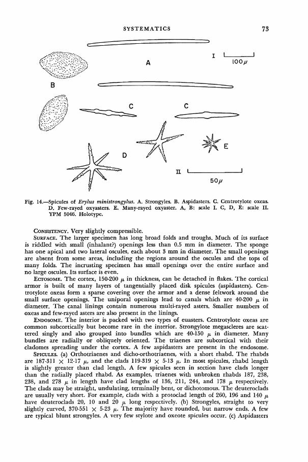
SYSTEMATICS 73
Fig. 14.—Spicules of Erylus ministrongylus. A. Strongyles. B. Aspidasters. C. Centrotylote oxeas. D. Few-rayed oxyasters. E. Many-rayed oxyaster. A, B: scale I. C, D, E: scale II. YPM 5046. Holotype.
CONSISTENCY. Very slightly compressible. SURFACE. The larger specimen has long broad folds and troughs. Much of its surface
is riddled with small (inhalant?) openings less than 0.5 mm in diameter. The sponge has one apical and two lateral oscules, each about 3 mm in diameter. The small openings are absent from some areas, including the regions around the oscules and the tops of many folds. The incrusting specimen has small openings over the entire surface and no large oscules. Its surface is even.
ECTOSOME. The cortex, 150-200 LL in thickness, can be detached in flakes. The cortical armor is built of many layers of tangentially placed disk spicules (aspidasters). Centrotylote oxeas form a sparse covering over the armor and a dense feltwork around the small surface openings. The uniporal openings lead to canals which are 40-200 LL in diameter. The canal linings contain numerous multi-rayed asters. Smaller numbers of oxeas and few-rayed asters are also present in the linings.
ENDOSOME. The interior is packed with two types of euasters. Centrotylote oxeas are common subcortically but become rare in the interior. Strongylote megascleres are scattered singly and also grouped into bundles which are 40-150 p in diameter. Many bundles are radially or obliquely oriented. The triaenes are subcortical with their cladomes spreading under the cortex. A few aspidasters are present in the endosome.
SPICULES, (a) Orthotriaenes and dicho-orthotriaenes, with a short rhabd. The rhabds are 187-311 X 12-17 LL, and the clads 119-319 x 5-13 LL. In most spicules, rhabd length is slightly greater than clad length. A few spicules seen in section have clads longer than the radially placed rhabd. As examples, triaenes with unbroken rhabds 187, 238, 238, and 278 LL in length have clad lengths of 136, 211, 244, and 178 LL respectively. The clads may be straight, undulating, terminally bent, or dichotomous. The deuteroclads are usually very short. For example, clads with a protoclad length of 260, 196 and 140 LL have deuteroclads 20, 10 and 20 LL long respectively, (b) Strongyles, straight to very slightly curved, 370-551 x 5-23 LL. The majority have rounded, but narrow ends. A few are typical blunt strongyles. A very few stylote and oxeote spicules occur, (c) Aspidasters

74 A SYSTEMATIC STUDY OF T H E DEMOSPONGIAE OF JAMAICA
(disk-spicules). Thin, elliptical, 87 X 43 to 181 X 87 /x. The spicules are only 7-14 //, in thickness. Typical sizes are 137 X 79, 145 x 72, and 166 x 79 //,. Their outline is usually irregular but seldom jagged. The minute, thickly-strewn tubercles give the surface a granular appearance, (d) Smooth centrotylote oxeas, nearly straight to slightly curved, 37-80 X 3-5 jx. The central inflation is elongate and ill-defined. For example, a spicule with a mid-length diameter of 4 ^ has a diameter of 3 fi at a point adjacent to the swelling. A few spicules have lateral as well as mid-length swellings. The ends are almost always sharp-pointed. A single spicule found in the incrusting specimen had a roughened surface, (e) Few-rayed oxyasters, with 3-6 rays. Ray length varies between 9 and 33 /x. The rays have a basal diameter of 2-3 p. They are straight, pointed and at least distally roughened. A few spicules have very thin rays which seem to be smooth. The central meeting point of the rays is only 3 //, in diameter, (f) Multi-rayed oxyasters, 10-25 p in overall diameter. About a dozen rays project from a central region which is 2-3 fM in diameter. The rays have a basal diameter of 1 /x. The larger spicules at least are minutely roughened. Individual analyses, lengths in microns: (holotype listed first in each category).
Strongyloxeas Aspidasters Centrotylote oxeas
398-534 (50)* 106 X 70-158 X 64 (25)* 53-80 (50)* 370-551 (50) 87 X 43-181 X 87 (50) 37-61 (50)
Few-rayed oxyasters (ray length) Multi-rayed oxyasters (total diam.)
13-33 (25)* 10-25 (25)*
9-26 (50) 12-24 (50)
Triaene clad length Rhabd length
136-260 (30)* 187-278 (5)* 119-319 (25) 217-311 (5)
DISCUSSION. The monaxonid megascleres are unusually small for the genus. The Jamaican sponge is related to Erylus euastrum (Schmidt, 1868) which, together with its apparent synonym, E. transiens (Weltner), has been reported from the West Indies. The Jamaican species differs from Schmidt's (as described by Babi£, 1922, p. 287 and Topsent, 1928, p. 115) in having smaller monaxonid spicules and roughened rather than smooth-rayed large asters. E. alleni de Laubenfels 1934, of Puerto Rico has smaller aspidasters, oxeote megascleres, and very small multi-rayed oxyasters. Its large asters have smooth rays.
FAMILY CHONDRILLIDAE Gray, 1867 GENUS CHONDRILLA Schmidt
Chondrilla nucula Schmidt
Chondrilla nucula Schmidt, 1862, p. 39 [type: Quarnero, Yugoslavia; repository unknown]; idem, 1870, p. 26; Schulze, 1877a, p. 108; Carter, 1881, p. 249; idem, 1882, p. 268; idem, 1885, p. 200; idem, 1886a, p. 277; idem, 1890, p. 565; Topsent, 1890a, p. 27; idem, 1892, p. 56; idem, 1918, p. 601; idem, 1925a, p. 630; Keller, 1891a, p. 327; Lendenfeld, 1897, p. 37; Wilson, 1902a, p. 386; Verrill, 1907, p. 334; Annandale, 1915, p. 470; Babic, 1922, p. 269; Burton, 1924b, p. 206; idem, 1934, p. 522; idem, 1936a, p. 7; Dendy, 1924, p. 314; Arndt, 1927, p. 138; Vosmaer, 1933, p. 293; de
Laubenfels, 1936b, p. 464; idem, 1949, p. 22: idem, 1950a; p. 133; idem, 1954a, p. 250 (?); idem, 1956, p. 2, 4; Sara, 1958a, p. 216; Sara and Siribelli, 1960, p. 31; LeVi, 1959, p. 121; Vacelet, 1959, p. 46; idem, 1961, p. 30; Little, 1963, p. 63.
[non] Chondrilla nucula, Kieschnick, 1896, p. 534 (fide Thiele, 1900, p. 63); de Laubenfels, 1935a, p. 12.
Chondrilla embolophora Schmidt, 1862, p. 39 (fide Topsent, 1918.) Chondrilla australiensis, de Laubenfels, 1954a, p. 249 (?) (not Carter, 1873.)

SYSTEMATICS 75 HABITAT. Common on pilings and rocks at Port Royal and at the cays. SHAPE. The sponge spreads horizontally over the substratum, attaining a thickness
of 1-2 mm. COLOR. A mottled brown to yellowish-brown, with pale areas around the oscules.
The interior is drab. In alcohol, the original coloration is retained to a certain extent. CONSISTENCY. Tough. SURFACE. Smooth, shiny. The oscules are small, scattered and contractile. ECTOSOME. A detachable, densely pigmented skin is present. Spherasters of all sizes
are scattered in it. Dermal pores could not be detected. ENDOSOME. The dense flesh is traversed by long, narrow, largely vertical canals, 10-25
H in diameter. Microscleres are abundant only in the periphery of the endosome and in the vicinity of the large canals.
SPICULES. Spherasters, typically with short, conical rays and with a definite centrum, 10-40 JJL in total diameter. A varied number of spicules have a relatively small centrum and long oxeote rays which are as much as 12 /A in length. Spicules with short blunt rays also occur, again in varied numbers. In some specimens ray length as a percentage of centrum diameter seldom exceeds 25 per cent. In other specimens ray length is often equal to 40-60 per cent of centrum diameter and may even be greater than the centrum diameter. Individual analyses, total diameter in microns: 10-34 (75); 15-37 (50); 20-40 (50); 12-33 (50); 20-40 (50).
DISTRIBUTION. Tropical Atlantic America: Florida—Schmidt, 1870, p. 26; West Coast, Florida—Little, 1963, p. 63; Atlantic coast of Panama—de Laubenfels, 1936b, p. 464; Venezuela—Carter, 1882, p. 268; Brazil—de Laubenfels, 1956, p. 2, 4; Bermuda— Verrill, 1907, p. 334; de Laubenfels—1950a, p. 133; Bahamas—de Laubenfels, 1949, p. 22; West Indies—Carter, 1881, p. 249; 1882, p. 268; Puerto Rico—Wilson, 1902a, p. 386; Antilles—Schmidt, 1870, p. 26; Curasao—Arndt, 1927, p. 138; Fernando Noronha, South Atlantic off Brazil—Carter, 1890, p. 565; South Trinidad Is., South Atlantic, off Brazil—Dendy, 1924, p. 314.
Mediterranean-Atlantic: French Mediterranean—Vacelet, 1959, p. 46-48; Ligurean Sea, Italy—Sara, 1958a, p. 216; Naples, Italy—Topsent, 1925a, p. 630; Vosmaer, 1933, p. 293; Sara and Siribelli, 1960, p. 31; Adriatic—Schmidt, 1862, p. 39; 1862, p. 39 (as Chondrilla embolophora); Schulze, 1877a, p. 108; Lendenfeld, 1897, p. 37; Babic\ 1922, p. 269; Mediterranean coast of Egypt—Burton, 1936a, p. 7; Corsica, Vacelet, 1961, p. 30; Azores—Topsent, 1890a, p. 27, 1892, p. 56; Gulf of Guinea—L^vi, 1959, p. 121; Sao Tom£, West Africa—Topsent, 1918, p. 601; Indo-Pacific: Red Sea—Carter, 1881, p. 249; Keller, 1891a, p. 327; Mauritus—Carter, 1881, p. 249; Gulf of Manaar, between India and Ceylon—Carter, 1881, p. 249; Mergui Archipelago, off Burma—Annandale, 1915, p. 470; Moluccas, East Indies—Carter, 1881, p. 249; Australia—Carter, 1885, p. 200, 1886a, p. 227; Great Barrier Reef, Australia—Burton, 1934, p. 522; West Central Pacific—de Laubenfels, 1954a, p. 250 (?); 1954a, p. 249 (as Chondrilla australiensis)}.
DISCUSSION. Long-rayed spherasters occur in Yale Peabody Museum specimens of Chondrilla nucula from Bermuda and the Virgin Islands. Schmidt (1870) reported them for his specimens from the Antilles and Florida. De Laubenfels (1935a) referred an aspiculous sponge from lower California to Chondrilla nucula. It was presumably a Chondrosia. De Laubenfels (1954a) recorded black Pacific sponges as C. nucula Schmidt and C. australiensis Carter (1873a). The latter identification is certainly incorrect, as the characteristic spined oxyasters of C. australiensis are absent. The long-rayed spicules of de Laubenfels' australiensis sponges are similar to those found in the above-mentioned West Indian specimens of C. nucula. It therefore seems probable that all of his black sponges are conspecific. As his sponges differ sharply from typical specimens of C. nucula in color (although not in spicule size), it is possible that they are a distinct species.
ORDER HOMOSCLEROPHORA Levi 1953 The Homosclerophora were dispersed in the classification of de Laubenfels (1936a).
LeVi (1953, 1956b), in reconstituting the group, has emphasized its morphological and embryological uniformity.

76 A SYSTEMATIC STUDY OF THE DEMOSPONGIAE OF JAMAICA
FAMILY PLAKINIDAE Schulze, sensu Lendenfeld, 1903
The families Plakinidae and Corticiidae as used by Sollas (1888) and LeVi (1953) are very similar and are here considered as a single group.
GENUS PLAKORTIS Schulze Plakortis zyggompha (de Laubenfels) new combination
Roosa zyggompha de Laubenfels, 1934, p. 2 [type: off Puerto Rico; USNM No. 22277]; idem, 1936a, p. 178.
Plakortis simplex, de Laubenfels (not Schulze, 1880), 1950a, p. 132.
HABITAT. Under rocks in shallow water at Maiden Cay. SHAPE. The seven specimens were small crusts, reaching a maximum thickness of
4.5 mm. COLOR. The exterior, in life and as preserved, is dark brown to drab. The interior
is of a lighter shade than the surface. CONSISTENCY. Tough, rubbery, very slightly compressible. SURFACE. Smooth to the touch, even but microtuberculate as seen under a dissecting
microscope. Five specimens have no obvious oscular openings. Each of the other two has a single small oscule.
ECTOSOME. NO detachable dermis and little dermal specialization. The flesh becomes more compact towards the surface. A few possible dermal pores, 20-60 /x in diameter, were observed in tangential sections taken at the surface. Narrow inhalant canals run downward toward the interior.
ENDOSOME. The peripheral inhalant canals are collected by larger ones. The compact flesh is thickly strewn with spicules. Large convoluted embryos (proper to the sponge?) are present in specimens collected in July and August, 1961.
SPICULES, (a) Diactinal spicules, with gradually pointed ends, 44-126 jx, in length and 2-3 n in midlength diameter. The spicule is divided into two rays by a swollen, twisted, notched or lumpy region. The rays are almost always at an angle, giving the spicule a curved or sharply bent outline. The rays themselves may be bent. Thinner, developmental stages may be present. Individual analyses, length in microns, 75 diactinal spicules each: 56-115; 54-126; 63-117; 44-119. (b) Triactinal spicules, always less frequent than the diactinal ones, and rare in some specimens. The triactinals are sometimes regular and equiangular, but are more frequently sagittal and inequiangular in some degree. The rays are sharp-pointed, straight to curved and often twisted or lumpy at the base. A lumpy midregion occasionally has one or more short projections in addition to the usual 3 rays. Normal rays are 24-46 x 2-3 /x. (c) Rare Y-shaped and roughened ovoid spicules, 5-10 ju, in length. A few of the roughened spicules have short rays, indicating a derivation from the diactinal spicules.
DISTRIBUTION. Tropical Atlantic America: Dry Tortugas, Florida—de Laubenfels, 1936a, p. 178 (as Roosa zyggompha); Bermuda—de Laubenfels, 1950a, p. 132 (as Plakortis simplex) ?; Puerto Rico—de Laubenfels, 1934, p. 2 (as Roosa zyggompha).
DISCUSSION. De Laubenfels (1934, p. 2) established Roosa zyggompha for Puerto Rican sponges similar in spiculation to Plakortis simplex Schulze. He later concluded (1950a, p. 132) that the West Indian sponges were conspecific with Schulze's species. W. D. Hartman (personal communication) has pointed out the presence of minute spicules in West Indian Plakortis-like sponges. Such spicules are present, although rare, in U. S. National Muesum specimen number 22490. The specimen was collected at the Dry Tortugas and identified as Roosa zyggompha by de Laubenfels. The West Indian sponge is unlikely to be con-specific with Plakortis simplex Schulze. If the small microscleres are consistently present, and not developmental stages, Roosa de Laubenfels could be treated as a distinct genus.

DISTRIBUTION BY HABITAT 77
GENUS CORTICIUM Schmidt Corticium tetralophum n. sp.
(Text-fig. 15)
HOLOTYPE. YPM 5047. Maiden Cay, Aug. 12, 1961. HABITAT. The single specimen was collected at Maiden Cay. SHAPE. A small incrustation with a maximum thickness of 2.5 mm. COLOR. The sponge was a dull yellow in life and is a grayish-beige in alcohol. CONSISTENCY. Compressible. SURFACE. The thin detachable ectosome contains numerous tetraloph spicules. A few
triradiates are also present. No ostia could be found. ENDOSOME. Tetralophs and a few triradiates are sparsely scattered in the flesh. The
ovoid flagellated chambers are 35-50 p in maximum diameter. Convoluted embryos (proper to the sponge?) are present in the specimen collected in early August.
I I
10// Fig. 15.—Spicules of Corticium tetralophum. Tetralophs. YPM 5047. Holotype.
SPICULES, (a) Tetralophs, 17-30 //, in total diameter (100 spicules). The protoclads are 3 ^ in diameter. They bear 2-6, typically 4, deuteroclads. The latter are about equal in length to the protoclads. The smooth protoclads are 1-2 fi in basal diameter. Smooth and rough or spiny deuteroclads may be present on the same spicule. A typical spicule has 4 similar deuteroclads on each of the 4 rays. A rhabd cannot be distinguished from 3 clads. (b) Very rare trilophate spicules, with the unbranched ray shortened or bent. The spicules are similar in size to the tetralophs. (c) Rare, thin triradiates and tetraradi-ates, 10-17 JJL in total diameter (15 spicules). Some or all of the rays are terminally branched. The rays are only 1 p in basal diameter. The spicules may be developmental stages of the tetralophs and trilophs.
DISCUSSION. The species is characterized by the lack of a special category of heterolophose candelabra. In addition, modified calthrops are absent. Two other species of Corticium occur in the West Indies. C. versatile Schmidt, 1880, has peculiar candelabras and variously modified calthrops. C. quadripartitum Topsent, 1923, has special dermal heterolophose candelabras. Its tetralophs are similar in form to those of the Jamaican sponge but are slightly larger in size.
DISTRIBUTION BY H A B I T A T
T h e sponge fauna of individual habi tats can now be summarized.
(A) Por t Royal Docks: T h e common species are:
Ircinia fasciculata Halichondria melanadocia Ircinia strobilina Tedania ignis Darwinella rosacea Microciona microchela Desmapsamma anchorata Microciona rarispinosa Iotrochota birotulata Mycale laevis Gelliodes areolata
T h e following species were present b u t less common:
Verongia fistularis Placospongia carinata Dysidea fragilis Zygomycale parishii

78 A SYSTEMATIC STUDY OF THE DEMOSPONGIAE OF JAMAICA
Sponges were the dominant members of the sessile seawall community, which also included mussels, barnacles and ascidians. The species exhibited no obvious zonation on the piers and seawalls. Very large specimens of Ircinia fasciculata, Ircinia strobilina and Verongia fistularis were restricted to the base of the seawall. At that depth (8-10 ft.), sponges are certainly less subject to the surface salinity changes noted by Goodbody (1961) after heavy rains.
(B) Port Royal Harbor:
Verongia longissima Spheciospongia vesparia
(C) Rasta's Wreck:
Iotrochota birotulata Tedania ignis Sigmadocia caerulea Microciona microchela Halichondria melanadocia Microciona rarispinosa Halichondria magniconulosa Mycale laevis
Sponges and ascidians were the most conspicuous elements of the wreck fauna. The microcionids incrusted mussel shells growing on the wreck. Microciona microchela covered large areas of the timbers as a thin film. Halichondria magniconulosa was found only in this habitat.
(D) Port Royal turtle grass bed:
Haliclona doria Mycale microsigmatosa Haliclona erina Tethya seychellenis
Mycale microsigmatosa and Tethya seychellensis grew on turtle grass blades. The other two species of Tethya at Port Royal were restricted to the rocky environment of the cays. Haliclona doria formed repent colonies over the mud and turtle grass with most of the branches above the level of the substratum. Most specimens of Haliclona erina grew at the border between turtle grass and sand, near Rasta's wreck.
(E) Mangroves bordering inlet:
Dysidea fragilis Tedania ignis Sigmodocia caerulea Mycale laevis Halichondria melanadocia
Sponges and ascidians covered many mangrove roots.
(F) Main Boat Channel: The common species were:
Darwinella rosacea Ulosa hispida Haliclona hogarthi Terpios zeteki Adocia implexiformis Geodia (Cydonium) papyracea Pellina coeliformis Geodia (Geodia) gibberosa Halichondria melanadocia
Less common species were:
Ircinia fasciculata Haliclona erina Dysidea fragilis Mycale microsigmatosa
Sponges and ascidians covered a majority of the mangrove roots bordering the channel. Most species grew only on the root systems. A few specimens of Geodia

DISTRIBUTION BY HABITAT 79
gibberosa and Terpios zeteki were found in the stream bed. Pellina coeliformis and a few specimens of Haliclona erina grew over the surface of Geodia gibberosa. A single root system often served as the substratum for several species of sponge. The species exhibited no obvious horizontal or vertical zonation.
The channel sponges were unaffected by the moderate autumn rains of 1961. Heavy seasonal rains are known to produce a surface layer of low salinity water in Kingston Harbor which is fatal to sponges (cf. Goodbody, 1961).
The mangrove channel fauna has a sharply limited distribution at Port Royal. Six of the nine common species were growing exclusively in the mangroves, and none were found at the cays. Three of the five common species of habitat E did not extend into the channels.
(G) Pigeon House Pile:
Ircinia fasciculata Oligoceras hemorrhages Darwinella rosacea Haliclona erina
(H) Shallow water at the cays:
Ircinia fasciculata Ircinia strobilina Neofibularia massa Adocia albifragilis Acanthacarnus souriei Spirastrella coccinea Anthosigmella varians Diplastrella megastellata Cliona vermifera
Most species, including Neofibularia massa, were incrustations growing on the underside of rocks. The heavy surf may exclude most massive species. Twelve of the seventeen species were collected only in this habitat. Only three species were also found in shallow water at Port Royal.
(I) Deep water near cays:
Ircinia fasciculata Ircinia strobilina Verongia fistularis (Ianthella ardis, in
St. Thomas Parish.) Haliclona rub ens Desmapsamma anchorata
Anthosigmella varians Placospongia carinata Geodia (Geodia) gibberosa
Cliona viridis Placospongia carinata Tethya actinia Tethya sp. Erylus ministrongylus Chondrilla nucula Plakortis zyggompha Corticium tetralophum
Neofibularia massa Iotrochota birotulata Gailyspongia fallax Callyspongia vaginalis
Callyspongia plicifera Thalyseurypon conulosa
Six of the species were restricted to this habitat. Most specimens of Neofibularia massa were distinctly vasiform, with upright growth, rather than thickly incrust-ing.
(J) Offshore turtle grass beds:
Oligoceras hemorrhages Dysidea fragilis Adocia carbonaria Sigmadocia caerulea
Tedania ignis Lissodendoryx isodictyalis Tethya seychellensis

80 A SYSTEMATIC STUDY OF THE DEMOSPONGIAE OF JAMAICA
The offshore turtle grass beds have a sponge fauna quite unlike that of the Port Royal turtle grass area. The offshore beds are sandy rather than muddy and subject to considerable surf. The sponges grew at the base of the turtle grass clumps and on stones; none grew on the turtle grass blades. Adocia carbonaria and Lis-sodendoryx isodictyalis were found only in this habitat.
FIELD KEY TO THE SPONGES OF PORT ROYAL
The key is designed for use as a preliminary guide in field work. It is based on the appearance in life of typical members of each species. The key must be used with caution, and all identifications should be confirmed by comparison with the description. Undescribed species may be found, particularly at the outer cays and in slightly deeper water. Interesting specimens should be preserved in alcohol, after completing field notes, and sent to the author.
The color in life is used for an initial division.
A—Black and grayish-black. B—Blue and blue-green. C—Brown or tan. D—Gray. E—Green. F—Orange. G—Pink. H—Purple. I—Red and red-orange. J—White and off-white. K—Yellow.
Glossary of terms:
Conule—a spike-shaped, thorn-like projection. Oscule—a large exhalant opening on the surface.
(A) Near black and grayish-black.
1—Conulose—2. 1—Not conulose—3. 2—Conules 3-7 mm high; massive; fetid—Ircinia strobilina. 2—Conules less than 1 mm high; vasiform—Verongia fistularis (Turns black
soon after collection; in life, has a yellow color.) 3—Old specimens much larger than fist-sized; with clusters of oscules—Sphecio-
spongia vesparia. 3—Fist-sized or smaller; without clusters of oscules—4. 4—Black; brittle; repent to lobate; stains alcohol blue-black—Adocia carbonaria. 4—Near-black, with greenish tinge in interior; compressible; massive—Hali-
chondria melanadocia.
(B) Blue or blue-green.
1—Conulose—Dysidea fragilis. 1—Not conulose—2. 2—Incrusting; blue-green, speckled with red-orange embryos—Mycale micro-
sigmatosa. 2—Massive or lobate—3.
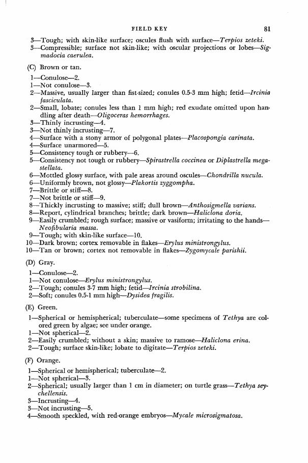
FIELD KEY 81
3—Tough; with skin-like surface; oscules flush with surface—Terpios zeteki. 3—Compressible; surface not skin-like; with oscular projections or lobes—Sig-
madocia caerulea.
(C) Brown or tan.
1—Conulose—2. 1—Not conulose—3. 2—Massive, usually larger than fist-sized; conules 0.5-3 mm high; fetid—Ircinia
fasciculata. 2—Small, lobate; conules less than 1 mm high; red exudate omitted upon han
dling after death—Oligoceras hemorrhages. 3—Thinly incrusting—4. 3—Not thinly incrusting—7. 4—Surface with a stony armor of polygonal plates—Placospongia carinata. 4—Surface unarmored—5. 5—Consistency tough or rubbery—6. 5—Consistency not tough or rubbery—Spirastrella coccinea or Diplastrella mega-
stellata. 6—Mottled glossy surface, with pale areas around oscules—Chondrilla nucula. 6—Uniformly brown, not glossy—Plakortis zyggompha. 7—Brittle or stiff—8. 7—Not brittle or stiff-—9. 8—Thickly incrusting to massive; stiff; dull brown—Anthosigmella varians. 8—Report, cylindrical branches; brittle; dark brown—Haliclona doria. 9—Easily crumbled; rough surface; massive or vasiform; irritating to the hands—
Neofibularia massa. 9—Tough; with skin-like surface—10.
10—Dark brown; cortex removable in flakes—Erylus ministrongylus. 10—Tan or brown; cortex not removable in flakes—Zygomycale parishii.
(D) Gray.
1—Conulose—2. 1—Not conulose—Erylus ministrongylus. 2—Tough; conules 3-7 mm high; fetid—Ircinia strobilina. 2—Soft; conules 0.5-1 mm high—Dysidea fragilis.
(E) Green.
1—Spherical or hemispherical; tuberculate—some specimens of Tethya are colored green by algae; see under orange.
1—Not spherical—2. 2—Easily crumbled; without a skin; massive to ramose—Haliclona erina. 2—Tough; surface skin-like; lobate to digitate—Terpios zeteki.
(F) Orange.
1—Spherical or hemispherical; tuberculate—2. 1—Not spherical—3. 2—Spherical; usually larger than 1 cm in diameter; on turtle grass—Tethya sey-
chellensis. 3—Incrusting—4. 3—Not incrusting—5. 4—Smooth speckled, with red-orange embryos—Mycale microsigmatosa.

82 A SYSTEMATIC STUDY OF THE DEMOSPONGIAE OF JAMAICA
4—Projecting spicules and fleshy tufts conspicuous; uniformly orange—Ulosa hispida.
5—Compressible; with rimmed oscules—Mycale laevis. 5—Tough; oscules flush with surface—Terpios zeteki.
(G) Pink,
1—Ramose—Desmapsamma anchorata. 1—Incrusting—2. 2—Conulose—Darwinella rosacea. 2—Smooth—3. 3—Tough; with pinkish-orange interior—Microciona microchela. 3—Soft, speckled, with red-orange embryos—Mycale microsigmatosa.
(H) Purple.
1—Conulose—2. 1—Not conulose—7. 2—With only a few thorn-like conules on an otherwise smooth surface; dull
purple—Callyspongia fallax. 2—With numerous conules—3. 3—Cheese-like consistency; bluish-purple—Ianthella ardis. 3—Characters otherwise—4. 4—Very dark purple—5. 4—Light purple—6. 5—Dark reddish purple—Thalyseurypon conulosa. 5—Nearly black, with a greenish tinge in life; orange zoanthids on surface—
Iotrochota birotulata. 6—Tough; reddish-purple; fetid—Ircinia fasciculata. 6—Soft; pale purple—Dysidea fragilis. 7—Vasiform—8. 7—Not vasiform—9. 8—With a paper-thin terminal collar—Callyspongia plicifera. 8—Without a collar—Callyspongia vaginalis. 9—Tough; with a skin-like surface—10. 9—Characters otherwise.
10—Lobate or digitate; on mangroves—Terpios zeteki. 10—Usually with elongate branches; on pilings—Zygomycale parishii. 11—Massive, with a fetid odor—Lissodendoryx isodictyalis. 11—Ramose or with oscular projections—12. 12—Easily crumbled, with oscular projections—Adocia implexiformis. 12—Not easily crumbled, cylindrical branches, oscules flush with surface—13. 13—Limp; an anastomosing repent complex of branches—Haliclona hogarthi. 13—Compressible; with a very rough surface; with stout, flattened erect branches
—Gelliodes areolata.
(I) Red-orange and red.
1—Conulose—2. 1—Not conulose—3. 2—Massive; reddish-purple; fetid odor—Ircinia fasciculata. 2—Cylindrical; dull red—Verongia longissima (Yellow in life, changing soon
after collection.) 3—Spherical; tuberculate—Tethya seychellensis.

ZOOGEOGRAPHICAL NOTES 83
3—Not spherical—4. 4—Boring, with papillae at surface of substratum—Cliona vermifera. 4—Not boring—5. 5—Incrusting—6. 5—Not incrusting—7. 6—Punctiform surface; on shells and pilings at Port Royal—Microciona rari-
spinosa. 6—Surface not punctiform; on rocks at cays—Acanthacarnus souriei (Spirastrella
coccinea has been reported as red in the literature.) 7—Massive or lobate; red-orange; with conspicuous red-orange gemmules in the
interior—Tedania ignis. 7—Characters otherwise-—8. 8—With a skin-like surface—Terpios zeteki. 8—Without a skin-like surface—9. 9—Dark red, retaining that color in preservative—Haliclona rubens. 9—Pale color, dull red or reddish-purple—10.
10—Reddish-purple; limp, a repent complex of branches—Haliclona hogarthi. 10—Dull red; compressible; with stout flattened erect branches; with a rough sur
face—Gelliodes areolata.
(J) White and off-white.
1—Massive—2. 1—Incrusting—3. 2—Leathery to stony; with a thick rigid cortex; with conspicuous sieve areas—
Geodia gibber osa. 2—Easily crushed; with a paper-like cortex; without oscular areas—Geodia
papyracea. 3—Oscules flush with the surface—Adocia albifragilis. 3—Oscules on high paper-thin tubes—Pellina coeliformis.
(K) Yellow.
1—Conulose—2. 1—Not conulose—4. 2—Vasiform—Verongia fistularis. 2—Not vasiform—3. 3—Cylindrical—Verongia longissima. 3—Massive—Ianthella ardis. 4—Boring sponges with papillae at the surface of the substratum—Cliona viridis. 4—Not boring—5. 5—Incrusting—Corticium tetralophum. 5—Not incrusting—6. 6—Lobate, soft—Callyspongia pallida. 6—Massive, tough—Halichondria magniconulosa.
ZOOGEOGRAPHICAL NOTES
According to present records, most Jamaican sponges have a limited distribution. Of the 57 recorded species, (including three not found in the present study), 18 have not been collected elsewhere. An additional 20 species, including the partially classified one, are known only from tropical Atlantic American waters. About two-thirds of the Jamaican fauna (38 species) are therefore endemic to the

84 A SYSTEMATIC STUDY OF THE DEMOSPONGIAE OF JAMAICA
region. Five other species, Ircinia fasciculata, 7. strobilina, Verongia lacunosa, Adocia carbonaria, and Tedania ignis, may or may not be conspecific with morphologically similar sponges in other parts of the world.
Ten species are widely distributed outside the West Indies. Dysidea fragilis, Lissodendoryx isodictyalis, Cliona viridis, and Chondrilla nucula are circum-tropical or nearly so, and the first three have also been reported from temperate waters. Other widespread species are Desmapsamma anchorata (West Africa, Indo-Pacific), Zygomycale parishii (Indo-Pacific), Spirastrella coccinea (Tropical Pacific America, West Africa, Mediterranean-Atlantic), Cliona vermifera (Mediterranean, Central Pacific), Placospongia carinata (Indo-Pacific, tropical Pacific America), and Tethya seychellensis (Indo-Pacific). The disjunct patterns indicate the fragmentary state of our present knowledge. In addition, several of the supposed species, such as Lissodendoryx isodictyalis and Cliona viridis, may prove to be complexes of closely related species.
Of the remaining five species, Tethya actinia has been recorded from the West Central Pacific. Acanthacarnus souriei has been previously recorded only from West Africa and the Mediterranean. Geodia gibberosa and Mycale microsigma-tosa also occur in West Africa. The former species has been collected at the Pacific end of the Panama canal. The latter species has been recorded from the Pacific coast of Panama and from the Hawaiian Islands. Terpios zeteki is known from both coasts of Panama, the Gulf of California and the Hawaiian Islands. The coasts of Central America require careful distributional studies. Several other West Indian species are found on the Pacific side of Panama (cf. de Lauben-fels, 1936b) and a few reach the Gulf of California, (cf. Dickinson, 1945).
De Laubenfels (1950b, 1951a) recorded a number of species which are common to the West Indies and Hawaii. The Jamaican specimens of Mycale micro-sigmatosa and Terpios zeteki agree closely with his Hawaiian descriptions. The presence of a considerable number of species common to both sides of the Isthmus of Panama, a barrier in existence since the Pliocene, raises the possibility of transport by ship in very recent times. De Laubenfels indeed found a Panamanian sponge, presumably Mycale cecilia = M. microsigmatosa, on dry-docked ships at Pearl Harbor (cf. de Laubenfels, 1950b, p. 4).
The present study suggests that the Jamaican shallow-water fauna has close affinities with those of the southern Caribbean. Nine species have been previously recorded only from that little-known area. Burton (1930) has suggested that the distribution of sponges is closely related to ocean currents which can transport larvae. Jamaica lies in the path of surface currents which run from the Lesser Antilles and the Venezuelan coast toward the Yucatan Channel. Only a few Jamaican species have been recorded from Bermuda and North Carolina which are rather distant from Jamaica in terms of the paths of ocean currents. Burton's theory could be tested further by studying the sponge fauna of the Lesser Antilles; a strong West African element would be expected through the transport of larvae by the equatorial currents.
SUMMARY
A total of 54 species have been recorded in a systematic study of the shallow water sponges of Port Royal, Jamaica. The new genus Neofibularia is proposed for the preoccupied name Fibularia Carter. Sixteen of the species are new to science, namely: Darwinella rosacea, Haliclona hogarthi, Adocia implexiformis, Adocia albifragilis, Pellina coeliformis, Sigmadocia caerulea, Callyspongia pallida,
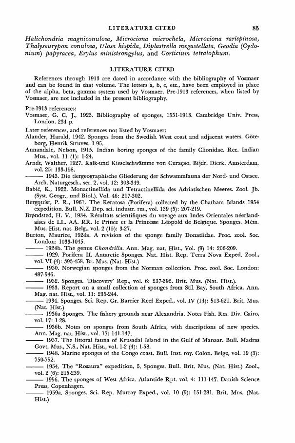
L I T E R A T U R E CITED 85 Halichondria magniconulosa, Microciona microchela, Microciona rarispinosa, Thalyseurypon conulosa, Ulosa hispida, Diplastrella megastellata, Geodia (Cydo-nium) papyracea, Erylus ministrongylus, and Corticium tetralophum.
LITERATURE CITED
References through 1913 are dated in accordance with the bibliography of Vosmaer and can be found in that volume. The letters a, b, c, etc., have been employed in place of the alpha, beta, gamma system used by Vosmaer. Pre-1913 references, when listed by Vosmaer, are not included in the present bibliography.
Pre-1913 references: Vosmaer, G. C. J., 1923. Bibliography of sponges, 1551-1913. Cambridge Univ. Press,
London. 234 p.
Later references, and references not listed by Vosmaer: Alander, Harald, 1942. Sponges from the Swedish West coast and adjacent waters. Gote-
borg, Henrik Struves. 1-95. Annandale, Nelson, 1915. Indian boring sponges of the family Clionidae. Rec. Indian
Mus., vol. 11 (1): 1-24. Arndt, Walther, 1927. Kalk-und Kieselschwamme von Curasao. Bijdr. Dierk. Amsterdam,
vol. 25: 133-158. 1943. Die tiergeographische Gliederung der Schwammfauna der Nord- und Ostsee.
Arch. Naturgesch., ser. 2, vol. 12: 303-349. Babic\ K., 1922. Monactinellida und Tetractinellida des Adriatischen Meeres. Zool. Jb.
(Syst. Geogr., und Biol.), Vol. 46: 217-302. Bergquist, P. R., 1961. The Keratosa (Porifera) collected by the Chatham Islands 1954
expedition. Bull. N.Z. Dep. sci. industr. res., vol. 139 (5): 207-219. Br0ndsted, H. V., 1934. R&ultats scientifiques du voyage aux Indes Orientales neerland-
aises de LL. AA. RR. le Prince et la Princesse Leopold de Belgique. Sponges. Mem. Mus. Hist. nat. Belg., vol. 2 (15): 3-27.
Burton, Maurice, 1924a. A revision of the sponge family Donatiidae. Proc. zool. Soc. London: 1033-1045.
1924b. The genus Chondrilla. Ann. Mag. nat. Hist., Vol. (9) 14: 206-209. 1929. Porifera II. Antarctic Sponges. Nat. Hist. Rep. Terra Nova Exped. Zool.,
vol. VI (4): 393-458. Br. Mus. (Nat. Hist.) 1930. Norwegian sponges from the Norman collection. Proc. zool. Soc. London:
487-546. 1932. Sponges. 'Discovery* Rep., vol. 6: 237-392. Brit. Mus. (Nat. Hist.). 1933. Report on a small collection of sponges from Stil Bay, South Africa. Ann.
Mag. nat. Hist., vol. 11: 235-244. 1934. Sponges. Sci. Rep. Gr. Barrier Reef Exped., vol. IV (14): 513-621. Brit. Mus.
(Nat. Hist.) 1936a Sponges. The fishery grounds near Alexandria. Notes Fish. Res. Div. Cairo,
vol. 17: 1-28. 1936b. Notes on sponges from South Africa, with descriptions of new species.
Ann. Mag. nat. Hist., vol. 17: 141-147. 1937. The littoral fauna of Krusadai Island in the Gulf of Manaar. Bull. Madras
Govt. Mus., N.S., Nat. Hist., vol. 1-2 (4): 1-58. 1948. Marine sponges of the Congo coast. Bull. Inst. roy. Colon. Beige, vol. 19 (3):
750-752. 1954. The "Rosaura" expedition, 5. Sponges. Bull. Brit. Mus. (Nat. Hist.) Zool.,
vol. 2 (6): 215-239. 1956. The sponges of West Africa. Atlantide Rpt. vol. 4: 111-147. Danish Science
Press, Copenhagen. 1959a. Sponges. Sci. Rep. Murray Exped., vol. 10 (5): 151-281. Brit. Mus. (Nat.
Hist.)
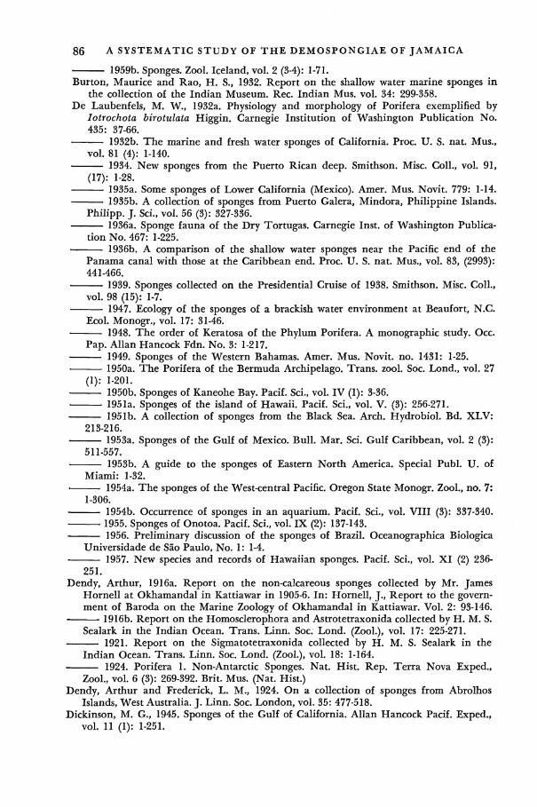
86 A SYSTEMATIC STUDY OF T H E DEMOSPONGIAE OF JAMAICA
1959b. Sponges. Zool. Iceland, vol. 2 (3-4): 1-71. Burton, Maurice and Rao, H. S., 1932. Report on the shallow water marine sponges in
the collection of the Indian Museum. Rec. Indian Mus. vol. 34: 299-358. De Laubenfels, M. W., 1932a. Physiology and morphology of Porifera exemplified by
Iotrochota birotulata Higgin. Carnegie Institution of Washington Publication No. 435: 37-66.
1932b. The marine and fresh water sponges of California. Proc. U. S. nat. Mus., vol. 81 (4): 1-140.
1934. New sponges from the Puerto Rican deep. Smithson. Misc. Coll., vol. 91, (17): 1-28.
1935a. Some sponges of Lower California (Mexico). Amer. Mus. Novit. 779: 1-14. 1935b. A collection of sponges from Puerto Galera, Mindora, Philippine Islands.
Philipp. J. Sci., vol. 56 (3): 327-336. 1936a. Sponge fauna of the Dry Tortugas. Carnegie Inst, of Washington Publica
tion No. 467: 1-225. 1936b. A comparison of the shallow water sponges near the Pacific end of the
Panama canal with those at the Caribbean end. Proc. U. S. nat. Mus., vol. 83, (2993): 441-466.
1939. Sponges collected on the Presidential Cruise of 1938. Smithson. Misc. Coll., vol. 98 (15): 1-7.
1947. Ecology of the sponges of a brackish water environment at Beaufort, N.C. Ecol. Monogr., vol. 17: 31-46.
1948. The order of Keratosa of the Phylum Porifera. A monographic study. Occ. Pap. Allan Hancock Fdn. No. 3: 1-217.
1949. Sponges of the Western Bahamas. Amer. Mus. Novit. no. 1431: 1-25. 1950a. The Porifera of the Bermuda Archipelago. Trans, zool. Soc. Lond., vol. 27
(1): 1-201. 1950b. Sponges of Kaneohe Bay. Pacif. Sci., vol. IV (1): 3-36.
. 1951a. Sponges of the island of Hawaii. Pacif. Sci., vol. V. (3): 256-271. 1951b. A collection of sponges from the Black Sea. Arch. Hydrobiol. Bd. XLV:
213-216. 1953a. Sponges of the Gulf of Mexico. Bull. Mar. Sci. Gulf Caribbean, vol. 2 (3):
511-557. 1953b. A guide to the sponges of Eastern North America. Special Publ. U. of
Miami: 1-32. « 1954a. The sponges of the West-central Pacific. Oregon State Monogr. Zool., no. 7:
1-306. 1954b. Occurrence of sponges in an aquarium. Pacif. Sci., vol. VIII (3): 337-340. 1955. Sponges of Onotoa. Pacif. Sci., vol. IX (2): 137-143. 1956. Preliminary discussion of the sponges of Brazil. Oceanographica Biologica
Universidade de Sao Paulo, No. 1: 1-4. 1957. New species and records of Hawaiian sponges. Pacif. Sci., vol. XI (2) 236-
251. Dendy, Arthur, 1916a. Report on the non-calcareous sponges collected by Mr. James
Hornell at Okhamandal in Kattiawar in 1905-6. In: Hornell, J., Report to the government of Baroda on the Marine Zoology of Okhamandal in Kattiawar. Vol. 2: 93-146.
1916b. Report on the Homosclerophora and Astrotetraxonida collected by H. M. S. Sealark in the Indian Ocean. Trans. Linn. Soc. Lond. (Zool.), vol. 17: 225-271.
1921. Report on the Sigmatotetraxonida collected by H. M. S. Sealark in the Indian Ocean. Trans. Linn. Soc. Lond. (Zool.), vol. 18: 1-164.
1924. Porifera 1. Non-Antarctic Sponges. Nat. Hist. Rep. Terra Nova Exped., Zool., vol. 6 (3): 269-392. Brit. Mus. (Nat. Hist.)
Dendy, Arthur and Frederick, L. M., 1924. On a collection of sponges from Abrolhos Islands, West Australia. J. Linn. Soc. London, vol. 35: 477-518.
Dickinson, M. G., 1945. Sponges of the Gulf of California. Allan Hancock Pacif. Exped., vol. 11 (1): 1-251.

LITERATURE CITED 87
Ekman, Sven, 1953. Zoogeography of the Sea. Sidgewick and Jackson, London. 417 p. Goodbody, I. M., 1961. Mass mortality of a marine fauna following tropical rains. Ecol
ogy, vol. 42: 150-155. George, W. C. and Wilson, H. V., 1919. Sponges of Beaufort (N.C.) harbor and vicinity.
Bull. U. S. Bur. Fish, vol. 36: 133-179. Hallmann, E. F., 1914. A revision of the monaxonid species described as new in Lenden-
feld's "Catalogue of the sponges," in the Australian Museum. Proc. Linn. Soc. N.S.W., vol. 39: 263-315, 327-376, 398-446.
Hartman, W. D., 1955. A collection of sponges from the West Coast of the Yucatan Peninsula with descriptions of two new species. Bull. Mar. Sci. Gulf Caribbean, vol. 5 (3): 161-189.
1958. Natural History of the Marine Sponges of Southern New England. Bull. Peabody Mus., vol. 12: 1-155.
Hentschel, Ernst, 1923. Porifera. In: Handbuch der Zoologie, von W. Kukenthal. Berlin. vol. 1: 307-418.
1929. Die Kiesel-und Hornschwamme des Nordlichen Eismeers. Fauna Arct., vol. V: 859-1042.
Hernandez, F. F., 1916. Fauna del Mediterraneo occidental esponjas espanolas. Trab. Mus. Cienc. nat., Zool., vol. 27: 1-52.
1917. Esponjas del litoral de Asturias. Trab. Mus. cienc. nat., Zool., vol. 36: 5-39, Hopkins, S. H., 1956. The boring sponges which attack South Carolina Oysters, with
notes on some associated organisms. Contr. Bears Bluff Lab., vol. 23: 3-30. Kumar, A., 1925. Report on some tetraxonid sponges in the collection of the Indian
Museum. Rec. Indian Mus., vol. 27: 211-227. Levi, Claude, 1952. Spongiaires de la cote du S£n£gal. Bull. Inst, franc,. Afr. Noire, vol.
14 (1): 34-59. 1953. Description de Plakortis nigra n. sp. et remarques sur les Plakinidae. (Demo-
sponges). Bull. Mus. Hist. nat. Paris., ser. 2, vol. 25 (3): 320-327. 1956a. Spongiaires des cotes de Madagascar. M&n. Inst. sci. Madagascar, vol. (4)
10: 2-23. 1956b. £tude des Halisarca de Roscoff. Embryologie et Systematique des De*mo-
sponges. Arch. Zool. exp. gen., vol. 93 (1): 1-181. 1957. Spongiaires des cotes d'Israel. Bull. Res. Council Israel, vol. 6 (B) (3-4): 201-
212. 1959. Spongiaires. Result, sci. Camp. "Calypso," fasc. 4, Ann. Inst, oceanogr.
Monaco, vol. 37: 115-141. 1960. Spongiaires des cotes occidentales africaines. Bull. Inst. fran$. Afr. noire,
vol.22 (A) (3): 743-769. Little, F. J., Jr., 1963. The sponge fauna of the St. George's Sound, Apalachee Bay, and
Panama City regions of the Florida Gulf Coast. Tulane Stud. Zool., vol. 11: 31-71. McDougall, K. D., 1943. Sessile marine invertebrates of Beaufort, N.C. Ecol. Monogr.,
vol. 13 (3): 321-374. Rao, M. S., 1941. Indian and Ceylon sponges of the Naturhistoriska Riksmuseet Stock
holm collected by K. Fristedt. Rec. Indian Mus., vol. 43: 417-469. Rutzler, Klaus, and Sara, Michele, 1962. Diplastrella ornata, eine neue mediterrane Art
der Familie Spirastrellidae (Demospongiae). Zool. Anz., vol. 169: 231-236. Sara, Michele, 1958a. Contributo alia conoscenze dei Porifera del Mar Ligure. Ann. Mus.
Civ. Sc. Nat. Genova, vol. 70: 207-244. 1958b. Studio sui Porifera di una grotta di marea del golfo di Napoli. Arch.
Zool. (ital)., vol. 43: 203-280. 1960a. Poriferi del litorale dell Isola d'Ischia e loro ripartizione per ambienti.
Pubbl. Staz. zool. Napoli, vol. 31: 421-472. 1960b. Poriferi di scogliera artificiale nel Golfo di Napoli. Ann. Pontificio Isti-
tuto Superiore di Scienze e lettere "Santa Chiara". Napoli, vol. 10: 259-269. 1961a. La fauna di Poriferi delle grotte delle isole Tremiti. Studio ecologico e
sistematico. Arch. zool. (ital.), vol. 46: 1-57.

88 A SYSTEMATIC STUDY OF T H E DEMOSPONGIAE OF JAMAICA
1961b. Zonazione dei Poriferi nella grotta della Gaiola. Ann. Inst. Mus. Zool. Univ. Napoli, vol. 13 (1): 1-32.
Sara, Michele and Siribelli, L., 1960. La fauna di Poriferi delle "secche" del golfo di Napoli. I. La "Secca" della Gaiola. Ann. Inst. Mus. Zool. Univ. Napoli, vol. 12 (3): 1-93.
Stephens, Jane, 1915. Atlantic sponges collected by the Scottish National Antarctic Expedition. Trans, roy. Soc. Edinb., vol. L: 423-467.
1917. Sponges collected by the dredging expeditions of the Royal Irish Academy and Royal Dublin Society. Proc. R. Irish Acad., vol. 34. (B): 1-16.
Topsent, £mile, 1918. Sponges de San Thome\ Essai sur les genres Spirastrella, Donatia, et Chondrilla. Arch. Zool. exp. g£n., vol. 57 (6): 535-618.
1920a. Spongiaires du Muse*e zoologique de Strasbourg. Monaxonides. Bull. Inst. oc^anogr. Monaco, 381: 1-34.
1920b. Sur quelques sponges du cabinet du Professeur Hermann, demerits et figured par Esper en 1794. Bull. Soc. zool. Fr., vol. 45: 314-327.
1923. Spongiaires du Mus£e zoologique de Strasbourg. Choristides. Bull. Inst. oceanogr. Monaco., 435: 1-66.
1924. Revision des Mycale de l'Europe occidentale. Ann. Inst, oceanogr. Monaco, vol. 1 (3): 77-118.
1925a. Etude des spongiaires du Golfe de Naples. Arch. Zool. exp. gen., vol. 63 (5): 623-725.
1925b. sponges de l'Etang de Thau. Bull. Inst, oceanogr. Monaco, vol. 452: 1-19. 1928. Spongiaires de l'Atlantique et de la M£diterran£e provenant des croisieres
du Prince Albert I de Monaco. Result. Camp. sci. Monaco, vol. LXXIV: 1-376. 1929. Chondrocladia yatsui, n. sp. de la Baie de Sagami. Annot. Zool. Jap. vol.
12: 421-432. 1930. Sponges de Lamarck conserves au Museum de Paris. Arch. Mus. Hist. nat.
Paris, s£r. 6, vol. 5: 1-56. 1932. Sponges de Lamarck conserves au Museum de Paris. 2. Arch. Mus. Hist.
nat. Paris, s£r. 6, vol. 8: 61-124. 1933a. Sponges de Lamarck conserves au Museum de Paris. 3. Arch. Mus. Hist.
nat. Paris, s£r. 6, vol. 10: 1-60. 1933b. Notes sur les Clionides. Arch. Zool. exp. g£n., vol. 74 (28): 549-579. 1934. Sponges observees dans les parages de Monaco. 1. Bull. Inst, oceanogr.
Monaco, 650: 1-42. 1936. Sponges observers dans les parages de Monaco. 2. Bull. Inst, oceanogr.
Monaco, 686: 1-70. 1938. Contribution nouvelle a la connaissance des sponges des cotes d'Algesic
Les especes nouvelles d'O. Schmidt, 1868. Bull. Inst, oceanogr. Monaco, 758: 1-32. Topsent, £mile and Oliver, Luc, 1943. Sponges observers dans les parages de Monaco.
Fin. Bull. Inst, oceanogr. Monaco 854: 1-12. Uliczka, E., 1929. Die tetraxonen Schwamme West Indiens. Zoologischen Forschungsreise
nach West Indien. Zool. jb., Suppl. 16: 3-62. Vacelet, Jean, 1959. Repartition generate des eponges et syst^matique des Sponges
corners de la region de Marseille et quelques stations Mediterranean. Rec. Trav. sta. mar. Endoume, vol. 26 (16): 39-101.
1960. Sponges de la M£diterran£e nord-occidentale recoltees par la "Pr&ident-Th£odore-Tissier" (1958). Rec. Trav. Inst. Peches Marit., vol. 24, (2): 257-272. — 1961. Spongiaires (De*mosponges) de la region de Bonifacio (Corse). Rec. Trav.
Sta. mar. d'Endoume, vol. 22 (36): 21-45. Verrill, A. E., 1907. Porifera of the Bermuda Islands. Trans. Conn. Acad. Arts Sci., vol.
12: 330-344. Volz, Peter, 1939. Die Bohrschamme (Clioniden) der Adria. Thalassia, vol. 3: 1-64. Vosmaer, G. C. J., 1933-35. The sponges of the Bay of Naples. Porifera Incalcarea. The
Hague, Martinus Nijhoff. 3 vols., 828 p.

L I T E R A T U R E CITED 89
Wells, H. W., Wells, M. J., and Gray, I. E., 1960. Marine sponges of North Carolina. J. Elisha Mitchell sci. Soc, vol. 76 (2): 200-245.
Wilson, H. V., 1925. Siliceous and horny sponges collected by the U. S. Fisheries steamer "Albatross" during the Philippine expedition, 1907-10. Bull. U. S. nat. Mus., vol. 100 (2): 273-532.

SYSTEMATIC INDEX
Numbers in boldface refer to detailed descriptions of species; numbers in italics refer to text illustrations; plate references are in capital Roman numerals.
Acanthacarnus, 40; levii, 40; souriei, 7, 40, 79, 83-84
acanthifolium, Pandaros, 45 acerata, Iotrochota, 24 aceratoobtusa, Microciona, 44 actinia, Tethya, 7, 66-67, 79, 84 Adocia, 26, 27; albifragilis, vi, 1, 6, 28-29, 28,
79, 83-84; baeri, 28; carbonaria, vi, 6, 26-27, 79-80, 84, III; chilensis, 28; grossa, 28; im-plexiformis, vi, 1, 6, 27-28, 27, 78, 82, 84, II; neens, 29; tubifera, 28
Adociidae, v, 1, 5, 6, 26 albifragilis, Adocia, vi, 1, 6, 28-29, 28, 79, 83-84 Alcyonium, purpureum, 54; vesparium, 57 Alemo seychellensis, 65-66 alleni, Erylus, 74 Amphilectidae, 1, 5, 45 Amphimedon arborescens, 19 anchorata, Desmapsamma, vi, 6, 21-22, 77, 79,
82, 84, II; Fibularia, 21-22 andrewsii, Spirastrella, 4, 57, 58 angulata, Hymeniacidon, 61-62 angulosa, Spongia, 51 angulospiculata, Epipolasis, 4 angulospiculatus, Suberites, 4, 6 anhelans, Tedania, 37-38 Anthosigmella, 55, 57; coronarius, 4, 56; varians,
vi, 4, 7, 55-57, 79, 81, VII Aplysillidae, v, 6, 17 Aplysina, 14; cauliformis, 13-14; fenestrata, 13;
fistularis, 12; flagelliformis, 4, 13-14; flagelli-formis rugosa, 13; hirsuta, 12; longissima, 13; praetexta, 13; spengelii, 4
arborescens, Amphimedon, 19 arcuata, Papillina, 56-57 ardw, Ianthella, 6, 16-17, 79, 82-83 areolata, Gelliodes, vi, 5-6, 25-26, 77, 82-83, II;
Halichondria, 15; Haliclona, 25; Pachycha-lina, 25
aspinosa, Callyspongia, 37 atlantica, Poterion, 57-58 aurantiaca, Pachychalina, 26 aurea, Verongia, 14 australiensis, Chondrilla, 74-75 Axocielita, 44; kilauea, 44; linda, 44 Axociella, 43-44; cfl/Ja, 44
6<zen, Adocia, 28 fcasta, Ianthella, 16-17 bermuda, Fibulia, 23 birotulata, Halichondria, 4, 24; Iotrochota, 4-6,
24-25, 77-79, 82 bistellata, Diplastrella, 54
bowerbanki, Halichondria, 53 brucei, Tedania, 37-38
Cacochalina rubiginosa, 19 Cacospongia spongeliformis, 11 caerulea, Sigmadocia, vi, 1, 5-6, 26, 30-31, 50,
78-79, 81, 84, III c<zZ/<z, Axociella, 44 Callyspongia, 31-32; aspinosa, 37; escknchtii,
32; /a/Zax, vi, 6, 31-32, 35, 79, 82, IV; paZ/fda, vi, 1, 6, 36-37, 36, 83-84, III; papillaris, 32; plicifera, vi, 4, 6, 32, 34-36, 79, 82, IV; scrobiculata, 32; vaginalis, 6, 32-34, 35, 79, 82
Callyspongiidae, v, 6, 31 carbonaria, Adocia, vi, 6, 26-27, 79-80, 84, III;
Pachychalina, 26-27; Pellina, 26-27, 30; P/zZo-eodictyon, 26; Spongia, 26-27; Thalysias, 26-27
caribboea, Clinona, 61-62 cariboa, Geodia, 69-70 carinata, Geodia, 62, 64; Placospongia, vi, 7,
62-64, 77, 79, 81, 84, VII carnosa, Fibulia, 22-23 carolinensis, Lissodendoryx, 39-40 carteri, Thalyseurypon, 45 carterianum, Desmacidon, 21-22 cauliformis, Aplysina, 13-14; Luffaria, 14 cecilia, Mycale, 47-48, 84 celata, Cliona, 61 Ceraochalina vanderhorsti, 32 Chalina rubens, 18-19 chilensis, Adocia, 28 Chondrilla, 74; australiensis, 74-75; embolo-
phora, 74-75; nucuZa, 7, 74-75, 79, 81, 84; phyllodes, 54-55
Chondrillidae, v, 7, 74 Chondrillina phyllodes, 54 Chondrosia, 75 Cinachyra rhizophyta, 4, 6 Clathria favosa, 42 Clathria copiosa curacaoensis, 42 Cliona, 60; caribboea, 61-62; celata, 61; coro-
naria, 56; orientalis, 57; subulata, 61-62; vermifera, 7, 60-61, 79, 83-84; viridis, 7, 61-62, 79, 83-84; t/mdw caribboea, 61-62
Clionidae, v, 7, 60 coccinea, Iotrochota, 25; Spirastrella, 7, 54-55,
79, 81, 83-84; Thalysias, 54-55 coela, Pellina, 30 coeliformis, Pellina, vi, 1, 6, 29-30, 2P, 78-79,
83-84, III collectrix, Oligoceras, 11; Oligoceras, hemor
rhages, 11
90
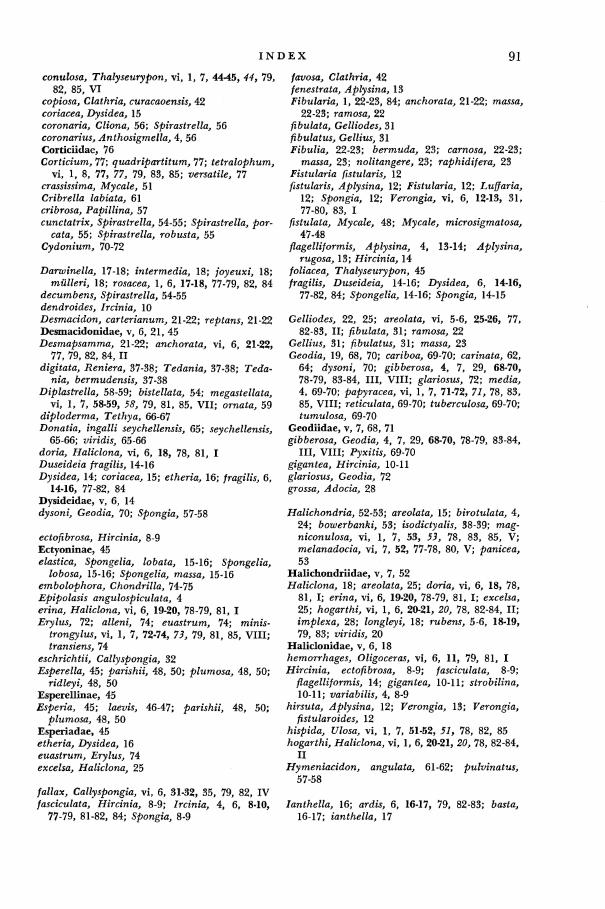
I N D E X 91
conulosa, Thalyseurypon, vi, 1, 7, 44-45, 44, 79, 82, 85, VI
copiosa, Clathria, curacaoensis, 42 coriacea, Dysidea, 15 coronaria, Cliona, 56; Spirastrella, 56 coronarius, Anthosigmella, 4, 56 Corticiidae, 76 Corticium, 77; quadripartitum, 77; tetralophum,
vi, 1, 8, 77, 77, 79, 83, 85; versatile, 77 crassissima, Mycale, 51 Cribrella labiata, 61 cribrosa, Papillina, 57 cunctatrix, Spirastrella, 54-55; Spirastrella, por-
cata, 55; Spirastrella, robusta, 55 Cydonium, 70-72
Darwinella, 17-18; intermedia, 18; joyeuxi, 18; miilleri, 18; rosacea, 1, 6, 17-18, 77-79, 82, 84
decumbens, Spirastrella, 54-55 dendroides, Ircinia, 10 Desmacidon, carterianum, 21-22; reptans, 21-22 Desmacidonidae, v, 6, 21, 45 Desmapsamma, 21-22; anchorata, vi, 6, 21-22,
77, 79, 82, 84, II digitata, Reniera, 37-38; Tedania, 37-38; Tecfa-
ma, bermudensis, 37-38 Diplastrella, 58-59; bistellata, 54; megastellata,
vi, 1, 7, 58-59, 5S, 79, 81, 85, VII; ornata, 59 diploderma, Tethya, 66-67 Donatia, ingalli seychellensis, 65; seychellensis,
65-66; viridis, 65-66 doria, Haliclona, vi, 6, 18, 78, 81, I Duseideia fragilis, 14-16 Dysidea, 14; coriacea, 15; etheria, 16; fragilis, 6,
14-16, 77-82, 84 Dysideidae, v, 6, 14 dysoni, Geodia, 70; Spongia, 57-58
ectofibrosa, Hircinia, 8-9 Ectyoninae, 45 elastica, Spongelia, lobata, 15-16; Spongelia,
lobosa, 15-16; Spongelia, massa, 15-16 embolophora, Chondrilla, 74-75 Epipolasis angulospiculata, 4 erina, Haliclona, vi, 6, 19-20, 78-79, 81, I Erylus, 72; alleni, 74; euastrum, 74; minis-
trongylus, vi, 1, 7, 72-74, 75, 79, 81, 85, VIII; transiens, 74
eschrichtii, Callyspongia, 32 Esperella, 45; parishii, 48, 50; plumosa, 48, 50;
ridleyi, 48, 50 Esperellinae, 45 Esperia, 45; laevis, 46-47; parishii, 48, 50;
plumosa, 48, 50 Esperiadae, 45 etheria, Dysidea, 16 euastrum, Erylus, 74 excelsa, Haliclona, 25
/a/ZflX, Callyspongia, vi, 6, 31-32, 35, 79, 82, IV fasciculata, Hircinia, 8-9; Ircinia, 4, 6, 8-10,
77-79, 81-82, 84; Spongia, 8-9
favosa, Clathria, 42 fenestrata, Aplysina, 13 Fibularia, 1, 22-23, 84; anchorata, 21-22; mawfl,
22-23; ramosa, 22 fibulata, Gelliodes, 31 fibulatus, Gellius, 31 Fibulia, 22-23; bermuda, 23; carnosa, 22-23;
massa, 23; nolitangere, 23; raphidifera, 23 Fistularia fistularis, 12 fistularis, Aplysina, 12; Fistularia, 12; Luffaria,
12; Spongia, 12; Verongia, vi, 6, 12-13, 31, 77-80, 83, I
fistulata, Mycale, 48; Mycale, microsigmatosa, 47-48
flagelliformis, Aplysina, 4, 13-14; Aplysina, rugosa, 13; Hircinia, 14
foliacea, Thalyseurypon, 45 fragilis, Duseideia, 14-16; Dysidea, 6, 14-16,
77-82, 84; Spongelia, 14-16; Spongia, 14-15
Gelliodes, 22, 25; areolata, vi, 5-6, 25-26, 77, 82-83, II; fibulata, 31; ramosa, 22
Gellius, 31; fibulatus, 31; massa, 23 Geodia, 19, 68, 70; cariboa, 69-70; carinata, 62,
64; d;y5om, 70; gibberosa, 4, 7, 29, 68-70, 78-79, 83-84, III, VlII; glariosus, 72; medsa, 4, 69-70; papyracea, vi, 1, 7, 71-72, 71, 78, 83, 85, VIII; reticulata, 69-70; tuberculosa, 69-70; tumulosa, 69-70
Geodiidae, v, 7, 68, 71 gibberosa, Geodia, 4, 7, 29, 68-70, 78-79, 83-84,
III, VIII; P y x t o , 69-70 gigantea, Hircinia, 10-11 glariosus, Geodia, 72 grossa, Adocia, 28
Halichondria, 52-53; areolata, 15; birotulata, 4, 24; bowerbanki, 53; isodictyalis, 38-39; mag-niconulosa, vi, 1, 7, 53, 55, 78, 83, 85, V; melanadocia, vi, 7, 52, 77-78, 80, V; panicea, 53
Halichondriidae, v, 7, 52 Haliclona, 18; areolata, 25; doria, vi, 6, 18, 78,
81, I; erma, vi, 6, 19-20, 78-79, 81, I; excelsa, 25; hogarthi, vi, 1, 6, 20-21, 20, 78, 82-84, II; implexa, 28; longleyi, 18; rubens, 5-6, 18-19, 79, 83; vmdw, 20
Haliclonidae, v, 6, 18 hemorrhages, Oligoceras, vi, 6, 11, 79, 81, I Hircinia, ectofibrosa, 8-9; fasciculata, 8-9;
flagelliformis, 14; gigantea, 10-11; strobilina, 10-11; variabilis, 4, 8-9
hirsuta, Aplysina, 12; Verongia, 13; Verongia, fistularoides, 12
hispida, Ulosa, vi, 1, 7, 51-52, 51, 78, 82, 85 hogarthi, Haliclona, vi, 1, 6, 20-21, 20, 78, 82-84,
II Hymeniacidon, angulata, 61-62; pulvinatus,
57-58
lanthella, 16; ardis, 6, 16-17, 79, 82-83; fcasfa, 16-17; ianthella, 17

92 I N D E X
ianthella, Ianthella, 17 ignis, Tedania, vi, 5, 7, 37-38, 39, 77-79, 83-84,
V; Tedania, pacifica, 37; Thalysias, 37-38 implexa, Haliclona, 28 implexiformis, Adocia, vi, 1, 6, 27-28, 27, 78,
82, 84, II incrustans, Spongelia, 15-16 ingalli, Donatia, seychellensis, 65; Tethya, 65-67 intermedia, Darwinella, 18; Placospongia, 62,
64; Strongylacidon, 51 Iotrochota, 24; acerata, 24; birotulata, 4-6,
24-25, 77-79, 82; coccinea, 25 Ircinia, 8-10, 39; dendroides, 10; fasciculata,
4, 6, 8-10, 77-79, 81-82, 84; muscarum, 11; oro$, 10; ramosa, 9; strobilina, 6, 9, 10-11, 77-81, 84; strobilina irregularis, 10-11; van"-abilis, 8-10
isochela, Mycale, 48, 50, 51 isodictyalis, Halichondria, 38-39; Lissodendoryx,
7, 38-40, 79-80, 82, 84; Lissodendoryx, jack-soniana, 38, 40; Lissodendoryx, paucispinosa, 38, 40; Myxi7/a, 38-39
jacksoniana, Lissodendoryx, 40 joyeuxi, Darwinella, 18
hilauea, Axocielita, 44
labiata, Cribrella, 61 lacunosa, Verongia, 4, 6, 13, 84 laevis, Esperia, 46-47; Mycale, vi, 7, 46-47, 49,
77-78, 82, VI Laxosuberites zeteki, 59-60 leptoderma, Lissodendoryx, 38; Tedania, 38-39 /ex/ft, Acanthacarnus, 40 linda, Axocielita, 44 linteiformis, Spongia, 10 Lissodendoryx, 38; carolinensis, 39-40; isodic
tyalis, 7, 38-40, 79-80, 82, 84; isodictyalis jacksoniana, 38, 40; isodictyalis paucispinosa, 38, 40; jacksoniana, 40; leptoderma, 38; similis, 38-40
longissima, Aplysina, 13; Verongia, 4, 6, 13-14, 78, 82-83
longleyi, Haliclona, 18 Luffaria, 14; cauliformis, 14; fistularis, 12; sebae,
13 lyncurium, Tethya, 65
magniconulosa, Halichondria, vi, 1, 7, 53, 55, 78, 83, 85, V
mflwfl, Fibularia, 22-23; Fibulia, 23; Gellius, 23; Neofibularia, 6, 23, 79, 81
maunakea, Mycale, 48 maunaloa, Microciona, 42 maxima, Spinosella, 4, 34-35 maza, Tethya, 7, 65-66, 67-68 media, Geodia, 4, 69-70; Synops (?), 69 megastellata, Diplastrella, vi, 1, 7, 58-59, 55, 79,
81, 85, VII melanadocia, Halichondria, vi, 7, 52, 77-78,
80, V
melobesioides, Placospongia, 63-64 membranacea, Ophlitaspongia, 44 microchela, Microciona, vi, 1, 5, 7, 41-42, 42,
77-78, 82, 85 Microciona, 41-44; aceratoobtusa, 44; maunaloa,
42; microchela, vi, 1, 5, 7, 41-42, 42, 77-78, 82, 85; parthena, 42; rarispinosa, vi, 1, 7, 42-44, 45, 77-78, 83, 85; «m*7w, 43-44; spinosa, 43
Microcionidae, v, 7, 41 microsigmatosa, Mycale, 7, 47-48, 78, 80-82, 84 millepora, Pachychalina, 26 ministrongylus, Erylus, vi, 1, 7, 72-74, 75, 79,
81, 85, VIII mixta, Placospongia, 62, 64 mollis, Spirastrella, 54-55 miilleri, Darwinella, 18 muscarum, Ircinia, 11 Mycale, 45-46; cecilia, 47-48, 84; crassissima, 51;
fistulata, 48; fistulata microsigmatosa, 47, 48; isochela, 48, 50-51; /aery's, vi, 7, 46-47, 49, 77-78, 82, VI; maunakea, 48; microsigmatosa, 7, 47-48, 78, 80-82, 84; parishii, 48, 50; pecrt-nicola, 48, 50-51; phyllophila, 48; senegalensis, 47-48'
Mycalidae, v, 1, 5, 7, 45 Mycalinae, 45 Myxilla isodictyalis, 38-39
neens, Adocia, 29 Neofibularia, 1, 6, 22-23, 84; masja, 6, 23, 79, 81 nigrescens, Tedania, 37 nigricans, Papillina, 61 nolitangere, Fibulia, 23 nucula, Chondrilla, 7, 74-75, 79, 81, 84
Oligoceras, 11; collectrix, 11; collectrix hemorrhages, 11; hemorrhages, vi, 6, 11, 79, 81, I; spongeliformis, 11
Ophlitaspongia membranacea, 44 Ophlitaspongiidae, 44-45 orientalis, Cliona, 57 ornata, Displastrella, 59 oras, Ircinia, 10 Osculina polystomella, 61
Pachychalina, areolata, 25; aurantiaca, 26; car-bonaria, 26-27; millepora, 26; rubens, 18-19
pallescens, Spongelia, 15-16 pallida, Callyspongia, vi, 1, 6, 36-37, 55, 83-84,
III Pandaros, 45; acanthifolium, 45 panicea, Halichondria, 53 papillaris, Callyspongia, 32; Spongia, 32 Papillina, arcuata, 56-57; cribrosa, 57; nigricans,
61 papyracea, Geodia, vi, 1, 7, 71-72, 72, 78, 83, 85,
VIII; Siphonochalina, 34-35 parishii, Esperella, 48, 50; Esperia, 48, 50; My-
ca/e, 48, 50; Raphiodesma, 48, 50; Zygomycale, vi, 7,48-51,77,81-82, 84, V
parthena, Microciona, 42 Patuloscula, plicifera, 34; procumbens, 32

I N D E X 93 pectinicola, Mycale, 48, 50-51 Pellina, 27, 29-30; carbonaria, 26-27, 30; coela,
30; coeliformis, vi, 1, 6, 29-30, 29, 78-79, 83-84, III; semitubulosa, 30
Phloeodictyon carbonaria, 26 phyllodes, Chondrilla, 54-55; Chondrillina, 54 phyllophila, Mycale, 48 Placospongia, 62; carinata, vi, 7, 62-64, 77, 79,
81, 84, VII; intermedia, 62, 64; melobesioides, 63-64; m/xta, 62, 64; sp., 62, 64
Placospongiidae, v, 7, 62 Plakinidae, v, 8, 76 Plakortis, 4, 76; simplex, 76; zyggompha, 8, 76,
79,81 plicifera, Callyspongia, vi, 4, 6, 32, 34-36, 79, 82,
IV; Patuloscula, 34; Spinosella, 34-35; Spongia, 34-35; Tu&a, 34-35
Plumocolumella, 23 plumosa, Esperella, 48, 50; Esperia, 48, 50 polystomella, Osculina, 61 Poterion atlantica, 57-58 praetexta, Aplysina, 13 procumbens, Patuloscula, 32 Prosuberites, 56 proxima, Thalysias, 26-27 pulvinata, Spirastrella, 57 pulvinatus, Hymeniacidon, 57-58 purpurea, Spirastrella, 54-55, 57 purpureum, Alcyonium, 54 Pyxitis gibberosa, 69-70
quadripartitum, Corticium, 77
ramosa, Fibularia, 22; Gelliodes, 22; Ircinia, 9 raphidifera, Fibulia, 23 Raphiodesma parishii, 48, 50 rarispinosa, Microciona, vi, 1, 7, 42-44, 43, 77-78,
83, 85 Reniera digitata, 37-38 reptans, Desmacidon, 21-22 reticulata, Geodia, 69-70 rhizophyta, Cinachyra, 4, 6 rhoda, Ulosa, 51-52 ridleyi, Esperella, 48, 50 Rodsa, 76; zyggompha, 76 rosacea, Darwinella, 1, 6, 17-18, 77-79, 82, 84 rubens, Chalina, 18-19; Haliclona, 5, 6, 18-19,
79, 83; Pachychalina, 18-19; Spongia, 18-19 rubiginosa, Cacochalina, 19
Sarcotragus, 10 scrobiculata, Callyspongia, 32; Spinosella, 34-35;
Spongia, 34-35; Tu&a, 32, 34-35 sebae, Luff aria, 13 semitubulosa, Pellina, 30 senegalensis, Mycale, 47-48 seychellensis, Alemo, 65-66; Donatia, 65-66;
7 > % a , 7, 65-66, 67, 78-79, 81-82, 84 Sigmadocia, 30-31; caerulea, vi, 1, 5-6, 26, 30-31,
30, 78-79, 81, 84, III similis, Lissodendoryx, 38-40; Microciona, 43-44
simplex, Plakortis, 76 Siphonochalina, papyracea, 34-35; stolonifera, 34 saroria, Spinosella, 33; Tuba, 33 souriei, Acanthacarnus, 7, 40, 79, 83-84 spengelii, Aplysina, 4 Spheciospongia, 57-58; vesparia, 4, 7, 57-58, 78,
80 spinosa, Microciona, 43 Spinosella, maxima, 4, 34-35; plicifera, 34-35;
scrobiculata, 34-35; sororia, 33 Spirastrella, 54, 58; andrewsii, 4, 57-58; coccinea,
7, 54-55, 79, 81, 83-84; coronaria, 56; cuncta-trix, 54-55; cunctatrix porcata, 55; cunctatrix robusta, 55; decumbens, 54-55; mollis, 54-55; pulvinata, 57; purpurea, 54-55, 57
Spirastrellidae, v, 7, 54 Spongelia, elastica lobata, 15-16; elastica lobosa,
15-16; elastica massa, 15-16; fragilis, 14-16; incrustans, 15-16; pallescens, 15-16
spongeliformis, Cacospongia, 11; Oligoceras, 11 Spongia, angulosa, 51; carbonaria, 26-27; cty-
5onz, 57-58; fasciculata, 8-9; fistularis, 12; fragilis, 14-15; linteiformis, 10; papillaris, 32; plicifera, 34-35; rubens 18-19; scrobiculata, 34-35; strobilina, 10; tubaeformis, 12; vagin a l , 32-33
spongia, Ulosa, 51-52 Spongiidae, v, 6, 8 stolonifera, Siphonochalina, 34 strobilina, Hircinia, 10-11; Ircinia, 6, 9, 10-11,
77-81, 84; Ircinia, irregularis, 10-11; Spongia, 10
Strongylacidon intermedia, 51 Suberites, angulospiculatus, 4, 6; coronarius, 4,
56; tuberculosus 56-57; undulatus, 62 Suberitidae, v, 7, 59 subulata, Cliona, 61-62 Synops (?) media, 69
Tedania, 37-38; anhelans, 37-38; orwcei, 37-38; digitata, 37-38; digitata bermudensis, 37-38; ignw, vi, 5, 7, 37-38, 39, 77-79; 83-84, V; ignw pacifica, 37; leptoderma, 38-39; nigrescent, 37
Tedaniidae, v, 7, 37 tenuissima, Verongia, 12 Terpios, 59; ztteftt, vi, 5, 7, 59-60, 78-79, 81-84,
VII Tethya, 65-66, 81; actinia, 7, 66-67, 79, 84; dip/o-
derma, 66-67; ingalli, 65-67; lyncurium, 65; maza, 7, 65-66, 67-68, seychellensis, 7, 65-66, 67, 78-79, 81-82, 84; sp., 7, 67, 68, 79
Tethyidae, v, 7, 65 tetralophum, Corticium, vi, 1, 8, 77, 77, 79, 83,
85 Thalyseurypon, 44-45; carteri, 45; conulosa, vi,
1, 7, 44-45, 44, 79, 82, 85, VI; foliacea, 45; vasiformis, 45
Thalysias, carbonaria, 26-27; coccinea, 54-55; fgnt5, 37-38; proxima, 26-27; varians, 55-57; vesparia, 57-58
transiens, Erylus, 74

94 I N D E X
Tuba, plicifera, 34-35; scrobiculata, 32, 34-35; sororia, 33; vaginalis, 32-33
tubaeformis, Spongia, 12 tuberculosa, Geodia, 69-70 tuberculosus, Suberites, 56-57 tubifera, Adocia, 28 tumultosa, Geodia, 69-70
£/Z<wa, 51-52; TiwpWa, vi, 1, 7, 51-52, 5 i , 78, 82, 85; rhoda, 51-52; spongia, 51-52
undulatus Suberites, 62
vaginalis, Callyspongia, 6, 32-34, 35, 79, 82; Spongia, 32-33; Tw&a, 32-33
vanderhorsti, Ceraochalina, 32 variabilis, Hircinia, 4, 8-9; Ircinia, 8-10 varians, Anthosigmella, vi, 4, 7, 55-57, 79, 81,
VII; Thalysias, 55-57 vasiformis, Thalyseurypon, 45
vermifera, Cliona, 7, 60-61, 79, 83-84; Fioa, 60 Verongia, 12, 14; aurea, 14; fistularis, vi, 6,
12-13, 31, 77-80, 83, I; hirsuta, 13; hirsuta fistularoides, 12; lacunosa, 4, 6, 13, 84; longissima, 4, 6, 13-14, 78, 82-83; tenuissima, 12
versatile, Corticium, 77 vesparia, Spheciospongia, 4, 7, 57-58, 78, 80;
Thalysias, 57-58 vesparium, Alcyonium, 57 F7oa, vermifera, 60; viridis, 61 viridis, Cliona, 7, 61-62, 79, 83-84; Cliona, carib-
boea, 61-62; Donatia, 65-66; Haliclona, 20; Fioa, 61
zeteki, Laxosuberites, 59-60; Terpios, vi, 5, 7, 59-60, 78-79, 81-84, VII
zyggompha, Plakortis, 8, 76, 79, 81; Roosa, 76 Zygomycale, 45, 48; parishii, vi, 7, 48-51, 77
81-82, 84, V
PLATES
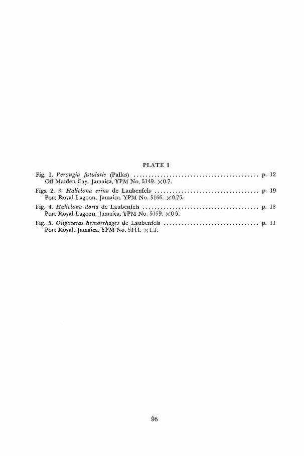
PLATE I
Fig. 1. Verongia fistularis (Pallas) . . . . . . . . . . . . . . . . . . . . . . . . . . . . . . . . . . . . . . . . . . p. 12 Ojff Maiden Cay, Jamaica. YPM No. 5149. x0.7.
Figs. 2, 3. Haliclona erina de Laubenfels . . . . . . . . . . . . . . . . . . . . . . . . . . . . . . . . . . . p. 19 Port Royal Lagoon, Jamaica, YPM No. 5166. x0.75.
Fig. 4. Haliclona doria de Laubenfels . . . . . . . . . . . . . . . . . . . . . . . . . . . . . . . . . . . . . . . p. 18 Port Royal Lagoon, Jamaica. YPM No. 5159.- x0.9.
Fig. 5. Oligoceras hemorrhages de Laubenfels . . . . . . . . . . . . . . . . . . . . . . . . . . . . . . . . p. 11 Port Royal, Jamaica. YPM No. 5144. X 1.1.
96

w*

i ,-T - . • S T *
«
t *
• •v .
* ***» •
*
I ^ v * -• '».*-- • J-.**" fal! •* mw-~
^ j
/
4* •:jj£5^ • 9.S'*
• • * & • • t-&+»&\

PLATE II
Fig. 1. Haliclona hogarthi n. sp. . . . . . . . . . . . . . . . . . . . . . . . . . . . . . . . . . . . . . . . . . . . p. 20 Port Royal, Jamaica. Holotype. YPM No. 5033. x0.8.
Fig. 2. Adocia implexiformis n. sp. . . . . . . . . . . . . . . . . . . . . . . . . . . . . . . . . . . . . . . . . . . p. 27 Port Royal, Jamaica. Holotype. YPM No. 5034. x0.9.
Fig. 3. Gelliodes areolata (Wilson) . . . . . . . . . . . . . . . . . . . . . . . . . . . . . . . . . . . . . . . . . . p. 25 Port Royal, Jamaica. YPM No. 5186. x0.6.
Fig. 4. Desmapsamma anchomta (Carter) p. 21 Port Royal, Jamaica. YPM No. 5174. xO.93.
§7

PLATE III
Fig. 1. PelUna coeliformis n. sp. (growing on Geodia gibberosa Lamarck) . . . . . . . . p Port Royal, Jamaica. Holotype. YPM No. 5086. x0.9.
Figs. 2, 8. Adocia carbonaria (Lamarck) . . . . . . . . . . . . . . . . . . . . . . . . . . . . . . . . . . . . . . p Dunn's River, Jamaica. YPM No. 5191. xO.75.
Fig. 4. Sigmadocia caerulea n. sp. . . . . . . . . . . . . . . . . . . . . . . . . . . . . . . . . . . . . . . . . . . . p Port Royal, Jamaica. Holotype. YPM No. 5037, X LI.
Fig. 5. Callyspongia pallida n. sp. . . . . . . . . . . . . . . . . . . . . . . . . . . . . . . . . . . . . . . . . . . . p Port Royal, Jamaica. Holotype. YPM No. 5088. _x0.8.
98
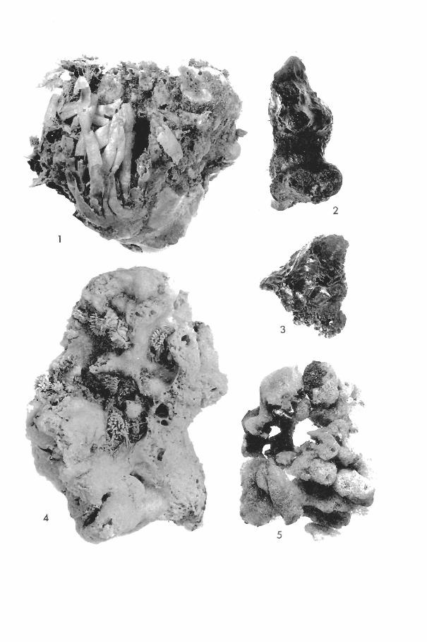
fr -•*„
Jj< ».. jl
^ ^
w. *<%&m
r?r*r
WP" • * * * .
<~#5
*v
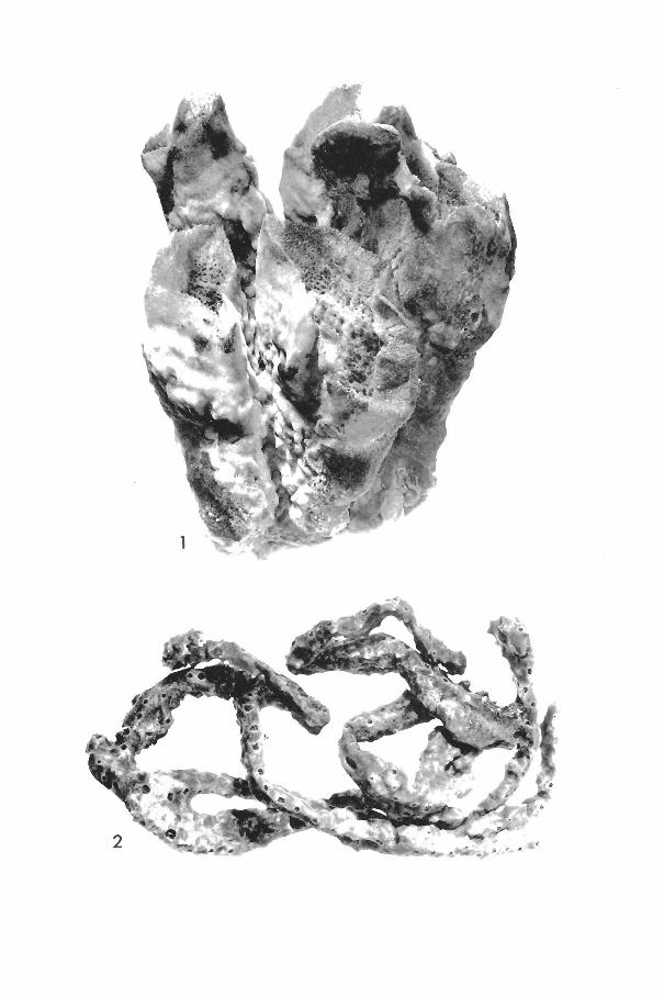
• > •
% •
fflA \^
^••^r%i*i
: ^
r* ^ V ; W
, R -
Vt
• • X V
PLATE IV
Fig. 1. Callyspongia plicifera (Lamarck) p. 34 Off Maiden Cay, Jamaica. YPM No. 5215. xO.55.
Fig. 2. Callyspongia fallax Duchassaing and Michelotti . . . . . . . . . . . . . . . . . . . . . . . p. 31 Off Maiden Cay, Jamaica. YPM No. 5205. xO.55.
f t

PLATE ¥
Fig. 1. HaMchondria mogniconulosa n. sp. . . . . . . . . . . . . . . . . . . . . . . . . . . . . . . . . . . . p. 58 Port Royal, Jamaica. Holotype. YPM No. 5089. x0.75.
Fig. 2. HaMchondria melanadocia de Laubenfels . . . . . . . . . . . . . . . . . . . . . . . . . . . . . . p. 52 Port Royal, Jamaica. YPM No. 5219. x0.85.
Fig. 8. Zygomycale parishii (Bowerbank). . . . . . . . . . . . . . . . . . . . . . . . . . . . . . . . . . . . . p. 48 Port Royal, Jamaica. YPM No. 5258. x0.8.
Fig. 4, Tedania ignis (Duchassaing and Michelotti) . . . . . . . . . . . . . . . . . . . . . . . . . . p. 87 Port Royal, Jamaica. YPM No. 5229. x0.85.
100
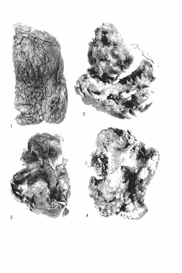
->-" .*^-^; .'-fcL
•"i-S;-"- i-Vi-" . • v9r£K* •'•'
•^SCf-k
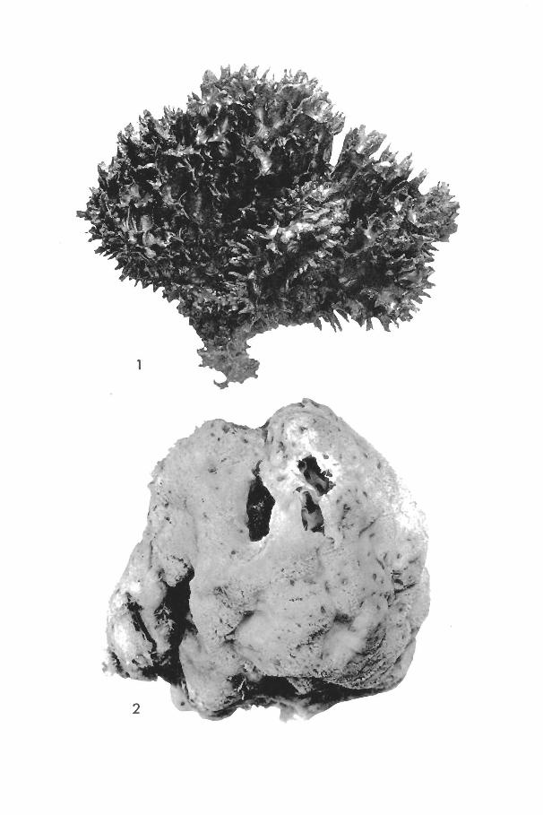
~ArW$^ £-
^ J I L

PLATE ¥1
Fig. 1. Thalyseurypon conulosa n. sp. . . . . . . . . . . . . . . . . . . . . . . . . . . . . . . . . . . . . . . . p. 44 Off Maiden Cay, Jamaica. Holotype. YPM No. 5042. X1.
Fig. 2. Mycale laevis (Carter) . . . . . . . . . . . . . . . . . . . . . . . . . . . . . . . . . . . . . . . . . . . . . . . p. 46 Port Royal, Jamaica. YPM No. 5240. x l . l .
101

PLATE VII
Fig. 1. Placospongia carinata (Bowerbank) . . . . . . . . . . . . . . . . . . . . . . . . . . . . . . . . . . p Port Royal, Jamaica. YPM No. 5279. x L
Fig. -.2. Diplastrella megastellata n. sp. . . . . . . . . . . . . . . . . . . . v . . . - . . . . . ' . ' . . . ' . . ' . ' . . . . . ' . . • p Off Maiden Cay, Jamaica. Holotype. YPM No. 5044. x 0.9..
Fig. S. Terpios zeteki (de Laubenfels) . . . . . . . . . . . . . . . . . . . . . . . . . . . . . . . . . . . . . . . . p Port Royal, Jamaica. YPM. No. 5270. X1.'
Figs. 4, 5. Anthosigmella varians (Duchassaing and Michelotti). . . . . . . . . . . . . . . . . . p Port Royal, Jamaica. YPM No. 5262. x0.6.
102
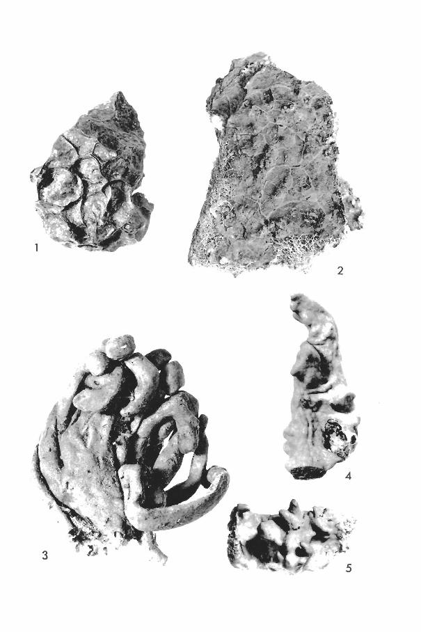
-^Kjm .#y • . •*&'
$ • $ . - ' J 3 T • . , . - • » • • *
^
• j * ^ *

<M S(^-v
, / &
> A
»**
•'" > W *, ^ '.•?% • Jf

PLATE VIII
Fig. 1. Geodia papyracea n. sp. p. 71 Port Royal, Jamaica. Paratype. YPM No. 5311. x 1.1.
Fig. 2. Geodia papyracea n. sp. (specimen on lower right, growing on Geodia gibberosa Lamarck) . . . . . . . . . . . . . . . . . . . . . . . . . . . . . . . . . . . . . . . . . . . . . . . . . . . . . p. 71 Port Royal, Jamaica. Paratype. YPM No. 5311. x 1.1.
Fig. 3. Erylus ministrongylus n. sp. . . . . . . . . . . . . . . . . . . . . . . . . . . . . . . . . . . . . . . . . . p. 72 Drunkenman's Cay, Jamaica. Holotype. YPM No. 5046. x 1.6.
103