Embed Size (px)
Citation preview
A Testis Specific Isoform of Endophilin B1, Endophilin B1t, Interacts Specificallywith Protein Phosphatase-1cγ2 in Mouse Testis and Is Abnormally Expressed in
PP1cγ Null Mice
Christopher Hrabchak, Hannah Henderson, and Susannah Varmuza*
Department of Cell and Systems Biology, UniVersity of Toronto, Toronto, Ontario, Canada
ReceiVed December 15, 2006; ReVised Manuscript ReceiVed February 8, 2007
ABSTRACT: Male mice homozygous for a null mutation in the protein phosphatase-1cγ (PP1cγ) gene areinfertile, displaying a severe impairment in spermatogenesis that is not compensated by the presence ofPP1cR and PP1câ in mutant testes. A lack of the PP1cγ2 splice variant seems the most likely cause ofthe mutant phenotype, as it is the most heavily expressed PP1cγ isoform in wild type testes. Yeast two-hybrid screening using PP1cγ2 has identified several new binding partners, including endophilin B1t, atestis enriched isoform of endophilin B1a which differs from the somatic form by virtue of a carboxyterminal deletion spanning the last 10 amino acids. The testis isoform did not show an interaction withPP1cR, or with a truncated PP1cγ2 mutant lacking the unique carboxy terminus. In contrast, somaticendophilin B1a did not interact with any of the PP1c isoforms. Sedimentation and co-immunoprecipitationexperiments using native testis proteins verified binding of endophilin B1t to PP1cγ2. Immunohistochem-istry on wild type testis sections revealed a stage specific expression pattern for endophilin that appearedconcentrated at discrete puncta throughout the seminiferous epithelium. Punctate endophilin expressionin cells adjacent to the lumen was absent in PP1cγ null mice. Phosphatase assays indicate that chimericendophilin B1t is able to inhibit recombinant PP1cγ2 activity toward phosphorylasea while having littleeffect on the activity of PP1cR. A potential role for endophilin B1t in mammalian spermatogenesis isdiscussed within the context of the PP1cγ knockout testis phenotype.
Protein phosphatase 1 (PP11) is a major eukaryotic serine/threonine phosphatase composed of a single catalytic subunit(PP1c) in complex with one or more regulatory subunits.Catalytic subunits are highly conserved across phyla, whileregulatory subunits are varied and thought to confer sub-cellular localization and substrate specificity (1). Highereukaryotes typically contain multiple PP1c isoforms showingan overall sequence similarity of∼90% (2). Mutations inPP1c genes from different organisms have resulted in distinctphysiological defects, suggesting that different isoforms havethe potential for unique functions despite close sequencesimilarity (3-6). One way in which functional independencecan be achieved is through preferential interaction withparticular regulatory subunits. To date more than 50 regula-tory subunits have been identified which target PP1 to diversecellular processes including glycogen metabolism, proteinsynthesis, mitosis, synaptic depression, and smooth musclecontraction (7). Evidence suggests that regulatory subunitsbind PP1c through multiple points of interaction, with manycontaining a degenerate “RVxF” motif (the more general
consensus sequence is (R/K)X0-1(V/I){P}(F/W), where X0-1
represents the possible presence of an amino acid, whoseidentity varies, while{P} is any amino acid except proline)(8). This motif has been implicated in an interaction with aconserved hydrophobic channel (9). Isolated PP1 holoen-zymes typically contain only one inhibitor or targetingsubunit, indicating that interaction of different regulatorysubunits is often mutually exclusive (10). Of the known PP1regulatory proteins only a small subset, such as the neurabins,actin-binding proteins that are thought to regulate synapticdepression in brain neurons, are known to preferentiallyinteract with specific PP1c isoforms (11, 12).
In mammalian systems, four different PP1c isoforms havebeen identified: PP1cR, PP1câ, PP1cγ1, and PP1cγ2, wherethe latter two variants are derived through alternate splicingof the PP1cγ gene (13). The isoforms are over 98% identicalexcluding the extreme carboxy termini where most of thedivergence in sequence occurs (14). Male mice homozygousfor a null mutation in the PP1cγ gene are infertile, displayinga severe impairment in spermatogenesis despite the continuedpresence of PP1cR and PP1câ (3). The phenotype iscompletely penetrant, with 100% of mutant males beingsterile despite the occasional presence of mature, if grosslyabnormal, sperm. Heterozygotes show no difference fromwild type, suggesting that defects in homozygous mutantsare not the result of a general decrease in PP1c gene dosage.Histochemical analysis of PP1cγ mutant testicular sectionsreveals gross reductions in numbers of condensing and
* Corresponding author. Address for correspondence: Departmentof Cell and Systems Biology, University of Toronto, 25 Harbord Street,M5S 3G5, Toronto, Ontario, Canada. Tel: 416 978-2759. Fax: 416978-8532. E-mail: [email protected].
1 Abbreviations: PP1, protein phosphatase 1; PP1c, protein phos-phatase-1 catalytic subunit; GST, glutathioneS-transferase; PMSF,phenylmethylsulfonyl fluoride; HA, hemagglutinin; EST, expressedsequence tag; 3-AT, 3-aminotriazole.
4635Biochemistry2007,46, 4635-4644
10.1021/bi6025837 CCC: $37.00 © 2007 American Chemical SocietyPublished on Web 03/24/2007
elongate spermatids, as well as increased levels of apoptosis(15) and aneuploidy (16) in developing germ cells. Inaddition to the observed germ cell defects large “holes” mayalso be observed in the seminiferous epithelium that maybe either vacuoles in the Sertoli cells, indicative of Sertolicell dysfunction (17), or gaps left by dead germ cells. A lackof the PP1cγ2 splice variant seems the most likely cause ofthe mutant phenotype, as it is the most abundant PP1cγisoform in the testis (18). To date, known physiologicalbinding partners of PP1cγ2, including the glutamate receptormGluR7b (19), the 78 kDa glucose-regulated protein (20)and a mammalian homologue of yeast Sds22p (21) are alsoknown to bind other PP1c isoforms, or have not beencharacterized in terms of their specificity.
To explain the phenotype of PP1cγ null mice, we havehypothesized the presence of one or more regulatory subunitsin the testis that interact specifically with PP1cγ2, but notPP1cR or PP1câ, targeting the enzyme to a function requiredfor spermatogenesis. Such a regulatory subunit would likelycontain novel sequences that allowed for an interaction withthe unique C-terminus of PP1cγ2. Our studies identified asmall number of isoform specific interactors, including threeunknown proteins, Spz1 (22) and a testis-enriched variantof endophilin designated as endophilin B1t. Here wecharacterize endophilin B1t expression in the testis andconfirm the interaction between PP1cγ2 and endophilin B1t.We propose that endophilin, whose function in other cellsis associated with endocytosis, may be involved in some ofthe dynamic tissue remodeling that occurs in the testis, andthat the loss of PP1cγ may impair the activity of the testicularendophilin variant.
MATERIALS AND METHODS
Yeast Two-Hybrid Screening and Assays.To create two-hybrid vectors, coding sequences for mouse PP1cγ2, PP1cγ1,PP1cR, and a truncated PP1cγ2 lacking 18 amino acids fromthe C-terminus were amplified by PCR from pGEM7Zvectors containing full-length cDNAs (23). PCR was per-formed using the following primer sets (Table 1): forPP1cγ2, primers 1 and 2; for PP1cγ1, primers 1 and 3; forPP1cR, primers 4 and 5; for truncated PP1cγ2, primers 1and 6 (primer sequences are summarized in Table 1).Resulting fragments were digested withNcoI andEcoRI and
ligated separately into pGBKT7 (Clontech). Two versionsof endophilin were cloned. Endophilin B1t was cloned byRT-PCR using primers 7 and 8 on RNA extracted from testisusing Trizol (Invitrogen). The endophilin B1t fragment wasdigested withSmaI and XhoI and ligated into the multiplecloning site of pGADT7 (Clontech). Somatic endophilin B1awas cloned by two rounds of RT-PCR on testis RNA usingprimers 7 and 9. Two rounds of PCR were necessary togenerate sufficient yield of the low abundance somatic splicevariant from testis RNA. All two-hybrid vectors weresequenced to ensure coding sequences had been inserted inframe.
Bait vectors containing PP1c constructs were transformedseparately into yeastSaccharomyces cereVisiae AH109(MATa, trp1-901, leu2-3, 112, ura3-52, his3-200, gal4∆,gal80∆, LYS2::GAL1UAS-GAL1TATA-HIS3, GAL2UAS-GAL2TATA-ADE2, ura3::MEL1UAS-MEL1TATA-lacZ) and maintained onsynthetic dropout media without tryptophan. Screening wasconducted using a mouse testis expression library constructedin pACT2 from pooled 8-12 week BALB/c males (Clon-tech). The library was amplified inEscherichia colistrainBNN132 as in Elledge et al. (24), and plasmid DNA waspurified using a maxi-prep kit from Qiagen. Library plasmidDNA (500µg) was transformed into yeast AH109 expressingPP1cγ2 by the LiAc method as described in the ClontechYeast Protocols Handbook (2001). Transformed cells wereselected after 5-7 days incubation at 30°C on minimalmedia. Activation of thelacZ reporter gene was assessedvia colony lift assay using X-gal as described in the Clontechmanual. Plasmid DNA from positively interacting clones wasextracted using a yeast plasmid purification kit (CPG Inc.)and electroporated (25) into previously prepared electro-competentE. coli DH5R cells (26). Plasmids recovered fromE. coli were retransformed into yeast expressing PP1cγ2 toconfirm two-hybrid interactions, then separately into strainsexpressing bait constructs for PP1cR, PP1cγ1, lamin C, andtruncated PP1cγ2.
Antibody Production and Western Blotting.Polyclonalantibodies recognizing both PP1cγ splice variants weregenerated in rabbits against the peptide “KPAEKKKP-NATRPVT”. The peptide was synthesized commercially andcoupled to keyhole limpet hemocyanin using an Imjectsulfhydryl reactive antibody production kit (Pierce). Antibod-
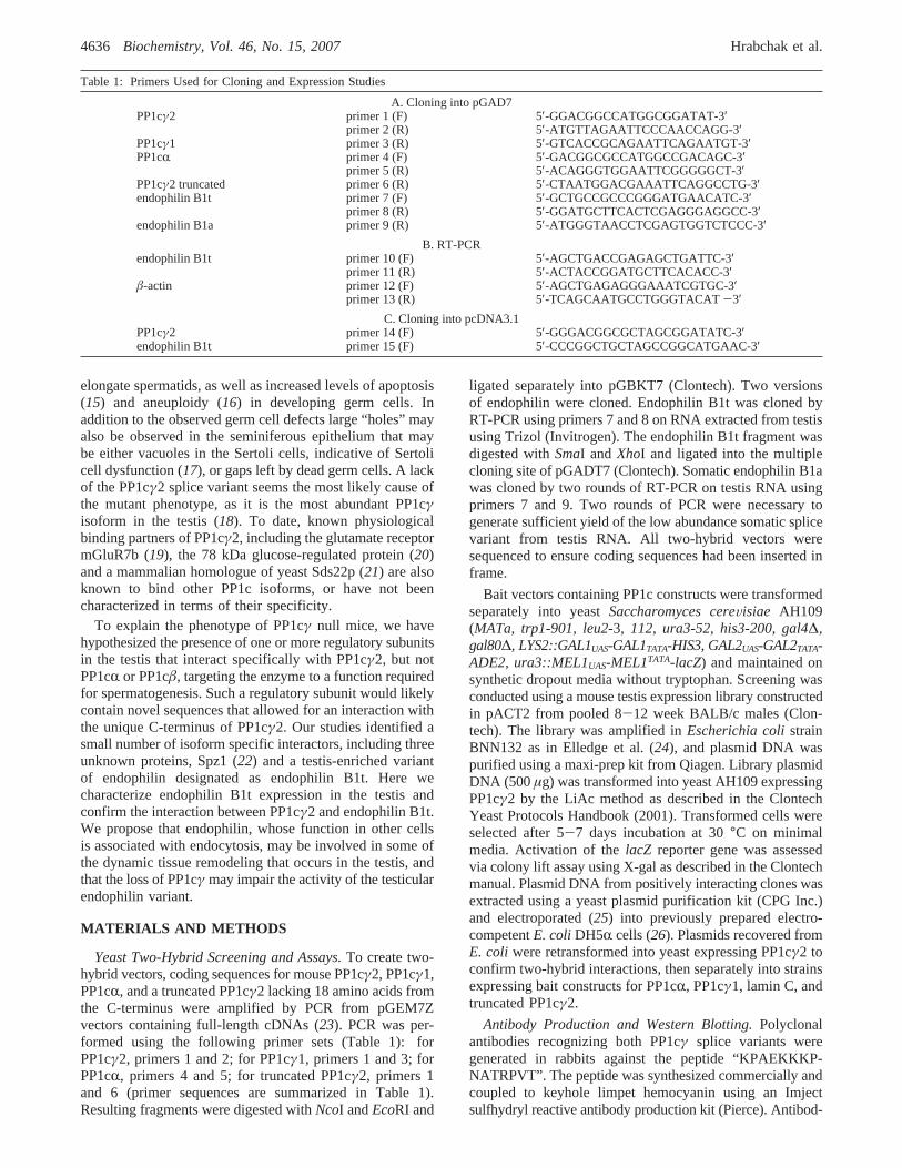
Table 1: Primers Used for Cloning and Expression Studies
A. Cloning into pGAD7PP1cγ2 primer 1 (F) 5′-GGACGGCCATGGCGGATAT-3′
primer 2 (R) 5′-ATGTTAGAATTCCCAACCAGG-3′PP1cγ1 primer 3 (R) 5′-GTCACCGCAGAATTCAGAATGT-3′PP1cR primer 4 (F) 5′-GACGGCGCCATGGCCGACAGC-3′
primer 5 (R) 5′-ACAGGGTGGAATTCGGGGGCT-3′PP1cγ2 truncated primer 6 (R) 5′-CTAATGGACGAAATTCAGGCCTG-3′endophilin B1t primer 7 (F) 5′-GCTGCCGCCCGGGATGAACATC-3′
primer 8 (R) 5′-GGATGCTTCACTCGAGGGAGGCC-3′endophilin B1a primer 9 (R) 5′-ATGGGTAACCTCGAGTGGTCTCCC-3′
B. RT-PCRendophilin B1t primer 10 (F) 5′-AGCTGACCGAGAGCTGATTC-3′
primer 11 (R) 5′-ACTACCGGATGCTTCACACC-3′â-actin primer 12 (F) 5′-AGCTGAGAGGGAAATCGTGC-3′
primer 13 (R) 5′-TCAGCAATGCCTGGGTACAT-3′C. Cloning into pcDNA3.1
PP1cγ2 primer 14 (F) 5′-GGGACGGCGCTAGCGGATATC-3′endophilin B1t primer 15 (F) 5′-CCCGGCTGCTAGCCGGCATGAAC-3′
4636 Biochemistry, Vol. 46, No. 15, 2007 Hrabchak et al.
ies were produced in rabbits, and sera were harvested usingstandard techniques (27).
Antibodies directed against endophilin (mouse anti-Bif-1IgG) were obtained commercially (Imgenex). Antibodiesdirected against Bax (rabbit anti-Bax IgG) were obtainedcommercially (Santa Cruz Biotech). Antiserum directedagainst PP1cγ2 was kindly provided by S. Vijayaraghavan.
For Western blotting, testis protein extracts were preparedby homogenizing whole mouse testes with mortar and pestlein protein extraction buffer (100 mM NaCl, 1 mM EDTApH 8.0, 10 mM Tris-HCl pH 7.5, 1 mM PMSF, with 1µg/mL each of aprotinin, leupeptin, and pepstatin A) using500µL of buffer per 50µg of tissue. Following centrifugationat 10500g for 10 min at 4°C, the resulting supernatants werecollected and 25µg samples were boiled in SDS buffer priorto SDS-PAGE using 12.5% (w/v) acrylamide gels. Proteinswere transferred to nitrocellulose with a Novex Westerntransfer apparatus and immunoblotted as described (28).PP1cγ, endophilin, and Bax antibodies were used at con-centrations of 1:500. Goat anti-PP1cR, rat anti-GATA1, andHRP-coupled secondary antibodies (Santa Cruz Biotech)were used at 1:1000. Bound complexes were visualized usingan enhanced chemiluminescence detection system (Amer-sham). For Western blotting with Bax antibodies, heartprotein extracts were prepared as above due to the lowconcentration of Bax in testis and brain protein extracts.
ReVerse Transcriptase-PCR and Northern Blotting.TotalRNA from multiple mouse tissues was isolated using Trizolaccording to manufacturer’s instructions. All tissues weredissected from a 15 week old wild type male CD-1 mouseexcept ovary, which was pooled from three female litter-mates. Reverse transcription was carried out on 3µg RNAsamples using Superscript II (Invitrogen). Resulting cDNAwas diluted 1:10 and used in separate PCR reactions toamplify a 726 bp endophilin B1t fragment with primers 10and 11 (Table 1). Aâ-actin fragment was amplified as apositive control using primers 12 and 13.
Co-Immunoprecipitation from Testis Lysates.Wild typetestis protein lysates were prepared as described above.500µg of testis lysate at 1µg/1 µL was incubated with 5µgof anti-PP1cγ or 5µg of anti-endophilin for 2 h at 4°C withgentle rotation. Antibody and bound proteins were precipi-tated with 50µL of Protein G: Sepharose 4B (Sigma) for 1h at 4 °C. Protein complexes were washed 5 times withprotein extraction buffer before the addition of SDS-PAGEsample buffer and Western blotting.
Co-Immunoprecipitation in COS-1 Cells.COS-1 sells werecultured on plates in Dulbecco’s modified Eagle’s mediumwith 10% fetal calf serum according to standard procedures(29). For expression constructs, PP1cγ2 cDNA was amplifiedby PCR using primers 14 and 2 from a pGEM7Z vector asbefore. Endophilin B1t cDNA was amplified from testis RNAusing primers 8 and 15 (Table 1). The cDNA fragments weredigested withNheI andEcoRI (for PP1cγ2) orNheI andXhoI(for endophilin B1t) and cloned separately intomyc- andHA-pcDNA3.1 plasmid vectors (Invitrogen). The PP1cγ2 con-struct was introduced into COS-1 cells by electroporationby stable transfection using a Bio-Rad Gene-Pulser accordingto the method of Misumi et al. (30), and transformed cellswere selected in 0.8 mg/mL G418 for one week. Two60 mm culture dishes were grown to 90% confluency beforetransient transfection withHA-pcDNA3.1-endophilin as
before. Resulting cells were lysed after 3 days in 1 mL ofimmunoprecipitation buffer (50 mM Tris-HCL, pH 8.0,120 mM NaCl, 0.5% NP40, 1 mM PMSF, and 1µg/mL eachof leupeptin, aprotinin, and pepstatin A) for 15 min at 4°C.Lysates were centrifuged at 10500g for 10 min at 4°C, andthe supernatant was split into 500µL samples. PP1cγ2 andendophilin B1t were precipitated separately using 5µg ofchicken anti-mycor anti-HA antibodies (Aves Labs) at 4°Cwith gentle rotation for 1.5 h. Antibody and bound proteinswere precipitated with 50µL of anti-chicken IgY im-mobilized on agarose beads (Promega). Protein complexeswere washed extensively before addition of SDS-PAGEsample buffer and Western blotting. For control experiments,5 µg of anti-GATA1 or anti-PP1cR was used with COS-1lysates as before. Bound proteins were precipitated with10µL of 50% Protein A: Agarose beads (Transduction Labs)before Western analysis.
Sedimentation Assays.Wild type testis protein lysates wereprepared as described above. Varying amounts of GST-endophilin B1t were bound to 40µL samples of glutathionesepharose 4B beads (Amersham) according to manufacturer’sinstructions. Binding of GST-endophilin B1t was verifiedvia Coomassie Blue staining following electrophoresis. Beadswere washed 2 times with PBS before addition of 5 mg totaltestis protein and incubation for 2 h at 4 °C. Proteincomplexes were washed 3 times with extraction buffer(100 mM NaCl, 1 mM EDTA pH 8.0, 10 mM Tris-HCl pH7.5, 1 mM PMSF) before elution in SDS sample buffer andWestern analysis. Relative amounts of PP1cγ2 were quanti-fied using Optiquant software (Packard Biosciences). Interac-tion between both GST-endophilin B1t and GST-endophilinB1a, and Bax was performed as described above, except thatheart extract was used instead of testis extract due to lowexpression of Bax in testis.
Protein Phosphatase-1 ActiVity Assays.To produce ex-pression vectors for recombinant PP1c isoforms, codingsequences for PP1cγ2 and PP1cR were first obtained throughrestriction digest of plasmid DNA from respective two-hybridbaits usingNcoI andSalI. Respective cDNA fragments werecloned into corresponding sites on an NpT7-5 plasmid vector(generously provided by Dr. S. Shenolikar). Recombinantplasmids were transformed intoE. coli BL21, and lysatesexpressing PP1c were prepared as in Ludlow (31).
To produce radiolabeled substrate, phosphorylaseb wasphosphorylated using phosphorylase kinase (Sigma) and6000 Ci/mmol [γ-32P]ATP (Amersham) as described (32).Unincorporated32P was dialyzed over a period of 2.5 daysat 4°C in 50 mM Tris-HCl pH 7.5, 1 mM MnCl2, and 0.1%â-mercaptoethanol. A full length GST-endophilin B1t con-struct was prepared through digestion of the respective two-hybrid plasmid to release a cDNA insert, followed by cloninginto pGEX-6P-2. GST-endophilin B1t protein was preparedas described previously. Phosphatase assays using 1:100 ofbacterial lysates were performed for 15 min at 37°C in50 mM Tris-HCl, pH 7.5, 1 mg/mL bovine serum albumin,1 mM MnCl2, and 0.1%â-mercaptoethanol using 20µMradiolabeled phosphorylasea (31, 32). Specifically, 20µLof diluted PP1c source was premixed with 20µL of chimericPP1c-binding protein or buffer before addition of 20µL ofphosphorylasea to start the reaction. Reactions wererestricted to a maximum of 20% release of labeled phosphateto ensure linearity of phosphate release over time. GST-
PP1cγ2 Interacts Specifically with Endophilin in Testis Biochemistry, Vol. 46, No. 15, 20074637
neurabin 1 containing amino acids 374-516 of neurabin I,and GST-GM containing amino acids 1-240 of GM, weregenerous gifts from Dr. S. Shenolikar.
Immunohistochemistry.Immunohistochemistry was per-formed on testes from 14-16 week old wild type CD-1 andPP1cγ null mice. In each case testes were dissected and fixedfor 24 h in 4% formalin. Resulting fixed tissue wasdehydrated in a graded ethanol series and embedded inparaffin, dewaxed, and hydrated as described (33). In orderto unmask potential antigenic sites, heat antigen retrieval wasperformed in a microwave with the slides immersed in0.01 M Tris-HCl pH 9.0 buffer. Slides were microwavedfor two separate 8 min periods on medium high heat, witha 3 min cooling down period between. Following microwavetreatment the slides were washed 10 min in PBS with 0.4%Photoflo (Kodak), 10 min in PBS with 0.005% Triton X-100,and 10 min in PBS with 10% antibody dilution buffer (10%goat serum, 3% bovine serum albumin, 0.05% Triton X-100).Incubation with bif-1 (endophilin) antibody (1:500) wasperformed overnight at 4°C, after which slides were washedand blocked in dilution buffer as before. Secondary antibodytreatment was for 4 h at 4°C using 1:4000 Cy3 goat anti-rabbit IgG (Jackson Labs). Secondary antibodies wereremoved for 20 min in PBS with 0.4% Photoflo, beforecounterstaining with DAPI at 0.1µg/mL for 10 min. Aftertwo final washes for 10 min in 0.4% Photoflo, the slideswere mounted in 50% glycerol for viewing. Sections wereexamined with an Olympus BX60 microscope equipped withrhodamine and DAPI filters. Slides were imaged using anOlympus UPlanFl 0.75 N.A. lens. Images were capturedusing Cool Snap software and a CCD camera (RS Photo-metrics). Images were adjusted for brightness and contrastin Photoshop 6.0 (Adobe).
Animal Protocols.Mice were bred using standard animalhusbandry. To maintain the PP1cγ mutant colony, individualmice were genotyped from genomic DNA isolated from tailsusing a PCR-based method previously described (3). In ourlaboratory, the PP1cγ mutant allele has been propagated ona CD-1 background (Charles River Labs). Rabbits used forantibody production were also maintained using standardanimal husbandry procedures. All procedures involvinglaboratory animals were approved by The Canadian Councilon Animal Care.
Sequence Information.The accession number for theendophilin B1t sequence is ABA54268.
RESULTS
Isolation and Identification of Endophilin B1t.In order toidentify putative regulatory subunits of PP1cγ2, we haveperformed a yeast two-hybrid screen using the PP1cγ2isoform as bait to screen a mouse testis expression libraryfrom 8-12 week males. Two-hybrid screening identifiedseven different clones as endophilin, and were thought tocorrespond to the B1a isoform. Initial attempts to amplifyendophilin B1a from testis RNA using published sequencesfrom the 5′ and 3′ ends were unsuccessful. Closer comparisonof the published sequence with sequence derived from theclones isolated during screening revealed the presence of adivergent 3′ end. Using primers corresponding to the novelsequence, we were able to amplify the variant endophilincDNA from testis RNA. Sequence comparisons of testis
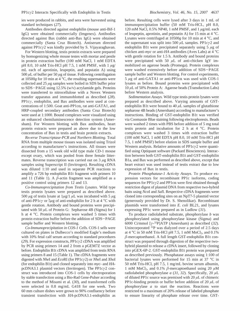
endophilin cDNA with genomic sequence revealed thepresence of an additional exon approximately 6 kb down-stream of the last exon in endophilin B1a. The splice donorsite for this testis specific exon resides within the codingsequence of the last endophilin B1a exon, and results in aputative protein that is identical with endophilin B1a, withthe exception that it is missing the last 10 amino acids(Figure 1; GenBank accession number DQ205686). Allendophilin B1 isoforms possess a canonical RVxF motif(RAVQF) near the amino terminus. CrossMatch alignmentreveals the presence of numerous mouse ESTs which haveundergone this differential splice, and also the location ofthe most likely polyA addition signal. We have designatedthis new isoform endophilin B1t.
Endophilin B1t Is Predominantly Expressed in Testis.Endophilin B1 is a well-characterized protein, and is widelyexpressed (34). However, we had difficulty amplifyingendophilin B1a from testis RNA, and were only successfulusing a 3′ primer from the newly discovered exon. Wetherefore assessed expression of this new isoform by RT-PCR on total RNA from a variety of mouse tissues usinggene-specific primers (Figure 2). A DNA fragment corre-sponding to endophilin B1t was amplified robustly from testisand liver. Faint bands were also observed following RT-PCR on stomach, skin, and ovary. Brain, spleen, lung, heart,and kidney were completely negative for endophilin B1t inthis assay.
FIGURE 1: Endophilin B1t results from differential splicing thatgenerates a slightly truncated protein. (A) The endophilin B1 geneencodes several different isoforms through differential splicing.Illustrated here is the variant splice that generates endophilin B1t,through the use of a cryptic splice donor site in the last exon ofendophilin B1a (short open bar) and an exon approximately 6 kbdownstream. (B) This splice event introduces a stop codon at thenew splice junction, resulting in a protein that is missing the last10 amino acids (small italics). The RVxF motif is underlined.
FIGURE 2: Endophilin B1t is expressed in a subset of tissues. RNAfrom indicated tissues was subjected to RT-PCR using endophilinB1t specific primers. Strong expression was observed in testis andliver, while weak but detectable expression could be seen in skin,stomach, and ovary.â-Actin was amplified from each sample as acontrol.
4638 Biochemistry, Vol. 46, No. 15, 2007 Hrabchak et al.
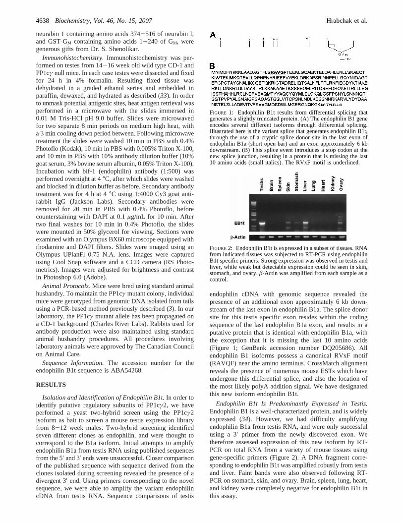
Endophilin B1t Interacts Specifically with PP1cγ2 in aYeast Two-Hybrid Assay.We have hypothesized the presenceof PP1 regulatory subunits that interact specifically with theγ2 catalytic subunit of PP1 to explain the phenotype ofPP1cγ null mice. To determine if endophilin B1t was ableto interact with multiple PP1c isoforms, two-hybrid assayswere performed using full length endophilin B1t in combina-tion with PP1cR, PP1cγ1, and a truncated version of PP1cγ2lacking 18 amino acids from the C-terminus. A constructencoding a portion of human lamin C was used as a negativecontrol. Expression of all fusion proteins in yeast wasconfirmed by Western blotting. A positive interaction wasassessed through the ability of transformed cells to grow onminimal media, activating expression ofADE2 and HIS3reporter genes. Expression of a lacZ reporter gene was alsoassessed after cell growth. Using these criteria endophilinB1t was found to interact solely with PP1cγ2 and not withPP1cR, PP1cγ1, or the truncated PP1γ2 (Table 2). Interactionwith PP1cγ2 was strong as judged by the white color ofresulting yeast colonies, indicating robust activation of theAde2reporter in theade2-101background. A bait constructexpressing amino acids 148-486 of the mousestaufengenewas used as a control during testing, and found to interactstrongly with all PP1c isoforms with no coloration due toade2-101. Staufen is an RNA-binding protein and hasrecently been recognized as a regulatory subunit of PP1 inrat brain neurons (35). Staufen is also expressed in male germcells during spermatogenesis in a stage-specific manner (35)and was isolated by us in the same two-hybrid screen asendophilin B1t. The strength of the interaction betweenendophilin B1t and PP1cγ2 was further demonstrated by theability of cells containing both constructs to survive on mediacontaining 10 mM 3-amino-1,2,4-triazole (3-AT), a competi-tive inhibitor of theHIS3 gene product. In addition, lacZcolony lift assays demonstrated robust expression of the thirdreporter construct in the yeast two-hybrid system (data notshown).
Endophilin B1a did not interact with any of the PP1cisoforms in a yeast two-hybrid assay.
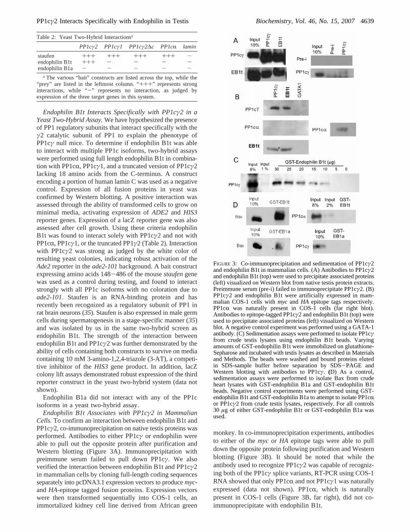
Endophilin B1t Associates with PP1cγ2 in MammalianCells.To confirm an interaction between endophilin B1t andPP1cγ2, co-immunoprecipitation on native testis proteins wasperformed. Antibodies to either PP1cγ or endophilin wereable to pull out the opposite protein after purification andWestern blotting (Figure 3A). Immunoprecipitation withpreimmune serum failed to pull down PP1cγ. We alsoverified the interaction between endophilin B1t and PP1cγ2in mammalian cells by cloning full-length coding sequencesseparately into pcDNA3.1 expression vectors to producemyc-andHA-epitope tagged fusion proteins. Expression vectorswere then transformed sequentially into COS-1 cells, animmortalized kidney cell line derived from African green
monkey. In co-immunoprecipitation experiments, antibodiesto either of themyc or HA epitope tags were able to pulldown the opposite protein following purification and Westernblotting (Figure 3B). It should be noted that while theantibody used to recognize PP1cγ2 was capable of recogniz-ing both of the PP1cγ splice variants, RT-PCR using COS-1RNA showed that only PP1cR and not PP1cγ1 was naturallyexpressed (data not shown). PP1cR, which is naturallypresent in COS-1 cells (Figure 3B, far right), did not co-immunoprecipitate with endophilin B1t.
Table 2: Yeast Two-Hybrid Interactionsa
PP1cγ2 PP1cγ1 PP1cγ2∆c PP1cR lamin
staufen +++ +++ +++ +++ -endophilin B1t +++ - - - -endophilin B1a - - - - -
a The various “bait” constructs are listed across the top, while the“prey” are listed in the leftmost column. “+++” represents stronginteractions, while “-” represents no interaction, as judged byexpression of the three target genes in this system.
FIGURE 3: Co-immunoprecipitation and sedimentation of PP1cγ2and endophilin B1t in mammalian cells. (A) Antibodies to PP1cγ2and endophilin B1t (top) were used to precipitate associated proteins(left) visualized on Western blot from native testis protein extracts.Preimmune serum (pre-i) failed to immunoprecipitate PP1cγ2. (B)PP1cγ2 and endophilin B1t were artificially expressed in mam-malian COS-1 cells withmyc and HA epitope tags respectively.PP1cR was naturally present in COS-1 cells (far right blot).Antibodies to epitope-tagged PP1cγ2 and endophilin B1t (top) wereused to precipitate associated proteins (left) visualized on Westernblot. A negative control experiment was performed using a GATA-1antibody. (C) Sedimentation assays were performed to isolate PP1cγfrom crude testis lysates using endophilin B1t beads. Varyingamounts of GST-endophilin B1t were immobilized on glutathione-Sepharose and incubated with testis lysates as described in Materialsand Methods. The beads were washed and bound proteins elutedin SDS-sample buffer before separation by SDS-PAGE andWestern blotting with antibodies to PP1cγ. (D) As a control,sedimentation assays were performed to isolate Bax from crudeheart lysates with GST-endophilin B1a and GST-endophilin B1tbeads. Negative control experiments were performed using GST-endophilin B1t and GST-endophilin B1a to attempt to isolate PP1cRor PP1cγ2 from crude testis lysates, respectively. For all controls30 µg of either GST-endophilin B1t or GST-endophilin B1a wasused.
PP1cγ2 Interacts Specifically with Endophilin in Testis Biochemistry, Vol. 46, No. 15, 20074639
Validity of the interaction between endophilin B1t andPP1cγ2 was illustrated using a GST-sedimentation assay onnative testis proteins. Specifically, varying amounts of full-length GST-endophilin B1t were covalently linked to glu-tathione-Sepharose beads and used to precipitate total proteinfrom wild type mouse testis lysates (Figure 3C). Under ourconditions, 30µg of GST-endophilin B1t was shown toprecipitate approximately 6% of the input PP1cγ. Bycomparison, 20µg of neurabin I was recently found to bind9-20% of input PP1cR/γ1 from brain lysates under similarconditions (11). We cannot exclude the possibility thatendophilin B1t binding to PP1cγ2 is stage specific, in whichcase only a subset of whole testis protein would be availablefor binding in our assay. GST-endophilin B1a was not ableto precipitate PP1cγ from wild type testis lysates. Similarly,GST-endophilin B1t was unable to isolate PP1cR from wildtype testis lysates (Figure 3D). These observations furtherconfirmed the specificity of the endophilin B1t/PP1cγ2interaction. However, both GST-endophilin B1t and GST-endophilin B1a were able to precipitate Bax, a bindingpartner of endophilin B1a (36), from heart extracts, whereBax is expressed abundantly, indicating that the GST fusionproteins have normal conformation.
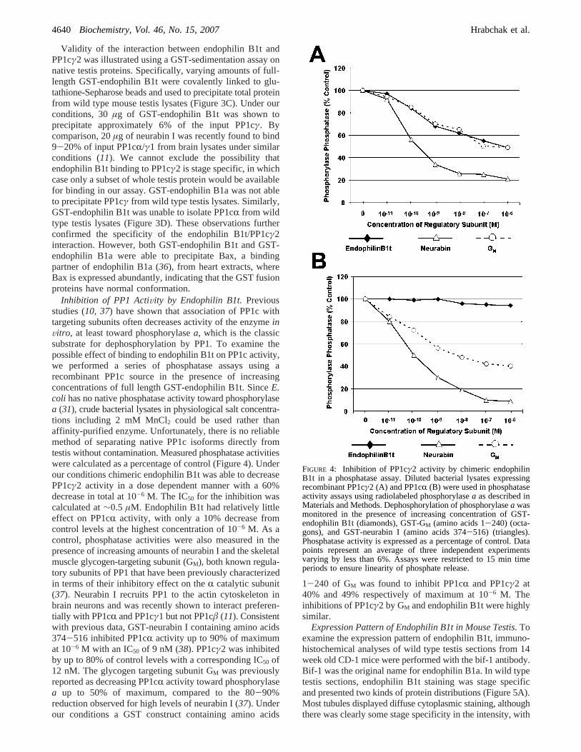
Inhibition of PP1 ActiVity by Endophilin B1t.Previousstudies (10, 37) have shown that association of PP1c withtargeting subunits often decreases activity of the enzymeinVitro, at least toward phosphorylasea, which is the classicsubstrate for dephosphorylation by PP1. To examine thepossible effect of binding to endophilin B1t on PP1c activity,we performed a series of phosphatase assays using arecombinant PP1c source in the presence of increasingconcentrations of full length GST-endophilin B1t. SinceE.coli has no native phosphatase activity toward phosphorylasea (31), crude bacterial lysates in physiological salt concentra-tions including 2 mM MnCl2 could be used rather thanaffinity-purified enzyme. Unfortunately, there is no reliablemethod of separating native PP1c isoforms directly fromtestis without contamination. Measured phosphatase activitieswere calculated as a percentage of control (Figure 4). Underour conditions chimeric endophilin B1t was able to decreasePP1cγ2 activity in a dose dependent manner with a 60%decrease in total at 10-6 M. The IC50 for the inhibition wascalculated at∼0.5 µM. Endophilin B1t had relatively littleeffect on PP1cR activity, with only a 10% decrease fromcontrol levels at the highest concentration of 10-6 M. As acontrol, phosphatase activities were also measured in thepresence of increasing amounts of neurabin I and the skeletalmuscle glycogen-targeting subunit (GM), both known regula-tory subunits of PP1 that have been previously characterizedin terms of their inhibitory effect on theR catalytic subunit(37). Neurabin I recruits PP1 to the actin cytoskeleton inbrain neurons and was recently shown to interact preferen-tially with PP1cR and PP1cγ1 but not PP1câ (11). Consistentwith previous data, GST-neurabin I containing amino acids374-516 inhibited PP1cR activity up to 90% of maximumat 10-6 M with an IC50 of 9 nM (38). PP1cγ2 was inhibitedby up to 80% of control levels with a corresponding IC50 of12 nM. The glycogen targeting subunit GM was previouslyreported as decreasing PP1cR activity toward phosphorylasea up to 50% of maximum, compared to the 80-90%reduction observed for high levels of neurabin I (37). Underour conditions a GST construct containing amino acids
1-240 of GM was found to inhibit PP1cR and PP1cγ2 at40% and 49% respectively of maximum at 10-6 M. Theinhibitions of PP1cγ2 by GM and endophilin B1t were highlysimilar.
Expression Pattern of Endophilin B1t in Mouse Testis.Toexamine the expression pattern of endophilin B1t, immuno-histochemical analyses of wild type testis sections from 14week old CD-1 mice were performed with the bif-1 antibody.Bif-1 was the original name for endophilin B1a. In wild typetestis sections, endophilin B1t staining was stage specificand presented two kinds of protein distributions (Figure 5A).Most tubules displayed diffuse cytoplasmic staining, althoughthere was clearly some stage specificity in the intensity, with
FIGURE 4: Inhibition of PP1cγ2 activity by chimeric endophilinB1t in a phosphatase assay. Diluted bacterial lysates expressingrecombinant PP1cγ2 (A) and PP1cR (B) were used in phosphataseactivity assays using radiolabeled phosphorylasea as described inMaterials and Methods. Dephosphorylation of phosphorylasea wasmonitored in the presence of increasing concentration of GST-endophilin B1t (diamonds), GST-GM (amino acids 1-240) (octa-gons), and GST-neurabin I (amino acids 374-516) (triangles).Phosphatase activity is expressed as a percentage of control. Datapoints represent an average of three independent experimentsvarying by less than 6%. Assays were restricted to 15 min timeperiods to ensure linearity of phosphate release.
4640 Biochemistry, Vol. 46, No. 15, 2007 Hrabchak et al.
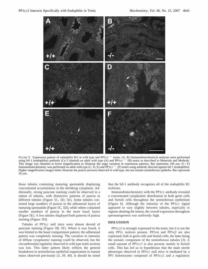
those tubules containing maturing spermatids displayingconcentrated accumulation in the shrinking cytoplasm. Ad-ditionally, strong punctate staining could be observed in asubset of tubules, with distinctive patterns of puncta indifferent tubules (Figure 5C, 5D, 5E). Some tubules con-tained large numbers of puncta in the adlumenal layers ofmaturing spermatids (Figure 5C, 5D), while others containedsmaller numbers of puncta in the more basal layers(Figure 5E). A few tubules displayed both patterns of punctastaining (Figure 5D).
Tubules of PP1cγ null mice were almost devoid ofpunctate staining (Figure 5B, 5F). Where it was found, itwas limited to the basal compartment pattern; the adlumenalpattern was completely missing. The variation in intensityof diffuse cytoplasmic staining could be observed, but thecircumferential regularity observed in wild type testis sectionswas lost. This latter pattern likely reflects the generalbreakdown in seminiferous epithelial organization in mutanttestes observed previously (3, 39, 40). It should be noted
that the bif-1 antibody recognizes all of the endophilin B1isoforms.
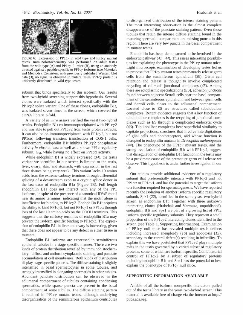
Immunohistochemistry with the PP1cγ antibody revealeda concentrated cytoplasmic distribution in both germ cellsand Sertoli cells throughout the seminiferous epithelium(Figure 6). Although the intensity of the PP1cγ signalappeared to vary slightly between tubules, especially inregions abutting the lumen, the overall expression throughoutspermatogenesis was uniformly high.
DISCUSSION
PP1cγ2 is strongly expressed in the testis, but it is not theonly PP1c isoform present. PP1cR and PP1câ are alsoexpressed, both in germ cells and Sertoli cells, the latter beingthe somatic component of the seminiferous tubules (3). Asmall amount of PP1cγ1 is also present, mainly in Sertolicells. This has led us to hypothesize that the male sterilephenotype observed in PP1cγ null mice is mediated by aPP1 holoenzyme composed of PP1cγ2 and a regulatory
FIGURE 5: Expression pattern of endophilin B1t in wild type and PP1cγ-/- testes. (A, B) Immunohistochemical analyses were performedusing bif-1 (endophilin) antibody (Cy-3 labeled) on adult wild type (A) and PP1cγ-/- (B) testes as described in Materials and Methods.This image was obtained at lower magnification to illustrate the stage variation in expression patterns. Bar represents 100µm. (C-F)Immunohistochemistry was performed on adult wild type (C, D, E) and PP1cγ-/- (F) testes using antibody directed against bif-1 (endophilin).Higher magnification images better illustrate the puncta (arrows) observed in wild type, but not mutant seminiferous epithelia. Bar represents50 µm.
PP1cγ2 Interacts Specifically with Endophilin in Testis Biochemistry, Vol. 46, No. 15, 20074641
subunit that binds specifically to this isoform. Our resultsfrom two-hybrid screening support this hypothesis. Severalclones were isolated which interact specifically with thePP1cγ2 splice variant. One of these clones, endophilin B1t,was isolated seven times in the screen, which covered thecDNA library 3-fold.
A variety of in Vitro assays verified the yeast two-hybridresults. Endophilin B1t co-immunoprecipitated with PP1cγ2and was able to pull out PP1cγ2 from testis protein extracts.It can also be co-immunoprecipitated with PP1cγ2, but notPP1cR, following transient transfection of COS-1 cells.Furthermore, endophilin B1t inhibits PP1cγ2 phosphataseactivity in Vitro at least as well as a known PP1c regulatorysubunit, GM, while having no effect on PP1cR activity.
While endophilin B1 is widely expressed (34), the testisvariant we identified in our screen is limited to the testis,liver, ovary, skin, and stomach, with expression in the lastthree tissues being very weak. This variant lacks 10 aminoacids from the extreme carboxy terminus through differentialsplicing of a downstream exon to a cryptic splice donor inthe last exon of endophilin B1a (Figure 1B). Full lengthendophilin B1a does not interact with any of the PP1isoforms, in spite of the presence of a canonical RVxF motifnear its amino terminus, indicating that the motif alone isinsufficient for binding to PP1cγ2. Endophilin B1t acquiresthe ability to bind PP1cγ2, but not PP1cγ1 or PP1cR throughloss of the last 10 amino acids on the COOH terminus. Thissuggests that the carboxy terminus of endophilin B1a mayprevent the isoform specific binding to PP1cγ2. The expres-sion of endophilin B1t in liver and ovary is interesting, giventhat there does not appear to be any defect in either tissue inmutants.
Endophilin B1 isoforms are expressed in seminiferousepithelial tubules in a stage specific manner. There are twokinds of protein distribution revealed by immunohistochem-istry: diffuse and uniform cytoplasmic staining, and punctateaccumulation at cell membranes. Both kinds of distributiondisplay stage specific patterns. The diffuse staining is slightlyintensified in basal spermatocytes in some tubules, andstrongly intensified in elongating spermatids in other tubules.Abundant punctate distribution can be observed in theadlumenal compartment of tubules containing condensingspermatids, while sparse puncta are present in the basalcompartment of some tubules. The diffuse staining patternis retained in PP1cγ mutant testes, although underlyingdisorganization of the seminiferous epithelium contributes
to disorganized distribution of the intense staining pattern.The most interesting observation is the almost completedisappearance of the punctate staining pattern. Even thosetubules that retain the intense diffuse staining found in thematuring spermatid compartment are missing puncta in thisregion. There are very few puncta in the basal compartmentin mutant testes.
Endophilin has been demonstrated to be involved in theendocytic pathway (41-44). This raises interesting possibili-ties for explaining the phenotype in the PP1cγ mutant mice.Light microscopic examination of developing testes led usto propose that PP1cγ mutant testes prematurely release germcells from the seminiferous epithelium (39). Germ cellretention and release is thought to involve complicatedrecycling of cell-cell junctional complexes (45). Amongthese are ectoplasmic specializations (ES), adhesion junctionsfound between adjacent Sertoli cells near the basal compart-ment of the seminferous epithelium, and between germ cellsand Sertoli cells closer to the adlumenal compartment.Located close to ES are structures called tubulobulbarcomplexes. Recent evidence suggests that a key function oftubulobulbar complexes is the recycling of junctional com-plexes such as ES through a complicated endocytic cycle(46). Tubulobulbar complexes bear superficial similarity tocapitate projections, structures that involve interdigitationsof glial cells and photoreceptors, and whose function isdisrupted in endophilin mutants inDrosophila melanogaster(44). The phenotype of the PP1cγ mutant testes, and thestrong association of endophilin B1t with PP1cγ2, suggestthat disregulation of endophilin B1t function in the testis maybe a proximate cause of the premature germ cell release weobserve. This hypothesis is under further investigation in ourlab.
Our studies provide additional evidence of a regulatorysubunit that preferentially interacts with PP1cγ2 and notPP1cR or PP1cγ1, and has the potential to target the isoformto a function required for spermatogenesis. We have reportedrecently the isolation of another isoform specific regulatorysubunit, Spz1 (22), identified in the same yeast two-hybridscreen as endophilin B1t. Together with three unknowninteracting clones (Hrabchak and Varmuza, unpublished),endophilin B1t and Spz1 are part of a growing list of PP1cisoform specific regulatory subunits. They represent a smallproportion of the PP1cγ2 interacting clones identified in thescreen (see Table 1, Supporting Information). Examinationof PP1cγ null mice has revealed multiple testis defectsincluding increased aneuploidy (16) and apoptosis (15),secondary to the central defect(s) resulting in infertility. Toexplain this we have postulated that PP1cγ2 plays multipleroles in the testis governed by a varied subset of regulatoryproteins, some of which are isoform specific. Combinatorialcontrol of PP1cγ2 by a subset of regulatory proteinsincluding endophilin B1t and Spz1 has the potential to bestexplain the phenotype of PP1cγ null mice.
SUPPORTING INFORMATION AVAILABLE
A table of all the isoform nonspecific interactors pulledout of the testis library in the yeast two-hybrid screen. Thismaterial is available free of charge via the Internet at http://pubs.acs.org.
FIGURE 6: Expression of PP1cγ in wild type and PP1cγ mutanttestes. Immunohistochemistry was performed on adult testesfrom the wild type (A) and PP1cγ-/- mice (B), using an antibodydirected against a peptide specific to PP1cγ isoforms (see Materialsand Methods). Consistent with previously published Western blotdata (3), no signal is observed in mutant testes. PP1cγ protein isuniformly distributed in wild type testes.
4642 Biochemistry, Vol. 46, No. 15, 2007 Hrabchak et al.
REFERENCES
1. Cohen, P. T. (2002) Protein Phosphatase 1sTargeted in ManyDirections,J. Cell. Sci. 115, 241-256.
2. Bollen, M. (2001) Combinatorial Control of Protein Phosphatase-1, Trends Biochem. Sci. 26, 426-431.
3. Varmuza, S., Jurisicova, A., Okano, K., Hudson, J., Boekelheide,K., and Shipp, E. B. (1999) Spermiogenesis is Impaired in MiceBearing a Targeted Mutation in the Protein Phosphatase 1cgammaGene,DeV. Biol. 205, 98-110.
4. Axton, J. M., Dombradi, V., Cohen, P. T., and Glover, D. M.(1990) One of the Protein Phosphatase 1 Isoenzymes in Drosophilais Essential for Mitosis,Cell 63, 33-46.
5. Cheng, A., Dean, N. M., and Honkanen, R. E. (2000) Serine/threonine Protein Phosphatase Type 1gamma1 is Required for theCompletion of Cytokinesis in Human A549 Lung CarcinomaCells,J. Biol. Chem. 275, 1846-1854.
6. Raghavan, S., Williams, I., Aslam, H., Thomas, D., Szoor, B.,Morgan, G., Gross, S., Turner, J., Fernandes, J., VijayRaghavan,K., and Alphey, L. (2000) Protein Phosphatase 1beta is Requiredfor the Maintenance of Muscle Attachments,Curr. Biol. 10, 269-272.
7. Ceulemans, H., and Bollen, M. (2004) Functional Diversity ofProtein Phosphatase-1, a Cellular Economizer and Reset Button,Physiol. ReV. 84, 1-39.
8. Wakula, P., Beullens, M., Ceulemans, H., Stalmans, W., andBollen, M. (2003) Degeneracy and Function of the UbiquitousRVXF Motif that Mediates Binding to Protein Phosphatase-1,J.Biol. Chem. 278, 18817-18823.
9. Zhao, S., and Lee, E. Y. (1997) A Protein Phosphatase-1-BindingMotif Identified by the Panning of a Random Peptide DisplayLibrary, J. Biol. Chem. 272, 28368-28372.
10. Aggen, J. B., Nairn, A. C., and Chamberlin, R. (2000) Regulationof Protein Phosphatase-1,Chem. Biol. 7, R13-23.
11. Terry-Lorenzo, R. T., Carmody, L. C., Voltz, J. W., Connor, J.H., Li, S., Smith, F. D., Milgram, S. L., Colbran, R. J., andShenolikar, S. (2002) The Neuronal Actin-Binding Proteins,Neurabin I and Neurabin II, Recruit Specific Isoforms of ProteinPhosphatase-1 Catalytic Subunits,J. Biol. Chem. 277, 27716-27724.
12. Colbran, R. J., Bass, M. A., McNeill, R. B., Bollen, M., Zhao, S.,Wadzinski, B. E., and Strack, S. (1997) Association of BrainProtein Phosphatase 1 with Cytoskeletal targeting/regulatorySubunits,J. Neurochem. 69, 920-929.
13. Shima, H., Hatano, Y., Chun, Y. S., Sugimura, T., Zhang, Z., Lee,E. Y., and Nagao, M. (1993) Identification of PP1 CatalyticSubunit Isotypes PP1 Gamma 1, PP1 Delta and PP1 Alpha invarious Rat Tissues,Biochem. Biophys. Res. Commun. 192, 1289-1296.
14. Okano, K., Heng, H., Trevisanato, S., Tyers, M., and Varmuza,S. (1997) Genomic Organization and Functional Analysis of theMurine Protein Phosphatase 1c Gamma (Ppp1cc) Gene,Genomics45, 211-215.
15. Jurisicova, A., Lopes, S., Meriano, J., Oppedisano, L., Casper, R.F., and Varmuza, S. (1999) DNA Damage in Round Spermatidsof Mice with a Targeted Disruption of the Pp1cgamma Gene andin Testicular Biopsies of Patients with Non-Obstructive Azoosper-mia, Mol. Hum. Reprod. 5, 323-330.
16. Oppedisano, L., Haines, G., Hrabchak, C., Fimia, G., Elliott, R.,Sassone-Corsi, P., and Varmuza, S. (2002) The Rate of Aneup-loidy is Altered in Spermatids from Infertile Mice,Hum. Reprod.17, 710-717.
17. Griswold, M. D. (1995) Interactions between Germ Cells andSertoli Cells in the Testis,Biol. Reprod. 52, 211-216.
18. Shima, H., Haneji, T., Hatano, Y., Kasugai, I., Sugimura, T., andNagao, M. (1993) Protein Phosphatase 1 Gamma 2 is Associatedwith Nuclei of Meiotic Cells in Rat Testis,Biochem. Biophys.Res. Commun. 194, 930-937.
19. Enz, R. (2002) The Metabotropic Glutamate Receptor mGluR7bBinds to the Catalytic Gamma-Subunit of Protein Phosphatase 1,J. Neurochem. 81, 1130-1140.
20. Chun, Y. S., Shima, H., Nagasaki, K., Sugimura, T., and Nagao,M. (1994) PP1 Gamma 2, a Testis-Specific Protein-serine/threonine-Phosphatase Type 1 Catalytic Subunit, is Associatedwith a Protein having High Sequence Homology with the 78-kDa Glucose-Regulated Protein, a Member of the 70-kDa HeatShock Protein Family,Proc. Natl. Acad. Sci. U. S. A. 91, 3319-3323.
21. Chun, Y. S., Park, J. W., Kim, G. T., Shima, H., Nagao, M., Kim,M. S., and Chung, M. H. (2000) A sds22 Homolog that isAssociated with the Testis-Specific serine/threonine Protein Phos-phatase 1gamma2 in Rat Testis,Biochem. Biophys. Res. Commun.273, 972-976.
22. Hrabchak, C., and Varmuza, S. (2004) Identification of theSpermatogenic Zip Protein Spz1 as a Putative Protein Phos-phatase-1 (PP1) Regulatory Protein that Specifically Binds thePP1cgamma2 Splice Variant in Mouse Testis,J. Biol. Chem. 279,37079-37086.
23. Mann, M., Latham, K. E., and Varmuza, S. (1995) Identificationof Genes Showing Altered Expression in Preimplantation andEarly Postimplantation Parthenogenetic Embryos,DeV. Genet. 17,223-232.
24. Elledge, S. J., Mulligan, J. T., Ramer, S. W., Spottswood, M.,and Davis, R. W. (1991) Lambda YES: A Multifunctional cDNAExpression Vector for the Isolation of Genes by Complementationof Yeast and Escherichia Coli Mutations,Proc. Natl. Acad. Sci.U. S. A. 88, 1731-1735.
25. Nickoloff, J. A. (1995)Electroporation Protocols for Microorgan-isms,Humana Press, Totowa, NJ.
26. Sharma, R. C., and Schimke, R. T. (1996) Preparation ofElectrocompetent E. Coli using Salt-Free Growth Medium,BioTechniques 20, 42-44.
27. Harlow, E., and Lane, D. (1988)Antibodies: A LaboratoryManual, Cold Spring Harbor Laboratory Press, Cold SpringHarbor, NY.
28. Sambrook, J., Fritsch, E. F., and Maniatis, T. (1989)MolecularCloning: A Laboratory Manual, 2nd ed., Cold Spring HarborLaboratory Press, Cold Spring Harbor, NY.
29. Gluzman, Y. (1981) SV40-Transformed Simian Cells Support theReplication of Early SV40 Mutants,Cell 23, 175-182.
30. Misumi, Y., Oda, K., Fujiwara, T., Takami, N., Tashiro, K., andIkehara, Y. (1991) Functional Expression of Furin Demonstratingits Intracellular Localization and Endoprotease Activity forProcessing of Proalbumin and Complement Pro-C3,J. Biol. Chem.266, 16954-16959.
31. Ludlow, J. W. (1998)Protein Phosphatase Protocols,HumanaPress, Totowa, NJ.
32. Shenolikar, S., and Ingebritsen, T. S. (1984) Protein (Serine andThreonine) Phosphate Phosphatases,Methods Enzymol. 107, 102-129.
33. Iwamura, M., Abrahamsson, P. A., Benning, C. M., Cockett, A.T., and di Sant’Agnese, P. A. (1994) Androgen Receptor Immu-nostaining and its Tissue Distribution in Formalin-Fixed, Paraffin-Embedded Sections After Microwave Treatment,J. Histochem.Cytochem. 42, 783-788.
34. Modregger, J., Schmidt, A. A., Ritter, B., Huttner, W. B., andPlomann, M. (2003) Characterization of Endophilin B1b, a Brain-Specific Membrane-Associated Lysophosphatidic Acid AcylTransferase with Properties Distinct from Endophilin A1,J. Biol.Chem. 278, 4160-4167.
35. Monshausen, M., Rehbein, M., Richter, D., and Kindler, S. (2002)The RNA-Binding Protein Staufen from Rat Brain Interacts withProtein Phosphatase-1,J. Neurochem. 81, 557-564.
36. Cuddeback, S. M., Yamaguchi, H., Komatsu, K., Miyashita, T.,Yamada, M., Wu, C., Singh, S., and Wang, H. G. (2001)Molecular Cloning and Characterization of Bif-1. A Novel SrcHomology 3 Domain-Containing Protein that Associates with Bax,J. Biol. Chem. 276, 20559-20565.
37. Liu, J., Wu, J., Oliver, C., Shenolikar, S., and Brautigan,D. L. (2000) Mutations of the Serine Phosphorylated in theProtein Phosphatase-1-Binding Motif in the Skeletal MuscleGlycogen-Targeting Subunit,Biochem. J. 346(Part 1), 77-82.
38. Terry-Lorenzo, R. T., Elliot, E., Weiser, D. C., Prickett, T. D.,Brautigan, D. L., and Shenolikar, S. (2002) Neurabins RecruitProtein Phosphatase-1 and Inhibitor-2 to the Actin Cytoskeleton,J. Biol. Chem. 277, 46535-46543.
39. Varmuza, S., and Ling, L. (2003) Increased RecombinationFrequency Showing Evidence of Loss of Interference is Associatedwith Abnormal Testicular Histopathology,Mol. Reprod. DeV. 64,499-506.
40. Oppedisano-Wells, L., and Varmuza, S. (2003) Protein Phosphatase1cgamma is Required in Germ Cells in Murine Testis,Mol.Reprod. DeV. 65, 157-166.
41. Song, W., and Zinsmaier, K. E. (2003) Endophilin and Synapto-janin Hook Up to Promote Synaptic Vesicle Endocytosis,Neuron40, 665-667.
PP1cγ2 Interacts Specifically with Endophilin in Testis Biochemistry, Vol. 46, No. 15, 20074643

42. Schuske, K. R., Richmond, J. E., Matthies, D. S., Davis, W. S.,Runz, S., Rube, D. A., van der Bliek, A. M., and Jorgensen, E.M. (2003) Endophilin is Required for Synaptic VesicleEndocytosis by Localizing Synaptojanin,Neuron 40, 749-762.
43. Verstreken, P., Koh, T. W., Schulze, K. L., Zhai, R. G., Hiesinger,P. R., Zhou, Y., Mehta, S. Q., Cao, Y., Roos, J., and Bellen, H.J. (2003) Synaptojanin is Recruited by Endophilin to PromoteSynaptic Vesicle Uncoating,Neuron 40, 733-748.
44. Fabian-Fine, R., Verstreken, P., Hiesinger, P. R., Horne, J. A.,Kostyleva, R., Zhou, Y., Bellen, H. J., and Meinertzhagen, I. A.(2003) Endophilin Promotes a Late Step in Endocytosis at Glial
Invaginations in Drosophila Photoreceptor Terminals,J. Neurosci.23, 10732-10744.
45. Russell, L., and Clermont, Y. (1976) Anchoring Device betweenSertoli Cells and Late Spermatids in Rat Seminiferous Tubules,Anat. Rec. 185, 259-278.
46. Guttman, J. A., Takai, Y., and Vogl, A. W. (2004) Evidence thatTubulobulbar Complexes in the Seminiferous Epithelium areInvolved with Internalization of Adhesion Junctions,Biol. Reprod.71, 548-559.
BI6025837
4644 Biochemistry, Vol. 46, No. 15, 2007 Hrabchak et al.