Embed Size (px)
Citation preview
A to Z ORTHODONTICS Volume: 21
Dr. Mohammad Khursheed Alam BDS, PGT, PhD (Japan)
RETENTION AND RELAPSE
1
First Published August 2012
© Dr. Mohammad Khursheed Alam
© All rights reserved. No part of this publication may be reproduced stored in a retrieval system, or transmitted, in any form or by any means, electronic, mechanical, photocopying, recording or
otherwise, without prior permission of author/s or publisher.
ISBN: 978-967-0486-10-9 Correspondance:
Dr. Mohammad Khursheed Alam
Senior Lecturer
Orthodontic Unit
School of Dental Science
Health Campus, Universiti Sains Malaysia.
Email:
Published by:
PPSP Publication
Jabatan Pendidikan Perubatan, Pusat Pengajian Sains Perubatan,
Universiti Sains Malaysia.
Kubang Kerian, 16150. Kota Bharu, Kelatan.
Published in Malaysia

2
Contents
1. What is Retention...................................................3
2. Why Retention is necessary...................................3
3. Types of Retention….……………………………...3-10
4. Requirements of a good retainer…………………..10
5. Theories of Retention………..………………….....10-12
6. Advantages of fixed retainer………………………..12
7. Disadvantages of fixed retainer…………………….12-13
8. What is Relapse......................................................13
9. Causes of Relapse..................................................13-15
10. Prevention of Relapse……….……………………...15
3
No orthodontic treatment should be done when long standing result cannot be achieved.
Retention
Retention is holding of teeth in an ideal aesthetic and functional position at
the end of orthodontic treatment.
Why retention is necessary?
1. The gingival and periodontal tissues are affected by orthodontic
tooth movement arc require time for reorganization when the
appliances are removed.
2. The teeth may be in an inherently unstable position after treatment,
so that soft tissue pressure constantly produces a relapse tendency.
3. Changes produced by growth may alter the orthodontic treatment
result.
Types of retention
According to Duration
o Group I –No retention
o Group II- Limited retention
o Group III- Prolonged retention
o Group IV- Permanent retention
According to malocclusion
o Class II
o Class III
4
o Deep bite
o Anterior open bite
According to Duration
Group I –No retention
Period of retention: Not applicable
Indications:
1. Anterior and posterior cross bite
2. Treatment after extraction or serial extraction
3. Highly placed canine
4. Impacted mandibular 2nd premolar
Group II- Limited retention
Period of retention:
Three to six (3-6) months. First 3 months full time retention, and next 3
months only at night
Indications:
1. Class I proclination and spacing of maxillary incisor
2. Class I and Class II extraction cases
3. Corrected deepbite cases
4. Class II division 2 cases
5
Group III - Prolonged retention
Period of retention: Rotation cases require 232 days or more
Indications:
1. Forces produced by lips, cheeks and tongue during rest position-
Soft tissue acts as a mould for the teeth. If any tooth moves out of
the muscle balance, then relapse occurs
2. Persisting abnormal habits
3. Failure to upright the roots can open up extraction spaces.
4. Lack of balance between buccal and lingual forces
5. Presence of excess tooth material to arch size
6. Poor patient cooperation, by not wearing the appliance regularly
7. Completion of the treatment before completion of growth;
especially in skeletal Class III and open bite, deep bite etc.
Group IV- Permanent retention
Period of retention: Lifelong
Indications:
1. Severe rotation
2. Midline diastema
3. Cleft palate cases
4. Generalized spacing
6
5. Expansion of lower arch
According to Malocclusion
Class II
1. Overcorrection of occlusal relationship as a finishing procedure
2. It is better not to move the lower incisors too far forward. If more than
2mm forward repositioning of the lower incisors occurred during
treatment, permanent retention wilt be required
3. For moderate to severe Class II cases- Fixed appliance along with
headgear to the upper arch or activator-bionator type functional
appliance at night after active treatment till the growing age subside
Class III
1. In mild Class III problems a functional appliance or a positioner may
be sufficient
2. For moderate to severe Class III cases surgical correction after the
growth is complete is the only stable treatment. Chin cap or Class III
functional appliance as a retainer rotate the mandible downward
causing the growth to be expressed more vertically and less
horizontally
Deep bite
Upper removable appliance with a bite plane, which does not separate the
posterior teeth, at night for several years after completion of the active
treatment, may be required.
Anterior open bite
7
1. Correction of the thumb sucking and tongue-thrust habit
2. Open bite activator or bionator or high pull headgear along with
removable retainer to prevent elongation of posterior teeth at
night time
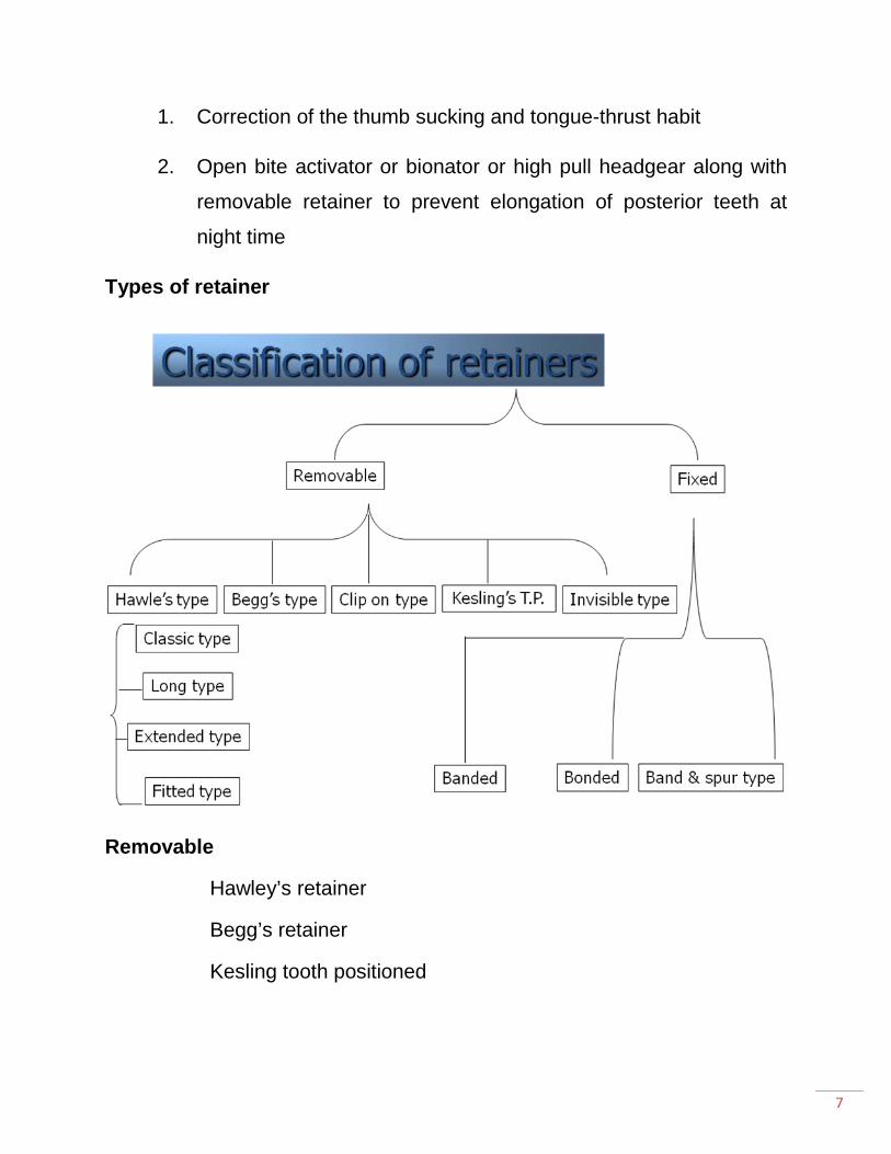
Types of retainer
Removable
Hawley’s retainer
Begg’s retainer
Kesling tooth positioned
8
Hawley’s appliance
- Designed in 1920 by Charles Hawley.
Most frequently used retainer
Consists of claps on molars and a short labial bow extending from
canine to canine having adjustment loops
Begg’s retainer
Consists of a labial wire that extends till the last erupted molar and curves
around it to get embeded in acrylic that spans the palate.
Advantage:
There is no cross over wire that extends between the canine and premolar
thereby eliminating the risk of space opening.
Clip – on retainer / spring aligner
This appliance is made of a wire frame work that runs labially over the
incisors and then passes b/w the canine and premolar and is reserved to lie
over the lingual surface. Both the labial as well as lingual segments are
embedded in a strip of clear acrylic.
Use: To bring about corrections of rotations commonly seen in lower
anterior region.
Wrap around retainer
Extended version of spring aligner
9
Consists of wire that passes along the labial as well as lingual surfaces of
all erupted teeth which is embedded in a strip of acrylic.
Use: In stabilizing a periodontally weak dentition.
Kesling tooth positioner
Made of thermoplastic rubber like material that spans the inter – occlusal
space and covers the clinical crowns of the U/L portion of teeth and a small
portion of the gingiva.
Needs no activation at regular intervals and is durable.
Drawbacks: - difficulty in speech
- risk of TMJ problems
Invisible retainers
Fully cover the clinical crowns and a part of the gingival tissue.
Made of ultra thin transparent thermoplastic sheets using a Biostar
machine.
Esthetical and go unnoticed.
Fixed
Band and Spur
Bonded canine to canine retainer
Bonded lingual retainer
Banded canine to canine retainer:
-Commonly used in lower anterior region.
10
-Canines are banded and a thick wire is contoured over the lingual aspects and soldered to canine bands
Bonded lingual retainers:
-Bonded on lingual aspects following anterior curvature.
-Ends are curved over the canines where it is bonded.
Band and spur retainers
Used where a single tooth has been orthodontcally treated for rotation
correction or labiolingual displacement.
The tooth that has been moved banded and spurs are soldered onto the
bands so as to overlap the adjacent teeth.
Requirements of a good retainer • It should restrain each tooth that has been moved into the desired
position in directions where there are tendencies toward recurring
movement.
• It should permit the forces associated with functional activity to act
freely on the retained tooth, permitting them to respond in as nearly a
physiologic manner as possible.
• It should be as self cleaning as possible and should be easy to
maintain optimal hygiene.
* Theories of Retention
Riedels nine theories Moyers added another theory as the tenth theorem.
Theorem 1:
Teeth that have been moved tend to return to their former position.
11
Theorem 2:
Eliminating of the cause of malocclusion will prevent relapse.
Theorem 3:
Malocclusion should be over corrected as a safety factor.
Theorem 4:
Malocclusion should be over corrected as a safety factor.
Theorem 4:
Proper occlusion is a potent factor in holding teeth in their corrected
positions.
Theorem 5:
Bone and adjacent tissues must be allowed time to reorganize around
newly positioned teeth.
Theorem 6:
If the lower incisors are placed upright over basal bone they are more likely
to remain in good alignment.
Theorem 7:
Corrections carried out during periods of growth are less likely to relapse.
Theorem 8:
The farther the teeth have been moved the lesser is the risk of relapse.
Theorem 9:

12
Arch form, particularly in the mandibular arch cannot be permanently
altered by appliance therapy.
Theorem 10:
Many treated malocclusions require permanent retaining devices.
Advantages of fixed retainer
Reduced need for patient co-operation
Can be used when conventional retainers cannot provide same degree
of stability.
Bonded retainers are more esthetics
No tissue irritation unlike what may been seen in tissue bearing areas of
Hawley’s retainer
Can be used for permanent and semi permanent retention.
Do not affect speech.
Disadvantages of fixed retainers
More cumbersome to insert
Increased chair side time
More expensive
Loss of healthy tooth material
13
Tend to discolor
Relapse
Definition: Relapse has been defined as “The loss of any correction
achieved by orthodontic treatment.”
Causes of relapse
1. Tension produced by periodontal ligament
2. Whenever teeth are moved orthodontically, the periodontal fibers are
stretched. The stretched fibers contract and cause relapse. The
principal fibers of the periodontal ligament reorganize in about 4
weeks time but the supra alveolar gingival fibers takes more time to
rearrange around the new position.
3. Occlusal forces associated with faulty interdigitation of teeth
4. Occlusal force associated with abnormal reduction of the
interocclusal spaces.
In most cases relapse occurs due to a combination of causes.
Causes attributing to relapse:
1. Periodontal ligament Tractions:
Inadequate retention after active orthodontic treatment. Whenever teeth are
moved orthodontically, the periodontal principal fibers and the gingival
fibers that encircle the teeth are stretched. These stretched fibers can
contract and are thus a potent cause of relapse.
2. Relapse due to growth related changes:
14
Patients with skeletal problems associated with class II, class III, open bite
or deep bite malocclusion may exhibit relapse.
3. Bone adaptation:
Teeth that have been moved recently are surrounded by lightly calcified
osteoid bone. Thus the teeth are not adequately stabilized and have a
tendency to move to their original position.
4. Muscular Forces:
Teeth are encapsulated in all directions by a blanket of muscles. Muscle
imbalance at the end of the orthodontic therapy can result in reappearance
of the malocclusion.
5. Failure to eliminate the original cause:
Failure to remove the etiology can result in relapse.
Role of 3rd molar:
The pressure exerted by the erupting 3rd molars in believed to cause
late anterior crowding predisposing to relapse.
Role of occlusion:
Presence of certain occlusal mannerisms such as clenching, grinding, nail
biting, lip biting etc... are important cause of relapse.
Inadequate retention:
After active Rx of orthodontic tooth movement if there is inadequate
retention. We need to stay teeth in retention position. If not there is chance
of relapse.
15
6. Living the tooth in undesirable area
Tooth should be placed in occlusal, cuspal & oro-facial balance.
7. Placing the tooth in a crowded position
8. Persistent abnormal habit
9. Poor patient’s co-operation
Prevention of Relapse:
(1) Over rotation.
(2) Prolonged retention.
(3) Treatment of rotated tooth should be performed at early age.
(4) Placement of teeth in oro–facial soft tissue balance.
(5) Placement of teeth in occlusal equilibrium.
(6) Pericision – surgical resection of stretched fiber around gingival
socket margin (supra-alveolar fibers).

16
Bibilography:
1. Bhalajhi SI. Orthodontics – The art and science. 4th edition. 2009
2. Gurkeerat Singh. Textbook of orthodontics. 2nd edition. Jaypee, 2007
3. Houston S and Tulley, Textbook of Orthodontics. 2nd Edition. Wright, 1992.
4. Iida J. Lecture/class notes. Professor and chairman, Dept. of Orthodontics, School of dental science, Hokkaido University, Japan.
5. Lamiya C. Lecture/class notes. Ex Associate Professor and chairman, Dept. of Orthodontics, Sapporo Dental College.
6. Laura M. An introduction to Orthodontics. 2nd edition. Oxford University Press, 2001
7. McNamara JA, Brudon, WI. Orthodontics and Dentofacial Orthopedics. 1st edition, Needham Press, Ann Arbor, MI, USA, 2001
8. Mitchel. L. An Introduction to Orthodontics. 3 editions. Oxford University Press. 2007
9. Mohammad EH. Essentials of Orthodontics for dental students. 3rd edition, 2002
10. Proffit WR, Fields HW, Sarver DM. Contemporary Orthodontics. 4th edition, Mosby Inc., St.Louis, MO, USA, 2007
11. Sarver DM, Proffit WR. In TM Graber et al., eds., Orthodontics: Current Principles and Techniques, 4th ed., St. Louis: Elsevier Mosby, 2005
12. Samir E. Bishara. Textbook of Orthodontics. Saunders 978-0721682891, 2002
13. T. M. Graber, R.L. Vanarsdall, Orthodontics, Current Principles and Techniques, "Diagnosis and Treatment Planning in Orthodontics", D. M. Sarver, W.R. Proffit, J. L. Ackerman, Mosby, 2000
14. Thomas M. Graber, Katherine W. L. Vig, Robert L. Vanarsdall Jr. Orthodontics: Current Principles and Techniques. Mosby 9780323026215, 2005
15. William R. Proffit, Raymond P. White, David M. Sarver. Contemporary treatment of dentofacial deformity. Mosby 978-0323016971, 2002
16. William R. Proffit, Henry W. Fields, and David M. Sarver. Contemporary Orthodontics. Mosby 978-0323040464, 2006
17. Yoshiaki S. Lecture/class notes. Associate Professor and chairman, Dept. of Orthodontics, School of dental science, Hokkaido University, Japan.
18. Zakir H. Lecture/class notes. Professor and chairman, Dept. of Orthodontics, Dhaka Dental College and hospital.

17
Dedicated To
My Mom, Zubaida Shaheen
My Dad, Md. Islam
&
My Only Son
Mohammad Sharjil
18
Acknowledgments I wish to acknowledge the expertise and efforts of the various teachers for their help and inspiration:
1. Prof. Iida Junichiro – Chairman, Dept. of Orthodontics, Hokkaido University, Japan.
2. Asso. Prof. Sato yoshiaki –Dept. of Orthodontics, Hokkaido University, Japan.
3. Asst. Prof. Kajii Takashi – Dept. of Orthodontics, Hokkaido University, Japan.
4. Asst. Prof. Yamamoto – Dept. of Orthodontics, Hokkaido University, Japan.
5. Asst. Prof. Kaneko – Dept. of Orthodontics, Hokkaido University, Japan.
6. Asst. Prof. Kusakabe– Dept. of Orthodontics, Hokkaido University, Japan.
7. Asst. Prof. Yamagata– Dept. of Orthodontics, Hokkaido University, Japan.
8. Prof. Amirul Islam – Principal, Bangladesh Dental college 9. Prof. Emadul Haq – Principal City Dental college 10. Prof. Zakir Hossain – Chairman, Dept. of Orthodontics,
Dhaka Dental College. 11. Asso. Prof. Lamiya Chowdhury – Chairman, Dept. of
Orthodontics, Sapporo Dental College, Dhaka. 12. Late. Asso. Prof. Begum Rokeya – Dhaka Dental College. 13. Asso. Prof. MA Sikder– Chairman, Dept. of Orthodontics,
University Dental College, Dhaka. 14. Asso. Prof. Md. Saifuddin Chinu – Chairman, Dept. of
Orthodontics, Pioneer Dental College, Dhaka.
19
Dr. Mohammad Khursheed Alam has obtained his PhD degree in Orthodontics from Japan in 2008. He worked as Asst. Professor and Head, Orthodontics department, Bangladesh Dental College for 3 years. At the same time he worked as consultant Orthodontist in the Dental office named ‘‘Sapporo Dental square’’. Since then he has worked in several international projects in the field of Orthodontics. He is the author of more than 50 articles published in reputed journals. He is now working as Senior lecturer in Orthodontic unit, School of Dental Science, Universiti Sains Malaysia.
Volume of this Book has been reviewed by: Dr. Kathiravan Purmal BDS (Malaya), DGDP (UK), MFDSRCS (London), MOrth (Malaya), MOrth RCS( Edin), FRACPS. School of Dental Science, Universiti Sains Malaysia. Dr Kathiravan Purmal graduated from University Malaya 1993. He has been in private practice for almost 20 years. He is the first locally trained orthodontist in Malaysia with international qualification. He has undergone extensive training in the field of oral and maxillofacial surgery and general dentistry.