Embed Size (px)
Citation preview
Version 1 Last Updated 30 August 2013
Instructions for Use
For the qualitative measurement of IgG class antibodies against Epstein Barr virus (EBV- EBNA) in Human serum and plasma (citrate).
This product is for research use only and is not intended for diagnostic use.
ab108731 – Anti-Epstein Barr virus (EBV-EBNA) IgG Human ELISA Kit
Discover more at www.abcam.com 1
Table of Contents
INTRODUCTION1. BACKGROUND 22. ASSAY SUMMARY 4
GENERAL INFORMATION3. PRECAUTIONS 54. STORAGE AND STABILITY 55. MATERIALS SUPPLIED 56. MATERIALS REQUIRED, NOT SUPPLIED 67. LIMITATIONS 68. TECHNICAL HINTS 7
ASSAY PREPARATION9. REAGENT PREPARATION 810. SAMPLE COLLECTION AND STORAGE 811. SAMPLE PREPARATION 812. PLATE PREPARATION 9
ASSAY PROCEDURE13. ASSAY PROCEDURE 10
DATA ANALYSIS14. CALCULATIONS 1215. TYPICAL SAMPLE VALUES 1416. ASSAY ANALYTICAL SPECS 14
RESOURCES17. INTERFERENCES 1518. TROUBLESHOOTING 1519. NOTES 17
Discover more at www.abcam.com 2
PRODUCT INFORMATION
1. BACKGROUNDAbcam’s anti-Epstein Barr virus (EBV-EBNA) IgG Human in vitro ELISA (Enzyme-Linked Immunosorbent Assay) kit is designed for the accurate qualitative measurement of IgG class antibodies against Epstein Barr virus in Human serum and plasma. A 96-well plate has been precoated with Epstein Barr virus antigens to bind cognate antibodies. Controls or test samples are added to the wells and incubated. Following washing, a horseradish peroxidase (HRP) labelled anti-Human IgG conjugate is added to the wells, which binds to the immobilized Epstein Barr virus-specific antibodies. TMB is then catalyzed by the HRP to produce a blue color product that changes to yellow after adding an acidic stop solution. The density of yellow coloration is directly proportional to the amount of Epstein Barr virus IgG sample captured in plate.Epstein-Barr virus (EBV) is a member of the herpesvirus family (Gamma subgroup, DNA virus of 120-200 nm) and one of the most common Human viruses. The virus occurs worldwide, and most people become infected with EBV sometime during their lives. Transmission of the virus is almost impossible to prevent since many healthy people can carry and spread the virus intermittently for life. Infants become susceptible to EBV as soon as maternal antibody protection disappears. Infection of children usually causes no symptoms, while infection during adolescence or young adulthood causes infectious mononucleosis 35% to 50% of the time.Infectious mononucleosis is almost never fatal. There are no known associations between active EBV infection and problems during pregnancy, such as miscarriages or birth defects. Although the symptoms of infectious mononucleosis usually resolve in 1 or 2 months, EBV remains dormant or latent in a few cells in the throat and blood for the rest of the person’s life. Periodically, the virus can reactivate and is commonly found in the saliva of infected persons. This reactivation usually occurs without symptoms of illness.
Discover more at www.abcam.com 3
PRODUCT INFORMATION
EBV also establishes a lifelong dormant infection in some cells of the body’s immune system. A late event in a very few carriers of this virus is the emergence of Burkitt´s lymphoma and nasopharyngeal carcinoma, but EBV is probably not the sole cause of these malignancies.
Species Disease Symptoms Mechanism of Infection
Epstein-Barr Virus
Infectious mononucleosis
Fever, sore throat, swollen lymph nodes
Person to person Transmission
EBV requires intimate contact with the saliva of an infected person, but the virus is also found in the saliva of healthy people.
The presence of viral infection may be identified by
PCR Serology; ‘mono spot’ test, Detection of antibodies by ELISA.
The optimal combination of serologic testing consists of the titration of four markers: IgM and IgG to the viral capsid antigen (VCA), IgM to the early antigen, and antibody to EBV nuclear antigen (EBNA). IgM to VCA appears early in infection and disappears within 4 to 12 weeks. IgG to VCA appears in the acute phase, peaks at 2 to 4 weeks after onset, declines slightly, and then persists for life.If antibodies to the viral capsid antigen are not detected, the patient is susceptible to EBV infection.
Discover more at www.abcam.com 4
PRODUCT INFORMATION
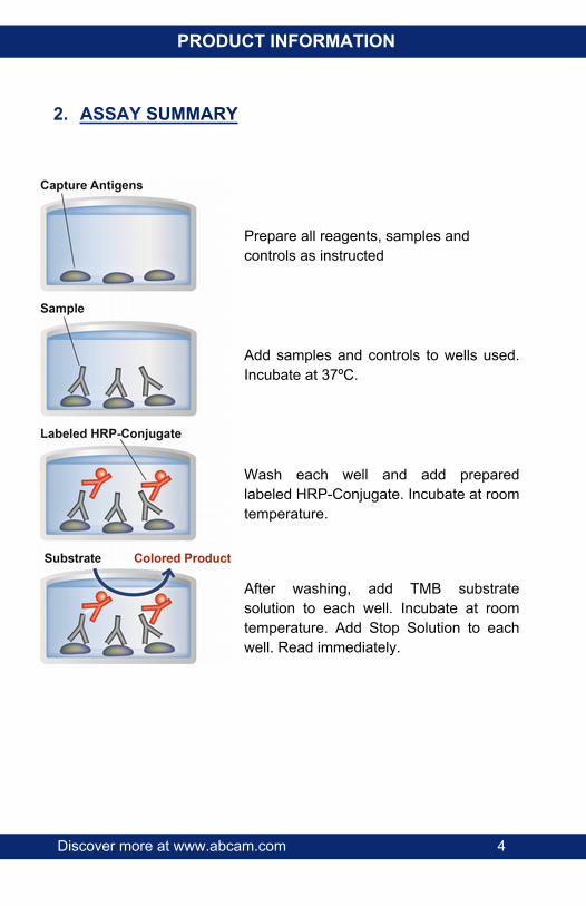
2. ASSAY SUMMARY
Prepare all reagents, samples and controls as instructed
Add samples and controls to wells used. Incubate at 37ºC.
Wash each well and add prepared labeled HRP-Conjugate. Incubate at room temperature.
After washing, add TMB substrate solution to each well. Incubate at room temperature. Add Stop Solution to each well. Read immediately.
Discover more at www.abcam.com 5
GENERAL INFORMATION
3. PRECAUTIONSPlease read these instructions carefully prior to beginning the assay.All kit components have been formulated and quality control tested to function successfully as a kit. Modifications to the kit components or procedures may result in loss of performance.
4. STORAGE AND STABILITYStore kit at 2-8°C immediately upon receipt.Refer to list of materials supplied for storage conditions of individual components. Observe the storage conditions for individual prepared components in section 9. Reagent Preparation.
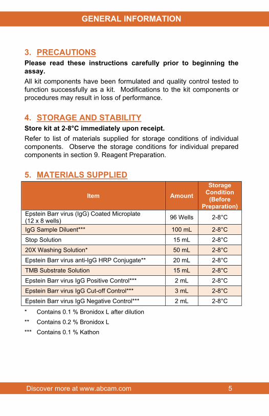
5. MATERIALS SUPPLIED
Item AmountStorage
Condition(Before
Preparation)Epstein Barr virus (IgG) Coated Microplate (12 x 8 wells) 96 Wells 2-8°C
IgG Sample Diluent*** 100 mL 2-8°C
Stop Solution 15 mL 2-8°C
20X Washing Solution* 50 mL 2-8°C
Epstein Barr virus anti-IgG HRP Conjugate** 20 mL 2-8°C
TMB Substrate Solution 15 mL 2-8°C
Epstein Barr virus IgG Positive Control*** 2 mL 2-8°C
Epstein Barr virus IgG Cut-off Control*** 3 mL 2-8°C
Epstein Barr virus IgG Negative Control*** 2 mL 2-8°C
* Contains 0.1 % Bronidox L after dilution** Contains 0.2 % Bronidox L*** Contains 0.1 % Kathon
Discover more at www.abcam.com 6
GENERAL INFORMATION
6. MATERIALS REQUIRED, NOT SUPPLIEDThese materials are not included in the kit, but will be required to successfully utilize this assay:
Microplate reader capable of measuring absorbance at 450 nm or 620 nm
Incubator at 37°C
Multi and single channel pipettes to deliver volumes between 10 and 1,000 µL
Optional: Automatic plate washer for rinsing wells
Vortex tube mixer
Deionised or (freshly) distilled water
Disposable tubes
Timer
7. LIMITATIONS ELISA kit intended for research use only. Not for use in diagnostic
procedures
All components of Human origin used for the production of these reagents have been tested for anti-HIV antibodies, anti-HCV antibodies and HBsAg and have been found to be non-reactive. Nevertheless, all materials should still be regarded and handled as potentially infectious
Use only clean pipette tips, dispensers, and lab ware.
Do not interchange screw caps of reagent vials to avoid cross-contamination
Close reagent vials tightly immediately after use to avoid evaporation and microbial contamination
After first opening and subsequent storage check conjugate and control vials for microbial contamination prior to further use

Discover more at www.abcam.com 7
GENERAL INFORMATION
To avoid cross-contamination and falsely elevated results pipette patient samples and dispense conjugate, without splashing, accurately to the bottom of wells
8. TECHNICAL HINTS Avoid foaming or bubbles when mixing or reconstituting
components
Avoid cross contamination of samples or reagents by changing tips between sample, standard and reagent additions.
Ensure plates are properly sealed or covered during incubation steps
Complete removal of all solutions and buffers during wash steps is necessary for accurate measurement readings
This kit is sold based on number of tests. A ‘test’ simply refers to a single assay well. The number of wells that contain sample, control or standard will vary by product. Review the protocol completely to confirm this kit meets your requirements. Please contact our Technical Support staff with any questions
Discover more at www.abcam.com 8
ASSAY PREPARATION
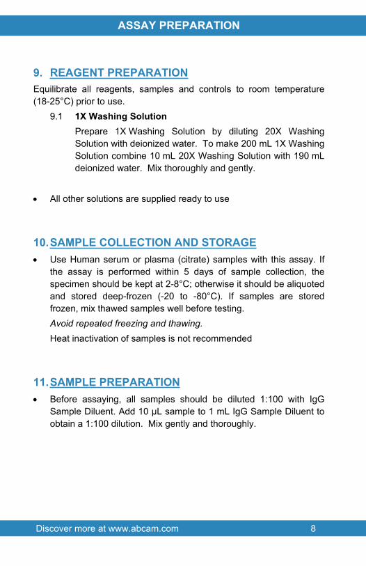
9. REAGENT PREPARATIONEquilibrate all reagents, samples and controls to room temperature (18-25°C) prior to use.
9.1 1X Washing SolutionPrepare 1X Washing Solution by diluting 20X Washing Solution with deionized water. To make 200 mL 1X Washing Solution combine 10 mL 20X Washing Solution with 190 mL deionized water. Mix thoroughly and gently.
All other solutions are supplied ready to use
10.SAMPLE COLLECTION AND STORAGE Use Human serum or plasma (citrate) samples with this assay. If
the assay is performed within 5 days of sample collection, the specimen should be kept at 2-8°C; otherwise it should be aliquoted and stored deep-frozen (-20 to -80°C). If samples are stored frozen, mix thawed samples well before testing. Avoid repeated freezing and thawing.Heat inactivation of samples is not recommended
11.SAMPLE PREPARATION Before assaying, all samples should be diluted 1:100 with IgG
Sample Diluent. Add 10 µL sample to 1 mL IgG Sample Diluent to obtain a 1:100 dilution. Mix gently and thoroughly.
Discover more at www.abcam.com 9
ASSAY PREPARATION
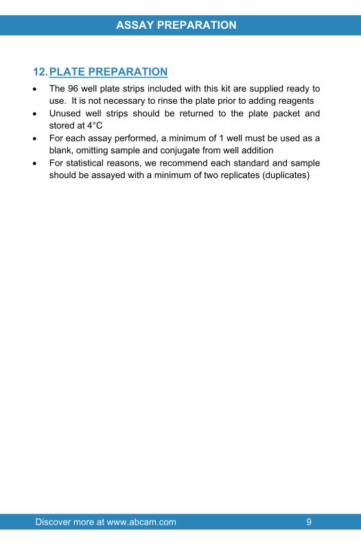
12.PLATE PREPARATION The 96 well plate strips included with this kit are supplied ready to
use. It is not necessary to rinse the plate prior to adding reagents Unused well strips should be returned to the plate packet and
stored at 4°C For each assay performed, a minimum of 1 well must be used as a
blank, omitting sample and conjugate from well addition For statistical reasons, we recommend each standard and sample
should be assayed with a minimum of two replicates (duplicates)
Discover more at www.abcam.com 10
ASSAY PROCEDURE
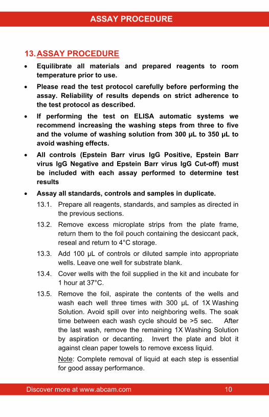
13.ASSAY PROCEDURE Equilibrate all materials and prepared reagents to room
temperature prior to use. Please read the test protocol carefully before performing the
assay. Reliability of results depends on strict adherence to the test protocol as described.
If performing the test on ELISA automatic systems we recommend increasing the washing steps from three to five and the volume of washing solution from 300 µL to 350 µL to avoid washing effects.
All controls (Epstein Barr virus IgG Positive, Epstein Barr virus IgG Negative and Epstein Barr virus IgG Cut-off) must be included with each assay performed to determine test results
Assay all standards, controls and samples in duplicate.13.1. Prepare all reagents, standards, and samples as directed in
the previous sections.13.2. Remove excess microplate strips from the plate frame,
return them to the foil pouch containing the desiccant pack, reseal and return to 4°C storage.
13.3. Add 100 µL of controls or diluted sample into appropriate wells. Leave one well for substrate blank.
13.4. Cover wells with the foil supplied in the kit and incubate for 1 hour at 37°C.
13.5. Remove the foil, aspirate the contents of the wells and wash each well three times with 300 µL of 1X Washing Solution. Avoid spill over into neighboring wells. The soak time between each wash cycle should be >5 sec. After the last wash, remove the remaining 1X Washing Solution by aspiration or decanting. Invert the plate and blot it against clean paper towels to remove excess liquid.Note: Complete removal of liquid at each step is essential for good assay performance.
Discover more at www.abcam.com 11
ASSAY PROCEDURE
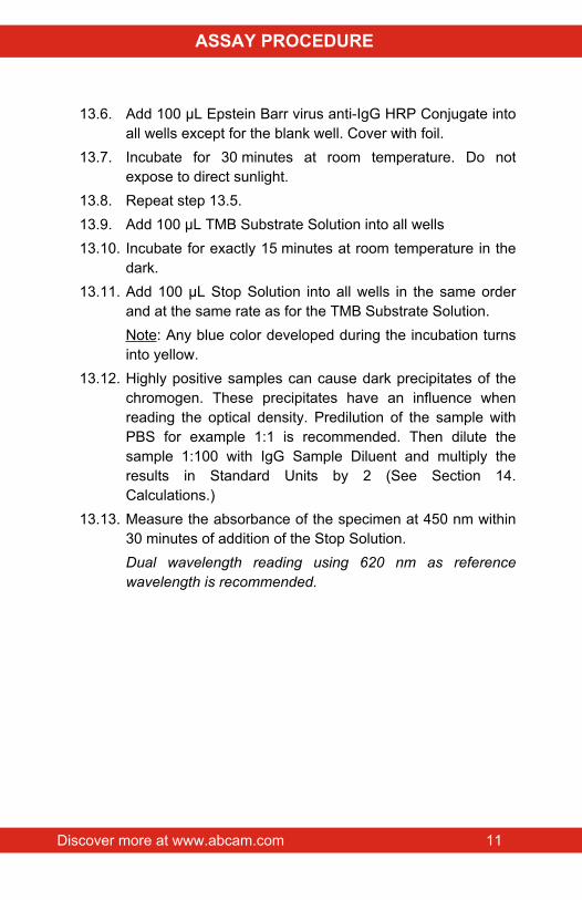
13.6. Add 100 µL Epstein Barr virus anti-IgG HRP Conjugate into all wells except for the blank well. Cover with foil.
13.7. Incubate for 30 minutes at room temperature. Do not expose to direct sunlight.
13.8. Repeat step 13.5.13.9. Add 100 µL TMB Substrate Solution into all wells13.10. Incubate for exactly 15 minutes at room temperature in the
dark.13.11. Add 100 µL Stop Solution into all wells in the same order
and at the same rate as for the TMB Substrate Solution. Note: Any blue color developed during the incubation turns into yellow.
13.12. Highly positive samples can cause dark precipitates of the chromogen. These precipitates have an influence when reading the optical density. Predilution of the sample with PBS for example 1:1 is recommended. Then dilute the sample 1:100 with IgG Sample Diluent and multiply the results in Standard Units by 2 (See Section 14. Calculations.)
13.13. Measure the absorbance of the specimen at 450 nm within 30 minutes of addition of the Stop Solution.Dual wavelength reading using 620 nm as reference wavelength is recommended.
Discover more at www.abcam.com 12
DATA ANALYSIS
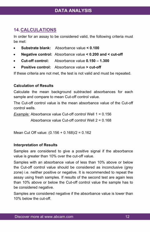
14.CALCULATIONSIn order for an assay to be considered valid, the following criteria must be met:
Substrate blank: Absorbance value < 0.100 Negative control: Absorbance value < 0.200 and < cut-off Cut-off control: Absorbance value 0.150 – 1.300 Positive control: Absorbance value > cut-offIf these criteria are not met, the test is not valid and must be repeated.
Calculation of ResultsCalculate the mean background subtracted absorbances for each sample and compare to mean Cut-off control value. The Cut-off control value is the mean absorbance value of the Cut-off control wells.Example: Absorbance value Cut-off control Well 1 = 0.156
Absorbance value Cut-off control Well 2 = 0.168
Mean Cut Off value: (0.156 + 0.168)/2 = 0.162
Interpretation of ResultsSamples are considered to give a positive signal if the absorbance value is greater than 10% over the cut-off value.Samples with an absorbance value of less than 10% above or below the Cut-off control value should be considered as inconclusive (grey zone) i.e. neither positive or negative. It is recommended to repeat the assay using fresh samples. If results of the second test are again less than 10% above or below the Cut-off control value the sample has to be considered negative.Samples are considered negative if the absorbance value is lower than 10% below the cut-off.
Discover more at www.abcam.com 13
DATA ANALYSIS
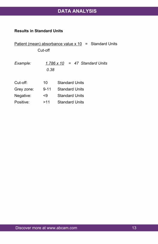
Results in Standard Units
Patient (mean) absorbance value x 10 = Standard Units Cut-off
Example: 1.786 x 10 = 47 Standard Units 0.38
Cut-off: 10 Standard UnitsGrey zone: 9-11 Standard UnitsNegative: <9 Standard UnitsPositive: >11 Standard Units
Discover more at www.abcam.com 14
DATA ANALYSIS
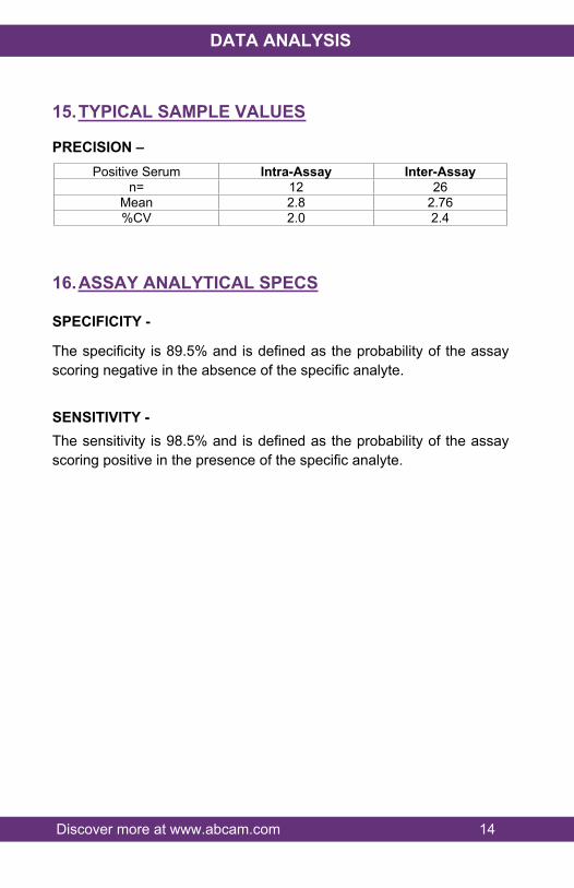
15.TYPICAL SAMPLE VALUES
PRECISION – Positive Serum Intra-Assay Inter-Assay
n= 12 26Mean 2.8 2.76%CV 2.0 2.4
16.ASSAY ANALYTICAL SPECS
SPECIFICITY -
The specificity is 89.5% and is defined as the probability of the assay scoring negative in the absence of the specific analyte.
SENSITIVITY -The sensitivity is 98.5% and is defined as the probability of the assay scoring positive in the presence of the specific analyte.
Discover more at www.abcam.com 15
RESOURCES
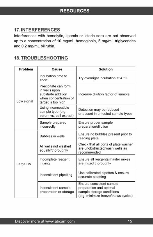
17. INTERFERENCESInterferences with hemolytic, lipemic or icteric sera are not observed up to a concentration of 10 mg/mL hemoglobin, 5 mg/mL triglycerides and 0.2 mg/mL bilirubin.
18.TROUBLESHOOTING
Problem Cause Solution
Incubation time to short Try overnight incubation at 4 °C
Precipitate can form in wells upon substrate addition when concentration of target is too high
Increase dilution factor of sample
Using incompatiblesample type (e.g. serum vs. cell extract)
Detection may be reducedor absent in untested sample types
Low signal
Sample prepared incorrectly
Ensure proper sample preparation/dilution
Bubbles in wells Ensure no bubbles present prior to reading plate
All wells not washedequally/thoroughly
Check that all ports of plate washer are unobstructed/wash wells as recommended
Incomplete reagent mixing
Ensure all reagents/master mixes are mixed thoroughly
Inconsistent pipetting Use calibrated pipettes & ensure accurate pipetting
Large CV
Inconsistent samplepreparation or storage
Ensure consistent samplepreparation and optimalsample storage conditions(e.g. minimize freeze/thaws cycles)
Discover more at www.abcam.com 16
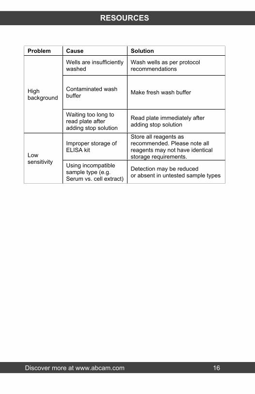
RESOURCES
Problem Cause Solution
Wells are insufficientlywashed
Wash wells as per protocol recommendations
Contaminated wash buffer Make fresh wash bufferHigh
background
Waiting too long to read plate after adding stop solution
Read plate immediately after adding stop solution
Improper storage ofELISA kit
Store all reagents as recommended. Please note all reagents may not have identical storage requirements.Low
sensitivity Using incompatiblesample type (e.g. Serum vs. cell extract)
Detection may be reducedor absent in untested sample types

Discover more at www.abcam.com 17
RESOURCES
19.NOTES

Discover more at www.abcam.com 18
RESOURCES

RESOURCES 19
UK, EU and ROWEmail: [email protected] | Tel: +44-(0)1223-696000
AustriaEmail: [email protected] | Tel: 019-288-259
FranceEmail: [email protected] | Tel: 01-46-94-62-96 GermanyEmail: [email protected] | Tel: 030-896-779-154 SpainEmail: [email protected] | Tel: 911-146-554 SwitzerlandEmail: [email protected] Tel (Deutsch): 0435-016-424 | Tel (Français): 0615-000-530
US and Latin AmericaEmail: [email protected] | Tel: 888-77-ABCAM (22226)
CanadaEmail: [email protected] | Tel: 877-749-8807
China and Asia Pacific Email: [email protected] | Tel: 108008523689 (中國聯通) JapanEmail: [email protected] | Tel: +81-(0)3-6231-0940
www.abcam.com | www.abcam.cn | www.abcam.co.jp
Copyright © 2013 Abcam, All Rights Reserved. The Abcam logo is a registered trademark.
All information / detail is correct at time of going to print.