Embed Size (px)
Citation preview
28
Turkish Journal of CancerVol.32/ No. 1/2002
Abdominal inflammatory myofibroblastic tumor:Review of the literature by means of a case reportHURŞİT APA1, GÜLDEN DİNİZ2, TÜRKAY SARITAŞ1, SAFİYE AKTAŞ2,
RAGIP ORTAÇ2, İRFAN KARACA3, AYTAÇ KARKIRAN3,CANAN VERGİN1
Departments of 1Paediatric Oncology and Hematology, 2Pathology and3Paediatric Surgery, Dr. Behçet Uz Children�s Hospital,
İzmir-Turkey
Inflammatory myofibroblastic tumor occurring atintraabdominal sites in children has rarely been described. Inthis paper, an 18-month old girl with intraabdominalinflammatory myofibroblastic tumor who presented with fever,anemia, constipation, weight loss and an abdominal mass isreported. A comprehensive review of the literature is alsodocumented. [Turk J Cancer 2002;32(1):28-31]
Key words: Inflammatory myofibroblastic tumor,abdominal mass, children
Inflammatory myofibroblastic tumors (IMT) are rare proliferative lesionsclinically resembling a malignant neoplasm. Their classification is controversialand confusing. IMTs are well described in the lung and upper respiratory tract ofyoung adults and children; but may occur at any age and affect any organsystem (1,2,3).
Typically, it is a circumscribed but nonencapsulated lesion containingspindle cells proliferating in a background of fibrosis, with lymphocytes,plasmacytes, histiocytes, foamy macrophages, and occasionally eosinophilsand neutrophils nuclear pleomorphism and atypical mitoses are absent (4,5).
At one time thought to be reactive in nature, the inflammatorymyofibroblastic tumor has come to be considered a true neoplasm with thepotential for recurrence and multifocality; clonal cytogenetic findings supportthis view. Complete excision is necessary to avoid local recurrence (6,7).
Case Report
An 18-month old girl with a 35-day history of intermittent fever, constipationand profound weight loss was referred for investigation of a large form, mobile,left-sided abdominal mass. Investigations revealed microcytic hypochromicanemia (haemoglobin level 5.3 g/dl) and thrombocytosis (platelet count1106x109/L). Ultrasonography and abdominal CT demonstrated an 8x10 cmmass adjacent to the left kidney. After a short period of investigations, a
APA et. al 29
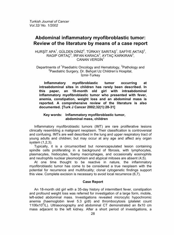
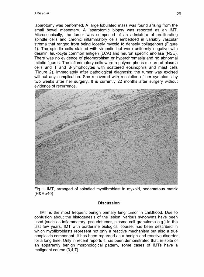
laparotomy was performed. A large lobulated mass was found arising from thesmall bowel mesentery. A laparotomic biopsy was reported as an IMT.Microscopically, the tumor was composed of an admixture of proliferatingspindle cells and chronic inflammatory cells embedded in variably vascularstroma that ranged from being loosely myxoid to densely collagenous (Figure1). The spindle cells stained with vimentin but were uniformly negative withdesmin, leukocyte common antigen (LCA) and neuron specific enolase (NSE).There was no evidence of pleomorphism or hyperchromasia and no abnormalmitotic figures. The inflammatory cells were a polymorphous mixture of plasmacells and T and B-lymphocytes with scattered eosinophils and mast cells(Figure 2). Immediately after pathological diagnosis; the tumor was excisedwithout any complication. She recovered with resolution of her symptoms bytwo weeks after her surgery. It is currently 22 months after surgery withoutevidence of recurrence.
Fig 1. IMT, arranged of spindled myofibroblast in myxoid, oedematous matrix(H&E x40)
Discussion
IMT is the most frequent benign primary lung tumor in childhood. Due toconfusion about the histogenesis of the lesion, various synonyms have beenused (such as inflammatory, pseudotumor, plasma cell granuloma e.g.) In thelast few years, IMT with borderline biological course, has been described inwhich myofibroblasts represent not only a reactive mechanism but also a trueneoplastic component. It has been regarded as a benign and reactive disorderfor a long time. Only in recent reports it has been demonstrated that, in spite ofan apparently benign morphological pattern, some cases of IMTs have amalignant course (3,4,7).
ABDOMINAL INFLAMMATORY MYOFIBROBLASTIC TUMOR30
Fig 2. Inflammatory infiltrate with plasma cells and lymphocytes of IMT (H&Ex400)
The myofibroblast was eventually recognised as the principal spindle-celltype in this tumor, which led to the new term IMT. Myofibroblasts are spindlecells having ultra-structural features in common with smooth muscle cells andfibroblasts. The important function of the myofibroblast in tissue repair isconsistent with the hypothesis that an aberrant response to tissue injury is thepathogenesis of IMT, however in most cases there is no identifiableprecipitating factor (2,6).
Evidence to support a directly infectious etiology is scanty but animmunological pathogenesis remains possible. The role of cytokines,particularly interleukin-6 (IL-6) in pathogenesis and the possibility for a specifictherapeutic approach has been described. Many authors postulate a postinflammatory process; although the presence of clonal chromosomalabnormalities suggests a neoplastic process (1,5,7).
Until 1992, a total of 21 children, predominantly female, with IMTs affectingintra-abdominal sites have been reported in the literature. These tumors areoften large. Multi-centric lesions are rare. There are no reports of malignancyarising in an IMT; nevertheless, the clinical, radiological and histologicalfeatures of these may cause confusion with malignant lesions. Most patientspresented with fever, anemia, trombocytosis, hyperglobulinemia and weightloss. These systemic features resolve after tumor excision but may enable thediagnosis to be suspected before operation. Misdiagnosis has led somepatients to be inappropriately treated with chemotherapy and radical surgery(2,4).
APA et. al 31
In IMTs, high cellularity with large, plump, active myofibroblasts withprominent nucleoli can cause confusion with malignancy, in particularrhabdomyosarcoma. However the lack of atypia, hyperchromasia and abnormalmitotic figures are pointers toward a benign lesion. IMT should be diagnosed byroutine staining because special stains and immunocytochemistry can bemisleading. Preliminary biopsy and full histological evaluation is recommendedin cases where resection may be particularly hazardous (5,6,7).
Other reasons for confusion of IMTs with malignant neoplasms include theircapacity for local tissue infiltration, occasional rapid growth and thedevelopment of local recurrence. Mediastinal and esophageal involvement fromadjacent pulmonary pathology appears to be particularly aggressive. Theircourse is complicated, which ranges from spontaneous regression throughgradual enlargement to rapid growth with local invasion. Tumor resolution orregression has been reported after radiotherapy, chemotherapy and steroidtherapy. Local recurrences after incomplete excision are recognised, may occurmany years later and may be fatal. This underlines the importance of completesurgical resection whenever possible (1,3).
References
1. Gangopadhyay K, Mahasin ZZ, Kfoury H, et al. Inflammatory myofibroblastictumour of the tonsil. J Laryngol Otol 1997;111:880-2.
2. Stringer MD, Ramani P, Yeung CK, et al. Abdominal IMTs in children. Br JSurg 1992;79:1357-60.
3. Verbeke J, Verberne A, Hollander J, et al. IMT of the lung manifesting asprogressive atelectasis. Pediatr Radiol 1999;29:816-9.
4. Treissman SP, Gillis DA, Lee CLY, et al. Omental-mesenteric inflammatorypseudotumor cytogenetic demonstration of genetic changes andmonoclonality in one tumor. Cancer 1994;73:1433-7.
5. Brooks JS. Disorders of soft tissue. In: Stenberg SS, editor. DiagnosticSurgical Pathology. 3rd ed. Philadelphia: Lippincott Williams&Wilkins,1999;131-221.
6. Pettinano G, Manivel JL, De Rosa N, et al. IMT (plasma cell granuloma);clinicopathologic study of 20 cases with immunohistochemical andultrastructural observations. Am J Clin Pathol 1990;94:538-46.
7. Ro Goodlad JR, Fletcher CD. Recent developments in soft tissue tumours.Histopathology 1995;27:103-20.