Embed Size (px)
Citation preview
pISSN: 2234-8646 eISSN: 2234-8840http://dx.doi.org/10.5223/pghn.2014.17.2.116Pediatr Gastroenterol Hepatol Nutr 2014 June 17(2):116-120 PGHNCase Report
PEDIATRIC GASTROENTEROLOGY, HEPATOLOGY & NUTRITION
Two Cases of Infantile Intra-abdominal Inflammatory Myofibroblastic Tumor
Soo-Hong Kim, Yong Hoon Cho and Hae Young Kim
Division of Pediatric Surgery, Department of Surgery, Pusan National University Children’s Hospital, Pusan National University School of Medicine, Yangsan, Korea
Inflammatory myofibroblastic tumor (IMT) is rare mesenchymal solid tumor that consists of proliferating myofibro-blasts with an inflammatory infiltrate background. It has a very low prevalence in infants and occurs mainly in children and young adults. IMT are mainly located in the thoracic cavity, but intra-abdominal lesions are rare. IMT can exhibit locally aggressive neoplastic processes and metastases similar to malignancies, so, have clinical importance. Herein, we describe two infantile intra-abdominal IMT cases presenting with incidentally found palpable abdominal mass. A 4-month-old male infant had IMT at the ileal mesentery and a 5-month-old male infant had IMT at liver. Both cases were successfully treated by complete surgical resection without complication or recurrence. Considering the biological behavior of the intermediate type of neoplasm in IMT, we expect good survivals when achieving appro-priate surgical resection without adjuvant therapy in infantile intra-abdominal IMT.
Key Words: Inflammatory myofibroblastic tumor, Intraabdominal, Infant
Received:Mar 25, 2014, Revised:April 2, 2014, Accepted:April 14, 2014
Corresponding author: Yong Hoon Cho, Division of Pediatric Surgery, Department of Surgery, Pusan National University Children's Hospital, 20,Geumo-ro, Mulgeum-eup, Yangsan 626-770, Korea. Tel: +82-55-360-2124, Fax: +82-55-360-2154, E-mail: [email protected]
Copyright ⓒ 2014 by The Korean Society of Pediatric Gastroenterology, Hepatology and NutritionThis is an openaccess article distributed under the terms of the Creative Commons Attribution NonCommercial License (http://creativecommons.org/licenses/by-nc/3.0/) which permits unrestricted noncommercial use, distribution, and reproduction in any medium, provided the original work is properly cited.
INTRODUCTION
Inflammatory myofibroblastic tumor (IMT) is rare mesenchymal solid tumor that consists of proliferat-ing myofibroblasts with an inflammatory infiltrate background. It occurs mainly in children and young adults, but has a low prevalence in infants [1,2]. IMTs may be located in various organs, mainly those in the thorax, but intra-abdominal lesions are rela-tively rare [3]. Intra-abdominal IMTs have clinical importance
due to their relationship to malignancy. Intra-ab-dominal IMTs are difficult to distinguish pre-operatively from other malignancies, such as sarco-mas, lymphomas, and metastases [4]. IMTs were originally reported to be benign tumors, but can ex-hibit locally aggressive neoplastic processes and metastases similar to malignancies. Based on such findings, surgical excision of IMTs is recommended [2]. In this report we describe two cases of infantile in-tra-abdominal IMT that were histopathologically di-
www.pghn.org 117
Soo-Hong Kim, et al:Intra-abdominal IMT of Infant
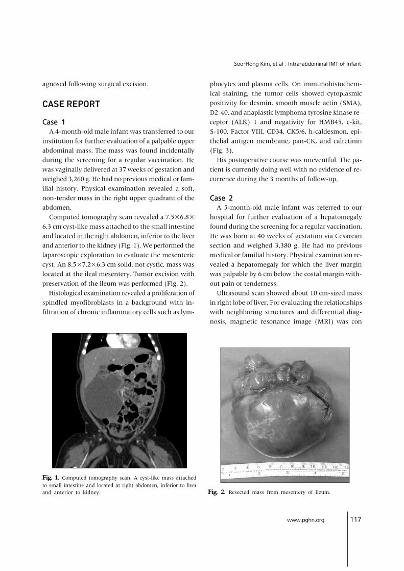
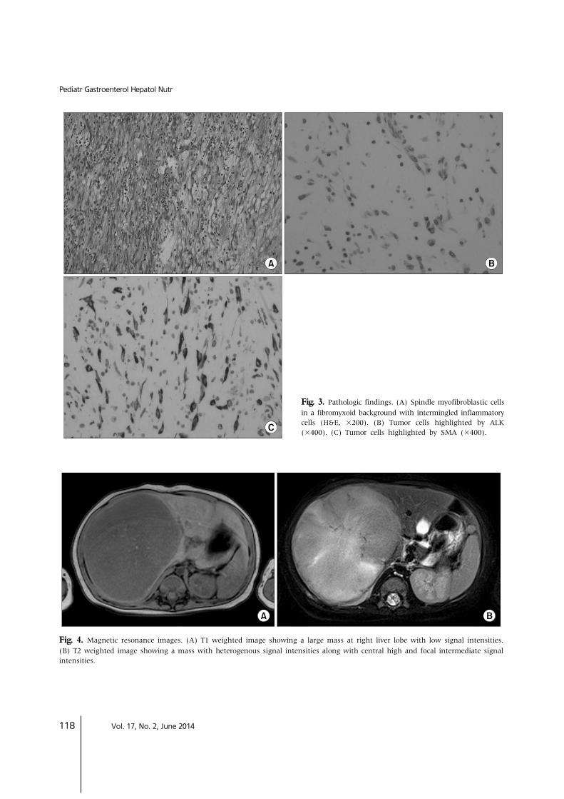
Fig. 1. Computed tomography scan. A cyst-like mass attached to small intestine and located at right abdomen, inferior to liverand anterior to kidney. Fig. 2. Resected mass from mesentery of ileum.
agnosed following surgical excision.
CASE REPORT
Case 1 A 4-month-old male infant was transferred to our institution for further evaluation of a palpable upper abdominal mass. The mass was found incidentally during the screening for a regular vaccination. He was vaginally delivered at 37 weeks of gestation and weighed 3,260 g. He had no previous medical or fam-ilial history. Physical examination revealed a soft, non-tender mass in the right upper quadrant of the abdomen. Computed tomography scan revealed a 7.5×6.8× 6.3 cm cyst-like mass attached to the small intestine and located in the right abdomen, inferior to the liver and anterior to the kidney (Fig. 1). We performed the laparoscopic exploration to evaluate the mesenteric cyst. An 8.5×7.2×6.3 cm solid, not cystic, mass was located at the ileal mesentery. Tumor excision with preservation of the ileum was performed (Fig. 2). Histological examination revealed a proliferation of spindled myofibroblasts in a background with in-filtration of chronic inflammatory cells such as lym-
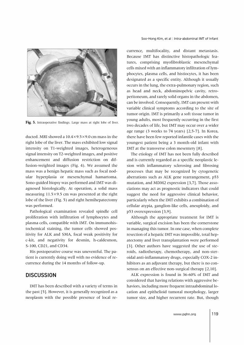
phocytes and plasma cells. On immunohistochem-ical staining, the tumor cells showed cytoplasmic positivity for desmin, smooth muscle actin (SMA), D2-40, and anaplastic lymphoma tyrosine kinase re-ceptor (ALK) 1 and negativity for HMB45, c-kit, S-100, Factor VIII, CD34, CK5/6, h-caldesmon, epi-thelial antigen membrane, pan-CK, and calretinin (Fig. 3). His postoperative course was uneventful. The pa-tient is currently doing well with no evidence of re-currence during the 3 months of follow-up.
Case 2 A 5-month-old male infant was referred to our hospital for further evaluation of a hepatomegaly found during the screening for a regular vaccination. He was born at 40 weeks of gestation via Cesarean section and weighed 3,380 g. He had no previous medical or familial history. Physical examination re-vealed a hepatomegaly for which the liver margin was palpable by 6 cm below the costal margin with-out pain or tenderness. Ultrasound scan showed about 10 cm-sized mass in right lobe of liver. For evaluating the relationships with neighboring structures and differential diag-nosis, magnetic resonance image (MRI) was con
118 Vol. 17, No. 2, June 2014
Pediatr Gastroenterol Hepatol Nutr
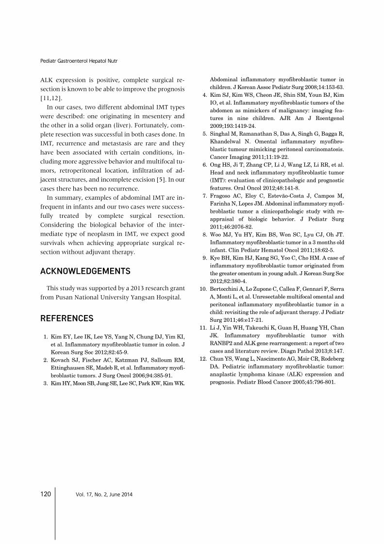
Fig. 4. Magnetic resonance images. (A) T1 weighted image showing a large mass at right liver lobe with low signal intensities.(B) T2 weighted image showing a mass with heterogenous signal intensities along with central high and focal intermediate signalintensities.
Fig. 3. Pathologic findings. (A) Spindle myofibroblastic cells in a fibromyxoid background with intermingled inflammatory cells (H&E, ×200). (B) Tumor cells highlighted by ALK (×400). (C) Tumor cells highlighted by SMA (×400).
www.pghn.org 119
Soo-Hong Kim, et al:Intra-abdominal IMT of Infant
Fig. 5. Intraoperative findings. Large mass at right lobe of liver.
ducted. MRI showed a 10.4×9.5×9.0 cm mass in the right lobe of the liver. The mass exhibited low signal intensity on T1-weighted images, heterogeneous signal intensity on T2-weighted images, and positive enhancement and diffusion restriction on dif-fusion-weighted images (Fig. 4). We assumed the mass was a benign hepatic mass such as focal nod-ular hyperplasia or mesenchymal hamartoma. Sono-guided biopsy was performed and IMT was di-agnosed histologically. At operation, a solid mass measuring 11.5×9.5 cm was presented at the right lobe of the liver (Fig. 5) and right hemihepatectomy was performed. Pathological examination revealed spindle cell proliferation with infiltration of lymphocytes and plasma cells, compatible with IMT. On immunohis-tochemical staining, the tumor cells showed pos-itivity for ALK and SMA, focal weak positivity for c-kit, and negativity for desmin, h-caldesmon, S-100, CD21, and CD34. His postoperative course was uneventful. The pa-tient is currently doing well with no evidence of re-currence during the 14 months of follow-up.
DISCUSSION
IMT has been described with a variety of terms in the past [5]. However, it is generally recognized as a neoplasm with the possible presence of local re-
currence, multifocality, and distant metastasis. Because IMT has distinctive histopathologic fea-tures, comprising myofibroblastic mesenchymal cells mixed with an inflammatory infiltration of lym-phocytes, plasma cells, and histiocytes, it has been designated as a specific entity. Although it usually occurs in the lung, the extra-pulmonary region, such as head and neck, abdominopelvic cavity, retro-peritoneum, and rarely solid organs in the abdomen, can be involved. Consequently, IMT can present with variable clinical symptoms according to the site of tumor origin. IMT is primarily a soft tissue tumor in young adults, most frequently occurring in the first two decades of life, but IMT may occur over a wider age range (3 weeks to 74 years) [2,5-7]. In Korea, there have been few reported infantile cases with the youngest patient being a 3 month-old infant with IMT at the transverse colon mesentery [8]. The etiology of IMT has not been fully described and is currently regarded as a specific neoplastic le-sion with inflammatory sclerosing and fibrosing processes that may be recognized by cytogenetic aberrations such as ALK gene rearrangement, p53 mutation, and MDM2 expression [3,7]. Those asso-ciations may act as prognostic indicators that could suggest the need for aggressive clinical behavior, particularly when the IMT exhibits a combination of cellular atypia, ganglion-like cells, aneuploidy, and p53 overexpression [3,9]. Although the appropriate treatment for IMT is variable, surgical excision has been the cornerstone in managing this tumor. In one case, when complete resection of a hepatic IMT was impossible, total hep-atectomy and liver transplantation were performed [3]. Other authors have suggested the use of ste-roids, radiotherapy, chemotherapy, and non-ster-oidal anti-inflammatory drugs, especially COX-2 in-hibitors as an adjuvant therapy, but there is no con-sensus on an effective non-surgical therapy [2,10]. ALK expression is found in 36-60% of IMT and considered that having relations with aggressive be-haviors, including more frequent intraabdominal lo-cation and epithelioid tumoral morphology, larger tumor size, and higher recurrent rate. But, though
120 Vol. 17, No. 2, June 2014
Pediatr Gastroenterol Hepatol Nutr
ALK expression is positive, complete surgical re-section is known to be able to improve the prognosis [11,12]. In our cases, two different abdominal IMT types were described: one originating in mesentery and the other in a solid organ (liver). Fortunately, com-plete resection was successful in both cases done. In IMT, recurrence and metastasis are rare and they have been associated with certain conditions, in-cluding more aggressive behavior and multifocal tu-mors, retroperitoneal location, infiltration of ad-jacent structures, and incomplete excision [5]. In our cases there has been no recurrence. In summary, examples of abdominal IMT are in-frequent in infants and our two cases were success-fully treated by complete surgical resection. Considering the biological behavior of the inter-mediate type of neoplasm in IMT, we expect good survivals when achieving appropriate surgical re-section without adjuvant therapy.
ACKNOWLEDGEMENTS
This study was supported by a 2013 research grant from Pusan National University Yangsan Hospital.
REFERENCES
1. Kim EY, Lee IK, Lee YS, Yang N, Chung DJ, Yim KI, et al. Inflammatory myofibroblastic tumor in colon. J Korean Surg Soc 2012;82:45-9.
2. Kovach SJ, Fischer AC, Katzman PJ, Salloum RM, Ettinghausen SE, Madeb R, et al. Inflammatory myofi-broblastic tumors. J Surg Oncol 2006;94:385-91.
3. Kim HY, Moon SB, Jung SE, Lee SC, Park KW, Kim WK.
Abdominal inflammatory myofibroblastic tumor in children. J Korean Assoc Pediatr Surg 2008;14:153-63.
4. Kim SJ, Kim WS, Cheon JE, Shin SM, Youn BJ, Kim IO, et al. Inflammatory myofibroblastic tumors of the abdomen as mimickers of malignancy: imaging fea-tures in nine children. AJR Am J Roentgenol 2009;193:1419-24.
5. Singhal M, Ramanathan S, Das A, Singh G, Bagga R, Khandelwal N. Omental inflammatory myofibro-blastic tumour mimicking peritoneal carcinomatosis. Cancer Imaging 2011;11:19-22.
6. Ong HS, Ji T, Zhang CP, Li J, Wang LZ, Li RR, et al. Head and neck inflammatory myofibroblastic tumor (IMT): evaluation of clinicopathologic and prognostic features. Oral Oncol 2012;48:141-8.
7. Fragoso AC, Eloy C, Estevão-Costa J, Campos M, Farinha N, Lopes JM. Abdominal inflammatory myofi-broblastic tumor a clinicopathologic study with re-appraisal of biologic behavior. J Pediatr Surg 2011;46:2076-82.
8. Woo MJ, Yu HY, Kim BS, Won SC, Lyu CJ, Oh JT. Inflammatory myofibroblastic tumor in a 3 months old infant. Clin Pediatr Hematol Oncol 2011;18:62-5.
9. Kye BH, Kim HJ, Kang SG, Yoo C, Cho HM. A case of inflammatory myofibroblastic tumor originated from the greater omentum in young adult. J Korean Surg Soc 2012;82:380-4.
10. Bertocchini A, Lo Zupone C, Callea F, Gennari F, Serra A, Monti L, et al. Unresectable multifocal omental and peritoneal inflammatory myofibroblastic tumor in a child: revisiting the role of adjuvant therapy. J Pediatr Surg 2011;46:e17-21.
11. Li J, Yin WH, Takeuchi K, Guan H, Huang YH, Chan JK. Inflammatory myofibroblastic tumor with RANBP2 and ALK gene rearrangement: a report of two cases and literature review. Diagn Pathol 2013;8:147.
12. Chun YS, Wang L, Nascimento AG, Moir CR, Rodeberg DA. Pediatric inflammatory myofibroblastic tumor: anaplastic lymphoma kinase (ALK) expression and prognosis. Pediatr Blood Cancer 2005;45:796-801.