Embed Size (px)
Citation preview
Emerg Med Clin N Am
21 (2003) 909–935
Abdominal surgical emergencies in infantsand young children
Maureen McCollough, MD, MPHa,b,*,Ghazala Q. Sharieff, MDc,d
aDepartment of Emergency Medicine, Keck School of Medicine, University of Southern
California, 1200 North State Street, Room G1011, Los Angeles, CA 90033, USAbDepartment of Pediatrics, Keck School of Medicine, University of Southern California,
Los Angeles, CA 90033, USAcDepartment of Emergency Medicine, Shands Jacksonville,
655 West Eighth Street, Jacksonville, FL 32009, USAdDepartment of Emergency Medicine, Palomar-Pomerado Health System, 555 East Valley
Parkway, San Diego, CA 92025, USA
Infants and children commonly present to the emergency department (ED)with abdominal and gastrointestinal (GI) symptoms. In most cases thesesymptoms are caused by a self-limited process such as viral gastroenteritis;however, they might also be the harbingers of life-threatening surgicalemergencies. Because symptoms such as vomiting, diarrhea, abdominal pain,and fever are so common and so nonspecific in children, the recognition ofsurgical emergencies is frequently delayed or missed altogether. When onealso considers the difficulties inherent to the pediatric examination, it is notsurprising that the diagnoses of intussusception, pyloric stenosis, malrotationwith volvulus, and bowel obstruction continue to be among the most elusivediagnoses for the emergency physician (EP). Appendicitis in the infant oryoung child is especially difficult to detect in its early stages and carriessignificant morbidity and mortality. Testicular torsion, another surgicalemergency, might also present with vague abdominal complaints. This articlereviews abdominal surgical emergencies in infants and young children thatare often mistaken for more benign, self-limited illnesses.
* Corresponding author. Department of Emergency Medicine, Keck School of Medicine,
University of Southern California, 1200 North State Street, Room G1011, Los Angeles, CA
90033.
E-mail address: [email protected] (M. McCollough).
0733-8627/03/$ - see front matter � 2003 Elsevier Inc. All rights reserved.
doi:10.1016/S0733-8627(03)00090-7

910 M. McCollough, G.Q. Sharieff / Emerg Med Clin N Am 21 (2003) 909–935
General approach to the child who has abdominal pain
Important information can often be elicited even before speaking to theparents or laying hands on a child. Infants and young toddlers are usuallyafraid of strangers. Older children might associate a ‘‘clinic’’ environment ora ‘‘man in a white coat’’ with immunizations and pain. The difficulty ofphysical examination increases when the physician enters the examinationroom and the child bursts into tears. Observing the child’s behavior beforeany interaction might reveal the reassuring signs of a young child ambulatingcomfortably around the ED or of an older infant sitting up on a gurneyinterested in his surroundings. An older child who walks slowly downa corridor in the ED holding his right lower quadrant has similarly given theexaminer a great deal of information. When the child is approached, usinga nonthreatening manner might pay dividends during the assessment. Forexample, sitting down or kneeling to bring the examiner closer to the child’seye level makes the examiner appear less intimidating.
If a child is found to be poorly responsive or displays other signs of shock,the ongoing assessment of the abdomen will need to occur simultaneouslywith the immediate priorities of resuscitation. A patent and secure air-way must be ensured. Ventilation should be assisted, if necessary, andsupplemental oxygen delivered. Vascular assess should be achieved using theintravenous or intraosseous routes, and fluid boluses of normal saline shouldbe administered as necessary. The child should be placed on a cardiacmonitor. Immediate bedside tests should include a blood glucose andhemoglobin determination. The delivery of intravenous antibiotics should notbe delayed if there is a reasonable suspicion of underlying sepsis.
Children and parents are often poor historians. Trying to elicit thechronology of symptoms with questions such as ‘‘did the pain start beforethe vomiting or vice versa?’’ might be difficult. Parents of young infantsmight only describe their child as irritable and not realize that the abdomenis the source of pain. Adolescents might be embarrassed to talk about bodilyfunctions or sexual issues, especially with physicians of the opposite sex. It isalso important to question adolescents about their medical history separatefrom their parents because they might be more forthcoming when assured oftheir privacy.
Attempting to bond with the child or using a toy as a distraction beforeauscultation or palpation can often improve the reliability of theabdominal examination. Infants might be distracted by a set of car keys.Hand and finger puppets can also be used for this purpose. Allowing thechild to remain in a parent’s arms or lap for as long as possible is alsohelpful. For older children, examining the mother first might show the childthat the examination is nothing to be feared. An older child can also beallowed to place his or her hand on top of the examiner’s and simul-taneously apply pressure, and they can also be questioned about school orplay activities.
911M. McCollough, G.Q. Sharieff / Emerg Med Clin N Am 21 (2003) 909–935
Before touching the abdomen, the examiner should look for any obviousabnormalities such as distension, masses, or peristaltic waves. If a child iscrying, it should be remembered that the abdomen is relatively soft duringthe child’s inhalation, which might be the best time to detect masses. Toelicit areas of tenderness or peritoneal signs, a quieter, calm child is helpful.If the examiner has difficulty, in some cases it might be possible to have themother gently push on different areas of the abdomen, with the examinermerely observing the child’s response. Another technique is to have themother hold the child over her shoulder with the child facing away. Theexaminer can then stand behind the child and slip a hand between motherand child to palpate the abdomen. Peritoneal signs can also be elicited byhaving the mother bounce the child up and down on her lap. Fussiness orcrying while this maneuver is performed raises the suspicion of peritonitis.Older children can simply be asked to jump up and down.
Rectal examinations are not imperative in a child presenting withabdominal pain. In particular, they have been shown to not be helpful inthe diagnosis of appendicitis [6]. Rectal examinations can, however, aid in thediagnosis of a GI bleed, intussusception, rectal abscess, or impaction. If arectal examination is necessary, it can be done by partially introducing a smallfinger. Inspection of the genitalia might reveal a hair tourniquet, hernia, orsigns of testicular torsion, and is an important part of the examination.
A thorough extra-abdominal examination is indicated in most childrenwho have abdominal pain. For example, failure to examine the throat mightlead to a missed diagnosis of pharyngitis, which can be associated withabdominal pain. Lower lobe pneumonias can also mimic an abdominalemergency. The general examination also includes an assessment of thechild’s hydration status. Classic signs and symptoms of dehydration ininfants and young children are dry mucous membranes, decreased tearing,sunken eyes and fontanelles, decreased skin turgor, prolonged capillaryrefill, and decreased urine output. Most of these signs have not been wellstudied, and some might not be reliable.
For the surgical disease processes discussed in this article, pain is typicallythe chief complaint. Management of the child’s pain during the evaluation isof paramount importance. The use of pain medication in children who haveabdominal pain does not appear to increase the risk of misdiagnosis [1]. Abetter physical examination can often be accomplished when the patient’spain has been addressed.
Appendicitis
Epidemiology
Appendicitis is the most common surgical cause of abdominal pain inchildhood, affecting four of every 1000 children. Appendicitis is the cause ofpain in 2.3% of all children seen in ambulatory clinics or EDs with abdominal
912 M. McCollough, G.Q. Sharieff / Emerg Med Clin N Am 21 (2003) 909–935
pain. Of the children admitted to the hospital with abdominal pain, 82% arediagnosed with appendicitis [2]. Because of the difficulty in evaluating youngchildren who have abdominal pain, perforation rates for appendicitis arehigher than in the general adult population (30–65%).Moreover, because theomentum is less developed in children, perforations are less likely to be‘‘walled-off’’ or localized, leading to generalized peritonitis.
Presentation
The ‘‘classic’’ presentation of appendicitis, consisting of generalizedabdominal pain migrating to the right lower quadrant associated withnausea, vomiting, and fever, is seen less often in pediatric patients [3]. Inaddition, children often present earlier in their clinical course than adults,when only mild or less specific symptoms are present; however, limited dataappear to indicate that individual signs such as rebound tenderness andRovsing’s sign have a high sensitivity and specificity in children [4].
The most common findings of appendicitis in children are right lowerquadrant pain, abdominal tenderness, guarding, and vomiting [4]. Ifavailable, a history that abdominal pain preceded vomiting can be helpfulin distinguishing appendicitis from acute gastroenteritis. Young childrencommonly have diarrhea as the presenting symptom [5]. Bearing in mind thespecial techniques discussed previously for eliciting peritoneal irritation, theEP should also remember that position of the appendix can vary greatly,and tenderness might be found locations other than the classic McBurney’spoint. Although the rectal examination is not usually helpful in making thediagnosis of appendicitis [6], some authors advocate rectal examination ininfants, in whom there might be a palpable rectal mass in up to 30% of cases[7]. Changes in skin temperature over the area of the appendix have not beenshown to be helpful in the diagnosis of appendicitis [8].
Differential diagnosis
Gastroenteritis is the most common diagnosis in cases of missedappendicitis. Although Yersinia enterocolitica and Y pseudotuberculosisenteritis have been termed the ‘‘great imitators’’ of appendicitis, the amountof diarrhea in gastroenteritis is usually more pronounced. Appendicitis isalso frequently mistaken for a urinary tract infection (UTI), which mightalso present with abdominal pain and vomiting. A study reported byReynolds [9] in 1993 showed that missed cases of appendicitis were morelikely to have diarrhea, to not be anorexic, and to be afebrile.
Laboratory evaluation
No laboratory test is 100% sensitive and specific for appendicitis. Thewhite blood cell (WBC) count can be helpful in the diagnosis, although byitself it is neither specific nor sensitive for appendicitis and cannot therefore
913M. McCollough, G.Q. Sharieff / Emerg Med Clin N Am 21 (2003) 909–935
be used alone to rule in or rule out the disease [10]. WBC count can be usedas an adjunct after the clinical suspicion of appendicitis has been estimated.If clinical suspicion is low before any laboratory or other investigations (eg,in a child who has vomiting and diarrhea but minimal abdominaltenderness) and the WBC count is normal, the likelihood of appendicitisis low. If the WBC count is high, the likelihood of appendicitis is sufficientlyhigh to warrant further tests or observation.
A urinalysis (UA) should be performed, but caution must be exercised inits interpretation, remembering that mild pyuria, hematuria, and bacteriuriacan be present if an inflamed appendix is located adjacent to a ureter. C-reactive protein has also been studied as a marker for appendicitis [11–14],but it is not significantly more sensitive or specific than WBC count.
Diagnostic radiology
Plain film abdominal series typically have nonspecific findings and are oflow yield in cases of appendicitis [15]. Appendicoliths are only present inapproximately 10% of true appendicitis cases. Barium enemas have also beenused with the principle that an inflamed appendix will fail to fill and will notbe visualized, but 10% to 30% of normal appendices are not visualized withbarium studies, creating a high number of false-positive results [15].
Ultrasound is considered by many clinicians to be the imaging test ofchoice in children. Ultrasound is noninvasive, rapid, and can be performedat the bedside. It does not require oral contrast, which is an advantage forpatients who might require surgery. It also spares the pediatric patientexposure to radiation. The normal appendix in pediatric patients is readilyvisualized by ultrasound because they usually have less abdominal wall fatthan adults. Graded compression of the appendix is used to determine thepresence or absence of inflammation. An inflamed appendix is usuallyaperistaltic, difficult to compress, and is larger than 6 mm in diameter. It isimportant for the ultrasonographer to visualize the entire appendix to avoida false-negative reading because sometimes only the distal tip of theappendix is inflamed. The mucosal lining might be intact or poorly defined,and a fecolith might or might not be present. Periappendiceal fluid collectionmight indicate an early perforation but might result simply frominflammation. Experienced ultrasonographers can achieve sensitivities of85% to 90% and specificities of 95% to 100% in acute appendicitis [16–24],but studies have not shown an improvement in outcome measures such asa decrease in negative laparotomies or time to the operating room [25,26].Color flow Doppler is now being added to increase the accuracy ofsonographic examinations. Doppler measurement demonstrates an increasein blood flow to the area of an inflamed appendix [27].
In recent years computed tomography (CT) has become the test of choicefor pediatric surgeons when ultrasound fails to give a definitive diagnosis[28]. Everything from triple-contrast (intravenous, oral, and rectal) CT

914 M. McCollough, G.Q. Sharieff / Emerg Med Clin N Am 21 (2003) 909–935
scanning to noncontrast, unenhanced CT has been used [29,30]. CT offersthe advantage of greater accuracy, the ability to identify alternativediagnoses, and (in some studies) lower negative laparotomy rates [31].Although CT appears to be better than ultrasound for making the diagnosisof appendicitis in children [32], it is slower, requires oral contrast in mostcenters, and exposes the young child to significant radiation. If the child isvomiting, keeping the oral contrast in the GI tract can be a challenge, andantiemetics might be required.
Leukocyte imaging studies [33] and technetium scans [34] have been usedfor equivocal cases of abdominal pain in children. Their overall sensitivity,specificity, and accuracy, however, are lower than CT. Magnetic resonanceimaging (MRI) is also superior in its ability to diagnose appendicitis inchildren [35], but it might not be available or practical. No study can berelied upon for 100% accuracy. If clinical suspicion is high and imagingstudies are negative, the child should be hospitalized for observation andserial examinations.
Management
When the clinical suspicion for appendicitis is high, consultation witha surgeon before any radiologic study is warranted. Nonetheless, manysurgeons request a diagnostic study to decrease the likelihood of a negativelaparotomy. When the diagnosis of appendicitis has been made, preparingthe child for the operating room is essential. The oral intake of thesechildren has usually been limited during the day or days before presentation,and intravenous fluids are necessary. Electrolyte imbalances should also beaddressed, although significant abnormalities are not common in childrenwho have appendicitis.
If there are clinical or radiologic signs of perforation, antibiotics withgram-negative and anaerobic coverage should be started in the ED [36]. Afew studies have also shown a benefit to antibiotic therapy in decreasinginfectious complications in children who have uncomplicated, nonperfo-rated appendicitis [37]. Diagnosing appendicitis early is the key to a betteroutcome. Any child who is evaluated in the ED with a chief complaint ofabdominal pain who is considered to be well enough to go home, but inwhom the diagnosis of appendicitis has not been ruled out, should be askedto return to the ED within 8 hours for another evaluation of the abdomen.
Hypertrophic pyloric stenosis
Pathophysiology
Hypertrophic pyloric stenosis (HPS) is caused by a narrowing of thepyloric canal caused by hypertrophy of the musculature. The etiology of thiscondition remains unclear, but some experts theorize that HPS is caused by
915M. McCollough, G.Q. Sharieff / Emerg Med Clin N Am 21 (2003) 909–935
Helicobacter pylori, the same bacterium associated with peptic ulcer disease.This theory is based on nonspecific evidence such as the temporaldistribution, seasonality, and familial clustering of HPS along with thepathological finding of leukocytic infiltrates and the increased incidence seenin association with bottle-feeding [38].
Epidemiology
HPS occurs in one of every 250 births and has a male predominance(male to female ratio 4:1). The condition also has racial variation; it isobserved to be more common in Caucasians than in African-Americans,and it is rare in Asians. Originally, first-born males were thought to beaffected more often, but it is now known that birth order is not a factor. Achild of an affected parent has an increased risk for HPS, with the risk beingstill higher if the mother is affected [39].
Presentation
HPS usually presents during the third to fifth week of life. Symptomsbegin rather benignly with occasional vomiting at the end of feeding or soonthereafter—this is when HPS is often confused with a viral syndrome,gastroesophageal (GE) reflux, or milk intolerance. Emesis is nonbiliousbecause the stenosis is proximal to the duodenum. As the disease progresses,vomiting increases to follow every feed and can become projectile.Comparing birth weight to current weight is a key element in the evaluationof a vomiting neonate. After the first week, healthy neonates should gainapproximately 20 to 30 g (1 ounce) per day. Healthy, normal infants who‘‘spit up’’ (regurgitate) will continue to gain weight and grow well. Infantswho have HPS will continue to be hungry but because of repeated vomitingthey might plateau or even lose weight. An infant who has HPS might alsobecome constipated because of dehydration and decreased intake.
On examination, the neonate who has HPS might appear normal, onlyhungry, or might have signs of dehydration, which can lead to the ap-pearance of jaundice. Peristaltic waves moving from left to right can be seenin the left upper quadrant after feeding. A palpable ‘‘olive,’’ or small massin the right upper or middle quadrant at the lateral margin of the rightrectus muscle just below the liver edge, might also be detected duringphysical examination. Decompressing the stomach with a nasogastric tubefirst and using a lubricant on the fingertips might improve the ability topalpate this ‘‘olive.’’ Clinicians’ ability to palpate the pylorus ‘‘olive’’has decreased over the years, probably because of the addition of ultra-sound in confirming the diagnosis. In 1999 Abbas [40] reported that manyinfants who have HPS who have palpable masses on examination stillundergo one or more unnecessary and redundant tests, which is associatedwith a delay in diagnosis, increased costs, and, possibly, adverse clinicalhealth problems.
916 M. McCollough, G.Q. Sharieff / Emerg Med Clin N Am 21 (2003) 909–935
Differential diagnosis
The differential diagnosis for a vomiting neonate includes the life-threatening disorder of volvulus with or without associated malrotation ofthe intestine. Infants who have volvulus deteriorate rapidly, and thevomiting is bilious, eventually exhibiting signs of sepsis and bowel necrosis.Incarcerated hernias can present similarly, as can intussusception (althoughit occurs less commonly in neonates).
Viral gastroenteritis can occur in the neonate, but caution is advisedwhen making this diagnosis in infants less than 6 weeks old. At a minimum,significant diarrhea and the presence of ill contacts should be present beforeviral gastroenteritis should be considered.
GE reflux is much more common than pyloric stenosis, and vomiting inthe neonatal period is often attributed to GE reflux when other diagnosesshould be considered. Vomiting caused by GE reflux usually occurs duringfeeds or immediately afterward. The amount of vomitus is smaller and theneonate continues to gain weight. Infections, especially in the urinary tract,can also present with vomiting as a chief complaint, and examination of thegenitalia and urine is imperative in any infant who presents with vomiting.
Laboratory tests
Prolonged vomiting in HPS causes the infant to lose large quantities ofgastric secretions rich in H+ and Cl� ions. As a result of dehydration, thekidney attempts to conserve Na+ ions by exchanging them for K+ ions. Thenet result is a loss of H+ and K+ ions. The infant who has HPS willtherefore initially demonstrate a hypokalemic, hypochloremic metabolicalkalosis [41]. If the infant remains dehydrated for a long period of time, thealkalosis can eventually turn to acidosis.
Imaging studies
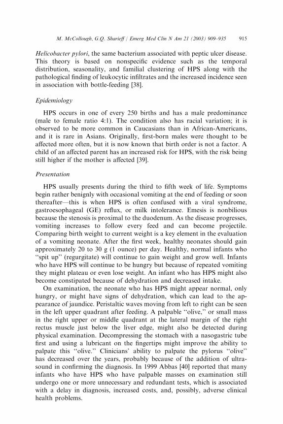
If no smallmass or ‘‘olive’’ is palpable in the right upper ormiddle quadrantof a young infant who has a clinical picture suggestive of HPS, further studiesare warranted. Ultrasound measures the thickness of the pyloric wall(normal\2.0 mm; HPS[4.0 mm) and length of the pyloric canal (normal\10.0mm;HPS[14–16mm) to diagnosisHPS (Fig. 1).Ultrasound has beenshown to have a sensitivity and specificity as high as 100% [42,43]. A false-negative result can occur if the ultrasonographer measures through the distalstomach or antrum and not through the pylorus itself. A false-positive resultmight occur if pyloric spasm is present but not pyloric stenosis.
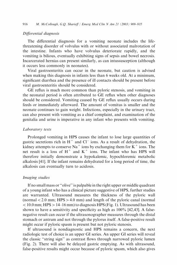
If ultrasound is nondiagnostic and HPS remains a concern, the nextradiologic test of choice is an upper GI series. An upper GI series will revealthe classic ‘‘string sign’’ as contrast flows through narrowed pyloric lumen(Fig. 2). There will also be delayed gastric emptying. As with ultrasound,false-positive results might occur because of pyloric spasm, which also gives
917M. McCollough, G.Q. Sharieff / Emerg Med Clin N Am 21 (2003) 909–935
the appearance of a string sign. Endoscopy can also be used to diagnoseHPS, but it is not used commonly [44].
Management
When the diagnosis of HPS has been made, admission to the hospital isindicated. These infants are often dehydrated, so hydration and correction ofany electrolyte abnormalities should be started in the ED. The surgicalprocedure required to correct the stenosis is the Ramstedt procedure, whichinvolves incising and separating the hypertrophic muscle fibers of the pylorus.
In Japan, intravenous atropine to decrease the spasm of the pylorus hasbeen used as an alternative to surgery. Atropine is then administered orallyfor several weeks until the child ‘‘outgrows’’ the stenosis. Surgery has beenavoided in many cases [45]; however, surgical treatment remains thestandard treatment in the United States.
Malrotation with midgut volvulus
Pathophysiology
Congenital malrotation of the midgut portion of the intestine is often thecause of volvulus in the neonatal period. Malrotation occurs during the fifth
Fig. 1. Sonogram of infant presenting with vomiting outlines enlarged pylorus and thickened
muscle (arrows) typical of pyloric stenosis. (From Heller RM, Hernanz-Schulman M.
Applications of new imaging modalities to the evaluation of common pediatric conditions.
J Pediatr 1999;135(5):632–9; with permission.)
918 M. McCollough, G.Q. Sharieff / Emerg Med Clin N Am 21 (2003) 909–935
to eighth week in embryonic life when the intestine projects out of theabdominal cavity, rotates 270�, then returns into the abdomen. If therotation is not correct, the intestine will not be ‘‘fixed down’’ correctly atthe mesentery. The vascular mesentery will appear to be more ‘‘stalk-like’’ inits structure and is at risk for later twisting, called volvulus. Volvulus is thetwisting of a loop of bowel about its mesenteric base ‘‘stalk’’ attachment.Ischemia subsequently develops, which constitutes a true surgical emergencybecause bowel necrosis can occur within hours. The entire small bowel is atrisk for ischemia and necrosis.
Epidemiology
The incidence of volvulus peaks during the first month of life but canpresent anytime in childhood. The male to female ratio is 2:1, and thedisorder is rarely familial. The exact frequency of midgut malrotation is notknown because it is frequently asymptomatic.
Congenital adhesions (Ladd’s bands), which extend from the cecum tothe liver, are associated with congenital malrotation, which can causeexternal compression of the duodenum and obstruction. This condition isnot generally considered to be a surgical emergency, but it eventuallyrequires surgical intervention to lyse the bands.
Presentation
Volvulus can present is one of three ways: (1) sudden onset of biliousvomiting and abdominal pain in a neonate, (2) history of feeding problemswith bilious vomiting that appears like a bowel obstruction, and, lesscommonly, (3) failure to thrive with severe feeding intolerance [46]. Biliousvomiting in a neonate is always worrisome and is a surgical emergency untilproven otherwise. If the bowel is already ischemic or necrotic, the neonatemight present pale and grunting. The abdomen might or might not be
Fig. 2. Upper GI series demonstrating the ‘‘string sign’’ in pyloric stenosis. (From Juhl JH,
Crummy AB, Kuhlman JE. Essentials of radiologic imaging. Lippincott–Raven Publishers;
1998; with permission.)

919M. McCollough, G.Q. Sharieff / Emerg Med Clin N Am 21 (2003) 909–935
distended depending upon location of the volvulus. If the obstruction isproximal, there might be no distension. The abdominal wall might appearblue if the bowel is already ischemic or necrotic. The pain is constant, notintermittent, and the neonate will appear irritable. Jaundice might also bepresent. Hematochezia is a late sign that indicates intestinal necrosis.Neonates who have volvulus will gradually deteriorate if the bowel remainsischemic.
Differential diagnosis
Bilious vomiting in a neonate is considered to be a surgical emergencyuntil proven otherwise; however, in early presentations of volvulus, vomituscan be nonbilious, and a misdiagnosis of acute gastroenteritis might result.As mentioned previously, the diagnosis of acute gastroenteritis should bemade cautiously in young infants. In pyloric stenosis, vomitus is alwaysnonbilious. The duration of symptoms with pyloric stenosis is usuallylonger, and the child usually appears well, although possibly dehydrated andhungry. Incarcerated hernias might also present with bilious vomiting. It istherefore imperative to examine a vomiting neonate thoroughly for signs ofa hernia. A more rare cause of bilious vomiting is duodenal or ileal atresia,although this is typically discovered in the newborn nursery or soon after.With intestinal atresia, the neonate will not be as ill-appearing as withvolvulus. Necrotizing enterocolitis can also rarely appear in term neonates.Intestinal hematomas can occur in cases of child abuse.
Congenital adrenal hyperplasia (CAH) can cause bilious vomitingwithout anatomical obstruction, and it can present in the first few weeksof life. CAH results in adrenal insufficiency with decreased cortisol levelsand salt wasting. Infants will present with hypotension and electrolyteimbalance (low Na+, high K+). It is more likely that CAH will be seen inmale infants in the ED. Female newborns who have this condition are lesscommonly missed in the newborn nursery because the accumulation ofandrogenic compounds affects the external genitalia to a greater extent.
Hirschsprung’s disease, or congenital intestinal aganglionosis, might alsopresent with bilious vomiting. In this condition there should be a history ofdecreased stool output since birth.
Laboratory tests
Laboratory tests are nonspecific for volvulus. Typically, blood tests willshow signs of dehydration and acidosis.
Diagnostic imaging
The classic finding on abdominal plain films is the ‘‘double bubble sign,’’which shows a paucity of gas (airless abdomen) with two air bubbles—onein the stomach and one in the duodenum. Other findings might include
920 M. McCollough, G.Q. Sharieff / Emerg Med Clin N Am 21 (2003) 909–935
air–fluid levels, a paucity of gas distally, or dilated loops over the livershadow. Plain film can also be entirely normal.
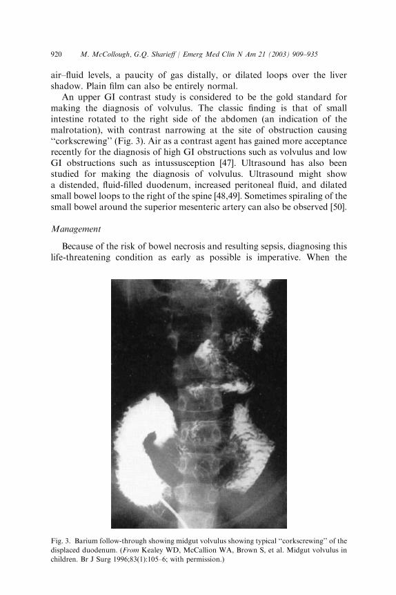
An upper GI contrast study is considered to be the gold standard formaking the diagnosis of volvulus. The classic finding is that of smallintestine rotated to the right side of the abdomen (an indication of themalrotation), with contrast narrowing at the site of obstruction causing‘‘corkscrewing’’ (Fig. 3). Air as a contrast agent has gained more acceptancerecently for the diagnosis of high GI obstructions such as volvulus and lowGI obstructions such as intussusception [47]. Ultrasound has also beenstudied for making the diagnosis of volvulus. Ultrasound might showa distended, fluid-filled duodenum, increased peritoneal fluid, and dilatedsmall bowel loops to the right of the spine [48,49]. Sometimes spiraling of thesmall bowel around the superior mesenteric artery can also be observed [50].
Management
Because of the risk of bowel necrosis and resulting sepsis, diagnosing thislife-threatening condition as early as possible is imperative. When the
Fig. 3. Barium follow-through showing midgut volvulus showing typical ‘‘corkscrewing’’ of the
displaced duodenum. (From Kealey WD, McCallion WA, Brown S, et al. Midgut volvulus in
children. Br J Surg 1996;83(1):105–6; with permission.)

921M. McCollough, G.Q. Sharieff / Emerg Med Clin N Am 21 (2003) 909–935
diagnosis has been made, aggressive resuscitation of the neonate using 10mL/kg boluses as needed of normal saline and placement of a nasogastrictube should occur. Antibiotics should be administered to cover gram-positive, gram-negative, and anaerobic flora (eg, ampicillin, gentamycin,and clindamycin). Consultation with a pediatric surgeon should not bedelayed for diagnostic studies; the sooner the child is admitted to theoperating room, the lower the morbidity and mortality of this condition.Some pediatric surgeons will take an ill-appearing neonate who has biliousvomiting directly to the operating room without any additional diagnostictests.
Intussusception
Pathophysiology
Intussusception, which was first described more than 300 years ago, is theprolapse of one part of the intestine into the lumen of an immediately distaladjoining part. The most common type is ileocolic invagination. During theinvagination, the mesentery is dragged along into the distal lumen andvenous return is obstructed, which leads to edema, bleeding of the mucosa,increased pressure in the area, and eventually obstruction to arterial flow.Gangrene and perforation result.
Epidemiology
Intussusception is most frequently seen in children between the ages of3 months and 5 years, with 60% of cases occurring in the first year of life,and a peak incidence at 6 to 11 months of age. This disorder has a malepredominance and was once believed to occur more often in the spring andautumn, but recent reports do not support this assertion [51,52]. Although itis usually idiopathic in the younger age groups, children older than 5 yearsoften have a pathologic ‘‘lead point’’ for intussusception such as polyps,lymphoma, Meckel’s diverticulum, or Henoch-Schonlein purpura andrequire a workup to determine the underlying etiology.
Presentation
The classic triad of intermittent colicky abdominal pain, vomiting, andbloody mucous stools is encountered only 20% to 40% of the time. At leasttwo of these findings will be present in about 60% of patients. The vomit isnot necessarily bilious because the level of obstruction is low, in the ileocecalarea. A palpable abdominal mass in the right upper or lower quadrant is anuncommon finding [53].
Abdominal pain associated with intussusception is colicky, lasting forapproximately 1 to 5 minutes at a time then abating for 5 to 20 minutes.During episodes of pain, the child cries and might draw the knees upward
922 M. McCollough, G.Q. Sharieff / Emerg Med Clin N Am 21 (2003) 909–935
toward the chest. Although the child often looks better between episodes, heor she still usually appears ill, quiet, or exhausted. Gradually, irritabilityincreases and vomiting becomes more frequent and sometimes bilious. Fevercan also develop as the child deteriorates.
If the ED staff does not witness a colicky episode, the EP should ask theparents to describe or demonstrate what the child does during the episodes.Most parents of a child who has gastroenteritis do not indicate that theirchild is in pain. Parents of a child presenting with intussusception usuallybelieve that the child is in pain before or during episodes of vomiting.Intussusception can also present with lethargy, pallor, and unresponsive-ness. It is important to keep this diagnosis in mind when dealing with aninfant who has an altered mental status [54].
The abdomen might be distended and tender, but usually the painappears to be out of proportion to the physical examination. There might bean elongated mass in the right upper or lower quadrants. Any type of bloodin the stool might be caused by intussusception. Rectal examination mightreveal occult blood or frankly bloody foul-smelling stool, classicallydescribed as ‘‘currant jelly’’ [55], but frank rectal bleeding is a late andunreliable sign; its absence should not deter the EP in the pursuit of thediagnosis. It should also be noted that what appears to be blood in a child’sstool might be something else, such as red fruit punch or gelatin. Guiaictesting might prevent this error when there is some question regarding thesource of the redness.
A period of observation in the ED for the recurrence of a pain episode ishelpful in equivocal cases. Specifically noting the absence of such episodesduring ED observation is good practice and should be documented in theclinical record.
Differential diagnosis
Gastroenteritis typically presents with more diarrhea than intussuscep-tion, and the child usually has ill contacts. The presence of any degree ofblood in the stool should also raise suspicion for a more serious condition.Bleeding from a Meckel’s diverticulum is usually painless unless thediverticulum becomes inflamed.
An incarcerated hernia or testicular or ovarian torsion might also presentwith sudden abdominal pain and vomiting. Inspection of the genitalia,especially in males, is vital. With torsion, the rectal examination does notshow occult or frank blood. Renal colic presenting with pain and vomiting isgenerally not seen in young children.
Laboratory tests
No laboratory test rules in or rules out the diagnosis of intussusceptionreliably. If the bowel has become ischemic or necrotic, acidosis might bepresent.

923M. McCollough, G.Q. Sharieff / Emerg Med Clin N Am 21 (2003) 909–935
Diagnostic imaging
Plain abdominal films are neither sensitive nor specific for intussuscep-tion [56,57]. Plain films might initially be normal. As the disease progresses,a variety of abnormalities can be seen, including a visible abdominal mass,abnormal distribution of gas and fecal contents, air–fluid levels, and dilatedloops of small intestine. A ‘‘target sign’’ on plain film consists of concentriccircles of fat density, similar in appearance to a doughnut, visualized to theright of the spine. This sign is caused by layers of peritoneal fat surroundingand within the intussusception alternating with layers of mucosa andmuscle. Less commonly, the soft tissue mass of the intussusception (leadingedge) can be seen projecting into the colon. Large areas of gas with the headof the intussuscepted bowel might take the shape of a crescent, althoughother patterns might be seen.
Ultrasound is used in some institutions for the diagnosis of intussuscep-tion and to confirm reduction after treatment [58]. Sonographic findings inintussusception include the ‘‘target’’ sign, a single, hypoechoic ring witha hyperechoic center and the ‘‘pseudokidney’’ sign, superimposed hypo- andhyperechoic areas representing the edematous walls of the intussusceptum,and layers of compressed mucosa. Doppler flow can be used to identifybowel ischemia. If signs of intussusception are not identified by ultrasoundin patients in whom the diagnosis is suspected clinically, proceeding witha barium or air enema should be still be considered.
Management
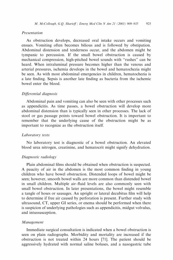
The main focus in management of a child who has intussusception isemergent reduction of the obstructed bowel. Classically, this is accom-plished by a barium enema, which acts as a diagnostic and therapeuticradiologic study (Fig. 4). The barium enema has been the gold standard fordiagnosis and treatment of intussusception for decades [59]. Saline enemashave also been employed successfully [60,61], and newer modalities such asair enemas and ultrasound-guided enemas have emerged.
Many centers in the United States are moving toward air enemas [62–67].This modality was first introduced to the Western world at the AmericanPediatric Surgical Association in 1985 with the presentation of a series of6396 successfully treated patients [68]. Air enemas offer several advantagesover barium. They are also easier to administer, and in most studies they havea higher rate of successful reduction. Air enemas using fluoroscopic guidancedeliver much less radiation than barium studies, and there is no exposure ifultrasound guidance is used. Limiting radiation exposure is important toconsider when dealing with infants and their susceptible reproductive organs.Moreover, if a perforation occurs during these investigations, air is much lessdangerous to the peritoneum and abdominal contents than barium.
Visualization of the entire colon to the terminal ileum is mandatory toexclude ileocolic intussusception. Ileo–ileo intussusception can be much
924 M. McCollough, G.Q. Sharieff / Emerg Med Clin N Am 21 (2003) 909–935
harder to diagnose and to reduce. Spontaneous reduction of intussusceptedbowel has been reported, although therapeutic intervention should not bedelayed in a patient who has significant symptoms [69].
Not every child who has intussusception should undergo bowel reductionby enema. Clinical signs of peritonitis, perforation, or hypovolemic shockare clear contraindications to enemas. These signs mandate surgicalexploration. Relative contraindications to enemas include prolongedsymptoms ([ 24 h), evidence of obstruction (eg, air–fluid levels on plainabdominal films), and ultrasound findings of intestinal ischemia or trappedfluid.
Even in well-selected patients, enemas can cause reduction of necroticbowel, perforation, and sepsis. After a successful reduction the child shouldbe admitted for observation. A small percentage of patients (0.5–15%) willhave a recurrence of the intussusception, usually within 24 hours, butsometimes after days or weeks. Even after reduction by laparotomy, therecurrence rate is 2% to 5% [52].
Small bowel obstruction
Pathophysiology
Small bowel obstruction can result from intrinsic, extrinsic, or intra-luminal disease. Although the most common causes of small bowelobstruction are adhesions from previous abdominal surgery and incarcer-ation of a hernia [70], intussusception, appendicitis, Meckel’s diverticulum,and malrotation with midgut volvulus and tumors should be considered aspossible etiologies. In addition to inguinal hernias, umbilical, obturator, andfemoral canal hernias can also lead to small bowel obstruction [56].
Fig. 4. Barium enema in ileocolic intussusception. The intussusceptum is visible in the
ascending colon (arrows). (From Lazar J. Greenfield surgery: scientific principles and practice.
Baltimore (MD): Lippincott Williams & Wilkins; 2001; with permission.)
925M. McCollough, G.Q. Sharieff / Emerg Med Clin N Am 21 (2003) 909–935
Presentation
As obstruction develops, decreased oral intake occurs and vomitingensues. Vomiting often becomes bilious and is followed by obstipation.Abdominal distension and tenderness occur, and the abdomen might betympanic to percussion. If the small bowel obstruction is caused bymechanical compression, high-pitched bowel sounds with ‘‘rushes’’ can beheard. When intraluminal pressure becomes higher than the venous andarterial pressures, ischemia develops in the bowel and hematochezia mightbe seen. As with most abdominal emergencies in children, hematochezia isa late finding. Sepsis is another late finding as bacteria from the ischemicbowel enter the blood.
Differential diagnosis
Abdominal pain and vomiting can also be seen with other processes suchas appendicitis. As time passes, a bowel obstruction will develop moreabdominal distension than is typically seen in other processes. The lack ofstool or gas passage points toward bowel obstruction. It is important toremember that the underlying cause of the obstruction might be asimportant to recognize as the obstruction itself.
Laboratory tests
No laboratory test is diagnostic of a bowel obstruction. An elevatedblood urea nitrogen, creatinine, and hematocrit might signify dehydration.
Diagnostic radiology
Plain abdominal films should be obtained when obstruction is suspected.A paucity of air in the abdomen is the most common finding in youngchildren who have bowel obstruction. Distended loops of bowel might beseen; however, smooth bowel walls are more common than distended bowelin small children. Multiple air–fluid levels are also commonly seen withsmall bowel obstruction. In later presentations, the bowel might resemblea tangle of hoses or sausages. An upright or lateral decubitus film will helpto determine if free air caused by perforation is present. Further study withultrasound, CT, upper GI series, or enema should be performed when thereis suspicion of underlying pathologies such as appendicitis, midgut volvulus,and intussusception.
Management
Immediate surgical consultation is indicated when a bowel obstruction isseen on plain radiographs. Morbidity and mortality are increased if theobstruction is not treated within 24 hours [71]. The patient should beaggressively hydrated with normal saline boluses, and a nasogastric tube
926 M. McCollough, G.Q. Sharieff / Emerg Med Clin N Am 21 (2003) 909–935
should be placed for gastric decompression. Broad-spectrum antibiotics areindicated, particularly if peritonitis is suspected.
Necrotizing enterocolitis
Epidemiology
Necrotizing enterocolitis (NEC) is typically seen in the neonatal intensivecare unit (NICU), occurring in premature infants in their first few weeks oflife. Occasionally, it is encountered in the term infant, usually within the first10 days after birth. The cause of NEC is not known, but history of an anoxicepisode at birth and other neonatal stressors are associated with thediagnosis [72,73].
Pathophysiology
The pathologic finding of NEC is that of a necrotic segment of bowelwith gas accumulation in the submucosa. Necrosis can lead to perforation,sepsis, and death. The distal ileum and proximal colon are most commonlyinvolved. Clostridium spp, Escherichia coli, Staphylococcus epidermidis, andRotavirus are the most commonly recovered pathogens [72,73].
Presentation
Infants who have NEC typically present appearing quite ill, withlethargy, irritability, decreased oral intake, distended abdomen, and bloodystools. Symptoms might present fairly mildly, with only occult blood-positive stools, to a much more critically ill presentation. Although thisdiagnosis is typically made in the NICU, it nonetheless must be consideredin a term infant who experienced significant stress at birth (eg, anoxia).
Imaging studies
The plain abdominal film finding of pneumatosis intestinalis, caused bygas in the intestinal wall, is diagnostic of NEC.
Management
Management includes fluid resuscitation, bowel rest, and broad-spectrumantibiotic coverage. Early surgical consultation is imperative.
Incarcerated hernia
Epidemiology
Inguinal hernias occur in 1% to 3% of all children, more often in males(6:1), and more often on the right side (2:1). Premature infants are at higher
927M. McCollough, G.Q. Sharieff / Emerg Med Clin N Am 21 (2003) 909–935
risk for inguinal hernias, with an overall incidence of 3% to 5%. Inaddition, more than two thirds of incarcerated inguinal hernias requiringoperative reduction occur during the first year of life [74]. Umbilical herniasare also more common in premature infants, but unlike inguinal herniasumbilical hernias rarely become incarcerated and usually close spontane-ously by 2 to 3 years of age [74,75].
Other conditions that place children at increased risk for abdominalhernias include connective tissue diseases (eg, Marfan’s syndrome, Ehlers-Danlos syndrome), cystic fibrosis, and metabolic disorders (eg, mucopoly-saccharidoses). Patients who have ventriculoperitoneal shunts or patientswho are receiving peritoneal dialysis are also at increased risk [75,76].
Presentation
Hernias usually present with an asymptomatic bulge in the groin orumbilical area made more prominent with crying, straining, or laughing.The first sign of incarceration of an inguinal hernia is an abrupt onset ofirritability in the young infant. Refusal to eat soon follows, followed byvomiting, which can become bilious and sometimes feculent.
Inguinal hernias can be palpated as smooth, firm, sausage-shaped, mildlytender masses in the groin; the hernia originates proximal to inguinal ringand can extend into the scrotum. The ‘‘silk glove sign’’ occurs when theindex finger rubs over the proximal spermatic cord, and sometimes twolayers of hernial sac can be felt rubbing together. If the child is well-appearing without vomiting, fever, or redness to the inguinal area, thehernia is not likely incarcerated.
Differential diagnosis
Many conditions can mimic an inguinal hernia, but the most common isa hydrocele. A hydrocele is a result of incomplete obliteration of the processvaginalis, which allows an outpocketing of peritoneum to appear in thescrotum. This fluid-filled sac can appear anywhere from the spermatic cord tothe testis, and if large it can be transilluminated. Hydroceles can be palpatedseparate from the testes and are freely movable. A hydrocele usually appearsin the first few months of life and disappears by 1 year of age.
Diagnostic imaging
If the diagnosis is uncertain, a scrotal ultrasound can differentiate aninguinal hernia from a hydrocele.
Management
If signs of incarceration are not present, reduction should be attempted inthe ED. Reduction of the hernia can usually be accomplished by placing the
928 M. McCollough, G.Q. Sharieff / Emerg Med Clin N Am 21 (2003) 909–935
child in Trendelenberg position with ice packs to the area and administeringpain medication. If reduction is not possible or if the hernia appears to beincarcerated or ischemic, emergent surgical consultation is required.Umbilical hernias rarely become incarcerated and often close withoutsurgery.
Meckel’s diverticulum
Pathophysiology and epidemiology
Meckel’s diverticulum is the most common congenital abnormality of thesmall intestine. A Meckel’s diverticulum is a remnant of the omphalome-senteric (vitelline) duct, which normally disappears by the seventh week ofgestation. It is a true diverticulum, containing all layers of the bowel wall.Up to 60% of these diverticulae contain heterotopic gastric tissue, andheterotopic pancreatic, endometrial, and duodenal mucosa have also beenreported [77,78]. The features of Meckel’s diverticulum are commonlydescribed by ‘‘the rule of 2s’’ [79]: it is present in approximately 2% of thepopulation with only 2% of affected patients becoming symptomatic. Forty-five percent of symptomatic patients are less than 2 years of age [80]. Themost common location is 2 feet (40–100 cm) from the ileocecal valve and thediverticulum typically is 2 inches long.
Clinical presentation
The classic presentation of a Meckel’s diverticulum is that of painless orminimally painful rectal bleeding. Isolated red rectal bleeding is common,particularly in boys less than 5 years of age [81]. Such painless bleeding isa result of heterotopic gastric tissue in the diverticulum or in the adjacentileum. Abdominal pain, distension, and vomiting can occur if obstructionhas occurred, and the presentation can mimic appendicitis or diverticulitis.A Meckel’s diverticulum can also ulcerate and perforate, presenting asa bowel perforation or acting as a lead point, resulting in intussusception.
Differential diagnosis
The differential diagnosis includes painful and nonpainful conditions.Rectal bleeding associated with abdominal pain can be caused by pepticulcer disease, intussusception, and volvulus. Polyps, arteriovenous malfor-mations, and tumors can cause nonpainful rectal bleeding.
Laboratory tests
Although no laboratory test is diagnostic for a Meckel’s diverticulum,children who have GI bleeding should undergo screening laboratory testssuch as a complete blood count, coagulation profile, and a type and screen.
929M. McCollough, G.Q. Sharieff / Emerg Med Clin N Am 21 (2003) 909–935
Diagnostic radiology
Abdominal films might show signs of obstruction such as dilated loops ofbowel or a paucity of bowel gas. A Meckel’s scan involves injection ofTechnetium–pertechnetate intravenously. This test relies on the presenceof gastric mucosa in or near the diverticulum, which has an affinity for theradionucleotide. A Meckel’s scan can detect the presence of gastric mucosawithin the diverticulae with up to 85% accuracy [82]. Mesenteric arte-riography can detect the site of active bleeding if bleeding is profuse.
Management
As in any patient who has active bleeding, fluid resuscitation starting with20 mL/kg boluses of normal saline is warranted. A blood transfusion mightbe necessary with a packed red blood cell increments of 10 mL/kg. Thepatient should have nothing by mouth and a nasogastric tube should beplaced. Antibiotics must be initiated if there are peritoneal signs. Surgicalconsultation should be obtained emergently. Surgical intervention caninvolve a diverticulectomy or a more extensive small bowel segmentalresection if there is irreversible bowel ischemia.
Ovarian and testicular torsion
Epidemiology
While ovarian torsion in the ED is rare, a missed diagnosis can result insignificant patient morbidity and medicolegal consequences. Both con-ditions are more common in preadolescent and adolescent patients, but theycan occur in all age groups, even in utero.
Torsion of the testes or spermatic cord is more common than ovariantorsion, occurring in one of 4000 males, with a peak incidence at age 13 [83].Delay in diagnosis and treatment can result in a loss of spermatogenesis ornecrotic, gangrenous testes. Testicular salvage rates are time-dependent,with a 96% success rate if detorsion is performed within 4 hours of symptomonset decreasing to less than 10% at 24 hours.
Pathophysiology
Older girls can have an ovarian torsion secondary to a corpus luteal orfollicular cyst, whereas younger children might have no underlying ovarianabnormality or a mature cystic teratoma [84]. Bilateral ovarian torsion hasbeen reported [85].
The testes enter the scrotum through the inguinal canal after descentfrom the abdomen. The peritoneum invaginates through the canal andpartially covers the testis and epididymis, forming the tunica vaginalis.Typically, the tunica vaginalis attaches to the posterior wall of the
930 M. McCollough, G.Q. Sharieff / Emerg Med Clin N Am 21 (2003) 909–935
hemiscrotum and the superior pole of the testes to achieve testicularfixation. If the tunica completely covers the testis and attaches higher up onthe spermatic cord (bell clapper deformity), proper testicular fixation doesnot occur and there is a predisposition to torsion. In intravaginal torsion,the testis can rotate within the tunica vaginalis and thereby constrict arterialblood flow. Extravaginal torsion, seen more commonly in prematureneonates, can also occur antenatally. If torsion is prolonged, testicularinfarction and atrophy has been reported after 4 to 6 hours of ischemia.
Clinical presentation
Ovarian torsion typically presents with an acute onset of pain on the sideof the torsion; however, pain can radiate to the flank, back, or groin.Associated symptoms might include nausea, vomiting, constipation, urinarytract symptoms, and fever. Pelvic examination in older girls will revealadnexal tenderness with a mass. Cervical motion tenderness might also bepresent. In younger children, when a pelvic examination is not possible,ultrasound will be diagnostic.
Testicular torsion can present as an acutely tender and swollenhemiscrotum, but it might present more subtly, especially in younger boys,underscoring the importance of the genital examination in the evaluation ofpediatric abdominal pain. Although in younger boys the only present-ing symptoms might be abdominal pain, vomiting, and a low-grade fever,examination will reveal scrotal tenderness and swelling with an elevatedtestis. The loss of the cremasteric reflex is another important sign; it wasnotably absent in 100% of the patients who had torsion in one series [86].Torsion should also be considered in boys who have an undescended testis.
Differential diagnosis
Other disease processes that can present with sudden abdominal pain andvomiting include intussusception and renal colic. In girls, ectopic pregnancyand ruptured corpus luteal cyst or tubo-ovarian abscess must also beconsidered.
Laboratory tests
Leukocytosis is present in up to 50% of patients, and UA is typicallynormal.
Diagnostic radiology
Diagnostic studies might be appropriate in certain cases, but they shouldnever delay surgical consultation, especially in patients who present with lessthan 12 hours of symptoms. A suggestive history and physical examinationare all that are necessary to prompt emergent surgical exploration.
931M. McCollough, G.Q. Sharieff / Emerg Med Clin N Am 21 (2003) 909–935
Color-flow Doppler ultrasound has a sensitivity of 82% to 86% witha specificity of almost 100% for testicular torsion. Sensitivity and specificityare lower for ovarian torsion. Scintigraphy for testicular torsion hasa sensitivity ranging from 80% to 100% and a specificity of 89% to 100%[87]. Decreased blood flow to the affected gonad is considered to bediagnostic of torsion.
Because torsion can be intermittent, diagnostic tests might be negative atthe time of examination. If suspicion for the diagnosis still exists,consultation with a urologist or gynecologist and admission to the hospitalis recommended.
Management
If ovarian torsion is suspected or confirmed on ultrasound with Dopplerflow studies [88], laparoscopy should be performed to attempt ovariansalvage. Unfortunately, ovarian salvage is rare, even when the intervalbetween diagnosis and operative intervention is short [89], which might becaused by a delay in the initial diagnosis. Symptom onset has varied from 6hours to 3 weeks before ultimate diagnosis [90,91] Although most torsedovaries are necrotic on exploration, there have been reports of ovariansalvage after ovarian detorsion and oophoropexy [92].
If a male patient presents within 12 hours of symptoms, immediatesurgical exploration is indicated. While awaiting surgery, an attempt atmanual detorsion in the ED is appropriate, which is accomplished byrotating the testis in an ‘‘open-book’’ fashion from medial to lateral afteradministration of adequate analgesia. Orchidopexy of both testes is usuallyperformed after detorsion to avoid recurrence; approximately 40% ofpatients have a bellclapper deformity of the contralateral testis.
Summary
Surgical emergencies can be missed easily in children, who are not alwaysable to volunteer relevant information. Awareness of the entities discussedin this review might help the EP uncover subtle clues to early diagnoses thatmight not be initially apparent.
Ill-appearing children who have abdominal pain and vomiting shouldbe considered to have ischemic or necrotic bowel until proven other-wise. Possible diagnoses include volvulus, intussusception, and necrotizingenterocolitis. Bilious vomiting, especially in a young infant, should beconsidered to be an indication of a high bowel obstruction such as midgutvolvulus, which warrants immediate surgical consultation. Significant rectalbleeding with abdominal pain can result from intussusception, volvulus, oran inflamed Meckel’s diverticulum. Rectal bleeding with unstable vital signscan result from an upper GI bleed (eg, peptic ulcer disease). Painless rectalbleeding can result from a Meckel’s diverticulum, polyps, arteriovenous
932 M. McCollough, G.Q. Sharieff / Emerg Med Clin N Am 21 (2003) 909–935
malformation, or a tumor. Examination of the genitalia is imperative,especially in boys, to exclude the possibility of an incarcerated hernia ortesticular torsion.
References
[1] Kim M, Strait RT, Sato TT, et al. A randomized clinical trial of analgesia in children with
acute abdominal pain. Acad Emerg Med 2002;9(4):281–7.
[2] Wagner JM, McKinner WP, Carpenter JL. Does this patient have appendicitis? JAMA
1996;276(19):1589–94.
[3] Williams N, Bello M. Perforation rates relates to delayed presentation in childhood acute
appendicitis. J Royal College Surg Edinburgh 1998;43(2):101–2.
[4] Saidi RF, Ghasemi M. Role of Alvarado score in diagnosis and treatment of suspected
acute appendicitis. Amer J Emerg Med 2000;18(2):230–1.
[5] Horwitz JR, Gursoy M, Jaksic T, et al. Importance of diarrhea as a presenting symptom of
appendicitis in very young children. Amer J Surgery 1997;173(2):80–2.
[6] Scholer SJ, Pituch K, Orr DP, et al. Use of the rectal examination on children with acute
abdominal pain. Clin Pediatr [Phila] 1998;37(5):311–6.
[7] Rothrock SG, Pagane J. Acute appendicitis in children: emergency department diagnosis
and management. Ann Emerg Med 2000;36(1):39–51.
[8] Emery M, Jones J, Brown M. Clinical application of infrared thermography in the
diagnosis of appendicitis. Am J Emerg Med 1994;42(1):48–50.
[9] Reynolds SL. Missed appendicitis in a pediatric emergency department. Pediatr Emerg
Care 1993;9(1):1–3.
[10] Coleman C, Thompson JE, Bennion RS, et al. White blood cell count is poor predictor of
severity of disease in the diagnosis of appendicitis. Amer Surgeon 1998;64(10):983–5.
[11] Chung JL, Kong MS, Lin SL, et al. Diagnostic value of C-reactive protein in children with
perforated appendicitis. European J Peds 1996;155(7):529–31.
[12] Paajanen H, Mansikka A, Laato M, et al. Are serum inflammatory markers age dependent
in acute appendicitis? Amer College Surgeons J 1997;184(3):303–8.
[13] Hallan S, Asberg A, Edna TH. Additional value of biochemical tests in suspected acute
appendicitis. Eur J Surgery 1997;163(7):533–8.
[14] Andersson RE, Hugander AP, Ghazi SH, et al. Diagnostic value of disease history, clinical
presentation, and inflammatory parameters in appendicitis. World J Surgery 1999;23(2):
133–40.
[15] Albiston E. The role of radiological imaging in the diagnosis of acute appendicitis. Can J
Gastroenterol 2002;16(7):451–63.
[16] Orr RK, Porter D, Hartman D. Ultrasonography to evaluate adults for appendicitis:
decision making based on meta-analysis and probabilistic reasoning. Acad Emerg Med
1995;2(7):644–50.
[17] Wong ML, Casey SO, Leonidas JC, et al. Sonographic diagnosis of acute appendicitis in
children. Pediatr Surg J 1994;29(10):1356–60.
[18] Ramachandran P, Sivit CJ, Newman KD, et al. Ultrasonography as an adjunct in the
diagnosis of acute appendicitis: a 4-year experience. J Pediatr Surg 1996;31(1):164–7
[discussion 167–9].
[19] Crady SK, Jones JS, Wyn T, Luttenton CR. Clinical validity of ultrasound in children with
suspected appendicitis. Ann Emerg Med 1993;22(7):1125–9.
[20] Zaki AM, MacMahon RA, Gray AR. Acute appendicitis in children: when does
ultrasound help? Aust N Z J Surg 1994;64(10):695–8.
[21] Hahn HB, Hoepner FU, Kalle T, et al. Sonography of acute appendicitis in children: 7
years experience. Pediatr Radiol 1998;28(3):147–51.
933M. McCollough, G.Q. Sharieff / Emerg Med Clin N Am 21 (2003) 909–935
[22] Carrico CW, Fenton LZ, Taylor GA, et al. Impact of sonography on the diagnosis and
treatment of acute lower abdominal pain in children and young adults. Am AJR
Roentgenol J 1999;172(2):513–6.
[23] Lessin MS, Chan M, Catallozzi M, et al. Selective use of ultrasonography for acute
appendicitis in children. Am J Surg 1999;177(3):193–6.
[24] Rice HE, Arbesman M, Martin DJ, et al. Does early ultrasonography affect management
of pediatric appendicitis? A prospective analysis. Pediatr Surg J 1999;34(5):754–8.
[25] Rice HE, Arbesman M, Martin DJ, et al. Does early ultrasonography affect management
of pediatric appendicitis? A prospective analysis. J Pediatr Surg 1999;34(5):754–8
[discussion 758–9].
[26] Roosevelt GE, Reynolds SL. Does the use of ultrasonography improve the outcome of
children with appendicitis? Acad Emerg Med 1998;5(11):1071–5.
[27] Quillin SP, Siegel MJ. Diagnosis of appendiceal abscess in children with acute appendicitis:
value of color Doppler sonography. AJR Am J Roentgenol 1995;164(5):1251–4.
[28] Funaki B, Grosskreutz SR, Funaki CN. Using unenhanced helical CT with enteric contrast
material for suspected appendicitis in patients treated at a community hospital. AJR Am J
Roentgenol 1998;171(4):997–1001.
[29] Garcia Pena BM, Mandl KD, Kraus SJ, et al. Ultrasonography and limited computed
tomography in the diagnosis and management of appendicitis in children. JAMA 1999;
282(11):1041–6.
[30] Pena BM, Taylor GA, Lund DP, et al. Effect of computed tomography on patient
management and costs in children with suspected appendicitis. Pediatrics 1999;104(3 Pt
1):440–6.
[31] Rao PM, Rhea JT, Rattner DW. Introduction of appendiceal CT: impact on negative
appendectomy and appendiceal perforation rates. Ann Surg 1999;229(3):344–9.
[32] Jabra AA, Shalaby-Rana EI, Fishman EK. CT of appendicitis in children. J Comput Assist
Tomogr 1997;21(4):661–6.
[33] Kanegaye JT, Vance CW, Parisi M, et al. Failure of technetium-99m hexamethylpropylene
amine oxime leukocyte scintigraphy in the evaluation of children with suspected
appendicitis. Ped Emerg Care 1995;11(5):285–90.
[34] Turan C, Tutus A, Ozokutan BH, et al. The evaluation of technetium 99m-citrate
scintigraphy in children with suspected appendicitis. J Pediatr Surg 1999;34(8):1272–5.
[35] Hormann M, Paya K, Eibenberger K, et al. MR imaging in children with nonperforated
acute appendicitis: value of unenhanced MR imaging in sonographically selected cases. Am
AJR Roentgenol J 1998;171(2):467–70.
[36] Banani SA, Talei A. Can oral metronidazole substitute parenteral drug therapy in acute
appendicitis? A new policy in the management of simple or complicated appendicitis with
localized peritonitis: a randomized controlled clinical trial. Am Surg 1999;65(5):411–6.
[37] Soderquist-Elinder C, Hirsch K, Bergdahl S, et al. Prophylactic antibiotics in un-
complicated appendicitis during childhood—a prospective randomised study. Eur J Pediatr
Surg 1995;5(5):282–5.
[38] Paulozzi LJ. Is Helicobacter pylori a cause of infantile hypertrophic pyloric stenosis? Med
Hypotheses 2000;55(2):119–25.
[39] Spicer RD. Infantile hypertrophic pyloric stenosis: a review. Br J Surg 1982;69(3):
128–35.
[40] Abbas AE, Weiss SM, Alvear DT. Infantile hypertrophic pyloric stenosis: delays in
diagnosis and overutilization of imaging modalities. Am Surg 1999;65(1):73–6.
[41] Smith GA, Mihalov L, Shields BJ. Diagnostic aids in the differentiation of pyloric stenosis
from severe gastroesophageal reflux during early infancy: the utility of serum bicarbonate
and serum chloride. Am J Emerg Med 1999;17(1):28–31.
[42] Hernanz-Schulman M, Sells LL, Ambrosino MM, et al. Hypertrophic pyloric stenosis in
the infant without a palpable olive: accuracy of sonographic diagnosis. Radiology
1994;193(3):771–6.
934 M. McCollough, G.Q. Sharieff / Emerg Med Clin N Am 21 (2003) 909–935
[43] Neilson D, Hollman AS. The ultrasonic diagnosis of infantile hypertrophic pyloric stenosis:
technique and accuracy. Clin Radiol 1994;49(4):246–7.
[44] De Backer A, Bove T, Vandenplas Y, et al. Contribution of endoscopy to early diagnosis of
hypertrophic pyloric stenosis. Pediatr Gastroenterol Nutr J 1994;18(1):78–81.
[45] Yamamoto A, Kino M, Sasaki T, et al. Ultrasonographic follow-up of the healing process
of medically treated hypertrophic pyloric stenosis. Pediatr Radiol 1998;28(3):177–8.
[46] Lin JN, Lou CC, Wang KL. Intestinal malrotation and midgut volvulus: a 15-year review.
J Formos Med Assoc 1995;94(4):178–81.
[47] Harrison RL, Set P, Brain AJ. Persistent value of air-augmented radiograph in neonatal
high gastrointestinal obstruction, despite more modern techniques. Acta Paediatr 1999;
88(11):1284–6.
[48] Shimanuki Y, Aihara T, Takano H, et al. Clockwise whirlpool sign at color Doppler US:
an objective and definite sign of midgut volvulus. Radiology 1996;199(1):261–4.
[49] Weinberger E, Winters WD, Liddell RM, et al. Sonographic diagnosis of intestinal
malrotation in infants: importance of the relative positions of the superior mesenteric vein
and artery. Am AJR Roentgenol J 1992;159(4):825–8.
[50] Zerin JM, DiPietro MA. Superior mesenteric vascular anatomy at ultrasound in patients
with surgically proved malrotation of the midgut. Radiology 1992;183(3):693–4.
[51] Parashar UD, Holman RC, Cummings KD, et al. Trends in intussusception-associated
hospitalizations and deaths among US infants. Pediatrics 2000;106(6):1413–21.
[52] Ugwu BT, Legbo JN, Dakum NK, et al. Childhood intussusception: a 9-year review. Ann
Trop Paediatr 2000;20(2):131–5.
[53] Kuppermann N, O’Dea T, Pinckney L, et al. Predictors of intussusception in young
children. Arch Pediatr Adolesc Med 2000;154(3):250–5.
[54] Heldrich FJ. Lethargy as a presenting symptom in patients with intussusception. Clin
Pediatr 1986;25(7):363–5.
[55] Yamamoto LG, Morita SY, Boychuk R, et al. Stool appearance in intussusception:
assessing the value of the term ‘‘currant jelly.’’ Am J Emerg Med 1997;15(3):293–8.
[56] Smith DS, Bonadio WA, Losek JD, et al. The role of abdominal x-rays in the diagnosis and
management of intussusception. Pediatr Emerg Care 1992;8(6):325–7.
[57] Yang ST, Tsai CH, Chen JA. Differential diagnosis between intussusception and
gastroenteritis by plain film. Zhonghua Min Guo Xiao Er Ke Yi Xue Hui Za Zhi
1995;36(3):170–5.
[58] Bhisitkul DM, Listernick R, Shkolnik A, et al. Clinical application of ultrasonography in
the diagnosis of intussusception. Pediatr J 1992;121(2):182–6.
[59] Campbell JB. Contrast media in intussusception. Pediatr Radiol 1989;19(5):293–6.
[60] Gonzalez-Spinola J, Del Pozo G, Tejedor D, et al. Intussusception: the accuracy of
ultrasound-guided saline enema and the usefulness of a delayed attempt at reduction.
Pediatr Surg J 1999;34(6):1016–20.
[61] Rohrschneider WK, Troger J. Hydrostatic reduction of intussusception under US
guidance. Pediatr Radiol 1995;25(7):530–4.
[62] Wang G, Liu XG, Zitsman JL. Nonfluoroscopic reduction of intussusception by air enema.
World J Surg 1995;19(3):435–8.
[63] Daneman A, Alton DJ, Ein S, et al. Perforation during attempted intussusception
reduction in children—a comparison of perforation with barium and air. Pediatr Radiol
1995;25(2):81–8.
[64] Gu L, Alton DJ, Daneman A, et al. John Caffey Award. Intussusception reduction in
children by rectal insufflation of air. Am AJR Roentgenol J 1988;150(6):1345–8.
[65] Schmit P, Rohrschneider WK, Christmann D. Intestinal intussusception survey
about diagnostic and nonsurgical therapeutic procedures. Pediatr Radiol 1999;29(10):
752–61.
[66] Sandler AD, Ein SH, Connolly B. Unsuccessful air-enema reduction of intussusception: is
a second attempt worthwhile? Pediatr Surg Int 1999;15(3–4):214–6.

935M. McCollough, G.Q. Sharieff / Emerg Med Clin N Am 21 (2003) 909–935
[67] Lui KW, Wong HF, Cheung YC, et al. Air enema for diagnosis and reduction of
intussusception in children: clinical experience and fluoroscopy time correlation. J Pediatr
Surg 2001;36(3):479–81.
[68] Guo JZ, Ma XY, Zhou QH. Results of air pressure enema reduction of intussusception:
6,396 cases in 13 years. J Pediatr Surg 1986;21(12):1201–3.
[69] Kornecki A, Daneman A, Navarro O, et al. Spontaneous reduction of intussusception:
clinical spectrum, management and outcome. Pediatr Radiol 2000;30(1):58–63.
[70] Vicario S, Price T. Intestinal obstruction. In: Tintinelli J, Kelen G, Stapczynski J, editors.
Emergency medicine. A comprehensive study guide. New York: McGraw-Hill; 2000.
[71] Brolin RE, Krasna MJ, Mast B. Use of tubes and radiographs in bowel obstruction. Ann
Surg 1987;206:126.
[72] Kliegman R, Walsh M. Neonatal necrotizing enterocolitis. Pathogenesis, classification, and
spectrum of illness. Curr Prob Pediatr 1987;27:215.
[73] Kulkarni A, Vigneswaran R. Necrotizing enterocolitis. Indian J Pediatr 2001;68(9):847–53.
[74] Sato TT, Oldham KT. Pediatric abdomen. In: Lazar J, editor. Surgery: scientific principles
and practice. 3rd edition. Greenfield: Lippincott Williams & Wilkins; 2001.
[75] Kapur P, Caty MG, Glick PL. Pediatric hernias and hydroceles. Pediatr Clin N Am
1998;45(4):773–89.
[76] Grosfeld JL. Current concepts in inguinal hernia in infants and children. World J Surg
1989;13(5):506–15.
[77] Kusamoto H, Yoshida M, Takahashi I, et al. Complications and diagnosis of Meckel’s
diverticulum in 776 patients. Am J Surgery 1992;164:382.
[78] Murali VP, Divaker D, Thachil MV, et al. Meckel’s diverticulum in adults. J Indian Med
Assoc 1989;87:116.
[79] Trier JS, Winter HS. Anatomy, embryology and developmental abnormalities of the small
intestine and colon. In: Sleisenger MH, Fordtran, editors. Gastrointestinal disease:
pathophysilogy, diagnosis, management. Philadephia: WB Saunders; 1989.
[80] Amoury RA. Meckel’s dieverticulum. In: Welch KJ, et al, editors. Pediatric surgery.
Chicago: Mosby; 1986.
[81] Hostetler M, Bracikowski A. Pediatric gastrointestinal disorders. In: Marx J, Hockberger
R, Walls R, editors. Rosen’s emergency medicine concepts and clinical practice. 5th
edition. St. Louis (MO): Mosby; 2002.
[82] St-Vil D, Brandt ML, Panic S, et al. Meckel’s diverticulum in children: a 20 year review.
J Pediatr Surg 1991;26(11):1289–92.
[83] Knight PJ, Vassy LE. The diagnosis and treatment of the acute scrotum in children and
adolescents. Ann Surg 1984;200:664–73.
[84] Kokoska ER, Keller MS, Weber TR. Acute ovarian torsion in children. Am J Surg
2000;180:452–65.
[85] Dunnihoo DR, Wolff J. Bilateral torsion of the adnexa: a case report and a review of the
world literature. Obstet Gynecol 1985;64:55S–9S.
[86] Rabinowitz R. The importance of the cremasteric reflex in acute scrotal swelling in
children. J Urol 1984;132(1):89–90.
[87] Paltiel HJ. Acute scrotal symptoms in boys with an indeterminate clinical presentation:
comparison of color Doppler sonography and scintigraphy. Radiology 1998;207:223–31.
[88] Davis LG, Gerscovich EO, Anderson MW, et al. Ultrasound and Doppler in the diagnosis
of ovarian torsion. Eur J Radiol 1995;20:133–6.
[89] HouryD,Abbott JT. Ovarian torsion: a fifteen-year review. AnnEmergMed 2001;38:156–9.
[90] Mordehai J, Mares AJ, Barki Y, et al. Torsion of uterine adnexa in neonates and children:
a report of 20 cases. J Pediatr Surg 1991;26:1195–9.
[91] Kienstra AM, Ward M. Three year old with ovarian torsion. J Emerg Med 2002;
23(7):375–7.
[92] Templeman C, Hertweck SP, Fallat ME. The clinical course of unresected ovarian torsion.
J Pediatr Surg 2000;35:624–6.


![Anaesthesia for abdominal emergencies in children - · PDF file · 2017-08-28ANAESTHESIA FOR ABDOMINAL EMERGENCIES IN CHILDREN* T. ]. MCCAUGHEY, ... as in 'an intussusception 6f several](https://img.pdfslide.net/doc/110x75/5aabe8157f8b9ac55c8c7185/anaesthesia-for-abdominal-emergencies-in-children-2017-08-28anaesthesia.jpg)