Embed Size (px)
Citation preview
1
UNIVERSITY OF MEDICINE AND PHARMACY OF CRAIOVA
Ph.D SCHOOL
ABSTRACT OF Ph.D PAPER ENTITLED:
CUTANEOUS MANIFESTATIONS
INDUCED BY STREPTOCOCCAL TONSILLAR
FOCAL INFECTION
SCIENTIFIC LEADERSHIP:
PROFESSOR DOCTOR. ELENA IONIŢĂ
Ph.D STUDENT:
CRISTINA TUTUNARU
CRAIOVA
2014

2
Contents
Introduction
Part I – GENERAL CONSIDERATIONS
CHAPTER I IMPLICATIONS OF FOCAL INFECTION IN CUTANEOUS PATHOLOGY..3
1.1 Definition……………………………………………………………………….. 3
1.2 Classification.......................................................................................................... 4
CHAPTER II TONSILLAR AND PHARYNGEAL FOCAL INFECTION ………………. 5
2.1 Anatomy of palatine tonsil…………………………………………………………... 6
2.2 Diffused chronic pharyngitis………………………………………………………... 9
2.3 Chronic tonsillitis …………………………………………………………………... 10
2.3.1 Chronic follicular tonsillitis………………………………………………………. 10
2.3.2 Chronic fibroid tonsillitis…………………………………………………………. 11
2.3.3 Chronic parenchymatous tonsillitis………………………………………………. 11
2.3.4 Treatment of chronic tonsillitis………………………………………………….. 12
2.4 Adenoid vegetations……………………………………………………………… 12
CHAPTER III ASPECTS OF MICROBIAL FLORA OF FOCAL INFECTION................... 14
3.1 Group A beta-hemolytic streptococcus…………………………………………….. 14
CHAPTER IV DERMATOLOGICAL MANIFESTATIONS INDUCED BY
STREPTOCOCCAL FOCAL INFECTION ............................................................................. 19
4.1 Impetigo.................................................................................................................... 19
4.2 Erythema nodosum................................................................................................... 26
4.3 Guttate psoriasis........................................................................................................ 32
4.4 Erythema multiforme................................................................................................. 36
4.5 Dyshidrotic eczema.................................................................................................... 40
4.6 Eczematid- like eruptions........................................................................................... 41

3
Part II – PERSONAL CONTRIBUTIONS
CHAPTER V MATERIAL AND METHOD............................................................................ 42
5.1 Clinical criteria used for selection of cases.............................................................. 43
5.2 Laboratory criteria used for selection of cases......................................................... 44
5.3 Bacterial culture....................................................................................................... 45
5.4 Rapid streptococcal antigen detection tests............................................................. 46
5.5 ASO titre ................................................................................................................ 48
CHAPTER VI MORBIDITY DATA – RESULTS ............................................................... 52
CHAPTER VII CLINICAL FORMS OF CUTANEOUS CONDITIONS SECONDARY TO
STREPTOCOCCAL INFECTION........................................................................................... 70
CHAPTER VIII DISCUSSIONS.......................................................................................... 112
FINAL CONCLUSIONS........................................................................................................ 121
REFERENCES ....................................................................................................................... 124

4
Abstract
The theme of my PhD dissertation concerning ‘Cutaneous manifestations induced by
outbreak of streptococcal etiology of tonsillar infection’ represents an interesting and important
issue from a doctrinary point of view, and also from the practical point of view for almost all
specialities, especially when we think that dermatology ranks first. It is essential to remind that
the founders of the focal infection theory in Romania were the representatives of the Medical
School of Cluj (I. Goia, I Hateganu, A. Moga) who demonstrated the causality relation between
focal infection and numerous severe conditions like rheumatic fever, slow malignant
endocarditis, diffuse or focal glomerulonephritis, bronchial asthma, etc.
From the large list of cutaneous manifestation due to focal infection we mention:
impetigo (most frequent), erythema nodosum, erythema multiforme, dyshidrotic eczema,
eczematid-like eruptions, guttate psoriasis. Atopic skin is more prone to chronic prurigo,
nummular eczema, neurodermatitis, vasculitis associated or not with purpura, urticaria,
Quincke’s edema, etc.
The group of diseases caused by group A beta-haemolytic streptococcal tonsillar
infection is extensive, we monitored only the most frequent conditions that occurred in our 204
pacients study group who committed to the dermatology department of Craiova, over a period of
3 years (2010-2012) compounded by the outpatients’ dermatology department.
We find it useful to insist more over the morphoclinical, etiopathogenic and evolutive
features of these conditions which are relatively frequent and often make difficult to put the
correct diagnosis and especially to recommend to most suitable theraphy and to prevent relapses.
With this occasion, I want to pay my respect to professor doctor Elena Ioniţă, who stood
by me all along the line, offering me all the support I needed.
I want to thank particularly to professor doctor Ion Ţolea for the valuable guidance in
selecting and ordering the documentary.

5
I also want to thank to doctor Simona Ianoşi and doctor Ion Florea of the Dermatology
Department for the advices and the colaboration in obtaining the iconography.
The author
Craiova, 2014

6
In the first part of my paper I have tried to bring up to date the information concerning
the focal infection and its implications in the general pathology and in particular in the cutaneous
pathology.
A general accepted definition establishes that focal infection represent a local
inflammatory process, usually, secluded, without possibility for drainage, that periodically
disseminates microorganisms, their toxins or other products of tissue disintegration to distal
locations.
The focal infection theory is ancient and was described (Ebers şi Ninive – 650 î.e.n)
when it was reported the cure of some organic disease after treating the focus of infection. Later
on authors like Kocher (1828), Possler (1906) and Billings (1912) spoke in their articles about
the implications of the focal infection in development and sustainance of certain systemic
conditions. The Romanian School of Medicine (professors Goia, Haţeganu and Moga from Cluj
–in 1930’s) introduced and developed the focal infection theory publishing a complete set of
papers regarding rheumatic fever induced by of focus of infection.
In this so called foci are mainly identified streptococcus species with variate biologic
features, partially acquired through their prolonged existence into organism. The dissemination
of microorganisms or their toxins can initiate, sustain, or worsen systemic diseases. In other
words, any chronic infection, regardless of its location in the body, can act at some point as a
focus of infection.
In view of the above and analysing existing literature data, currently we cannot talk yet
about a complete classification. Current medical practice shows that the sites particularly
susceptible to focus of infection are the following:
- tonsills – being the most commun focal infection called tonsillar foci;
- facial sinuses: maxillary, ethmoidal, frontal – rhinosinusal foci;
- middle ear – ear foci;
- teeths – dental foci;
- tracheobronchial tree – bronchopulmonary foci;
7
- gall bladder - gallbladder foci;
- digestive system – intestinal foci;
- urinary tract – anexial foci
and the list could continue because, as we have seen earlier, any cutaneous or visceral infection
can become at a time a focus of infection.
We must underline the fact that over 90% of the foci are located at the cephalic
extremity, the most agressive being the tonsillar, dental and sinusal foci.
The tonsillar focal infection is one of the most frequent foci, accounting about 60-70% of
the cases.
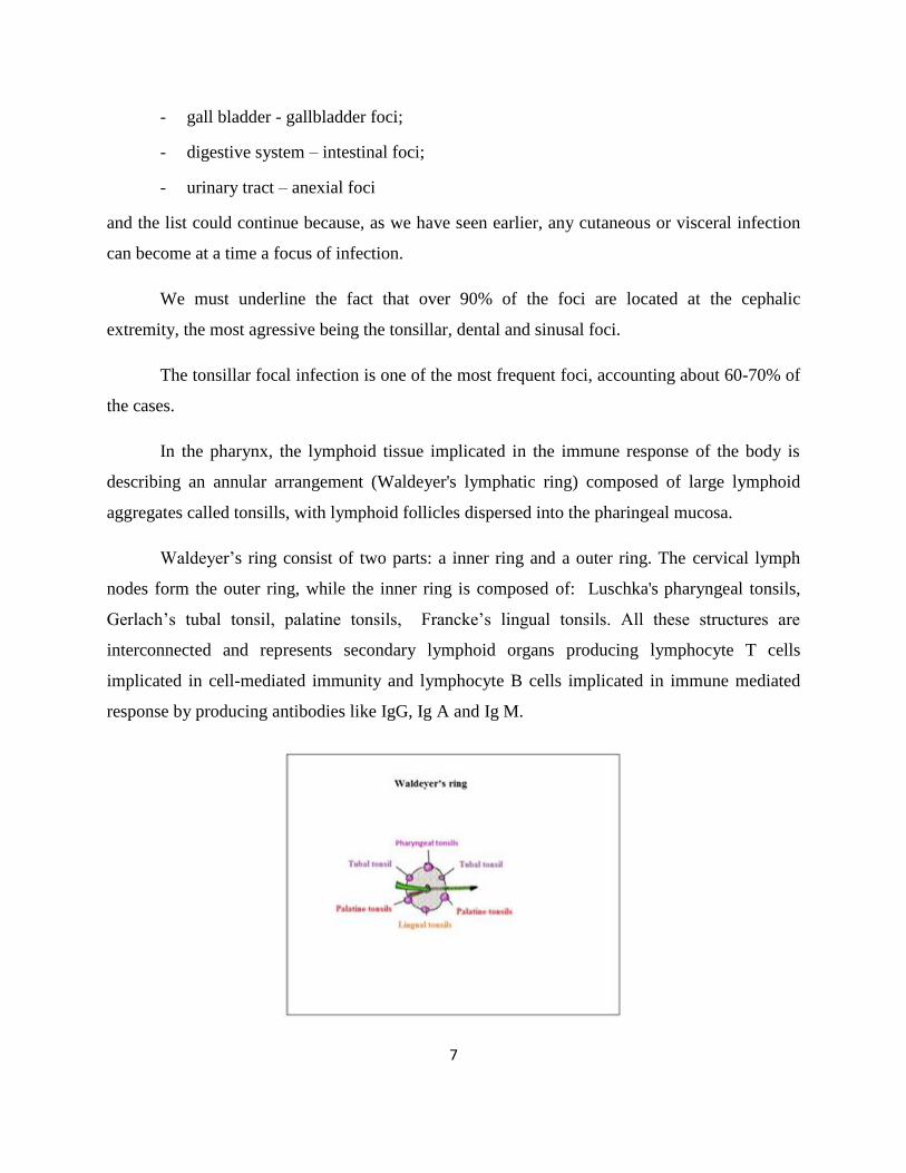
In the pharynx, the lymphoid tissue implicated in the immune response of the body is
describing an annular arrangement (Waldeyer's lymphatic ring) composed of large lymphoid
aggregates called tonsills, with lymphoid follicles dispersed into the pharingeal mucosa.
Waldeyer’s ring consist of two parts: a inner ring and a outer ring. The cervical lymph
nodes form the outer ring, while the inner ring is composed of: Luschka's pharyngeal tonsils,
Gerlach’s tubal tonsil, palatine tonsils, Francke’s lingual tonsils. All these structures are
interconnected and represents secondary lymphoid organs producing lymphocyte T cells
implicated in cell-mediated immunity and lymphocyte B cells implicated in immune mediated
response by producing antibodies like IgG, Ig A and Ig M.

8
In the second part of my paper, which contains the personal contributions, we started to
correlate the tonsillar focal infection with the cutaneous manifestations, conducting a complex
retrospective and prospective study, which consisted of 204 cases with various patology,
represented by patients who checked in and outpatients’ dermatology department of Craiova,
over a period of 3 years (2010-2012). Based on the diagnosis, our cases were distributed as
following:
impetigo occuping the first place with 87 patients accountig for almost half of the
study group;
second is erythema nodosum with 36 cases;
and in descending order:
erythema multiforme – 29 cases;
dyshidrotic eczema – 19 cases;
eczematid like eruptions – 17 cases;
guttate psoriasis – 16 cases. (see graph 1)
Graf. 1 Patients’ distribution according to cutaneous manifestation
9
As we have seen, the patology related to focal infections includes many other cutaneous
manifestations (erythroderma, eczemas especially nummular eczema, pityriasis lichenoides
chronica, etc) but we didn’t take them into account because they didn’t occur in our study group.
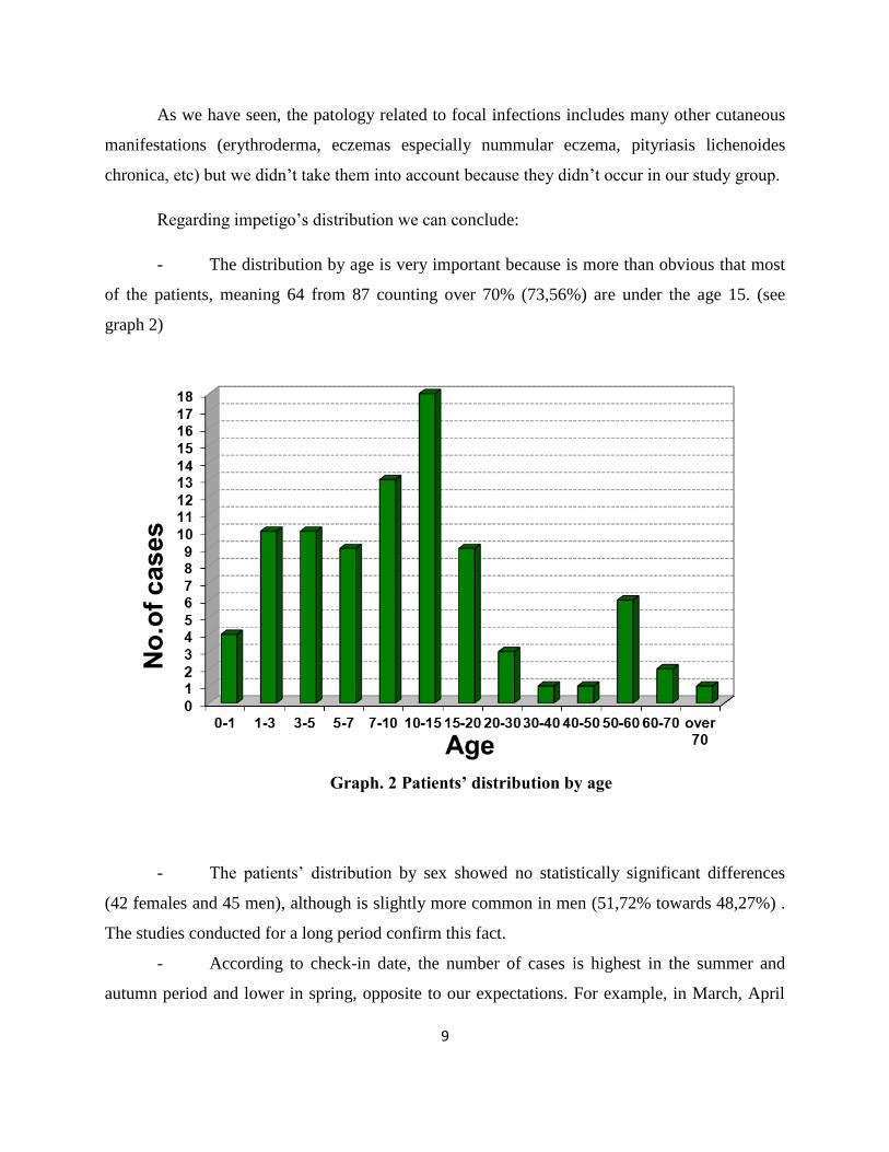
Regarding impetigo’s distribution we can conclude:
- The distribution by age is very important because is more than obvious that most
of the patients, meaning 64 from 87 counting over 70% (73,56%) are under the age 15. (see
graph 2)
Graph. 2 Patients’ distribution by age
- The patients’ distribution by sex showed no statistically significant differences
(42 females and 45 men), although is slightly more common in men (51,72% towards 48,27%) .
The studies conducted for a long period confirm this fact.
- According to check-in date, the number of cases is highest in the summer and
autumn period and lower in spring, opposite to our expectations. For example, in March, April

10
and May, the overall cases were 9, meaning 10,34% in comparison with June, July and August
when were registered almost 4 times more cases (39,08%). If we add the other 25 cases
registered in September and October (28,73%) to the latter, we notice that approximately 70% of
the cases presented to the doctor in this period. (see graph 3). These clinical – statistical data
seems unreal, but it is important to underline that specialized literature reports superimposable
values.
Graph. 3 Patients’ distribution by date of presentation
The bacteriological investigation shows:
staphylococcus aureus in pure culture in 34 patients (39,08%)
streptococcus beta-haemolytic in pure culture in 8 patients (9.19%),
both bacteria in 40 patients (45,97%)
in 5 patients (5,74%) were discovered other microorganisms (other staphylococci
types, corynebacterium, candida, etc) (see graph 4).

11
Graf. 4 Bacteriological investigation results
The group with erythema nodosum (EN) consisted of 36 patients, aged 16 – 76 years, the
majority being owned by patients aged between 18 and 34 years. It’s important to mention that
nearly all were females, 33 cases (91,6%).
In more than a third of the cases studied we couldn’t identify the etiologic agent, the rest
of the cases being distributed as following:
- infections: post-streptococcal EN was responsible for 10 out of 13 infections, the
other being diagnosed with EN secondary to ganglionic and pulmonary tuberculosis and another
with HBs antigen and HCV antibodies present.
- in 7 cases (19,4%) EN secondary to chronic drug exposure. Drugs responsible for
inducing EN were found to be: anti-inflammatory drugs, birth control pills, antidepresive drugs,
hipotensors.

12
- other causes: 1 case of chronic lupus erythematosus, 1 case of hypothyroidism
(Hashimoto’s thyroidis). (see graph. 5)
Graph. 5 Cases distribution according to etiological factors
Regarding erythema multiforme, this condition with extremely variable clinical
appearance, was represented in our study group by 29 patients, showing almost all the clinical
presentation described in literature, ranged from minor papulovesicular eruptions located on the
photoexposed areas to severe forms with mucosal involvement and extended lesions like
ectodermosis pluriorificialis or Steven Johnson syndrome. Statistically, the majority of the cases
occurrs in adults, whereas 72.4% of the patients were aged between 21 and 60 years, meaning
that were exposed to most of the etiological factors. It’s important to mention that there are no
significant differences concerning the distribution by age and by area of residence.
We also been interesed in discovering conditions induced by streptococcal tonsillar
infection and we found 3 cases with repeted pharyngotonsillitis and 3 cases with acute infections
of the upper respiratory tract.

13
The cases with dyshidrotic eczema studied (19 cases) displayed lesions on the palms (16
cases), 3 of them reporting feet involvement. These data confirms the rarity of the cases which
presents eruption affecting both the hands and feet. In 6 cases the patients recalled repeat
pharyngotonsillitis, some being treated more or less empirically, other being neglected for a long
period.
We must stress that 7 out of all 19 cases presented interdigital tinea pedis, some patients
completely ignoring these manifestation.
The 17 cases with eczematid-like eruptions could be correlated with streptococcal and
staphylococcal focal infections. Between them the pityriasiform and microbial eruptions were the
most, each with 5 cases, followed by seborrheic eruptions with 4 cases and last psoriasiform
eruption with 3 cases. The distribution by age, area of residence and sex has nothing specific
whereas this has no relevance.
Another clinical entity, being a part of our study, was represented by the 28 cases of
psoriasis, aged under 20 years from which 16 cases presented with classic form of guttate
psoriasis. From those, in 9 cases we were able to demonstrate the presence of tonsillar focal
infection. These data was verified along time by the beneficial effect that tonsillectomy may
have on different variants of psoriasis, especially in streptococcal carrier and in patients with
recurrent streptococcal pharyngitis or tonsillitis.
That being mentioned, we reccomended tonsillectomy to all guttate psoriasis cases
associated or not with hypertrophic tonsils and frequent relapses.
Based on our results and on clinical observations supported by de statistical data and
inconography, we conclud:
1. Our study reveals the leading role played as an etiologic factor by the focal
infection. The large number of these clinical manifestation with unpredictable evolution, with
tendency to relapse and sometimes difficult to treat, confirms the practical significance of this
paper for the practitioner regardless of his specialisation.
14
2. The large number of patients studied, accounting over 200 people, allowed us to
obtain accurate results, the majority being superimposable with the specialised literature data,
noting that the study group was heterogeneous, aged between 2 months and 76 years.
3. Considering the theme of our paper, we tried to find in each case a correlation
between a focal infection and the cutaneous manifestation and we always noticed the sequence
between the focus of infection and clinical skin lesion, highly variable in time, with worsening of
the local phenomena whenever the activation of the foci occurred.
4. Given the importance of the bacteriological investigation especially in cases of
impetigo, we managed to identify in more than a third of the cases (40%) Staphylococcus aureus
in pure culture, streptococcus beta-haemolytic in pure culture in 9%, and mixed infection in
46%.
5. The bacteriological data allowed us to notice that staphylococcal involvement in
triggering impetigo is not mandatory, the bacterium being present in cultures due to its high
virulence by developing exotoxin that inhibits the growth of streptococci.
6. In chronic infectious foci developed streptococcus species with variate biologic
features, partially acquired through their prolonged existence into organism. Those bacteria can
initiate systemic diseases and/or dermatological symptomatology (sometimes discrete) according
to patient’s immune system. In other words, any chronic infection, regardless of its location in
the body, can act at some point depending on patient’s immunity as a focus of infection.
7. Erythema nodosum represented in our study by 36 cases, exceeded by a multitude
a etiologic factors often impossible to identify. Concerning the subject of my paper, we were
able to demonstrate that streptococcal infection was responsible for almost one third of cases
(27,7%).
8. The predilection of erythema nodosum for females (91,6%) was partially explain
by the large comsumption of drugs, the self medication custom, associated with an impresive
variety of pathologies, and/or the worldwide usage of birth control pills, that are known to
produce a lot of side effects.
15
9. Erythema multiforme is a condition with a incompletly understood
etiopathogenesis, being ultimately considered a cutaneous and mucosal reaction triggered by a
variety of stimuli: acute and chronic infections, repeated drug administration, prolonged
corticotherapy, immunosuppression, collagen diseases, etc which appears to be a delayed
hypersensitivity cell mediated reaction (type IV).
10. The recurrences of erythema multiforme at the same individual, in the following
years, usually in spring, can’t be etiopathogenically classified for now.
11. Taking into consideration that aproximatevely one third of the cases were
proceded by herpes virus infection type I or II, it worth trying prophylactic measures for 6-12
months by reccomanding immunomodulators.
12. Dyshidrotic eczema has a multifactorial etiology (unknown cause) which
contribute in different ways from case to case both endogenous and exogenous factors.
13. Regarding the etiopathogenesis of our 19 cases we mention:
- Streptococcal and staphylococcal infections were responsible for aproximatevely
one third of the cases, figures much higher than those reported in the specialised literature
- Cutaneous hypersensibility reported by most patients is due to the atopic
dermatitis identified in more than a half of cases.
14. Eczematid like eruptions represent cutaneous manifestations frequently induced
by a focus of infection, with similar clinical appearance, without subjective symptoms and highly
variable evolution. The proposed classification is incompletely justified and difficult to make
mainly due to the clinical resemblance and because they constantly change aspect day by day.
15. Our cases match the current classification and are distributed like following:
- pityriasiform eruptions - 5 cases;
- psoriasiform eruptions – 3 cases;
- seborrheic eruptions - 4 cases
- microbial eruptions -5 cases.
16. Regarding psoriasis, our data shows 28 cases of psoriasis, aged under 20 years
from which 16 cases presented with classic form of guttate psoriasis (tear drop or rain drop
psoriasis). From those, in 9 cases we were able to demonstrate the presence of streptococcal
16
tonsillar infection; the treatment of the focus of infection led to remission of the psoriatic
eruption.
17. Both our data and specialised literature mention the beneficial effect of
tonsillectomy in improving psoriasis and/or long term antibiotic treatments.
18. In cases with guttate psoriasis we consider that a local and systemic treatment
addressing both rash and combating any infectious process is mandatory even if it does not fit
perfectly into the classic definition of a focus of infection.

17
References:
1. Cornean-Santa Corina, Cornean Corina Iulia – “Boala de focar amigdaliană”, Conexiuni
medicale nr.1, martie 2009, pag. 51-53.
2. Bisno AL, Stevens DL. Streptococcus pyogenes. In: Mandell GL, Bennett JE, Dolin R,
eds. Principles and Practice of Infectious Diseases. 7th ed. Philadelphia, Pa: Elsevier Churchill
Livingstone; 2009:chap 198.
3. Morelli JG. Cutaneous Bacterial Infections. In: Kliegman RM, Behrman RE, Jenson HB,
Stanton BF, eds. Nelson Textbook of Pediatrics. 19th ed. Philadelphia, Pa: Saunders Elsevier;
2011:chap 657.
4. Geria AN, Schuartz RA. Impetigo Update: New Challenges in the Era of Methicillin
Resistance. Cutis. 2010;85(2):65-70.
5. Mert A, Ozaras R, Tabak F, Pekmezci S, Demirkesen C, Ozturk R. Erythema nodosum:
an experience of 10 years. Scand J Infect Dis. 2004;36(6-7):424-7
6. Habif, Thomas "Erythma Nodosum" Clinical Dermatology A color Guide to Diagnosis
and Therapy. , 5th ed. Ed. Thomas Habif, MD. New York: Mosby, 2010. 720-721
7. Farhi D, Cosnes J, Zizi N, et al. Significance of erythema nodosum and pyoderma
gangrenosum in inflammatory bowel diseases: a cohort study of 2402 patients. Medicine
(Baltimore). Sep 2008;87(5):281-93.
8. McFadden JP, Baker BS, Powles AV, Fry L. Psoriasis and streptococci: the natural
selection of psoriasis revisited. Br J Dermatol. May 2009;160(5):929-37.
9. Telfer NR, Chalmers RJ, Whale K, Colman G. The role of streptococcal infection in the
initiation of guttate psoriasis. Arch Dermatol. Jan 1992;128(1):39-42.
10. Mallbris L, Wolk K, Sánchez F. HLA-Cw*0602 associates with a twofold higher
prevalence of positive streptococcal throat swab at the onset of psoriasis: a case control study.
BMC Dermatol. 2009;9:5.
11. Nahary L, Tamarkin A, Kayam N, Sela S, Fry L, Baker B, et al. An investigation of
antistreptococcal antibody responses in guttate psoriasis. Arch Dermatol Res. Sep
2008;300(8):441-9.

18
12. Dogan B, Karabudak O, Harmanyeri Y. Antistreptococcal treatment of guttate psoriasis:
a controlled study. Int J Dermatol. Sep 2008;47(9):950-2.
13. Sokumbi O, Wetter DA. Clinical features, diagnosis, and treatment of erythema
multiforme: a review for the practicing dermatologist. Int J Dermatol. Aug 2012;51(8):889-902.
14. Fritsch PO, Ruiz-Maldonado R, Erythema multiforme. Stevens-Johnson syndrome and
toxic epidermal necrolysis. In: Freedberg IM, Irwin M, Eisen AZ, Wolff K, Austen KF,
Goldsmith LA, Katz SI, editors. Fitzpatrick's Dermatology in General Medicine. 7th
edition. New
York: McGraw-Hill; 2008:343-54.


![Radiation-induced swelling and hardening of 316L stainless ...iint.nuaa.edu.cn/_upload/article/files/8b/e6/f1c5a4de40f6a9e2f454e247… · hardening [12], the usual manifestations](https://img.pdfslide.net/doc/110x75/60678d5b4b21297cc4043b99/radiation-induced-swelling-and-hardening-of-316l-stainless-iintnuaaeducnuploadarticlefiles8be6f1c5a4de40f6a9e2f454e247.jpg)