Embed Size (px)
Citation preview
Acclarent Professional Education
ACCLARENT AERA® Eustachian Tube Balloon Dilation System
1
This professional education activity is brought to you by Acclarent, Inc. and is not certified for continuing medical education. Dr. X is compensated by and presenting on behalf of Acclarent, Inc., and must present information in accordance with applicable Regulatory requirements. This presentation may include demonstrations of the use of surgical devices; it is not intended to be used as a surgical training guide. Other surgeons may employ different techniques. The steps demonstrated may not be the complete steps of the procedure. Individual surgeon preference and experience, as well as patient needs, may dictate variation in procedure steps. Before using any medical device, review all relevant package inserts with particular attention to the indications, contraindications, warnings and precautions, and steps for use of the device(s).
ACCLARENT AERA® Eustachian Tube Balloon Dilation System is intended for use by physicians who are trained on Acclarent technology. Eustachian tube balloon dilation has associated risks, including tissue and mucosal trauma, infection, or possible carotid artery injury. Prior to use, it is important to read the Instructions for Use and to understand the contraindications, warnings, and precautions associated with these devices. The safety of the device as used under local anesthesia has not been evaluated.
2
Caution: Federal (US) law restricts the sale, distribution or use of these devices to, by or on the order of a physician. Third party trademarks used herein are trademarks of their respective owners. This site is intended for visitors from the United States and published by Acclarent, Inc., which is solely responsible for its contents.
Indication For Use: The ACCLARENT AERA® Eustachian Tube Balloon Dilation system is intended to dilate the Eustachian tube for treatment of persistent Eustachian tube dysfunction in patients age 18 and older.
Surgeon training must include simulated use on cadavers to ensure users can follow the instructions for use to allow safe use of the device.
3
Eustachian Tube DysfunctionAnatomy, Physiology, Diagnosis and Treatment
4
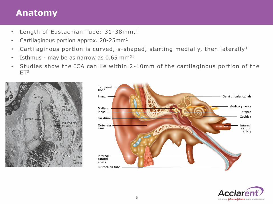
• Length of Eustachian Tube: 31-38mm,1
• Cartilaginous portion approx. 20-25mm1
• Cartilaginous portion is curved, s-shaped, starting medially, then laterally1
• Isthmus - may be as narrow as 0.65 mm21
• Studies show the ICA can lie within 2-10mm of the cartilaginous portion of the ET2
Anatomy
5
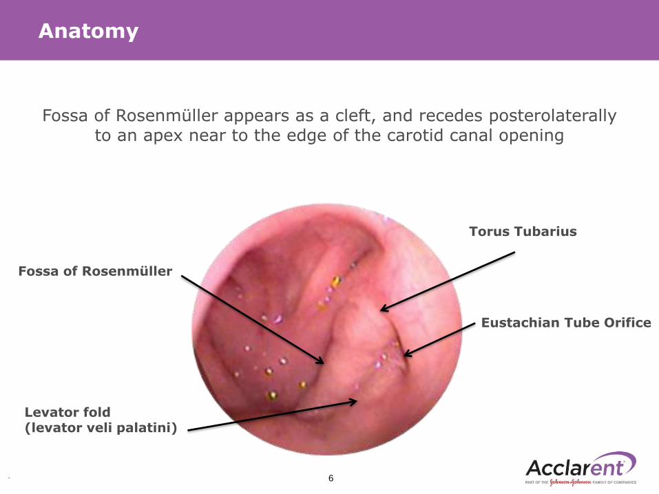
Fossa of Rosenmüller appears as a cleft, and recedes posterolaterally to an apex near to the edge of the carotid canal opening
Anatomy
Fossa of Rosenmüller
Levator fold(levator veli palatini)
Eustachian Tube Orifice
Torus Tubarius
. 6
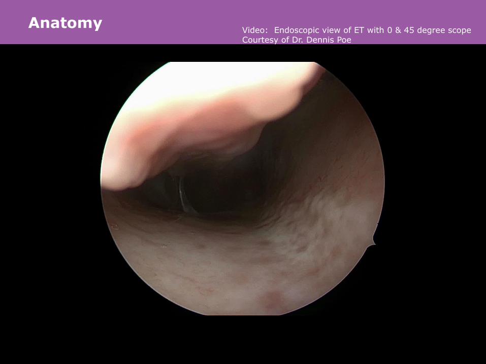
Anatomy
059266-160831 | 7
Video: Endoscopic view of ET with 0 & 45 degree scopeCourtesy of Dr. Dennis Poe
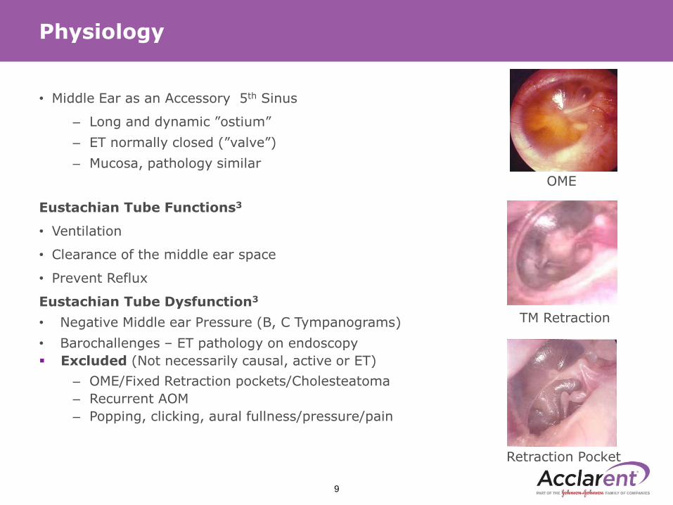
• Middle Ear as an Accessory 5th Sinus
– Long and dynamic ”ostium”
– ET normally closed (”valve”)
– Mucosa, pathology similar
Eustachian Tube Functions3
• Ventilation
• Clearance of the middle ear space
• Prevent Reflux
Eustachian Tube Dysfunction3
• Negative Middle ear Pressure (B, C Tympanograms)
• Barochallenges – ET pathology on endoscopy
▪ Excluded (Not necessarily causal, active or ET)
– OME/Fixed Retraction pockets/Cholesteatoma
– Recurrent AOM
– Popping, clicking, aural fullness/pressure/pain
Physiology
9
OME
TM Retraction
Retraction Pocket
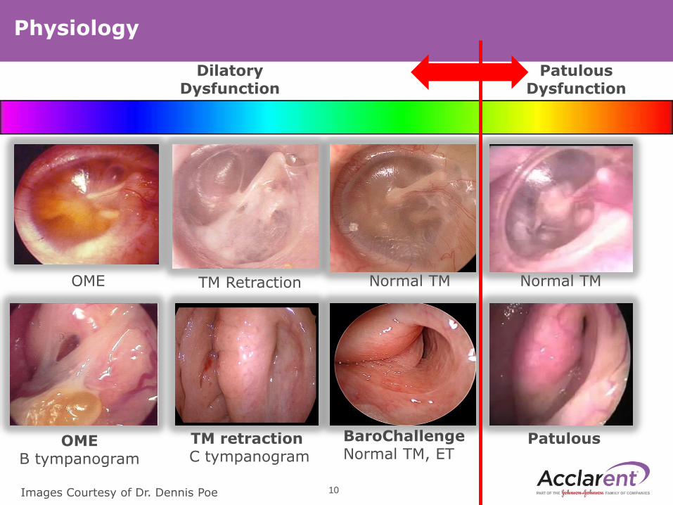
Physiology
OMEB tympanogram
TM retractionC tympanogram
BaroChallengeNormal TM, ET
Patulous
Dilatory Dysfunction
Patulous Dysfunction
10
TM Retraction Normal TM Normal TMOME
Images Courtesy of Dr. Dennis Poe
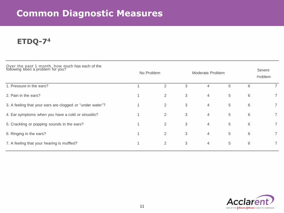
ETDQ-74
Common Diagnostic Measures
Over the past 1 month, how much has each of thefollowing been a problem for you?
No Problem Moderate ProblemSevere
Problem
1. Pressure in the ears? 1 2 3 4 5 6 7
2. Pain in the ears? 1 2 3 4 5 6 7
3. A feeling that your ears are clogged or ‘‘under water’’? 1 2 3 4 5 6 7
4. Ear symptoms when you have a cold or sinusitis? 1 2 3 4 5 6 7
5. Crackling or popping sounds in the ears? 1 2 3 4 5 6 7
6. Ringing in the ears? 1 2 3 4 5 6 7
7. A feeling that your hearing is muffled? 1 2 3 4 5 6 7
11
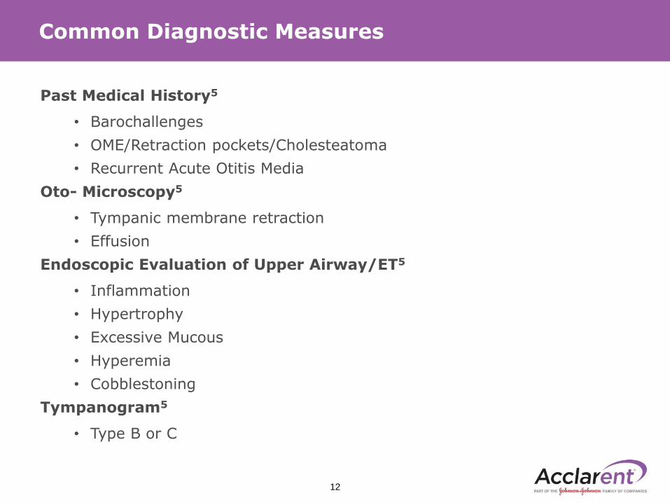
Past Medical History5
• Barochallenges
• OME/Retraction pockets/Cholesteatoma
• Recurrent Acute Otitis Media
Oto- Microscopy5
• Tympanic membrane retraction
• Effusion
Endoscopic Evaluation of Upper Airway/ET5
• Inflammation
• Hypertrophy
• Excessive Mucous
• Hyperemia
• Cobblestoning
Tympanogram5
• Type B or C
Common Diagnostic Measures
12
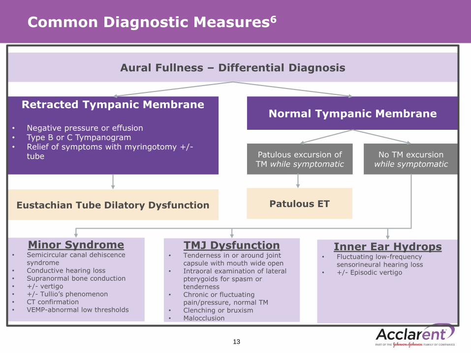
Common Diagnostic Measures6
13
Aural Fullness – Differential Diagnosis
Retracted Tympanic Membrane
• Negative pressure or effusion• Type B or C Tympanogram• Relief of symptoms with myringotomy +/-
tube
Normal Tympanic Membrane
Patulous excursion of TM while symptomatic
No TM excursion while symptomatic
Patulous ETEustachian Tube Dilatory Dysfunction
Minor Syndrome• Semicircular canal dehiscence
syndrome• Conductive hearing loss• Supranormal bone conduction• +/- vertigo• +/- Tullio’s phenomenon• CT confirmation• VEMP-abnormal low thresholds
TMJ Dysfunction• Tenderness in or around joint
capsule with mouth wide open• Intraoral examination of lateral
pterygoids for spasm or tenderness
• Chronic or fluctuating pain/pressure, normal TM
• Clenching or bruxism• Malocclusion
Inner Ear Hydrops• Fluctuating low-frequency
sensorineural hearing loss• +/- Episodic vertigo
Medical Management1
• Effectiveness for medical therapies remains uncertain.
• There is no FDA approved medical therapy for nonspecific ETD
Surgical Management5
• Tympanostomy tube placement
• Adenoidectomy
• ACCLARENT AERA™ Eustachian Tube Balloon Dilation
Surgical Indications5
• Persistent OME or Non-adherent atelectasis
AND Type B or C tympanogram
AND ET pathology on endoscopy usually inflammation
• Flight or Scuba barochallenge AND ET pathology
• Symptomatic- CHL, pain/blockage w pressure change
• Symptoms improved with tympanostomy tube if done
• Absence of autophony
Common Treatment Approaches
14
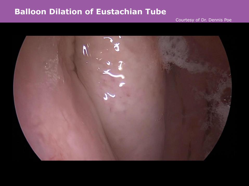
Balloon Dilation of Eustachian Tube
059266-160831 | 15
Courtesy of Dr. Dennis Poe

Mechanism of Action: Why does it Work?
Eustachian Tube Mucous Membrane7
• The ET lumen is lined with pseudostratified, columnar epithelium of the ciliated type, which sweeps material from the middle ear into the nasopharynx
• The mucosa is continuous with the lining of the tympanic cavity at its distal end, as it is with the nasopharynx at its proximal end
16
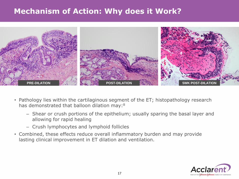
Mechanism of Action: Why does it Work?
PRE-DILATION POST-DILATION 5WK POST-DILATION
• Pathology lies within the cartilaginous segment of the ET; histopathology research has demonstrated that balloon dilation may:8
– Shear or crush portions of the epithelium; usually sparing the basal layer and allowing for rapid healing
– Crush lymphocytes and lymphoid follicles
• Combined, these effects reduce overall inflammatory burden and may provide lasting clinical improvement in ET dilation and ventilation.
17
ACCLARENT AERA® Eustachian Tube Balloon Dilation System
Clinical Trial Overview
18
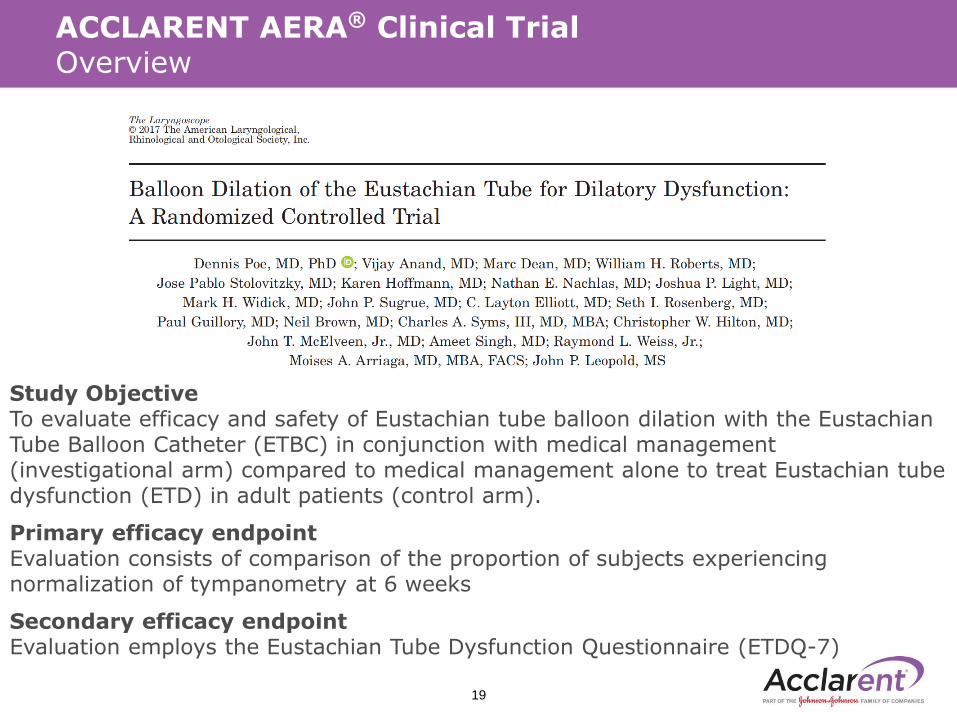
Study ObjectiveTo evaluate efficacy and safety of Eustachian tube balloon dilation with the Eustachian Tube Balloon Catheter (ETBC) in conjunction with medical management (investigational arm) compared to medical management alone to treat Eustachian tube dysfunction (ETD) in adult patients (control arm).
Primary efficacy endpointEvaluation consists of comparison of the proportion of subjects experiencing normalization of tympanometry at 6 weeks
Secondary efficacy endpointEvaluation employs the Eustachian Tube Dysfunction Questionnaire (ETDQ-7)
ACCLARENT AERA® Clinical Trial Overview
19
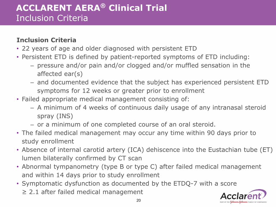
Inclusion Criteria
• 22 years of age and older diagnosed with persistent ETD
• Persistent ETD is defined by patient-reported symptoms of ETD including:
– pressure and/or pain and/or clogged and/or muffled sensation in the
affected ear(s)
– and documented evidence that the subject has experienced persistent ETD
symptoms for 12 weeks or greater prior to enrollment
• Failed appropriate medical management consisting of:
– A minimum of 4 weeks of continuous daily usage of any intranasal steroid
spray (INS)
– or a minimum of one completed course of an oral steroid.
• The failed medical management may occur any time within 90 days prior to
study enrollment
• Absence of internal carotid artery (ICA) dehiscence into the Eustachian tube (ET)
lumen bilaterally confirmed by CT scan
• Abnormal tympanometry (type B or type C) after failed medical management
and within 14 days prior to study enrollment
• Symptomatic dysfunction as documented by the ETDQ-7 with a score
≥ 2.1 after failed medical management
ACCLARENT AERA® Clinical Trial Inclusion Criteria
20
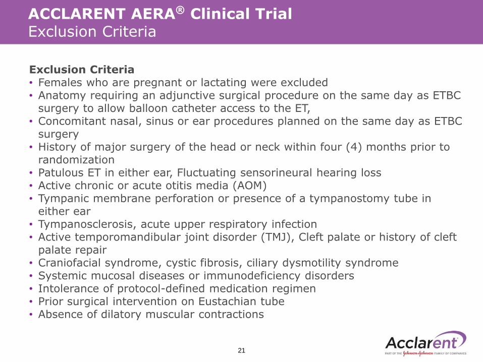
Exclusion Criteria• Females who are pregnant or lactating were excluded• Anatomy requiring an adjunctive surgical procedure on the same day as ETBC
surgery to allow balloon catheter access to the ET, • Concomitant nasal, sinus or ear procedures planned on the same day as ETBC
surgery • History of major surgery of the head or neck within four (4) months prior to
randomization • Patulous ET in either ear, Fluctuating sensorineural hearing loss • Active chronic or acute otitis media (AOM) • Tympanic membrane perforation or presence of a tympanostomy tube in
either ear • Tympanosclerosis, acute upper respiratory infection • Active temporomandibular joint disorder (TMJ), Cleft palate or history of cleft
palate repair• Craniofacial syndrome, cystic fibrosis, ciliary dysmotility syndrome • Systemic mucosal diseases or immunodeficiency disorders • Intolerance of protocol-defined medication regimen • Prior surgical intervention on Eustachian tube • Absence of dilatory muscular contractions
ACCLARENT AERA® Clinical Trial Exclusion Criteria
21
ACCLARENT AERA® Clinical Trial Exclusion Criteria
22
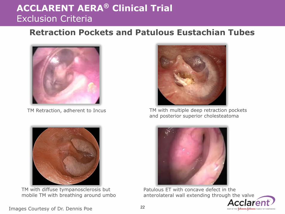
Retraction Pockets and Patulous Eustachian Tubes
TM Retraction, adherent to Incus
Patulous ET with concave defect in the anterolateral wall extending through the valve
TM with multiple deep retraction pockets and posterior superior cholesteatoma
TM with diffuse tympanosclerosis but mobile TM with breathing around umbo
Images Courtesy of Dr. Dennis Poe
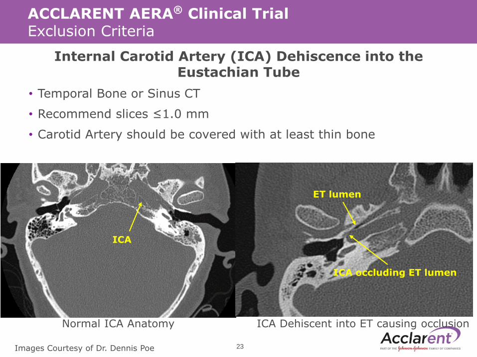
Internal Carotid Artery (ICA) Dehiscence into the Eustachian Tube
• Temporal Bone or Sinus CT
• Recommend slices ≤1.0 mm
• Carotid Artery should be covered with at least thin bone
ACCLARENT AERA® Clinical Trial Exclusion Criteria
ICA
ET lumen
ICA occluding ET lumen
Normal ICA Anatomy ICA Dehiscent into ET causing occlusion
23Images Courtesy of Dr. Dennis Poe
• For 1 week post-randomization (control arm) or post-procedure (lead-ins and investigational arm), subjects were required to adhere to the following:
– Avoid performing the Valsalva maneuver
– Avoid nose blowing
– Avoid using Continuous Positive Airway Pressure (CPAP) machines (if possible)
– Sleep with an extra pillow to elevate the head
• After 1 week post-randomization (control arm) or post-procedure (lead-ins and investigational arm), it is recommended that all subjects perform the Valsalva maneuver one time per hour.
• Proper Valsalva maneuver technique consists of holding the nose, blowing slowly to build pressure, and then swallowing hard.
• For 6 weeks post-randomization (control arm) or post-procedure (lead-ins and investigational arm), subjects are required to adhere to the recommended Nasacort labeling dosage and frequency.
ACCLARENT AERA® Clinical Trial Post-treatment and follow-up care
24
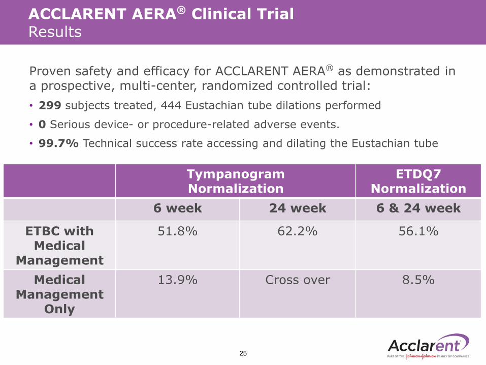
Proven safety and efficacy for ACCLARENT AERA® as demonstrated in a prospective, multi-center, randomized controlled trial:
• 299 subjects treated, 444 Eustachian tube dilations performed
• 0 Serious device- or procedure-related adverse events.
• 99.7% Technical success rate accessing and dilating the Eustachian tube
ACCLARENT AERA® Clinical Trial Results
25
Tympanogram Normalization
ETDQ7Normalization
6 week 24 week 6 & 24 week
ETBC with Medical
Management
51.8% 62.2% 56.1%
Medical Management
Only
13.9% Cross over 8.5%
ACCLARENT AERA® Clinical Trial Results
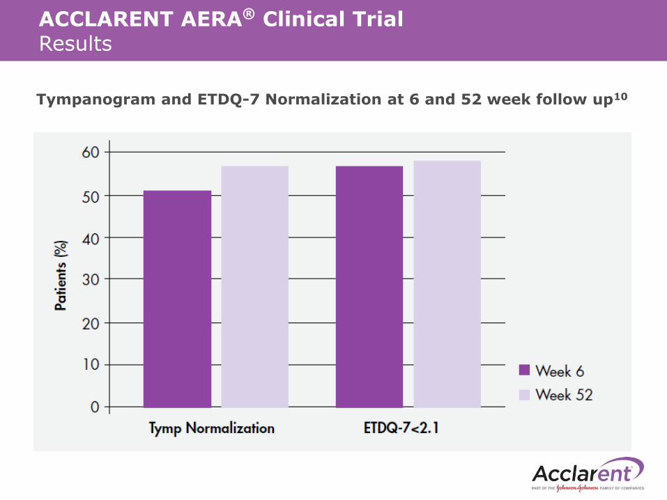
Tympanogram and ETDQ-7 Normalization at 6 and 52 week follow up10
ACCLARENT AERA®
Eustachian Tube Balloon Dilation System
27
Indication For Use
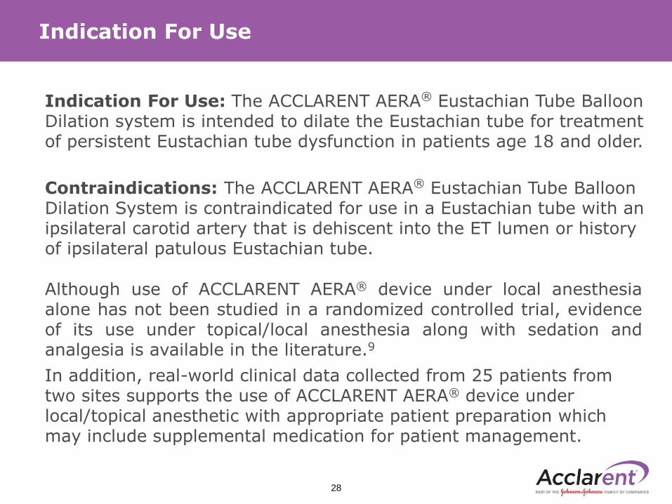
Indication For Use: The ACCLARENT AERA® Eustachian Tube Balloon Dilation system is intended to dilate the Eustachian tube for treatment of persistent Eustachian tube dysfunction in patients age 18 and older.
Contraindications: The ACCLARENT AERA® Eustachian Tube Balloon Dilation System is contraindicated for use in a Eustachian tube with an ipsilateral carotid artery that is dehiscent into the ET lumen or history of ipsilateral patulous Eustachian tube.
28
Although use of ACCLARENT AERA® device under local anesthesiaalone has not been studied in a randomized controlled trial, evidenceof its use under topical/local anesthesia along with sedation andanalgesia is available in the literature.9
In addition, real-world clinical data collected from 25 patients from two sites supports the use of ACCLARENT AERA® device under local/topical anesthetic with appropriate patient preparation which may include supplemental medication for patient management.
• Introduction of false passages and rupture or damage to carotid artery
• Injury to mucosal tissue
– Due to misuse of device on patulous Eustachian tube or following skull base surgery
– Due to catheter mechanical failure
– Due to balloon rupture
– Due to mishandling of device with respect to excessive force and/or incorrect positioning
Device Safety Identified Risks
29
• Intended for single patient use only. DO NOT REUSE.
• Patients with a history of skull base surgery, prior ear surgery, skull fracture, or anatomic abnormalities may have elevated risk of complications and should be radiographically screened before treatment.
• DO NOT use product if the integrity of the sterile packaging has been compromised or if the device appears damaged.
• DO NOT use if the device becomes damaged or touches a non-sterile object outside of the operating field.
• Never advance or retract the device against unknown resistance, as this could cause tissue trauma or device damage.
• Advancing the device into the Eustachian tube against resistance may cause injury.
• DO NOT exceed the recommended maximum balloon inflation pressure of 12 atmospheres (ATM).
• Use only sterile saline or sterile water for inflation. DO NOT inflate with air.
Warnings
30
• DO NOT move the balloon while it is inflated. Ensure balloon is fully deflated during insertion and withdrawal.
• Radiographic assessment of the targeted Eustachian tube is recommended prior to any procedure involving balloon tuboplasty.
• Certain nasal anatomy such as a deviated nasal septum may preclude access to the Eustachian tube/s resulting in failure to treat the target anatomy.
• DO NOT inflate the Balloon Catheter until it has exited the Guide Catheter.
• DO NOT bend the Guide Catheter shaft.
Precautions
31
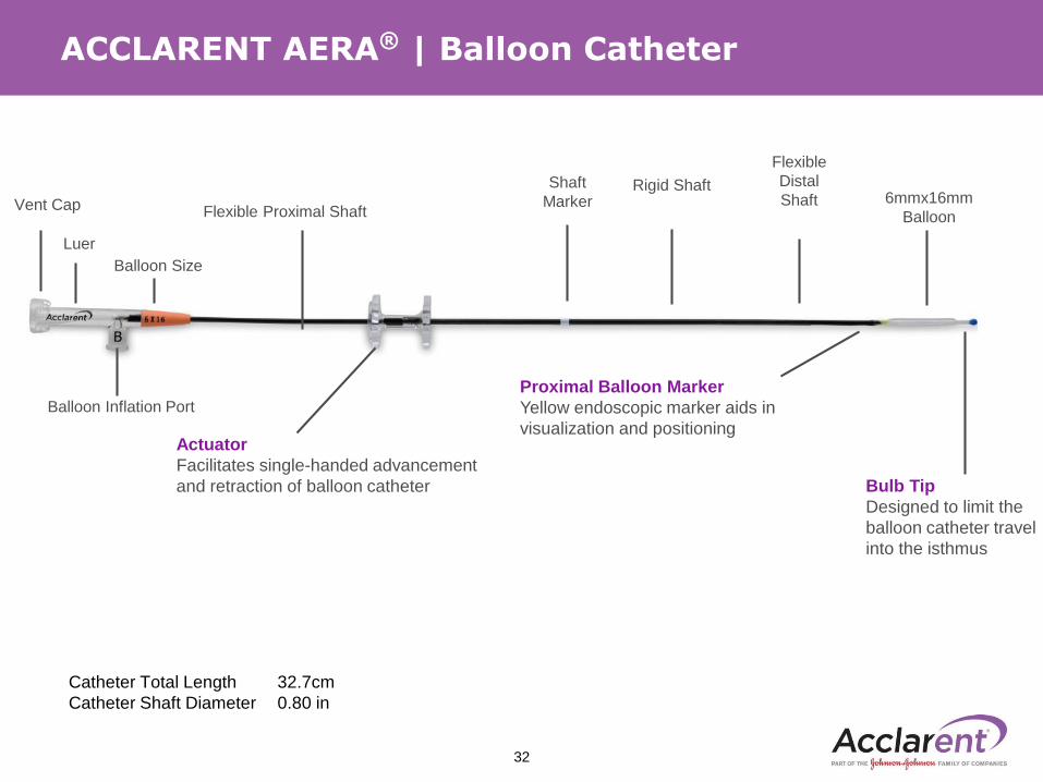
ACCLARENT AERA® | Balloon Catheter
Balloon Inflation Port
Actuator
Facilitates single-handed advancement
and retraction of balloon catheter
Shaft
Marker
Proximal Balloon Marker
Yellow endoscopic marker aids in
visualization and positioning
Flexible
Distal
ShaftFlexible Proximal Shaft
6mmx16mm
Balloon
Bulb Tip
Designed to limit the
balloon catheter travel
into the isthmus
Rigid Shaft
Balloon Size
Vent Cap
Luer
Catheter Total Length 32.7cm
Catheter Shaft Diameter 0.80 in
32
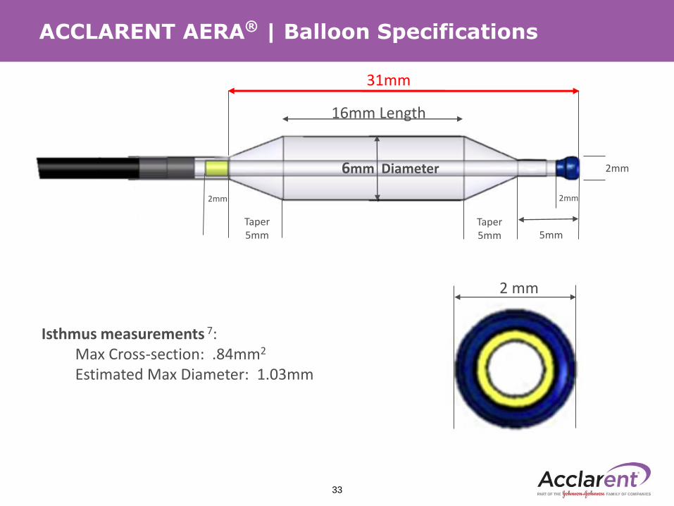
ACCLARENT AERA® | Balloon Specifications
2 mm
Isthmus measurements 7: Max Cross-section: .84mm2
Estimated Max Diameter: 1.03mm
31mm
6mm Diameter
16mm Length
2mm 2mm
Taper 5mm
Taper 5mm
2mm
5mm
33
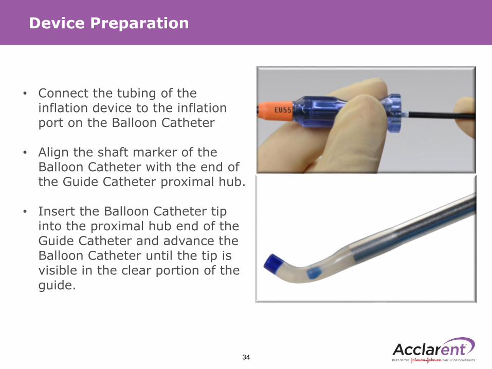
Device Preparation
• Connect the tubing of the inflation device to the inflation port on the Balloon Catheter
• Align the shaft marker of the Balloon Catheter with the end of the Guide Catheter proximal hub.
• Insert the Balloon Catheter tip into the proximal hub end of the Guide Catheter and advance the Balloon Catheter until the tip is visible in the clear portion of the guide.
34
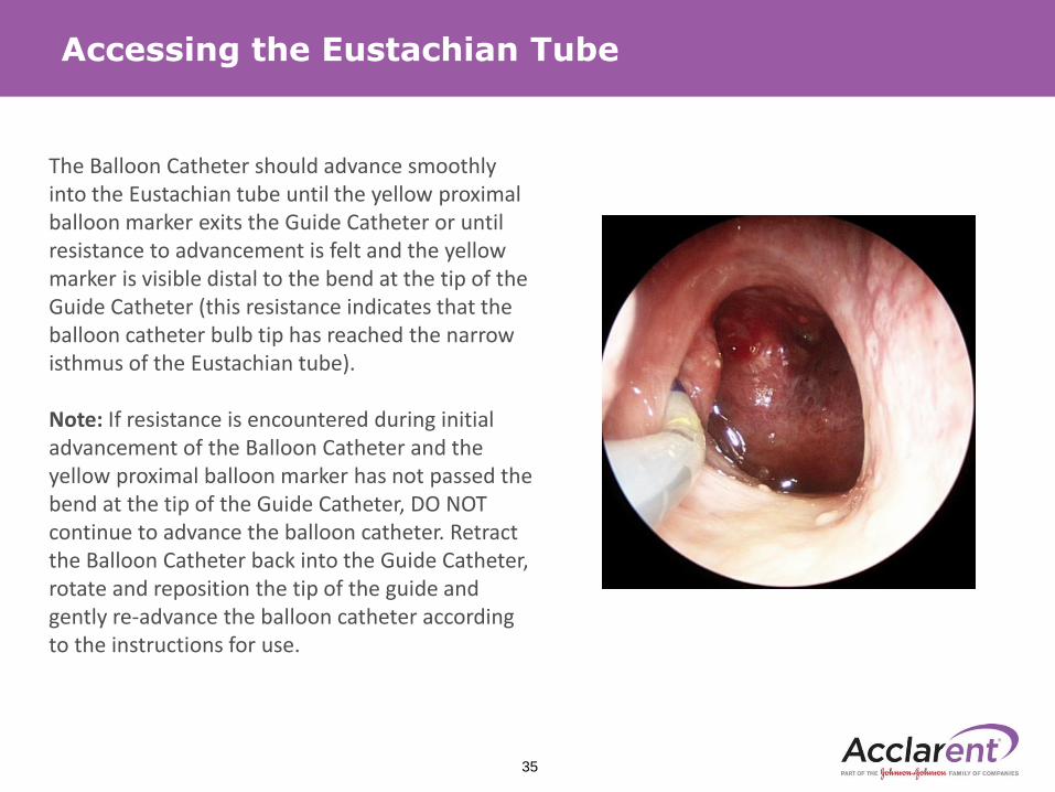
Accessing the Eustachian Tube
The Balloon Catheter should advance smoothly into the Eustachian tube until the yellow proximal balloon marker exits the Guide Catheter or until resistance to advancement is felt and the yellow marker is visible distal to the bend at the tip of the Guide Catheter (this resistance indicates that the balloon catheter bulb tip has reached the narrow isthmus of the Eustachian tube).
Note: If resistance is encountered during initial advancement of the Balloon Catheter and the yellow proximal balloon marker has not passed the bend at the tip of the Guide Catheter, DO NOT continue to advance the balloon catheter. Retract the Balloon Catheter back into the Guide Catheter, rotate and reposition the tip of the guide and gently re-advance the balloon catheter according to the instructions for use.
35
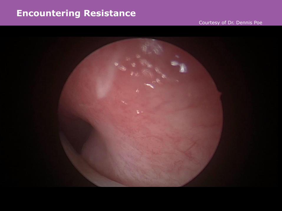
Encountering Resistance
059266-160831 | 36
Courtesy of Dr. Dennis Poe
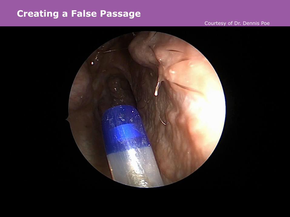
Creating a False Passage
059266-160831 | 37
Courtesy of Dr. Dennis Poe
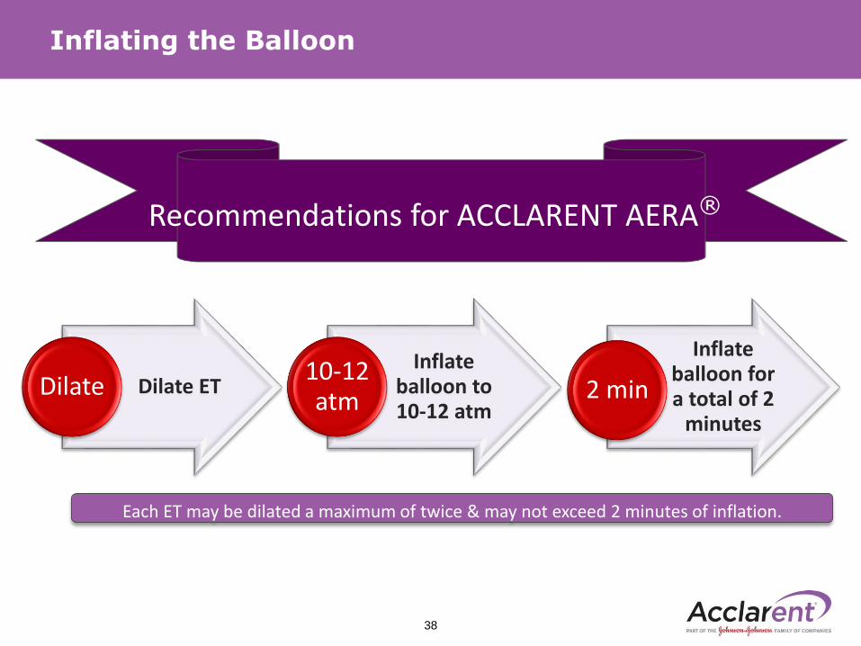
Inflating the Balloon
Recommendations for ET DilationRecommendations for ACCLARENT AERA®
38
Dilate ETDilateInflate
balloon to 10-12 atm
10-12 atm
Inflate balloon for a total of 2
minutes
2 min
Each ET may be dilated a maximum of twice & may not exceed 2 minutes of inflation.
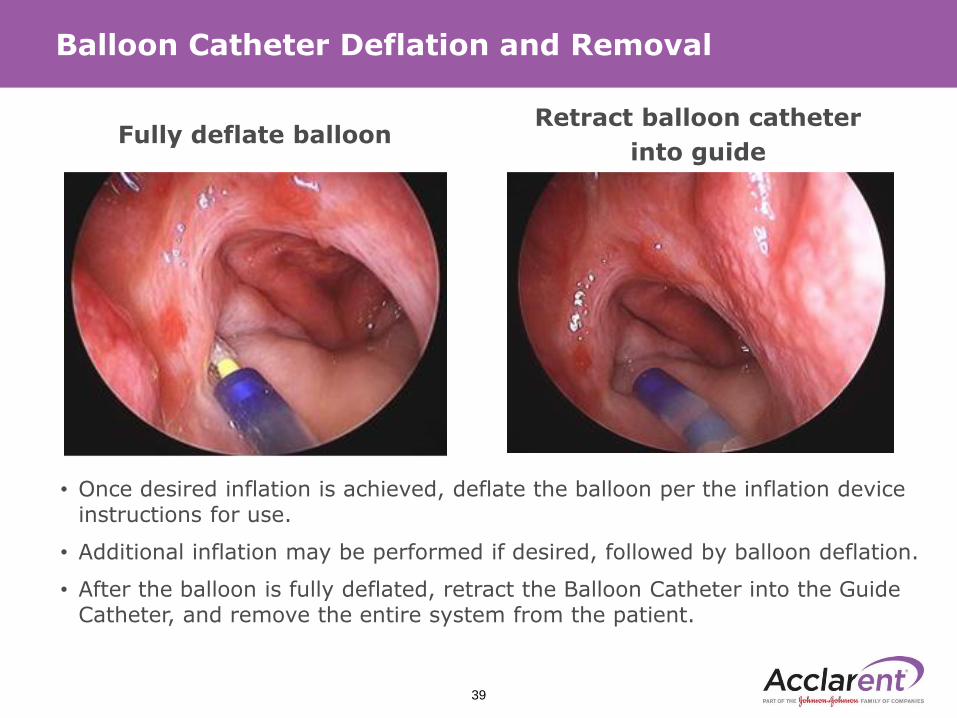
Balloon Catheter Deflation and Removal
• Once desired inflation is achieved, deflate the balloon per the inflation device instructions for use.
• Additional inflation may be performed if desired, followed by balloon deflation.
• After the balloon is fully deflated, retract the Balloon Catheter into the Guide Catheter, and remove the entire system from the patient.
Fully deflate balloonRetract balloon catheter
into guide
39

40
Thank you
059266-180228 | 41
References
1. McCoul, E., Lucente, F., and Anand, V. Evolution of ET Surgery. The Laryngoscope, 2011. 121:661–6662. Olander, H., Jarnstedt, J., Poe, D., and Kiveka, I. Critical distance between the cartilaginous Eustachian
tube and the internal carotid artery. European Archive of Otol-Rhino-Laryngology, 2016. pp. 1-5 3. Schilder, A.G.M., Bhutta, M.F., Butler, C.C., Holy, C., Levine, L.H., Kvaerner, K.J., Norman, G., Pennings,
R.J., Poe, D., Silvola, J.T., Sudhoff, H. & Lund, V.J.. Eustachian tube dysfunction: consensus statement on definition, types, clinical presentation and diagnosis. Clinical Otolaryngology, 2015 . 40 pp. 407-411
4. McCoul, E., Anand, V., and Christos, P. Validating the clinical assessment of Eustachian tube dysfunction: the Eustachian tube dysfunction questionnaire (ETDQ-7). Laryngoscope, 2012. 122(5): 1137–1141.
5. Adil, E.., and Poe, D. What is the full range of medical and surgical treatments available for patients with Eustachian tube dysfunction? Current Opinion in Otolaryngology and Head and Neck Surgery, 2014.22 (1), pp. 8-15
6. Otologic Surgery. Diagnosis and Management of Patulous Eustachian Tube. Brackman MD, Derald;2010;chapter 7
7. Eustachian Tube Structure, Function, Role in Otitis Media. Bluestone, M.D., Charles D; 2005; chapter 3 page 34
8. Kivekäs I1, Chao WC, Faquin W, Hollowell M, Silvola J, Rasooly T, Poe D. Adil, E., and Poe, D. Histopathology of balloon-dilation Eustachian tuboplasty. Laryngoscope. 2015 Feb;125(2):436-41
9. Luukkainen V., Kivekas I, Hammaren-Malmi S, Rautiainen M, Poyhonen L, Aarnisalo AA, Jero J, Sinkkonen ST. Balloon eustachian tuboplasty under local anesthesia. Is it feasible? Laryngoscope. 2017 Feb 03. doi:10.1002/lary.26488
10. Anand V, Leopold J, Poe D. (2017, September). Balloon dilation of the Eustachian tube: 12M follow-up. Oral presentation presented at the American Academy of Otolaryngology- Head and Neck Surgery, Chicago, IL.
www.acclarent.com, ©2018 Acclarent, Inc. Intended for use by or under the direction of a physician. It is important to read the Instructions for Use and to understand the contraindications, warnings, and precautions. All rights reserved.