Embed Size (px)
Citation preview

Originally published as: Achazi, K., Patel, P., Paliwal, R., Radonić, A., Niedrig, M., Donoso-Mantke, O. RNA interference inhibits replication of tick-borne encephalitis virus in vitro (2012) Antiviral Research, 93 (1), pp. 94-100. DOI: 10.1016/j.antiviral.2011.10.023
This is an author manuscript. The definitive version is available at: http://www.sciencedirect.com/

1
RNA interference inhibits replication of tick-borne encephalitis virus in vitro
Katharina Achazia,b, Pranav Patela, Ravish Paliwala, Aleksandar Radonića, Matthias
Niedriga, Oliver Donoso-Mantkea
a Centre for Biological Security 1 (ZBS 1), Robert Koch Institute, Nordufer 20, 13353
Berlin, Germany
b Corresponding author
Katharina Achazi: [email protected]; Tel.: +49 (0)30-18 754-2917; Fax.: +49 (0)30-18
754-2604
Pranav Patel: [email protected]
Ravish Paliwal: [email protected]
Aleksandar Radonić: [email protected]
Matthias Niedrig: [email protected]
Oliver Donoso-Mantke: [email protected]
Katharina Achazi
Robert Koch Institute
Centre for Biological Security 1 (ZBS 1)
Nordufer 20
D-13353 Berlin
Germany
Tel.: +49 (0)30-18 754-2917
Fax.: +49 (0)30-18 754-2604
Email: [email protected]

2
Abstract
Each year, up to 10,000 cases of infections with the flavivirus tick-borne encephalitis
(TBE) virus that affect the central nervous system are reported in Europe and Asia.
Due to the potentially severe adverse effects of post-exposure prophylaxis with TBE
virus hyperimmunoglobulin, TBE can currently only be treated symptomatically. An
RNA interference (RNAi) approach to inhibiting TBE virus replication was therefore
developed. In this study we demonstrate for the first time that small interfering RNAs
(siRNA) targeted to the TBE virus genome reduce the quantity of infectious TBE virus
particles, TBE virus genome, and TBE virus protein in vitro by up to 85%. The 50%
inhibitory dose (DI50]) of the shRNA plasmid was only 0.05 µg/ml. As RNAi-based
therapeutics for other diseases are already being evaluated in phases II and III
clinical trials, it is possible that RNAi could become valuable tool for controlling TBE
virus infection.
Keywords
Flavivirus, tick-borne encephalitis virus, tick-borne encephalitis, RNA interference,
small interfering RNA, antiviral drug
Abbreviations
DAPI; 4',6-diamidino-2-phenylindole
DI50; 50% inhibitory dose
DMEM; Dulbecco's modified Eagle medium
dsRNA; double stranded RNA
EGFP; enhanced green fluorescent protein
GE; genome equivalent
3
HIV; human immunodeficiency virus
MOI; multiplicity of infection
PFU; plaque forming units
RISC; RNA-induced silencing complex
RNAi; RNA interference
RT-qPCR; quantitative real-time RT-PCR
shRNA; short hairpin RNA
siRNA; small interfering RNA
TBE; tick-borne encephalitis
1. Introduction
Viruses of the genus Flavivirus are small enveloped viruses with a single positive-
stranded RNA genome of about 11 kilobases. The genome codes for three structural
(E, M/preM, and C) and seven non-structural proteins (NS1, NS2a, NS2b, NS3,
NS4a, NS4b and NS5) (Mansfield et al., 2009). Flaviviruses, transmitted mainly by
mosquito or tick vectors and a cause of serious health issues throughout the world,
are divided into three groups: mosquito-borne flaviviruses, tick-borne flaviviruses, and
flaviviruses with unknown vector (Grard et al., 2007).
The prototype of tick-borne flaviviruses, tick-borne encephalitis (TBE) virus, is the
causative agent of TBE that, with an annual average of 10,000 cases, is the most
important viral encephalitis in Europe and Asia (Süss, 2008). Three mainly human-
pathogenic TBE virus subtypes are known: the European, the Siberian, and the Far
Eastern subtypes, each associated with varying degrees of disease severity (Grard et
al., 2007; Gritsun et al., 2003). To date, there is no specific therapy to treat TBE virus
infection although a vaccine is available (Lindquist and Vapalahti, 2008). Post-
exposure prophylaxis with TBE virus hyperimmunoglobulin is no longer
4
recommended because it can cause adverse events in children and adults and there
is no evidence for efficacy in adults (Aebi and Schaad, 1994; Noack, 1997; Valdueza
et al., 1996; Waldvogel et al., 1996).
The use of RNA interference (RNAi) to regulate gene expression and defend against
bacterial and viral pathogens is common to all eukaryotic species (Katiyar-Agarwal et
al., 2006; Navarro et al., 2006). RNAi is induced by double-stranded RNA (dsRNA)
that in the eukaryotic cell is recognized by an enzyme complex (Dicer) able to cut it
into pieces of 21 to 23 nucleotides in length (Fire et al., 1998). These fragments,
known as small interfering RNA (siRNAs), are incorporated into a multi-protein
complex termed the RNA-induced silencing complex (RISC) that degrades or
inactivates complementary mRNAs and viral RNAs.
Although siRNAs can be synthesized chemically and transfected into cells it is also
possible to clone a synthetic shRNA gene cassette into a plasmid or viral vector for
subsequent transfection. Such a vector encodes the shRNA that is processed by
Dicer in the cell into siRNA (Bernstein et al., 2001; Guil and Esteller, 2009; Hajeri and
Singh, 2009; Hammond et al., 2001; Mathonnet et al., 2007; Stevenson, 2004;
Valencia-Sanchez et al., 2006).
Many studies have shown RNAi to be a useful tool for inhibiting replication of viruses
such as the human immunodeficiency virus (HIV) in vitro or viral hepatitis in vitro and
in vivo (Coburn and Cullen, 2002; Eekels et al., 2011; Hannon and Rossi, 2004;
Huang et al., 2010; Jacque et al., 2002; Song et al., 2003; Weinberg and Arbuthnot,
2010; Zhang et al., 2010). Reduction of virus replication by RNAi in vitro and in vivo
has also been described for many flaviviruses such as Dengue virus, West Nile virus,
Japanese encephalitis virus and Yellow fever virus (Bai et al., 2005; Haasnoot and
Berkhout, 2006; Haasnoot et al., 2007; Haasnoot et al., 2003; Hajeri and Singh,
2009; Kumar et al., 2006; Murakami et al., 2005; Pacca et al., 2009; Singh and

5
Hajeri, 2009; Stein and Shi, 2008). However, so far no studies describing the effects
of RNAi on TBE virus infection have been reported.
We therefore designed and tested in vitro a number of siRNAs targeted to the TBE
virus genome. The siRNAs were found to reduce the quantity of infectious virus
particles, viral genomes and virus proteins in cultures of infected cells.
2. Material and Methods
2.1. Virus propagation
Three cell culture-derived TBE virus strains, one strain for each subtype, were used
to test the efficacy of the siRNAs: European subtype K23 (GenBank accession no.
AM600965), Siberian subtype Aina (GenBank accession no. AF091006) and Far
Eastern subtype Sofjin (GenBank accession no. X03870). Viruses were propagated
in Vero E6 cells (ATCC CRL-1586) cultivated in Dulbecco's modified Eagle medium
(DMEM) supplemented with 10% fetal calf serum, 1% glutamine, 1% penicillin and
1% streptomycin after infection with TBE virus-containing cell culture supernatants at
a multiplicity of infection (MOI) of 1. Supernatants were harvested after 3 to 5 days
and the TBE virus titer determined by plaque assay as described below. Unless
otherwise stated, the TBE virus strain K23 was used as a prototype of all TBE virus
strains.
2.2. SiRNA target sequence selection and cloning
Suitable siRNA target sequences in the TBE virus E-protein-coding region (prototype
strain K23) were identified using the siRNA Target Designer (Promega, Madison, WI,
USA) and analyzed using the BLAST program (http://blast.ncbi.nlm.nih.gov/Blast.cgi)
for homology with human and mouse genes. Only sequences that showed a
6
difference to the human or murine genome of more than three base pairs were
considered. siRNA-targeted sequences were also compared to those of TBE virus
strains described in the NCBI database (http://blast.ncbi.nlm.nih.gov). SiRNA
sequences targeting the most conserved viral sequences (no more than two
mismatches compared to the siRNA target sequences) were selected (Table 1) and
used to design the corresponding shRNA gene cassettes.
The shRNA plasmids were produced using the Promega siStrike U6 hairpin cloning
system kit according to the manufacturer’s instructions. Briefly, the shRNA gene
cassettes obtained from Invitrogen (Karlsruhe, Germany) were cloned into the
psiStrike vector using the Promega protocol. Incorporation of the shRNA gene
cassette into the plasmid was confirmed by a PstI digestion.
ShRNA plasmids were checked for errors by sequencing. The plasmid SC#1
containing an shRNA gene cassette based on a randomized siRNA sequence (SC#1)
was used as negative control.
2.3. Transfection of cells
5x10⁵ HEK293T cells in 200 µl medium without antibiotics were seeded into each well
of poly-L-lysine coated (Sigma, Steinheim, Germany) 24-well culture plates one day
before transfection. Cells were approximately 90% confluent after 24 h.
Transfection of shRNA plasmids was performed using Lipofectamine 2000
(Invitrogen, Karlsruhe, Germany) according to the manufacturer’s instructions. Briefly,
for each well, 1 µg shRNA plasmid and 1 µl Lipofectamine 2000 were diluted in
200 µl Opti-MEM (Invitrogen, Karlsruhe, Germany), added to the pre-seeded
HEK293T cells and incubated for 48 h at 37°C and 5% CO2. The transfection
efficiency was determined by counting the total cell number and the number of cells
expressing enhanced green fluorescent protein (EGFP).

7
2.4. Infection of cells
To analyze the effect of the siRNAs on TBE virus infection, HEK293T cells transiently
transfected with shRNA plasmids, and therefore transiently expressing shRNAs that
are processed to siRNAs targeted against the TBE virus genome, were infected with
TBE virus-containing cell culture supernatant. After removing the transfection media,
200 µl of virus-containing medium were added to each well to give an MOI of 0.1 for
the K23 and Aina strains and 0.01 for the Sofjin strain. Cells were incubated for 30
min at 37°C and 5% CO2 before removing the virus-containing medium, washing the
cells twice with 200 µl of PBS (140 mM NaCl, 2 mM KCl, 10 mM Na2HPO4, 2 mM
KH2PO4) and adding 200 µl fresh medium. After 24 h, 48 h, and/or 72 h of
incubation, cells were analyzed by immunofluorescence microscopy or harvested to
be further analyzed by RT-qPCR, Western blot and plaque assay. As a negative
control, cells were transfected with an shRNA plasmid containing an shRNA gene
cassette based on a nonspecific randomized siRNA sequence (SC#1).
2.5. Isolation of viral nucleic acids
Total RNA was prepared according to the manufacturer’s instructions from cells and
cell culture supernatants using the Qiagen RNeasy mini kit (Qiagen, Hilden,
Germany) or the Qiagen viral extraction kit (Qiagen, Hilden, Germany), respectively.
To avoid DNA contamination, RNA was also treated with the Turbo DNase Kit
(Ambion, Foster City, CA, USA) according to the manufacturer’s instructions.
2.6. Virus detection by quantitative real-time RT-PCR
cDNA was reverse-transcribed from 5 µl RNA in a final reaction volume of 20 µl using
the Superscript II kit (Invitrogen, Karlsruhe, Germany). The RNA was first incubated
8
for 10 min at 65°C and reverse transcription performed at 37°C for 60 min and at
93°C for 10 min using a Biometra thermoblock cycler (Biometra, Göttingen,
Germany). The presence of viral genomes and tubulin genes (as reference) in the
cDNA was tested by TBE virus-specific quantitative real-time RT-PCR (RT-qPCR) or
by a tubulin-specific in-house RT-qPCR as described previously (Achazi et al., 2011;
Radonic et al., 2004).
2.7. Detection of infectious virus particles by plaque assay
Titers of virus in culture supernatants and cells were determined by plaque assay as
described by Bae et al. (2003). Culture supernatants were used directly whereas
cells were first resuspended in 250 µl PBS, frozen and thawed twice (30 min at -
80°C, 30 min at 37°C) and centrifuged for 5 min at 5,000×g to yield supernatants to
be tested in the plaque assay.
2.8. Isolation of membrane proteins, SDS page, and Western blot
To isolate membrane proteins from HEK293T cells, culture medium was removed
and the cells washed twice with 200 µl ice-cold PBS before lysing in 100 µl RIPA
buffer (150 mM NaCl, 1% Igepal CA-630, 0.5% sodium deoxycholate, 0.1% SDS,
50 mM Tris, pH 8, 1 mM PMSF, 1% protease inhibitor cocktail III, 25 units / ml
Benzonase, 1 mM DTT) and centrifugation at 4°C for 10 min at 15,000× g. The
protein-containing supernatant was then transferred to a new reaction tube for
storage at -80°C. Protein concentrations were determined using the Invitrogen
Quant-iT Protein Assay Kit and a Qubit fluorometer (Invitrogen, Karlsruhe, Germany)
according to the manufacturer’s instructions.
Protein extracts were subjected to SDS-PAGE and Western blotting as described by
Muller et al. (2007). Detection of the TBE virus E-protein was used to confirm the

9
presence of virus proteins in the infected cells with β-actin serving as a positive
control. The murine monoclonal anti-TBE virus antibody MAB 1367 described by
Niedrig et al. (1994) diluted 1:1,000 was used as first antibody and an HRP-labeled
anti-mouse immunoglobulin G antibody (Pierce, Rockford, USA, order no. 31450)
diluted 1:5,000 served as the secondary antibody. The SuperSignal West Femto
Chemiluminescent system was used for detection (Pierce Biotechnology, Rockford,
IL, USA) with 1 min exposure of the membrane to the X-ray film.
2.9. Immunofluorescence staining and microscopy
Immunofluorescence staining was performed as described by Niedrig et al. (1999)
using MAB 1367 (see above) diluted 1:40 as first antibody and an Alexa 594-labeled
anti-mouse immunoglobulin G antibody (Invitrogen, Karlsruhe, Germany, order no.
A11005) diluted 1:500 as secondary antibody.
Cells were observed using a fluorescence microscope Axioskop 20 (Zeiss, Jena,
Germany) or a confocal laser scanning microscope cLSM 510 Meta (Zeiss, Jena,
Germany).
2.10. Statistical analysis
Laboratory data were handled and analyzed using PASW statistics 17 (version
17.0.3; SPSS Inc., Chicago, IL, USA).
3. Results
3.1. Identification of siRNA target sequences

10
A thorough analysis of all available TBE virus sequences was performed to allow
development of siRNA molecules binding to the RNA of all three human-pathogenic
TBE virus subtypes. However, the 15% to 20% diversity between the genomes
precluded the identification of an siRNA target sequence perfectly fitting all TBE virus
subtypes. Three siRNA-targeted sequences (Table 1) with no more than one or two
mismatches to the genome sequences of all TBE virus strains were therefore
selected.
Because most sequences available on NCBI are for the E-protein-coding region, the
selected target sequences are complementary to this region. Furthermore, as the E-
protein is the most important flaviviral structural protein, being responsible for virus
adherence and absorption, it has been previously targeted by RNAi in studies of
other flaviviruses (Kumar et al., 2006; Murakami et al., 2005; Pacca et al., 2009).
3.2. Exclusion of aspecific effects on TBE virus infection and transfection
efficiency
To exclude potential aspecific effects of shRNA plasmids, the degree of virus
replication in non-transfected cells was compared to that in cells transfected with the
control shRNA plasmid SC#1. The quantity of virus was standardized to the amount
of tubulin expression in the cells.
The SC#1 control shRNA plasmid had no statistically significant effect (t-test >0.05)
on the virus genome content of infected cells (data not shown) and was therefore
used as a negative control in subsequent experiments.
The transfection efficiency with 5 µg/ml of shRNA plasmid was found to be 47%,
dropping to 34% with 0.5 µg/ml and 2.5% with 0.05 µg/ml. There was no significant
difference between experiments using the same amount of shRNA plasmid (t-test p
<0.05).

11
3.3. Effects of the siRNAs on a TBE virus infection in cells
First, cells infected with the TBE virus strain K23 (MOI=0.1) and transfected with the
shRNA plasmid (5 µg/ml) were sampled every day for 72 h to determine the optimal
sampling time. siRNA TBEV#2 and TBEV#3 were most effective after one day,
reducing the virus genome yield by up to 85%. In contrast, siRNA TBEV#1 showed
the best effect after 72 h with a 65% reduction (Figure 1), although already at 24 h
inhibition was as high as 58%. In subsequent experiments, the cells were therefore
harvested 24 h after transfection.
Different concentrations of shRNA plasmid used to transfect cells (5, 0.5 and 0.05
µg/ml) were tested to find the optimum and to demonstrate the dose-dependent
effects of the siRNAs. All three siRNAs significantly reduced the quantity of TBE virus
genome generated in the cells (t-test <0.05) compared to the control SC#1 (Figure
1). SiRNA TBEV#2 was most effective (Figure 2) with 0.05 µg/ml being sufficient to
inhibit the quantity of virus genome by more than 50%, compared to TBEV#3 and
TBEV#1 that required at least 0.5 µg/ml and 5 µg/ml respectively to achieve the
same degree of inhibition.
A plaque assay was used to verify that the RNAi had reduced not only the quantity of
virus genome but also the number of infectious particles produced (Figure 2). All
three siRNA tested (TBEV#1, #2, and #3) inhibited the production of infectious
particles by up to approximately 80%. Although inhibition by the different siRNAs
varied by about 5%, the siRNAs TBEV#2 and TBEV#3 were again most effective.
Whether or not shRNA-expressing cells were susceptible to infection was
investigated by immunofluorescence staining of transfected, TBE virus-infected cells.
Transfected cells fluoresce green (due to expression of GFP in the shRNA plasmid)
and an Alexa 594-labeled anti-mouse immunoglobulin G antibody (red) was used to

12
visualise the monoclonal anti-TBE virus antibody bound to virus. Cells that were both
infected and transfected therefore appeared orange. In addition, cell nuclei were
stained blue using 4',6-diamidino-2-phenylindole (DAPI).
TBE virus was present in a proportion of the cells transfected with the control shRNA
plasmid SC#1. Virus was also observed in cells expressing the siRNA TBEV#1, albeit
at a lower frequency compared to the control SC#1. In contrast, virus could not be
detected in any of the cells expressing the shRNA plasmids TBEV#2 or TBEV#3
(Figure 3A-D).
Western blot analysis was used to give a semi-quantitative measure of the TBE virus
E-protein content in shRNA plasmid-treated cells (Figure 4). ShRNA plasmids
TBEV#2 and TBEV#3 were used as they have been the most effective against the
TBE virus strain K23 in previous experiments and β-actin was used as a reference
protein.
Although 43 kDa bands for the β-actin protein were present in lysates of non-infected
and infected cells, the TBE virus E-protein could only be detected in infected cells.
The intensity of the band from infected cells treated with the control SC#1 was
stronger than those from shRNA plasmid TBEV#2- and TBEV#3-treated cells,
indicating an inhibition of E-protein production by the siRNAs.
3.4. Effects of the siRNAs on different TBE virus subtypes
Finally, we infected cells with three different TBE virus strains, one for each subtype:
K23 (European), Aina (Siberian), and Sofjin (Far Eastern) to determine the specificity
of the antiviral effect of the siRNAs. The efficacy of the siRNAs varied according to
the TBE virus subtype (Figure 5). SiRNA TBEV#1 was most effective for all strains,
reducing virus content by up to 85%. SiRNA TBEV#3 also reduced virus production
for all strains observed, although the inhibition was not statistically significant (t-test

13
>0.05) for the Siberian subtype (strain Aina). Due to mismatches between the
Siberian and Far Eastern subtypes, siRNA TBEV#2 only inhibited replication of the
European subtype (strain K23).
In summary, the results demonstrate that siRNAs targeted against the TBE virus
genome efficiently reduced the TBE virus load in cell culture with regard to quantity of
infectious virus particles, viral genome and virus protein.
4. Discussion
The results of this study demonstrate that siRNAs can inhibit the replication of TBE
virus in infected cell culture. The efficacy of the three different siRNAs varied
according to each subtype, which is partly due to mismatches between the siRNA
and TBE virus strain sequences and possibly also to secondary structures in the
virus genome that are known to influence the effects (Westerhout and Berkhout,
2007). SiRNA TBEV#1 exactly matches the TBE virus strains Sofjin (Far Eastern
subtype) and Aina (Siberian subtype) and most efficiently reduced the yield of virus
genome for these two strains compared to siRNA TBEV#2 and siRNA TBEV#3. For
example, siRNA TBEV#1 has two mismatches with TBE virus strain K23 (European
subtype) and is therefore less inhibitory for this strain. However, some mismatches
can be tolerated (Amarzguioui and Prydz, 2004; Lin et al., 2003), and indeed the
siRNA TBEV#3 inhibited the TBE virus strains Aina and Sofjin despite having up to
two mismatches.
Complete inhibition of TBE virus by the siRNAs was not achieved with the method
used here, possibly because cells were only transiently transfected and with an
average efficiency of only 47% (when using the highest concentration of shRNA
plasmid). This efficiency is, however, comparable to that reported for other RNAi-
based experiments described in the literature (Wu et al., 2010).
14
siRNAs reduced the yield of virus genome by up to 85% and the production of
infectious virus particles by 80%. These values are similar to those achieved by
Pacca et al. (2009) with yellow fever virus and Murakami et al. (2005) with Japanese
encephalitis virus, who showed a decrease in virus production of between 12% and
97% depending on the siRNA used. This reduction in virus production in our study
was also confirmed at the protein level by Western blot analysis.
Furthermore, similar to the study by Murakami et al. (2005) with Japanese
encephalitis virus, we showed siRNA to act on TBE virus in a dose-dependent
manner. The dose of the most efficient siRNA (TBEV#2) required to achieve 50%
virus inhibition (inhibitory dose [DI50]) of the prototype strain (K23) was 0.05 µg/ml of
the shRNA plasmid. For the other two shRNA plasmids TBEV#1 and TBEV#3 the
DI50s were 5 µg/ml and 0.5 µg/ml, respectively. Since high concentrations of siRNAs
are toxic and could induce non-specific immune responses in vivo, from a medical
point of view it is important to minimize the amount of siRNA used (Hornung et al.,
2005; Sioud, 2004). SiRNA TBEV# 2 would therefore seem to be the most
appropriate candidate to serve as a basis for developing antiviral therapies against
the European subtype, whereas TBEV#1 would be better for all three subtypes. In
future experiments, all three siRNA could be combined to improve reduction of viral
replication and to reduce the possibility of TBE virus escape mutants developing.
The next step will be to test whether the shRNA plasmids are effective when
administered after the infection of the cells. If successful, the molecules could then be
tested in vivo (mouse model) to evaluate their efficacy and toxicity.
The main challenge facing the use of RNAi in living organisms is delivery of the
siRNAs and plasmid or viral shRNA vectors to their site of action. For the treatment of
tick-borne encephalitis, the siRNAs or shRNA vectors need to be introduced into the
brain and different approaches have been developed. In mice, siRNA molecules or

15
even plasmid and viral shRNA vectors can be injected directly into the brain or with
high pressure into the tail vein (Bai et al., 2005; Harper et al., 2005; Kumar et al.,
2006; Van den Haute et al., 2003) and delivery strategies more appropriate for
humans have been described in the literature. For example, siRNA molecules can be
targeted by coupling them to aptamers that bind specifically to certain cell types (Chu
et al., 2006; McNamara et al., 2006). In addition, modified lentiviral vectors
expressing specific envelope glycoproteins can be used to transduce cells in the
CNS, or cell specific promotors can be used to allow cell specific expression. The use
and safety of the different approaches involving modified lentivirus vectors have
recently been summarized by Manfredsson and Mandel (2011). A further option to
introduce siRNA molecules into neural cells is to couple them to a rabies virus
glycoprotein that passes the blood-brain barrier and binds specifically to neuronal
cells (Kumar et al., 2007).
The successful inhibition of virus replication in vivo (mice) has been shown for other
flaviviruses. Indeed, therapeutic approaches have been developed against many
other viruses such as hepatitis C virus or HIV and against other diseases such as
cancer or Alzheimer's disease (Bai et al., 2005; Kumar et al., 2006; Murakami et al.,
2005; Pacca et al., 2009; Ray and Shi, 2006). Finally, RNAi-based drugs against
human respiratory syncytial virus and age-related macular degeneration have been
tested in phase II and III clinical trials (Federici et al., 2007; Kim and Rossi, 2007).
In conclusion, RNAi is a new approach that may develop into a suitable tool for
controlling TBE virus infection and a therapeutic agent for post-exposure prophylaxis.
5. Acknowledgements
We thank M. Böthe, N. Litzba, N. Stock, A. Teichmann, R. Schädler, U. Erikli, and S.
Norley for excellent support. The work was supported by a grant from the German
16
National Academic Foundation and by the grant 01 KI 0714 of the Federal Ministry of
Education and Research (BMBF).
6. References
Achazi, K., Nitsche, A., Patel, P., Radonic, A., Mantke, O.D., Niedrig, M., 2011.
Detection and differentiation of tick-borne encephalitis virus subtypes by a reverse
transcription quantitative real-time PCR and pyrosequencing. J Virol Methods 171,
34-39.
Aebi, C., Schaad, U.B., 1994. [TBE-immunoglobulins--a critical assessment of
efficacy]. Schweiz Med Wochenschr 124, 1837-1840.
Amarzguioui, M., Prydz, H., 2004. An algorithm for selection of functional siRNA
sequences. Biochem Biophys Res Commun 316, 1050-1058.
Bae, H.G., Nitsche, A., Teichmann, A., Biel, S.S., Niedrig, M., 2003. Detection of
yellow fever virus: a comparison of quantitative real-time PCR and plaque assay. J
Virol Methods 110, 185-191.
Bai, F., Wang, T., Pal, U., Bao, F., Gould, L.H., Fikrig, E., 2005. Use of RNA
interference to prevent lethal murine west nile virus infection. J Infect Dis 191, 1148-
1154.

17
Bernstein, E., Caudy, A.A., Hammond, S.M., Hannon, G.J., 2001. Role for a
bidentate ribonuclease in the initiation step of RNA interference. Nature 409, 363-
366.
Chu, T.C., Twu, K.Y., Ellington, A.D., Levy, M., 2006. Aptamer mediated siRNA
delivery. Nucleic Acids Res 34, e73.
Coburn, G.A., Cullen, B.R., 2002. Potent and specific inhibition of human
immunodeficiency virus type 1 replication by RNA interference. J Virol 76, 9225-
9231.
Eekels, J.J., Geerts, D., Jeeninga, R.E., Berkhout, B., 2011. Long-term inhibition of
HIV-1 replication with RNA interference against cellular co-factors. Antiviral Res 89,
43-53.
Federici, T., Liu, J.K., Teng, Q., Yang, J., Boulis, N.M., 2007. A means for targeting
therapeutics to peripheral nervous system neurons with axonal damage.
Neurosurgery 60, 911-918.
Fire, A., Xu, S., Montgomery, M.K., Kostas, S.A., Driver, S.E., Mello, C.C., 1998.
Potent and specific genetic interference by double-stranded RNA in Caenorhabditis
elegans. Nature 391, 806-811.
Grard, G., Moureau, G., Charrel, R.N., Lemasson, J.J., Gonzalez, J.P., Gallian, P.,
Gritsun, T.S., Holmes, E.C., Gould, E.A., de Lamballerie, X., 2007. Genetic

18
characterization of tick-borne flaviviruses: new insights into evolution, pathogenetic
determinants and taxonomy. Virology 361, 80-92.
Gritsun, T.S., Frolova, T.V., Zhankov, A.I., Armesto, M., Turner, S.L., Frolova, M.P.,
Pogodina, V.V., Lashkevich, V.A., Gould, E.A., 2003. Characterization of a siberian
virus isolated from a patient with progressive chronic tick-borne encephalitis. J Virol
77, 25-36.
Guil, S., Esteller, M., 2009. DNA methylomes, histone codes and miRNAs: tying it all
together. Int J Biochem Cell Biol 41, 87-95.
Haasnoot, J., Berkhout, B., 2006. RNA interference: its use as antiviral therapy.
Handb Exp Pharmacol, 117-150.
Haasnoot, J., Westerhout, E.M., Berkhout, B., 2007. RNA interference against
viruses: strike and counterstrike. Nat Biotechnol 25, 1435-1443.
Haasnoot, P.C., Cupac, D., Berkhout, B., 2003. Inhibition of virus replication by RNA
interference. J Biomed Sci 10, 607-616.
Hajeri, P.B., Singh, S.K., 2009. siRNAs: their potential as therapeutic agents--Part I.
Designing of siRNAs. Drug Discov Today 14, 851-858.
Hammond, S.M., Boettcher, S., Caudy, A.A., Kobayashi, R., Hannon, G.J., 2001.
Argonaute2, a link between genetic and biochemical analyses of RNAi. Science 293,
1146-1150.

19
Hannon, G.J., Rossi, J.J., 2004. Unlocking the potential of the human genome with
RNA interference. Nature 431, 371-378.
Harper, S.Q., Staber, P.D., He, X., Eliason, S.L., Martins, I.H., Mao, Q., Yang, L.,
Kotin, R.M., Paulson, H.L., Davidson, B.L., 2005. RNA interference improves motor
and neuropathological abnormalities in a Huntington's disease mouse model. Proc
Natl Acad Sci U S A 102, 5820-5825.
Hornung, V., Guenthner-Biller, M., Bourquin, C., Ablasser, A., Schlee, M., Uematsu,
S., Noronha, A., Manoharan, M., Akira, S., de Fougerolles, A., Endres, S., Hartmann,
G., 2005. Sequence-specific potent induction of IFN-alpha by short interfering RNA in
plasmacytoid dendritic cells through TLR7. Nat Med 11, 263-270.
Huang, F., Zhou, J., Yang, Z., Cui, L., Zhang, W., Yuan, C., Yang, S., Zhu, J., Hua,
X., 2010. RNA interference inhibits hepatitis E virus mRNA accumulation and protein
synthesis in vitro. Vet Microbiol 142, 261-267.
Jacque, J.M., Triques, K., Stevenson, M., 2002. Modulation of HIV-1 replication by
RNA interference. Nature 418, 435-438.
Katiyar-Agarwal, S., Morgan, R., Dahlbeck, D., Borsani, O., Villegas, A., Jr., Zhu,
J.K., Staskawicz, B.J., Jin, H., 2006. A pathogen-inducible endogenous siRNA in
plant immunity. Proc Natl Acad Sci U S A 103, 18002-18007.

20
Kim, D.H., Rossi, J.J., 2007. Strategies for silencing human disease using RNA
interference. Nat Rev Genet 8, 173-184.
Kumar, P., Lee, S.K., Shankar, P., Manjunath, N., 2006. A single siRNA suppresses
fatal encephalitis induced by two different flaviviruses. PLoS Med 3, e96.
Kumar, P., Wu, H., McBride, J.L., Jung, K.E., Kim, M.H., Davidson, B.L., Lee, S.K.,
Shankar, P., Manjunath, N., 2007. Transvascular delivery of small interfering RNA to
the central nervous system. Nature 448, 39-43.
Lin, D., Li, L., Dick, D., Shope, R.E., Feldmann, H., Barrett, A.D., Holbrook, M.R.,
2003. Analysis of the complete genome of the tick-borne flavivirus Omsk
hemorrhagic fever virus. Virology 313, 81-90.
Lindquist, L., Vapalahti, O., 2008. Tick-borne encephalitis. Lancet 371, 1861-1871.
Manfredsson, F.P., Mandel, R.J., 2011. The development of flexible lentiviral vectors
for gene transfer in the CNS. Experimental neurology 229, 201-206.
Mansfield, K.L., Johnson, N., Phipps, L.P., Stephenson, J.R., Fooks, A.R., Solomon,
T., 2009. Tick-borne encephalitis virus - a review of an emerging zoonosis. J Gen
Virol 90, 1781-1794.
Mathonnet, G., Fabian, M.R., Svitkin, Y.V., Parsyan, A., Huck, L., Murata, T., Biffo,
S., Merrick, W.C., Darzynkiewicz, E., Pillai, R.S., Filipowicz, W., Duchaine, T.F.,
21
Sonenberg, N., 2007. MicroRNA inhibition of translation initiation in vitro by targeting
the cap-binding complex eIF4F. Science 317, 1764-1767.
McNamara, J.O., 2nd, Andrechek, E.R., Wang, Y., Viles, K.D., Rempel, R.E., Gilboa,
E., Sullenger, B.A., Giangrande, P.H., 2006. Cell type-specific delivery of siRNAs
with aptamer-siRNA chimeras. Nat Biotechnol 24, 1005-1015.
Muller, M.A., Paweska, J.T., Leman, P.A., Drosten, C., Grywna, K., Kemp, A.,
Braack, L., Sonnenberg, K., Niedrig, M., Swanepoel, R., 2007. Coronavirus
antibodies in African bat species. Emerg Infect Dis 13, 1367-1370.
Murakami, M., Ota, T., Nukuzuma, S., Takegami, T., 2005. Inhibitory effect of RNAi
on Japanese encephalitis virus replication in vitro and in vivo. Microbiol Immunol 49,
1047-1056.
Navarro, L., Dunoyer, P., Jay, F., Arnold, B., Dharmasiri, N., Estelle, M., Voinnet, O.,
Jones, J.D., 2006. A plant miRNA contributes to antibacterial resistance by
repressing auxin signaling. Science 312, 436-439.
Niedrig, M., Klockmann, U., Lang, W., Roeder, J., Burk, S., Modrow, S., Pauli, G.,
1994. Monoclonal antibodies directed against tick-borne encephalitis virus with
neutralizing activity in vivo. Acta Virol 38, 141-149.
Niedrig, M., Lademann, M., Emmerich, P., Lafrenz, M., 1999. Assessment of IgG
antibodies against yellow fever virus after vaccination with 17D by different assays:
22
neutralization test, haemagglutination inhibition test, immunofluorescence assay and
ELISA. Trop Med Int Health 4, 867-871.
Noack, R., 1997. Postexpositionelle FSME-Immunglobulingabe im Kindesalter nicht
mehr zu empfehlen. Monatsschr Kinderheilkd 145, 416-417.
Pacca, C.C., Severino, A.A., Mondini, A., Rahal, P., D'Avila S, G., Cordeiro, J.A.,
Nogueira, M.C., Bronzoni, R.V., Nogueira, M.L., 2009. RNA interference inhibits
yellow fever virus replication in vitro and in vivo. Virus Genes 38, 224-231.
Radonic, A., Thulke, S., Mackay, I.M., Landt, O., Siegert, W., Nitsche, A., 2004.
Guideline to reference gene selection for quantitative real-time PCR. Biochem
Biophys Res Commun 313, 856-862.
Ray, D., Shi, P.Y., 2006. Recent advances in flavivirus antiviral drug discovery and
vaccine development. Recent Pat Antiinfect Drug Discov 1, 45-55.
Singh, S.K., Hajeri, P.B., 2009. siRNAs: their potential as therapeutic agents--Part II.
Methods of delivery. Drug Discov Today 14, 859-865.
Sioud, M., 2004. Ribozyme- and siRNA-mediated mRNA degradation: a general
introduction. Methods Mol Biol 252, 1-8.
Song, E., Lee, S.K., Wang, J., Ince, N., Ouyang, N., Min, J., Chen, J., Shankar, P.,
Lieberman, J., 2003. RNA interference targeting Fas protects mice from fulminant
hepatitis. Nat Med 9, 347-351.

23
Stein, D.A., Shi, P.Y., 2008. Nucleic acid-based inhibition of flavivirus infections.
Front Biosci 13, 1385-1395.
Stevenson, M., 2004. Therapeutic potential of RNA interference. N Engl J Med 351,
1772-1777.
Süss, J., 2008. Tick-borne encephalitis in Europe and beyond--the epidemiological
situation as of 2007. Euro Surveill 13.
Valdueza, J.M., Weber, J.R., Harms, L., Bock, A., 1996. Severe tick borne
encephalomyelitis after tick bite and passive immunisation. J Neurol Neurosurg
Psychiatry 60, 593-594.
Valencia-Sanchez, M.A., Liu, J., Hannon, G.J., Parker, R., 2006. Control of
translation and mRNA degradation by miRNAs and siRNAs. Genes Dev 20, 515-524.
Van den Haute, C., Eggermont, K., Nuttin, B., Debyser, Z., Baekelandt, V., 2003.
Lentiviral vector-mediated delivery of short hairpin RNA results in persistent
knockdown of gene expression in mouse brain. Hum Gene Ther 14, 1799-1807.
Waldvogel, K., Bossart, W., Huisman, T., Boltshauser, E., Nadal, D., 1996. Severe
tick-borne encephalitis following passive immunization. Eur J Pediatr 155, 775-779.
Weinberg, M.S., Arbuthnot, P., 2010. Progress in the use of RNA interference as a
therapy for chronic hepatitis B virus infection. Genome Med 2, 28.

24
Westerhout, E.M., Berkhout, B., 2007. A systematic analysis of the effect of target
RNA structure on RNA interference. Nucleic Acids Res 35, 4322-4330.
Wu, X., Hong, H., Yue, J., Wu, Y., Li, X., Jiang, L., Li, L., Li, Q., Gao, G., Yang, X.,
2010. Inhibitory effect of small interfering RNA on dengue virus replication in
mosquito cells. Virol J 7, 270.
Zhang, Y.L., Cheng, T., Cai, Y.J., Yuan, Q., Liu, C., Zhang, T., Xia, D.Z., Li, R.Y.,
Yang, L.W., Wang, Y.B., Yeo, A.E., Shih, J.W., Zhang, J., Xia, N.S., 2010. RNA
Interference inhibits hepatitis B virus of different genotypes in vitro and in vivo. BMC
Microbiol 10, 214.
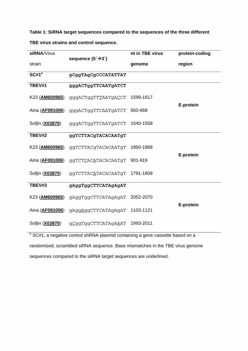
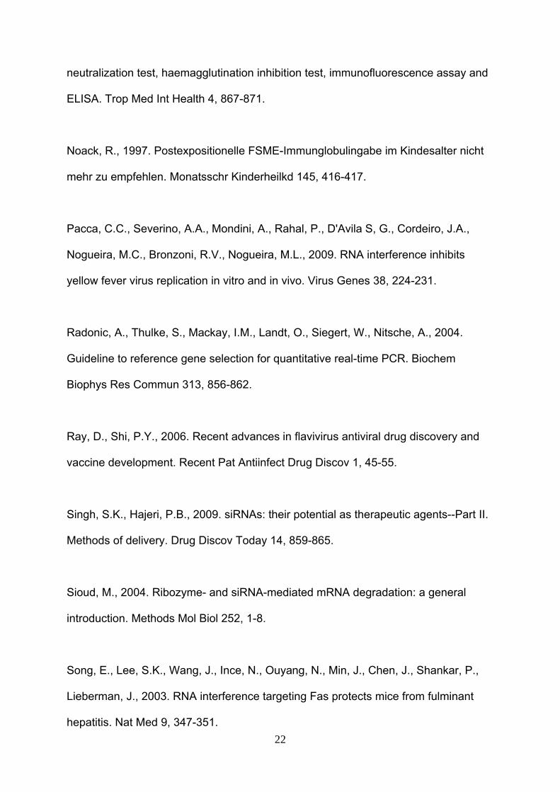
Table 1: SiRNA target sequences compared to the sequences of the three different
TBE virus strains and control sequence.
siRNA/Virus
strain sequence (5´3´)
nt in TBE virus
genome
protein-coding
region
SC#1a gCggTAgCgCCCATATTAT
TBEV#1 gggACTggTTCAATgATCT
K23 (AM600965) gggACTggTTTAATgACCT 1599-1617
Aina (AF091006) gggACTggTTCAATgATCT 650-668
Sofjin (X03870) gggACTggTTCAATgATCT 1540-1558
E-protein
TBEV#2 ggTCTTACgTACACAATgT
K23 (AM600965) ggTCTTACgTACACAATgT 1850-1868
Aina (AF091006) ggTCTCACATACACAATgT 901-919
Sofjin (X03870) ggTCTTACATACACAATgT 1791-1809
E-protein
TBEV#3 gAggTggCTTCATAgAgAT
K23 (AM600965) gAggTggCTTCATAgAgAT 2052-2070
Aina (AF091006) gAggAggCTTCATAgAgAT 1103-1121
Sofjin (X03870) gCggTggCTTCATAgAAAT 1993-2011
E-protein
a SC#1, a negative control shRNA plasmid containing a gene cassette based on a
randomized, scrambled siRNA sequence. Base mismatches in the TBE virus genome
sequences compared to the siRNA target sequences are underlined.
Figure 1: Time course of TBE virus genome equivalent (GE) copy number in infected
(strain K23) HEK293T cells transfected with shRNA plasmids TBEV#1, #2, or #3
compared to HEK293T cells transfected with the control shRNA plasmid SC#1.


Figure 2: Effects of different shRNA plasmid concentrations on TBE virus plaque-
forming units (PFU, white bars) and TBE virus genome equivalent copy number (GE,
hatched bars) in HEK293T cells infected with TBE virus (strain K23) at 24h post
infection.
All three siRNAs (TBEV#1, #2, and #3) significantly reduced (t-test <0.05) the quantity of
TBE virus genome compared to the control SC#1.


Figure 3: Immunofluorescence staining of HEK293T cells infected 24h earlier with TBE
virus and transfected with shRNA plasmids TBEV#1, #2, and #3.
Cells are expressing either the control shRNA SC#1 (A), or shRNAs TBEV#1 (B), TBEV#2
(C), or TBEV#3 (D). Viral proteins are stained red, shRNA-expressing cells fluoresce green,
and the nuclei are stained blue (DAPI). Cells expressing shRNAs and containing TBE virus
appear orange and are indicated by white arrows. The bars represent a length of 20 µm.
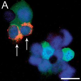
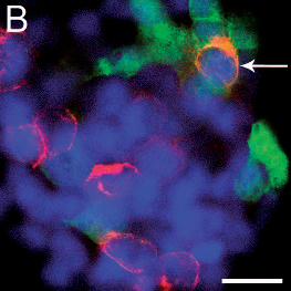

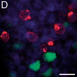
Figure 4: Effect of siRNAs on the quantity of E-protein in TBE virus (strain K23)-
infected HEK293T cells at 24h post infection.
Western Blot analysis was performed using lysates from cells transfected with the control
shRNA plasmid SC#1 or with the most efficient inhibitory plasmids TBEV#2, and TBEV#3.


Figure 5: Effects of siRNAs TBEV#1, #2, and #3 on different TBE virus subtypes.
Experiments with every siRNA and strain were performed at least three times in duplicate (5
times for TBEV strain K23). Standard deviations were calculated for all the experiments and
replicates. The TBE virus genome equivalent (GE) copy number was normalized to the
reference gene tubulin at 24h post infection. Statistically significant differences (t-test p <
0.05) are marked with an asterisk.


>We developed small interfering RNAs (siRNA) targeted to the TBE virus genome.
>siRNAs reduce the quantity of TBE virus particles, genome, and protein up to 85%.
>The 50% inhibitory dose of the shRNA plasmid was 0.05 µg/ml. >RNA interference
could become valuable tool for controlling TBE virus infections.


![[Paliwal, Manisha] Business Ethics(BookFi.org)[1]](https://img.pdfslide.net/doc/110x75/5695d1ba1a28ab9b0297b41d/paliwal-manisha-business-ethicsbookfiorg1.jpg)