Embed Size (px)
Citation preview
www.ketabdownload.com

ACNE AND ITS
THERAPY
www.ketabdownload.com

BASIC AND CLINICAL DERMATOLOGY
Series EditorsALAN R. SHALITA, M.D.
Distinguished Teaching Professor and ChairmanDepartment of Dermatology
SUNY Downstate Medical CenterBrooklyn, New York
DAVID A. NORRIS, M.D.Director of Research
Professor of DermatologyThe University of Colorado
Health Sciences CenterDenver, Colorado
1. Cutaneous Investigation in Health and Disease: Noninvasive Methods andInstrumentation, edited by Jean-Luc Leveque
2. Irritant Contact Dermatitis, edited by Edward M. Jackson and Ronald Goldner3. Fundamentals of Dermatology: A Study Guide, Franklin S. Glickman and
Alan R. Shalita4. Aging Skin: Properties and Functional Changes, edited by Jean-Luc Leveque
and Pierre G. Agache5. Retinoids: Progress in Research and Clinical Applications, edited by
Maria A. Livrea and Lester Packer6. Clinical Photomedicine, edited by Henry W. Lim and Nicholas A. Soter7. Cutaneous Antifungal Agents: Selected Compounds in Clinical Practice
and Development, edited by John W. Rippon and Robert A. Fromtling8. Oxidative Stress in Dermatology, edited by Jurgen Fuchs and Lester Packer9. Connective Tissue Diseases of the Skin, edited by Charles M. Lapiere and
Thomas Krieg10. Epidermal Growth Factors and Cytokines, edited by Thomas A. Luger and
Thomas Schwarz11. Skin Changes and Diseases in Pregnancy, edited by Marwali Harahap and
Robert C. Wallach12. Fungal Disease: Biology, Immunology, and Diagnosis, edited by
Paul H. Jacobs and Lexie Nall13. Immunomodulatory and Cytotoxic Agents in Dermatology, edited by
Charles J. McDonald14. Cutaneous Infection and Therapy, edited by Raza Aly, Karl R. Beutner, and
Howard I. Maibach15. Tissue Augmentation in Clinical Practice: Procedures and Techniques,
edited by Arnold William Klein16. Psoriasis: Third Edition, Revised and Expanded, edited by Henry H. Roenigk,
Jr., and Howard I. Maibach17. Surgical Techniques for Cutaneous Scar Revision, edited by Marwali Harahap18. Drug Therapy in Dermatology, edited by Larry E. Millikan19. Scarless Wound Healing, edited by Hari G. Garg and Michael T. Longaker20. Cosmetic Surgery: An Interdisciplinary Approach, edited by Rhoda S. Narins
www.ketabdownload.com
21. Topical Absorption of Dermatological Products, edited by Robert L. Bronaughand Howard I. Maibach
22. Glycolic Acid Peels, edited by Ronald Moy, Debra Luftman, and Lenore S. Kakita23. Innovative Techniques in Skin Surgery, edited by Marwali Harahap24. Safe Liposuction and Fat Transfer, edited by Rhoda S. Narins25. Pyschocutaneous Medicine, edited by John Y. M. Koo and Chai Sue Lee26. Skin, Hair, and Nails: Structure and Function, edited by Bo Forslind and
Magnus Lindberg27. Itch: Basic Mechanisms and Therapy, edited by Gil Yosipovitch, Malcolm
W. Greaves, Alan B. Fleischer, and Francis McGlone28. Photoaging, edited by Darrell S. Rigel, Robert A. Weiss, Henry W. Lim, and
Jeffrey S. Dover29. Vitiligo: Problems and Solutions, edited by Torello Lotti and Jana Hercogova30. Photodamaged Skin, edited by David J. Goldberg31. Ambulatory Phlebectomy, Second Edition, edited by Mitchel P. Goldman,
Mihael Georgiev, and Stefano Ricci32. Cutaneous Lymphomas, edited by Gunter Burg and Werner Kempf33. Wound Healing, edited by Anna Falabella and Robert Kirsner34. Phototherapy and Photochemotherapy for Skin Disease, Third Edition,
Warwick L. Morison35. Advanced Techniques in Dermatologic Surgery, edited by Mitchel P. Goldman
and Robert A. Weiss36. Tissue Augmentation in Clinical Practice, Second Edition, edited by Arnold
W. Klein37. Cellulite: Pathophysiology and Treatment, edited by Mitchel P. Goldman,
Pier Antonio Bacci, Gustavo Leibaschoff, Doris Hexsel, and Fabrizio Angelini38. Photodermatology, edited by Henry W. Lim, Herbert Honigsmann, and
John L. M. Hawk39. Retinoids and Carotenoids in Dermatology, edited by Anders Vahlquist and
Madeleine Duvic40. Acne and Its Therapy, edited by Guy F. Webster and Anthony V. Rawlings
www.ketabdownload.com

www.ketabdownload.com
Edited by
Guy F. WebsterJefferson Medical College of Thomas Jefferson University
Philadelphia, Pennsylvania, USA
Anthony V. RawlingsAVR Consulting LTD
Northwich, Cheshire, UK
ACNE AND ITS
THERAPY
www.ketabdownload.com
DK4962-Webster-FM_R2_020407
Informa Healthcare USA, Inc.52 Vanderbilt AvenueNew York, NY 10017
# 2007 by Informa Healthcare USA, Inc.Informa Healthcare is an Informa business
No claim to original U.S. Government worksPrinted in the United States of America on acid-free paper10 9 8 7 6 5 4 3 2 1
International Standard Book Number-10: 0-8247-2971-4 (Hardcover)International Standard Book Number-13: 978-0-8247-2971-4 (Hardcover)
This book contains information obtained from authentic and highly regarded sources. Reprintedmaterial is quoted with permission, and sources are indicated. A wide variety of referencesare listed. Reasonable efforts have been made to publish reliable data and information, but theauthor and the publisher cannot assume responsibility for the validity of all materials or for theconsequence of their use.
No part of this book may be reprinted, reproduced, transmitted, or utilized in any form by any elec-tronic, mechanical, or other means, now known or hereafter invented, including photocopying,microfilming, and recording, or in any information storage or retrieval system, without writtenpermission from the publishers.
For permission to photocopy or use material electronically from this work, please access www.copyright.com (http://www.copyright.com/) or contact the Copyright Clearance Center, Inc.(CCC) 222 Rosewood Drive, Danvers, MA 01923, 978-750-8400. CCC is a not-for-profit organizationthat provides licenses and registration for a variety of users. For organizations that have beengranted a photocopy license by the CCC, a separate system of payment has been arranged.
Trademark Notice: Product or corporate names may be trademarks or registered trademarks, andare used only for identification and explanation without intent to infringe.
Library of Congress Cataloging-in-Publication Data
Acne and its therapy / [edited by] Guy F. Webster, Anthony V. Rawlings.p. ; cm. -- (Basic and clinical dermatology ; 40)
Includes bibliographical references and index.ISBN-13: 978-0-8247-2971-4 (hardcover : alk. paper)ISBN-10: 0-8247-2971-4 (hardcover : alk. paper)1. Acne. 2. Acne--Treatment. I. Webster, Guy F. II. Rawlings, Anthony V., 1958- III.
Series.[DNLM: 1. Acne Vulgaris. 2. Acne Vulgaris--therapy. W1 CL69L v.40 2007/WR 430
A187 2007]
RL131.A2568 2007616.503--dc22 2007005325
Visit the Informa Web site atwww.informa.com
and the Informa Healthcare Web site atwww.informahealthcare.com
www.ketabdownload.com
DK4962-Webster-FM_R2_020407
B Introduction
During the past 25 years, there has been a vast explosion in new informationrelating to the art and science of dermatology as well as fundamental cutaneousbiology. Furthermore, this information is no longer of interest only to the smallbut growing specialty of dermatology. Clinicians and scientists from a widevariety of disciplines have come to recognize both the importance of skin infundamental biological processes and the broad implications of understandingthe pathogenesis of skin disease. As a result, there is now a multidisciplinary andworldwide interest in the progress of dermatology.
With these factors in mind, we have undertaken this series of books specifi-cally oriented to dermatology. The scope of the series is purposely broad, withbooks ranging from pure basic science to practical, applied clinical dermatology.Thus, while there is something for everyone, all volumes in the series willultimately prove to be valuable additions to the dermatologist’s library.
The latest addition to the series, volume 40, edited by Drs. Guy F. Webster andAnthony V. Rawlings, is both timely and pertinent. The editors are internationallyrespected for their basic science and clinical expertise in the pathogenesis andtreatment of acne, and have assembled an outstanding group of contributorsfor this latest addition to our series. We trust that this volume will be of broadinterest to scientists and clinicians alike.
Alan R. Shalita, MDDistinguished Teaching Professor and Chairman
Department of DermatologySUNY Downstate Medical Center
Brooklyn, New York, U.S.A.
iii
www.ketabdownload.com

DK4962-Webster-FM_R2_020407
www.ketabdownload.com
DK4962-Webster-FM_R2_020407
B Preface
The intention of this book is to review the latest developments in the understandingof acne and its treatment. The contents cover the molecular and cell biologicalaspects of sebocytes, sebaceous glands, and the pilosebaceous unit through to thepathogenesis of acne, its treatment with hormones, antimicrobials, retinoids, andlaser. Novel actives are reviewed, such as the effect of octadecenedioic acid, sphin-golipids, and enzyme inhibitors. Formulation principles and the importance of fol-licular delivery through sebum are overviewed and, finally, in vitro testingmethods. This book is an invaluable resource for dermatologists as well as scientistsworking in the pharmaceutical and skin care industries. Each chapter reviews themost relevant literature and gives personal insight into tackling the problemsassociated with the treatment of acne, its underlying pathophysiology, and itstherapy.
The book is a result of the contributions of experts in their own areas and is thework of an international team representing scientists from many disciplines. Der-matologists, cosmetic scientists, and researchers will find Acne and Its Therapy aninvaluable and in-depth analysis of the pathogenesis of acne and its treatment.
Guy F. WebsterAnthony V. Rawlings
v
www.ketabdownload.com

DK4962-Webster-FM_R2_020407
www.ketabdownload.com
DK4962-Webster-FM_R2_020407
B Contents
Introduction Alan R. Shalita . . . . iii
Preface . . . . v
Contributors . . . . ix
PART I: THE BIOLOGY OF THE SEBACEOUS GLANDAND PATHOPHYSIOLOGY OF ACNE
1. Overview of the Pathogenesis of Acne 1Guy F. Webster
2. Cell Biology of the Pilosebaceous Unit 9Helen Knaggs
3. Sebum Secretion and Acne 37Philip W. Wertz
4. The Molecular Biology of Retinoids and Their Receptors 45Anthony V. Rawlings
5. Molecular Biology of Peroxisome Proliferator-Activated Receptorsin Relation to Sebaceous Glands and Acne 55Michaela M. T. Downie and Terence Kealey
6. Antimicrobial Peptides and Acne 75Michael P. Philpott
PART II: ACNE TREATMENTS
7. Hormonal Influences in Acne 83Diane Thiboutot
8. Antimicrobial Therapy in Acne 97Guy F. Webster
9. Topical Retinoids 103Daniela Kroshinsky and Alan R. Shalita
10. Phototherapy and Laser Therapy of Acne 113Guy F. Webster
vii
www.ketabdownload.com
DK4962-Webster-FM_R2_020407
11. Benzoyl Peroxide and Salicylic Acid Therapy 117Gabi Gross
12. Treating Acne with Octadecenedioic Acid: Mechanism of Action,Skin Delivery, and Clinical Results 137Johann W. Wiechers, Anthony V. Rawlings, Nigel Lindner, and William J. Cunliffe
13. The Effect of Sphingolipids as a New TherapeuticOption for Acne Treatment 155Saskia K. Klee, Mike Farwick, and Peter Lersch
14. 5a-Reductase and Its Inhibitors 167Rainer Voegeli, Christos C. Zouboulis, Peter Elsner, and Thomas Schreier
PART III: ACTIVE DELIVERY, FORMULATION, AND TESTING
15. Sebum: Physical–Chemical Properties, MacromolecularStructure, and Effects of Ingredients 203Linda D. Rhein, Joel L. Zatz, and Monica R. Motwani
16. Targeted Delivery of Actives from Topical Treatment Products to thePilosebaceous Unit 223Linda D. Rhein, Joel L. Zatz, and Monica R. Motwani
17. Topical Therapy and Formulation Principles 253Steve Boothroyd
18. In Vitro Models for the Evaluation of Anti-acne Technologies 275John Bajor
Index . . . . 303
viii Contents
www.ketabdownload.com
DK4962-Webster-FM_R2_020407
B Contributors
John Bajor Department of Research and Development, Home and Personal Care
Division, Unilever PLC, Trumbull, Connecticut, U.S.A.
Steve Boothroyd Department of Research and Development, Reckitt Benckiser
PLC, Hull, U.K.
William J. Cunliffe Department of Dermatology, Leeds Foundation for
Dermatological Research, Leeds General Infirmary, Leeds, U.K.
Michaela M. T. Downie Sequenom GmbH, Hamburg, Germany
Peter Elsner Department of Dermatology and Allergology, Friedrich-Schiller-University Jena, Jena, Germany
Mike Farwick Degussa, Goldschmidt Personal Care, Essen, Germany
Gabi Gross Department of Research and Development, Reckitt Benckiser PLC,
Hull, U.K.
Terence Kealey The Clore Laboratories, University of Buckingham,
Buckingham, U.K.
Saskia K. Klee Degussa, Goldschmidt Personal Care, Essen, Germany
Helen Knaggs Department of Research and Development, Nu Skin Enterprises,
Provo, Utah, U.S.A.
Daniela Kroshinsky Department of Dermatology, SUNY Downstate Medical
Center, Brooklyn, New York, U.S.A.
Peter Lersch Degussa, Goldschmidt Personal Care, Essen, Germany
Nigel Lindner Department of Research and Development, Uniqema, Gouda,The Netherlands
Monica R. Motwani College of Pharmacy, Rutgers University, Piscataway,New Jersey, U.S.A.
Michael P. Philpott Centre for Cutaneous Research, Institute of Cell andMolecular Science, Barts and the London, Queen Mary’s School of Medicine
and Dentistry, University of London, London, U.K.
Anthony V. Rawlings AVR Consulting Ltd., Northwich, Cheshire, U.K.
Linda D. Rhein School of Natural Sciences, Fairleigh Dickinson University,Teaneck, New Jersey, U.S.A.
ix
www.ketabdownload.com
DK4962-Webster-FM_R2_020407
Thomas Schreier Department of Research and Development, Pentapharm Ltd.,
Basel, Switzerland
Alan R. Shalita Department of Dermatology, SUNY Downstate Medical Center,
Brooklyn, New York, U.S.A.
Diane Thiboutot Department of Dermatology, Pennsylvania State University
College of Medicine, Hershey, Pennsylvania, U.S.A.
Rainer Voegeli Department of Research and Development, Pentapharm Ltd.,
Basel, Switzerland
Guy F. Webster Jefferson Medical College of Thomas Jefferson University,
Philadelphia, Pennsylvania, U.S.A.
Philip W. Wertz Dows Institute, University of Iowa, Iowa City, Iowa, U.S.A.
Johann W. Wiechers JW Solutions, Gouda, The Netherlands
Joel L. Zatz College of Pharmacy, Rutgers University, Piscataway,
New Jersey, U.S.A.
Christos C. Zouboulis Departments of Dermatology and Immunology, Dessau
Medical Center, Dessau, Germany
x Contributors
www.ketabdownload.com
DK4962-Webster-ch1_R2_020407
Part I: The Biology of the Sebaceous Gland andPathophysiology of Acne
B1 Overview of the Pathogenesis of Acne
Guy F. WebsterJefferson Medical College of Thomas Jefferson University, Philadelphia,Pennsylvania, U.S.A.
INTRODUCTION
Acne is an extremely complex disease with elements of pathogenesis involvingdefects in epidermal keratinization, androgen secretion, sebaceous function,bacterial growth, inflammation, and immunity. In the past 30 years, much hasbeen worked out, and we now have a fairly detailed understanding of the eventsthat result in an acne pimple, although there is also much left to be discovered.
COMEDO FORMATION
The initial event in acne is the formation of comedo, a plug in the follicle, which istermed “open” if a black tip is visible in the follicular orifice and “closed” if theopening has not distended enough to be visible without magnification. Patients(and their mothers) erroneously conclude that this black tip is due to dirt in thefollicle. Rather, it represents oxidized melanin and perhaps certain sebaceouslipids (1,2). The earliest lesion is termed microcomedo and is clinically inapparent,but is the lesion that gives rise to inflammatory acne. Microcomedones are bestvisualized by harvesting them using cyanoacrylate glue (3). By this method, micro-comedones are seen to be numerous on the skin of acne patients, and much lessprevalent and less robust on the skin of normal individuals.
Comedo formation begins with faulty desquamation of the follicular lining.Instead of shedding as fine particles, the epithelium comes off in sheets that areincapable of exiting through the follicular orifice, and hence a plug results. Con-centric laminae of keratinous material fill and distend the follicle. This process isfirst detectable at the junction of the sebaceous duct and the follicular epitheliumand involves in distal cells later. The granular layer becomes prominent, tono-filaments increase, and lipid inclusions form the desquamated keratin (1,4).
Most comedones contain hairs, usually small vellus hairs, and the age of acomedo may be reflected by the number of hairs that it contains (5). Terminalhairs are almost never seen in comedones. It may be that the presence of a stouthair in the follicle provides a mechanical opening that prevents comedo distention.Is it possible that the conversion of vellus to terminal hairs as acne patients matureis the explanation for the decrease in acne in the late teens and early 20s?
The cause of the faulty desquamation that leads to comedo formation is notknown. Comedones have been demonstrated before puberty, so activation of sebac-eous secretion cannot be the key event (1). Many compounds have been shown toinduce comedones in experimental systems (e.g., coal tar, sulfur, squalene, haloge-nated biphenyls, and cutting oils), but none are obviously relevant to the naturalcourse of acne formation (6–8). Two experimental systems exist for studyingcomedo formation: the rabbit ear model and the backs of human volunteers.
1
www.ketabdownload.com
DK4962-Webster-ch1_R2_020407
In general, the rabbit ear is more sensitive and forms plugs easily, but there isgenerally good agreement between the two systems for most compounds (6–8).
Physical agents may also enhance comedogenesis. Favre–Racouchaut syn-drome consists of severe photodamage accompanied by open comedones on theface (9). Mills et al. (10,11) have demonstrated that UV irradiation will enhancethe comedo formation in the rabbit ear engendered by squalene, cocoa butter,sebum, and some sunscreens.
Inflammation may also play a role in the formation of comedones. A ring ofcomedones may be occasionally seen around a large inflammatory nodule on theback of patients with severe acne. In vitro studies have shown that Propionibacteriumacnes cell walls will induce follicular plugging in proportion to the degree of inflam-mation triggered by bacteria in the skin of rats (12). More recent studies in an invitro model of the acne follicle show that cytokines such as interleukin (IL)1-amodulate the cornification of the epidermis and may be involved in the inflamma-tory induction of comedones (13,14).
Another potential cause of comedo formation is the lipid contents of the fol-licle itself. Bacterial lipolysis will liberate fatty acids from sebaceous triglyceridesthat are comedogenic, but the presence of microcomedones in the skin of prepuber-tal children (who have no follicular microflora and no sebum) argues against amajor role of bacterial action in early comedogenesis (15). Strauss et al. (16) haveshown that the sebum of acne patients is relatively deficient in linoleic acid,perhaps reflective of high sebum secretion rates and have suggested that locallinoleic acid deficiency may be involved in comedo formation. Further study ofthis possibility is warranted.
BACTERIAL FACTORS
The skin microflora is greatly influenced by the onset of puberty. Before this hormonalflood, the sebaceous gland is inactive and bacterial populations are low. The arrival of alipid product with about 50% triglyceride on the skin greatly stimulates bacterialgrowth and selects bacteria that can effectively metabolize triglycerides. A oncesterile follicle becomes the residence of P. acnes, an anaerobe, it metabolizes the glycerolfraction of triglycerides, which sterile follicle liberates with an extracellular lipase (17).Lipase cleaves triglycerides into fatty acids and glycerol, and the fatty acids remain insebum in proportion to the P. acnes population (18). It was once thought that these fattyacids were the primary stimulants for inflammation in acne, but now they are believedto be a relatively minor contributor to the process.
Although tens of millions of P. acnes present in a square centimeter area on theface (19,20), yet infection with the organism is rare and is typically postsurgical. It istruly a commensal, incapable of surviving in skin without unusual conditions. Wemay derive some benefits from P. acnes colonization. Group A streptococci areinhibited by fatty acids produced by P. acnes (21), which may account for therarity of facial streptococcal impetigo after puberty.
P. acnes populations are proportional to the amount of sebum produced butthere is variation amongst the cutaneous microenvironments. Sebum-rich areassuch as the face and upper trunk carry mean log populations between 4.8 and5.5 cm22, whereas the lipid deficient legs harbor only 0.5 cm22 (20). Animal skindoes not support the growth of P. acnes, because animal sebum does not containtriglyceride (22), a major reason why there is no satisfactory animal model availablefor inflammatory acne. The distribution of active sebaceous glands and high
2 Webster
www.ketabdownload.com
DK4962-Webster-ch1_R2_020407
P. acnes populations are reason for the distribution of inflammatory acne lesions. Thelargest and most active sebaceous glands are located on the face, upper trunk, andarms, regions where acne is common (23). The lower trunk and distal extremitieshave negligible sebaceous activity, trivial P. acnes populations, and no acne (19).
The severity of acne is also somewhat linked to sebaceous secretion andP. acnes populations. Teenage acne patients have higher levels of bacteria in theirfollicles than do age-matched controls (24). Although there is a good degree ofoverlap between acne and nonacne groups, in general, teenage acne patientshave higher sebum production than their normal counterparts, accounting fortheir greater bacterial populations (25). Interestingly, this difference is less pro-nounced in older individuals with the disease.
INFLAMMATION IN ACNE
Formation of acne pimples and pustules typically begins at the microcomedonesformation. Kligman (1) has observed that visible comedones only, rarely, becomeinflamed and microcomedones have been shown to contain evidence of neutrophilactivity, even though they came from areas of the skin with no acne lesions (26). Thetrigger for the inflammation of the microcomedo is the comedonal resident P. acnesthat has many characteristics that incite the inflammatory and immune responses.
The organism P. acnes is a potent activator of many facets of the innateimmune system, and under the archaic name of Corynebacterium parvum, P. acneshas been found to be a potent macrophage activator similar to BCG (27). P. acnesmakes chemotactic substances that attract neutrophils and monocytes. Low mol-ecular weight peptides are produced as a consequence of postsynthetic protein pro-cessing by the organism. Neutrophils recognize these peptides by the same receptoras other bacterial chemotactic peptides (28,29) (Tables 1 and 2). These peptides are,2 kDa in mass and accumulate as the organism grows. Presumably small enoughto leach out from an intact follicle, these compounds may be part of the initialstimulus for inflammation. P. acnes produces at least one other chemotaxin; thelipase that cleaves triglycerides in sebum is also attractive to leukocytes (30).
P. acnes is a potent activator of the classic and complement pathways. It is themajor and perhaps sole activator in the comedo (31) and complement depositionaround the inflamed acne lesions is great (32). The alternative pathway activatoris a mannose-containing cell-wall polysaccharide that shares characteristics withthe macrophage-activating factor in P. acnes cell wall (33–35). In the classicalpathway, the activation is through the formation of immune complexes with anti-P. acnes antibody. The more the antibody present, the more the activation occurs(36). Thus, complement activation and the subsequent generation of C5-derived che-motactic factors are greatest in patients with high levels of anti-P. acnes immunity.
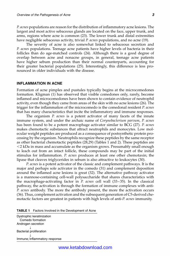
TABLE 1 Factors Involved in the Development of Acne
Dystrophic keratinizationComedo formation
Androgen secretion#
Bacterial proliferation#
Immune/inflammatory response
Overview of the Pathogenesis of Acne 3
www.ketabdownload.com
DK4962-Webster-ch1_R2_020407
Toll-like receptors (TLRs) are more recently discovered components of innateimmunity, which involve cell-mediated defenses in response to the pathogens in theabsence of an immune response. Vowels et al. (37) have demonstrated that P. acnesstimulates proinflammatory cytokines such as IL-8, tumor necrosis factor (TNF)-a,and IL1-b in monocytes. Lee et al. (38) have shown that P. acnes cell-wallcomponents activate TLR-2 in monocytes, resulting in the production of TNF-a,IL1-b, and IL-8 that attract both neutrophils and lymphocytes to the follicle. Thisprocess that is involved in acne is supported by the identification of monocytesin inflamed acne lesions, expressing TLR2 on their surfaces. Activation of TLRs byP. acnes also accounts for the observation that CD4-bearing lymphocytes appear atthe comedo, early in the initiation of acne inflammation (39).
RESOLUTION OF ACNE LESIONS
Surprisingly, little is known about the processes involved in the healing of acnelesions, which often takes weeks to occur. Kligman (1) observed the evolutionand healing of acne lesions and noted a late influx of lymphocytes and the for-mation of granulomas. Electron microscopy has shown these cells to be syntheti-cally and metabolically active (40). The stimulus for the inflammation is probablypersistence of P. acnes. The organism is unusually difficult for leukocytes todegrade. Injected P. acnes will remain in tissue for weeks, inciting ongoing inflam-mation (41,42). In vitro studies find that the organism is far more resistant to degra-dative enzymes from neutrophils and monocytes than a genuine pathogen such asStaphylococcus aureus (43) that is degraded within hours. In contrast, P. acnes degra-dation procedes at a glacial pace, requiring 24 hours for the release of only 10% ofcell-wall mass supporting the observation of persistence of injected organisms aftermany weeks.
THE ROLE OF PROPIONIBACTERIUM ACNES–SPECIFICIMMUNITY IN ACNE
The presence of elevated immunity to P. acnes may be the factor that determinesthe severity of a patient’s acne. Other potential explanations such as elevatedandrogens and subsequent increased sebum secretion clearly may play a role indetermining acne severity, but their influence is probably not the primary issue.
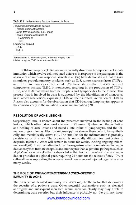
TABLE 2 Inflammatory Factors Involved in Acne
Propionibacterium acnes-derivedPeptide chemoattractantsLarge MW molecules, e.g., lipaseInnate immune activators of
ComplementTLR
Leukocyte-derivedIL1-bTNF-aIL-8
Abbreviations: IL, interleukin; MW, molecular weight; TLR,toll-like receptors; TNF, tumor necrosis factor.
4 Webster
www.ketabdownload.com
DK4962-Webster-ch1_R2_020407
It is known that virilized women may have more severe acne (44), but not allhyperandrogenic women fit this stereotype. In fact, many hirsuite, hyperandrogenicwomen have no acne at all, and among those who do have acne, it tends not to beparticularly severe (45,46). Moreover, correction of the hyperandrogenicity typicallyresults in an improvement, but not a complete resolution of the acne (47). Thus,virilization is permissive for severe acne, but not the prime factor that causes it.
There is substantial evidence that a patient’s anti-P. acnes immunity maybe the factor that determines acne severity. Agglutinating and complement-fixingantibodies to P. acnes are elevated in proportion to the severity of acne inflammation(48–51). Lymphocyte proliferation in response to P. acnes antigens is likewise elev-ated (52,53). Skin test reactivity to comedonal contents and to P. acnes fractions isproportional to acne severity as well (54).
There is substantial evidence that elevated immunity makes P. acnes a morepotent inflammatory stimulus. Complement activation by comedonal contents isincreased by the addition of anti-P. acnes antibody (31). Complement activationby P. acnes organisms in vitro is intensified by increasing amounts of anti-P. acnesantibody (33) and results in the generation of increased amounts of neutrophilchemoattractants. When neutrophils encounter the organism, they release destruc-tive hydrolases into tissue in proportion to the amount of anti-P. acnes antibodypresent in the system (55). Thus, humoral immunity to the organism is proinflam-matory, rather than protective of infection, and most likely serves to intensifyinflammation and tissue damage. Which then comes first, immunity or acne? Inthe absence of direct experimental data, the author would contend that the hyper-sensitivity to P. acnes is an inherited tendency and is the factor that accounts formany cases of severe acne in unfortunate families.
What is the future of acne research? Much is left to be understood regardingthe role of endogenous antimicrobial peptides and TLRs in controlling the inflam-matory response in acne, and methods to decrease severe scarring are lacking.
REFERENCES
1. Kligman AM. An overview of acne. J Invest Dermatol 1974; 62:268–287.2. Blair C, Lewis CA. The pigment of comedones. Br J Dermatol 1970; 82:572–583.3. Marks R, Dawber RPR. Skin surface biopsy: an improved technique for examination of
the horny layer. Br J Dermatol 1971; 84:117–123.4. Knutson DD. Ultrastructural observations in acne vulgaris: the normal sebaceous follicle
and acne lesions. J Invest Dermatol 1974; 62:288–307.5. Leyden JJ, Kligman AM. Hairs in acne comedones. Arch Dermatol 1972; 106:851–853.6. Kaidbey KH, Kligman AM. A human model for coal tar acne. Arch Dermatol 1974;
109:212–215.7. Morris WE, Kwan SC. Use of the rabbit ear model in evaluating the comedogenic poten-
tial of cosmetic ingredients. J Soc Cosmet Chem 1983; 34:215–225.8. Kligman AM, Kowng T. An improved rabbit ear model for assessing comedogenic
substances. Br J Dermatol 1979; 100:699–702.9. Izumi A, Marples RR, Kligman AM. Senile comedones. J Invest Dermatol 1973; 61:46–50.
10. Mills OH, Kligman AM. Comedogenicity of sunscreens. Arch Dermatol 1982; 118:417–419.
11. Mills OH, Porte M, Kligman AM. Enhancement of comedogenic substances by ultra-violet readiation. Br J Dermatol 1978; 98:145–150.
12. Deyoung LM, Spires DA, Ballaron SJ. Acne like chronic inflammatory activity ofPropionibacterium acnes preparations in an animal model. J Invest dermatol 1985; 85:255–258.
Overview of the Pathogenesis of Acne 5
www.ketabdownload.com
DK4962-Webster-ch1_R2_020407
13. Guy R, Kealey T. The effects of inflammatory cytokines on the isolated human sebaceousepithelium. J Invest Dermatol 1998; 110:410–415.
14. Guy R, Green MR, Kealey T. Modeling acne invitro. J Invest Dermatol 1996; 106:176–182.
15. Lavker RM, Leyden JJ, McGinley KJ. The relationship between bacteria and the abnor-mal follicular keratinization in acne vulgaris. J Invest Dermatol 1981; 77:325–330.
16. Morello AM, Downing DT, Strauss JS. Octadecaenoic acids in the skin surface lipids ofacne patients and normal controls. J Invest Dermatol 1976; 66:319–332.
17. Leyden JJ, McGinley KJ, Webster GF. Cutaneous bacteriology. In: Goldsmith L, ed. ThePhysiology and Biochemistry of the Skin. London: Oxford Press, 1983:1153–1165.
18. Marples RR, Downing DT, Kligman AM. Control of free fatty acids in skin surface lipidby Corynebacterium acnes. J Invest Dermatol 1971; 56:127–131.
19. McGinley KJ, Webster GF, Leyden JJ. Regional variations of cutaneous propionibacteria.Appl Environ Microbiol 1978; 35:62–66.
20. McGinley KJ, Webster GF, Ruggieri MR, Leyden JJ. Regional variations of cutaneouspropionibacteria, correlation of Propionibacterium acnes populations with sebaceoussecretion. J Clin Microbiol 1980; 12:672–675.
21. Speert DP, Wannamaker LW, Gray ED, Clawson CC. Related articles, Bactericidal effectof oleic acid on group A streptococci: mechanism of action. Infect Immun 1979;26(3):1202–1210.
22. Webster GF, Ruggieri MR, McGinley KJ. Correlation of Propionibacterium acnes popu-lations with the presence of triglycerides on non-human skin. Appl Environ Microbiol1981; 41:1269–1270.
23. Cunliffe WJ, Perera WDH, Thackray P. Pilosebaceous duct physiology III. Observationson the number and size of pilosebaceous ducts in acne vulgaris. Br J Dermatol 1970;82:572–583.
24. Leyden JJ, McGinley KJ, Mills OH, Kligman AM. Propionibacterium levels in patientswith and without acne vulgaris. J Invest Dermatol 1975; 65:382–384.
25. Pochi P, Strauss JS, Rao RS. Plasma testosterone and sebum production in males withacne vulgaris. J Clin Endocrinol Metab 1965; 51:287–291.
26. Webster GF, Kligman AM. A method for the assay of inflammatory mediators in follicu-lar casts. J Invest Dermatol 1979; 73:266–268.
27. Cummins CS, Johnson JL. Corynebacterium parvum: a synonym for Propionibacteriumacnes? J Gen Microbiol 1974; 80(2):433–442.
28. Webster GF, Leyden JJ, Tsai C-C. Characterization of serum independent polymorpho-nuclear leukocyte chemotactic factors produced by Propionibacterium acnes. Inflam-mation 1980; 4:261–271.
29. Puhvel SM, Sakamoto M. Cytotoxin production by comedonal bacteria. J InvestDermatol 1978; 71:324–329.
30. Lee WL, Shalita AR, Sunthralingam K. Neutrophil chemotaxis to P. acnes lipase and itsinhibition. Infect Immun 1982; 35:71–78.
31. Webster GF, Leyden JJ, Nilsson UR. Complement activation by in acne vulgaris,consumption of complement by comedones. Infect Immun 1979; 26:186–188.
32. Leeming JP, Ingham E, Cunliffe WJ. Microbial contents and complement C3 cleavingactivity of comedones in acne vulgaris. Acta Derm Venereol 1988; 68:469–473.
33. Webster GF, Nilsson UR, McArthur WR. Activation of the alternative pathway of comp-lement by Propionibacterium acnes cell fractions. Inflammation 1981; 5:165–176.
34. Webster GF, McArthur WR. Activation of components of the alternative pathway ofcomplement by Propionibacterium acnes cell wall carbohydrate. J Invest Dermatol 1982;79:137–140.
35. Cummins CS, Linn DM. Related articles. Reticulostimulating properties of killed vac-cines of anaerobic coryneforms and other organisms. J Natl Cancer Inst 1977;59(6):1697–1708.
36. Webster GF, Leyden JJ, Norman ME, Nilsson UR. Complement activation in acne vul-garis: in vitro studies with Propionibacterium acnes and Propionibacterium granulosum.Infect Immun 1978; 22:523–529.
6 Webster
www.ketabdownload.com
DK4962-Webster-ch1_R2_020407
37. Vowels BR, Yang S, Leyden JJ. Induction of proinflammatory cytokines by a solublefactor of Propionibacterium acnes implications for chronic inflammatory acne. InfectImmun 2000; 63:3158–3165.
38. Kim J, Ochoa M-T, Krutzik SR, et al. Activation of toll-like receptor 2 in acne tiggersinflammatory cytokine responses. J Immunol 2002; 169:1535–1541.
39. Norris JFB, Cunliffe WJ. A histological and immunocytochemical study of early acnelesions. Br J Dermatol 1988; 118:651–659.
40. Lavker RM, Leyden JJ, Kligman AM. The anti-inflammatory activity of isotretinoin is amajor factor in the clearing of acne conglobata. In Marks R, Plewig G, eds. Acne andRelated Disorders. London: Dunitz, 1989:207–216.
41. Sadler TE, Crump WA, Castro JE. Radiolabelling of Corynebacterium parvum and itsdistribution in mice. Br J Cancer 1977; 35:357–368.
42. Dimitrov NV, Greenberg CS, Denny T. Organ distribution of Corynebacterium parvumlabeled with I-125. J Nat Canc Inst 1977; 58:287–294.
43. Webster GF, Leyden JJ, Musson RA, Douglas SD. Susceptibility of Propionibacteriumacnes to killing and degradation by human monocytes and neutrophils in vitro. InfectImmun 1985; 49:116–121.
44. Marynick SP, Chakmakjian ZH, McCaffree DL, Herndon JH. Androgen excess in cysticacne. N Engl J Med 1983; 308:981–986.
45. Vexiau P, Husson C, Chivot M. Androgen excess in women with acne alone compared towomen with acne and or hirsuitism. J Invest Derm 1990; 94:279–283.
46. Steinberger E, Smith KD, Rodrigues-Ridau LJ. Testosterone, dehydroepiandrosteroneand dehydroepiandrosterone sulfate in hyperandrogenic women. J Clin EndocrinolMetab 1984; 59:47–477.
47. Nader S, Rodriguez-Rigau LJ, Smith KD, Sternberger E. Acne and hyperandrogenism.Impact of lowering androgen levels with glucocorticoid treatment. J Am Acad Derm1984; 11:256–259.
48. Puhvel SM, Barfatani M, Warnick M. Study of antibody levels to Corynebacterium acnes.Arch Derm 1964; 90:421–427.
49. Puhvel SM, Hoffamn CK, Sternberg TH. Presence of complement fixing antibodies toCorynebacterium acnes in the sera of acne patients. Arch Derm 1966; 93:364–368.
50. Webster GF, Indrisano JP, Leyden JJ. Antibody titers to Propionibacterium acnes cell wallcarbohydrate in nodulocystic acne patients. J Invest Derm 1985; 84:496–500.
51. Holland KT, Holland DB, Cunliffe WJ, Cutcliffe AG. Detection of Propionibacteriumacnes polypeptides which have stimulated an immune response in acne patients butnot in normal individuals. Exp Derm 1993; 2:12–16.
52. Puhvel SM, Amirian DA, Weintraub J. Lymphocyte transformation in subjects withnodulocystic acne. Br J Derm 1977; 97:205–210.
53. Gowland G, Ward RM, Holland KT, Cunliffe WJ. Cellular immunity to P. acnes in thenormal population and in patients with acne. Br J Derm 1978; 99:43–48.
54. Kersey P, Sussman M, Dabl M. Delayed skin test reactivity to P. acnes correlates with theseverity of inflammation in acne vulgaris. Br J Derm 1980; 103:651–655.
55. Webster GF, Leyden JJ, Tsai CC, McArthur WP. Polymorphonuclear leukocyte lysosomalenzyme release in response to Propionibacterium acnes in vitro and its enhancement bysera from patients with inflammatory acne. J Invest Der 1980; 74:398–401.
Overview of the Pathogenesis of Acne 7
www.ketabdownload.com

DK4962-Webster-ch1_R2_020407
www.ketabdownload.com
DK4962-Webster-ch2_R2_240307
B2 Cell Biology of the Pilosebaceous Unit
Helen KnaggsDepartment of Research and Development, Nu Skin Enterprises, Provo, Utah, U.S.A.
INTRODUCTION
This chapter reviews the structure and function of the pilosebaceous unit andthe controlling influences on the pilosebaceous unit and sebum secretion. Thechapter is divided into three sections. Section I gives an account of the structureand function of the normal pilosebaceous unit; Section II describes the biochemistryand regulation of pilosebaceous unit biology; and finally, Section III dealsbriefly with the biochemical changes occurring in the pilosebaceous duct in acne.
SECTION ONE: ANATOMYStructure of the Pilosebaceous UnitIn humans, pilosebaceous units or pilosebaceous follicles are found on all skin sur-faces, apart from the palms of the hands and soles of the feet. Essentially, they areinvaginations of the epidermis into the dermis. Each comprises a duct, which endsin the dermal papilla, a hair fiber (or pilus) produced by the dermal papilla, a sebac-eous gland and its associated sebaceous duct. The duct supports and protects thehair fiber and also drains sebum produced by the sebaceous gland and carries itto the skin surface. In addition, in split thickness wounds, the cells of the ductal epi-thelium are a source of proliferating keratinocytes, which migrate to re-epithelializethe wound (1). A specialized population of epithelial cells called stem cells, locatedin the bulge region situated below the sebaceous gland, are believed to be crucial forthis (2). These cells are pluripotent and can also differentiate in some circumstancesto produce ductal keratinocytes and sebocytes (3,4). Both the hair and sebumare products of pilosebaceous follicles, emerging onto the skin surface. Sebum isa holocrine secretion from the sebaceous gland cells or sebocytes, which meansthat the cells are destroyed when sebum is released. The function of sebum inhumans is unclear, but as will be discussed later it may play a role in severalskin functions (5,6).
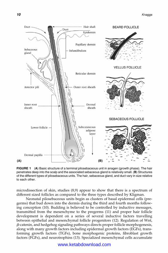
According to Kligman (7), three types of pilosebaceous units may be distin-guished histologically based on the relative proportions of duct, gland, and hairin each: the terminal follicle, the vellus follicle, and the sebaceous follicle. Terminalfollicles produce long hairs and are found, for example, on the scalp (Fig. 1). Thesepilosebaceous ducts are long, relative to those of other follicles, penetrating deepinto the dermis, and the associated sebaceous glands are relatively small. Thehair acts as a wick facilitating the passage of sebum and possibly also desquamatingductal cells to the surface of the skin. In contrast, vellus and sebaceous follicles havesmall, vestigial hairs and relatively large sebaceous glands. Sebaceous follicles aredistinguished from vellus follicles by their large sebaceous gland and large follicu-lar orifices (pores), which are visible clinically to the naked eye. However, based on
9
www.ketabdownload.com
DK4962-Webster-ch2_R2_240307
microdissection of skin, studies (8,9) appear to show that there is a spectrum ofdifferent sized follicles as compared to the three types described by Kligman.
Neonatal pilosebaceous units begin as clusters of basal epidermal cells (pre-germs) that bud down into the dermis during the third and fourth months follow-ing conception (10). Budding is believed to be controlled by inductive messages,transmitted from the mesenchyme to the pregerms (11) and proper hair follicledevelopment is dependent on a series of several inductive factors travellingbetween epithelial and mesenchymal follicle progenitors (12). Regulation of Wnt,b-catenin, and hedgehog signaling pathways directs proper follicle morphogenesis,along with many growth factors including epidermal growth factors (EGFs), trans-forming growth factors (TGFs), bone morphogenic proteins, fibroblast growthfactors (FGFs), and neurotrophins (13). Specialized mesenchymal cells accumulate
FIGURE 1 (A) Basic structure of a terminal pilosebaceous unit in anagen (growth phase). The hairpenetrates deep into the scalp and the associated sebaceous gland is relatively small. (B) Structuresof the different types of pilosebaceous units. The hair, sebaceous gland, and duct vary in size relativeto each other.
10 Knaggs
www.ketabdownload.com

DK4962-Webster-ch2_R2_240307
beneath and around the pregerms and direct the elongation of the cells toform oblique hair pegs in the dermis. By approximately the fifth month, thepilosebaceous units are clearly visible and the keratinization of the duct begins.At this stage there are no follicular orifices. As cells in the center of what is to bethe duct senesce, a central canal forms allowing the development of the hairfiber. The growth of the hair upwards removes the cellular debris from the canaland produces the follicular pore. It is believed that no new pilosebaceous unitsare formed after birth (14). However, recent work indicates that the epidermiscan still maintain its capacity to produce follicles, if it is combined with embryonictrichogenic dermis by transplantation (15).
Sebaceous glands form from the outgrowths of the outer root sheaths of hairfollicles and are clearly visible by the 15th week of fetal life (16). The factors sur-rounding the development of sebaceous glands are complex, although there aredata supporting a critical role for the hedgehog-signaling pathway. Thus, inhibitionof hedgehog signaling blocked the development of sebocytes, while activation ledto a striking increase in size and number of sebaceous glands in transgenic mice(17,18). In the embryo, sebum secretion contributes to the vernix caseosa and theamniotic fluid. During the neonatal period, sebaceous gland function is regulatedby both fetal steroid synthesis and maternal androgens. At parturition, the sebac-eous glands are well developed, largely due to the influence of the maternal andro-gens. However, during the first six months of life they become small and atrophic,remaining so until puberty, when they mature and become active under theinfluence of pubertal androgens.
DistributionAs mentioned earlier, all skin areas excluding the glabrous skin, that is, the palmar(palms) and plantar (soles), possess pilosebaceous units. Although the number ofsecreting follicles, and consequently sebum output, varies greatly between individ-uals, the distribution and shape of follicles tend to follow the same pattern over thehuman body (19). The highest density of sebaceous and vellus follicles is found onthe face, especially on the forehead, where there may be as many as 900 glands/cm2 in some areas (20), but the number varies according to the study: Blume et al.(21) determined a density of 423 follicles/cm2, Pagnoni et al. (22) found 455 fol-licles/cm2 on the lateral forehead and up to 1220 follicles/cm2 in the nose area,and most recently, Otberg et al. (23) determined a number of 292 follicles/cm2 onthe forehead. Nevertheless, there is agreement that sebum output is maximal onthe forehead, nose, and chin, the so-called “t-zone” and decreases toward the outeredges of the face (22). Elsewhere on the body, there may be fewer than 100pilosebaceous units/cm2 (23). The face contains the largest sebaceous glands, butdespite this the ducts serving these glands are smallest on the face, particularly,on the forehead and have a significantly smaller pore compared to those foundon the back. It has been calculated that the smaller duct creates a five timesgreater resistance to sebum output on the forehead compared to the back (24).This greater resistance exerted by the small duct on sebum flow, may, in part,explain the prevalence of pilosebaceous diseases on the face, involving highsebum secretion, such as acne.
Other types of sebaceous glands are found in humans distributed over epithelialsurfaces and open directly onto the surfaces upon which they secrete. These so-calledfree sebaceous glands are particularly prevalent in transitional areas between skin and
Cell Biology of the Pilosebaceous Unit 11
www.ketabdownload.com
DK4962-Webster-ch2_R2_240307
mucosal membranes for example, anogenital region, lips, and meibomian glands inthe eyelids. Sebaceous glands are also located in oral mucosa (called Fordycespots), the digestive and respiratory tract, and the areoles of the nipples.
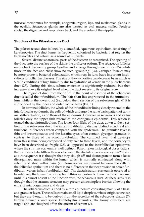
Structure of the Pilosebaceous Duct
The pilosebaceous duct is lined by a stratified, squamous epithelium consisting ofkeratinocytes. The duct lumen is frequently colonized by bacteria that rely on thekeratinocytes and sebum as a source of nutrients.
Several distinct anatomical parts of the duct can be recognized. The opening ofthe duct onto the surface of the skin is the orifice or ostium. The sebaceous follicleson the back frequently group together and emerge through one orifice (25), whilethose on the face and chest show no such “grouping” (26). Grouped follicles maybe more prone to bacterial colonization, which may, in turn, have important impli-cations for follicular diseases. The size of the duct orifice can decrease by as much as50% in conditions of high humidity due to hydration of keratin in the pilosebaceousduct (27). During this time, sebum excretion is significantly reduced, but thenincreases above its original level when the duct reverts to its original size.
The region of duct from the orifice to the point of insertion of the sebaceousduct is called the infundibulum. The hair shaft lies unprotected in the infundibu-lum, while in the lower duct (i.e., below the insertion of the sebaceous gland) it issurrounded by the inner and outer root sheaths (Fig. 1).
In terminal follicles, the whole of the infundibular lining closely resembles theinterfollicular epidermis, the cells of which undergo the same basic pattern of term-inal differentiation, as do those of the epidermis. However, in sebaceous and vellusfollicles only the upper fifth resembles the contiguous epidermis. This region istermed the acroinfundibulum. The lower four-fifths of the duct, down to the inser-tion of the sebaceous duct, the infrainfundibulum, display distinct structural andfunctional differences when compared with the epidermis. The granular layer isthin and inconspicuous and the keratinocytes often contain glycogen granules incontrast to those of the acroinfundibulum. The cornified layer that developsfrom this is thin, being composed of only two to three layers, and the corneocyteshave been described as fragile (28), as opposed to the interfollicular epidermis,where the stratum corneum is well defined. Based upon histological observations,there appears to be little adherence between the ductal cells or corneocytes (28), andas a consequence it is thought that they slough off and disintegrate to form a loosedisorganized mass within the lumen which is normally eliminated along withsebum and shed vellus hairs (7). Desmosomes are present between the cells ofthe follicular epithelium and there is no difference in their expression in acroinfun-dibulum versus infrainfundibulum (29). The ductal stratum corneum is observed tobe relatively thick near the orifice, but it thins as it extends down the follicular canaluntil it is almost absent at the junction with the sebaceous duct. At these sites, it isthought that the stratum corneum may present an incomplete barrier, allowing theentry of microorganisms and drugs.
The sebaceous duct is lined by a thin epithelium consisting mainly of a basaland granular layer. These cells contain small lipid droplets, whose origin is unclear,but they are thought to be derived from the secretion of the sebaceous glands (7),keratin filaments, and sparse keratohyalin granules. The horny cells here arefragile and are sloughed off in the stream of sebum (7).
12 Knaggs
www.ketabdownload.com
DK4962-Webster-ch2_R2_240307
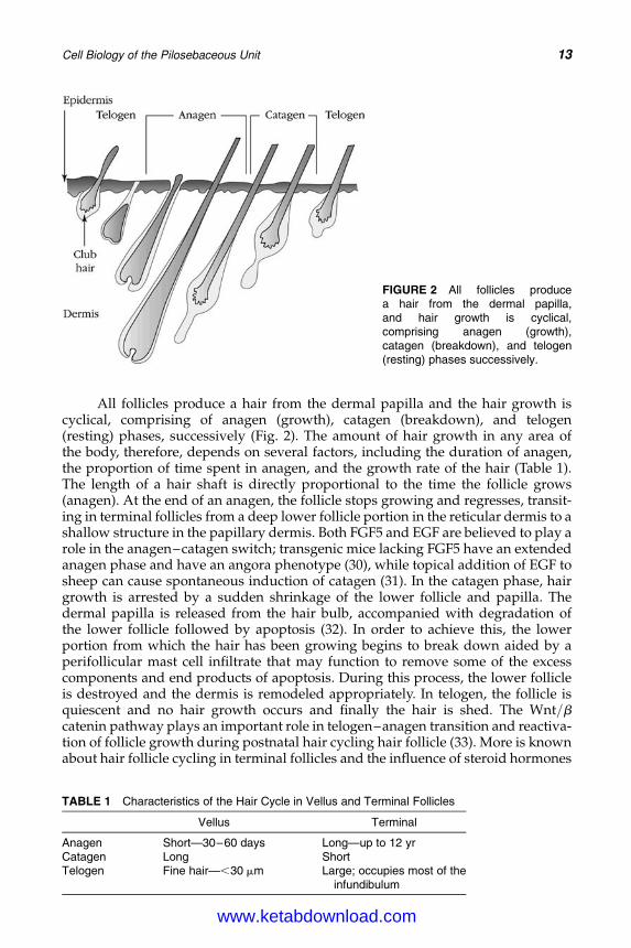
All follicles produce a hair from the dermal papilla and the hair growth iscyclical, comprising of anagen (growth), catagen (breakdown), and telogen(resting) phases, successively (Fig. 2). The amount of hair growth in any area ofthe body, therefore, depends on several factors, including the duration of anagen,the proportion of time spent in anagen, and the growth rate of the hair (Table 1).The length of a hair shaft is directly proportional to the time the follicle grows(anagen). At the end of an anagen, the follicle stops growing and regresses, transit-ing in terminal follicles from a deep lower follicle portion in the reticular dermis to ashallow structure in the papillary dermis. Both FGF5 and EGF are believed to play arole in the anagen–catagen switch; transgenic mice lacking FGF5 have an extendedanagen phase and have an angora phenotype (30), while topical addition of EGF tosheep can cause spontaneous induction of catagen (31). In the catagen phase, hairgrowth is arrested by a sudden shrinkage of the lower follicle and papilla. Thedermal papilla is released from the hair bulb, accompanied with degradation ofthe lower follicle followed by apoptosis (32). In order to achieve this, the lowerportion from which the hair has been growing begins to break down aided by aperifollicular mast cell infiltrate that may function to remove some of the excesscomponents and end products of apoptosis. During this process, the lower follicleis destroyed and the dermis is remodeled appropriately. In telogen, the follicle isquiescent and no hair growth occurs and finally the hair is shed. The Wnt/bcatenin pathway plays an important role in telogen–anagen transition and reactiva-tion of follicle growth during postnatal hair cycling hair follicle (33). More is knownabout hair follicle cycling in terminal follicles and the influence of steroid hormones
FIGURE 2 All follicles producea hair from the dermal papilla,and hair growth is cyclical,comprising anagen (growth),catagen (breakdown), and telogen(resting) phases successively.
TABLE 1 Characteristics of the Hair Cycle in Vellus and Terminal Follicles
Vellus Terminal
Anagen Short—30–60 days Long—up to 12 yrCatagen Long ShortTelogen Fine hair—,30 mm Large; occupies most of the
infundibulum
Cell Biology of the Pilosebaceous Unit 13
www.ketabdownload.com
DK4962-Webster-ch2_R2_240307
and receptor expression, compared to sebaceous and vellus follicles. Terminal hairfollicle cycling is well reviewed by Stenn and Paus (34).
The Cells of the Pilosebaceous Duct
The duct is lined with keratinocytes, which undergo the same basic pattern of term-inal differentiation as do those of the epidermis. This process of terminal differen-tiation of keratinocytes has been well characterized for the epidermis, but is lesswell understood for the pilosebaceous duct, largely due to the small amounts ofmaterial available for experimentation. Most of the information comes from immuno-histochemical studies. Our present understanding of keratinocyte biochemistrywithin the infundibulum of terminal, vellus, and sebaceous follicles is outlinedbriefly in the next paragraph.
In the epidermis, cytokeratins form the major structural proteins of keratino-cytes, forming the cytoskeleton. Reports on the expression of keratins in the ducts ofterminal follicles vary according to the antisera used (35–37). Moll et al. (36)reported that the expression of keratins in the infundibulum of terminal folliclesresembled that of the interfollicular epidermis. However, other researchers madevery specific antibodies to the highly variable C-terminal region of keratins andreported finding the hyperproliferative keratin 16 suprabasally in the infundibu-lum, indicating that follicular epithelium may naturally have a higher cell turnovercompared to interfollicular epithelium.
Keratins 1 and 10, normally found suprabasally in epidermis, are expressedthroughout the infundibulum of sebaceous and vellus follicles, except for thearea around the point of insertion of the sebaceous duct, where expression of K16and K17 was noted, suprabasally (9). It is surprising that no gross differenceswere reported between the acroinfundibulum and the infrainfundibulum despitethe fact that the latter is reported to produce only a thin stratum granulosum andstratum corneum in the sebaceous follicle (38). In comparison with terminalfollicles, sebaceous follicles were found to have less extensive expression of keratins16 and 19 in the outer root sheath of the lower duct (39,40), suggesting that thefollicular epithelia of lower ducts in terminal follicles are more hyperproliferativethan in other follicles. The keratin pairs 1 and 10, 5 and 14, and 16 and 17 wereexpressed in the sebaceous duct cells.
Histological observations made by Plewig and Kligman (28) led them todescribe the cornified envelopes formed in the infrainfundibulum as fragile,since they disintegrated more readily than did those of the acroinfundibulum.These purely histological observations have never been followed up by isolatingand examining the shapes of cornified envelopes from ducts. In fact, ductal corni-fied envelopes were shown to be antigenically identical throughout the infundibu-lum, but different from interfollicular cornified envelopes (41). Rupniak et al. (42)also showed that psoriatic keratinocytes express epitopes similar to those expressedby the cornified envelopes of keratinocytes in the follicular duct.
Immunohistochemical staining for transglutaminase demonstrated that thisenzyme is expressed throughout the follicular epithelium of sebaceous follicles(H. Knaggs, unpublished observations). A hair follicle specific transglutaminase,which is structurally distinct from the other types of transglutaminase, exists inthe inner root sheath and in the medulla of the terminal hair follicles (43,44).Transglutaminase catalyzes the crosslinking of various precursor proteins via
14 Knaggs
www.ketabdownload.com
DK4962-Webster-ch2_R2_240307
g-glutamyl-1-lysine bonds in a calcium-dependent acyl transfer reaction. Proteinsthat can be incorporated into the cornified envelope include desmosomal com-ponents, compounds obtained following organelle destruction, and involucrin.Staining for involucrin in the duct is restricted to the upper spinous and granularlayers at the cell boundaries (45). Other transglutaminase substrates such asloricrin, keratolinin, pancornulins (46), and sciellin (47) may be more importantthan involucrin in cornified envelope formation, although the location of these pro-teins within the sebaceous follicle has not been reported. In terminal follicles of rats,loricrin is found predominantly in the upper duct (48).
The lumen of the duct provides a suitable environment for bacterial coloniza-tion, but recently it has been demonstrated that the keratinocytes of follicles are asource of antimicrobial peptides called defensins (49).
Structure of the Pilosebaceous GlandAll sebaceous glands are similar in structure. They consist of either a singlelobule (acinus) or a collection of acini. The glands are separated from the dermisby a connective tissue capsule, consisting of fine collagen fibers, fibroblasts, anda capillary plexus. The ultrastructure of human sebaceous cells does not varysignificantly from one skin site to another nor does the ultrastructure of sebocytesof prepubertal children differ significantly from that of adults, implying thatincreased levels of androgens at puberty do not induce gross ultrastructuralchanges (50).
In human skin, the cells of sebaceous glands, called sebocytes, which aremodified keratinocytes, can be divided into three major cell types determined bystructure: undifferentiated or dividing, differentiated, and mature. The undifferen-tiated cells are attached to the basement membrane by hemidesmosomes. Thesecells tend to be cuboidal and are characterized by the possession of large nuclei,numerous mitochondria, tonofilaments, small golgi bodies, a high free ribosome,and glycogen granule content. No sebum is apparent in these cells. A secondpool of dividing cells has been described to occur near the insertion of the sebac-eous duct. These cells have a higher labeling index with tritiated thymidine(19224%) compared to the germinative cells at the periphery of the gland(8210%), implying that these cells have a higher turnover rate. Langerhans cells,which participate in the immune function of skin, are also found among the undif-ferentiated layer.
The sebocytes differentiate centripetally, that is, toward the center of thelobule. They take on a rounded appearance and the volume of cytoplasm decreasesas the cells become filled with lipid containing vacuoles. The cells develop an exten-sive golgi apparatus, smooth endoplasmic reticulum, numerous mitochondria, freeribosomes, and glycogen. As differentiation progresses, lysosomes become appar-ent, which are thought to originate from the golgi apparatus. These are enrichedwith acid phosphatase activity. The mature cells in the center of the gland, nearthe insertion of the sebaceous duct, are approximately 100 to 150 times larger involume than the basal cells. At this point, cytoplasmic organelles and nuclei degen-erate and the mature cells disintegrate to produce the oily liquid sebum, a so-calledholocrine secretion.
In sebaceous glands, the release of sebum from the mature cells into the sebac-eous duct is thought to be a consequence of physical displacement of mature cellsby new cells from the basal layer. Acid esterases and phosphatases have been
Cell Biology of the Pilosebaceous Unit 15
www.ketabdownload.com
DK4962-Webster-ch2_R2_240307
demonstrated histochemically in the central portion of sebaceous glands (51),where they may be involved in holocrine secretion (52).
It was generally accepted that sebaceous glands were not innervated (53) untila very specific silver staining procedure was used on sections of sebaceous glands.This showed that nerve fibers do, in fact, penetrate the connective tissue capsule ofthe gland and enter between the lobules, reaching the inner part of the sebaceouslobule (54). These fibers may play a role in the holocrine secretion of sebum orthey may secrete neuropeptides into sebum before the sebum exits from thegland. Certainly in acne, there is evidence of increased innervation of the sebaceousgland with increased expression of nerve growth factor (NGF) (55).
SECTION TWO: SEBUMSebum ProductionSebum is an oily liquid containing triglycerides, free fatty acids, wax esters, squa-lene, and a little cholesterol produced by the gland. It is modified by bacteria thathydrolyze triglycerides to produce free fatty acids, thus a sample of skin surfacelipids has a different composition compared to sebum produced by the gland(Table 2).
The delay between sebum synthesis, as measured by the incorporation ofinjected 14-C acetate into forehead skin of four healthy male subjects, and the sub-sequent excretion of radiolabeled sebum was determined to be eight days (56). Thiswas similar to the five days reported for the delay between the onset of fasting andinitial change in composition of skin surface lipids reported by Pochi et al. in 1970(57). However, the overall process from sebocyte cell division to cell rupture islonger, about 14 days (58). In the beard and scalp follicles, it is believed that thehairs act as a wick to facilitate the passage of sebum to the surface of the skin. Infollicles, which possess a vellus hair, the sebum may pool and form a follicularreservoir (59). Thus, the rate of sebum excretion onto the skin surface is a functionof the rate of sebocyte proliferation, lipid synthesis, cell lysis, and the rate of flowthrough the follicular reservoir.
Sebum secretion is increased around puberty under the influence of andro-gens, concomitant with sebaceous gland enlargement. In human males, sebumsecretion continues until the age of 80, but in females it drops significantly in thedecade after menopause (60). In elderly individuals, sebaceous glands mayundergo hyperplasia, but this does not seem to result in an increase in sebumoutput (61).
TABLE 2 Composition of Sebum
Sebum producedby gland (%)
Sebum obtained fromskin surface (%)
Triglycerides 60 40Free fatty acids 40 20Wax esters 25 25Squalene 15 15Cholesterolþ cholesterol
esters1–2 1–2
16 Knaggs
www.ketabdownload.com
DK4962-Webster-ch2_R2_240307
Function of SebumIn animals, sebum may provide odor: it contains pheromones and may also con-dition the hair. However, in humans, the function of sebum is unclear (5) and ithas been speculated that the sebaceous glands are vestigial (20). There is now anincreasing body of evidence indicating that the sebaceous gland and thecomponents of sebum play a crucial role in the homeostasis of skin (6).Sebum transports vitamin E, a lipophilic antioxidant, to the skin surface where itmay play a key role in protecting skin surface lipids from peroxidation (62). Gly-cerol, a major component of sebaceous triglycerides, may play a role in maintainingepidermal barrier function, since asebic mice lacking sebum and, therefore, glycerolhave a poorly functioning epidermal barrier (63). Other proposed functions includeantibacterial, lubrication, and/or to provide precursor substrates for epidermalmetabolism and synthesis of lipids and/or vitamin D (5). In equine follicles, thesebaceous gland and sebum are required to regulate growth of the hair sheath(64) and an intact sebaceous gland in sheep and human terminal follicles wasshown to be required for the inner root sheath breakdown (65,66). Whether thisholds true for human sebaceous and vellus follicles, remains to be determined. Alo-pecia is present in asebic mice with hypoplastic sebaceous glands, that may alsoindicate a role of the sebaceous gland in hair follicle growth (67).
It seems unlikely that lack of sebum is a causative factor in the production ofdry skin. The distribution of sebaceous glands and amount of sebum produceddoes not correlate with dry skin. Dry skin in the elderly is, apparently, notrelated to sebum output (68), nor does a low sebum output in prepubertal childrenlead to dry skin (69). In addition, subjective self-assessment of skin type as dry,normal, or oily does not always correlate with the amount of sebum as measuredusing a sebumeter (70).
LipogenesisHuman sebum is quite distinct in composition and biological complexity comparedto that of other animals, and also compared to epidermal lipids synthesized bykeratinocytes. It is unique in containing high levels of squalene and characteristicfree fatty acids, for which the rate-limiting enzymes of the biosynthetic pathwaysare 3-hydroxy-3-methylglutaryl (HMG) CoA reductase and acetyl CoA carboxy-lase, respectively. Both enzymes are inactivated by phosphorylation through acAMP-activated protein kinase. Comparison of the kinetic parameters for theseenzymes with those previously described in the literature found in other organs,indicates that the sebaceous gland enzymes have similar affinities for substratesand similar responses to allosteric effectors such as citrate. However, there arekey points of differences and these will be discussed in turn.
Squalene BiosynthesisHuman sebum contains a high percentage of squalene, in contrast to the epidermallipids, which contain a higher proportion of cholesterol. In the sebaceous glands,therefore, the cholesterol biosynthetic pathway appears to be partly arrested inthe steps after the production of squalene. This may be due to low activity of squa-lene epoxidase, or other enzyme of cholesterol biosynthesis, or the availability ofsubstrates. For example, a ready supply of acetate appears to direct lipogenesistoward squalene biosynthesis, while glucose, glutamine, and isoleucine preferen-tially result in more triacylglyceride production in vitro (71). Limiting levels of
Cell Biology of the Pilosebaceous Unit 17
www.ketabdownload.com

DK4962-Webster-ch2_R2_240307
NADPH had previously been suggested, but this is not believed to be responsiblefor the low levels of cholesterol produced. Since squalene is unique to sebum, it isoften used as a marker to differentiate sebaceous lipids from epidermal lipids.
The activity of HMG CoA reductase in sebocytes regulates the amount ofsqualene produced. This can be reduced by 200 mg/mL of low-density lipoproteins(LDL) in isolated glands (72), similar to LDL downregulation of cholesterol pro-duction in fibroblasts, which may work via HMG CoA reductase suppression.Cholesterol itself is a weak repressor of HMG CoA reductase, with the oxygenatedcholesterol derivatives mediating suppression of the enzyme. Thus, low concen-trations of 25-hydroxycholesterol (2 mg/mL) did not have an effect on squalene syn-thesis in sebocytes. However, synthesis was reduced by 65% by a 10-fold higherconcentration of 25-hydroxycholesterol (73). Mevalonate, the end product of thereaction catalyzed by HMG CoA reductase, can downregulate the reaction and astructurally related compound, mevinolin (lovastatin), inhibits the synthesis ofsqualene and cholesterol in isolated sebaceous glands (74).
Other compounds that inhibit cholesterol synthesis such as oral aluminumnicotinate and clofibrate had no effect on sebum production (75). Eicosa-5:8:11:14-tetraynoic acid, given orally, suppressed squalene synthesis by 13% to 64%between subjects (76), but its site of action has never been determined.
Free Fatty AcidsSebum contains numerous fatty acids and many are quite unique in structure,including a wide variety of straight, branched, saturated, and unsaturated fattyacids (77). In human sebum, about 27% of fatty acids chains are saturated, withthe greatest proportion being unsaturated (68%). The vast majority of these aremonounsaturated (64%) and about 4% are diunsaturated, with the remainderbeing composed of fatty acid chains longer than 22 carbons (78). Monounsaturatedfatty acids of sebum have a double bond usually inserted at position D9. However,in human sebum there is an unusual placing of the double bond at D6 to producesapienic acid. Sapienic acid is a very abundant and important monounsaturatedfatty acid with 16-carbons and a cis double bond located at the sixth carbon fromthe carboxyl terminal. This fatty acid has been implicated in acne and is producedby an enzyme unique to sebaceous glands, the D6 desaturase, which has recentlybeen isolated from human skin, and its expression is demonstrated in human sebac-eous glands (79). This is the first time that a sebaceous gland-specific functionalmarker has been demonstrated.
In mice, a similar enzyme has been located in sebaceous glands producingunsaturation between carbons 9 and 10, in other words, a D9 desaturase. Loss ofexpression of this enzyme in mice results in profound effects with hypoplasticsebaceous glands and an asebic phenotype (80). Although the reason for the pre-sence of these unique fatty acids is not understood, it is speculated that they mayimpart fluidity on the sebum allowing it to reach the skin surface. An area ofactive interest is, whether subtle changes in these lipids occur causing blockagein the duct leading to skin diseases such as acne (81) or not. The ratio of D6:D9fatty acids depends on the rate of sebum secretion—in prepubertal children, andin sebum from the vernix caeosa, as well as elderly individuals the D9 fatty acid pre-dominates. The D6 form is found most commonly in sebum from adults andincreases concomittantly with sebum excretion rate. This is thought to be due toa higher level of the D6 desaturase enzyme located in differentiating sebocytes,
18 Knaggs
www.ketabdownload.com
DK4962-Webster-ch2_R2_240307
compared to the enzyme responsible for producing the D9 unsaturated fatty acids,which predominates in undifferentiated sebocytes (82). Thus, as sebum secretionincreases, the D6 fatty acid increases and dilutes the D9 fraction. Since high levelsof sebum production are implicated in acne, the increased content of sapienatemay be relevant to the pathogenesis of this disease.
Diunsaturated fatty acids in sebum have double bonds at positions D5 and D8or at positions D7 and D10. The major form of dienoic acid was identified as18:2 D5:8 and named sebaleic acid. This is presumably synthesized by action ofthe D6 desaturase on palmitic acid (16:0) to produce 16:1 D6, which then undergoesfurther desaturation at the 5,6 position following chain elongation to 18 carbons(83). Sebaleic acid is thought to be a major component of sebaceous membranephospholipids and this may explain the increased levels associated with increasedrates of sebum excretion and acne. Another important dienoic acid of sebum islinoleic acid (18:2 D9,12), and the levels are inversely related to sebum excretion,with lower levels being found in subjects with high sebum excretion rates (84).As the cells enlarge during differentiation, they may need additional diunsaturatedfatty acids for membrane synthesis and presumably synthesize sebaleic acid as asubsititute for linoleic acid, thus explaining the change in ratio of these fattyacids accompanying high sebum excretion (81). There is evidence thatlinoleic acid from sebum is incorporated into epidermal acylceramides, but whenlevels of sebum are low it is replaced by sapienic acid, and this could impairbarrier function.
There is evidence to suggest that the types of fatty acids synthesized by indi-viduals is controlled by genotype (85) and is unchanged by fluctuations in diet ormetabolism. Site to site variations in free fatty acids in skin surface lipids havealso been reported (86). It is important to note that the types of fatty acids syn-thesized seem to vary significantly during the life of an individual: there areseveral reported examples of this. In addition to those mentioned earlier, asimilar effect is seen with some branched chain fatty acids (82). Although humanfatty acids are predominantly straight chain, branched chain components weredetected with gas chromatography, and saturated fatty acids of human sebumwere found to contain methyl branches on one or more of the even numberedcarbon atoms throughout the chain (87). The terminally isobranched 15 and 17carbon species, often comprising wax esters, occur in higher proportions in thesebum of young children, when sebum secretion is low, and in lower quantitiesin adult sebum (88).
Another enzyme involved in fatty acid synthesis is acetyl CoA carboxylaseand this controls the rate-limiting step of this biosynthetic pathway. It exists in differ-ent isoforms, but it is not known whether any of these forms predominates in sebo-cytes. These cells incorporate fatty acids into triglycerides and wax esters. Palmitate,palmitoleate, stearate, and oleate seem to be the main fatty acids used for furtheresterification in the gland (89). Of all the fatty acids investigated, linoleic acid wasunique, since it was preferentially broken down to 2-carbon units to fuel furtherfatty acid synthesis, such as palmitic and oleic acids, and squalene synthesis.
The processes of lipid biosynthesis and terminal differentiation in humansebocytes are quite well understood, but the pathways that control holocrinesecretion are still unknown. Immortalized humans (SZ95) sebocytes were foundto exhibit DNA fragmentation after a six-hours culture followed by increasedlactate dehydrogenase release after 24 hours, indicating cell damage, indicatingat least in culture this cell line that lipid release is accompanied by apoptosis (90).
Cell Biology of the Pilosebaceous Unit 19
www.ketabdownload.com
DK4962-Webster-ch2_R2_240307
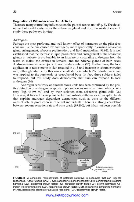
Regulation of Pilosebaceous Unit ActivityThere are many controlling influences on the pilosebaceous unit (Fig. 3). The devel-opment of model systems for the sebaceous gland and duct has made it easier tostudy these pathways in vitro.
AndrogensPerhaps the most profound and well-known effect of hormones on the pilosebac-eous unit is the one caused by androgens, more specifically in causing sebaceousgland enlargement, sebocyte proliferation, and lipid metabolism (91,92). It is wellestablished that the increase in lipid production and enlargement of the sebaceousglands at puberty is attributable to an increase in circulating androgens from thetestes in males, the ovaries in females, and the adrenal glands of both sexes.Androgen-insensitive subjects do not produce sebum (93). Furthermore, the localapplication of testosterone to skin resulted in a 15-fold increase in sebum excretionrate, although admittedly this was a small study in which 2% testosterone creamwas applied to the foreheads of prepubertal boys. In fact, three subjects failedto respond, but this study does demonstrate that skin can respond to localandrogens (94).
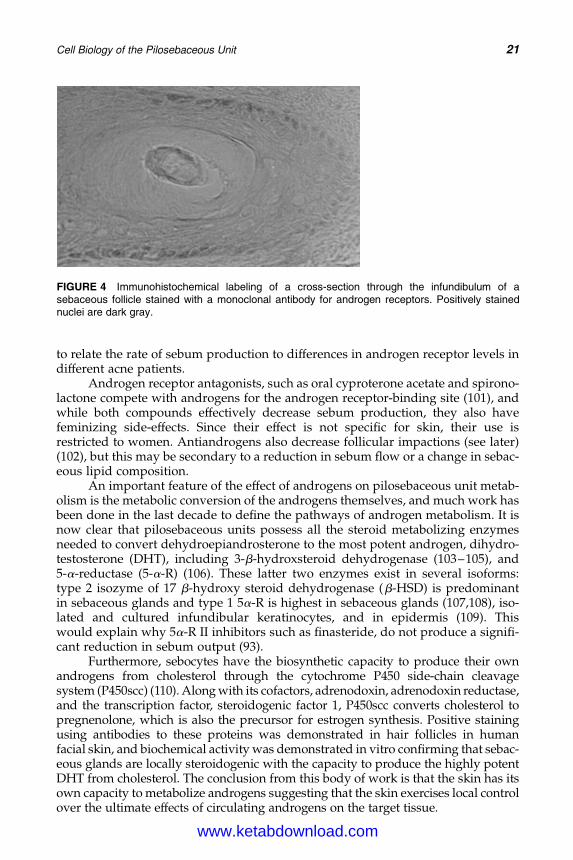
Androgen sensitivity of pilosebaceous units has been confirmed by the posi-tive detection of androgen receptors in pilosebaceous units by immunohistochem-istry (Fig. 4) (95–97) and by their isolation from sebaceous gland cells (98).However, it has not been possible to demonstrate differences in receptor levelsthat explain androgen dependent dermatoses, such as acne or the differentrates of sebum production in different individuals. There is a strong correlationbetween sebum excretion rate and acne grade (99,100), but it has not been possible
FIGURE 3 A schematic representation of potential pathways in sebocytes that can regulatelipogenesis. Abbreviations: CAMP, cyclic adenosine monophosphate; CRH, corticotrophic releasinghormone; EGF, epidermal growth factor; FGF, fibroblast growth factor; GH, growth hormone; IGF,insulin-like growth factors; KGF, keratinocyte growth factor; MSH, melanocyte stimulating hormone;PPARs, peroxisome proliferator-activated receptors; TGF, transforming growth factor.
20 Knaggs
www.ketabdownload.com
DK4962-Webster-ch2_R2_240307
to relate the rate of sebum production to differences in androgen receptor levels indifferent acne patients.
Androgen receptor antagonists, such as oral cyproterone acetate and spirono-lactone compete with androgens for the androgen receptor-binding site (101), andwhile both compounds effectively decrease sebum production, they also havefeminizing side-effects. Since their effect is not specific for skin, their use isrestricted to women. Antiandrogens also decrease follicular impactions (see later)(102), but this may be secondary to a reduction in sebum flow or a change in sebac-eous lipid composition.
An important feature of the effect of androgens on pilosebaceous unit metab-olism is the metabolic conversion of the androgens themselves, and much work hasbeen done in the last decade to define the pathways of androgen metabolism. It isnow clear that pilosebaceous units possess all the steroid metabolizing enzymesneeded to convert dehydroepiandrosterone to the most potent androgen, dihydro-testosterone (DHT), including 3-b-hydroxsteroid dehydrogenase (103–105), and5-a-reductase (5-a-R) (106). These latter two enzymes exist in several isoforms:type 2 isozyme of 17 b-hydroxy steroid dehydrogenase (b-HSD) is predominantin sebaceous glands and type 1 5a-R is highest in sebaceous glands (107,108), iso-lated and cultured infundibular keratinocytes, and in epidermis (109). Thiswould explain why 5a-R II inhibitors such as finasteride, do not produce a signifi-cant reduction in sebum output (93).
Furthermore, sebocytes have the biosynthetic capacity to produce their ownandrogens from cholesterol through the cytochrome P450 side-chain cleavagesystem (P450scc) (110). Along with its cofactors, adrenodoxin, adrenodoxin reductase,and the transcription factor, steroidogenic factor 1, P450scc converts cholesterol topregnenolone, which is also the precursor for estrogen synthesis. Positive stainingusing antibodies to these proteins was demonstrated in hair follicles in humanfacial skin, and biochemical activity was demonstrated in vitro confirming that sebac-eous glands are locally steroidogenic with the capacity to produce the highly potentDHT from cholesterol. The conclusion from this body of work is that the skin has itsown capacity to metabolize androgens suggesting that the skin exercises local controlover the ultimate effects of circulating androgens on the target tissue.
FIGURE 4 Immunohistochemical labeling of a cross-section through the infundibulum of asebaceous follicle stained with a monoclonal antibody for androgen receptors. Positively stainednuclei are dark gray.
Cell Biology of the Pilosebaceous Unit 21
www.ketabdownload.com
DK4962-Webster-ch2_R2_240307
Genes for lipid biosynthesis are regulated in sebaceous glands by androgen-dependent transcription (111). Topical application of 0.005% DHT to hamster earactivated the steroid regulating element (SRE) binding proteins in sebaceousglands, with an upregulation of the mRNA expression for enzymes involvedin lipid production, including HMG CoA reductase and synthase, glycerol3-phosphate acyltransferase, and stearoyl CoA desaturase. SREs are present inthe promoter regions of the genes for these enzymes (112).
EstrogensIn contrast to the stimulatory effect of androgens on the sebaceous glands, estrogensand compounds that have estrogenic activity, such as phenol red (113), reduce lipo-genesis in vitro. In vivo, the effect of estrogens is contradictory, although at pharma-cological doses estrogens are sebosuppressive in rat preputial gland (114) and inhumans also producing feminizing side effects (115). This action of estrogens isindirect and occurs by inhibiting the adrenals and gonads via the pituitary,thereby reducing the production of androgens. During hormonal treatments,such as hormone replacement therapy (HRT), the effect on skin surface lipidsdepends on the predominant hormone given. Skin surface lipids are increasedduring combined HRT, possibly reflecting stimulatory effects of the progestogencomponent on sebaceous gland activity, while estrogen alone has a sebum-suppressive action (116).
Local effects of estrogens have been demonstrated (115). Thus, ethinyl estra-diol is capable of reducing sebum secretion when applied to the forehead. Inaddition, sebum secretion was reduced at sites on the forehead distal to the siteof application. In agreement with a local effect, both a and b receptor subtypesfor estrogens are detected in sebaceous glands of human terminal follicles in bothbasal and partially differentiated sebocytes (117). There was also widespreadexpression of estrogen receptor b in the terminal hair follicle, where it was localizedto the nuclei of the outer root sheath, epithelial matrix, dermal papilla cells, andin the cells of the bulge (118).
RetinoidsRetinoids exert dramatic, dose-dependent effects on sebocytes in vitro and invivo. The effect of retinoids is mediated through nuclear receptors [retinoic acidreceptor (RAR), retinoid X receptor (RXR)] expressed in sebocytes (119), and invitro, sebocytes respond differently to retinoids depending upon whether RAR orRXR agonists are given. Activation of RAR by all-trans-retinoic acid (all-trans-RA)mediated the antiproliferative and antidifferentiation effects, while RXR agonistscaused differentiation and only weak proliferation in rat preputial cells (120).
Vitamin A was essential for the proliferation, lipogenesis, and differentiationof immortalized human sebocytes in vitro (121), but these cells showed differentresponses to other retinoids and the effects were dose dependent. In delipidizedserum (i.e., in the absence of vitamin A), isotretinoin [13-cis-retinoic acid (13-cis-RA)] and acitretin at concentrations �1027M enhanced sebocyte proliferation andlipid synthesis, whereas at concentrations .1027M–sebocyte proliferation was inhi-bited (121). In contrast, with cells cultured in media containing vitamin A, furtheraddition of either isotretinoin or to a lesser extent, all-trans-RA at concentrationsranging from 1028M to 1025M inhibited cell proliferation and lipid synthesis, andaltered keratin expression (122). For 13-cis-RA to affect sebocyte proliferation,
22 Knaggs
www.ketabdownload.com
DK4962-Webster-ch2_R2_240307
differentiation, and lipogenesis, it must apparently be first isomerized to all-trans-RA (123), so it is perhaps not surprising that these retinoids show similar effects.
Other culture systems support the inhibitory role for retinoids in sebaceousglands. Retinoids significantly inhibited proliferation in the rank order13-cis-RA . all-trans-RA� acitretin in immortalized sebocytes (124) and in sebac-eous gland organ culture, 13-cis-RA caused a significant decrease in lipogenesisover seven days (113,125), and reduced thymidine uptake in pilosebaceous ductorgan culture (126).
Clinically, 13-cis-RA is given orally for severe acne at doses of 1 mg/kg/dayfor four months and produces a rapid reduction in sebum excretion, for example,down to 20% of its former level within four weeks (127). This is due to sebaceousgland atrophy as demonstrated by histological methods (128) but the mechanisminvolved is unknown. 13-cis-RA and the derivatives, 3,4-didehydroretinoic acid,and 3,4-didehydroretinol are potent competitive inhibitors of the oxidative3 a-HSD, potentially downregulating the production of potent androgens, DHT,and androstandione, in vitro (129). 13-cis-RA does not however have any effecton 5-a-R responsible for the production of DHT (107). There is also a strong increasein cellular retinoic acid binding protein II expression in the sebaceous gland, and tosome extent, in the infundibular structures compared with the level of expression inthe epidermis in biopsies obtained from patients treated with 13-cis-RA. Strongeststaining was always found in layers of suprabasal sebocytes lacking lipid droplets,and staining was absent in the basal layers (130).
Vitamin DThe effect of topical vitamin D on human sebocytes and sebum secretion has notbeen documented, although receptors for vitamin D have been demonstrated inthe nuclei of basal sebocytes (131,132). In a culture system of hamster auricularsebocytes, 1-a,25-dihydroxyvitamin D3, decreased the triglyceride level by 34.3%,but augmented the accumulation of wax esters by 30%, with no difference in thelevel of cholesterol produced (133).
Peroxisome Proliferator-Activated ReceptorsPeroxisome proliferator-activated receptors (PPARs) are a subfamily of so-calledorphan receptors belonging to the nonsteroid receptor family of nuclear hormonereceptors. Several major types of PPARs have been identified. Activation ofPPARs regulates many of the lipid metabolic genes contained in peroxisomes, mito-chondria, and microsomes, with the net effect of stimulating lipogenesis. Adipo-cytes, for example, are induced to differentiate and accumulate lipid throughPPARg2. Therefore, there is interest in PPARs functioning as regulators of lipogen-esis in sebocytes (134,135). Certainly in rat preputial sebocytes, PPARg is selectivelyexpressed and has been implicated in the regulation of sebocyte differentiation(136). Fatty acids are PPAR ligands and act as regulators of lipogenesis (137). Forexample, linoleic acid incubation for 48 hours in immortalized sebocytes inducedabundant lipid synthesis. Furthermore, the effect of androgens appears to be addi-tive with the PPAR pathways: androgens appear to upregulate downstream PPARexpression, which in turn influences terminal differentiation of sebocytes (137,138).PPARs can also form heterodimers with RXRs and there is some evidence that thereis cooperation between RXR agonists and PPAR agonists in sebocyte growth anddevelopment (139). This is a complex area and the role of these receptors in lipid
Cell Biology of the Pilosebaceous Unit 23
www.ketabdownload.com
DK4962-Webster-ch2_R2_240307
production in sebocytes is just beginning to be understood with the help of modelsystems.
Thyroid HormonesThyroid hormones exert an effect on sebum secretion since thyroidectomydecreases the rate of sebum secretion in rats and administration of thyroxinereverses this effect (140). Increases in sebum secretion rate were observedwhen thyroxine was given to hypothyroid patients (141), and immunohistochem-ical localization of thyroid hormone nuclear receptors has been demonstrated inhuman scalp follicles in the nuclei of the outer root sheath cells, dermal papillacells, sheath cells, and sebaceous gland cells (142). Other than this, little attentionhas been paid to the effect of thyroid hormones on sebum secretion.
MelanocortinsMelanocortins are small peptides, including a-melanocyte stimulating hormone(aMSH) and adrenocorticotrophic hormone (ACTH), known to regulate immunefunctions, lipid metabolism, and pigmentation pathways. These small peptidesare derived from a larger precursor, proopiomelanocortin (POMC), a 241 aminoacid peptide secreted by the pituitary, which is cleaved to liberate the melanocor-tins. Evidence has existed since the 1970s demonstrating that the sebaceous glandis influenced by pituitary secretions. For example, removal of the posterior pituitaryin rats significantly reduced lipid secretion from rat preputial glands, addition ofaMSH stimulated a dose-dependent increase in sebum secretion in rats (143),and patients with hypopituitarism show reduced sebum secretion (141).The expression of POMC-derived peptides in hair follicles in mice varies duringthe hair cycle, with high levels of b-endorphin expressed during anagen andcatagen (144).
The biological effects of melanocortins are mediated through specific trans-membrane receptors, known as melanocortin receptors, which are coupled toG-proteins to modulate intracellular levels of cAMP. To date, five receptor subtypes(MCR1–5) have been cloned (145) and, of these, MCR-1 was detected in humansebaceous glands using immunostaining (146), and in an immortalized humansebocyte cell line (147). MCR-5 expression was demonstrated in microdissectedhuman sebaceous glands (148), but could not be detected using immunostainingof sections. Specific transcripts for MCR-1, but not for MCR-5, were present in animmortalized sebocyte cell line and these sebocytes responded to aMSH by modu-lating interleukin-8 (IL-8) secretion (147). In rat preputial cells, MCR-5 was detectedperhaps explaining the earlier observations of the effect of melanocortin on thesecells (143); and in transgenic mice, targeted disruption of MCR-5 gave markedreduction of sebum secretion (149). Other investigators showed that sebocytesderived from normal human facial skin were stimulated to differentiate andproduce prominent lipid droplets when POMC-derived peptides, aMSH, andACTH were added, and this correlated with an increase in expression of MCR-5(150). Together, these data support the regulation of human sebum production bymelanocortins and may offer an explanation for the link between stress and acne,with high circulating levels of ACTH acting directly on the sebaceous gland tomediate sebum production (148).
24 Knaggs
www.ketabdownload.com
DK4962-Webster-ch2_R2_240307
Growth Factors and NeuropeptidesEGF, TGF-a, basic FGF, and keratinocyte growth factor (KGF) all inhibit lipogenesisand with the exception of KGF are mitogenic in cultured hamster sebocytes(133,151). In organ culture of human sebaceous glands, EGF produced a dose-dependent inhibition of lipid synthesis and inhibited DNA synthesis (152), andremoval of EGF from the medium caused an increase in the rate of lipogenesiswithout affecting cell turnover (113). In vivo, the presence of EGF inhibitedthe sebaceous gland differentiation in sheep (153), and in hamster ears, itstimulated the sebaceous glands to proliferate (154). Receptors for EGF are foundin sebaceous glands (155). Based on this evidence, Kealey and colleagues proposedthat EGF may be, in part, responsible for sebaceous gland atrophy observed duringacne lesion formation (7,152). EGF can also induce changes in sebum composition.Ridden (156), reported that there was a fall in the amount of squalene and acorresponding rise in cholesterol, although this was not corroborated by otherresearchers (157).
Sebocyte proliferation is stimulated by insulin, thyroid-stimulating hormone,and hydrocortisone (158). In rat preputial cells, growth hormone (GH) increasedsebocyte differentiation and was more potent than either insulin-like growthfactors (IGFs) or insulin. Furthermore, GH enhanced the effect of DHT on sebocytedifferentiation perhaps complementing the effect of androgens in increasing sebumduring puberty. IGFs, in contrast, had a greater effect on increasing DNA synthesiscompared to GH, and had no effect on the response of the cell to DHT (159),although a role for IGF in causing the observed increase in sebum production atpuberty has not been ruled out. This suggests that antagonists of IGF may offer away of decreasing sebum synthesis.
Corticotrophic-releasing hormone (CRH), a key regulator in the centralnervous system, has been implicated in sebum production (160). CRH and its recep-tor are expressed in cultured sebocytes, where the hormone appears to upregulateexpression of 3 b-HSD—a key enzyme in the androgen biosynthetic pathways.Therefore, CRH could indirectly influence lipid synthesis by modifying the localproduction of androgens.
Incubation of sebaceous glands in organ culture with neuropeptides (e.g.,calcitonin gene-related peptide, substance P, vasoactive intestinal polypeptide, orneuropeptide Y), or NGF up to a final concentration of 1027M showed that onlysubstance P had any effect on sebaceous gland ultrastructure as observed in theelectron microscope, and this effect was dose dependent (161). Skin samplescultured with substance P showed sebaceous glands with an increased glandarea (in sections) compared with control samples. The sebocytes were engorgedwith numerous lipid droplets.
CytokinesExpression of IL-1a and b was demonstrated in sebaceous glands by immunohis-tochemistry (162), and mRNA for IL-1a was detected in cultured sebocytes (163).No change in staining pattern was observed when sebum was extracted from thesamples, indicating that IL-1 is not associated with sebum. The function of IL-1in normal sebaceous glands is unclear, although in vitro IL-1a and tumor necrosisfactor a, inhibited sebaceous lipogenesis in sebaceous gland organ culture andinduced de-differentiation of human sebocytes into a keratinocyte-like phenotype(152). In organ culture, IL-1a at 1 ng/mL promoted ductal cornification (164) and
Cell Biology of the Pilosebaceous Unit 25
www.ketabdownload.com
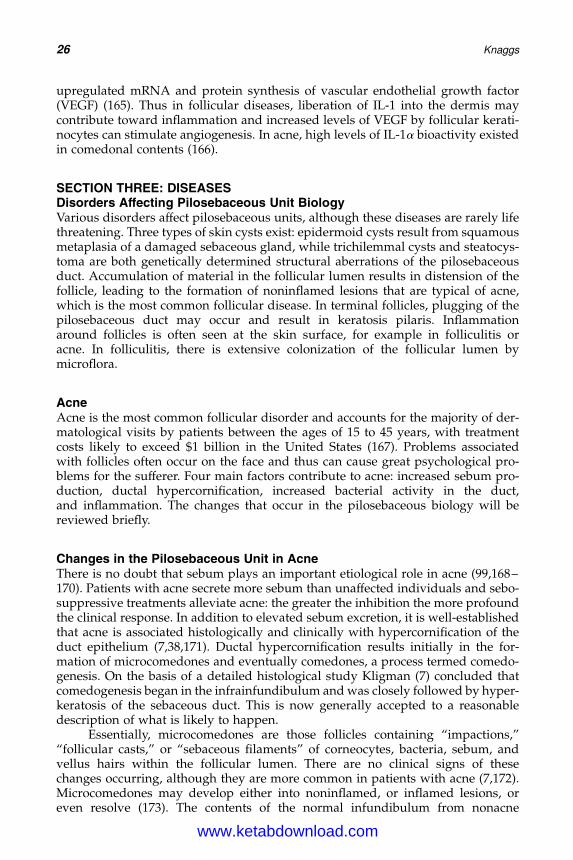
DK4962-Webster-ch2_R2_240307
upregulated mRNA and protein synthesis of vascular endothelial growth factor(VEGF) (165). Thus in follicular diseases, liberation of IL-1 into the dermis maycontribute toward inflammation and increased levels of VEGF by follicular kerati-nocytes can stimulate angiogenesis. In acne, high levels of IL-1a bioactivity existedin comedonal contents (166).
SECTION THREE: DISEASESDisorders Affecting Pilosebaceous Unit BiologyVarious disorders affect pilosebaceous units, although these diseases are rarely lifethreatening. Three types of skin cysts exist: epidermoid cysts result from squamousmetaplasia of a damaged sebaceous gland, while trichilemmal cysts and steatocys-toma are both genetically determined structural aberrations of the pilosebaceousduct. Accumulation of material in the follicular lumen results in distension of thefollicle, leading to the formation of noninflamed lesions that are typical of acne,which is the most common follicular disease. In terminal follicles, plugging of thepilosebaceous duct may occur and result in keratosis pilaris. Inflammationaround follicles is often seen at the skin surface, for example in folliculitis oracne. In folliculitis, there is extensive colonization of the follicular lumen bymicroflora.
AcneAcne is the most common follicular disorder and accounts for the majority of der-matological visits by patients between the ages of 15 to 45 years, with treatmentcosts likely to exceed $1 billion in the United States (167). Problems associatedwith follicles often occur on the face and thus can cause great psychological pro-blems for the sufferer. Four main factors contribute to acne: increased sebum pro-duction, ductal hypercornification, increased bacterial activity in the duct,and inflammation. The changes that occur in the pilosebaceous biology will bereviewed briefly.
Changes in the Pilosebaceous Unit in AcneThere is no doubt that sebum plays an important etiological role in acne (99,168–170). Patients with acne secrete more sebum than unaffected individuals and sebo-suppressive treatments alleviate acne: the greater the inhibition the more profoundthe clinical response. In addition to elevated sebum excretion, it is well-establishedthat acne is associated histologically and clinically with hypercornification of theduct epithelium (7,38,171). Ductal hypercornification results initially in the for-mation of microcomedones and eventually comedones, a process termed comedo-genesis. On the basis of a detailed histological study Kligman (7) concluded thatcomedogenesis began in the infrainfundibulum and was closely followed by hyper-keratosis of the sebaceous duct. This is now generally accepted to a reasonabledescription of what is likely to happen.
Essentially, microcomedones are those follicles containing “impactions,”“follicular casts,” or “sebaceous filaments” of corneocytes, bacteria, sebum, andvellus hairs within the follicular lumen. There are no clinical signs of thesechanges occurring, although they are more common in patients with acne (7,172).Microcomedones may develop either into noninflamed, or inflamed lesions, oreven resolve (173). The contents of the normal infundibulum from nonacne
26 Knaggs
www.ketabdownload.com
DK4962-Webster-ch2_R2_240307
control patients were easy to extrude and contained numerous empty spaces in thecells which appeared to have lost their contents and had no clear cytoskeleton. Incontrast, the contents of follicles from acne patients were hard to extrude and thecells were firm and compact (7).
An extensive ultrastructural study on the cornified material in follicularimpactions was carried out by Al Baghdadi (174). He showed that corneocytesare arranged into three layers in the follicular impaction: a loosely attached outerlayer; a densely packed middle layer; and a further loosely attached layer in themiddle of the cast. Lipid droplets occur inside the cornified cells (38,175) and asa result the corneocytes become balloon-like in appearance. Abnormal lamellarinclusions of lipid have been described as a marker for abnormal keratinization(176). Scanning electron microscopy of comedonal contents revealed a complexsystem of numerous internal channels or “canaliculi” composed of concentriclamellae of corneocytes enclosing bacteria and sebum (177) (Fig. 5).
The overall composition of follicular casts was reported to be water (20–60%),lipids (20%), proteins (15%), and microorganisms (178). Bladon et al. (179) analyzedthe protein content of comedones and found both epidermal keratins and degradedkeratins to be present in the comedones.
Biochemical Changes in the Infundibulum During ComedogenesisThe process of comedogenesis is thought to be due to hyperproliferation of ductalkeratinocytes, inadequate separation of the ductal corneocytes, or a combination ofboth factors, resulting in microcomedones. Hyperproliferation of basal keratino-cytes in acne has been demonstrated (180,181) and correlates with keratin 16expression, a marker for hyperproliferation suprabasally (182). Aldana et al. (183)proposed a cycling of normal follicles through different levels of expression ofKi-67 (a proliferation marker) and keratin 16 and that these represent differentstages of development of the microcomedone. The point at which both Ki-67 andkeratin 16 are coexpressed by the follicle is when the follicle is susceptible tocomedogenic changes (184).
FIGURE 5 Environmental scanning electron micrograph of a microcomedone removed from apilosebaceous duct. Keratinocytes are seen covering multiple hairs.
Cell Biology of the Pilosebaceous Unit 27
www.ketabdownload.com
DK4962-Webster-ch2_R2_240307
A more extensive investigation of adhesion in follicles is required. In epider-mis, intercellular adhesion is mediated by lipids, cellular adhesion molecules, anddesmosomes. At the present time, the relative importance of follicular intercellularlipids in normal and acne follicles is unclear due to the experimental difficultiesencountered obtaining sufficient material for analysis. Hypercornification fromthe retention of desmosomes was described to be a contributory factor to theformation of acne lesions. However, no differences were detected between stainingfor these epitopes in the wall of normal and comedonal follicles (29).
CONCLUSION
Although it is clear that we now know a great deal about the pilosebaceous unit atthe structural, biochemical, and physiological levels, there are still considerablegaps in our understanding. Much of the early work was descriptive biology ofthe structure obtained using immunohistochemical studies. Over the last decade,several in vitro models systems (isolated organ culture, cultured sebocytes) havemade it easier to study the cellular processes in the follicle, including sebum pro-duction and the effect of inhibitors and stimulators. There is also now a gooddeal of information about the chemistry and biosynthesis of sebum components,and we also have a much better understanding of the cell biology of the pilosebac-eous duct. Even so, we are still unable to precisely define the cause of one of themost common skin diseases, namely acne, which affects a very large proportionof the population globally.
ACKNOWLEDGMENTS
The author wishes to acknowledge and thank Professor W Cunliffe for kindlyallowing the use of Figure 1B, and the journal “Retinoids and Lipid SolubleVitamins in Clinical Practice” for kindly allowing the use of Figures 1A, 2, and 3.
REFERENCES
1. Eisen AZ, Holyoke JB, Lobitz WC. Responses of the superficial portion of the humanpilosebaceous apparatus to controlled injury. J Invest Dermatol 1955; 25:145–156.
2. Alonso L, Fuchs E. Stem cells of the skin epithelium. Proc Natl Acad Sci USA 2003;30(suppl 1):11830–11835.
3. Cotsarelis G, Sun TT, Lavker R. Label-retaining cells reside in the bulge area of pilo-sebaceous unit: implications for follicular stem cells, hair cycle and skin carcinogenesis.Cell 1990; 61:1329–1337.
4. Oshima H, Rochat A, Kedzia C, Kobayashi K, Barrandon Y. Morphogenesis andrenewal of hair follicles from adult multipotent stem cells. Cell 2001; 104:233–245.
5. Shuster S. Biological purpose of acne. Lancet 1976; 1:1328–1329.6. Zouboulis C. Sebaceous gland in human skin—the fantastic future of a skin appendage.
J Invest Dermatol 2003; 120(6):xiv-xv.7. Kligman AM. An overview of acne. J Invest Dermatol 1974; 62:268–287.8. Leeming JP, Holland KT, Cunliffe WJ. The microbiological ecology of pilosebaceous
units from human skin. J Gen Microbiol 1984; 130:803–807.9. Hughes BR. The Structure and Function of the Pilosebaceous Duct in Health and
Disease—with Special Reference to Acne Vulgaris. MD thesis, University of Leeds,UK, 1992.
28 Knaggs
www.ketabdownload.com
DK4962-Webster-ch2_R2_240307
10. Holbrook KA. Structure and function of the developing human skin. In: Goldsmith LA,ed. Physiology, Biochemistry and Molecular Biology of the Skin. Vol. 1. Oxford: OxfordUniversity Press, 1991:83–89.
11. Westgate GE, Shaw DA, Harrap GJ, Couchman JR. Immunohistochemical localisationof basement membrane components during hair follicle morphogenesis. J InvestDermatol 1984; 82:259–264.
12. Millar SE. Molecular mechanisms regulating hair follicle development. J Invest Derma-tol 2002; 118:216–225.
13. McElwee KJ, Hoffman R. Growth factors in early hair follicle morphogenesis. Eur JDermatol 2000; 10:341–350.
14. Ebling FJG. Embryology. In: Rook A, Wilkinson DS, Ebling FJG, eds. Textbook ofDermatology. Oxford: Blackwell Scientific, 1968:16–22.
15. Ferraris C, Bernard BA, Dhouailly D. Adult epidermal keratinocytes are endowed withpilosebaceous forming abilities. Int J Dev Biol 1997; 41:491–498.
16. Serri F, Huber WM. In Advances in Biology of Skin. Vol. 4. Oxford: Pergamon Press,1963:1–18.
17. Allen M, Grachtchouk M, Sheng H, et al. Hedgehog signaling regulates sebaceousgland development. Am J Path 2003; 163:2173–2178.
18. Niemann C, Unden AB, Lyle S, Zouboulis ChC, Toftgard R, Watt FM. Indianhedgehog and beta-catenin signaling: role in the sebaceous lineage of normal andneoplastic mammalian epidermis. Proc Natl Acad Sci USA 2003; 100(suppl 1):11873–11880.
19. Pierard GE. Follicle to follicle heterogeneity of sebum excretion. Dermatologica 1986;173:61–65.
20. Montagna W. Advances in Biology of the Skin. Vol. 4. Oxford, U.K.: Pergamon Press,1963.
21. Blume U, Ferracin J, Verschoore M, Czernielewski JM, Schaefer H. Physiology of thevellus hair follicle: hair growth and sebum excretion. Br J Dermatol 1991; 124: 21–28.
22. Pagnoni A, Kligman AM, el Gammal S, Stoudemayer T. Determination of density offollicles on various regions of the face by cyanoacrylate biopsy: correlation withsebum output. Br J Dermatol 1994; 131:862–865.
23. Otberg N, Richter H, Schaefer H, Blume-Peytavi U, Sterry W, Lademann J. Variations ofhair follicle size and distribution in different body sites. J Invest Dermatol 2004;122:14–19.
24. Cunliffe WJ, Perera WDH, Thackray P, Williams M, Forster RA, Williams SM. Pilo-sebaceous duct physiology III. Observations on the number and size of pilo-sebaceousducts in acne vulgaris. Br J Dermatol 1976; 95:153–156.
25. Price VH, Cappas A, Uno H. Compound follicles presenting as folliculitis and scarringalopecia [abstr]. J Invest Dermatol 1992; 98:582.
26. Cunliffe WJ, Cotterill JA. The Acnes-Clinical Features, Pathogenesis and Treatment.London: WB Saunders, 1975.
27. Williams M, Cunliffe WJ, Gould D. Pilosebaceous duct physiology I. Effect of hydrationon pilosebaceous duct orifice. Br J Dermatol 1974; 90:631–635.
28. Plewig G, Kligman AM. Acne, Morphogenesis and Treatment. Berlin: Springer-Verlag,1975.
29. Knaggs HE, Hughes BR, Morris C, Holland DB, Wood EJ, Cunliffe WJ. Immunohisto-chemical study of desmosomes in acne vulgaris. Br J Dermatol 1994; 130:731–737.
30. Rosenquist TA, Martin GA. Fibroblast growth factor signalling in the hair growth cycle:expression of the fibroblast growth factor receptor and ligand genes in the murine hairfollicle. Dev Dyn 1996; 205:379–386.
31. Danilenko DM, Ring BD, Pierce GF. Growth factors and cytokines in hair follicle devel-opment and cycling: recent insights from animal models and the potential for clinicaltherapy. Mol Med Today 1996; 2:460–467.
32. Paus R, Cotsarelis G. The biology of hair follicles. N Engl J Med 1999; 341:491–497.33. Van Mater D, Kolligs FT, Dlugosz AA, Fearon ER. Transient activation of b-catenin
signalling in cutaneous keratinocytes is sufficient to trigger the active growth phaseof the hair cycle in mice. Genes Dev 2003; 17:1219–1224.
Cell Biology of the Pilosebaceous Unit 29
www.ketabdownload.com
DK4962-Webster-ch2_R2_240307
34. Stenn K, Paus R. Controls of hair follicle cycling. Physiol Rev 2003; 81:449–494.35. Heid HW, Moll I, Franke WW. Patterns of expression of trichocytic and epithelial cyto-
keratins in mammalian tissues I. Human and bovine hair follicle. Differentiation 1988;91:553–559.
36. Moll R, Franke WW, Volk Platzer B, Koepler R. Different keratin polypeptides in epi-dermis and other epithelia of human skin: a specific cytokeratin of molecular weight46000 in epithelia of the pilosebaceous tract and basal cell epitheliomas. J Cell Biol1982; 95:285–295.
37. Wilson CL, Deane D, Wojnarowska F, Lane EB, Leigh IM. Follicular keratin expressionin normal and psoriatic scalp [abstr]. J Invest Dermatol 1990; 95:495.
38. Knutson DD. Ultrastructural observations in acne vulgaris. The normal sebaceousfollicle and acne lesions. J Invest Dermatol 1974; 62:288–307.
39. Kluznik AR, Cunliffe WJ. Keratin biosynthesis in the pilosebaceous ducts in acnevulgaris. Biochem Soc Trans 1988; 220:330–331.
40. Stark H-J, Breitkreutz, Limat A, Bowden PE, Fusenig NE. Keratins of the human hairfollicle: “Hyperproliferative” keratins consistently expressed in outer root sheathcells in vivo and in vitro. Differentiation 1987; 35:236–248.
41. Rupniak HT, Turner DM, Cunliffe WJ, Dhuna R, West MR. Cell envelope production inacne. In: Marks R, Plewig G, eds. Acne and Related Disorders. London: Martin Dunitz,1989:87–93.
42. Rupniak HT, Turner DM, Wood EJ, Cunliffe WJ. Characteristics of the cellenvelope system in normal and psoriatic epidermis [abstr]. J Invest Dermatol 1986;87:164.
43. Peterson LL, Wuepper KD. Epidermal and hair follicle transglutaminases and cross-linking in skin. Mol Cell Biochem 1984; 58:99–111.
44. Rothnagel JA, Rogers GE. Transglutaminase-mediated cross-linking in mammalianepidermis. Mol Cell Biochem 1984; 58:113–119.
45. Murphy GF, Flynn TC, Rice RH, Pinkus G. Involucrin expression in normal and neo-plastic skin: a marker for keratinocyte differentiation. J Invest Dermatol 1984;82:453–457.
46. Greco MA, Ladas D, Rudolph S, Baden H. Modulation of pancornulin expression bycalcium and relative confluence [abstr]. J Invest Dermatol 1992; 98:640.
47. Kvedar JC, Manabe M, Phillips SB, Ross BS, Baden HP. Characterisation of sciellin, aprecursor to the cornified envelope of human keratinocytes. Differentiation 1992;49:195–204.
48. Hohl D, Olana BR, Schnyder UW, Roop DR. Loricrin is a marker of squamous differen-tiation in rodents and a marker of epidermal differentiation in higher mammals [abstr].J Invest Dermatol 1991; 96:1030.
49. Philpott MP. Defensins and acne. Mol Immunol 2003; 40:457–462.50. Bell M. A comparative study of the ultrastructure of the sebaceous glands of man and
other primates. J Invest Dermatol 1974; 62:132–143.51. Brandes D, Bertini F. Role of lysosomes in cellular lytic processes II. Cell death during
holocrine secretion in sebaceous glands. Exp Mol Pathol 1965; 11:245–265.52. Im MJC, Hoopes JE. Enzymes of carbohydrate metabolism in normal human sebaceous
glands. J Invest Dermatol 1974; 62:153–160.53. Hurley HJ, Shelley WB, Koelle GB. The distribution cholinesterases in human skin,
with special reference to eccrine and apocrine sweat glands. J Invest Dermatol 1953;21:139–147.
54. Pawlowski A, Weddell G. The lability of cutaneous neural elements. Br J Dermatol1967; 79:14–19.
55. Toyoda M, Nakamura M, Morohashi M. Neuropeptides and sebaceous glands. Eur JDermatol 2002; 12:422–427.
56. Downing DT, Strauss JS, Ramasastry P, Abel M, Lees CW, Pochi PE. Measurement ofthe time between synthesis and surface excretion of sebaceous lipids in sheep andman. J Invest Dermatol 1975; 64:215–219.
57. Pochi PE, Downing DT, Strauss JS. Sebaceous gland response in man to prolonged totalcaloric deprivation. J Invest Dermatol 1970; 55:303–309.
30 Knaggs
www.ketabdownload.com
DK4962-Webster-ch2_R2_240307
58. Downing DT, Strauss JS. On the mechanism of sebum secretion. Arch Dermatol Res1982; 272:343–349.
59. Downing DT, Straniesi AM, Strauss JS. The effect of accumulated lipids on measure-ments of sebum secretion in human skin. J Invest Dermatol 1982; 79:226–228.
60. Pierard-Franchimont C, Pierard GE. Postmenopausal aging of the sebaceous follicle: acomparison between women receiving hormone replacement therapy or not. Derma-tology 2003; 204(1):17–22.
61. Zouboulis CC, Boschnakow A. Chronological ageing and photoageing of the humansebaceous gland. Clin Exp Dermatol 2001; 26(7):600–607.
62. Thiele JJ, Weber SU, Packer L. Sebaceous gland secretion is a major physiologic route ofvitamin E delivery to skin. J Invest Dermatol 1998; 113:390–395.
63. Fluhr JW, Man M-Q, Brown BE, et al. Glycerol regulates stratum corneum hydration insebaceous gland deficient (asebia) mice. J Invest Dermatol 2003; 120: 728–737.
64. Williams D, Siock P, Stenn K. 13-cis-Retinoic acid affects sheath-shaft interaction ofequine hair follicles in vitro. J Invest Dermatol 1996; 106(2):356–361.
65. Williams D, Stenn K. Transection level dictates the pattern of hair follicle sheath growthin vitro. Dev Biol 1994; 165:469–479.
66. Philpott MP, Sanders DA, Kealey T. Is the sebaceous gland important for inner rootsheath breakdown? In: Van Neste D, Randall VA, eds. Hair Research for the Next Mile-nnium. Amsterdam: Elsevier, 1996:393–395.
67. Gates AH, Karasek M. Hereditary absence of sebaceous glands in mouse. Science 1965;148:1471–1473.
68. Downing DT, Stewart ME, Strauss JS. Changes in sebum secretion and the sebaceousgland. Clin Geriatr Med 1989; 5(1):109–114.
69. Kligman AM. The uses of sebum. Br J Dermatol 1963; 75:307–319.70. Youn SW, Kim SJ, Hwang IA, Park KC. Evaluation of facial skin type by sebum
secretion: discrepancies between subjective descriptions and sebum secretion. SkinRes Technol 2002; 8(3):168–172.
71. Downie MMT, Kealey T. Lipogenesis in the human sebaceous gland: glycogen andglycerophosphate are substrates for the synthesis of sebum lipids. J Invest Dermatol1998; 1111:199–205.
72. Marsden JR, Middleton B. Lipogenesis in isolated human sebacoues glands: effects ofdifferent precursors and inhibitors of cholesterol synthesis. In: Marks R, Plewig G, eds.Acne and Related Disorders. London, U.K.: Martin Dunitz, 1989:35–38.
73. Smythe CDW, Greenall M, Kealey T. The activity of HMG-CoA reductase and actyl-CoA carboxylase in human apocrine sweat glands, sebaceous glands and hair folliclesis regulated by phosphorylation and by exogenous cholesterol. J Invest Dermatol 1998;111:139–148.
74. Marsden JR, Middleton B, Cox J. Lipogenesis in isolated human sebaceous glands:effect of inhibitors. Br J Dermatol 1986; 116:448.
75. Cunliffe WJ, Burton JL, Shuster S. Effect of aluminium nicotinate on sebum excretion.Br J Dermatol 1969; 81:867–868.
76. Strauss JS, Pochi PE, Whitman EN. Suppression of sebaceous gland activity witheicosa-5:8:11:14-tetraynoic acid. J Invest Dermatol 1967; 48(5):492–493.
77. Nicolaides N, Kellum RE, Woolley PV. The structures of the free unsaturated fatty acidsof human skin surface fat. Arch Biochem Biophys 1964; 105:634–639.
78. Nicolaides N, Fu HC, Ansari MN, Rice GR. The fatty acids of wax esters and sterolesters from vernix caesosa and from human skin surface. Lipids 1972; 7:506–517.
79. Ge L, Gordon JS, Hsuan C, Stenn K, Prouty SM. Identification of theD-6 desaturase of humansebaceous glands expression and enzyme activity. J Invest Dermatol 2003; 120:707–714.
80. Zheng Y, Eilertsen KJ, Ge L, et al. Scd1 is expressed in sebaceous glands and is dis-rupted in the asebia mouse. Nat Genet 1999; 23(3):268–270.
81. Downing DT, Stewart ME, Wertz PW, Strauss JS. Essential fatty acids and acne. J AmAcad Dermatol 1986; 14:221–225.
82. Stewart ME, Quinn MA, Downing DT. Variability in the fatty acid composition of waxesters from vernix caseosa and its possible relation to sebaceous gland activity. J InvestDermatol 1982; 78:291–295.
Cell Biology of the Pilosebaceous Unit 31
www.ketabdownload.com
DK4962-Webster-ch2_R2_240307
83. Thody A, Shuster S. Control and function of sebaceous glands. Physiol Rev 1989;69:383416.
84. Stewart ME, Grahek MO, Cambier LS, Wertz PW, Downing DT. Dilutional effect ofincreased sebaceous gland activity on the proportion of linoleic acid in sebaceouswax esters and in epidermal acylceramides. J Invest Dermatol 1986; 87:733–736.
85. Stewart ME, Greenwood R, Cunliffe WJ, Strauss JS, Downing DT. Effect of cyproteroneacetate-ethinyl estradiol treatment on the proportions of linoleic and sebaleic acids invarious skin surface lipid classes. Arch Dermatol Res 1986; 278:481–485.
86. Kotani A, Kusu F. HPLC with electrochemical detection for determining the distri-bution of free fatty acids in skin surface lipids from the human face and scalp. ArchDermatol Res 2002; 294(4):172–177.
87. Nicolaides N, Apon JM. The saturated methyl branched fatty acids of adult human skinsurface lipid. Biomed Mass Spectrom 1977; 4:337–347.
88. Stewart ME, Downing DT. Proportions of various straight and branched fatty acidchain types in the sebaceous wax esters of young children. J Invest Dermatol 1985;84:501–503.
89. Pappas A, Anthonavage M, Gordon JS. Metabolic fate and selective utilisation of majorfatty acids in human sebaceous gland. J Invest Dermatol 2002; 118:164–171.
90. Wrobel A, Seltmann H, Fimmel S, et al. Differentiation and apoptosis in human immor-talized sebocytes. J Invest Dermatol 2003; 120(2):175–181.
91. Hamilton JB. Male hormone substance: a prime factor in acne. J Clin Endocrinol 1941;1:570–592.
92. Rosenfield RL, Deplewski D. Role of androgens in the developmental biology of thepilosebaceous unit. Am J Med 1995; 16(suppl):80–88.
93. Imperato-McGinley J, Gautier T, Cai LQ, Yee B, Epstein J, Pochi P. The androgen controlof sebum production. Studies of subjects with dihydrotestosterone deficiency and com-plete androgen insensitivity. J Clin Endocrinol Metab 1993; 76:524–528.
94. Holland DB, Cunliffe WJ, Norris JFB. Differential response if sebaceous glands toexogenous testosterone. Br J Dermatol 1998; 139:102–103.
95. Henderson CA, Knaggs H, Clark A, Highet AS, Cunliffe WJ. Apert’s syndrome andandrogen receptor staining of the basal cells of sebaceous glands. Br J Dermatol1995; 132(1):139–143.
96. Whitaker SB, Vigneswaran N, Singh BB. Androgen receptor status of the oral sebaceousglands. Am J Dermatopathol 1997; 19(4):415–418.
97. Choudhry R, Hodgins MB, Van der Kwast TH, Brinkmann AO, Boersma WJ. Localiz-ation of androgen receptors in human skin by immunohistochemistry: implications forthe hormonal regulation of hair growth, sebaceous glands and sweat glands. J Endocri-nol 1992; 133(3):467–475.
98. Sawaya M. Purification of androgen receptors in human sebocytes and hair. J InvestDermatol 1992; 98(suppl):92–96.
99. Cunliffe WJ, Shuster S. Pathogenesis of acne. Lancet 1969; 1:685–687.100. Cunliffe WJ. In: Marks R, ed. Androgen abnormalities in acne subjects. Acne. London:
Martin Dunitz, 1989:158–160.101. Akamatsu H, Zouboulis CC, Orfanos CE. Spironolactone directly inhibits proliferation
of cultured human facial sebocytes and acts antagonistically to testosterone and5 alpha-dihydrotestosterone in vitro. J Invest Dermatol 1993; 100:660–662.
102. Cunliffe WJ, Forster RA. Androgen control of the pilosebaceous duct. [abstr]. Br JDermatol 1986; 116:449.
103. Sawaya ME, Penneys NS. Immunohistochemical distribution of aromatase and3b-hydroxysteroid dehydrogenase in human hair follicle and sebaceous gland.J Cutan Pathol 1992; 19(4):309–314.
104. Thiboutot D, Martin P, Volikos L, Gilliland K. Oxidative activity of the type 2 isozymeof 17beta-hydroxysteroid dehydrogenase (17beta-HSD) predominates in humansebaceous glands. J Invest Dermatol 1998; 111(3):390–395.
105. Thiboutot D, Knaggs H, Gilliland K, Lin G. Activity of 5-alpha-reductase and 17-beta-hydroxysteroid dehydrogenase in the infrainfundibulum of subjects with and withoutacne vulgaris. Dermatology 1998; 196(1):38–42.
32 Knaggs
www.ketabdownload.com
DK4962-Webster-ch2_R2_240307
106. Luu-The V, Sugimoto Y, Puy L, et al. Characterization, expression, and immunohisto-chemical localization of 5 alpha-reductase in human skin. J Invest Dermatol 1994;102(2):221–226.
107. Thiboutot D, Harris G, Iles V, Cimis G, Gilliland K, Hagari S. Activity of the type 15-alpha-reductase exhibits regional differences in isolated sebaceous glands andwhole skin. J Invest Dermatol 1995; 105:209–214.
108. Bayne EK, Flanagan J, Einstein M, et al. Immunohistochemical localization of types 1and 2 5-alpha-reductase in human scalp. Br J Dermatol 1999; 141(3):481–491.
109. Thiboutot DM, Knaggs H, Gilliland K, Hagari S. Activity of type 1 5 alpha-reductase isgreater in the follicular infrainfundibulum compared with the epidermis. Br J Dermatol1997; 136(2):166–171.
110. Thiboutot D, Jabara S, McAllister JM, et al. Human skin is a steroidogenic tissue: ster-oidogenic enymes and cofactors are expressed in epidermis, normal sebocytes and animmortalised sebocyte cell line (SEB-1). J Invest Dermatol 2003; 120:905–914.
111. Ideta R, Seki T, Adachi K, Nakayama Y. The isolation and characterization of androgen-dependent genes in the flank organs of golden Syrian hamsters. Dermatology 1998;196(1):47–50.
112. Rosignoli C, Nicolas JC, Jomard A, Michel S. Involvement of the SREBP pathway inthe mode of action of androgens in sebaceous glands in vivo. Exp Dermatol 2003;12:480–489.
113. Guy R, Ridden C, Kealey T. The improved organ maintenance of the human sebaceousgland: modeling in vitro the effects of epidermal growth factor, androgens, estrogens,13-cis retinoic acid, and phenol red. J Invest Dermatol 1996; 106(3):454–460.
114. Ebling FJ, Skinner J. The local effects of topically applied estradiol, cyproterone acetate andethanol on sebaceous secretion in intact male rats. J Invest Dermatol 1983; 81:448–451.
115. Strauss JS, Kligman AM, Pochi PE. Effects of androgens and estrogens on human sebac-eous glands. J Invest Dermatol 1962; 39:139–155.
116. Sator PG, Schmidt JB, Sator MO, Huber JC, Honigsmann H. The influence of hormonereplacement therapy on skin ageing: a pilot study. Maturitas 2001; 25:43–55.
117. Thornton MJ, Taylor AH, Mulligan K, et al. The distribution of estrogen receptor beta isdistinct to that of the estrogen receptor a and the androgen receptor in human skin andthe pilosebaceous unit. J Invest Dermatol Sym Proc 2003; 8:100–103.
118. Thornton MJ, Taylor AH, Mulligan K, et al. Oestrogen receptor beta is the predominantoestrogen receptor in human scalp skin. Exp Dermatol 2003; 12(2):181–190.
119. Doran TI, Lucas DA, Levin AA, et al. Biochemical and retinoid receptor activities inhuman sebaceous cells. In: Saurat J-H, ed. Retinoids: 10 Years On. Basel: Karger,1991:243–253.
120. Kim MJ, Ciletti N, Michel S, Reichert U, Rosenfield RL. The role of specific retinoidreceptors in sebocyte growth and differentiation in culture. J Invest Dermatol 2000;114:349–353.
121. Zouboulis CC, Korge BP, Miscke D, Orfanos CE. Altered proliferation, syntheticactivity, and differentiation of cultured human sebocytes in the absence of vitamin Aand their modulation by synthetic retinoids. J Invest Dermatol 1993; 101:628–633.
122. Zouboulis CC, Korge B, Akamatsu H, et al. Effects of 13-cis-retinoic acid, all trans-retinoic acid and acitretin on the proliferation, lipid synthesis and keratin expressionof cultured human sebocytes in vitro. J Invest Dermatol 1991; 96:792–797.
123. Tsukada M, Schroder M, Roos TC. 13 cis-retinoic acid exerts its specific activity onhuman sebocytes through selective intracellular isomerisation to all trans-retinoicacid and binding to retinoid acid receptors. J Invest Dermatol 2000; 115:321–327.
124. Zouboulis CC, Seltmann H, Neitzel H, Orfanos CE. Establishment and characterizationof an immortalized human sebaceous gland cell line (SZ95). J Invest Dermatol 1999;113(6):1011–1020.
125. Ridden J, Ferguson D, Kealey T. Organ maintenance of human sebaceous glands: invitro effects of 13-cis retinoic acid and testosterone. J Cell Sci 1990; 95:125–136.
126. Guy R, Ridden C, Barth J, Kealey T. Isolation and maintenance of the human pilosebac-eous duct: 13-cis retinoic acid acts directly on the duct in vitro. Br J Dermatol 1993;128(3):242–248.
Cell Biology of the Pilosebaceous Unit 33
www.ketabdownload.com

DK4962-Webster-ch2_R2_240307
127. Stewart ME, Benoit AM, Stranieri AM, Rapini RP, Strauss JS, Downing DT. Effect of oral13-cis-retinoic acid at three dose levels on sustainable rates of sebum secretion and onacne. J Am Acad Dermatol 1983; 8:532–538.
128. Dalziel K, Barton S, Marks R. The effects of isotretinoin on follicular and sebaceousgland differentiation. Br J Dermatol 1987; 117:317–323.
129. Karlsson T, Vahlquist A, Kedishvili N, Torma H. 13-cis-retinoic acid competitively inhi-bits 3 alpha-hydroxysteroid oxidation by retinol dehydrogenase RoDH-4: a mechanismfor its anti-androgenic effects in sebaceous glands? Biochem Biophys Res Commun2003; 303(1):273–278.
130. Sitzmann JH, Bauer FW, Cunliffe WJ, Holland DB, Lemotte PK. In situ hybridizationanalysis of CRABP II expression in sebaceous follicles from 13-cis retinoic acid-treated acne patients. Br J Dermatol 1995; 133(2):241–248.
131. Stumpf WE, Koike N, Hayakawa N, et al. Distribution of 1,25-dihydroxyvitamin D3[22-oxa] in vivo receptor binding in adult and developing skin. Arch Dermatol Res 1995;287:294–303.
132. Stumpf WE, Perez-Delgado MM, Li L, Bidmon HJ, Tuohimaa P. Vitamin D3 (soltriol)nuclear receptors in abdominal scent gland and skin of Siberian hamster (Phodopussungorus) localised by autoradiography and immunohistochemistry. Histochemistry1993; 100:115–119.
133. Sato T, Imai N, Akimoto N, Sakiguchi T, Kitamura K, Ito A. Epidermal growth factorand 1-25-dihydroxy-vitamin D3 suppress lipogenesis in hamster sebaceous glandcells in vitro. J Invest Dermatol 2001; 117:965–970.
134. Tontonoz P, Hu E, Spigelman B. Stimulation of adipogenesis in fibroblasts by PPARg2, alipid-activated transcription factor. Cell 1994; 79:1147–1156.
135. Tontonoz P, Hu E, Spigelman BM. Regulation of adipocyte gene expression and differ-entiation by peroxisome proliferators activated receptor. Curr Opin Genet Dev 1995;5:571–576.
136. Rosenfield RL, Kentsis A, Deplewski D, Ciletti N. Rat preputial sebocyte differentiationinvolves peroxisome proliferator-activated receptors. J Invest Dermatol 1999; 112:226–232.
137. Chen W, Yang CC, Sheu HM, Seltmann H, Zouboulis CC. Expression of peroxisomeproliferator-activated receptor and CCAAT/enhancer binding protein transcriptionfactors in cultured human sebocytes. J Invest Dermatol 2003; 121(3):441–447.
138. Rosenfield RL, Deplewski D, Kentsis, Ciletti N. Mechanisms of androgen induction ofsebocyte differentiation. Dermatology 1998; 196:43–46.
139. Kim MJ, Deplewski D, Ciletti N, Michel S, Reichert U, Rosenfield RL. Limitedcooperation between peroxisome proliferator-activated receptors and retinoid X recep-tor agonists in sebocyte growth and differentiation. Mol Genet Metab 2001; 74:362–369.
140. Thody AJ, Shuster S. A study of the relationship between the thyroid gland and sebumsecretion in the rat. J Endocrinol 1972; 54:239–244.
141. Goolamali SK, Evered D, Shuster S. Thyroid disease and sebaceous function. Br Med J1973; 1:432–433.
142. Ahsan MK, Urano Y, Kato S, Oura H, Arase S. Immunohistochemical localisationof thyroid hormone nuclear receptors in human hair follicles and in vitro effect ofL-triiodothyronine on cultured cells of hair follicles and skin. Lab Med Invest 1998;44:179–184.
143. Thody AJ, Shuster S. Control of sebum secretion by the posterior pituitary. Nature 1972;237:346–347.
144. Slominski A, Paus R, Mazurkiewicz JE. Proopiomelanocortin expression in the skinduring induced hair growth in mice. Experientia 1992; 48:50–54.
145. Cone RD, Lu D, Koppula S, et al. The melanocortin receptors, agonists, antagonists andthe hormonal control of pigmentation. Recent Prog Horm Res 1996; 51:287–318.
146. Stander S, Bohm M, Brzoska T, Zimmer KP, Luger T, Metze D. Expression of melano-cortin-1 receptor in normal, malformed and neoplastic skin glands and hair follicles.Exp Dermatol 2002; 11(1):42–51.
147. Bohm M, Schiller M, Stander S, et al. Evidence for expression of melanocortin-1 recep-tor in human sebocytes in vitro and in situ. J Invest Dermatol 2002; 118:533–539.
34 Knaggs
www.ketabdownload.com
DK4962-Webster-ch2_R2_240307
148. Thiboutot D, Sivarajah A, Gilliland K, Cong Z, Clawson G. The melanocortin-5-receptoris expressed in human sebaceous glands and rat preputial cells. J Invest Dermatol 2000;115:614–619.
149. Chen W, Kelly MA, Opitz-Araya X, Thomas RE, Low MJ, Cone RD. Exocrine gland dys-function in MC5-R-deficient mice: evidence for coordinated regulation of exocrinegland function by melanocortin peptides. Cell 1997; 91:789–798.
150. Zhang L, Anthonavage M, Huang Q, Li WH, Eisinger M. Proopiomelanocortin pep-tides and sebogenesis. Ann NY Acad Sci 2003; 994:154–161.
151. Akimoto N, Sato T, Sakiguchi T, Kitamura K, Kohno Y, Ito A. Cell proliferation and lipidformation in hamster sebaceous gland cells. Dermatology 2002; 204:118–123.
152. Downie MMT, Sandera DA, Kealey T. Modelling remission of individual acne lesions invitro. Br J Dermatol 2002; 147:869–878.
153. Moore GPM, Panaretto BA, Carter NB. Epidermal hyperplasia and wool follicleregression in sheep infused with epidermal growth factor. J Invest Dermatol 1985;84:172–175.
154. Matias JR, Orentriech N. Stimulation of hamster sebaceous glands by epidermal growthfactors. J Invest Dermatol 1983; 80:516–519.
155. Green MR, Basketter DA, Couchman JR, Rees DA. Distribution and number of epider-mal growth factor receptors in skin related to epithelial cell growth. Dev Biol 1983;100:506–512.
156. Ridden J. Ph.D. thesis, University of Cambridge, U.K., 1990.157. Guy R. Ph.D. thesis, University of Cambridge, U.K., 1994.158. Zouboulis CC, Xia L, Akamatsu H, et al. The human sebocyte culture model provides
new insights into development and management of seborrhoea and acne. Dermatology1998; 196:21–31.
159. Deplewski D, Rosenfield RL. Growth hormone and insulin-like growth factors havedifferent effects on sebaceous cell growth and differentiation. Endocrinology 1999;140:4089–4094.
160. Zouboulis CC, Seltmann H, Hiroi N, et al. Corticotrophin-releasing hormone: an auto-crine hormone that promotes lipogenesis in human sebocytes. Proc Natl Acad Sci 2002;99:7148–7153.
161. Toyoda M, Morohashi M. Pathogenesis of acne. Med Electron Microsc 2001; 34:29–40.162. Anttila HSI, Reitamo S, Saurat JH. Interleukin 1 immunoreactivity in sebaceous glands.
Br J Dermatol 1992; 127:585–588.163. Horneman S, Seltmann H, Kodelija V, Orfanos CE, Zouboulis C. Interleukin-1a mRNA
and protein are expressed in cultured human sebocytes at a steady state and their levelsare barely influenced by lipopolysaccharides [abstr]. J Invest Dermatol 1997; 108:382.
164. Guy R, Kealey T. Modelling the infundibulum in acne. Dermatology 1998; 196:32–37.165. Kozlowska U, Blume-Peytavi U, Kodelja V, et al. Vascular endothelial growth factor
expression induced proinflammatory cytokines (Interleukin 1a,b) in cells of thehuman pilosebaceous unit. Dermatology 1998; 196:89–92.
166. Ingham E, Eady EA, Goodwin CE, Cove JH, Cunliffe WJ. Pro-inflammatory levels ofinterleukin-1a 2 like bioactivity are present in the majority of open comedones inacne vulgaris. J Invest Dermatol 1992; 98:895–901.
167. Stern RS. Medication and medical service utilization for acne 1995-1198. J Am AcadDermatol 2000; 43:1042–1048.
168. Strauss JS, Pochi PE. The quantitative gravimetric determination of sebum production.J Invest Dermatol 1961; 36:293–298.
169. Burton JL, Shuster S. The relationship between seborrhoea and acne vulgaris. Br J Der-matol 1971; 85:197–198 (letter).
170. Cotterill JA, Cunliffe WJ, Williamson B. Severity of acne and sebum excretion rate. Br JDermatol 1971; 85:93–94.
171. Van Scott EJ, MacCardle RC. Keratinisation of the duct of the sebaceous gland andthe growth cycle of the hair follicle in the histogenesis of acne in human skin.J Invest Dermatol 1956; 27:405–429.
172. Holmes RL, Williams M, Cunliffe WJ. Pilosebaceous duct obstruction and acne. BrJ Dermatol 1972; 87:327–332.
Cell Biology of the Pilosebaceous Unit 35
www.ketabdownload.com
DK4962-Webster-ch2_R2_240307
173. Blake J, Cunliffe WJ, Holland KT. The development and regression of individual acnelesions [abstr]. J Invest Dermatol 1986; 87:130.
174. Al Baghdadi H. The Structure and Function of the Skin in Relation to Acne Vulgaris. AClinical and Functional Study Using Non-Intrusive Dermatological SamplingTechniques. Ph.D. thesis, University of Leeds, U.K., 1984.
175. Wolff HH, Plewig G, Braun-Falco O Ultrastructure of human sebaceous follicles andcomedones following treatment with vitamin A acid. Acta Derm Venereol 1975;74(suppl):99–110.
176. Lavker RM, Leyden JJ. Lamellar inclusions in follicular horny cells: a new aspect ofabnormal follicular keratinisation. J Ultra Res 1979; 69:362–370.
177. Hoyberg K, Knaggs HE. Environmental scanning electron microscopy of microcome-dones. Proc Ann Microsc Soc Am 1994; 52:370–371.
178. Lees CW, Strauss JS, Downing DT, Pochi PE, Bachta M. Analysis of soluble proteins incomedones. Acta Derm Venereol (Stockh) 1977; 57:117–120.
179. Bladon PT, Cooper NF, Cunliffe WJ, Wood EJ. Protein content of comedones frompatients with acne vulgaris. Acta Derm Venereol (Stockh) 1985; 65:413–418.
180. Plewig G, Fulton JE, Kligman AM. Cellular dynamics of comedo formation in acnevulgaris. Arch Dermatol Forsch 1971; 242:12–29.
181. Knaggs HE, Holland DB, Morris C, Wood EJ, Cunliffe WJ. Quantification of cellularproliferation in acne using the monoclonal antibody, Ki-67. J Invest Dermatol 1994;102:89–92.
182. Hughes BR, Cunliffe WJ, Morris C, Leigh IM, Lane EB. Keratin profiles of the pilosebac-eous unit in sites prone to acne vulgaris—an in situ study [abstr]. J Invest Dermatol1990; 95:473.
183. Aldana OL, Holland DB, Cunliffe WJ. Variation in pilosebaceous duct keratinocyteproliferation in acne patients. Dermatology 1998; 196:98–99.
184. Cunliffe WJ, Holland DB, Clark SM, Stables GI. Comedogenesis: some new aetiological,clinical and therapeutic strategies. BJD 2000; 142:1084–1091.
36 Knaggs
www.ketabdownload.com
DK4962-Webster-ch3_R2_220307
B3 Sebum Secretion and Acne
Philip W. WertzDows Institute, University of Iowa, Iowa City, Iowa, U.S.A.
COMPOSITION OF HUMAN SEBUM
Sebum is synthesized in sebaceous glands, which are part of the pilosebaceous unitsof the skin (1). Sebaceous glands are epidermal appendages found in all regions ofthe skin, except the palmar and plantar surfaces; however, the greatest density ofglands is found in the scalp and facial areas.
As sebocytes move from the basal layer at the periphery of the gland towardthe lumen, they synthesize neutral lipids, which accumulate as lipid droplets, andeventually all of the carbon-based components of the cell are converted into lipid.The composition of sebum is species-specific. Human sebum, as it is synthesized inthe gland (2), consists of squalene (15%), wax esters (25%), cholesterol esters (2%),triglycerides (57%), and cholesterol (1%). The small proportion of free cholesteroland cholesterol esters are thought to be derived from cholesterol in the basal sebo-cyte plasma membrane. Differentiating sebocytes do not express the enzymes of thecholesterol biosynthetic pathway beyond those leading to the production of squa-lene (3). This mixture of sebaceous lipids is liquid phase at skin tempertature.
As it flows out through the follicle and over the skin surface, the triglyceridesundergo at least partial hydrolysis to liberate free fatty acids (4). The first investi-gation of the free fatty acids derived from human sebum was conducted by Weit-kamp et al. in 1947 (5). Using fractional distillation, fatty acids ranging from 7through 22 carbons in length were detected. Shorter chains may have beenpresent, but the 7-carbon entity was the shortest that could be detected. In addition,series of D6- and D9-monoenes were identified, with the D6 series dominating.The 16- and 18-carbon species were the most abundant. Small proportions of1,2-diglycerides and 1,3-diglycerides are also produced through lipase action (4).The esterases responsible for sebaceous triglyceride hydrolysis are bacterial (6)and probably also of epidermal origin (7).
Of the fatty acids released from sebaceous triglycerides lauric acid (C12:0) andsapienic acid (C16:1D6) are the most potent antimicrobials (8; Drake DR and WertzPW, unpublished observations). Sapienic acid is the single most abundant fatty acidin human sebum and is not found in abundance in any other known source,whereas lauric acid is a relatively minor sebaceous fatty acid. These two fattyacids both have potent antimicrobial properties, especially against gram-positivebacteria.
Frequently, alkanes are found in lipid samples collected from the human skinsurface. Carbon dating has proven this material to be derived from petroleum (9),although it is not certain if it is simply surface contamination, as opposed to aninternalized contaminant that is delivered to the skin surface through the sebaceoussecretions.
In addition to the sebaceous lipids synthesized in the gland, hydrophobicmaterials from the circulation may partition into the sebaceous glands. This
37
www.ketabdownload.com
DK4962-Webster-ch3_R2_220307
includes the antioxidants vitamin E (10,11) and coenzyme Q10 (11). Sebum secretionappears to be the major route for delivery of these antioxidant vitamins to the skinsurface. This may be of particular significance for defense against reactive oxygenspecies and protection of the linoleate containing acylceramides in the stratumcorneum (12).
The major fatty acids in human sebum range in length from 12 carbonsthrough 20 carbons, with the 16- and 18-carbon species predominating (13,14). Inprepubertal children, some longer fatty chains are found (15). As noted, C16:1D6is the most abundant fatty acid in human sebum. C18:1D8 is produced fromC16:1D6 by chain extension. An unusual characteristic of human sebum is thepresence of various methyl branched fatty acids (13,14). These include saturatediso- and anteisomethyl branched chains, but also various other internal and multi-methyl branched chains. The methyl branching pattern of saturated fatty acidsvaries among individuals, but is invariant with time for a given individual (14).This indicates genetic control.
Saturated and monounsaturated fatty acids predominate in the sebaceousesters with only small proportions of dienes being present (4). Among the waxester fatty acids, the saturated-to-monounsaturated fatty acid ratio is approxi-mately 40 to 60; whereas in the cholesterol ester and triglyceride fractions thisratio is 65:35 and 70:30, respectively (13,16). The dienoic fatty acids include linoleicacid (C18:2 D9,12), derived from the diet, and an isomer thereof (C18:2 D5,8), whichis synthesized in the gland (17). The proportion of C18:2 D9,12 relative to C18:2 D5,8is decreased in acne. This is consistent with the suggestion that comedogenesis isinitiated by a localized essential fatty acid deficiency (18).
MEASUREMENT OF SEBUM SECRETIONCigarette Paper MethodOne of the most widely used methods for measurement of sebum secretion rates isthe cigarette paper method of Strauss and Pochi (19). In this method, the foreheadwas first cleansed to remove surface lipid. A previously extracted cigarette paperwas then placed against the skin of the forehead and held in place with an acebandage. After a three-hour collection period, the cigarette paper was removed.Lipids were extracted from the paper using ethyl ether and quantitated either byweighing (19) or by quantitative thin-layer chromatographic analysis (4,20).
Bentonite MethodSubsequently, bentonite has been used to adsorb sebum on the forehead (21). Afterwashing the forehead with soap and water and swabbing with an ethanol-soakedgauze pad, a thin layer of bentonite gel was applied to the forehead. In initialstudies, two 1.8-mm diameter circular disks of Dacron mesh were pressed intothe bentonite, and these were covered with additional bentonite. At three hoursinterval thereafter, the Dacron disks and adhering bentonite were replaced andsampling continued to 24 hours. The amount of sebum adsorbed per disk overthree hours decreased steadily for about the first 12 hours, after which the rate ofsebum secretion became constant. The excess sebum secreted during the initial 12hours of collection was interpreted as a reflection of a follicular reservoir. Onlyafter this reservoir was depleted was the sustainable sebum secretion rate
38 Wertz
www.ketabdownload.com
DK4962-Webster-ch3_R2_220307
measurable. This sustainable secretion rate is equal to the rate of synthesis in theglands. In later studies, bentonite was applied to the forehead, and a rectangularpiece of Dacron mesh large enough to cover most of the forehead was pressedinto the gel and covered with additional bentonite. After seven hours, the Dacronmesh on the forehead was replaced, and after another seven hours to deplete thereservoir, the rectangular Dacron was replaced with two circular disks of Dacronfor a final three-hour collection period, reflecting the sustainable rate of sebumsecretion. The lipids were extracted from the bentonite into ethyl ether and ana-lyzed by quantitative thin-layer chromatography. In a variant on this method, thefinal collection period after depletion of the follicular reservoir was extended tonine hours, and the extracted sebum was quantitated gravimetrically.
SebutapeMore recently, Sebutape (Cuderm Corporation, Dallas, Texas, U.S.A.) has beenintroduced for assessment of pore patterns and sebum secretion (22,23). This adsor-bant polymeric tape is white but turns transparent at the points where sebum isadsorbed. This tape is used after clearing sebum from the surface, but not depletionof the follicular reservoir, although there is no fundamental reason why this couldnot be done. Typically, the Sebutape is removed from the forehead after three hours,although it has been suggested that one hour would be adequate (24), and placed ona black background. The pore pattern then appears as black dots on a white back-ground, and the total black area determined by image analysis is proportional to theamount of sebum secreted (25). One can qualitatively assign the pore pattern to oneof five categories, referring to images provided by the manufacturer: prepubertal,pubertal, acne, mature, or senescent (26). It is also possible to extract lipids fromthe Sebutape and to analyze them by thin-layer chromatography (23).
Instrumental MethodsIn 1970, Schaefer and Kuhn-Bussius (27) demonstrated that sebum secretion couldbe measured by collecting it on a frosted glass plate and measuring the transpar-ency. As sebum is adsorbed on the rough surface, it spreads and fills the micro-scopic pockets within the glass. This smooths the surface and will result in lesslight scattering when the plate is illuminated with a beam of light. The relationshipbetween the amount of adsorbed lipid and light transmission was quantitated andshown to be nonlinear. Subsequently, several instruments based on this principlehave become commercially available. The first of this is the Lipometre introducedby L’Oreal (Aulnay’s Bois, France). More recently, the Sebumeter has been intro-duced by Courage & Khazaka (Koln, Germany). These instruments allow measure-ments of the amount of sebum on the skin surface or the sebum secretion rate,which can be made easily within a few minutes. However, calibration can be diffi-cult, and unless the sebaceous reservoir has been depleted, this will be the leastaccurate means for measuring sebum secretion rates.
Proposed Hybrid MethodThe biggest obstacle to accurate measurement of sebum secretion rates is the needto deplete the sebaceous follicles. This can be overcome by 12 to 14 hours of adsorp-tion onto bentonite prior to measurement. Once the reservoir is depleted, the
Sebum Secretion and Acne 39
www.ketabdownload.com
DK4962-Webster-ch3_R2_220307
Sebutape method is appealing for the actual sebum secretion measurement. Thecollection time could be shortened to one hour, and a digitized image of the Sebu-tape on its black backing should be captured immediately. Subsequently, this imagecan be analyzed to estimate the sebum secretion rate from the total black area. Inaddition, information on the number, the pattern and the activity of individualglands can be obtained. The Sebutape can be removed from the backing and thelipids can be extracted and examined by thin-layer chromatography in conjunctionwith photodensitometry. In this way, one could obtain two measures of the sebumsecretion rate, the overall lipid class composition and information on the poredensity and variation in the activity of individual glands.
HORMONAL CONTROL
At the time of birth, sebaceous glands are large and active, probably as a result ofandrogenic stimulation in utero (28). Shortly after birth, the sebaceous glandsundergo atrophy and remain relatively small until the onset of puberty. The rateof sebum secretion in prepubertal children is generally low, but there is a measur-able increase in sebum secretion beginning at about an age of seven (29). This isthought to reflect increasing secretion of dehydroepiandrosterone and its sulfatedform from the adrenal glands. At the onset of puberty in males, the testesproduce increased levels of testosterone. In the skin, this is metabolized to dihydro-testosterone, which binds to a cytoplasmic receptor protein. This complex trans-locates to the cell nucleus and modulates gene expression (30). In females, theovaries do produce testosterone, androstenedione, and dehydroepiandrosterone;however, adrenal hormones are the major circulating androgens in women (31).In general, sebum secretion rates reach a maximum in the mid-teen years andslowly decline thereafter as circulating androgen levels decline.
Hormonal control of sebaceous gland activity (Thiboutot) and the etiology ofacne (Strauss) are discussed at length in later chapters of this book.
SEBUM SECRETION AND AGE
As noted earlier, sebaceous glands are active in utero under the influence ofmaternal hormones, and sebaceous lipids are major components of the vernixcaseosa (13). Vernix caseosa also contains exfoliated corneocytes. In the laterstages of development, lung surfactant is produced and can be detected in theamniotic fluid. This surfactant at least partially dislodges vernix caseosa, and thismaterial is ingested (32). Whether this has significance beyond possible provisionof nutrients is unknown.
The mean rate of sebaceous wax ester secretion in six-year old boys and girlswas 5 mg/10 cm2/3 hr. This increased to about 25 mg/10 cm2/3 hr in seven-yearolds and 50 mg/10 cm2/3 hr in eight-year olds (29). There is a major, age-dependentincrease in the mean sebum secretion rate for both genders from ages 10 to 15 (33).After age 10, the mean sebum secretion rate at any given age was always greater formales. At age 15, the mean wax ester secretion rates were 0.75 mg/10 cm2/3 hr forboys and 0.55 mg/10 cm2/3 hr for girls (31,33). Sebum secretion rates appear todecline exponentially thereafter, reaching rates approximately half of secretionrates of 15-year olds at about age 50 (33). In individuals older than about 65years, sebum secretion rates are in the same range as prepubertal children.
40 Wertz
www.ketabdownload.com
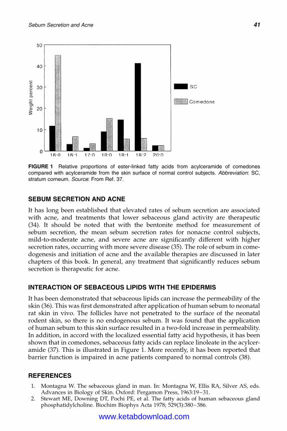
DK4962-Webster-ch3_R2_220307
SEBUM SECRETION AND ACNE
It has long been established that elevated rates of sebum secretion are associatedwith acne, and treatments that lower sebaceous gland activity are therapeutic(34). It should be noted that with the bentonite method for measurement ofsebum secretion, the mean sebum secretion rates for nonacne control subjects,mild-to-moderate acne, and severe acne are significantly different with highersecretion rates, occurring with more severe disease (35). The role of sebum in come-dogenesis and initiation of acne and the available therapies are discussed in laterchapters of this book. In general, any treatment that significantly reduces sebumsecretion is therapeutic for acne.
INTERACTION OF SEBACEOUS LIPIDS WITH THE EPIDERMIS
It has been demonstrated that sebaceous lipids can increase the permeability of theskin (36). This was first demonstrated after application of human sebum to neonatalrat skin in vivo. The follicles have not penetrated to the surface of the neonatalrodent skin, so there is no endogenous sebum. It was found that the applicationof human sebum to this skin surface resulted in a two-fold increase in permeability.In addition, in accord with the localized essential fatty acid hypothesis, it has beenshown that in comedones, sebaceous fatty acids can replace linoleate in the acylcer-amide (37). This is illustrated in Figure 1. More recently, it has been reported thatbarrier function is impaired in acne patients compared to normal controls (38).
REFERENCES
1. Montagna W. The sebaceous gland in man. In: Montagna W, Ellis RA, Silver AS, eds.Advances in Biology of Skin. Oxford: Pergamon Press, 1963:19–31.
2. Stewart ME, Downing DT, Pochi PE, et al. The fatty acids of human sebaceous glandphosphatidylcholine. Biochim Biophys Acta 1978; 529(3):380–386.
FIGURE 1 Relative proportions of ester-linked fatty acids from acylceramide of comedonescompared with acylceramide from the skin surface of normal control subjects. Abbreviation: SC,stratum corneum. Source: From Ref. 37.
Sebum Secretion and Acne 41
www.ketabdownload.com
DK4962-Webster-ch3_R2_220307
3. Bloch K. The biological synthesis of cholesterol. Science 1965; 150(692):19–28.4. Downing DT, Strauss JS, Pochi PE. Variability in the chemical composition of human
skin surface lipids. J Invest Dermatol 1969; 53(5):322–327.5. Weitkamp AW, Smiljanic AM, Rothman S. The free fatty acids of human hair fat. J Am
Chem Soc 1947; 69(8):1936–1939.6. Marples RR, Kligman AM, Lantis LR, et al. The role of the aerobic microflora in the
genesis of fatty acids in human surface lipids. J Invest Dermatol 1970; 55(3):173–178.
7. Gilbert D, Madison KC, Sando GN, et al. Acid lipase in lamellar granules. J Dent Res2000; 79(special issue):322.
8. Bergsson G, Arnfinnsson J, Steingrimsson O, et al. Killing of gram-positive cocci by fattyacids and monoglycerides. APMIS 2001; 109(10):670–678.
9. Bortz JT, Wertz PW, Downing DT. On the origin of alkanes found in human skin surfacelipids. J Invest Dermatol 1989; 93(6):723–727.
10. Thiele JJ, Schroeter C, Hsieh SN, et al. The antioxidant network of the stratum corneum.Curr Probl Dermatol 2001; 29:26–42.
11. Passi S, De Pita O, Puddu P, et al. Lipophilic antioxidants in human sebum and aging.Free Radic Res 2002; 36(4):471–477.
12. Ponec M, Weerheim A, Lankhorst P, et al. New acylceramide in native and reconstructedepidermis. J Invest Dermatol 2003; 120(4):581–588.
13. Nicolaides N, Fu HC, Ansari MNA, et al. The fatty acids of wax esters and sterol esters ofvernix caseosa and from human skin surface. Lipids 1972; 7(8):506–517.
14. .Green SC, Stewart ME, Downing DT. Variation in sebum fatty acid composition amongadult humans. J Invest Dermatol 1984; 83(2):114–117.
15. Stewart ME, Downing DT. Unusual cholesterol esters in the sebum of young children.J Invest Dermatol 1990; 95(5):603–606.
16. Downing DT, Pochi PE, Strauss JS. Synthesis and composition of surface lipids ofhuman skin. J Invest Dermatol 1974; 62(3):228–244.
17. Krakow R, Downing DT, Strauss JS, et al. Identification of a fatty acid in human skinsurface lipids apparently associated with acne vulgaris. J Invest Dermatol 1973;61(5):286–289.
18. Downing DT, Stewart ME, Wertz PW, et al. Essential fatty acids and acne. J Am AcadDermatol 1986; 14(2 Pt 1):221–225.
19. Strauss JS, Pochi PE. The quantitative gravimetric determination of sebum production.J Invest Dermatol 1961; 36(4):293–298.
20. Downing DT. Photodensitometry in the thin-layer chromatographic analysis of neutrallipids. J Chromatogr 1968; 38(1):91–99.
21. Downing DT, Stranieri AM, Strauss JS. The effect of accumulated lipids on measurementof sebum secretion in human skin. J Invest Dermatol 1982; 79(4):226–228.
22. Pierard GE. Follicle to follicle heterogeneity of sebum excretion. Dermatologica 1986;173(2):61–65.
23. Nordstrom KM, Schmus HG, McGinley KJ, et al. Measurement of sebum output using alipid adsorbent tape. J Invest Dermatol 1986; 87(2):260–263.
24. Kligman AM, Miller DL, McGinley K. Sebutape: a device for visualizing and measuringhuman sebaceous secretion. J Soc Cosmet Chem 1986; 37(4):369–374.
25. Pierard-Franchimont C, Pierard GE, Saint-Leger D, et al. Comparison of the kinetics ofsebum secretion in young women with and without acne. Dermatologica 1991;183(6):120–122.
26. Clarys P, Barel A. Quantitative evaluation of skin surface lipids. Clin Dermatol 1995;13(4):307–321.
27. Schaefer H, Kuhn-Bussius H. A method for the quantitative determination of humansebum secretion. Arch Klin Exp Dermatol 1970; 238(4):429–435.
28. Agache P, Blanc D, Barrand C, et al. Sebum levels during the first year of life. Br JDermatol 1980; 103(6):643–649.
29. Stewart ME, Downing DT. Measurement of sebum secretion rates in young children.J Invest Dermatol 1985; 84(1):59–61.
42 Wertz
www.ketabdownload.com
DK4962-Webster-ch3_R2_220307
30. Leyden JJ. New understandings of the pathogenesis of acne. J Am Acad Dermatol 1995;32(5 Pt 3):S15–S25.
31. Downing DT, Stewart ME, Strauss JS. Changes in sebum secretion and the sebaceousgland. Dermatol Clin 1986; 4(3):419–423.
32. Narendran V, Wickett RR, Pickens WL, et al. Interaction between pulmonary surfactantand vernix: a potential mechanism for induction of amniotic fluid turbidity. Pediatr Res2000; 48(1):120–124.
33. Jacobsen E, Billings JK, Frantz RA, et al. Age-related changes in sebaceous wax estersecretion rates in men and women. J Invest Dermatol 1985; 85(5):483–485.
34. Strauss JS. Sebaceous gland, acne and related disorders—an epilogue. Dermatology1998; 196(1):182–184.
35. Harris HH, Downing DT, Stewart ME, et al. Sustainable rates of sebum secretion. J AmAcad Dermatol 1983; 8(2):200–203.
36. Squier CA, Wertz PW, Williams DM, Cruchley AT. Permeability of oral mucosa and skinwith age. In: Squier CA, Hill MH, eds. The Effect of Aging in Oral Mucosa and Skin. BocaRaton: CRC Press, 1994:91–98.
37. Wertz PW, Miethke MC, Long SA, et al. The composition of ceramides from humanstratum corneum and from comedones. J Invest Dermatol 1985; 84(5):410–412.
38. Yamamoto A, Takenouchi K, Ito M. Impaired water barrier function in acne vulgaris.Arch Dermatol Res 1995; 287(2):214–218.
Sebum Secretion and Acne 43
www.ketabdownload.com

DK4962-Webster-ch3_R2_220307
www.ketabdownload.com
DK4962-Webster-ch4_R2_020407
B4 The Molecular Biology of Retinoidsand Their Receptors
Anthony V. RawlingsAVR Consulting Ltd., Northwich, Cheshire, U.K.
INTRODUCTION
Acne vulgaris is a multifactorial disease of the skin in areas rich in sebaceousfollicles. It is characterized by seborrhea, hypercornification of the infundibulum(the neck of the sebaceous gland), and the presence of comedones and inflamma-tory lesions such as papules and pustules. The inflammatory pathway is mediatedby antigenic and inflammatory products of Propionibacterium acnes (1). Althoughacne is mostly associated with puberty, persistent or late onset acne may besimilar in physiology to pubertal acne, with hyperandrogenicity and increasedsebogenesis, being the key factors (2). The hyperproliferation of the infundibulumkeratinocytes, characterized by the expression of the hyperproliferative markerproteins, Ki67 and K6/16, and the resulting immature stratum corneum does notdesquamate efficiently, leading to clumps of squames that attach to the hair folliclecausing a blockage to sebum flow. Hypercornification of the infrainfundibulum isan early feature of the microcomedones, preceding the clinically observable inflam-matory process (3).
Hypovitaminosis A has been proposed as an inducer of sebogenesis, asretinoids suppress sebogenesis. Follicles with a high sebum output are thought tobecome deficient in vitamin A, which will disturb the keratinization process ofthe infundibulum of the gland, culminating in the initial formation of microcome-dones in these glands (4).
Recently, comedogenesis has been related to a subclinical microinflammatorystate in the sebaceous gland (3). The increased interleukin-1 levels may influencethe production of other growth factors by fibroblasts that then cause the ductalhyperproliferation and faulty differentiation (5). Increased transcription factorsignaling occurs following binding of the cytokines to their receptors, activatorprotein-1 (AP-1) and nuclear factor kappa-b, thereby activating the signalingpathways. As a result, suppression of these signaling pathways will be beneficialfor acne. Retinoids can improve epidermal differentiation, reduce sebogenesis,and decrease AP-1 driven inflammatory pathways. It is not too surprising, there-fore, that this class of agents improve the acne status. This chapter reviews themolecular biology and biochemistry of this important class of molecules.
NUCLEAR HORMONE RECEPTORS AND THE RETINOID RECEPTORS
In the last several years, the structure and function of nuclear receptors have beendetermined (6). Based on the similarities among the sequences, it has been possibleto classify the receptor superfamilies. The endocrine receptors include the retinoicacid receptors (RARs) and the thyroid hormone receptors, and the adopted orphan
45
www.ketabdownload.com
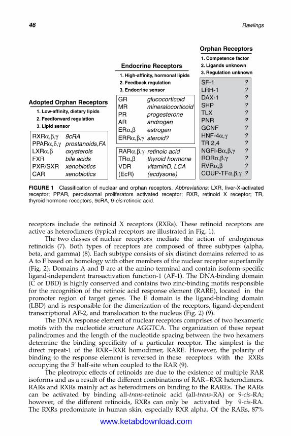
DK4962-Webster-ch4_R2_020407
receptors include the retinoid X receptors (RXRs). These retinoid receptors areactive as heterodimers (typical receptors are illustrated in Fig. 1).
The two classes of nuclear receptors mediate the action of endogenousretinoids (7). Both types of receptors are composed of three subtypes (alpha,beta, and gamma) (8). Each subtype consists of six distinct domains referred to asA to F based on homology with other members of the nuclear receptor superfamily(Fig. 2). Domains A and B are at the amino terminal and contain isoform-specificligand-independent transactivation function-1 (AF-1). The DNA-binding domain(C or DBD) is highly conserved and contains two zinc-binding motifs responsiblefor the recognition of the retinoic acid response element (RARE), located in thepromoter region of target genes. The E domain is the ligand-binding domain(LBD) and is responsible for the dimerization of the receptors, ligand-dependenttranscriptional AF-2, and translocation to the nucleus (Fig. 2) (9).
The DNA response element of nuclear receptors comprises of two hexamericmotifs with the nucleotide structure AGGTCA. The organization of these repeatpalindromes and the length of the nucleotide spacing between the two hexamersdetermine the binding specificity of a particular receptor. The simplest is thedirect repeat-1 of the RXR–RXR homodimer, RARE. However, the polarity ofbinding to the response element is reversed in these receptors with the RXRsoccupying the 50 half-site when coupled to the RAR (9).
The pleotropic effects of retinoids are due to the existence of multiple RARisoforms and as a result of the different combinations of RAR–RXR heterodimers.RARs and RXRs mainly act as heterodimers on binding to the RAREs. The RARscan be activated by binding all-trans-retinoic acid (all-trans-RA) or 9-cis-RA;however, of the different retinoids, RXRs can only be activated by 9-cis-RA.The RXRs predominate in human skin, especially RXR alpha. Of the RARs, 87%
Adopted Orphan Receptors
Endocrine Receptors
Orphan Receptors
1. Competence factor
1. High-affinity, hormonal lipids
1. Low-affinity, dietary lipids
2. Feedback regulation
2. Feedforward regulation
3. Endocrine sensor
GR
SF-1LRH-1DAX-1SHPTLXPNRGCNF
TR 2,4
MRPRARERα,βERRα,β,γ
RARα,β,γ
HNF-4α,γ
TRα,β
RXRα,β,γ
NGFI-Bα,β,γ RORα,β,γ RVRα,βCOUP-TFα,β,γ
?????????????
LXRα,βFXR
CARVDR(EcR)
PXR/SXR
PPARα,δ,γ
3. Lipid sensor
9cRAprostanoids,FAoxysterolsbile acidsxenobioticsxenobiotics (ecdysone)
vitaminD, LCAthyroid hormoneretinoic acid
steroid?estrogenandrogenprogesteronemineralocorticoidglucocorticoid
2. Ligands unknown
3. Regulation unknown
FIGURE 1 Classification of nuclear and orphan receptors. Abbreviations: LXR, liver-X-activatedreceptor; PPAR, peroxisomal proliferators activated receptor; RXR, retinoid X receptor; TR,thyroid hormone receptors, 9cRA, 9-cis-retinoic acid.
46 Rawlings
www.ketabdownload.com
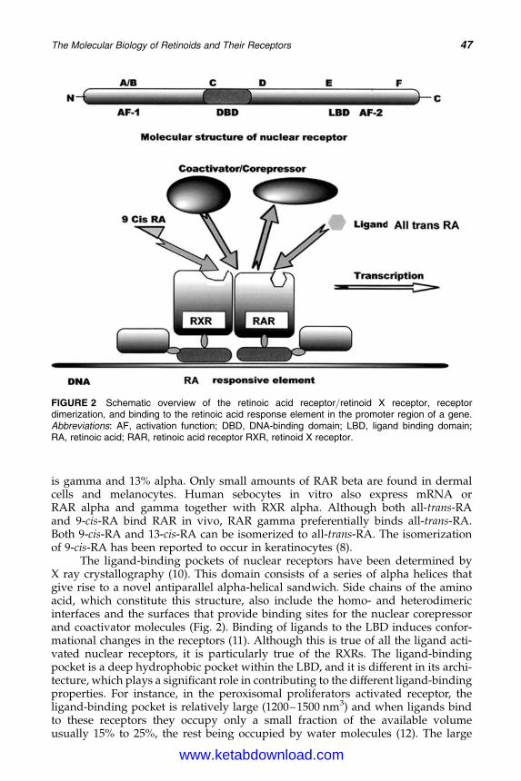
DK4962-Webster-ch4_R2_020407
is gamma and 13% alpha. Only small amounts of RAR beta are found in dermalcells and melanocytes. Human sebocytes in vitro also express mRNA orRAR alpha and gamma together with RXR alpha. Although both all-trans-RAand 9-cis-RA bind RAR in vivo, RAR gamma preferentially binds all-trans-RA.Both 9-cis-RA and 13-cis-RA can be isomerized to all-trans-RA. The isomerizationof 9-cis-RA has been reported to occur in keratinocytes (8).
The ligand-binding pockets of nuclear receptors have been determined byX ray crystallography (10). This domain consists of a series of alpha helices thatgive rise to a novel antiparallel alpha-helical sandwich. Side chains of the aminoacid, which constitute this structure, also include the homo- and heterodimericinterfaces and the surfaces that provide binding sites for the nuclear corepressorand coactivator molecules (Fig. 2). Binding of ligands to the LBD induces confor-mational changes in the receptors (11). Although this is true of all the ligand acti-vated nuclear receptors, it is particularly true of the RXRs. The ligand-bindingpocket is a deep hydrophobic pocket within the LBD, and it is different in its archi-tecture, which plays a significant role in contributing to the different ligand-bindingproperties. For instance, in the peroxisomal proliferators activated receptor, theligand-binding pocket is relatively large (1200–1500 nm3) and when ligands bindto these receptors they occupy only a small fraction of the available volumeusually 15% to 25%, the rest being occupied by water molecules (12). The large
FIGURE 2 Schematic overview of the retinoic acid receptor/retinoid X receptor, receptordimerization, and binding to the retinoic acid response element in the promoter region of a gene.Abbreviations: AF, activation function; DBD, DNA-binding domain; LBD, ligand binding domain;RA, retinoic acid; RAR, retinoic acid receptor RXR, retinoid X receptor.
The Molecular Biology of Retinoids and Their Receptors 47
www.ketabdownload.com
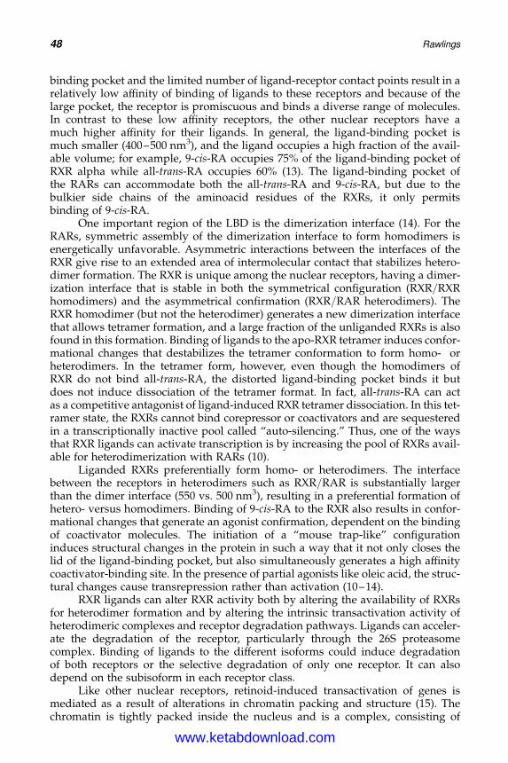
DK4962-Webster-ch4_R2_020407
binding pocket and the limited number of ligand-receptor contact points result in arelatively low affinity of binding of ligands to these receptors and because of thelarge pocket, the receptor is promiscuous and binds a diverse range of molecules.In contrast to these low affinity receptors, the other nuclear receptors have amuch higher affinity for their ligands. In general, the ligand-binding pocket ismuch smaller (400–500 nm3), and the ligand occupies a high fraction of the avail-able volume; for example, 9-cis-RA occupies 75% of the ligand-binding pocket ofRXR alpha while all-trans-RA occupies 60% (13). The ligand-binding pocket ofthe RARs can accommodate both the all-trans-RA and 9-cis-RA, but due to thebulkier side chains of the aminoacid residues of the RXRs, it only permitsbinding of 9-cis-RA.
One important region of the LBD is the dimerization interface (14). For theRARs, symmetric assembly of the dimerization interface to form homodimers isenergetically unfavorable. Asymmetric interactions between the interfaces of theRXR give rise to an extended area of intermolecular contact that stabilizes hetero-dimer formation. The RXR is unique among the nuclear receptors, having a dimer-ization interface that is stable in both the symmetrical configuration (RXR/RXRhomodimers) and the asymmetrical confirmation (RXR/RAR heterodimers). TheRXR homodimer (but not the heterodimer) generates a new dimerization interfacethat allows tetramer formation, and a large fraction of the unliganded RXRs is alsofound in this formation. Binding of ligands to the apo-RXR tetramer induces confor-mational changes that destabilizes the tetramer conformation to form homo- orheterodimers. In the tetramer form, however, even though the homodimers ofRXR do not bind all-trans-RA, the distorted ligand-binding pocket binds it butdoes not induce dissociation of the tetramer format. In fact, all-trans-RA can actas a competitive antagonist of ligand-induced RXR tetramer dissociation. In this tet-ramer state, the RXRs cannot bind corepressor or coactivators and are sequesteredin a transcriptionally inactive pool called “auto-silencing.” Thus, one of the waysthat RXR ligands can activate transcription is by increasing the pool of RXRs avail-able for heterodimerization with RARs (10).
Liganded RXRs preferentially form homo- or heterodimers. The interfacebetween the receptors in heterodimers such as RXR/RAR is substantially largerthan the dimer interface (550 vs. 500 nm3), resulting in a preferential formation ofhetero- versus homodimers. Binding of 9-cis-RA to the RXR also results in confor-mational changes that generate an agonist confirmation, dependent on the bindingof coactivator molecules. The initiation of a “mouse trap-like” configurationinduces structural changes in the protein in such a way that it not only closes thelid of the ligand-binding pocket, but also simultaneously generates a high affinitycoactivator-binding site. In the presence of partial agonists like oleic acid, the struc-tural changes cause transrepression rather than activation (10–14).
RXR ligands can alter RXR activity both by altering the availability of RXRsfor heterodimer formation and by altering the intrinsic transactivation activity ofheterodimeric complexes and receptor degradation pathways. Ligands can acceler-ate the degradation of the receptor, particularly through the 26S proteasomecomplex. Binding of ligands to the different isoforms could induce degradationof both receptors or the selective degradation of only one receptor. It can alsodepend on the subisoform in each receptor class.
Like other nuclear receptors, retinoid-induced transactivation of genes ismediated as a result of alterations in chromatin packing and structure (15). Thechromatin is tightly packed inside the nucleus and is a complex, consisting of
48 Rawlings
www.ketabdownload.com
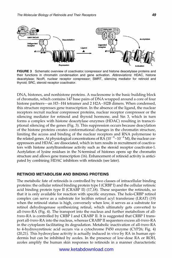
DK4962-Webster-ch4_R2_020407
DNA, histones, and nonhistone proteins. A nucleosome is the basic building blockof chromatin, which contains 147 base pairs of DNA wrapped around a core of fourhistone partners—an H3–H4 tetramer and 2 H2A–H2B dimers. When condensed,this structure represses gene transcription. In the absence of the ligand, the nuclearreceptors recruit nuclear corepressor proteins, nuclear receptor corepressor or thesilencing mediator for retinoid and thyroid hormone, and Sin 3, which in turnforms a complex with histone deacetylase enzymes (HDAC) resulting in transcri-ptional silencing of the genes (Fig. 3). This suppression occurs because deacylationof the histone proteins creates conformational changes in the chromatin structure,limiting the access and binding of the nuclear receptors and RNA polymerase tothe related genes. At physiological concentrations of RA (1029–1028 M), the nuclear cor-eppressors and HDAC are dissociated, which in turn results in recruitment of coactiva-tors with histone acetyltransferase activity such as the steroid receptor coactivator-1.Acetylation of lysine residues in the N-terminal of histones opens up the chromatinstructure and allows gene transcription (16). Enhancement of retinoid activity is antici-pated by combining HDAC inhibitors with retinoids (see later).
RETINOID METABOLISM AND BINDING PROTEINS
The metabolic fate of retinoids is controlled by two classes of intracellular bindingproteins: the cellular retinol binding protein type I (CRBP I) and the cellular retinoicacid binding protein type II (CRABP II) (17,18). These sequester the retinoids, sothat it is only available for reaction with specific enzymes. The holo-CRBP retinolcomplex can serve as a substrate for lecithin retinol acyl transferase (LRAT) (19)when the retinoid status is high, conversely when low, it serves as a substrate forretinol dehydrogenase synthesizing retinal, which ultimately gets converted toall-trans-RA (Fig. 4). The transport into the nucleus and further metabolism of all-trans-RA is controlled by CRBP I and CRABP II. It is suggested that CRBP I trans-port all-trans-RA into the nucleus, whereas CRABP II sequesters excess all-trans-RAin the cytoplasm facilitating its degradation. Metabolic inactivation of all-trans-RAto 4-hydroxyretinoic acid occurs via a cytochrome P450 enzyme (CYP26; Fig. 4)(20,21). This hydroxylase activity is actually induced in vivo by RA in human epi-dermis but can be inhibited by azoles. In the presence of low-dose RA or ROH,azoles amplify the human skin responses to retinoids in a manner characteristic
FIGURE 3 Schematic overview of coactivator/corepressor and histone deacetylase proteins andtheir functions in chromatin condensation and gene activation. Abbreviations: HDAC, histonedeacetylase; NcoR, nuclear receptor corepressor; SMRT, silencing mediator for retinoid andthyroid; SRC, steroid receptor coactivator.
The Molecular Biology of Retinoids and Their Receptors 49
www.ketabdownload.com
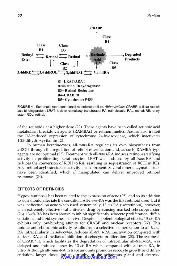
DK4962-Webster-ch4_R2_020407
of the retinoids at a higher dose (22). These agents have been called retinoic acidmetabolism breakdown agents (RAMBAs) or retinomimetics. Azoles also inhibitthe RA-induced expression of cytochrome 24-hydroxylase, which inactivates1,25-dihydroxyvitamin D3.
In human keratinocytes, all-trans-RA regulates its own biosynthesis fromatROH through the regulation of retinol esterification and, as such, RAMBA-typeagents are not optimal (23). Treatment with all-trans-RA induces retinol-esterifyingactivity in proliferating keratinocytes. LRAT was induced by all-trans-RA andreduces the conversion of ROH to RA, resulting in sequestration of ROH in REs.Acyl retinol acyl transferase activity is also present. Several other enzymatic stepshave been identified, which if manipulated can deliver improved retinoidresponses (24).
EFFECTS OF RETINOIDS
Hypovitaminosis has been related to the expression of acne (25), and so its additionto skin should alleviate the condition. All-trans-RA was the first retinoid used, but itwas ineffectual on acne when used systemically. 13-cis-RA (isotretinoin), however,is an extremely effective oral anti-acne drug by causing marked sebosuppression(26). 13-cis-RA has been shown to inhibit significantly sebocyte proliferation, differ-entiation, and lipid synthesis in vivo. Despite its potent biological effects, 13-cis-RAexhibits only low-binding affinity for CRABP and nuclear receptors (27). Thisunique antisebotrophic activity results from a selective isomerization to all-trans-RA intracellularly in sebocytes, reduces all-trans-RA inactivation compared withall-trans-RA, and mediates inhibition of sebocyte proliferation (28). The synthesisof CRABP II, which facilitates the degradation of intracellular all-trans-RA, wasdelayed and induced lesser by 13-cis-RA when compared with all-trans-RA, invitro. Although all-trans-RA in trace amounts promotes sebocyte growth and differ-entiation, larger doses induce atrophy of the sebaceous gland and decrease
FIGURE 4 Schematic representation of retinol metabolism. Abbreviations: CRABP, cellular retinoicacid binding protein; LRAT, lecithin retinol acyl transferase; RA, retinoic acid; RAL, retinal; RE, retinylester; ROL, retinol.
50 Rawlings
www.ketabdownload.com
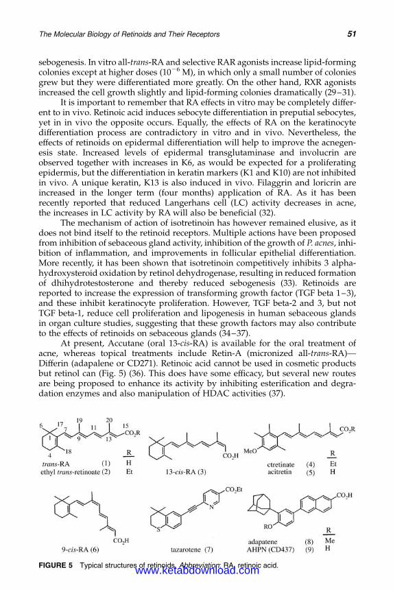
DK4962-Webster-ch4_R2_020407
sebogenesis. In vitro all-trans-RA and selective RAR agonists increase lipid-formingcolonies except at higher doses (1026 M), in which only a small number of coloniesgrew but they were differentiated more greatly. On the other hand, RXR agonistsincreased the cell growth slightly and lipid-forming colonies dramatically (29–31).
It is important to remember that RA effects in vitro may be completely differ-ent to in vivo. Retinoic acid induces sebocyte differentiation in preputial sebocytes,yet in in vivo the opposite occurs. Equally, the effects of RA on the keratinocytedifferentiation process are contradictory in vitro and in vivo. Nevertheless, theeffects of retinoids on epidermal differentiation will help to improve the acnegen-esis state. Increased levels of epidermal transglutaminase and involucrin areobserved together with increases in K6, as would be expected for a proliferatingepidermis, but the differentiation in keratin markers (K1 and K10) are not inhibitedin vivo. A unique keratin, K13 is also induced in vivo. Filaggrin and loricrin areincreased in the longer term (four months) application of RA. As it has beenrecently reported that reduced Langerhans cell (LC) activity decreases in acne,the increases in LC activity by RA will also be beneficial (32).
The mechanism of action of isotretinoin has however remained elusive, as itdoes not bind itself to the retinoid receptors. Multiple actions have been proposedfrom inhibition of sebaceous gland activity, inhibition of the growth of P. acnes, inhi-bition of inflammation, and improvements in follicular epithelial differentiation.More recently, it has been shown that isotretinoin competitively inhibits 3 alpha-hydroxysteroid oxidation by retinol dehydrogenase, resulting in reduced formationof dhihydrotestosterone and thereby reduced sebogenesis (33). Retinoids arereported to increase the expression of transforming growth factor (TGF beta 1–3),and these inhibit keratinocyte proliferation. However, TGF beta-2 and 3, but notTGF beta-1, reduce cell proliferation and lipogenesis in human sebaceous glandsin organ culture studies, suggesting that these growth factors may also contributeto the effects of retinoids on sebaceous glands (34–37).
At present, Accutane (oral 13-cis-RA) is available for the oral treatment ofacne, whereas topical treatments include Retin-A (micronized all-trans-RA)—Differin (adapalene or CD271). Retinoic acid cannot be used in cosmetic productsbut retinol can (Fig. 5) (36). This does have some efficacy, but several new routesare being proposed to enhance its activity by inhibiting esterification and degra-dation enzymes and also manipulation of HDAC activities (37).
FIGURE 5 Typical structures of retinoids. Abbreviation: RA, retinoic acid.
The Molecular Biology of Retinoids and Their Receptors 51
www.ketabdownload.com
DK4962-Webster-ch4_R2_020407
REFERENCES
1. Stern RS. Acne therapy: medication use and sources of care in office-based practice. ArchDermatol 1996; 132:776–780.
2. Knaggs HE, Wood EJ, Rizer RL, et al. Post-adolescent acne. Int J Cosmet Chem 2004;26(3):129–138.
3. Jeremy AH, Holland DB, Roberts SG, et al. Inflammatory events are involved in acnelesion initiation. J Invest Dermatol 2003; 121(1):20–27.
4. Macdonald-Hull SM, Cunliffe WJ. Hypovitaminosis A of follicular duct as cause of acnevulgaris. Lancet 1988; 31(2):8626–8627.
5. Angel P, Szabowski A. Function of AP-1 target genes in mesenchymal-epithelial cross-talk in skin. Biochem Pharmacol 2002; 64(5–6):949–956.
6. Aranda A, Pascual A. Nuclear hormone receptors & gene expression. Physiol Rev 2001;81(3):1269–1304.
7. Mehta K. Retinoids as regulators of gene transcription. J Biol Regul Homeost Agents2003; 17:1–12.
8. Fisher GJ, Voorhees JJ. Molecular mechanisms of retinoid actions in skin. FASEB J 1996;10:1002–1013.
9. Rastinejad F. Retinoid X receptor and its partners in the nuclear receptor family. CurrOpin Struct Biol 2001; 11:33–38.
10. Ahuja HS, Szanto A, Nagy L, et al. The retinoid X receptor & its ligands: versatile reg-ulators of metabolic function, cell differentiation & cell death. J Biol Regul HomeostAgents 2003; 17(1):29–45.
11. Wurtz JM, Bourguet W, Renaud JP, et al. A canonical structure for the ligand bindingdomain of nuclear receptors. Nat Struct Biol 1996; 3:87–94.
12. Gampe RT, Montana VG, Lambert MH, et al. Asymmetry in the PPARgamma/RXRalphacrystal structure reveals the molecular basis of heterodimerization among nuclearreceptors. Mol Cell 2000; 5:545– 555.
13. Egea PF, Mitschler A, Rochel N, et al. Crystal structure of the human RXRalpha ligandbinding domain bound to its natural ligand: 9-cis retinoic acid. EMBO J 2000;19:2592–2601.
14. Bourguet W, Vivat V, Wurtz JM, et al. Crystal structure of a heterodimeric complex ofRAR & RXR ligand-binding domains. Mol Cell 2000; 5:289–298.
15. Khorasanizadeh S. The nucleosome: from genomic organization to genomic regulation.Cell 2004; 53:1003–1009.
16. Jung M. Inhibitors of histone deacetylase as new anticancer agents. Curr Med Chem2002; 8(12):1505–1511.
17. Roos TC, Jugert FK, Merk HF, et al. Retinoid metabolism in the skin. Pharmacol Rev1998; 50(2):315–333.
18. Napoli JL. Interactions of retinoid binding proteins & enzymes in retinoid metabolism.BBA 1999; 1440:139–162.
19. Kurlandsky SB, Duell EA, Kang S, et al. Autoregulation of retinoic acid biosynthesisthrough regulation of retinol esterification in human keratinocytes. J Biol Chem 1996;271(26):15346–15352.
20. Duell EA, Kang S, Voorhees JJ. Retinoic acid isomers applied to human skin in vivo eachinduce a 4-hydroxylase that inactivates only trans retinoic acid. J Invest Dermatol 1996;106(2):316–320.
21. Mirikar Y, Wang Z, Duell EA, et al. Retinoic acid receptors regulate expression of retinoicacid 4-hydroxylase that specifically inactivates all-trans retinoic acid in human keratino-cyte HaCaT cells. J Invest Dermatol 1998; 111(3):434–439.
22. Kang S, Duell EA, Kim KJ, et al. Liarozole in hibits human epidermal retinoic acid4-hydroxylase activity and differentially augments human skin responses to retinoicacid and retinol in vivo. J Invest Dermatol 1996; 107:183–187.
23. Iobst S, Feinberg C, Rawlings AV, et al. Manipulation of retinoid metabolism. J InvestDermatol 2003; 121:0563.
24. Granger S, Rawlings AV, Scott IR. Compositions to mimic retinoic acid. European PatentWO9813020.
52 Rawlings
www.ketabdownload.com

DK4962-Webster-ch4_R2_020407
25. Wolbach SB, Howe PR. Tissue changes following deprivation of fat soluble A vitamin.J Exp Med 1925; 42:753–777.
26. Goldstein JS, Socha-Szott A, Thomsen RJ, et al. Comparative effect of isotretinoin andetretinate on acne and sebaceous gland secretion. J Am Acad Dermatol 1982; 6:760–765.
27. Downie MMT, Guy R, Kealey T. Advances in sebaceous gland research: potential newapproaches to acne management. Int J Cosmet Chem 2004; 26(6):291–312.
28. Tsukada M, Schroder M, Roos TC, et al. 13-cis retinoic acid exerts its specific activity onhuman sebocytes through selective intracellular isomerization to all-trans retinoic acidand binding to retinoid receptors. J Invest Dermatol 2000; 115:321–327.
29. Zouboulis CC, Korge B, Akamatsu H, et al. Effects of 13-cis Retnoic acid, all trans retinoicacid and acitretin on the proliferation, lipid synthesis and keratin expression of culturedhuman sebocytes in vitro. J Invest Dermatol 1991; 96:792–797.
30. Zouboulis CC, Korge B, Mischke D, et al. Altered proliferation, synthetic activity &differentiation of cultured human sebocytes in the absence of vitamin A and theirmodulation by synthetic retinoids. J Invest Dermatol 1993; 101:628–633.
31. Kim MJ, Ciletti N, Michael S, et al. The role of specific retinoid receptors in sebocytegrowth & differentiation in culture. J Invest Dermatol 2000; 114:349–353.
32. Griffiths CEM, Voorhees JJ. Human in vivo pharmacology of topical retinoids. ArchDermatol Res 1994; 287:53–60.
33. Glick AB, McCune BK, Abdulkarem N, et al. Complex regulation of TGFbeta expressionby retinoic acid in the vitamin A deficient rat. Development 1991; 111:1081–1086.
34. Fisher GJ, Tavakkol A, Griffiths CEM, et al. Differential modulation of TGFbetaexpression & mucin deposition by retinoic acid & SLS in human skin. J Invest Dermatol1992; 98:102–108.
35. Han GR, Dohi DF, Lee HY, et al. All trans retinoic acid increases TGFbeta2 & IGFBP-3expression through a retinoic acid receptor alpha dependent signalling pathway. J BiolChem 1997; 272:13711–13716.
36. Dawson MI. Synthetic retinoids & their nuclear receptors. Curr Med Chem 2004; 4:199–230.37. Schehlmann V, Klock J, Maillan P, Vollhardt J, Rawlings AV, Beumer R. Composition
comprising an HDAC inhibitor in combination with a retinoid. European PatentWO2005092283, 2005.
The Molecular Biology of Retinoids and Their Receptors 53
www.ketabdownload.com

DK4962-Webster-ch4_R2_020407
www.ketabdownload.com
DK4962-Webster-ch5_R2_020407
B5 Molecular Biology of PeroxisomeProliferator-Activated Receptors in Relationto Sebaceous Glands and Acne
Michaela M. T. DownieSequenom GmbH, Hamburg, Germany
Terence KealeyThe Clore Laboratories, University of Buckingham, Buckingham, U.K.
INTRODUCTION
Peroxisome proliferators-activated receptors (PPARs) are nuclear hormonereceptors belonging to the superfamily of steroid/thyroid receptors. Three typesof nuclear PPARs, PPARa, PPARb/d, and PPARg1/g2, have been identified andcloned, each being encoded by separate genes (1–8). Alternative splicing of onegene product yields two transcripts of PPARg: PPARg1 was found to predominatein adipose tissue and large intestines, whereas PPAR g2 seemed to be a minorisoform (9). These receptors have been named according to their capacity to beactivated by many compounds, which share in common the property of inducingperoxisome proliferation, especially in rodent liver (10–12). They are involved inthe control of genes encoding lipid metabolism-associated proteins, particularlythose of peroxisomal b-oxidation (13–15), and are activated by physiologicalconcentrations of fatty acids (16–18), fibrate hypolipidemic drugs (10,19), andcertain plasticizers and herbicides (19,20).
The main response to peroxisomal proliferators is an increase inperoxisome proliferation, stimulation of peroxisomal fatty acidb-oxidation, andhepatocarcinogenesis. The oxidative degradation is responsible for theformation of hydrogen peroxide, which might lead to DNA damage andtumor initiation (20–22). Besides targeting peroxisomes, PPAR activation hasbeen shown to be important in the regulation of lipid metabolism in fat andliver cells (23,24) and also regulates the expression of target genes involvedin many cellular functions, including cell proliferation, differentiation, andimmune/inflammatory response (25). The three PPARs (a, b/d, g) show thecharacteristics of ligand binding, and ligand activation can be related to anumber of cellular processes: (i) fatty acid catabolism and modulation of theinflammatory response for PPARa; (ii) embryo implantation, cell proliferation,and apoptosis for PPARb/d; and (iii) adipocytic differentiation, monocyticdifferentiation, and cell-cycle withdrawal for PPARg (18). It has been shownthat 3T3 L1 cells can be induced to differentiate into adipocytes, demonstratingthat PPARs not only regulate the expression of lipid metabolism-associated pro-teins but are also directly involved in cell differentiation (26). This suggestionwas supported by the finding that PPARg expression is induced early in thedifferentiation of 3T3 L1 preadipocytes into adipocytes (27,28). Further,examining the effects of the PPARg ligands BRL49653, troglitazone, and
55
www.ketabdownload.com
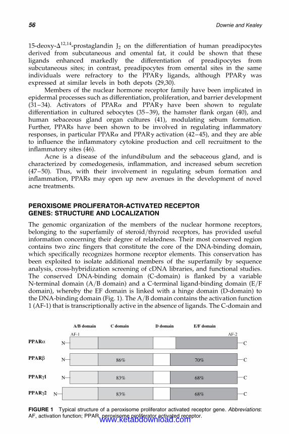
DK4962-Webster-ch5_R2_020407
15-deoxy-D12,14-prostaglandin J2 on the differentiation of human preadipocytesderived from subcutaneous and omental fat, it could be shown that theseligands enhanced markedly the differentiation of preadipocytes fromsubcutaneous sites; in contrast, preadipocytes from omental sites in the sameindividuals were refractory to the PPARg ligands, although PPARg wasexpressed at similar levels in both depots (29,30).
Members of the nuclear hormone receptor family have been implicated inepidermal processes such as differentiation, proliferation, and barrier development(31–34). Activators of PPARa and PPARg have been shown to regulatedifferentiation in cultured sebocytes (35–39), the hamster flank organ (40), andhuman sebaceous gland organ cultures (41), modulating sebum formation.Further, PPARs have been shown to be involved in regulating inflammatoryresponses, in particular PPARa and PPARg activation (42–45), and they are ableto influence the inflammatory cytokine production and cell recruitment to theinflammatory sites (46).
Acne is a disease of the infundibulum and the sebaceous gland, and ischaracterized by comedogenesis, inflammation, and increased sebum secretion(47–50). Thus, with their involvement in regulating sebum formation andinflammation, PPARs may open up new avenues in the development of novelacne treatments.
PEROXISOME PROLIFERATOR-ACTIVATED RECEPTORGENES: STRUCTURE AND LOCALIZATION
The genomic organization of the members of the nuclear hormone receptors,belonging to the superfamily of steroid/thyroid receptors, has provided usefulinformation concerning their degree of relatedness. Their most conserved regioncontains two zinc fingers that constitute the core of the DNA-binding domain,which specifically recognizes hormone receptor elements. This conservation hasbeen exploited to isolate additional members of the superfamily by sequenceanalysis, cross-hybridization screening of cDNA libraries, and functional studies.The conserved DNA-binding domain (C-domain) is flanked by a variableN-terminal domain (A/B domain) and a C-terminal ligand-binding domain (E/Fdomain), whereby the EF domain is linked with a hinge domain (D-domain) tothe DNA-binding domain (Fig. 1). The A/B domain contains the activation function1 (AF-1) that is transcriptionally active in the absence of ligands. The C-domain and
niamod C niamod F/E
%38 %86
%68
%38
%07
%86
PPARα
PPARγ1
PPARγ2
PPARβ
niamod B/A niamod D
1-FA 2-FA
N
N
N
N
C
C
C
C
FIGURE 1 Typical structure of a peroxisome proliferator activated receptor gene. Abbreviations:AF, activation function; PPAR, peroxisome proliferator activated receptor.
56 Downie and Kealey
www.ketabdownload.com
DK4962-Webster-ch5_R2_020407
the E/F-domain determine the specificity of promoters in DNA sequence recog-nition and ligand recognition, respectively. The ligand-binding domain, in additionto determining ligand specificity, contains a ligand-inducible transactivationfunction and a heptad-repeat motif involved in dimerization of nuclear receptors.This domain contains some regions that are highly conserved between mostmembers of the nuclear receptor superfamily, particularly in the Ti-domain andAF-2 region. The N-terminal domain varies both in length and amino acidcomposition, and is responsible for the interaction with other transcriptionfactors and transactivation. A distinctive feature of the PPAR subfamily is itsgreat divergence in the ligand-binding domain, allowing researchers to distinguishthe different PPAR subtypes pharmacologically (46,51,52).
The chromosomal localization of the human genes encoding PPARa,PPARb/d, and PPARg have been defined. They map on different chromosomes. ThePPARg gene is on chromosome 3 at position 3p25 and has six exons (53). ThePPARb/d gene is located on chromosome 6 at chromosomal region 6p21.2 andspans approximately 85 kb of DNA consisting of nine exons and eight introns(54). Finally, the PPARa gene has been mapped on chromosome 22 in the generalregion of 22q12-q13.1 and spans at least 80 kb with eight exons (6).
TRANSCRIPTIONAL ACTIVITY, DIMERIZATION, AND DNA BINDING
The transcriptional activity of the PPARs is regulated by post-translational modifi-cations, such as phosphorylation (55,56) and ubiquitination (57). Phosphorylationof PPARs is controlled by environmental factors, activating different kinase path-ways leading to the modulation of their activities (56). PPAR degradation by theubiquitin–proteasome system modulates the intensity of the ligand response bycontrolling the level of PPAR proteins in the cells (46,57). It has been shown, more-over, that the novel orphan nuclear hormone receptor, liver-X-activated receptor(LXR)a, which is distinct from retinoic-X-activated receptor (RXR)a, inhibitedPPAR signaling by competing with RXRa for PPARa heterodimerization,whereby the LXRa/PPARa heterodimer was unable to form a DNA-bindingcomplex (58).
Dimerization is essential for the function of PPARs (Fig. 2). It has been demon-strated in vitro that PPARs form heterodimers with RXRs and that activation ofPPARs can be increased in vitro by the addition of the RXR ligand, 9-cis-retinoicacid (59–63). As for other members of the nuclear hormone receptor family,PPARs regulate gene expression by binding to specific peroxisome proliferatorresponse elements (PPRE) in the promoter regions of target genes (64) (Fig. 2).PPARs recognize a hormone response element that comprises a direct repeat(DR) of the hexameric AGGTCA half-site motif, with a one nucleotide spacerbetween the half elements (DR-1). The complex formed in vitro on such a responseelement is a PPAR–RXR heterodimer (59–63). The first PPRE characterized wasfound in the promoter of the acyl-coenzyme A (CoA) oxidase gene and wasdefined as DR-1 (2). Further, it has been shown that PPARs can achieve gene/tissue specific activity by being selectively bound as heterodimers to PPRE,whereby specific heterodimers such as PPARg/RXRa or PPARg/RXRg bind morestrongly to certain PPREs than other heterodimer combinations (65). Binding ofPPAR/RXR to certain PPREs mediates transcriptional activation, but this can benegatively controlled by the presence of other heterodimers such as the thyroidhormone receptor/RXR heterodimer, as they can act as competitive inhibitors forPPRE binding that do not activate transcription (66).
Molecular Biology of Peroxisome Proliferator-Activated Receptors 57
www.ketabdownload.com
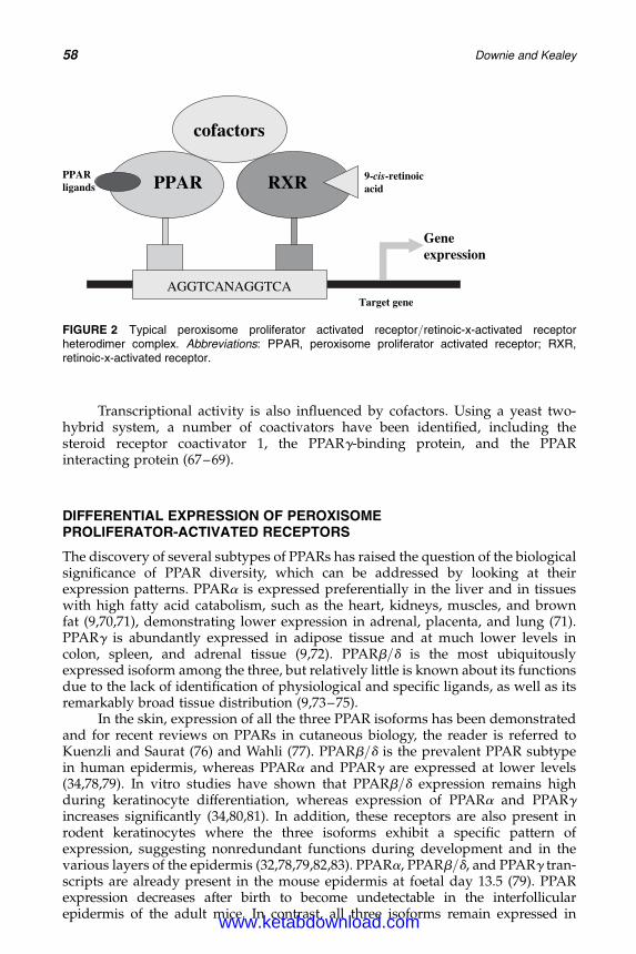
DK4962-Webster-ch5_R2_020407
Transcriptional activity is also influenced by cofactors. Using a yeast two-hybrid system, a number of coactivators have been identified, including thesteroid receptor coactivator 1, the PPARg-binding protein, and the PPARinteracting protein (67–69).
DIFFERENTIAL EXPRESSION OF PEROXISOMEPROLIFERATOR-ACTIVATED RECEPTORS
The discovery of several subtypes of PPARs has raised the question of the biologicalsignificance of PPAR diversity, which can be addressed by looking at theirexpression patterns. PPARa is expressed preferentially in the liver and in tissueswith high fatty acid catabolism, such as the heart, kidneys, muscles, and brownfat (9,70,71), demonstrating lower expression in adrenal, placenta, and lung (71).PPARg is abundantly expressed in adipose tissue and at much lower levels incolon, spleen, and adrenal tissue (9,72). PPARb/d is the most ubiquitouslyexpressed isoform among the three, but relatively little is known about its functionsdue to the lack of identification of physiological and specific ligands, as well as itsremarkably broad tissue distribution (9,73–75).
In the skin, expression of all the three PPAR isoforms has been demonstratedand for recent reviews on PPARs in cutaneous biology, the reader is referred toKuenzli and Saurat (76) and Wahli (77). PPARb/d is the prevalent PPAR subtypein human epidermis, whereas PPARa and PPARg are expressed at lower levels(34,78,79). In vitro studies have shown that PPARb/d expression remains highduring keratinocyte differentiation, whereas expression of PPARa and PPARgincreases significantly (34,80,81). In addition, these receptors are also present inrodent keratinocytes where the three isoforms exhibit a specific pattern ofexpression, suggesting nonredundant functions during development and in thevarious layers of the epidermis (32,78,79,82,83). PPARa, PPARb/d, and PPARg tran-scripts are already present in the mouse epidermis at foetal day 13.5 (79). PPARexpression decreases after birth to become undetectable in the interfollicularepidermis of the adult mice. In contrast, all three isoforms remain expressed in
RAPP RXR
ACTGGANACTGGA
eneGnoisserpxe
srotcafoc
RAPP sdnagil
-9 sic cioniter-dica
eneg tegraT
FIGURE 2 Typical peroxisome proliferator activated receptor/retinoic-x-activated receptorheterodimer complex. Abbreviations: PPAR, peroxisome proliferator activated receptor; RXR,retinoic-x-activated receptor.
58 Downie and Kealey
www.ketabdownload.com
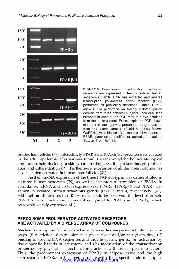
DK4962-Webster-ch5_R2_020407
murine hair follicles (79). Interestingly, PPARa and PPARb/d expression is reactivatedin the adult epidermis after various stimuli (tetradecanoylphorbol acetate topicalapplication, hair plucking, or skin wound healing), resulting in keratinocyte prolifer-ation and differentiation (79). Furthermore, expression of all the three isoforms hasalso been demonstrated in human hair follicles (84).
Further, mRNA expression of the three PPAR subtypes was demonstrated incultured human sebocytes (39), as well as the protein expression of PPARg. Inaccordance, mRNA and protein expression of PPARa, PPARb/d, and PPARg wasshown in isolated human sebaceous glands (Figs. 3 and 4, respectively) (41).Although no differences in mRNA levels could be observed, the level of proteinPPARb/d was much more abundant compared to PPARa and PPARg, whichwere only weakly expressed (41).
PEROXISOME PROLIFERATOR-ACTIVATED RECEPTORSARE ACTIVATED BY A DIVERSE ARRAY OF COMPOUNDS
Nuclear transcription factors can achieve gene- or tissue-specific activity in severalways: (i) restriction of expression to a given tissue and/or at a given time, (ii)binding to specific DNA sequences and thus to specific genes, (iii) activation bytissue-specific ligands or activators, and (iv) modulation of the transactivationproperties by physical or functional interactions with tissue specific cofactors.Thus, the predominant expression of PPARg in adipose tissue and the highexpression of PPARa in the liver correlate with their specific role in adipose
1000
750
750
500
350
750
500
350
1500
1000
750
1500
PPARα
321M
PPARβ/δ
PPARγ
HDPAG
FIGURE 3 Peroxisome proliferator activatedreceptors are expressed in freshly isolated humansebaceous glands. RNA was extracted and reversetranscription polymerase chain reaction (PCR)performed as previously described. Lanes 1 to 3show PCRs performed on freshly isolated glandsderived from three different subjects. Individual lanenumbers in each of the PCR refer to cDNA obtainedfrom the same subject. For example the PCR shownin lane 1 in each gel was performed using an aliquotfrom the same sample of cDNA. Abbreviations:GAPDH, glyceraldehyde-3-phosphate dehydrogenase;PPAR, peroxisome proliferator activated receptors.Source: From Ref. 41.
Molecular Biology of Peroxisome Proliferator-Activated Receptors 59
www.ketabdownload.com
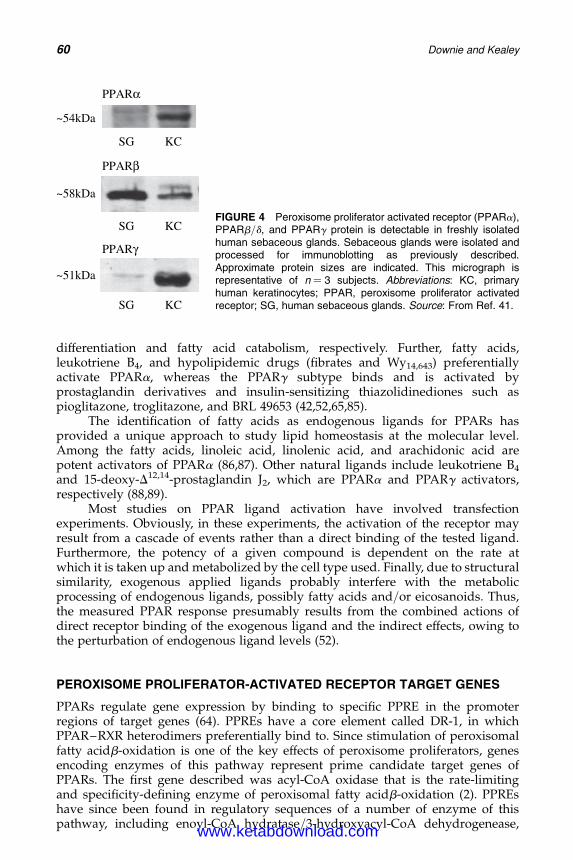
DK4962-Webster-ch5_R2_020407
differentiation and fatty acid catabolism, respectively. Further, fatty acids,leukotriene B4, and hypolipidemic drugs (fibrates and Wy14,643) preferentiallyactivate PPARa, whereas the PPARg subtype binds and is activated byprostaglandin derivatives and insulin-sensitizing thiazolidinediones such aspioglitazone, troglitazone, and BRL 49653 (42,52,65,85).
The identification of fatty acids as endogenous ligands for PPARs hasprovided a unique approach to study lipid homeostasis at the molecular level.Among the fatty acids, linoleic acid, linolenic acid, and arachidonic acid arepotent activators of PPARa (86,87). Other natural ligands include leukotriene B4
and 15-deoxy-D12,14-prostaglandin J2, which are PPARa and PPARg activators,respectively (88,89).
Most studies on PPAR ligand activation have involved transfectionexperiments. Obviously, in these experiments, the activation of the receptor mayresult from a cascade of events rather than a direct binding of the tested ligand.Furthermore, the potency of a given compound is dependent on the rate atwhich it is taken up and metabolized by the cell type used. Finally, due to structuralsimilarity, exogenous applied ligands probably interfere with the metabolicprocessing of endogenous ligands, possibly fatty acids and/or eicosanoids. Thus,the measured PPAR response presumably results from the combined actions ofdirect receptor binding of the exogenous ligand and the indirect effects, owing tothe perturbation of endogenous ligand levels (52).
PEROXISOME PROLIFERATOR-ACTIVATED RECEPTOR TARGET GENES
PPARs regulate gene expression by binding to specific PPRE in the promoterregions of target genes (64). PPREs have a core element called DR-1, in whichPPAR–RXR heterodimers preferentially bind to. Since stimulation of peroxisomalfatty acidb-oxidation is one of the key effects of peroxisome proliferators, genesencoding enzymes of this pathway represent prime candidate target genes ofPPARs. The first gene described was acyl-CoA oxidase that is the rate-limitingand specificity-defining enzyme of peroxisomal fatty acidb-oxidation (2). PPREshave since been found in regulatory sequences of a number of enzyme of thispathway, including enoyl-CoA hydratase/3-hydroxyacyl-CoA dehydrogenease,
~51kDa
~58kDa
KC SG
KCSG
KCSG
~54kDa
PPARα
PPARβ
PPARγ
FIGURE 4 Peroxisome proliferator activated receptor (PPARa),PPARb/d, and PPARg protein is detectable in freshly isolatedhuman sebaceous glands. Sebaceous glands were isolated andprocessed for immunoblotting as previously described.Approximate protein sizes are indicated. This micrograph isrepresentative of n ¼ 3 subjects. Abbreviations: KC, primaryhuman keratinocytes; PPAR, peroxisome proliferator activatedreceptor; SG, human sebaceous glands. Source: From Ref. 41.
60 Downie and Kealey
www.ketabdownload.com
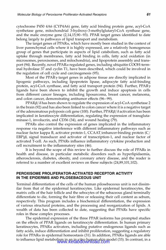
DK4962-Webster-ch5_R2_020407
cytochrome P450 4A6 (CYP4A6) gene, fatty acid binding protein gene, acyl-CoAsynthetase gene, mitochondrial 3-hydroxy-3-methylglutaryl-CoA synthase gene,and the malic enzyme gene (2,14,15,90–93). PPAR target genes identified to datebelong largely to pathways of lipid transport and metabolism.
The target genes of PPARa, which have mostly been studied in the context ofliver parenchymal cells where it is highly expressed, are a relatively homogenousgroup of genes that participate in aspects of lipid catabolism, such as fatty aciduptake through membranes, fatty acid binding in cells, fatty acid oxidation (inmicrosomes, peroxisomes, and mitochondria), and lipoprotein assembly and trans-port (94). Recently, novel PPARa regulated genes, including ubiquitin COOH-term-inal hydrolase 37 and cyclin T1, have been described, which could be involved inthe regulation of cell cycle and carcinogenesis (95).
Most of the PPARg target genes in adipose tissue are directly implicated inlipogenic pathways, including lipoprotein lipase, adipocyte fatty acid-bindingprotein, acyl-CoA synthase, and fatty acid transport protein (94). Further, PPARgligands have been shown to inhibit the growth and induce apoptosis in cellsfrom different cancer lineages, including liposarcoma, breast cancer, melanoma,and colon cancer, demonstrating antitumor effects (96–99).
PPARb/d has been shown to regulate the expression of acyl-CoA synthetase 2in the brain (92) and has also been linked to colon cancer where it is a negative targetof the adenomatous polyposis coli gene (100). Further, PPARb/d activation has beenimplicated in keratinocyte differentiation, regulating the expression of transgluta-minase-1, involucrin, and CD36 (34), and wound healing (79).
PPARs also control the expression of genes implicated in the inflammatoryresponse via negative interference with different inflammatory pathways such asnuclear factor kappa B, activator protein-1, CCAAT/enhancer-binding protein (C/EBP)b, signal transducer and activator of transcription-1, and nuclear factor ofactivated T-cell. As such, PPARs influence inflammatory cytokine production andcell recruitment to the inflammatory sites (46).
It is beyond the scope of this review to further discuss the role of PPARs inhealth and disease, in particular metabolic disorders such as hyperlipidaemia,atherosclerosis, diabetes, obesity, and coronary artery disease, and the reader isreferred to a number of excellent reviews on these subjects (24,89,101,102).
PEROXISOME PROLIFERATOR-ACTIVATED RECEPTOR ACTIVITYIN THE EPIDERMIS AND PILOSEBACEOUS UNIT
Terminal differentiation of the cells of the human pilosebaceous unit is not dissim-ilar from that of the epidermal keratinocytes. Like epidermal keratinocytes, thematrix cells of the hair follicle and the sebocytes of the sebaceous gland terminallydifferentiate to die, forming the hair fibre or releasing their cell content as sebum,respectively. This program includes a biochemical differentiation, the expressionof various structural proteins, and the processing and reorganization of lipids. Awealth of data has been collected to date, suggesting that PPARs have specificroles in these complex processes.
The epidermal expression of the three PPAR isoforms has prompted studieson the effects of PPAR ligands on keratinocyte differentiation. In human primarykeratinocytes, PPARa activators, including putative endogenous ligands such asfatty acids, induce differentiation and inhibit proliferation, suggesting a regulatoryrole for PPARa in epidermal homeostasis (31). In addition, PPARa ligands are ableto influence lipid metabolism in an in vitro human skin model (33). In contrast, in a
Molecular Biology of Peroxisome Proliferator-Activated Receptors 61
www.ketabdownload.com

DK4962-Webster-ch5_R2_020407
more recent study, PPARa activators, like PPARg activators, seemed to have littleeffect on human keratinocyte differentiation (30). However, a selective PPARb/dligand induced the expression of keratinocyte differentiation markers (30). Theseapparent discrepancies remain to be elucidated. In the rat, PPARa ligands accelerateepidermal maturation in vitro (31,103,104) and in utero (32), whereas PPARb/d andPPARg activators had no effects. In addition, PPARa ligands induce epidermaldifferentiation and restore epidermal homeostasis in hyperproliferative mouseepidermis (105).
PPAR mutant mouse models have contributed to our understanding of therole of PPARs in epidermal homeostasis. Using PPARa, PPARb/d, and PPARgmutant mice, it was shown that PPARa and PPARb/d are important for the rapidepithelialization of a skin wound and that each of them plays a specific role inthis process. PPARa was mainly involved in the early inflammation phase of thehealing, whereas PPARb/d was implicated in the control of keratinocyte prolifer-ation. In addition and very interestingly, PPARb/d mutant primary keratinocytesshowed impaired adhesion and migration properties (77,79). However, PPARa,PPARb/d, and PPARg mutations appear to have no obvious effects duringnormal foetal development of the epidermis (79,106,107).
Unfortunately, epidermal and sebaceous gland functions were not examinedin the mouse mutant models. However, in rat preputial sebocytes, PPARg andPPARa activation induced sebocyte lipogenesis in vitro but not in keratinocytes,whereas activators of PPARb/d induced lipid formation in both sebocytes andkeratinocytes (36). PPARg ligands promoted the differentiative effects of androgenson sebocytes and PPARg1 was shown to be expressed more strongly in freshlydispersed sebocytes compared to cultured sebocytes, suggesting that androgeninfluences an early step in sebocyte differentiation, which is related to but distinctfrom that influenced by PPARg1. Further, PPARb/d was abundantly expressed insebocytes and keratinocytes (36). RXR agonists augmented the stimulation ofsebocyte differentiation by PPAR agonists, as expected from PPAR–RXR hetero-dimerization; however, the evidence for PPAR–RXR cooperativity was limited(37). Moreover, in cultured human sebocytes, PPARb/d activation had a stimulatoryeffect on lipid production, whereas PPARg and PPARa activation was ineffective(39). In addition, treatment with arachidonic acid resulted in increased lipid for-mation in cultured human sebocytes (38). Furthermore, treatment with arachidonicacid enhanced apoptosis in cultured human sebocytes, as determined by the pre-sence of fragmented nuclei, indicating that apoptosis is part of the sebocyte differ-entiation process (38). Using a PPARb/d mutant mouse model, proinflammatorycytokines initiated the production of endogenous PPARb/d ligands, which wereessential for PPARb/d activation and action, and activated PPARb/d regulatedthe expression of genes associated with apoptosis (108).
In contrast, in organ maintained human sebaceous glands, PPARg and PPARaselective ligands inhibited total sebaceous lipogenesis (Table 1) and also reducedthe production of the sebum-specific lipids, squalene and triacylglycerol(Table 2). Further, arachidonic, linoleic, linolenic, and oleic acid, which are lessspecific in their PPAR subtype activation, also inhibited sebaceous lipogenesis(41). Similar observations were made using the hamster flank organ wherePPARa activation with clofibric acid produced a dose-dependent inhibition of lipo-genesis (40). In addition, it has been demonstrated that the oral administration ofeicosatetraynoic acid to human male volunteers significantly reduced sebumsecretion (109). These findings indicate that PPAR effects are differentiation
62 Downie and Kealey
www.ketabdownload.com
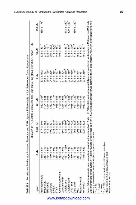
DK4962-Webster-ch5_R2_020407
TA
BL
E1
Pero
xis
om
eP
rolif
era
tor
Activ
ate
dR
ecepto
rand
FX
RLig
ands
Diff
ere
ntia
llyIn
hib
itS
ebace
ous
Gla
nd
Lip
ogenesi
s
8m
M[1
,2-1
4C
]aceta
teupta
ke
into
tota
llip
id(p
mol/
mg
gla
nd
wet
wt/
hr)
,m
ean+
SD
Lig
and
0m
M0.0
1m
M0.1
mM
1m
M10m
M100m
M
Ara
chid
onic
aci
d1318+
814
877+
546
562+
361
a870+
612
635+
421
a385+
175
b
Bezafibra
te1253+
310
1132+
462
1194+
628
1142+
497
971+
520
BR
L49653
1454+
524
1175+
467
1224+
568
991+
735
c322+
131
a
Clo
fibra
te1470+
478
1400+
521
1332+
696
1043+
296
901+
292
d-P
GJ
21764+
952
1931+
1187
1196+
1174
1097+
838
716+
606
a
ET
YA
1284+
498
1418+
524
986+
651
616+
497
d(4
8+
46
b)
Juve
nile
horm
one
III
1353+
745
1414+
897
1190+
285
1015+
386
1404+
528
Leuko
trie
ne
B4
2001+
949
1564+
756
1224+
861
1217+
804
Lin
ole
icacid
1467+
1089
632+
580
d332+
186
b549+
258
d535+
361
d513+
233
d
Lin
ole
nic
acid
1900+
659
1073+
802
d1143+
626
a1035+
469
d678+
410
b809+
327
b
LY
171883
1376+
321
1782+
722
1085+
406
1223+
639
474+
199
a
Ole
icacid
1761+
756
963+
802
c691+
329
d536+
145
b718+
417
d860+
360
a
PG
J2
1251+
519
1095+
420
1350+
365
1273+
574
752+
441
Tra
ns-f
arn
esol
1042+
409
899+
379
1060+
627
994+
351
818+
327
WY
14643
1415+
682
1224+
622
1062+
369
923+
289
c480+
371
b
No
tes:S
eba
ceo
us
gla
nds
we
reis
ola
ted
an
dm
ain
tain
ed
,in
cub
ate
dw
ith
8m
M[1
,2-1
4C
]ace
tate
,an
dsu
bseq
ue
ntly
lipid
sw
ere
an
aly
zed
,as
de
scri
bed
inM
ate
ria
lsan
dM
eth
ods.
Experim
ents
were
carr
ied
outf
orn¼
5subje
cts
.Data
are
expre
ssed
as
mean+
SD
.Sta
tisticals
ignifi
cance
was
dete
rmin
ed
usin
gsin
gle
facto
rA
NO
VA
with
blo
ckin
g(s
ubje
cts
were
blo
cked)
follo
wed
by
Dunnett’s
multip
lecom
parison
pro
cedure
.aP
,0.0
1.
bP
,0.0
01.
cP
,0.0
5.
dP
,0.0
05;
(),
com
pound
toxic
at
this
concentr
ation.
Abbre
via
tion
:E
TY
A,
eic
osate
trayno
icacid
.S
ourc
e:
Fro
mR
ef.
41.
Molecular Biology of Peroxisome Proliferator-Activated Receptors 63
www.ketabdownload.com
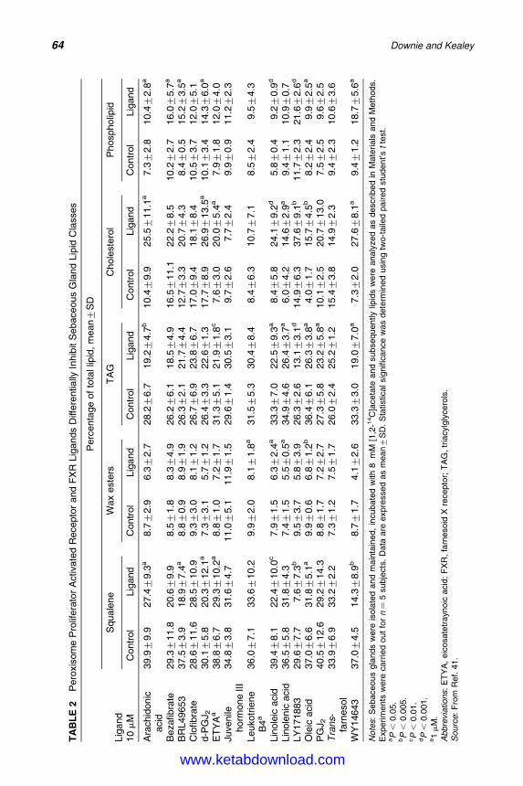
DK4962-Webster-ch5_R2_020407
TA
BL
E2
Pero
xis
om
eP
rolif
era
tor
Activ
ate
dR
ecepto
rand
FX
RLig
ands
Diff
ere
ntia
llyIn
hib
itS
ebace
ous
Gla
nd
Lip
idC
lasses
Perc
enta
ge
of
tota
llip
id,
mean+
SD
Lig
and
Squale
ne
Wax
este
rsT
AG
Chole
ste
rol
Phospholip
id
10m
MC
ontr
ol
Lig
and
Contr
ol
Lig
and
Contr
ol
Lig
and
Contr
ol
Lig
and
Contr
ol
Lig
and
Ara
chid
onic
acid
39.9+
9.9
27.4+
9.3
a8.7+
2.9
6.3+
2.7
28.2+
6.7
19.2+
4.7
b10.4+
9.9
25.5+
11.1
a7.3+
2.8
10.4+
2.8
a
Bezafibra
te29.3+
11.8
20.6+
9.9
8.5+
1.8
8.3+
4.9
26.2+
6.1
18.5+
4.9
16.5+
11.1
22.2+
8.5
10.2+
2.7
16.0+
5.7
a
BR
L49653
37.5+
3.9
18.9+
7.4
a8.8+
0.9
8.9+
1.9
26.3+
2.1
21.7+
4.4
12.7+
3.3
20.7+
4.3
8.4+
0.5
15.2+
3.5
a
Clo
fibra
te28.6+
11.6
28.5+
10.9
9.3+
3.0
8.1+
1.2
26.7+
6.9
23.8+
6.7
17.0+
9.4
18.1+
8.4
10.5+
3.7
12.0+
5.1
d-P
GJ
230.1+
5.8
20.3+
12.1
a7.3+
3.1
5.7+
1.2
26.4+
3.3
22.6+
1.3
17.7+
8.9
26.9+
13.5
a10.1+
3.4
14.3+
6.0
a
ET
YA
a38.8+
6.7
29.3+
10.2
a8.8+
1.0
7.2+
1.7
31.3+
5.1
21.9+
1.8
c7.6+
3.0
20.0+
5.4
a7.9+
1.8
12.0+
4.0
Juve
nile
horm
one
III
34.8+
3.8
31.6+
4.7
11.0+
5.1
11.9+
1.5
29.6+
1.4
30.5+
3.1
9.7+
2.6
7.7+
2.4
9.9+
0.9
11.2+
2.3
Leuko
trie
ne
B4
a36.0+
7.1
33.6+
10.2
9.9+
2.0
8.1+
1.8
a31.5+
5.3
30.4+
8.4
8.4+
6.3
10.7+
7.1
8.5+
2.4
9.5+
4.3
Lin
ole
icacid
39.4+
8.1
22.4+
10.0
c7.9+
1.5
6.3+
2.4
a33.3+
7.0
22.5+
9.3
a8.4+
5.8
24.1+
9.2
d5.8+
0.4
9.2+
0.9
d
Lin
ole
nic
acid
36.5+
5.8
31.8+
4.3
7.4+
1.5
5.5+
0.5
a34.9+
4.6
26.4+
3.7
a6.0+
4.2
14.6+
2.9
a9.4+
1.1
10.9+
0.7
LY
171883
29.6+
7.7
7.6+
7.3
b9.5+
3.7
5.8+
3.9
26.3+
2.6
13.1+
3.1
d14.9+
6.3
37.6+
9.1
b11.7+
2.3
21.6+
2.6
d
Ole
icacid
37.0+
6.6
31.8+
5.1
a9.9+
0.6
6.8+
1.2
b36.4+
6.1
26.3+
3.8
a4.0+
1.7
15.7+
4.5
b8.2+
2.4
9.9+
2.5
a
PG
J2
40.5+
12.6
29.2+
14.3
8.8+
1.7
7.2+
2.7
27.3+
5.8
23.2+
5.8
a10.1+
2.5
20.7+
13.0
7.5+
2.5
9.6+
2.5
Tra
ns-
farn
esol
33.9+
6.9
33.2+
2.2
7.3+
1.2
7.5+
1.7
26.0+
2.4
25.2+
1.2
15.4+
3.8
14.9+
2.3
9.4+
2.3
10.6+
3.6
WY
14643
37.0+
4.5
14.3+
8.9
b8.7+
1.7
4.1+
2.6
33.3+
3.0
19.0+
7.0
a7.3+
2.0
27.6+
8.1
a9.4+
1.2
18.7+
5.6
a
Note
s:
Sebaceous
gla
nds
were
isola
ted
and
main
tain
ed,
incubate
dw
ith
8m
M[1
,2-1
4C
]aceta
teand
subsequently
lipid
sw
ere
analy
zed
as
described
inM
ate
rials
and
Meth
ods.
Experim
ents
were
carr
ied
out
for
n¼
5subje
cts
.D
ata
are
expre
ssed
as
mean+
SD
.S
tatisticalsig
nifi
cance
was
dete
rmin
ed
usin
gtw
o-t
aile
dpaired
stu
dent’s
tte
st.
aP
,0.0
5.
bP
,0.0
05.
cP
,0.0
1.
dP
,0.0
01.
e1m
M.
Abbre
via
tions:
ET
YA
,eic
osate
traynoic
acid
;F
XR
,fa
rnesoid
Xre
cepto
r;T
AG
,tr
iacylg
lycero
ls.
Sourc
e:
Fro
mR
ef.
41.
64 Downie and Kealey
www.ketabdownload.com
DK4962-Webster-ch5_R2_020407
sensitive, that is, one might postulate that structures that are missing in sebocytemonolayers but are present in whole organ cultures may be involved in PPARagonist activity.
Further, there is an evidence that cutaneous inflammation can be reduced byPPAR agonists, such as linoleic acid, with clinical effects comparable to that ofglucocorticoids (110). In addition, the PPARa activators, WY-14,643 and clofibrate,were shown to reverse ultraviolet-B-light-mediated expression of inflammatorycytokines, that is, interleukin (IL)-6 and IL-8 (43). This work suggests the possibilityof PPARa activators as novel nonsteroidal anti-inflammatory drugs in the topicaltreatment of inflammatory skin diseases (43). Further, PPARa activation withclofibrate treatment resulted in the reduction of inflammatory cells and a decreasein tumor necrosis factora and IL-1a in the epidermis of a mouse model of irritantand allergic contact dermatitis (44).
ACNE
Acne is a disease unique to humans. It affects up to 80 % of young adults, in whomit can induce stress, depression, and anxiety, as determined by psychometricscoring (47,111,112). Acne is a disease of the infundibulum or “pore” of thehuman sebaceous pilosebaceous unit and of the gland itself. This unit developsat puberty, appears only at the site of acne lesions (the chest, back, and face),and seems to have no other function than to sometimes produce acne lesions.The earlier forms of acne are characterized by microcomedones, which representan intrainfundibular scaling-like phenomenon. As the disease progresses in sever-ity, comedogenesis develops, as does inflammation. Duct rupture is a late event inthe development of most inflammatory lesions (47–50).
Acne is generally accepted by clinicians to be a multifactorial disease and it isbelieved that acne is associated with seborrhea, the excess production of sebum bythe sebaceous gland (49,113). Further, Propionibacterium acnes is a major bacteriumimplicated in the pathogenesis of acne (114). Acne is characterized by the presenceof noninflammatory lesions, termed comedones (47,50). The earliest pathologicalchange in acne is altered follicular epithelial differentiation, resulting in afollicular retention hyperkeratosis—the microcomedo (50). In addition to the non-inflammatory lesions, inflammatory lesions can develop when such microcomedosare colonized by P. acnes. For inflammatory lesions such as papules, pustules, andnodulocystic lesions to occur, a variety of pathogenic factors are involved, includingbesides P. acnes, inflammatory mediators, and host immunity (115). In vitro exper-iments on organ maintained human infudibula have identified a number of cyto-kines that can model the morphological and inflammatory aspects of acne (116).These cytokines not only induce the infundibular changes in acne, namely (i) hyper-cornification (scaling) of the infundibulum, (ii) expression of intercellular adhesionmolecule-1 and human leukocyte antigen-D related, and (iii) disruption of infun-dibular morphology, but will also inhibit the secretion of lipid from the sebaceousgland (117). Since acne seems to be provoked by P. acnes in sebum and since P. acnesdepends on sebum for nutrition, the inhibition of sebum secretion would beexpected to promote the remission of the disease by inhibiting P. acnescolonization (117).
A number of acne therapies are currently in use, including retinoids (e.g., iso-tretinoin, adapalene), antiandrogens (e.g., cyproterone acetate), contraceptives,antibacterials (e.g., clindamycin, erythromycin), benzoyl peroxide, and salicylicacid (118,119). In milder cases, topical therapy is sufficient, but in more severe
Molecular Biology of Peroxisome Proliferator-Activated Receptors 65
www.ketabdownload.com
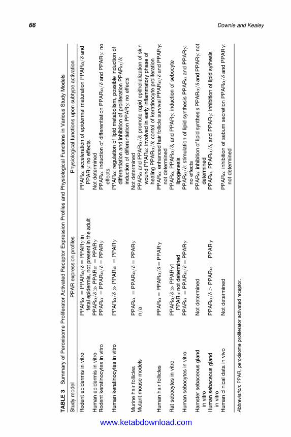
DK4962-Webster-ch5_R2_020407
TA
BL
E3
Sum
mary
of
Pero
xisom
eP
rolif
era
tor
Activate
dR
ecepto
rE
xpre
ssio
nP
rofile
sand
Physio
logic
alF
unct
ions
inV
arious
Stu
dy
Models
Stu
dy
model
PP
AR
expre
ssio
npro
file
sP
hysi
olo
gic
alfu
nct
ions
upon
subty
pe
activatio
n
Rodent
epid
erm
isin
vitro
PP
ARa¼
PP
ARa
/d¼
PP
ARg
infe
tale
pid
erm
is,notpre
sentin
the
adult
PP
ARa
:acce
lera
tion
of
epid
erm
alm
atu
ration
PP
ARa
/d
and
PP
ARg:
no
effects
Hum
an
epid
erm
isin
vitro
PP
ARa
/d�
PP
ARa¼
PP
ARg
Not
dete
rmin
ed
Rodent
kera
tinocyt
es
invitr
oP
PA
Ra¼
PP
ARa
/d¼
PP
ARg
PP
ARa
:in
duct
ion
of
diff
ere
ntiation
PP
ARa
/d
and
PP
ARg:
no
effects
Hum
an
kera
tinocyt
es
invitr
oP
PA
Ra
/d�
PP
ARa¼
PP
ARg
PP
ARa
:re
gula
tion
of
lipid
meta
bolis
m,
possib
lein
duct
ion
of
diff
ere
ntia
tion
and
inhib
itio
nof
pro
lifera
tion
PP
ARa
/d:
induct
ion
of
diff
ere
ntiatio
nP
PA
Rg:
no
effect
sM
urine
hair
folli
cle
sP
PA
Ra¼
PP
ARa
/d¼
PP
ARg
Not
dete
rmin
ed
Muta
nt
mouse
models
n/a
PP
ARa
and
PP
ARa
/d:
pro
mote
rapid
epith
elia
lizatio
nof
skin
wound
PP
ARa
:in
volv
ed
inearly
inflam
mato
ryphase
of
healin
gP
PA
Ra
/d:
conto
lof
kera
tinocy
tepro
lifera
tion
Hum
an
hair
folli
cle
sP
PA
Ra¼
PP
ARa
/d¼
PP
ARg
PP
ARa
:enhance
dhair
folli
cle
surv
ivalP
PA
Ra
/dand
PP
ARg:
not
dete
rmin
ed
Rat
sebocyte
sin
vitro
PP
ARa
/d�
PP
ARg1
PP
ARa
not
dete
rmin
ed
PP
ARa
,P
PA
Ra
/d,
and
PP
ARg:
induction
of
sebocyt
elip
ogenesis
Hum
an
sebocyte
sin
vitro
PP
ARa¼
PP
ARa
/d¼
PP
ARg
PP
ARa
/d:
stim
ula
tion
of
lipid
synth
esis
PP
ARa
and
PP
ARg:
no
effects
Ham
ster
sebaceous
gla
nd
invitro
Not
dete
rmin
ed
PP
ARa
:in
hib
itio
noflip
idsyn
thesis
PP
ARa
/dand
PP
ARg:not
dete
rmin
ed
Hum
an
sebaceous
gla
nd
invitro
PP
ARa
/d.
PP
ARa¼
PP
ARg
PP
ARa
,P
PA
Ra
/d,
and
PP
ARg:
inhib
itio
nof
lipid
syt
hesi
s
Hum
an
clin
icaldata
inviv
oN
ot
dete
rmin
ed
PP
ARa
:in
hib
ition
of
sebum
secr
etion
PP
ARa
/d
and
PP
ARg:
not
dete
rmin
ed
Abbre
via
tion
:P
PA
R,
pero
xis
om
epro
lifera
tor
activate
dre
cepto
r.
66 Downie and Kealey
www.ketabdownload.com
DK4962-Webster-ch5_R2_020407
cases where papulopustular or nodulocystic acne is present, there is a need of sys-temic treatment. However, there is a continued need for effective drugs for thetherapy of acne, although judicious combined use of existing topical and systemictherapies offers great relief to many patients and a recent review of these therapiesindicated that approximately 68% of treatment is effective (120). Nonetheless,PPARs may offer a solution for the development of novel effective drugs for acnetherapy.
CONCLUSIONS
The study of PPAR expression profiles and the identification of target genes andligands in the skin and its appendages as well as the utilization of PPAR mousemutant models have unveiled distinct physiological functions of the PPARs(Table 3). In particular, activation of PPARs regulates differentiation and prolifer-ation, lipid metabolism, inflammation, and apoptosis. However, the molecularmechanisms by which PPARs coordinate the regulation of these processes remainlargely unknown, and thus PPARs represent a major research target for the under-standing and treatment of many skin diseases including acne.
The research reviewed here focused on PPAR activity in human epidermisand its appendages. The data demonstrates that PPARs regulate many of the phys-iological processes that are involved in acne. However, it is also apparent that thereis some discrepancies with regard to the physiological effects of PPAR ligands onsebaceous lipogenesis, which may be attributed to the different study modelsused, but this requires further elucidation. It would be of particular interest toevaluate the efficacy of PPAR ligands on acne and other skin disorders in the clinic.
Since the completion of this manuscript, more research has been done intoexamining the effects of PPAR ligands on sebaceous lipogenesis. The PPARligands GW7647, GW0742, GW2433, rosiglitazone, and GW4148 significantlyincreased lipogenesis in SEB-1 sebocytes (121), confirming earlier reports refer-enced in this chapter. Interestingly, patient data collected in the same papershowed increased sebum secretion in patients receiving fibrates for hyperlipidemiaor thialzolidienediones for diabetes. However, this is in contrast to observation inhealthy volunteers, and one might argue that these patients are treated for meta-bolic diseases of the liver and pancreas, which may also effect sebaceous glandmetabolism, and therefore these data should be treated with caution. In addition,PPAR involvement in the prostaglandin pathway has been shown in a recentpaper where PPARg activation has been implicated in oxidative stress-mediatedprostaglandin E(2) production in SX95 human sebaceous cells (122).
REFERENCES
1. Issemann I, Green S. Activation of a member of the steroid hormone receptor superfam-ily by peroxisome proliferators. Nature 1990; 347:645–650.
2. Dreyer C, Krey G, Keller H, et al. Control of the peroxisomal beta-oxidation pathway bya novel family of nuclear hormone receptors. Cell 1992; 68:879–887.
3. Gottlicher M, Widmark E, Li Q, et al. Fatty acids activate a chimera of the clofibric acid-activated receptor and the glucocorticoid receptor. Proc Natl Acad Sci U S A 1992;89:4653–4657.
4. Schmidt A, Endo N, Rutledge SJ, et al. Identification of a new member of the steroidhormone receptor superfamily that is activated by a peroxisome proliferator andfatty acids. Mol Endocrinol 1992; 6:1634–1641.
Molecular Biology of Peroxisome Proliferator-Activated Receptors 67
www.ketabdownload.com
DK4962-Webster-ch5_R2_020407
5. Chen F, Law SW, O ’Malley BW. Identification of two mPPAR related receptors and evi-dence for the existence of five subfamily members. Biochem Biophys Res Commun1993; 196:671–677.
6. Sher T, Yi HF, Mcbridge OW, et al. cDNA cloning, chromosomal mapping, and func-tional characterization of the human peroxisome proliferator activated receptor. Bio-chemistry 1993; 32:5598–5604.
7. Zhu Y, Alvars K, Huang Q, et al. Cloning of a new member of the peroxisome prolif-erator-activated receptor gene family from mouse liver. J Biol Chem 1993;258:26817–26820.
8. Mukherjee R, Jow L, Croston GE, et al. Identification, characterization, and tissue dis-tribution of human peroxisome proliferator-activated receptor (PPAR) isoformsPPARg2 versus PPARg1 and activation with retinoid X receptor agonist and antagonist.J Biol Chem 1997; 272:8071–8076.
9. Auboeuf D, Rieusset J, Fajas L, et al. Tissue distribution and quantification of theexpression of mRNAs of peroxisome proliferator-activated receptors and liver X recep-tor-alpha in humans: no alteration in adipose tissue of obese and NIDDM patients. Dia-betes 1997; 46:1319–1327.
10. Hess R, Staubli W, Riess W. Nature of the hepatomegalic effect produced by ethyl-chlorophenoxy-isobutyrate in the rat. Nature 1965; 208:856–858.
11. Reddy JK, Rao MS, Azarnoff DL, et al. Mitogenic and carcinogenic effects of a hypolipi-demic peroxisome proliferator, [4-chloro-6-(2,3-xylidino)-2-pyrimidinylthio]acetic acid(WY-14,643), in rat and mouse liver. Cancer Res 1979; 39:152–161.
12. Styles JA, Kelly M, Pritchard NR, et al. A species comparison of acute hyperplasiainduced by the peroxisome proliferator methylclofenapate: involvement of the binu-cleated hepatocyte. Carcinogenesis 1988; 9:1647–1655.
13. Zhang X-K, Lehman J, Hoffmann B, et al. Homodimer formation of retinoid X receptorinduced by 9-cis retinoic acid. Nature 1992; 358:587–591.
14. Bardot O, Aldridge TC, Latruffe N, et al. PPAR-RXR heterodimer activates a peroxi-some proliferator response element upstream of the bifunctional enzyme gene.Biochem Biophys Res Commun 1993; 192:37–45.
15. Krey G, Keller H, Mahfoudi A, et al. Xenopus peroxisome proliferator activated recep-tors: genomic organization, response element recognition, heterodimer formation withretinoid X receptor and activation by fatty acids. J Steroid Biochem Mol Biol 1993;47:65–73.
16. Dreyer C, Keller H, Mahfoudi A, et al. Positive regulation of the peroxisomalb-oxidation pathway by fatty acids through activation of peroxisome proliferatoractivated receptors (PPAR). Biol Cell 1993; 77:67–76.
17. Desvergene B, Wahli W. PPAR: a key nuclear factor in nutrient/gene interactions?Inducible Gene Expr 1995; 1:142–176.
18. Hihi AK, Michalik L, Wahli W. PPARs: transcriptional effectors of fatty acids and theirderivatives. Cell Mol Life Sci 2002; 59:790–798.
19. Lock EA, Mitchell AM, Elcombe CR. Biochemical mechanisms of induction of hepaticperoxisome proliferation. Annu Rev Pharmacol Toxicol 1989; 29:145–163.
20. Reddy JK, Lalwani ND. Carcinogenesis by hepatic peroxisome proliferators: evalu-ation of the risk of hypolipidemic drugs and industrial plasticizers to humans. CRCCrit Rev Toxicol 1983; 12:1–58.
21. Conway JG, Tomaszewski KE, Olson MJ, et al. Relationship of oxidative damage to thehepatocarcinogenicity of the peroxisome proliferators di(2-ethylhexyl)phthalate andWY-14,643. Carcinogenesis 1989; 10:513–519.
22. Kasai H, Okada Y, Nishimura S, et al. Formation of 8-hydroxydeoxyguanosine in liverDNA of rats following long-term exposure to a peroxisome proliferator. Cancer Res1989; 49:2603–2605.
23. Shoonjans K, Staels B, Auwerx J. The peroxisome proliferator activated receptors(PPARs) and their effects on lipid metabolism and adipocyte differentiation. BiochimBiophys Acta 1996; 1302:93–109.
24. Lee CH, Olson P, Evans RM. Minireview: lipid metabolism, metabolic diseases, andperoxisome proliferator activated receptors. Endocrinology 2003; 144:2201–2207.
68 Downie and Kealey
www.ketabdownload.com
DK4962-Webster-ch5_R2_020407
25. Kliewer SA, Lehmann JM, Willson TM. Orphan nuclear receptors: shifting endrocrinol-ogy into reverse. Science 1999; 284:757–760.
26. Chawala A, Lazar MA. Peroxisome proliferator and retinoids signaling pathwaysco-regulate preadipocyte phenotype and survival. Proc Natl Acad Sci USA 1994;91:1786-1790.
27. Tontonoz P, Hu E, Spiegelman BM. Stimulation of adipogenesis in fibroblasts byPPARg2, a lipid-activated transcription factor. Cell 1994; 79:1147–1156.
28. Tontonoz P, Hu E, Spiegelman BM. Regulation of adipocyte gene expression and differ-entiation by peroxisome proliferators activated receptorg. Curr Opin Genet Dev 1995;5:571–576.
29. Adams M, Montague CT, Prins JB, et al. Activators of peroxisome proliferator-activatedreceptor gamma have depot-specific effects on human preadipocyte differentiation.J Clin Invest 1997; 100:3149–3153.
30. Sewter C, Blows F, Considine R, et al. Differential effects of adiposity on peroxisomalproliferator-activated receptor gamma1 and gamma2 messenger ribonucleic acidexpression in human adipocytes. J Clin Endocrinol Metab 2002; 87:4203–4207.
31. Hanley K, Jiang Y, He SS, et al. Keratinocyte differentiation is stimulated by activatorsof the nuclear hormone receptor PPARa. J Invest Dermatol 1998; 110:368–375.
32. Hanley K, Komuves LG, Bass NM, et al. Fetal epidermal differentiation and barrierdevelopment in vivo is accelerated by nuclear hormone receptor activators. J InvestDermatol 1999; 113:788–795.
33. Rivier M, Castiel I, Safonova I, et al. Peroxisome proliferator-activated receptor-aenhances lipid metabolism in a skin equivalent model. J Invest Dermatol 2000;114:681–687.
34. Westergaard M, Henningsen J, Svendsen ML, et al. Modulation of keratinocyte geneexpression and differentiation by PPAR selective ligands and tetradecylthioaceticacid. J Invest Dermatol 2001; 116:702–712.
35. Rosenfield RL, Deplewski D, Kentsis A, et al. Mechanisms of androgen induction ofsebocyte differentiation. Dermatology 1998; 196:43–46.
36. Rosenfield RL, Kentsis A, Deplewski D, et al. Rat preputial sebocytes differentiationinvolves peroxisome proliferator-activated receptors. J Invest Dermatol 1999;112:226–232.
37. Kim MJ, Deplewski D, Ciletti N, et al. Limited cooperation between peroxisomeproliferator-activated receptors and retinoid X receptor agonists in sebocyte growthand development. Mol Genet Metab 2001; 74:362–369.
38. Wr obel A, Seltmann H, Fimmel S, et al. Differentiation and apoptosis in human immor-talized sebocytes. J Invest Dermatol 2003; 120:175–181.
39. Chen W, Yang CC, Sheu HM, et al. Expression of peroxisome proliferator-activatedreceptor and CCAAT/enhancer binding protein transcription factors in culturedhuman sebocytes. J Invest Dermatol 2003; 121:441–447.
40. Venencie PY, Vantrou-Delaunay M, Bougaret S, et al. Modification of lipogenesis inthe isolated hamster flank organ through clofibric acid. Skin Pharmacol 1995;8:203–206.
41. Downie MMT, Sanders DA, Maier LM, et al. PPAR and FXR ligands differentially regu-late sebaceous differentiation in human sebaceous gland organ cultures in vitro. Br JDermatol 2004; 151:766–775.
42. Devchand PR, Keller H, Peters JM, et al. The PPARa-leukotriene B4 pathway to inflam-mation control. Nature 1996; 384:39–43.
43. Kippenberger S, Loitsch SM, Grundmann-Kollmann M, et al. Activators of peroxisomeproliferator-activated receptors protect human skin from ultraviolet-B-light-inducedinflammation. J Invest Dermatol 2001; 117:1430–1436.
44. Sheu MY, Fowler AJ, Kao J, et al. Topical peroxisome proliferator activated receptor-alpha activators reduce inflammation in irritant and allergic contact dermatitismodels. J Invest Dermatol 2002; 118:94–101.
45. Houseknecht KL, Cole BM, Steele PJ. Peroxisome proliferator-activated receptor gamma(PPARgamma) and its ligands: a review. Domest Anim Endocrinol 2002; 22:1–23.
Molecular Biology of Peroxisome Proliferator-Activated Receptors 69
www.ketabdownload.com
DK4962-Webster-ch5_R2_020407
46. Blanquart C, Barbier O, Fruchart JC, et al. Peroxisome proliferator-activated receptors:regulation of transcriptional activities and roles in inflammation. J Steroid BiochemMol Biol 2003; 85:267–273.
47. Kligman AM. An overview of acne. J Invest Dermatol 1974; 62:268–287.48. Strauss JS, Kligman AM. Pathological patterns of the sebaceous gland. J Invest Derma-
tol 1958; 30:51–61.49. Cunliffe WJ, Shuster S. Pathogenesis of acne. Lancet 1969; ii:685–687.50. Knutson DD. Ultrastructural observations in acne vulgaris: the normal sebaceous
follicle and acne lesions. J Invest Dermatol 1974; 62:288–307.51. Enmark E, Gustafsson JA. Orphan nuclear receptors—the first eight years. Mol Endo-
crinol 1996; 10:1293–1307.52. Lemberger T, Desvergne B, Wahli W. Peroxisome proliferator-activated receptors: a
nuclear receptor signalling pathway in lipid physiology. Annu Rev Cell Dev Biol1996; 12:335–363.
53. Beamer BA, Negri C, Yen CJ, et al. Chromosomal localization and partial genomicstructure of the human peroxisome proliferator activated receptor-gamma (hPPARgamma) gene. Biochem Biophys Res Commun 1997; 33:756–759.
54. Skogsberg J, Kannisto K, Roshani L, et al. Characterization of the human peroxisomeproliferator activated receptor delta gene and its expression. Int J Mol Med 2000;6:73–81.
55. Camp HS, Tafuri SR. Regulation of peroxisome proliferator-activated receptorg activityby mitogen-activated protein kinase. J Biol Chem 1997; 272:10811–10816.
56. Shalev A, Siegrist-Kaiser CA, Yen PM, et al. The peroxisome proliferator-activatedreceptora is a phosphoprotein: regulation by insulin. Endocrinology 1996;137:4499–4502.
57. Blanquart C, Barbier O, Fruchart JC, et al. Peroxisome proliferator-activated receptoralpha (PPARalpha) turnover by the ubiquitin-proteasome system controls the ligand-induced expression level of its target genes. J Biol Chem 2002; 277:37254–37259.
58. Miyata KS, McCaw SE, Patel HV, et al. The orphan nuclear hormone receptor LXLainteracts with the peroxisome proliferator-activated receptor and inhibits peroxisomeprolifarator signaling. J Biochem Chem 1996; 271:9189–9192.
59. Kliewer SA, Umesono K, Noonan DJ, et al. Convergence of 9-cis retinoic acid and per-oxisome proliferator signaling pathways through heterodimer formation of their recep-tors. Nature 1992; 358:771–774.
60. Gearing KL, Gottlicher M, Teboul M, et al. Interaction of the peroxisome-proliferator-activated receptor and retinoid X receptor. Proc Natl Acad Sci USA 1993; 90: 1440–1444.
61. Issemann I, Prince RA, Tugwood JD, et al. The peroxisome proliferator activated recep-tor: retinoid X receptor heterodimer is activated by fatty acids and fibrate hypo-lipidemic drugs. J Mol Endocrinol 1993; 11:37–47.
62. Keller H, Dreyer C, Medin J, et al. Fatty acids and retinoids control lipid metabolismthrough activation of peroxisome proliferator-activated receptor-retinoid X receptorheterodimers. Proc Natl Acad Sci U S A 1993; 90:2160–2164.
63. Chawla A, Repa JJ, Evans RM, et al. Nuclear receptors and lipid physiology: openingthe X-files. Science 2001; 294:1866–1870.
64. Green S. Promiscuous liaisons. Nature 1993; 361:590–591.65. Juge-Aubry C, Pernin A, Favez T, et al. DNA binding properties of peroxisome prolif-
erator-activated receptor subtypes on various natural peroxisome proliferator responseelements. J Biol Chem 1997; 272:25252–25259.
66. Miyamoto T, Kaneko A, Kakizawa T, et al. Inhibition of peroxisome proliferator signal-ing pathways by thyroid hormone receptor. J Biol Chem 1997; 272:7752–7758.
67. Zhu Y, Qi C, Calandra C, et al. Cloning and identification of mouse steroid receptorcoactivator-1 (mSRC-1), as a coactivator of peroxisome proliferator-activated receptorgamma. Gene Expr 1996; 6:185–195.
68. Zhu Y, Qi C, Jain S, et al. Isolation and characterization of PBP, a protein that interactswith peroxisome proliferator-activated receptor. J Biol Chem 1997; 272:25500–25506.
69. Zhu Y, Kan L, Qi C, et al. Isolation and characterization of peroxisome proliferator-activated receptor (PPAR) interacting protein (PRIP) as a coactivator for PPAR. J BiolChem 2000; 275:13510–13516.
70 Downie and Kealey
www.ketabdownload.com
DK4962-Webster-ch5_R2_020407
70. Gonzales FJ. The role of peroxisome proliferator activated receptora in peroxisomeproliferation, physiological homeostasis, and chemical cacinogenesis. Adv Exp MedBiol 1997; 422:109–125.
71. Su JL, Simmons CJ, Wisely B, et al. Monitoring of PPAR alpha protein expression inhuman tissue by the use of PPAR alpha-specific Mabs. Hybridoma 1998; 17:47–53.
72. Rosen ED, Spiegelman BM. PPARgamma: a nuclear regulator of metabolism, differen-tiation, and cell growth. J Biol Chem 2001; 276:37731–37734.
73. Peters JM, Lee SS, Li W, et al. Growth, adipose, brain, and skin alterations resultingfrom targeted disruption of the mouse peroxisome proliferator activated receptorbeta (delta). Mol Cell Biol 2000; 20:5119–5128.
74. Lim H, Gupta RA, Ma WA, et al. Cyclo-oxygenase-2-derived prostacyclin mediatesembryo implantation in the mouse via PPARdelta. Genes Dev 1999; 13:1561–1574.
75. Leibowitz MD, Fievet C, Hennuyer N, et al. Activation of PPAR-delta alters lipidmetabolism in db/db mice. FEBS Lett 2000; 473:333–336.
76. Kuenzli S, Saurat JH. Peroxisome proliferator-activated receptor in cutaneous biology.Br J Dermatol 2003; 149:229–236.
77. Wahli W. Peroxisome proliferator-activated receptors (PPARs): from metabolic controlto epidermal wound healing. Swiss Med Wkly 2002; 132:83–91.
78. Braissant O, Wahli W. Differential expression of peroxisome proliferator activatedalpha, beta and gamma during rat embryonic development. Endrocrinology 1998;139:2748–2754.
79. Michalik L, Desvergne B, Tan NS, et al. Impaired skin wound healing in peroxisomeproliferator-activated receptor (PPAR) alpha and PPARbeta mutant mice. J Cell Biol2001; 154:799–814.
80. Matsuura H, Adachi H, Smart RC, et al. Correlation between expression of peroxisomeproliferator activated receptorb and squamous differentiation in epidermal and tra-cheobronchial epithelial cells. Mol Cell Endocrinol 1999; 147:85–92.
81. Rivier M, Safonova I, Lebrun P, et al. Differential expression of peroxisome proliferator-activated receptor subtypes during the differentiation of human keratinocyte. J InvestDermatol 1998; 111:1116–1121.
82. Michalik L, Desvergne B, Dreyer C, et al. PPAR expression and function during ver-tebrate development. Int J Dev Biol 2002; 46:105–114.
83. Braissant O, Foufelle F, Scotto C, et al. Differential expression of peroxisome prolifera-tor-activated receptors (PPARs): tissue distribution of PPAR-alpha, -beta, and -gammain the adult rat. Endocrinology 1996; 137:354–366.
84. Billoni N, Buan B, Gautier B, et al. Expression of peroxisome proliferator activatedreceptors (PPARs) in human hair follicles and PPAR alpha involvement in hairgrowth. Acta Derm Venereol 2000; 80:329–334.
85. Lehmann JM, Moore LB, Smith-Oliver TA, et al. An antidiabetic thiazolidinedione is ahigh affinity ligand for peroxisome proliferatior-activated receptorg (PPARg). J BiolChem 1995; 270:12953–12956.
86. Forman BM, Chen J, Evans RM. Hypolipidemic drugs, polyunsaturated fatty acids,and eicosanoids are ligands for peroxisome proliferator-activated receptorsa and d.Proc Natl Acad Sci USA 1997; 94:4312–4317.
87. Lin Q, Ruuska SE, Shaw NS, et al. Ligand selectivity of PPARa. Biochemistry 1999;38:185–190.
88. Yu K, Bayona W, Kallen C, et al. Differential activation of PPAR by eicosanoids. J BiolChem 1995; 270:23975–23983.
89. Forman BM, Tontonoz P, Chen J, et al. 15-Deoxy-delta 12, 14-prostaglandin J2 is aligand for the adipocyte determination factor PPAR gamma. Cell 1995; 83:803–812.
90. Zhang B, Marcus SL, Sajjadi FG, et al. Identification of a peroxisome proliferator-responsive element upstream of the gene encoding rat peroxisomal enoyl-CoAhydratase/3-hydroxyacyl-CoA dehydrogenase. Proc Natl Acad Sci USA 1992;89:7541–7545.
91. Meertens LM, Miyata KS, Cechetto JD, et al. A mitrochondral ketogenic enzyme regu-lates its gene expression by association with the nuclear hormone receptor PPARa.EMBO J 1998; 17:6972–6978.
Molecular Biology of Peroxisome Proliferator-Activated Receptors 71
www.ketabdownload.com
DK4962-Webster-ch5_R2_020407
92. Basu-Modak S, Braissant O, Escher P, et al. Peroxisome proliferator-activated receptorbregulates acyl-CoA synthetase 2 in reaggreated rat brain cell cultures. J Biol Chem 1999;274:35881–35888.
93. IJpenberg A, Jeannin E, Wahli W, et al. Polarity and specific sequence requirements ofperoxisome proliferator-activated receptor (PPAR)/retinoid X receptor heterodimerbinding to DNA. J Biol Chem 1997; 272:20108–20117.
94. Kersten S, Desvergne B, Wahli W. Roles of PPARs in health and disease. Nature 2000;405:421–424.
95. Tien ES, Gray JP, Peters JM, et al. Comprehensive gene expression analysis of peroxi-some proliferator-treated immortalized hepatocytes: identification of peroxisomeproliferator-activated receptor alpha-dependent growth regulatory genes. Cancer Res2003; 63:5767–5780.
96. Demetri GD, Fletcher CD, Mueller E, et al. Induction of solid tumor differentiation bythe peroxisome proliferator-activated receptor-gamma ligand troglitazone in patientswith liposarcoma. Proc Natl Acad Sci USA 1999; 96:3951–3956.
97. Lapillonne H, Konopleva M, Tsao T, et al. Activation of peroxisome proliferator-activated receptor gamma by a novel synthetic triterpenoid 2-cyano-3,12-dioxoo-leana-1,9-dien-28-oic acid induces growth arrest and apoptosis in breast cancer cells.Cancer Res 2003; 63:5926–5939.
98. Placha W, Gil D, Dembinska-Kiec A, et al. The effect of PPARgamma ligands on the pro-liferation and apoptosis of human melanoma cells. Melanoma Res 2003; 13:447–456.
99. Peek RM. The role of PPARgamma in colon cancer: is it as simple as APC? Gastroenter-ology 2003; 125:619–621.
100. He TC, Chan TA, Vogelstein B, et al. PPARdelta is an APC-regulated target of nonster-oidal anti-inflammatory drugs. Cell 1999; 99:335–345.
101. Plutzky J. The potential role of peroxisome proliferator-activated receptors oninflammation in type 2 diabetes mellitus and atherosclerosis. Am J Cardiol 2003;92:34J–41J.
102. Gurnell M, Savage DB, Chatterjee VK, et al. The metabolic syndrome: peroxisomeproliferator-activated receptor gamma and its therapeutic modulation J Clin Endocri-nol Metab 2003; 88:2412–2421.
103. Hanley K, Jiang Y, Crumrine D, et al. Activators of the nuclear hormone receptorsPPARalpha and FXR accelerate the development of the fetal epidermal permeabilitybarrier. J Clin Invest 1997; 100:705–712.
104. Komuves LG, Hanley K, Jiang Y, et al. Ligands and activators of nuclear hormonereceptors regulate epidermal differentiation during fetal rat skin development. JInvest Dermatol 1998; 111:429–433.
105. Komuves LG, Hanley K, Man MQ, et al. Keratinocyte differentiation in hyperprolifera-tive epidermis: topical application of PPARalpha activators restores tissue homeostasis.J Invest Dermatol 2000; 115:361–367.
106. Barak Y, Nelson MC, Ong ES, et al. PPAR gamma is required for placental, cardiac, andadipose tissue development. Mol Cell 1999; 4:585–595.
107. Rosen ED, Sarraf P, Troy AE, et al. PPAR gamma is required for the differentiation ofadipose tissue in vivo and in vitro. Mol Cell 1999; 4:611–617.
108. Tan NS, Michalik L, Noy N, et al. Critical roles of PPAR beta/delta in keratinocyteresponse to inflammation. Genes Dev 2001; 15:3263–3277.
109. Strauss JS, Pochi PE, Whitman EN. Supression of sebaceous gland activity witheicosa —5:8:11:14-tetraynoic acid. J Invest Dermatol 1967; 48:492–493.
110. Schurer NY. Implementation of fatty acid carriers to skin irritation and the epidermalbarrier. Contact Dermatitis 2002; 47:199–205.
111. Jowett S, Ryan T. Skin disease and handicap: an analysis of the impact of skin con-ditions. Soc Sci Med 1985; 20:425–429.
112. Shuster S, Fisher GH, Harris E, et al. The effects of skin disease on self image. Br JDermatol 1978; 99:18s–19s.
113. Pochi PE, Strauss JS. Sebum production, casual sebum levels, titratable acidity ofsebum, and urinary fractional 17-ketosteroid excretion in males with acne. J Invest Der-matol 1964; 43:383–388.
72 Downie and Kealey
www.ketabdownload.com
DK4962-Webster-ch5_R2_020407
114. Holland KT, Leeming JP. Follicular micro-organisms in acne. In: Marks R, Plewig G,eds. Acne and Related Disorders. London: Martin Dunitz Ltd, 1989:121–126.
115. Dolitsky C, Shalita AR. Pathogenesis of inflammatory acne. In: Marks R, Plewig G, eds.Acne and Related Disorders. London: Martin Dunitz Ltd, 1989:77–80.
116. Guy R, Green MR, Kealey T. Modeling acne in vitro. J Invest Dermatol 1996;106:176–182.
117. Downie MMT, Sanders D, Kealey T. Modelling the remission of individual acne lesionsin vitro. Br J Dermatol 2002; 147:869–878.
118. Gollnick H. Current concepts of the pathogenesis of acne: implications for drug treat-ment. Drugs 2003; 63:1579–1596.
119. Rosen MP, Breitkopf DM, Nagamani M. A randomized controlled trial of second-versus third-generation oral contraceptives in the treatment of acne vulgaris. Am JObstet Gynecol 2003; 188:1158–1160.
120. Larsen TH, Jemec GB. Acne: comparing hormonal approaches to antibiotics and isotre-tinoin. Expert Opin Pharmacother 2003; 4:1097–1103.
121. Trivedi NR, Cong Z, Nelson AM, et al. Peroxisome proliferator-activated receptorsincrease human sebum production. J Invest Dermatol 2006; 126(9):2002–2009. EpubMay 4, 2006.
122. Zhang Q, Seltmann H, Zouboulis CC, et al. Involvement of PPARgamma in oxidativestress-mediated prostaglandin E(2) production in SZ95 human sebaceous gland cells.J Invest Dermatol 2006; 126(1):42–48.
Molecular Biology of Peroxisome Proliferator-Activated Receptors 73
www.ketabdownload.com

DK4962-Webster-ch5_R2_020407
www.ketabdownload.com
DK4962-Webster-ch6_R2_220307
B6 Antimicrobial Peptides and Acne
Michael P. PhilpottCentre for Cutaneous Research, Institute of Cell and Molecular Science, Bartsand the London, Queen Mary’s School of Medicine and Dentistry,University of London, London, U.K.
INTRODUCTION
It is well known that the epidermis forms an effective structural barrier and is alsohighly resistant to pathological infection by the many different types of microorgan-ism that colonize its surface. If the epidermal barrier is breached by pathogens,the first line of defence is the hosts innate immune response followed by theadaptive immune response. The innate immune response consists of a number ofpre-existing defence mechanisms including phagocytic and natural killer cells,mast cells as well as epithelial cells themselves (1). These cells respond to microbialpathogens in a number of ways including the release of antimicrobial peptides.More than 500 antimicrobial peptides have been described in plants, insects,amphibians, and mammals, with broad-spectrum activity against bacteria, fungi,and viruses, representing an integral part of innate immunity (2,3). Of these antimi-crobial peptides, the defensins, adrenomedullin (AM), and cathelicidins areperhaps the most widely studied in skin, although to date only defensins andAM appear to have been investigated in acne.
DEFENSINS
Mammalian defensins are a family of cationic antimicrobial peptides, 28 to 42amino acids long, containing three disulfide bonds. They have been divided intotwo subtypes, the a-defensins and the b-defensins (4). The a-defensins are foundin neutrophil granules [human neutrophil proteins (HNP)1–3] or in the panethcells [human defensin (HD)5–6] of the small intestine (5). The four b-defensinsso far identified, human beta-defensin 1 to 4 (hBD1–4) are produced in variousepithelia including keratinocytes of the epidermis (6,7). In keratinocytes, hBD1 isconstitutive, whereas hBD2–4 are inducible and are produced by keratinocytes inresponse to pro-inflammatory stimuli such as interleukin 1 (IL-1), tumor necrosisfactor (TNF), and lipopolysaccharide (LPS).
The most widely studied defensins in skin are hBD1 and hBD2 (8,9). Strongconstitutive expression of both hBD1 and hBD2 mRNA and protein can be detectedin the distal outer root sheath (ORS) of the hair follicle, surrounding the hair canaland in the pilosebaceous duct (Fig. 1). It is of particular interest that hBD1 and hBD2proteins appear to be strongly expressed in more suprabasal cells. These patterns ofexpression are consistent with the concept that these regions are highly exposed tomicrobial organisms, and it is most likely that hBD1 and hBD2 play a key role inprotecting the pilosebaceous unit from microbial invasion. In contrast, hair folliclecompartments that are rarely exposed to microbial invasion such as the proximalORS and inner root sheath (IRS) as well as the hair follicle bulb, including the
75
www.ketabdownload.com
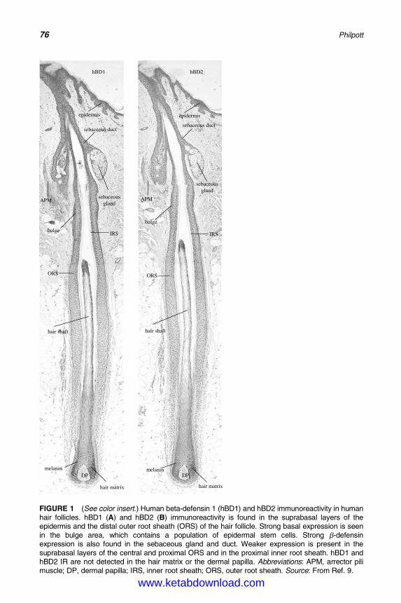
DK4962-Webster-ch6_R2_220307
DP DPmelanin melanin
hair matrix hair matrix
hair shafthair shaft
epidermisepidermis
sebaceous ductsebaceous duct
sebaceousgland
sebaceousgland
IRSIRS
ORS ORS
hBD1 hBD2
bulge
bulge
APMAPM
FIGURE 1 (See color insert.) Human beta-defensin 1 (hBD1) and hBD2 immunoreactivity in humanhair follicles. hBD1 (A) and hBD2 (B) immunoreactivity is found in the suprabasal layers of theepidermis and the distal outer root sheath (ORS) of the hair follicle. Strong basal expression is seenin the bulge area, which contains a population of epidermal stem cells. Strong b-defensinexpression is also found in the sebaceous gland and duct. Weaker expression is present in thesuprabasal layers of the central and proximal ORS and in the proximal inner root sheath. hBD1 andhBD2 IR are not detected in the hair matrix or the dermal papilla. Abbreviations: APM, arrector pilimuscle; DP, dermal papilla; IRS, inner root sheath; ORS, outer root sheath. Source: From Ref. 9.
76 Philpott
www.ketabdownload.com
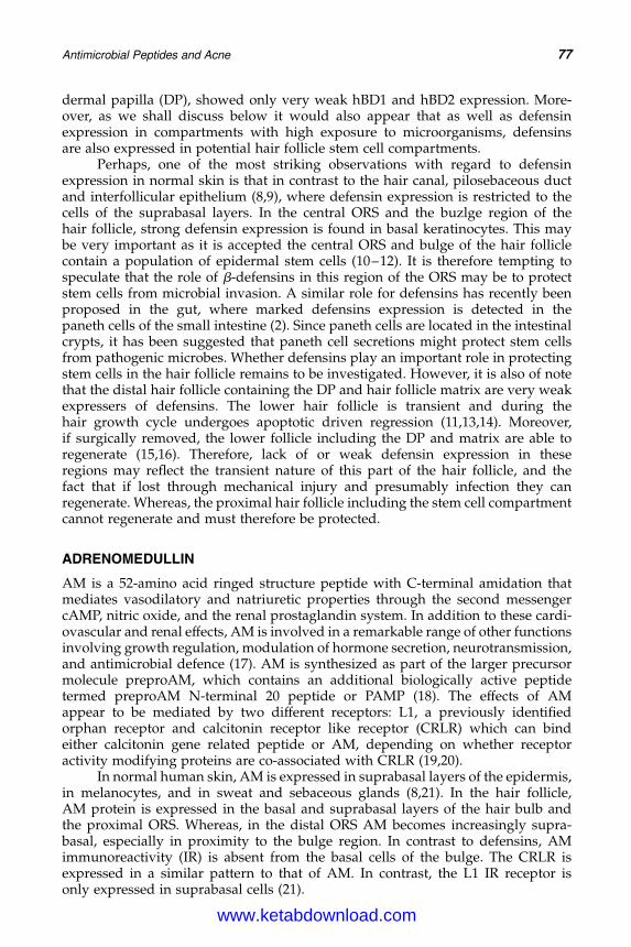
DK4962-Webster-ch6_R2_220307
dermal papilla (DP), showed only very weak hBD1 and hBD2 expression. More-over, as we shall discuss below it would also appear that as well as defensinexpression in compartments with high exposure to microorganisms, defensinsare also expressed in potential hair follicle stem cell compartments.
Perhaps, one of the most striking observations with regard to defensinexpression in normal skin is that in contrast to the hair canal, pilosebaceous ductand interfollicular epithelium (8,9), where defensin expression is restricted to thecells of the suprabasal layers. In the central ORS and the buzlge region of thehair follicle, strong defensin expression is found in basal keratinocytes. This maybe very important as it is accepted the central ORS and bulge of the hair folliclecontain a population of epidermal stem cells (10–12). It is therefore tempting tospeculate that the role of b-defensins in this region of the ORS may be to protectstem cells from microbial invasion. A similar role for defensins has recently beenproposed in the gut, where marked defensins expression is detected in thepaneth cells of the small intestine (2). Since paneth cells are located in the intestinalcrypts, it has been suggested that paneth cell secretions might protect stem cellsfrom pathogenic microbes. Whether defensins play an important role in protectingstem cells in the hair follicle remains to be investigated. However, it is also of notethat the distal hair follicle containing the DP and hair follicle matrix are very weakexpressers of defensins. The lower hair follicle is transient and during thehair growth cycle undergoes apoptotic driven regression (11,13,14). Moreover,if surgically removed, the lower follicle including the DP and matrix are able toregenerate (15,16). Therefore, lack of or weak defensin expression in theseregions may reflect the transient nature of this part of the hair follicle, and thefact that if lost through mechanical injury and presumably infection they canregenerate. Whereas, the proximal hair follicle including the stem cell compartmentcannot regenerate and must therefore be protected.
ADRENOMEDULLIN
AM is a 52-amino acid ringed structure peptide with C-terminal amidation thatmediates vasodilatory and natriuretic properties through the second messengercAMP, nitric oxide, and the renal prostaglandin system. In addition to these cardi-ovascular and renal effects, AM is involved in a remarkable range of other functionsinvolving growth regulation, modulation of hormone secretion, neurotransmission,and antimicrobial defence (17). AM is synthesized as part of the larger precursormolecule preproAM, which contains an additional biologically active peptidetermed preproAM N-terminal 20 peptide or PAMP (18). The effects of AMappear to be mediated by two different receptors: L1, a previously identifiedorphan receptor and calcitonin receptor like receptor (CRLR) which can bindeither calcitonin gene related peptide or AM, depending on whether receptoractivity modifying proteins are co-associated with CRLR (19,20).
In normal human skin, AM is expressed in suprabasal layers of the epidermis,in melanocytes, and in sweat and sebaceous glands (8,21). In the hair follicle,AM protein is expressed in the basal and suprabasal layers of the hair bulb andthe proximal ORS. Whereas, in the distal ORS AM becomes increasingly supra-basal, especially in proximity to the bulge region. In contrast to defensins, AMimmunoreactivity (IR) is absent from the basal cells of the bulge. The CRLR isexpressed in a similar pattern to that of AM. In contrast, the L1 IR receptor isonly expressed in suprabasal cells (21).
Antimicrobial Peptides and Acne 77
www.ketabdownload.com
DK4962-Webster-ch6_R2_220307
AM has antimicrobial effects against both Gram-positive and -negative bac-teria isolated from the skin and oral cavity. Moreover, antimicrobial activity ismost marked for Propionibacterium acnes and Micrococcus luteus with minimal inhibi-tory concentrations close to AM concentrations measured in sweat (22). AM alsoacts as an autocrine/paracrine growth factor in different tumor cell lines (23),Swiss 3T3 fibroblasts (24), vascular smooth muscle cells (25), as well as keratino-cytes of skin (26), and oral mucosa (27). Based on these data, possible roles forAM in the innate unspecific immune system of the skin and a possible participationof AM in the regulation of skin proliferation, wound repair, hair growth, and tumorprogression have been postulated.
ROLE OF ANTIMICROBIAL PEPTIDES IN ACNE
The epidermis forms an effective barrier, however, the hair canal, the distal ORS ofthe hair follicle, and the pilosebaceous duct constitute major ports of entry formicrobial invasion in humans and harbor a rich residential microflora such asP. acnes, Staphylococcus epidermidis, Demodex folliculorum, and Malassezia furfur. Thedistal ORS and the pilosebaceous duct are also characterized by many features ofinnate and adaptive immunological activity such as classical and nonclassicalMHC class 1 expression, ICAM-1 expression, and the presence of intraepithelialLangerhans cells and perifollicular macrophages (28–30). It is of considerable inter-est, therefore, that this area of the pilosebaceous unit is also a “hot spot” in thedevelopment of “acne vulgaris” lesions.
Acne, a disease of the pilosebaceous unit is characterized by hypercornifica-tion and hyperkeratosis of the ORS and sebaceous duct and perilesional infiltrate(PI). Lesions may be characterized as “non”inflammatory versus inflammatoryand their increasing inflammatory infiltrate may be classified as: comedones,papules, and pustules. In acne (Fig. 2), there is marked upregulation of hBD1 andhBD2 as follows: hBD1, healthy follicular skin � pustule � comedo , papule;hBD2, healthy follicular skin � comedo , papule , pustule. The fact that intensityof upregulation of hBD2 is identical to the classification of inflammatory infiltratecorrelates with other inflammatory disorders such as psoriasis and mastitis,which also exhibit marked upregulation of hBD2 (31–33).
From the published studies to date, it is clear that b-defensins are expressedin regions of the pilosebaceous unit that are exposed to microorganisms and more-over, in regions that may provide access routes for these organisms into the skin.Moreover, it is also apparent that some b-defensins are upregulated in acne (9).What, therefore, is the role of antimicrobial peptides in acne? Major factors in thepathogenesis of acne include hypercornification of the distal ORS and the pilosebac-eous duct in concert with increased sebum production and abnormalities of themicrobial flora (34).
There is increasing evidence that proinflammatory cytokines, such as IL-1b,TNF-a, and bacterial LPS, can upregulate b-defensins (4,6,32). P. acnes producesmany likely candidates for inflammation such as lipases, neuramidases, phospha-tases, and proteases (35). Therefore, the observed upregulation of b-defensins inacne vulgaris lesions is most likely a secondary response to the marked PI. It hasbeen suggested that, variation in the microenvironment of the duct is likely to influ-ence the production and activity of inflammatory mediators (36,37) and this mayinfluence defensin production. Although hBD1 has been reported to be constitu-tively expressed and not upregulated under inflammatory conditions in oral
78 Philpott
www.ketabdownload.com
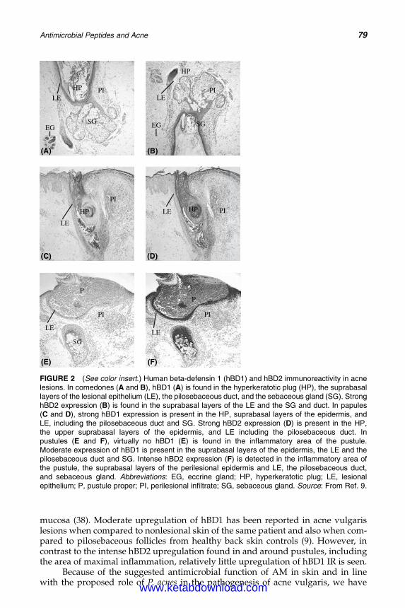
DK4962-Webster-ch6_R2_220307
mucosa (38). Moderate upregulation of hBD1 has been reported in acne vulgarislesions when compared to nonlesional skin of the same patient and also when com-pared to pilosebaceous follicles from healthy back skin controls (9). However, incontrast to the intense hBD2 upregulation found in and around pustules, includingthe area of maximal inflammation, relatively little upregulation of hBD1 IR is seen.
Because of the suggested antimicrobial function of AM in skin and in linewith the proposed role of P. acnes in the pathogenesis of acne vulgaris, we have
(A)
(C)
(E)
(B)
(D)
(F)
SG SG
HP
HP
HP HP
PP
PI
PI
PI PI
PI PILE LE
LE
LE
LELE
SG SG
EG EG
FIGURE 2 (See color insert.) Human beta-defensin 1 (hBD1) and hBD2 immunoreactivity in acnelesions. In comedones (A and B), hBD1 (A) is found in the hyperkeratotic plug (HP), the suprabasallayers of the lesional epithelium (LE), the pilosebaceous duct, and the sebaceous gland (SG). StronghBD2 expression (B) is found in the suprabasal layers of the LE and the SG and duct. In papules(C and D), strong hBD1 expression is present in the HP, suprabasal layers of the epidermis, andLE, including the pilosebaceous duct and SG. Strong hBD2 expression (D) is present in the HP,the upper suprabasal layers of the epidermis, and LE including the pilosebaceous duct. Inpustules (E and F), virtually no hBD1 (E) is found in the inflammatory area of the pustule.Moderate expression of hBD1 is present in the suprabasal layers of the epidermis, the LE and thepilosebaceous duct and SG. Intense hBD2 expression (F) is detected in the inflammatory area ofthe pustule, the suprabasal layers of the perilesional epidermis and LE, the pilosebaceous duct,and sebaceous gland. Abbreviations: EG, eccrine gland; HP, hyperkeratotic plug; LE, lesionalepithelium; P, pustule proper; PI, perilesional infiltrate; SG, sebaceous gland. Source: From Ref. 9.
Antimicrobial Peptides and Acne 79
www.ketabdownload.com
DK4962-Webster-ch6_R2_220307
investigated AM immunoreactivity in inflammatory acne lesions compared tohealthy pilosebaceous follicles. However, in contrast to b-defensins, AM does notappear to be upregulated in acne (21).
We have proposed (9,39) that acne vulgaris patients may suffer from a dysregu-lation of the production of innate and specific antimicrobial peptides. This is basedon our own observations that substantial variations in the intensity of hBD1 andhBD2 IR occur between sex- and age-matched patients, between face and backskin as well as between different hair follicle types (terminal hair follicles and pilo-sebaceous follicles). Colonization of the pilosebaceous duct is a feature of establishedcomedones and early inflammatory lesions (40,41). Why some colonized ductsbecome inflamed and others do not is uncertain. Variation in the microenvironmentof the duct could be important. Likewise, it is possible that differences in levels orinduction of defensins may explain why some colonized ducts become inflamedand others do not and may also explain why some individuals are prone to moresevere acne than others. P. acnes is likely to be the major organism since selective anti-biotic studies have shown that only those antibiotics which suppress P. acnes in vivo,are associated with clinical benefit. However, what is not clear is why some patientsare good responders to antibiotic treatment and other not. An interesting line ofstudy would be to investigate whether good responders to antibiotic antiacne treat-ment differ in their b-defensin levels and/or activity from bad responders.
From published studies, it is clear that there is marked variation in defensinexpression, both between different body sites as well as between individuals. Ithas been reported that hBD-1 promotes keratinocyte differentiation in vitro (42).It is intriguing, therefore, to speculate whether defensins may play a role in thehypercornification of the sebaceous duct and whether perhaps individuals thatare high producers of defensins may in fact be more prone to acne.
CONCLUSIONS
Defensins play a major role in innate immunity of epithelial tissues. It is clear thatthey are expressed in skin and are upregulated in inflammatory conditions. Theirrole in acne is still subject to much speculation and it is hoped that future studieswill address whether individuals who suffer from severe acne or are poor respon-ders to antibiotic treatment do produce lower levels of defensins.
ACKNOWLEDGMENTS
I would like to thank Dr. Sven Muller Roever and Catherine Chronnell for carryingout the research on defensins and acne in my laboratory; Professor Tony Quinn formany useful discussions; Dr. Bill Cunliffe and Dr. Diane Holland for supplying theacne biopsies; Dr. Thomas Ganz for donating the hBD1 and hBD2 antibodies,and Dr. Veronique Bataille for her valuable contributions to the study concept.I would also like to acknowledge the European Union (EU) for financial assistance.
REFERENCES
1. Oppenheim JJ, Biragyn A, Kwak LW, Yang D. Roles of antimicrobial peptides such asdefensins in innate and adaptive immunity. Ann Rheum Dis 2003; 62(suppl II):17–21.
2. Boman HG. Innate immunity and the normal microflora. Immunol Rev 2000; 173:5–16.3. Ganz T. Defensins and host defense. Science 1999; 286:420–421.
80 Philpott
www.ketabdownload.com
DK4962-Webster-ch6_R2_220307
4. Diamond G, Bevins CL. Beta-defensins: endogenous antibiotics of the innate hostdefense response. Clin Immunol Immunopathol 1998; 88:221–225.
5. Ganz T, Lehrer RI. Antimicrobial peptides of vertebrates. Curr Opin Immunol 1998;10:41–44.
6. Harder J, Bartels J, Christophers E, Schroder JM. A peptide antibiotic from human skin.Nature 1997; 387:861.
7. Valore EV, Park CH, Quayle AJ, Wiles KR, McCray PB, Ganz T. Human beta-defensin-1:an antimicrobial peptide of urogenital tissues. J Clin Invest 1998; 101:1633–1642.
8. Ali RS, Falconer A, Ikram M, Bissett CE, Cerio R, Quinn AG. Expression of the peptideantibiotics human beta defensin-1 and human beta defensin-2 in normal human skin.J Invest Dermatol 2001; 117:106–111.
9. Chronnell CMT, Ghali LR, Ali RS, et al. Human beta defensin-1 and -2 expression inhuman pilosebaceous units: upregulation in acne vulgaris lesions. J Invest Dermatol2001; 117:1120–1125.
10. Cotsarelis G, Sun TT, Lavker RM. Label-retaining cells reside in the bulge area of pilo-sebaceous unit: implications for follicular stem cells, hair cycle, and skin carcinogenesis.Cell 1990; 61:1329–1337.
11. Cotsarelis G, Kaur P, Dhouailly D, Hengge U, Bickenbach J. Epithelial stem cells in theskin: definition, markers, localization and functions. Exp Dermatol 1999; 8:80–88.
12. Rochat A, Kobayashi K, Barrandon Y. Location of stem cells of human hair follicles byclonal analysis. Cell 1994; 76:1063–1073.
13. Stenn K, Parimoo S, Prouty S. Growth of the hair follicle: a cycling and regeneratingbiological system. In: Chuong CM, ed. Molecular Basis of Epithelial AppendageMorphogenesis. Austin, TX: Landes Bioscience Publ., 1998:111–124.
14. Lindner G, Botchkarev VA, Botchkarev NV, Ling G, van der Veen C, Paus R. Analysis ofapoptosis during murine hair follicle regression (catagen). Am J Pathol 1997;151(6):1601–1617.
15. Oliver RF. Whisker growth after removal of the dermal papilla and lengths of the folliclein the hooded rat. J Embryol Exp Morphol 1966; 15:331–347.
16. Paus R. Control of the hair cycle and hair diseases as cycling disorders. Curr OpinDermatol 1996; 3:248–258.
17. Hinson JP, Kapas S, Smith DM. Adrenomedullin, a multifunctional regulatory peptide.Endocrine Rewiews 2000; 21:138–167.
18. Kitamura K, Sakata J, Kangawa K, et al. Cloning and characterization of cDNA encodinga precursor for human adrenomedullin [published erratum appears in Biochem BiophysRes Commun 1994; 202:643]. Biochem Biophys Res Commun 1993; 194:720–725.
19. McLatchie LM, Fraser NJ, Main MJ, et al. RAMPs regulate the transport and ligandspecificity of the calcitonin-receptor-like receptor. Nature 1998; 393:333–339.
20. Kapas S, Catt KJ, Clark AJ. Cloning and expression of cDNA encoding a rat adrenome-dullin receptor. J Biol Chem 1995; 270:25344–25347.
21. Muller FB, Muller-Rover S, Korge BP, Kapas S, Philpott MP. Adrenomedullin: expressionand possible role in human skin and hair growth. Br J Dermatol 2003; 148:30–38.
22. Allaker RP, Zihni C, Kapas S. An investigation into the antimicrobial effects of adreno-medullin on members of the skin, oral, respiratory tract and gut microflora. FEMSImmunol Med Microbiol 1999; 23:289–293.
23. MillerMJ,MartinezA,UnsworthEJ,etal.Adrenomedullinexpressioninhumantumorcelllines. Its potential role as an autocrine growth factor. J Biol Chem 1996; 271:23345–23351.
24. Withers DJ, Coppock HA, Seufferlein T, Smith DM, Bloom SR, Rozengurt E. Adrenomedullinstimulates DNA synthesis and cell proliferation via elevation of cAMP in Swiss 3T3 cells.FEBS Lett 1996; 378:83–87.
25. Iwasaki H, Eguchi S, Shichiri M, Marumo F, Hirata Y. Adrenomedullin as a novelgrowth-promoting factor for cultured vascular smooth muscle cells: role of tyrosinekinase-mediated mitogen-activated protein kinase activation. Endocrinology 1998;139:3432–3441.
26. Martinez A, Elsasser TH, Muro-Cacho C, et al. Expression of adrenomedullin and itsreceptor in normal and malignant human skin: a potential pluripotent role in the integu-ment. Endocrinology 1997; 138:5597–5604.
Antimicrobial Peptides and Acne 81
www.ketabdownload.com
DK4962-Webster-ch6_R2_220307
27. Kapas S, Brown DW, Farthing PM, Hagi-Pavli E. Adrenomedullin has mitogenic effectson human oral keratinocytes: involvement of cyclic AMP. FEBS Lett 1997; 418:287–290.
28. Paus R. Immunology of the hair follicle. In: Bos JD, ed. The Skin Immune System. BocaRaton: CRC Press, 1997.
29. Paus R, Muller-Rover S, Christoph T. Hair follicle immunology: a journey into terraincognita. J Invest Dermatol Symp Proc 1999; 4:226–234.
30. Christoph T, Muller-Rover S, Audring H, et al. The human hair follicle immune system:cellular composition and immune privilege. Br J Dermatol 2000; 142:1–13.
31. Schonwetter BS, Stolzenberg ED, Zasloff MA. Epithelial antibiotics induced at sites ofinflammation. Science 1995; 267:1645–1648.
32. Stolzenberg ED, Anderson GM, Ackermann MR, Whitlock RH, Zasloff M. Epithelialantibiotic induced in states of disease. Proc Natl Acad Sci USA 1997; 94:8686–8690.
33. Schroder JM, Harder J. Human beta-defensin-2. Int J Biochem Cell Biol 1999; 31:645–651.
34. Cunliffe WJ, Simpson NB. Disorders of the sebaceous gland [In: Rook, Wilkinson andEbling: Diamond G, Russell JP, Bevins CL. Inducible expression of an antibioticpeptide gene in lipopolysaccharide-challenged tracheal epithelial cells]. Proc NatlAcad Sci USA 1996; 93:5156–5160.
35. Greenman J, Holland KT, Cunliffe WJ. Effects of pH on biomass, maximum specificgrowth rate and extracellular enzyme production by three species of cutaneous propio-nibacteria grown in continuous culture. J Gen Microbiol 1983; 129:1301–1307.
36. Holland KT, Cunliffe WJ, Roberts CD. The role of bacteria in acne vulgaris: a newapproach. Clin Exp Dermatol 1978; 3:253–257.
37. Holland KT, Aldana O, Bojar RA, et al. Propionibacterium acnes and acne. Dermatology1998; 196:67–68.
38. Mathews M, Jia HP, Guthmiller JM, et al. Production of beta-defensin antimicrobialpeptides by the oral mucosa and salivary glands. Infect Immun 1999; 67:2740–2745.
39. Philpott MP. Defensins and acne. Mol Immunol 2003; 40:457–462.40. Leeming JP, Holland KT, Cunliffe WJ. The pathological and ecological significance of
microorganisms colonizing acne vulgaris comedones. J Med Microbiol 1985; 20:11–16.41. Leeming JP, Holland KT, Cunliffe WJ. The microbial colonisation of inflamed acne
vulgaris lesions. Br J Dermatol 1988; 118:203–208.42. Frye M, Bargon J, Gropp R. Expression of human beta-defensin-1 promotes differen-
tiation of keratinocytes. J Mol Med 2001; 79:275–282.
82 Philpott
www.ketabdownload.com
DK4962-Webster-ch7_R2_220307
Part II: Acne Treatments
B7 Hormonal Influences in Acne
Diane ThiboutotDepartment of Dermatology, Pennsylvania State University College of Medicine,Hershey, Pennsylvania, U.S.A.
INTRODUCTION
The effects of hormones in acne are most notable in women. Androgens such asdihydrotestosterone (DHT) and testosterone, the adrenal precursor dehydroepian-drosterone sulfate (DHEAS), estrogen, and other hormones, including growthhormone or insulin-like growth factors (IGFs), may each be important. It is likelythat the hormones affecting the sebaceous gland are both taken up from theserum in addition to being made locally within the gland.
Hormonal therapy is an option in women whose acne is not responding toconventional treatment or if signs of endocrine abnormalities are present. The great-est therapeutic benefit from hormonal therapy is achieved in combination withother effective anti-acne medications. This chapter focuses on the role of hormonesin acne, the clinical presentation of adult female acne, the work-up of a suspectedendocrine abnormality, and the available options for hormonal therapy.
ANDROGENS IN ACNE
Both clinical observation and experimental evidence confirm the importance ofandrogens in the pathophysiology of acne. The majority of circulating androgensare produced by the gonads and the adrenal gland. Androgens can also be producedlocally within the sebaceous gland from the adrenal precursor hormone, DHEAS.The main androgens that interact with the androgen receptor are testosteroneand DHT. Androgen receptors are found in the basal layer of the sebaceous glandand the outer root sheath keratinocytes of the hair follicle (1,2). DHTis approximatelyfive to 10 times more potent than testosterone in its interaction with the androgenreceptor.
An essential role for androgens in stimulating sebum production is supportedby several lines of evidence. For example, the development of acne in the prepuber-tal period has been associated with elevated serum levels of DHEAS, a precursor fortestosterone (3,4). Androgen-insensitive subjects who lack functional androgenreceptors do not produce sebum and do not develop acne (5). Tumors of theovary or the adrenal that produce androgens are often associated with thedevelopment of acne. Systemic administration of testosterone and dehydroepian-drosterone increases the size and secretion of sebaceous glands (6), and we knowthat severe acne is often associated with elevated serum androgens (7,8).
Androgen Metabolism Within the SkinAcne may be mediated by serum androgens, locally produced androgens, or a com-bination of both. Insights have been gained regarding the local metabolism of
83
www.ketabdownload.com
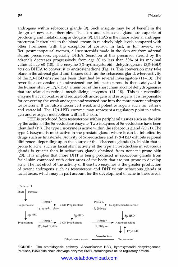
DK4962-Webster-ch7_R2_220307
androgens within sebaceous glands (9). Such insights may be of benefit in thedesign of new acne therapies. The skin and sebaceous gland are capable ofproducing and metabolizing androgens (9). DHEAS is the major adrenal androgenprecursor. It circulates in the blood stream in relatively high levels compared withother hormones with the exception of cortisol. In fact, in for review, seeRef. postmenopausal women, all sex steroids made in the skin are from adrenalsteroid precursors, especially DHEA. Secretion of this precursor steroid by theadrenals decreases progressively from age 30 to less than 50% of its maximalvalue at age 60 (10). The enzyme 3b -hydroxysteroid dehydrogenase (3b -HSD)acts on DHEA to convert it to androstenedione (Fig. 1). This conversion may takeplace in the adrenal gland and tissues such as the sebaceous gland, where activityof the 3b -HSD enzyme has been identified by several investigators (11–13). Thereversible conversion of androstenedione into testosterone is then catalyzed inthe human skin by 17b -HSD, a member of the short chain alcohol dehydrogenasesthat are related to retinol metabolizing enzymes (14–18). This is a reversibleenzyme that can oxidize and reduce both androgens and estrogens. It is responsiblefor converting the weak androgen androstenedione into the more potent androgentestosterone. It can also interconvert weak and potent estrogens such as estroneand estradiol. The 17b -HSD enzyme may represent a regulatory point in andro-gen and estrogen metabolism within the skin.
DHT is produced from testosterone within peripheral tissues such as the skinby the action of the 5a -reductase enzyme. Two isozymes of 5a -reductase have beenidentified (19). The type 1 isozyme is active within the sebaceous gland (20,21). Thetype 2 isozyme is most active in the prostate gland, where it can be inhibited bydrugs such as finasteride. Activity of 5a-reductase and 17b -HSD exhibits regionaldifferences depending upon the source of the sebaceous glands (9). In skin that isprone to acne, such as facial skin, activity of the type 1 5a-reductase in sebaceousglands is greater than in sebaceous glands obtained from nonacne-prone skin(20). This implies that more DHT is being produced in sebaceous glands fromfacial skin compared with other areas of the body that are not prone to developacne. The net effect of the activity of these two enzymes is the greater productionof potent androgens such as testosterone and DHT within sebaceous glands offacial areas, which may in part account for the development of acne in these areas.
Cholesterol
StAR P450scc
P450c17
P450c17 P450c17
Pregnenolone 17α-hydroxylase
17α-hydroxylase
3β-HSD 3β-HSD 3β-HSD
Progesterone
17β-HSD
5α-reductase
Dehydroepiandrosterone 17-OH-Pregnenolone P450c17
17, 20 lyase
17, 20 lyase Androstenedione 17-OH Progesterone
Dihydrotestosterone Testosterone
FIGURE 1 The steroidogenic pathway. Abbreviations: HSD, hydroxysteroid dehydrogenase;P450scc, P450 side chain cleavage enzyme; StAR, steroidogenic acute regulatory protein.
84 Thiboutot
www.ketabdownload.com
DK4962-Webster-ch7_R2_220307
The Sebaceous Gland Is a Steroidogenic TissueThe skin and sebaceous glands are capable of synthesizing cholesterol de novo fromacetate (22–24). Although this cholesterol is utilized in cell membranes, in the for-mation of the epidermal barrier, and is secreted in sebum, its use as a substrate forsteroid hormone synthesis had not been established until recently. In order forsteroid synthesis to occur, cholesterol needs to be translocated from the outer tothe inner mitochondrial membrane. This process is regulated by the steroidogenicacute regulatory protein (25). Additional enzymes and cofactors needed to convertcholesterol into a steroid include P450 cholesterol side chain cleavage, adrenodoxinreductase, cytochrome P450c17, and steroidogenic factor-1. Expression of each ofthese proteins was found in human facial skin, sebocytes, and in a recently devel-oped simian virus (SV) 40-immortalized human sebocyte cell line (SEB-1) (26).These data demonstrate that the skin is in fact a steroidogenic tissue. The clinicalsignificance of this finding in mediating androgenic skin disorders such as acne,hirsutism, or androgenetic alopecia remains to be established.
ESTROGENS IN ACNE
Very little is known about the role of estrogens in modulating sebum production. Anyestrogen given systemically in sufficient amounts will decrease sebum production.The dose of estrogen required to suppress sebum production, however, is greaterthan the dose required to suppress ovulation (27). The major active estrogen is estra-diol, which is produced from testosterone by the action of the enzyme aromatase.Aromatase is active in the ovary, adipose tissue, and other peripheral tissues. Estradiolcan be converted to the less potent estrogen, estrone, by the action of the 17b -HSDenzyme. Both aromatase and 17b -HSD are present in the skin (17,28). Estrogensmay act by several mechanisms; they may: (i) directly oppose the effects of androgenslocally within the sebaceous gland, (ii) inhibit the production of androgens by gonadaltissue via a negative feedback loop on pituitary gonadotrophin release, and (iii)regulate genes that negatively influence sebaceous gland growth or lipid production.
GROWTH HORMONE AND INSULIN-LIKE GROWTH FACTORS IN ACNE
Growth hormone is secreted by the pituitary gland. It acts on the liver and periph-eral tissues to stimulate the production of IGFs, formerly known as somatomedians.There are two forms of IGF, termed IGF-1 and IGF-2. IGF-1 is the more prevalentgrowth factor. It has been hypothesized that growth hormone may be involved inthe development of acne (29). Acne is most prevalent in adolescents during atime when growth hormone is maximally secreted and serum levels of IGF-1 arehighest. In addition, IGF-1 can be produced locally within the skin, where it caninteract with receptors on the sebaceous gland to stimulate its growth. Furthermore,conditions of growth hormone excess such as acromegaly are associated with sebor-rhea and the development of acne. In some tissues, the actions of IGF-1 can bemediated by androgens. It is possible that androgens may influence IGF-1 actionin the sebaceous gland as well.
CLINICAL PRESENTATION OF ADULT FEMALE ACNE
For reasons that are not understood, the distribution of facial acne in many adultfemales differs from that seen in adolescents and in males. Many adult women
Hormonal Influences in Acne 85
www.ketabdownload.com
DK4962-Webster-ch7_R2_220307
note that acne localizes to the lateral face, chin, and neck (Fig. 2). Oftentimes, acnein these women is not necessarily widespread or severe, but rather it may below-grade, persistent, and consist of a few isolated deep-seated tender nodules.
Many women note flares of their acne just prior to their menstrual period withreports ranging from 27–60% to 70% of women (30–32). One study has been pub-lished that provides quantitative documentation of acne lesion counts over themenstrual period (33). In this study of women, who were followed over two men-strual cycles, 63% showed a 25% increase in inflammatory acne lesions prior to themenstrual period. Some women may feel that intermittent acne therapy prior totheir menstrual period may be beneficial. There is no evidence for this approach.Since most acne therapy is designed to prevent the formation of lesions and sincethis process often takes several weeks, it seems unlikely that intermittent therapywould be beneficial. For this reason, it is important to have patients use their medi-cations consistently and to avoid spot treatment.
WHEN TO SUSPECT AN ENDOCRINE DISORDER IN ACNE PATIENTS
Although hormones influence acne, most acne patients do not have an endocrinedisorder. Hyperandrogenism should be considered in female patients whose acneis severe, sudden in its onset, or is associated with hirsutism, or irregular menstrualperiods. Additional clinical signs of hyperandrogenism include Cushinoid features,increased libido, clitoromegaly, deepening of the voice, acanthosis nigricans, orandrogenetic alopecia. Women with hyperandrogenism may also have insulinresistance. They are at risk for the development of diabetes and cardiovasculardisease. It is therefore important for the long-term health of these patients to ident-ify hyperandrogenism so that they can receive appropriate therapy from an endo-crinologist or gynecologist.
SCREENING FOR AN ENDOCRINE DISORDER
A medical history and physical examination should be performed that is directedtoward eliciting any symptoms or signs of hyperandrogenism. Screening laboratorytests for hyperandrogenism include a serum DHEAS, total testosterone, free testos-terone, and luteinizing hormone/follicle-stimulating hormone (LH/FSH) ratio.These tests should be obtained apart from the time of ovulation in order to avoidthe surge of hormones associated with ovulation. From a practical standpoint, itmay be easiest to suggest that women have these tests performed either just priorto or during the menstrual period. It is important to note that if a patient is onoral contraceptives at the time of hormonal testing, an underlying hyperandrogen-emia may be masked. This does not occur with antiandrogens such as cyproteroneor spironolactone. Therefore, it is best that patients discontinue oral contraceptivesfour to six weeks prior to the endocrine evaluation.
Excess androgens may be produced by either the adrenal gland or the ovary.Serum levels of DHEAS can be used to screen for an adrenal source of excess androgenproduction. Patients with a serum DHEAS greater than 8000 ng/mL (units may differdepending upon the laboratory) may have an adrenal tumor and should be referred toan endocrinologist for further evaluation. Some adrenal tumors may also produce tes-tosterone. Values of DHEAS in the range of 4000 ng/mL to 8000 ng/mL may be associ-ated with congenital adrenal hyperplasia, which is most commonly due to a partialdeficiency in the 21-hydroxylase or 11-hydroxylase enzyme in the adrenal gland.
86 Thiboutot
www.ketabdownload.com
DK4962-Webster-ch7_R2_220307
Such an enzyme deficiency results in the shunting of steroids from the cortisol biosyn-thetic pathway into the androgen biosynthetic pathway.
An ovarian source of excess androgens can be suspected in cases where theserum total testosterone is elevated. Serum total testosterone in the range of150 ng/dL to 200 ng/dL or an increased LH/FSH ratio (greater than 2 to 3) canbe found in cases of polycystic ovary disease. This condition is a spectrum and isoften, but not always, associated with irregular menstrual periods, reduced fertility,obesity, insulin resistance, or hirsutism. Greater elevations in serum testosteronemay indicate an ovarian tumor and appropriate referral should be made. In somecases, there can be modest elevations in both DHEAS and testosterone. A serumlevel of 17-hydroxypregneneolone can be obtained to discern between an ovarianor adrenal source of androgens. If 17-hydroxypregneneolone is elevated, it indicatesan adrenal source of excess androgens, most often secondary to late onset congeni-tal adrenal hyperplasia. Of note is that there is a significant amount of variation inan individual’s serum androgen levels. In cases where abnormal results areobtained, it is recommended to repeat the test before proceeding with therapy ora more extensive work-up.
Questions arise as to the importance of a pelvic ultrasound in the diagnosis ofpolycystic ovarian syndrome. This test can be nonspecific, in that women withnormal androgens may have ovarian cysts and conversely, women with hyperan-drogenism and other findings associated with polycystic ovarian syndromemay not have ovarian cysts at the time of pelvic ultrasound. For this reason, thediagnosis of polycystic ovarian syndrome is more heavily based upon the serumhormonal profile and associated clinical findings.
In the majority of women with acne, serum androgens are completely normal,yet these women will in fact respond if treated with hormonal therapy. Studies haveshown that, as a group, women with acne may have higher levels of serum DHEAS,testosterone, and DHT than those without acne (7,34). However, although higher,these laboratory values may still be within the normal range. Serum levels ofDHEAS, DHT, and IGF-1 are reported to correlate positively with acne lesioncounts in women, whereas androstenedione and DHEAS correlate with lesioncounts in men (35). Reduction of serum androgens or inhibition of their action, asobtained with oral contraceptives or antiandrogens, respectively, can lead toimprovement in acne in women with normal serum androgen levels.
OPTIONS FOR THE HORMONAL THERAPY OF ACNE IN WOMEN
Once the decision has been made to initiate hormonal therapy, the various optionsto choose from include: (i) androgen receptor blockers, or antiandrogens (this classof drugs block the effect of androgens on the sebaceous gland); (ii) inhibitors ofandrogen production by the ovary or adrenal gland such as oral contraceptivesor glucocorticosteroids, respectively; or (iii) in the future, it may be possible toinhibit the activity of androgen metabolizing enzymes in the skin or sebaceousgland itself.
Agents that Block the Androgen ReceptorWithin the class of androgen receptor blockers, therapeutic options include spirono-lactone, cyproterone acetate, and flutamide. In the United States, spironolactone isthe most commonly used drug, although flutamide is also available.
Hormonal Influences in Acne 87
www.ketabdownload.com
DK4962-Webster-ch7_R2_220307
SpironolactoneOral spironolactone decreases sebum excretion rate by 30% to 50%. Recommendeddoses are 50 to 100 mg taken with meals (36,37). However, many women withsporadic outbreaks of inflammatory lesions or isolated cysts respond well to25 mg twice daily and some even respond to just 25 mg a day. These low dosesin healthy young women are generally well-tolerated. However, if this drug isused in older women who may have other medical problems, or if higher dosesare used for conditions such as hirsutism or androgenetic alopecia, serum electro-lytes should be monitored. Side effects of spironolactone include breast tendernessand menstrual irregularities. Additionally, it is important that pregnancy beavoided during treatment with spironolactone due to the potential for abnormal-ities of the male fetal genitalia such as hypospadias.
Cyproterone AcetateCyproterone acetate is available in many parts of the world, but not in the UnitedStates. It possesses dual activity in that it serves as a progestogen in oral contracep-tives in addition to its direct inhibition of the androgen receptor. It can be given indoses of 2 to 100 mg per day as a single agent, in which case there can be improve-ment in 75% to 90% of women with acne. Cyproterone acetate, however, is mostcommonly used in the form of an oral contraceptive combined with ethinyl estra-diol in varying doses (38). Numerous clinical studies support the efficacy of theseoral contraceptive preparations in women with acne.
FlutamideFlutamide is a potent nonsteroidal antagonist of the androgen receptor. Although mostcommonly used to treat prostate cancer, flutamide has been reported to be efficaciousin the treatment of acne, hirsutism, and androgenic alopecia (39). It can be given indoses of 250 mg twice daily in combination with an oral contraceptive. Fatal hepatitishas been reported with this drug. Liver function tests should be monitored and seriousconsideration should be given to the risk/benefit ratio of its use in acne (40). Addition-ally, because it is an antiandrogen, pregnancy issues are a concern.
Inhibitors of Adrenal Androgen ProductionAnother option in hormone therapy is to block the production of androgens eitherby the adrenal gland or ovary, which can be accomplished through the use of low-dose glucocorticoids or oral contraceptives, respectively.
GlucocorticoidsLow-dose glucocorticoids are most commonly used to treat patients with late onsetcongenital adrenal hyperplasia, which is an inherent defect in the 21-hydroxylaseor the 11-hydroxylase enzyme. This defect causes a block in the cortisol biosyntheticpathway, which results in a buildup of steroid precursors that are shunted into theandrogen biosynthetic pathway. Low-dose prednisone (2.5 to 5 mg a day, at bedtime)can be used. Low doses of dexamethasone can also be used, but the risk of adrenalsuppression is higher. To ascertain if therapy with glucocorticoids is having thedesired effect, serum DHEAS can be monitored for a decrease or normalization ofthe level of DHEAS. To check for adrenal suppression, an adrenocorticotrophinhormone (ACTH) simulation test can be performed. This consists of injecting
88 Thiboutot
www.ketabdownload.com
DK4962-Webster-ch7_R2_220307
ACTH and assessing the plasma cortisol 30 minutes later. If plasma cortisol has risenby an appropriate amount, the adrenal gland is not suppressed.
Inhibition of Ovarian Androgen ProductionGonadotropin-Releasing AgonistsAndrogen production in the ovary can also be blocked by gonadotropin-releasinghormone agonists such as buserelin, nafarelin, or leuprolide. These gonadotropin-releasing agonists block ovulation by interrupting the cyclic release of FSH and LHfrom the pituitary. These drugs are efficacious in acne and hirsutism, and are availableas injectable drugs or nasal spray. However, in addition to suppressing the productionof ovarian androgens, these drugs also suppress the ovarian production of estrogens,thereby eliminating the function of the ovary. Thus, the patient could develop meno-pausal symptoms and suffer from hypoestrogenism. Headaches can also develop, aswell as the occurrence of bone loss, due to the reduction in estrogen.
Oral ContraceptivesOral contraceptives generally contain an estrogen (most commonly ethinyl estra-diol) and a progestin. In their early formulations, oral contraceptives containedover 100 mg of estrogen. In these and higher doses, estrogens themselves can sup-press sebum production. Estrogens also act on the liver to increase the synthesis ofsex hormone-binding globulin that binds testosterone and lowers the circulatinglevels of free testosterone. In addition, oral contraceptives inhibit the ovarian pro-duction of androgens by suppressing ovulation. This, in turn, decreases serumandrogen levels and reduces sebum production. The concentrations of estrogenin oral contraceptives have decreased over the years from 150 to 35 mg, and inthe most recent forms, to 20 mg, in order to reduce the side effects associatedwith estrogen (41). Oral contraceptives containing low doses of estrogen arelisted in Table 1.
ProgestinsThe progestins contained in oral contraceptives include estranges and gonanes,which are derivatives of 19-nortestosterone, cyprotereone acetate, and a novel pro-gestin, drosperinone. Members of the estrane and gonane class of progestins(Table 2) can cross-react with the androgen receptor, which can lead to increasedandrogenic effects and could aggravate acne, hirsutism, or androgenic alopecia.These progestins can also cause changes in lipid metabolism and can increaseserum glucose, leading to glucose intolerance, as well as possibly interfering withthe beneficial effect of estrogen on the sex hormone-binding globulin. However,the third generation progestins, including norgestimate, desogestrel, and gesto-dene, are more selective for the progesterone receptor rather than the androgenreceptor. The biological relevance of these differences, however, is uncertain. For
TABLE 1 Oral Contraceptives Containing Low Doses of Estrogen
Ethinyl estradiol 20 mg/Levonorgestrel 0.1 mg (AlesseTM)Ethinyl estradiol 20, 10 mg/Desogestrel 0.15 mg (MircetteTM)Ethinyl estradiol 20, 30, 35 mg/norethindrone 1.0 mg (Estrostepw)Ethinyl estradiol 20 mg/norethindrone acetate 1.0 mg (Lo-Estrinw 1/20)Ethinyl estradiol 20 mg/drospirenone 3.0 mg (YazTM)
Hormonal Influences in Acne 89
www.ketabdownload.com
DK4962-Webster-ch7_R2_220307
years, it has been known that almost all oral contraceptives are beneficial in thetreatment of acne (42). It is possible that some women are more sensitive to theandrogenic effects of a progestin, but it is more likely that the effect of progestinmay be offset by estrogen. All oral contraceptives, regardless of the type of proges-tin, will inhibit serum androgen levels. Moreover, although some progestins mightbe more androgenic than others, there is an increase in sex hormone-binding globu-lin with the use of any oral contraceptives and an improvement of the acne inwomen who are treated with them.
Drospirenone is a novel progestin that is derived from 17a-spironolactone. Itpossesses antiandrogenic and antimineralocorticoid activity, which can be ofbenefit in androgenic-related conditions such as acne and hirsutism and in theestrogen-related fluid retention associated with some oral contraceptives (43).
ORAL CONTRACEPTIVES STUDIED IN ACNE
Many oral contraceptives have been studied in the treatment of acne (Table 3).These include those containing ethinyl estradiol in combination with cyproter-one acetate (Diane, Dianette), ethynodiol diacetate (Demulen), levonorgestrel(TriPhasil, Alesse), norgestimate (Ortho Tri-Cyclenw), desogesterel (Desogen),and drosperinone (Yasmin, Yaz). Numerous studies point to the efficacy ofethinyl estradiol/cyproterone acetate oral contraceptives (Diane and Dianette) inthe treatment of acne. Reductions in inflammatory lesion count on the order of50% to 75% have been reported (44,45). Two large studies involving a total ofapproximately 500 women with moderate acne were conducted with ethinylestradiol 35 mg/norgestimate (Ortho Tri-Cyclen). Improvement in inflammatorylesions, total lesions, and global assessment was noted with this oral contraceptiveafter six months of treatment (46,47). There was a 50% to 60% improvement ininflammatory lesions. Decreases in serum free testosterone and an increase in sexhormone-binding globulin were noted in the active group. Two large, six-month,placebo-controlled trials (350 and 371 women, respectively) were conducted
TABLE 2 Progestins: 19-Nortestosterone Derivatives
Estranes GonanesNorethindrone NorgestrelNorethindrone acetate LevonorgestrelEthynodiol diacetate Desogestrel
GestodeneNorgestimate
TABLE 3 Oral Contraceptives Studied in Acne
Demulen (ethinyl estradiol 35 mg/ethynodiol diacetate)Diane, Dianette (ethinyl estradiol 50, 35 mg/cyproterone acetate)Estrostep (ethinyl estradiol 20, 25, 30 mg/norethindrone)Alesse (ethinyl estradiol 20 mg/levonorgestrel)Ortho Tri-Cyclen (ethinyl estradiol 35 mg/norgestimate)Desogen (ethinyl estradiol 30 mg/desogestrel)Yasmin (ethinyl estradiol 30 mg/drospirenone)Yaz (ethinyl estradiol 20 mg/drospirenone)Triphasil (ethinyl estradiol 30 mg, 40 mg/levonorgestrel)
90 Thiboutot
www.ketabdownload.com
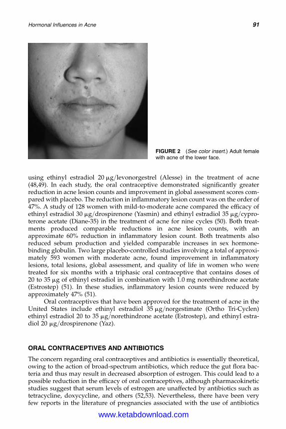
DK4962-Webster-ch7_R2_220307
using ethinyl estradiol 20 mg/levonorgestrel (Alesse) in the treatment of acne(48,49). In each study, the oral contraceptive demonstrated significantly greaterreduction in acne lesion counts and improvement in global assessment scores com-pared with placebo. The reduction in inflammatory lesion count was on the order of47%. A study of 128 women with mild-to-moderate acne compared the efficacy ofethinyl estradiol 30 mg/drospirenone (Yasmin) and ethinyl estradiol 35 mg/cypro-terone acetate (Diane-35) in the treatment of acne for nine cycles (50). Both treat-ments produced comparable reductions in acne lesion counts, with anapproximate 60% reduction in inflammatory lesion count. Both treatments alsoreduced sebum production and yielded comparable increases in sex hormone-binding globulin. Two large placebo-controlled studies involving a total of approxi-mately 593 women with moderate acne, found improvement in inflammatorylesions, total lesions, global assessment, and quality of life in women who weretreated for six months with a triphasic oral contraceptive that contains doses of20 to 35 mg of ethinyl estradiol in combination with 1.0 mg norethindrone acetate(Estrostep) (51). In these studies, inflammatory lesion counts were reduced byapproximately 47% (51).
Oral contraceptives that have been approved for the treatment of acne in theUnited States include ethinyl estradiol 35 mg/norgestimate (Ortho Tri-Cyclen)ethinyl estradiol 20 to 35 mg/norethindrone acetate (Estrostep), and ethinyl estra-diol 20 mg/drospirenone (Yaz).
ORAL CONTRACEPTIVES AND ANTIBIOTICS
The concern regarding oral contraceptives and antibiotics is essentially theoretical,owing to the action of broad-spectrum antibiotics, which reduce the gut flora bac-teria and thus may result in decreased absorption of estrogen. This could lead to apossible reduction in the efficacy of oral contraceptives, although pharmacokineticstudies suggest that serum levels of estrogen are unaffected by antibiotics such astetracycline, doxycycline, and others (52,53). Nevertheless, there have been veryfew reports in the literature of pregnancies associated with the use of antibiotics
FIGURE 2 (See color insert.) Adult femalewith acne of the lower face.
Hormonal Influences in Acne 91
www.ketabdownload.com
DK4962-Webster-ch7_R2_220307
in conjunction with oral contraceptives (54,55). Existing reports have focused ontetracycline, and the incidence was 1.2 to 1.4 pregnancies/100 woman years ofuse of the oral contraceptive. These data are no greater than the backgroundfailure rate of oral contraceptives (54,55).
NEWER FORMS OF CONTRACEPTIVES
Recently, newer forms of contraceptives have been developed, such as contracep-tive patches, vaginal rings, and injectable combination hormones. Each of these isdesigned to suppress ovulation and in this regard will lower the ovarian productionof androgens. As of yet, these formulations have not been studied in the treatmentof acne. The contraceptive patch (Ortho Evra) contains 20 mg of ethinyl estradioland 150 mg of the progestin, norelgestromin. The patch is worn for three weeksand removed for one week, during which time menstrual bleeding will occur.The advantages of this formulation are better patient compliance, dosing that isnot affected by gastrointestinal disturbances, and more consistent serum levels ofestrogen serum levels compared to oral dosing (56). The vaginal ring (NuvaRing)is a contraceptive vaginal ring that releases 15 mg of ethinyl estradiol and 120 mgof the progestin, etonogestrel. It is placed within the vagina for three weeks andremoved for one week. In one study, the incidence of irregular bleeding was lesscompared with an oral combination contraceptive (57). An injectable combinationof estradiol cypionate and medroxyprogesterone acetate (Lunelle) has been devel-oped. This is given as a monthly contraceptive injection. Contraceptive efficacy wasshown to be comparable to a triphasic oral contraceptive containing ethinyl estra-diol and norethindrone (Ortho 7/7/7) (58).
APPROACH TO HORMONAL THERAPY IN FEMALE ACNE
Hormonal therapy is an excellent option for treatment of women whose acne is notresponding to conventional therapy. If there are signs of hyperandrogenism, anendocrine evaluation is indicated, consisting of tests such as DHEAS, total- andfree-testosterone, and an LH/FSH ratio. Although hyperandrogenism is an indi-cation for hormonal therapy, women with normal serum androgen levels alsorespond well to treatment. The mainstays of hormonal therapy include oral contra-ceptives and spironolactone. Other agents to choose from include cyproteroneacetate, flutamide, and glucocorticoids. Hormonal agents work best as part of acombination regimen including topical retinoids or topical or oral antibioticsdepending upon the severity of the acne. In some women, the additional of hormo-nal therapy has improved acne to the point where subsequent treatment with iso-tretinoin was no longer necessary. As more is learned about the hormones involvedin acne, their source of production and the mechanisms by which they influencesebaceous gland growth and sebum production, new opportunities will arise forthe development of novel therapies aimed at the hormonal aspects of acne.
REFERENCES
1. Choudhry R, Hodgins M, Van der Kwast T, et al. Localization of androgen receptors inhuman skin by immunohistochemistry: implications for the hormonal regulation of hairgrowth, sebaceous glands and sweat glands. J Endocrinol 1991; 133:467–475.
92 Thiboutot
www.ketabdownload.com

DK4962-Webster-ch7_R2_220307
2. Liang T, Hoyer S, Yu R. Immunocytochemical localization of androgen receptors inhuman skin using monoclonal antibodies against the androgen receptor. J Invest Derma-tol 1993; 100:663–666.
3. Lucky AW, Biro FM, Huster GA, et al. Acne vulgaris in premenarchal girls. Arch Derma-tol 1994; 130:308–314.
4. Stewart ME, Downing DT, Cook JS, et al. Sebaceous gland activity and serum dehydroe-piandrosterone sulfate levels in boys and girls. Arch Dermatol 1992; 128:1345–1348.
5. Imperato-McGinley J, Gautier T, Cai LQ, et al. The androgen control of sebum pro-duction. Studies of subjects with dihydrotestosterone deficiency and complete androgeninsensitivity. J Clin Endocrinol Metab 1993; 76(2):524–528.
6. Pochi PE, Strauss JS. Sebaceous gland response in man to the administration of testoster-one,D4- androstenedione, and dehydroisoandrosterone. J Invest Dermatol 1969; 52:32–36.
7. Marynick SP, Chakmajian ZH, McCaffree DL, et al. Androgen excess in cystic acne. NEngl J Med 1983; 308:981–986.
8. Lucky A, McGuire J, Rosenfield R, et al. Plasma androgens in women with acne vulgaris.J Invest Dermatol 1983; 81:70.
9. Chen W, Thiboutot D, Zouboulis C. Cutaneous androgen metabolism: basic research andclinical perspectives. J Invest Dermatol 2002; 119:992–1007.
10. Labrie F, Luu-The V, Pelletier G, et al. Intracrinology and the skin. Horm Res 2000;54:213–229.
11. Baillie A, Calman K, Milne J. Histochemical distribution of hydroxysteroid dehydrogen-ases in human skin. Br J Dermatol 1965; 7:610–616.
12. Sawaya ME, Honig JLS, Garland LD, et al. D5-3b-Hydroxysteroid dehydrogenaseactivity in sebaceous glands of scalp in male-pattern baldness. J Invest Dermatol 1988;91:101–105.
13. Simpson NB, Cunliffe WJ, Hodgins MB. The relationship between the in vitro activityof 3b-hydroxysteroid dehydrogenase delta4-5-isomerase in human sebaceous glandsand their secretory activity in vivo. J Invest Dermatol 1983; 81(2):139–144.
14. Mason J. The 3b-hydroxysteroid dehydrogenase gene family of enzymes. TEM 1993;4:199–202.
15. Chai Z, Brereton P, Suzuki T, et al. 17b-hydroxysteroid dehydrogenase type 11 localizesto human steroidogenic cells. Endocrinology 2003; 144:2084–2091.
16. Andersson S, Moghrabi N. Physiology and molecular genetics of 17ß-hydroxysteroiddehydrogenases. Steroids 1997; 62:143–147.
17. Hay JB, Hodgins MB, Donnelly JB. Human skin androgen metabolism and preliminaryevidence for its control by two forms of 17 ß-hydroxysteroid oxidoreductase. J Endocri-nol 1982; 93(3):403–413.
18. Thiboutot D, Martin P, Volikos L, et al. Oxidative activity of the type 2 isozyme of 17b-hydroxysteroid dehydrogenase (17b-HSD) predominates in human sebaceous glands.J Invest Dermatol 1998; 111:390–395.
19. Jenkins EP, Andersson S, Imperato-McGinley J, et al. Genetic and pharmacol-ogical evidence for more than one human steroid 5a- reductase. J Clin Invest 1992;89:293–300.
20. Thiboutot D, Harris G, Iles V, et al. Activity of the type 1 5a-reductase exhibits regionaldifferences in isolated sebaceous glands and whole skin. J Invest Dermatol 1995;105(2):209–214.
21. Zouboulis C, Fritsch M, Seltmann H, et al. Allocated activity of androgen-metabolizingenzymes in human sebocytes and keratinocytes in vitro. J Invest Dermatol 2000; 114:796.
22. Cassidy D, Lee C, Laker M, et al. Lipogenesis in isolated human sebaceous glands. FEBSLett 1986; 200:173–176.
23. Proksch E, Feingold KR, Elias PM. Epidermal HMG CoA reductase activity in essentialfatty acid deficiency: barrier requirements rather than eicosanoid generation regulatecholesterol synthesis. J Invest Dermatol 1992; 99(2):216–220.
24. Smythe C, Greenall M, Kealey T. The activity of HMG-CoA reductase and acetyl- CoAcarboxylase in human apocrine sweat glands, sebaceous glands and hair follicles isregulated by phosphorylation and by exogenous cholesterol. J Invest Dermatol 1998;111:139–148.
Hormonal Influences in Acne 93
www.ketabdownload.com
DK4962-Webster-ch7_R2_220307
25. Sugawara T, Lin D, Holt JA, et al. Structure of the human steroidogenic acute regulatoryprotein (StAR) gene: StAR stimulates mitochondrial cholesterol 27-hydroxylase activity.Biochemistry 1995; 34(39):12506–12512.
26. Thiboutot D, Jabara S, McAllister J, et al. Human skin is a steroidogenic tissue: steroido-genic enzymes and cofactors are expressed in epidermis, normal sebocytes, and animmortalized sebocyte cell line (SEB-1). J Invest Dermatol 2003; 120:905–914.
27. Strauss J, Kligman A. Effect of cyclic progestin-estrogen therapy on sebum and acne inwomen. JAMA 1964; 190:815.
28. Sawaya ME, Price VH. Different levels of 5a-reductase type I and II, aromatase, andandrogen receptor in hair follicles of women and men with androgenetic alopecia.J Invest Dermatol 1997; 109:296–300.
29. Deplewski D, Rosenfield RL. Growth hormone and insulin-like growth factors havedifferent effects on sebaceous cell growth and differentiation. Endocrinology 1999;140(9):4089–4094.
30. Williams M, Cunliffe W. Explanation for pre-menstrual acne. Lancet 1972; 10:1055–1057.31. Shaw JC. Low-dose adjunctive spironolactone in the treatment of acne in women: a
retrospective analysis of 85 consecutively treated patients. J Am Acad Dermatol2000; 43:498 –503.
32. Stoll S, Shalita AR, Webster GF, Kaplan R, Danesh S, Penstein A. The effect of menstrualcycle on acne. J Am Acad Dermatol 2001; 45:957–960.
33. Lucky AW. Quantitative documentation of a premenstrual flare of facial acne in adultwomen. Arch Dermatol 2004; 140:423–424.
34. Thiboutot D, Gilliland K, Light J, et al. Androgen metabolism in sebaceous glandsfrom subjects with and without acne [see comments]. Arch Dermatol 1999; 135(9):1041–1045.
35. Cappel M, Mauger D, Thiboutot D. Correlation between serum levels of insulin-likegrowth factor 1, dehydroepiandrosterone sulfate and dihydrotestosterone and acnelesion counts in adult women. Arch Dermatol 2005; 141:333–338.
36. Shaw J. Spironolactone in dermatologic therapy. J Am Acad Dermatol 1991;24:236 – 243.
37. Lubbos HG, Hasinski S, Rose LI, et al. Adverse effects of spironolactone therapy inwomen with acne [letter]. Arch Dermatol 1998; 134(9):1162–1163.
38. Stewart ME, Greenwood R, Cunliffe WJ, et al. Effect of cyproterone acetate-ethinyl estra-diol treatment on the proportions of linoleic and sebaleic acids in various skin surfacelipid classes. Arch Dermatol Res 1986; 278(6):481–485.
39. Dodin S, Faure N, Cedrin I, et al. Clinical efficacy and safety of low-dose flutamide aloneand combined with an oral contraceptive for the treatment of idiopathic hirsutism.Clin Endocrinol 1995; 43:575–582.
40. Wysowski D, Freiman J, Tourtelot J, et al. Fatal and nonfatal hepatotoxicity associatedwith flutamide. Ann Intern Med 1993; 118:860–864.
41. Thorneycroft IH, Stanczyk FZ, Bradshaw KD, et al. Effect of low-dose oral contraceptiveson androgenic markers and acne. Contraception 1999; 60(5):255–262.
42. Leyden JJ. Therapy for acne vulgaris. N Engl J Med 1997; 336(16):1156–1162.43. Thorneycroft I. Evolution of progestins. Focus on the novel progestin drospirenone.
J Reprod Med 2002; 47(11 suppl):975– 980.44. van Vloten W, van Haselen C, van Zuuren E, et al. The effect of 2 combined oral contra-
ceptives containing either drospirenone or cyproterone acetate on acne and seborrhea.Cutis 2002; 69(4 suppl):2–15.
45. Greenwood R, Brummitt L, Burke B, et al. Acne: double blind clinical and laboratory trialof tetracycline, oestrogen-cyproterone acetate, and combined treatment. Br Med J 1985;291:1231–1235.
46. Lemay A, Dewailly SD, Grenier R, et al. Attenuation of mild hyperandrogenicactivity in postpubertal acne by a triphasic oral contraceptive containing lowdoses of ethynyl estradiol and d,l-norgestrel. J Clin Endocrinol Metab 1990; 71(1):8–14.
47. Lucky A, Henderson T, Olson W, et al. Effectiveness of norgestimate and ethinyl estra-diol in treating moderate acne vulgaris. J Am Acad Dermatol 1997; 37:746–754.
94 Thiboutot
www.ketabdownload.com
DK4962-Webster-ch7_R2_220307
48. Redmond GP, Olson WH, Lippman JS, et al. Norgestimate and ethinyl estradiol in thetreatment of acne vulgaris: a randomized, placebo-controlled trial. Obstet Gynecol1997; 89:615–622.
49. Thiboutot D, Archer D, Lemay A, et al. A randomized, controlled trial of a low-dosecontraceptive containing 20 mg of ethinylestradiol and 100 mg of levonorgestrel foracne treatment. Fertil Steril 2001; 76:461–468.
50. Leyden J, Shalita A, Hordinsky M, et al. Efficacy of a low-dose oral contraceptivecontaining 20 mg of ethinyl estradiol and 100 mg of levonorgestrel for the treatmentof moderate acne: a randomized, placebo-controlled trial. J Am Acad Dermatol 2002;47:399–409.
51. Maloney M, Arbit D, Flack M, et al. Use of a low-dose oral contraceptive containingnorethindrone acetate and ethinyl estradiol in the treatment of moderate acne vulgaris.Clin J Women’s Health 2001; 1:124–131.
52. Archer J, Archer D. Oral contraceptive efficacy and antibiotic interaction: a mythdebunked. J Am Acad Dermatol 2002; 46:917–923.
53. Weaver K, Glasier A. Interaction between broad-spectrum antibiotics and the combinedoral contraceptive pill. A literature review. Contraception 1999; 59:71–78.
54. Helms SE, Bredle DL, Zajic J, et al. Oral contraceptive failure rates and oral antibiotics.J Am Acad Dermatol 1997; 36(5 Pt 1):705–710.
55. London BM, Lookingbill DP. Frequency of pregnancy in acne patients taking oral anti-biotics and oral contraceptives [letter]. Arch Dermatol 1994; 130(3):392–393.
56. Dittrich R, Parker L, Rosen J, et al. Transdermal contraception: Evaluation of three trans-dermal norelgestromin/ethinyl estradiol doses in a randomized, multicenter, dose-response study. Am J Obstet Gynecol 2002; 186:15–20.
57. Bjarnadottir R, Tuppurainen M, Killick S. Comparison of cycle control with a combinedcontraceptive vaginal ring and oral levonorgestrel/ethinyl estradiol. Am J ObstetGynecol 2002; 186:389–395.
58. Kaunitz AM, Garceau R, Cromie M. Comparative safety, efficacy, and cycle control ofLunelle monthly contraceptive injection (medroxyprogesterone acetate and estradiolcypionate injectable suspension) and Ortho Novum 7/7/7 oral contraceptive (nor-ethindrone/ ethinyl estradiol triphasic). Lunelle Study Group. Contraception 1999;60:179–187.
Hormonal Influences in Acne 95
www.ketabdownload.com

DK4962-Webster-ch7_R2_220307
www.ketabdownload.com
DK4962-Webster-ch8_R2_020407
B8 Antimicrobial Therapy in Acne
Guy F. WebsterJefferson Medical College of Thomas Jefferson University, Philadelphia,Pennsylvania, U.S.A.
INTRODUCTION
Antibiotic therapy in acne is a time-honored practice whose mechanism is onlyrecently thoroughly understood. Initially, it was assumed that the reduction in Pro-pionibacterium acnes was the sole mechanism of antibiotic efficacy in acne, but it isnow understood that certain antibiotic drugs are also potent anti-inflammatoryagents via nonantibiotic mechanisms. In addition, the induction of resistance hasmade antibiotic therapy problematic in many patients.
BENZOYL PEROXIDE
Benzoyl peroxide (BP) is a topical disinfectant that was originally used as a peelingagent for acne. Its mechanism of action is through lowering P. acnes populationsby oxidative killing, and the drug is extremely effective as a topical agent. Whenapplied to the skin, BP breaks down into benzoic acid and hydrogen peroxide (1,2).It assumed that the peroxide accounts for the majority of bactericidal activity, butno studies have been performed to assess the activity of benzoic acid in acne.
The major side effect of BP is irritation, which usually is easily managed withmoisturizers. However, BP has been reported as a contact sensitizer in as many as4% of patients and can reach nearly 75% when applied to leg ulcers (3), but in clini-cal acne practice actual contact allergy is rarely noted. As a heavy prescriber of thedrug, I see, at most, a case every few years.
Various concentrations of BP are available, but there is no convincing data toprove that high concentrations are more effective than lower ones. P. acnes reductionis as effective by 2.5% as 10% BP (4), and one small study shows therapeutic equiv-alence between 2.5%, 5%, and 10% BP gels and a lower rate of irritation with 2.5%than the higher concentrations (5). BP washes are useful in particular for trunk acnesince they can cover a large area easily, but in the past have been of fairly lowpotency. Newer formulations have been designed to have greater substantivityand are capable of P. acnes reductions near that of traditional gels and creams. Asa single agent, BP is superior to clindamycin (6). Combination products of BPplus erythromycin or clindamycin have been developed and are more effectiveclinically than either product alone (6,7).
MACROLIDES
Topical and oral erythromycin and topical clindamycin have been well-establishedacne treatments for decades, but have become much less effective in the past15 years or so due to the acquisition of resistance by P. acnes. Resistant bacteria arenow induced quickly by macrolide therapy because most patients have a portion of
97
www.ketabdownload.com
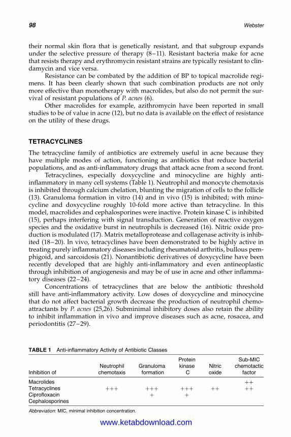
DK4962-Webster-ch8_R2_020407
their normal skin flora that is genetically resistant, and that subgroup expandsunder the selective pressure of therapy (8–11). Resistant bacteria make for acnethat resists therapy and erythromycin resistant strains are typically resistant to clin-damycin and vice versa.
Resistance can be combated by the addition of BP to topical macrolide regi-mens. It has been clearly shown that such combination products are not onlymore effective than monotherapy with macrolides, but also do not permit the sur-vival of resistant populations of P. acnes (6).
Other macrolides for example, azithromycin have been reported in smallstudies to be of value in acne (12), but no data is available on the effect of resistanceon the utility of these drugs.
TETRACYCLINES
The tetracycline family of antibiotics are extremely useful in acne because theyhave multiple modes of action, functioning as antibiotics that reduce bacterialpopulations, and as anti-inflammatory drugs that attack acne from a second front.
Tetracyclines, especially doxycycline and minocycline are highly anti-inflammatory in many cell systems (Table 1). Neutrophil and monocyte chemotaxisis inhibited through calcium chelation, blunting the migration of cells to the follicle(13). Granuloma formation in vitro (14) and in vivo (15) is inhibited; with mino-cycline and doxycycline roughly 10-fold more active than tetracycline. In thismodel, macrolides and cephalosporines were inactive. Protein kinase C is inhibited(15), perhaps interfering with signal transduction. Generation of reactive oxygenspecies and the oxidative burst in neutrophils is decreased (16). Nitric oxide pro-duction is modulated (17). Matrix metalloprotease and collagenase activity is inhib-ited (18–20). In vivo, tetracyclines have been demonstrated to be highly active intreating purely inflammatory diseases including rheumatoid arthritis, bullous pem-phigoid, and sarcoidosis (21). Nonantibiotic derivatives of doxycycline have beenrecently developed that are highly anti-inflammatory and even antineoplasticthrough inhibition of angiogenesis and may be of use in acne and other inflamma-tory diseases (22–24).
Concentrations of tetracyclines that are below the antibiotic thresholdstill have anti-inflammatory activity. Low doses of doxycycline and minocycinethat do not affect bacterial growth decrease the production of neutrophil chemo-attractants by P. acnes (25,26). Subminimal inhibitory doses also retain the abilityto inhibit inflammation in vivo and improve diseases such as acne, rosacea, andperiodontitis (27–29).
TABLE 1 Anti-inflammatory Activity of Antibiotic Classes
Inhibition ofNeutrophilchemotaxis
Granulomaformation
Proteinkinase
CNitricoxide
Sub-MICchemotactic
factor
Macrolides þþ
Tetracyclines þþþ þþþ þþþ þþ þþ
Ciprofloxacin þ þ
Cephalosporines
Abbreviation: MIC, minimal inhibition concentration.
98 Webster
www.ketabdownload.com
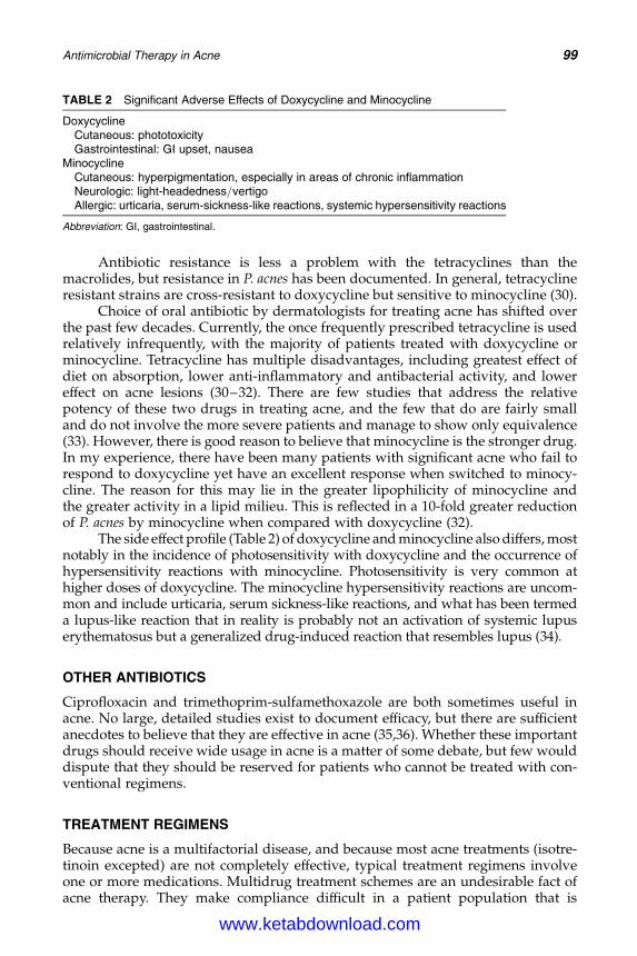
DK4962-Webster-ch8_R2_020407
Antibiotic resistance is less a problem with the tetracyclines than themacrolides, but resistance in P. acnes has been documented. In general, tetracyclineresistant strains are cross-resistant to doxycycline but sensitive to minocycline (30).
Choice of oral antibiotic by dermatologists for treating acne has shifted overthe past few decades. Currently, the once frequently prescribed tetracycline is usedrelatively infrequently, with the majority of patients treated with doxycycline orminocycline. Tetracycline has multiple disadvantages, including greatest effect ofdiet on absorption, lower anti-inflammatory and antibacterial activity, and lowereffect on acne lesions (30–32). There are few studies that address the relativepotency of these two drugs in treating acne, and the few that do are fairly smalland do not involve the more severe patients and manage to show only equivalence(33). However, there is good reason to believe that minocycline is the stronger drug.In my experience, there have been many patients with significant acne who fail torespond to doxycycline yet have an excellent response when switched to minocy-cline. The reason for this may lie in the greater lipophilicity of minocycline andthe greater activity in a lipid milieu. This is reflected in a 10-fold greater reductionof P. acnes by minocycline when compared with doxycycline (32).
The side effect profile (Table 2) of doxycycline and minocycline also differs, mostnotably in the incidence of photosensitivity with doxycycline and the occurrence ofhypersensitivity reactions with minocycline. Photosensitivity is very common athigher doses of doxycycline. The minocycline hypersensitivity reactions are uncom-mon and include urticaria, serum sickness-like reactions, and what has been termeda lupus-like reaction that in reality is probably not an activation of systemic lupuserythematosus but a generalized drug-induced reaction that resembles lupus (34).
OTHER ANTIBIOTICS
Ciprofloxacin and trimethoprim-sulfamethoxazole are both sometimes useful inacne. No large, detailed studies exist to document efficacy, but there are sufficientanecdotes to believe that they are effective in acne (35,36). Whether these importantdrugs should receive wide usage in acne is a matter of some debate, but few woulddispute that they should be reserved for patients who cannot be treated with con-ventional regimens.
TREATMENT REGIMENS
Because acne is a multifactorial disease, and because most acne treatments (isotre-tinoin excepted) are not completely effective, typical treatment regimens involveone or more medications. Multidrug treatment schemes are an undesirable fact ofacne therapy. They make compliance difficult in a patient population that is
TABLE 2 Significant Adverse Effects of Doxycycline and Minocycline
DoxycyclineCutaneous: phototoxicityGastrointestinal: GI upset, nausea
MinocyclineCutaneous: hyperpigmentation, especially in areas of chronic inflammationNeurologic: light-headedness/vertigoAllergic: urticaria, serum-sickness-like reactions, systemic hypersensitivity reactions
Abbreviation: GI, gastrointestinal.
Antimicrobial Therapy in Acne 99
www.ketabdownload.com
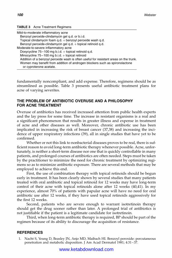
DK4962-Webster-ch8_R2_020407
fundamentally noncompliant, and add expense. Therefore, regimens should be asstreamlined as possible. Table 3 presents useful antibiotic treatment plans foracne of varying severites.
THE PROBLEM OF ANTIBIOTIC OVERUSE AND A PHILOSOPHYFOR ACNE TREATMENT
Overuse of antibiotics has received increased attention from public health expertsand the lay press for some time. The increase in resistant organisms is a real anda significant phenomenon that results in greater illness and expense in treatmentof acne and other diseases as well. Moreover, chronic antibiotic use has beenimplicated in increasing the risk of breast cancer (37,38) and increasing the inci-dence of upper respiratory infections (39), all in single studies that have yet to beconfirmed.
Whether or not this link to nonbacterial diseases proves to be real, there is suf-ficient reason to avoid long-term antibiotic therapy whenever possible. Acne, unfor-tunately, is neither a short-term disease nor one that is quickly controllable in manypatients, and prolonged courses of antibiotics are often needed. Steps must be takenby the practitioner to minimize the need for chronic treatment by optimizing regi-mens so as to minimize antibiotic exposure. There are several methods that may beemployed to achieve this end.
First, the use of combination therapy with topical retinoids should be begunearly in treatment. It has been clearly shown by several studies that many patientstreated with oral antibiotic and topical retinoid for 12 weeks may have long-termcontrol of their acne with topical retinoids alone after 12 weeks (40,41). In myexperience, almost 70% of patients with papular acne will have no need for oralantibiotic use after 12 weeks, if they have used topical retinoids aggressively forthe first 12 weeks.
Second, patients who are severe enough to warrant isotretinoin therapyshould get the drug sooner rather than later. A prolonged trial of antibiotics isnot justifiable if the patient is a legitimate candidate for isotretinoin.
Third, when long-term antibiotic therapy is required, BP should be part of theregimen because of its ability to discourage the acquisition of resistance.
REFERENCES
1. Nacht S, Yeung D, Beasley JN, Anjo MD, Maibach HI. Benzoyl peroxide: percutaneouspenetration and metabolic disposition. J Am Acad Dermatol 1981; 4:31–37.
TABLE 3 Acne Treatment Regimens
Mild-to-moderate inflammatory acneBenzoyl peroxide-clindamycin gel q.d. or b.i.d.Topical clindamycin foam q.d.þ benzoyl peroxide wash q.d.Benzoyl peroxide-clindamycin gel q.d.þ topical retinoid q.d.
Moderate-to-severe inflammatory acneDoxycycline 75–100 mg b.i.d.þ topical retinoid q.d.Minocycline 75–100 mg b.i.d.þ topical retinoidAddition of a benzoyl peroxide wash is often useful for resistant areas on the trunk.Women may benefit from addition of androgen blockers such as spironolactone
or cyproterone acetate.
100 Webster
www.ketabdownload.com
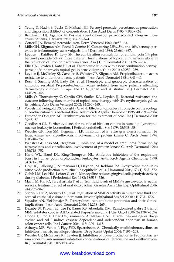
DK4962-Webster-ch8_R2_020407
2. Yeung D, Nacht S, Bucks D, Maibach HI. Benzoyl peroxide: percutaneous penetrationand disposition II:Effect of concentration. J Am Acad Derm 1983; 9:920–924.
3. Bandmann HJ, Agathos M. Post-therapeutic benzoyl peroxidecontact allergyin ulcuscruris patients. Hautarzt 1985; 36:670–674.
4. Cotterill JA. Benzoyl peroxide. Acta Derm Venereol 1980; 60:57–63.5. Mills OH, Kligman AM, Pochi P, Comite H. Comparing 2.5%, 5%, and 10% benzoyl per-
oxide in inflammatory acne vulgaris. Int J Dermatol 1986; 25:664–667.6. Leyden J, Kaidbey K, Levy SF. The combination formulation of clindamycin 1% plus
benzoyl peroxide 5% vs. three different formulations of topical clindamycin alone inthe reduction of Propionibacterium acnes. Am J Clin Dermatol 2001; 4:263–266.
7. Ellis CN, Leyden J, Katz HI, et al. Therapeutic studies with a new combination benzoylperoxide/clindamycin topical gel in acne vulgaris. Cutis 2001; 67:257–259.
8. Leyden JJ, McGinley KJ, Cavelieri S, Webster GF, Kligman AM. Propionibacterium acnesresistance to antibiotics in acne patients. J Am Acad Dermatol 1984; 8:41–45.
9. Ross JI, Snelling AM, Eady EA, et al. Phenotypic and genotypic characterization ofantibiotic resistant Propionibacterium acnes isolated from acne patients attendingdermatology clinicsin Europe, the USA, Japan and Australia. Br J Dermatol 2001;144:339–346.
10. Mills O, Thornsberry C, Cardin CW, Smiles KA, Leyden JJ. Bacterial resistance andoutcome following three months of topical acne therapy with 2% erythromycin gel vs.its vehicle. Acta Derm Venereol 2002; 82:260–265.
11. Vowels BR, Feingold DS, Sloughfy C, et al. Effects of topical erythromycin on the ecologyof aerobic cutaneous bacterial flora. Antimicrob Agents Chemother 1996; 40:2598–2604.
12. Fernandez-Obregon AC. Azithromycin for the treatment of acne. Int J Dermatol 2000;39:45–50.
13. Goodheart GL. Further evidence for the role of bivalent cations in human polymorpho-nuclear leukocyte locomotion. J Reticuloendothelial Soc 1979; 25:545–554.
14. Webster GF, Toso SM, Hegemann LR. Inhibition of in vitro granuloma formation bytetracyclines and ciprofloxacin: involvement of protein kinase C. Arch Derm 1994;130:748–752.
15. Webster GF, Toso SM, Hegeman L. Inhibition of a model of granuloma formation bytetracyclines and ciprofloxacin: involvement of protein kinase C. Arch Dermatol 1994;130:748–752.
16. Hand WL, Hand DL, King-Thompson NL. Antibiotic inhibition of the respiratoryburst in human polymorphonuclear leukocytes. Antimicrob Agents Chemother 1990;34:321–330.
17. Hoyt JC, Ballering J, Numanami H, Hayden JM, Robbins RA. Doxycycline modulatesnitric oxide production in murine lung epithelial cells. J Immunol 2006; 176(1): 567–572.
18. Golub LM, Lee HM, Lehrer G, et al. Minocycline reduces gingival collagenolytic activityduring diabetes. J Periodontal Res 1983; 18:516–526.
19. Maata M, Kari O, Tervahartiala T, et al. Tear fluid levels of MMP-8 are elevated in ocularrosacea: treatment effect of oral doxycycline. Graefes Arch Clin Exp Ophthalmol 2006;244:957–962.
20. Sobrin L, Liu Z, Monroy DC, et al. Regulation of MMP-9 activity in human tear fluid andcorneal epithelial culture supernatant. Invest Ophthalmol Vis Sci 2000; 41:1703–1709.
21. Sapadin AN, Fleishmajer R. Tetracyclines: non-antibiotic properties and their clinicalimplications. J Am Acad Dermatol 2006; 54:258–265.
22. Dezube BJ, Krown SE, Lee JY, Bauer KS, Aboulafia DM. Ramdomized pahse 2 trial ofMMP inhibitor col-3 in AIDS-related Kaposi’s sarcoma. J Clin Oncol 2006; 24:1389–1394.
23. Onoda T, Ono T, Dhar DK, Yamoanoi A, Nagasue N. Tatracyclines analogues doxy-cycline and col 3 induce caspase dependent and independent apoptosis in humancolon cancer cells. Int J Cancer 2006; 118:1309–1315.
24. Acharya MR, Venitz J, Figg WD, Sparreboom A. Chemically modifiedtetracyclines asinhibitors f matrix metalloproteinases. Drug Resist Updat 2004; 7:195–208.
25. Webster GF, McGinlery KJ, Leyden JJ. Inhibition of lipase production in Propionibacter-ium acnes by sub minimal inhibitory concentrations of tetracycline and erythromycin.Br J Dermatol 1981; 105:453–457.
Antimicrobial Therapy in Acne 101
www.ketabdownload.com
DK4962-Webster-ch8_R2_020407
26. Webster GF, Leyden JJ, McGinley KJ, McArthur WP. Suppression of polymorphonuclearleukocyte chemotactic factor production by sub minimal inhibitory concentrations oftetracycline, minocycline, ampicillin and erythromycin. Antimicrob Agents Chemother1982; 21:770–772.
27. Sanchez J, Somolinos AL, Almodovar PI, Webster G, Bradshaw M, Powala C. A random-ized, double-blind, placebo-controlled trial of the combined effect of doxycycline hyclate20-mg tablets and metronidazole 0.75% topical lotion in the treatment of rosacea. J AmAcad Dermatol 2005; 53(5):791–797.
28. Skidmore R, Kovach R, Walker C, et al. Effects of subantimicrobial-dose doxycycline inthe treatment of moderate acne. Arch Dermatol 2003; 139(4):459–464.
29. Emingil G, Atilla G, Sorsa T, Luoto H, Kirilmaz L, Baylas H. The effect of adjunctive low-dose doxycycline therapy on clinical parameters and gingival crevicular fluid matrixmetalloproteinase-8 levels in chronic periodontitis. J Periodontol 2004; 75(1):106–115.
30. Leyden JJ. Current issues in antimicrobial therapy for the treatment of acne. J Eur AcadDermatol Venereol 2001; 15(suppl 3):51–55.
31. Hubbell CG, Hobbs ER, Rist T, White JW Jr. Efficacy of minocycline compared with tetra-cycline in treatment of acne vulgaris. Arch Dermatol 1982; 118(12):989–992.
32. Leyden JJ. The antimicrobial effects in vivo of minocycline, doxycycline and tetracyclinein humans. J Dermatolog Treat 1996; 7:223–225.
33. Harrison PV. A comparison of doxycycline and minocycline in the treatment of acnevulgaris. Clin Exp Dermatol 1988; 13:242–244.
34. Elkayam O, Yaron M, Caspi D. Minocycline-induced autoimmune syndromes: an over-view. Semin Arthritis Rheum 1999; 28(6):392–397.
35. Bottomly WW, Cunliffe WJ. Oral trimethoprim as a third line antibiotic in the manage-ment of acne vulgaris. Dermatology 1993; 187:193–196.
36. Sykes NL, Webster GF. Acne: a review of optimum treatment. Drugs 1994; 48:59–70.37. Velicer CM, Heckbert SR, Rutter C, Lampe JW, Malone K. Association between antibiotic
use prior to breast cancer diagnosis and breast tumor characteristics. Cancer CausesControl 2006; 17:307–313.
38. Velicer CM, Heckbert SR, Lampe JW, Potter JD, Robertson CA, Taplin SH. Antibiotic usein relation to the risk of breast cancer. JAMA 2004; 291:827–835.
39. Margolis DJ, Bowe WP, Hoffstad O, Berlin JA. Antibiotic treatment of acne may be associatedwith upper respiratory tract infections. Arch Dermatol 2005; 141(9):1132–1136.
40. Leyden J, Thiboutot DM, Shalita AR, et al. Comparison of tazarotene and minocyclinemaintenance therapies in acne vulgaris: a multicenter, double-blind, randomized,parallel-group study. Arch Dermatol 2006; 142(5):605–612.
41. Thiboutot DM, Shalita AR, Yamauchi PS, et al. Adapalene gel, 0.1%, as maintenancetherapy for acne vulgaris: a randomized, controlled, investigator-blind follow-up of arecent combination study. Arch Dermatol 2006; 142(5):597–602.
102 Webster
www.ketabdownload.com
DK4962-Webster-ch9_R2_030407
B9 Topical Retinoids
Daniela Kroshinsky and Alan R. ShalitaDepartment of Dermatology, SUNY Downstate Medical Center, Brooklyn,New York, U.S.A.
INTRODUCTION
Topical retinoids are singularly important agents in the treatment of acne vulgaris.Any molecule having a biological effect through the binding and activation ofretinoid receptors is considered a retinoid (1). This class of medications includesvitamin A and all synthesized molecules that are derived from it (1). By influencingDNA transcription, retinoids can modify cellular growth and differentiation,immunomodulation, and tumor promotion. In turn, they improve acne vulgarisby inhibiting microcomedone formation, diminishing the number of mature come-dones as well as inflammatory lesions, and normalizing follicular epithelium matu-ration and desquamation (2).
CLASSIFICATION
Three generations of retinoids exist. First-generation topical retinoids includevitamin A and its derivatives retinaldehyde, all-trans-retinoic acid (all-trans-RA)or tretinoin, and 13-cis-retinoic acid (13-cis-RA) or isotretinoin (3). The second gen-eration retinoids comprise synthetic analogs where one aspect of the basic vitaminA structure has been changed, such as etretinate and acitretin, though no topicalforms of these forms exist (3). Third-generation retinoids have significant modifi-cation of the original molecule, such as adapalene, tazarotene, arotinoid, arotinoidmethyl sulfone, and arotinoid ethyl ester (3). Presently, there are seven topical reti-noids: tretinoin, adapalene, tazarotene, topical isotretinoin, motretinide, retinalde-hyde, and b-retinoyl glucuronide (3). Only all-trans-RA, adapalene, and tazaroteneare available in the United States and are discussed in the text that follows.
ABSORPTION
Absorption of topical medications occurs transepidermally or transfollicularly,influenced by the size of the particles involved (4). Molecules 3 to 10 mm in diameterpenetrate the follicular ducts, making this size the ideal target size for acne therapy(4). Particles larger than 10 mm become trapped on the skin surface, whereas par-ticles less than 3 mm disperse over the stratum corneum and hair follicles andare, therefore, less efficacious (4).
MECHANISM OF ACTION
Topical retinoids act to clear and prevent the formation of the microcomedo, theprecursor to acneiform lesions. The microcomedo is formed from the occlusion ofthe follicular ostium by the androgen-induced production of sebum and the
103
www.ketabdownload.com
DK4962-Webster-ch9_R2_030407
accumulation of stratum corneum cells (5). In normal skin, the corneocytes of thehair follicle’s infrainfundibular region are small and form a noncontinuous, inco-herent layer of cells that easily desquamate individually into the follicular canal(1,2,6). They then travel to the surface of the skin through the secretion of lipid-rich sebum (6). In contrast, the follicular epithelium of the microcomedo demon-strates abnormal, hyperactive keratinization, resulting in hypergranulosis andhyperkeratosis (2,5). Corneocytes are more cohesive and less able to migrate tothe skin surface and instead become lodged within the follicle, occluding the expul-sion of sebum, distending the follicular ostia, and thus forming the comedo (1,6).The anaerobic environment created therein favors the proliferation of Proprinibac-terium acnes (1,6), which subsequently creates an inflammatory response by secret-ing lipase, releasing chemotactic factors, and recruiting polymorphonuclearlymphocytes (PMNs) (1,2,6). Bacterial lipases break down sebum triglyceridesinto glycerol, a growth factor for the bacteria, and free fatty acids that are comedo-genic and proinflammatory (1,6). The lipases and cytokines produced by therecruited PMNs each contribute to the breakdown of the follicular wall, eventuallycausing microcomedo rupture. The release of sebum, keratin, and free fatty acidsinduces an inflammatory foreign body reaction, resulting in the formation ofinflammatory papules, pustules, and nodules (1,5,6). Therefore, by targeting themicrocomedo, topical retinoids also help prevent the formation of inflammatorylesions that have the potential to heal with scarring. By preventing the formationof new microcomedones, topical retinoids produce acne remissions that can be suc-cessfully maintained for extended periods of time (2,7). These effects occur in whathas be referred to as the “ideal state,” in which retinoids could be used in sufficientconcentrations and frequencies to obtain the desired effect. Unfortunately, for manypatients, the irritant side effects of these drugs—erythema, xerosis, burning,desquamation—can impede 100% adherence and improvement (2). Unlike oral reti-noids, the topical retinoids do not decrease production of sebum, but instead act bydecreasing inflammation, normalizing keratinocyte differentiation, and increasingkeratinocyte proliferation and migration (8,9).
Receptor ActionTopical retinoids regulate gene transcription through an interaction with nuclearretinoid receptors. Nuclear retinoid receptors are ligand-dependent transcriptionfactors that regulate the transcription of target genes in two ways (10). One mech-anism of action is via the direct binding to retinoid receptor cognate hormoneresponse elements, direct repeats of six nucleotides, found in the promoterregions of retinoid-receptive genes (10,11). The second means of transcription modi-fication is the antagonism of nuclear transcription factors that bind to alternateresponse elements (AREs), thereby preventing the expression of genes regulatedby these AREs (10,11). For instance, when a retinoid binds to the retinoic acid recep-tor (RAR) subtype complexed with the jun/fos transcription factors, it disrupts theinteraction between jun/fos and the ARE, activator protein-1 (AP-1), and preventstranscription of AP-1 regulated genes (11). It is thought that this results in the anti-proliferative and anti-inflammatory effects seen with some of the retinoids (10–12).
Retinoid receptors, like thyroid hormone receptors, vitamin D receptors, andsteroid receptors, are classified as nuclear receptors (12). There are two classes ofnuclear retinoid receptors: RARs and retinoid X receptor (RXRs) (12). Each classof receptors consist of three subtypes: alpha, beta, and gamma, and in vivo, these
104 Kroshinsky and Shalita
www.ketabdownload.com
DK4962-Webster-ch9_R2_030407
receptors exist as dimers (12,13). RXRs can dimerize with RAR, thyroid hormonereceptors, and vitamin D receptors. RARs, however, can only dimerize with RXR(12). RAR alpha subunits are found ubiquitously, RAR gamma subunits arefound predominantly in the skin, and RAR beta subtypes are inducible by retinoidsonly in certain tissues, including the skin (6,10). RAR and RXR subtypes bind asheterodimers along with the retinoid ligand to the cognate hormone responseelements. Each retinoid holds a different affinity for each receptor subtype. Theclinical response to each retinoid differs on the basis of the affinity for specific recep-tors, the expression pattern of receptor subtypes, and the cognate response element(10,11). Gamma receptor is the most important receptor involved in epidermaldifferentiation, and tretinoin has a high affinity for these receptors.
Retinoid TypesThe first developed topical retinoid was retinoic acid, or tretinoin. Tretinoin’s flex-ible molecular structure allows it to bind all RAR and RXR subtypes, whereas tazar-otene and adapalene are more rigid, and therefore demonstrate receptor specificity(13). Tretinoin also has the capacity to bind cytosolic retinoid-activating bindingproteins (CRABPs) that exist in two forms, I and II, and act as buffers for retinoicacid by controlling free intracellular concentration (3,10,11). Retinoid potency corre-lates with affinity for the RARs but not for CRABPs (11).
Tretinoin is available as 0.025%, 0.05%, and 0.1% creams, 0.01% and 0.025%gels, and 0.05% solution, with 0.025% cream having the least and 0.05% solutionhaving the most irritation potential (2,3,6). Newer microencapsulated tretinoingel and polyolprepolymer 2 gel forms are less irritating than other formulationsof tretinoin (2,6,14). The tretinoin microsphere, available in 0.4% and 0.1% gelforms, encapsulates the molecule within a porous acrylate copolyer microsphere(2,15). This packaging system allows selective, time-released delivery of tretinointo the follicle, leading to a decreased available concentration and lower irritationwithout diminishing the efficacy (2,15). In fact, the maximum comedolytic activityof tretinoin is reached at a concentration 5 to 10 times lower when the drug is usedin a microsphere form (15). In addition, potential percutaneous penetration to theblood vessels is diminished (2,15).
Similarly, novel vehicles can allow controlled release of topical retinoids.Polyolprepolymer-2 is a material designed to help retain drug molecules in andon the skin when applied topically and has been shown to distribute tretinoinover time with equivalent efficacy to vehicle-free analogous formulations (16).The incorporation of tretinoin into this vehicle prevents rapid and excess percuta-neous absorption of tretinoin, decreasing irritation (16). In vitro absorption studies,guinea pig irritation models, human patch test studies, and acne clinical trials of tre-tinoin gel and cream containing polyolprepolymer-2 demonstrate that the absorp-tion of tretinoin during the first six hours of delivery is significantly less than that ofstandard preparations of tretinoin, but becomes similar after this time (16).Additionally, the total penetration of polyolprepolymer-impregnated tretinoin isless than vehicle-free tretinoin (16). Human patch tests demonstrated that both tre-tinoin gel and cream formulated with polyolprepolymer-2 resulted in less irritationwhencomparedtotheir tretinoingelandcreamforms(16).Humandouble-blindclini-cal trials comparing tretinoin 0.025% gel and cream containing polyolprepolymer-2to their commercially available equivalents demonstrated diminished xerosis,erythema, and peeling in the tretinoin polyolpolymer gel and cream formulations
Topical Retinoids 105
www.ketabdownload.com
DK4962-Webster-ch9_R2_030407
(16). This decrease in irritation was achieved without compromising treatment effi-cacy (16). Although the exact mechanism for these actions is unknown, one pro-posed mechanism is that the degree of irritation from tretinoin is related to therate of initial penetration of drug and while the efficacy is more a consequence ofmean molecular flux (16).
The second topical retinoid developed for the treatment of acne is adapalene,a stable naphthoic acid derivative with both significant anti-inflammatory andcomedolytic properties (6,11,17). Adapalene has been shown to have a more favor-able sideffect profile than either tazarotene or tretinoin (6,17). It has a specific affi-nity for RAR beta and gamma subtypes in the terminal differentiation zone ofthe epidermis and does not bind to CRABP I and II due to steric hindrance(3,6,11,13,18,19). Adapalene treats acne by causing epidermal and follicularepithelium hyperplasia, increased desquamation, keratinocyte differentiation,and loosening of corneocyte connections (11,14,18).
Adapalene is available as 0.1% cream, gel, solution, and pledget forms and isin clinical trials as a newer 0.3% gel formulation (2,20). The 0.1% gel has been shownto be as effective as tretinoin 0.025% gel with decreased irritation (2,20). The 0.3%gel has been shown to be safe and well tolerated for long-term use with almostdoubled success over adapalene 0.1% gel without a significant increase in clinicallyrelevant side effects (20). The release of adapalene from solution formulations suchas lotions and hydroalcoholic gels is greater than when dispersed in creams oraqueous gels (18,21).
The anti-inflammatory effects of adapalene result from inhibition of the oxi-dative metabolism of arachidonic acid by the 5 and 15 lipoxygenase pathways andthrough the inhibition of PMN chemotactic and chemokinetic responses (3,11). Assuch, adapalene has demonstrated superiority when compared to reference anti-inflammatory agents such as indomethacin and betamethasone valerate (22). Othercontributing factors for the lower skin irritation induced by adapalene include itsdifferent RAR subtype-binding pattern and more neutral molecular structure,which makes it less likely to nonspecifically interact with cell membrane function(3). Its high melting point and low solubility result in low flux through the skin,leading to high concentrations in the stratum corneum and hair follicle, where itcan best act to prevent and treat acne (11).
Tazarotene is a topical retinoid that has demonstrated superior efficacy whencompared with other topical retinoids in the treatment of acne (23). Tazarotene isavailable in 0.05% and 0.1% cream and gel formulations (2). Tazarotene is selectivefor RAR beta and gamma subtypes (13). Although tazarotene does not directly bindto the RXRs, it does isomerize to 9-cis-RA, which weakly binds all RXRs leading to adegree of transactivation (21). The precise mechanism of action of tazarotene isunknown, however, it is postulated that through the activation of retinoid recep-tors, retinoid responsive genes are upregulated impacting the differentiation ofkeratinocytes. Similarly, by inhibiting proinflammatory transcription factors,tazarotene causes decreased cell proliferation and inflammation (21). Tazarotenepenetrates the skin but accumulates in the upper dermis (24). The moleculeremains concentrated here with very little absorption into blood vessels orlymphatics (24).
The topical retinoids induce specific changes in the structure and morphologyof the skin. Normal epithelial cell differentiation is a vitamin A-dependent process(25). The presence of vitamin A alters the size of the keratin molecules synthesizedby keratinocytes (25). When small keratin molecules are produced, a secretory-type
106 Kroshinsky and Shalita
www.ketabdownload.com
DK4962-Webster-ch9_R2_030407
epithelium is generated. A stratified squamous epithelium is induced when largerkeratin molecules are synthesized (25). In the absence of vitamin A, cells differen-tiate toward keratinizing epithelium (25).
Keratinocytes within each layer of epidermis possess a distinct set of proteinsunique to their stage of differentiation (26). As undifferentiated basal cells matureand migrate up through the epidermis, their pattern of protein expression changes(26). Once these cells arrive at the stratum corneum, the intracellular organelleshave been catabolized and cytoplasmic and membrane proteins altered so as toproduce a layer of dead corneocytes with thick keratin fibrils within a densematrix (26). As a group, the retinoids alter this process by inducing dose-specific ker-atinocyte proliferation and reversing the abnormal keratinocyte desquamationfound in acne (26). This helps form a thicker layer of suprabasal cells, but athinned, loose stratum corneum (2,3,26). Topical retinoids increase the rate of kerati-nocyte turnover resulting in an increased rate of follicular proliferation and differen-tiation (24). This results in decreased follicular occlusion and faster microcomedoneclearance (3,24). Specifically, topical retinoids cause basal and spinous epithelial cellproliferation and reduce the size and adhesion of corneocytes by decreasing filaggrinexpression and suppressing the normal proteolysis of keratins 1 and 14 (1). Inaddition, retinoids thin the stratum corneum by causing desmosomal shedding,decreasing tonofilaments, and increasing keratinocyte autolysis (1,8,27).
Epidermal cell commitment to proliferation and differentiation can be alteredby the topical retinoids. Normal keratinocyte differentiation involves the expressionof keratins K1 and K10 (26). An alternative pathway of differentiation compatiblewith hyperproliferation occurs upon suprabasal expression of K6, K16, and K17(26). Cells expressing K6 and K16 enlarge as they migrate to the surface and donot produce keratohyalin granules or lose their nuclei before desquamating (26).
The dose-dependent epidermal acanthosis results from increased numberof cell layers, not from hypertrophy of individual cells (9). There is no change instructure or size of sebaceous glands after treatment with topical retinoids (9).All of the above factors demonstrate that comedolysis is retinoid-specific, notsimply secondary to desquamation (3).
Retinoids also have secondary effects that facilitate acne clearance. By weak-ening and loosening the cornified layer and decreasing the number of corneocytes,skin permeability is increased (2). This facilitates the absorption of other topicalagents, such as antimicrobials or benzoyl peroxide (2). Increased cell turnoverwithin the follicular epithelium also allows more oral antibiotic to enter the follicu-lar canal where P. acnes is concentrated (2). This increases antimicrobial activity,lowering the potential for antibiotic resistance from inadequate concentrations(2). By increasing the rate of acne clearance, the overall duration of antibiotic treat-ment and chance for antibiotic resistance can be reduced (2,6). In addition, thismodification of skin morphology creates a more aerobic environment and decreasesthe ability of P. acnes to proliferate (2).
USE AND SIDE EFFECTS
Proper selection and usage of the topical retinoids is crucial to maximize efficacyand patient adherence and satisfaction. Treatment should begin with the lowestconcentration of medication, usually in a cream-based form if available (21). Appli-cation should occur at night to dry skin, 20 to 30 minutes after the face hasbeen washed with a mild, nonsoap cleanser (21). A pea-sized amount should be
Topical Retinoids 107
www.ketabdownload.com
DK4962-Webster-ch9_R2_030407
dispensed and equally divided over two index fingers, which then dab the medi-cation evenly onto opposite sides of the face and spread the medication into athin layer until no visible product remains (21). Hands should be washed after-wards to avoid retinoid dermatitis (21). Application should first occur everyother night for one to two weeks depending on the skin type so as to minimizethe initial irritation that may otherwise discourage adherence to the treatmentregimen (1,21). Oily skin is better able to tolerate the potential irritating effects ofthe retinoids and, as such, a shorter introductory period may be utilized (21).Non-comedogenic moisturizers can be used to minimize xerosis, erythema, andstinging (2). Patients should be instructed that as long as four to six weeks of usemay be required before the onset of efficacy and that an initial flare in acne mayoccur following two to four weeks of use due to an accelerated evolution of pre-existing microcomedones (3). Periodic encouragement and reassurance may benecessary. Four to six weeks of nightly application should occur, after which theconcentration may be increased if the desired results are not obtained (21). Anadditional benefit to topical retinoid use is the potential to improve and preventthe postinflammatory hyperpigmentation often seen in darker skin types as acnelesions heal.
MINIMIZING SIDE EFFECTS
Side effects of topical retinoid use include initial local irritation, includingerythema, burning, stinging, peeling, and xerosis (1). These symptoms usuallypeak after two weeks of use, and subsequently diminish and then resolve oncethe skin adapts to the use of the product (1,3,18). Factors influencing the extentand duration of irritation include concentration of the medication used, vehicleof delivery, frequency and amount of application, skin type, and environmentalfactors such as use of abrasive cleansers or other topical alcoholic agents,ambient xerosis, and exposure to the sun (1,6,14,18,28). Higher concentratationsor use of a gel or solution vehicle predisposes to the most irritation. Topicalretinoids produce more irritation when used by patients with eczema, rosacea, orother conditions of skin sensitivity, including exposure to extreme weather (2,14).In these patients and during the winter, lower concentrations and the milderforms and vehicles should be utilized (2,28). Individual patient variationdoes, however, occur and some patients may actually tolerate gels better thancreams, perhaps because they are more difficult to apply. Adapalene and tretinoinare photoirritants, not photosensitizers, meaning that there is an increasedsusceptibility to irritation upon sun exposure, and therefore minimizing sunexposure through sun avoidance and the use of sunscreen or physical blockers isimperative (14,28).
For patients who are unable to tolerate even the gradual introduction of themildest topical retinoids, short contact therapy can be utilized. Short contacttherapy is a safe and effective method that involves applying the topical agentfor a brief period and then washing it off (13). Patients should initially leave the reti-noid on the skin for two minutes each day, increasing by one minute at least everythree days as tolerated and without causing adverse effects (13). If irritation occurs,time of exposure should be decreased by 30 seconds for at least three days and thenincremental increases can be resumed (13). Studies have demonstrated somewhatdiminished efficacy than overnight tazarotene, but overall good results without irri-tation in patients who otherwise would be unable to benefit from topical retinoidtherapy (13).
108 Kroshinsky and Shalita
www.ketabdownload.com
DK4962-Webster-ch9_R2_030407
VEHICLES
The type of vehicle used to deliver the retinoid influences the degree of skinpenetration, the clinical effect, and the side effect profile, including the degree ofphotoirritance (1,6). Future formulations suspending topical retinoids in a foamvehicle could potentially create a nongreasy, no-residue, less-drying formulationto more effectively deliver the medication over hair-bearing sites such as the backand chest (29).
EFFICACY
Acne vulgaris is an almost ubiquitous condition. When antimicrobials are usedalone, there is a risk of creating widespread, multidrug resistance of P. acnes aswell as other skin flora such as Staphylococcus aureus (2,5,30). This potential resist-ance can be limited by combining agents that act on different steps in the mechan-ism of acne pathogenesis. When topical retinoids are used in combination with oralor topical antimicrobials, excessive ductal cornification, P. acnes proliferation, andinflammation can be simultaneously targeted for increased efficacy, faster onsetof effects, decreased total antibiotic use and risk of resistance, and shorter overallduration of treatment (2). The combination tretinoin with topical or oral antibioticshas been shown to be superior to either alone in decreasing lesion count, increasingrate of improvement, and decreasing levels of P. acnes and free fatty acids (8,14).Similarly, the combination of adapalene with topical doxycycline or clindamycinshows increased efficacy in improving total, inflammatory, and noninflammatorylesions including a faster onset of action without an augmentation in side effects(17). In addition, combination use of adapalene offers an anti-inflammatorybenefit, which minimizes the increased risk of irritation that occurs upon increasingthe number of topical treatments (17).
Similar results were found upon combining retinoic acid and antibiotics, witha reduction in the initial retinoid-induced flare of lesions through the antibiotic use(3). When combined with a benzoyl peroxide oxidizing wash—which demonstratesan effective bacteriostatic concentration after only 20 seconds of use—tretinoin 0.1%micro gel decreased P. acnes counts without increasing irritation (6,31).
With long-term use, however, overall improvement is similar with combi-nation therapy as compared with topical retinoid use alone (14,23,32). Increasedreductions in open and closed comedones, papules, and pustules were demon-strated when tazarotene or tretinoin creams were combined with clindamycin/benzoyl peroxide as compared to either retinoid alone (14,32).
Finally, it has been shown that combining topical retinoid therapy with oralantibiotics for inflammatory acne is more cost-effective than using an oral retinoidalone when accounting for pretreatment and treatment laboratory tests, pregnancytests, and diagnostic fetal tests after a potential unintended pregnancy (33).
A new combination clindamycin 1%/tretinoin 0.025% gel, Velac, has demon-strated greater and faster treatment success in initial clinical trials when comparedto clindaymycin or tretinoin gel alone and provides a convenient means of appli-cation, which may improve patient adherence (12). The side effect profile of Velacwas similar to that of tretinoin gel alone, with only slightly higher irritancy thanclindamycin gel alone (12).
And as previously discussed, the use of topical retinoid in combination withoral antibiotics can decrease time of use of antibiotics, decreasing risk of resistance,adverse response, or drug interaction.
Topical Retinoids 109
www.ketabdownload.com
DK4962-Webster-ch9_R2_030407
SYSTEMIC ABSORPTION AND TERATOGENICITY
The teratogenic effects of oral retinoid therapy are well-documented. Tretinoinpenetrates the skin and accumulates in the upper dermis with very little absorptioninto blood vessels or lymphatics (24). Several studies have evaluated the systemiceffects of topical retinoid therapy. Tretinoin 0.1% to 0.2% applied for extendedperiods demonstrates minimal to no increase in tretinoin levels above endogenousand these level changes have not been associated with teratogenicity (1,21).Worobec et al. (34) studied the percutaneous absorption of 0.05% tretinoin byusing radioactive labeling and measuring the levels of retinoid found in theurine, stool, and plasma, after repeated applications. An absorption level of 1.38%to 2.13% was found, correlating to an insignificant increase in the endogenouslevel of tretinoin and a very low risk of systemic toxicity (34). When 0.1% tretinoincream, 0.1% Tazarotene gel, and 0.1% adapalene gel were applied over 26 days, therespective mean maximal plasma levels of were 4.7, 0.14, 0.04 ng/mL, which are allwell below the mean endogenous level of 6.6 ng/mL of all-trans-, 13-cis-þ 13-cis-4-oxo-RA (21). Other studies have also found that overnight tazarotene use produceslevels of tazarotene and tazarotenic acid levels lower than endogenous, with thehighest rate of absorption shown to occur after tazarotene was applied under occlu-sion for 10 hours resulting in ,6% absorption of total applied dose (13). In fact,diurnal and nutritional factors alter endogenous plasma levels of retinoids morethan widespread topical use of all-trans-RA (35). Buchan et al. (35) evaluated thedaily use of radiolabeled 0.025% all-trans-RA over the face, shoulder, and upperchest and demonstrated a percutaneous absorption in the range of 0.1% to 7.2%,considered to be an insignificant increase in endogenous levels. Daily ingestion ofthe 50,000 IU of vitamin A, however, tripled the endogenous level of all-trans-RA,doubled the level of 4-oxo-13-cis-RA, and increased 13-cis-RA six times (35). Therecommended daily allowance of vitamin A for male adults is 900 mg/day andfornonpregnant, nonlactating women 700 mg/day of vitamin A, with the recommendeddosage actually increasing to 770 mg/day in preganancy and to 1300 mg/day in lactation(36). On the basis of this information, it is very unlikely that topical retinoid use couldcause systemic toxicity.
Although there is a lack of studies on humans, topical retinoids havenot demonstrated teratogenic effects in animals (24,37,38). Nevertheless, tretinoinshould be used cautiously in pregnancy and lactation while tazarotene iscontraindicated.
CONCLUSIONS
Topical retinoid therapy is a safe, effective, economical means of treating all but themost severe cases of acne vulgaris. The retinoids serve as the cornerstone of treat-ment and should be the initial therapeutic step, either alone or in combination withtopical or oral antibiotic therapy and benzoyl peroxides. Once a patient hasachieved adequate results, the topical retinoid should be continued alone as main-tenance therapy to maintain lesion remission (2,20,33).
REFERENCES
1. Biro DE, Shalita AR. Clinical aspects of topical retinoids. Skin Pharmacol 1993; 6(suppl 1):53–60.
2. Gollnick H, Cunliffe W, Berson D, et al. Management of acne: a report from a global alli-ance to improve outcomes in acne. J Am Acad Dermatol 2003; 49:S1–S373.
110 Kroshinsky and Shalita
www.ketabdownload.com
DK4962-Webster-ch9_R2_030407
3. Verschoore M, Bouclier M, Czernielewski J, et al. Topical retinoids: their uses in derma-tology. Dermatol Clin 1993; 11:107–115.
4. Allec J, Chatelus A, Wagner N. Skin distribution and pharmaceutical aspects of adapa-lene gel. J Am Acad Dermatol 1997; 36:S119–S125.
5. Wolff H, Plewig G, Braun-Falco O. Ultrastructure of human sebaceous follicles andcomedones following treatment with vitamin A acid. Acta Dermatovenerol (Stockholm)1975; 74:99–110.
6. Brown SK, Shalita AR. Acne vulgaris. Lancet 1998; 351:1871–1876.7. Leyden JJ. A review of the use of combination therapies in the treatment of acne vulgaris.
J Amer Acad Dermatol 2003; 49(3 suppl):S200–S210.8. Leyden JJ. Retinoids and Acne. J Am Acad Dermatol 1988; 19:164–168.9. Asthon RE, Connor MJ, Lowe NJ. Histologic changes in the skin of the rhino mouse
induced by retinoids. J Invest Dermatol 1984; 82:632–636.10. Shalita A. Clinical Management of Topical Retinoids for Acne. A presentation at the
Annual Valley of the Sun Meeting, Pheonix, AZ 2005.11. Shroot B, Michel S. Pharmacology and chemistry of adapalene. J Am Acad Dermatol
1997; 36(6):S96–S103.12. Velac Phase III study efficacy results (combined analysis): primary outcome measures,
Data on File Connetics Laboratories.13. Bershad S, Kranjac Singer G, Parente JE, et al. Successful treatment of acne vulgaris using
a new method: results of a randomized vehicle-controlled trial of short-contact therapywith 0.1% tazarotene gel. Arch Dermatol 2002; 138:481–489.
14. Weiss JS, Shavin JS. Adapalene for the treatment of acne vulgaris. J Am Acad Dermatol1998; 39(2):50–54.
15. Dinarvand R, Rahmani E, Farbod E. Gelatin microspheres for the controlled releaseof all-trans-retinoic acid topical formulation and drug delivery evaluation. IranianJ Pharm Res 2003; 2:47 – 50.
16. Quigley JW, Bucks DA. Reduced skin irritation with tretinoin containing polyolprepoly-mer-2, a new topical tretinoin delivery system: a summary of preclinical and clinicalinvestigations. J Am Acad Dermatol 1998; 38(4):S5–S10.
17. Thiboutot D, Shalita A. Combination therapy with adapalene gel 0.1% and doxycyclinefor severe acne vulgaris: a multicenter, investigator-blind, randomized, controlled-study.Skinmed 2005; 4(3):138–146.
18. Brogden R, Goa K. Adapalene: a review of its pharmacological properties and clinicalpotential in the management of mild to moderate acne. Drugs 1997; 53(3):511–519.
19. Cunliffe WJ, Poncet M, Loesche C, et al. A comparison of the efficacy and tolerability ofadapalene 0.1% gel versus tretinoin 0.025% gel in patients with acne vulgaris: a meta-analysis of five randomized trials. Br J Dermatol 1998; 139(suppl 52):48–56.
20. Thiboutot, D. Clinical Experience With Adapalene 0.3% Gel, Presented at Satellite Sym-posium at the American Academy of Dermatology Meeting 2006, San Francisco, CA.
21. Topical Retinoids in the Treatment of Acne Vulgaris. A presentation.22. Hensby C, Cavey D, Bouclier M, et al. The in vivo and in vitro anti-inflammatory activity
of CD271: A new retinoids-like modulator of cell differentiation. Agents and Actions1990; 29:56–58.
23. Shalita AR. Tazarotene cream in facial acne vulgaris the best in acne trial: balancing effi-cacy, speed, and tolerability. Cutis 2004; 74(suppl 4):4–8.
24. Stuttgen G. Historical perspectives of tretinoin. J Am Acad Dermatol 1986; 15(4):735–740.25. Fuchs E, Green H. Regulation of terminal differentiation of cultured human keratino-
cytes by vitamin A. Cell 1981; 25:617–625.26. Eichener MK, Capetola RJ, Gendimenico GJ, Mezick JA. Cytoskeletal effects of topical
retinoids. J Invest Dermatol 1992; 98:154–161.27. Bolognia JL, Jorizzo JL, Rapini RP. Dermatology. 1st ed. New York: Mosby, 2003.28. Skov MJ, Quigley JW, Bucks DW. Topical delivery system for tretinoin: research and
clinical implications. J Pharm Sci 1997; 86(10):1138–1143.29. Shalita A. A novel clindamycin foam formulation for the treatment of acne vulgaris:
focus of phase three clinical trials, presented at the 2004 American Academy of Derma-tology Summer Meeting, New York, NY.
Topical Retinoids 111
www.ketabdownload.com
DK4962-Webster-ch9_R2_030407
30. Iyer S, Jones D. Community-acquired methicillin-resistant Staphylococcus aureus skininfection: a retrospective analysis of clinical presentation and treatment of a local out-break. J Am Acad Dermatol 2004; 50(6):854–858.
31. Shalita AR, Elyse SR, Anderson DN, et al. Compared efficacy and safety of tretinoin 0.1%microsphere gel alone and in combination with benzoyl peroxide 6% cleanser for thetreatment of acne vulgaris. Ther Clin 2003; 72:167–172.
32. Shalita A, Tanghetti E, Abramouits W, Solomon B. Tazarotene versus tazarotene plusclindamycin/benzoyl peroxide in the treatment of acne vulgaris. Abstract for a posterpresentation at the 63rd annual meeting of the American Academy of Dermatology,New Orleans, L.A. February 2005.
33. Harper J. The use of topical retinoids for inflammatory acne: a review of the pharmacoe-conomic considerations. Manag Care Interface 2005; 18(3):51–55.
34. Worobec SM, Wong FA, Tolman EL, et al. Percutaneous absorption of 3H-tretinoin innormal volunteers. J Invest Dermatol 1991; 257:574.
35. Buchan P, Eckhoff C, Caron D, et al. Repeated topical administration of all-trans-retinoicacid and plasma levels of retinoic acids in humans. J Am Acad Dermatol 1994;30:428–434.
36. RDA Dietary Reference Intakes: Vitamins website http://www.iom.edu/Object.File/Master/7/296/0.pdf
37. Latriano L, Tzimas G, Wong F, et al. The percutaneous absorption of topically appliedtretinoin and its effect on endogenous concentrations of tretinoin and its metabolitesafter single doses or long-term use. J Am Acad Dermatol 1997; 36:S37–S46.
38. Johnson EM. A risk assessment of topical tretinoin as a potential human develop-mental toxin based on animal and comparative human data. J Am Acad Dermatol1997; 36:S86–S90.
112 Kroshinsky and Shalita
www.ketabdownload.com
DK4962-Webster-ch10_R2_020407
B10 Phototherapy and Laser Therapy of Acne
Guy F. WebsterJefferson Medical College of Thomas Jefferson University, Philadelphia,Pennsylvania, U.S.A.
INTRODUCTION
Light-based treatments for acne are not new. A tan has long been known to help teenswith pimples, and it would not be surprising to find reports of acne phototherapydating from the 1930s. In my youth, a local dermatologist was treating many of uswith what must have been a cold quartz lamp along with various creams and oral anti-biotics. He told me that he knew that the light was helpful, but could not say morethan that about its mechanism. Since that time, there have been great advancesin the understanding of acne and in laser and light technology, but the level ofunderstanding of how light might help acne has not progressed much.
In the past 10 years or so, there have been many reports of different light-based acne regimens (1), but all are small (or very small) studies and most canbest be categorized as organized anecdote. The typical report describes a handfulof patients who have treatment with light plus antibiotics plus other interventions(such as peels). Controls, blinding, and randomization are lacking, and the photo-therapy regimen differs from patient to patient. It is tempting to dismiss the reportsas worthless, but that would be missing what could be an important new directionin acne treatment.
POTENTIAL TARGETS FOR LIGHT IN ACNEPropionibacterium acnesPropionibacterium acnes is an obvious target for acne phototherapy since it is centralto the inflammatory process. The organism makes porphyrins, which are present inthe follicle, in proportion to its population (2). These photoactive compounds can beexcited by light to generate reactive oxygen, which is toxic to P. acnes (3). Aninherent limitation is that to reduce P. acnes effectively, the therapy would have tobe given very frequently since the organism proliferates rapidly and wouldquickly repopulate after being reduced by a phototoxic reaction. How frequentlymight be necessary? There are no data available to directly answer the question,but one can predict in vitro from the organism’s generation time that populationswould double every roughly 30 minutes, and a P. acnes suppressive therapywould have to be given at least every two or three days.
A study by Pollock et al. (4) sheds some light on the potential mechanism ofporphyrin phototherapy in acne. They studied the effect of topically applied amino-levulinic acid (ALA) on acne, which is a precursor to porphyrins that are convertedby skin enzymes and is more potent than the effects of endogenous porphyrin. Theporphyrin was excited by a 635-nm diode laser. Ten patients had four areas of theirback treated with ALA alone, ALAþ light, light alone, or no treatment. After threetreatments over three weeks, the acne improvement was noted only in the ALAþ
113
www.ketabdownload.com
DK4962-Webster-ch10_R2_020407
light sites. Although there was a clear clinical benefit, sebum secretion and P. acnespopulations were measured to be unchanged.
Similarly, blue-light treatment of acne patients reduces P. acnes levels afterseveral treatments, but acne begins to improve before bacterial reduction can benoted (5). So, theoretically, and in acne trials, porphyrin-based photodynamictherapy is unlikely to exert its effects through reducing P. acnes.
Inhibition of Sebum ProductionSebum is, in a sense, the central problem in acne. Without it, P. acnes cannot prolifer-ate and acne would not exist. The most effective drug for the disease isotretinoinexerts the majority of its effects on sebum secretion. A light-based treatment thattargets sebum production would have the potential to cure acne. Could one dowithout sebaceous glands? Most likely. The function of sebum is unknown; itmay serve to inhibit invading bacteria such as dermatophytes and streptococci,but children do well with no sebum and adults have little-to-no sebaceous activityon the extremities with no ill effects.
The challenge in a therapeutic photon attack on the sebaceous gland is its dis-tance from the skin surface. Between the gland and the surface is up to 3 mm oftissue that can absorb light and be damaged. The key would seem to be todevelop a chromophore that homes to the sebaceous gland and then can be acti-vated by an otherwise innocuous wavelength of light.
Anderson et al. (6) have reported a series of 22 patients with acne of the back,who were sensitized aggressively with ALA and then irradiated with a potent doseof broadband (550–700 nm) light. Pain, erythema, and evidence of epidermaldamage were common. Patients experienced a flare of acne that was suppressedby following treatments. Although the adverse effects were significant, there washistologic evidence of damage to sebaceous glands and patients had suppressedsebum production for at least 20 weeks after treatment. Although the adverseeffects of this regimen are probably too severe for a practicable therapy, thereport serves as a wonderful proof-of-principle study, documenting that the sebac-eous gland can be destroyed by a limited number of light treatments. Similarly,Lloyd and Mirkov (7) demonstrated that indocyanine green accumulates in thesebaceous gland and can be activated by 810-nm light to damage the sebaceousgland, yet relatively spare the epidermis. All that remains is to develop a chromo-phore that is sebaceous gland specific, cosmetically practical, and excitable by awavelength that can be delivered several millimeters into the skin.
Modulation of KeratinizationFormation of the comedo results from defective keratinization in the epidermallining of the follicle and is the target of topical retinoids in acne. Could light alterthe epidermis sufficiently to stop comedo formation and thus halt acne? So far,there is no evidence that this might be possible, but no theoretical reason why itmight not.
Modulation of Immune ResponsivenessToll-like receptors (TLRs) have been shown to be involved in the development ofacne lesions (8). Might light-based techniques be able to improve acne throughmodulation of epidermal innate immunity? There is some evidence that this maybe possible. It is well-described that sun and phototherapy decrease the reactivity
114 Webster
www.ketabdownload.com
DK4962-Webster-ch10_R2_020407
of epidermal Langerhans cells (9), which could be involved in acne inflammation atsome level. Omi et al. (10) have shown that a nonablative 585-nm laser increases IL-2 and IL-4. The relevance to acne of this observation is not certain.
Schnitkind et al. (5) studied the anti-inflammatory properties of narrow bandblue light on cultured keratinocytes. They found that cytokine-induced productionof IL-1a and ICAM-1 was inhibited, which gives credence to the concept thatcurrent phototherapy techniques exert an anti-inflammatory effect in acne.
NOTABLE OBSERVATIONS AND STUDIES
In contrast to a loosely controlled study showing that a single low-dose, nonpur-puric treatment with the pulsed-dye laser resulted in acne improvement up to12 weeks after therapy (11), Orringer et al. (12) performed a blinded split-facestudy of similar low-dose pulsed-dye laser therapy for acne and found no effecton the number of lesions or on the clinical grade of the acne.
Alexiades-Armenakas (13) reported a series of acne patients, among whom13 patients with what was described as severe or cystic acne were treated withlong-pulsed 585-nm pulsed-dye laser and ALA. All patients received either topicalretinoids or azaleic acid and either a sulfur- or benzoyl peroxide-based cleanser.All patients in this group achieved a complete clearance after a mean of 2.9 treat-ments and a mean follow-up of 6.9 months. Adverse effects were minimal andlimited to erythema and “slight” pain. Several clinical photographs were shown doc-umenting clearance of significant inflammatory acne [but acne that falls short (inthese pictures) of what many would usually classify as “cystic”]. In spite of quibblingabout the definition of cystic acne, and in spite of the use of other medications such astopical retinoids and benzoyl peroxide, it is clear that in this report, the use of ALAphotodynamic therapy (PDT) resulted in an improvement in significant acne.
Weigell and Wulf (14) evaluated the efficacy of methyl aminolevulenate PDTon moderate-to-severe acne in a randomized, controlled trial. Twenty-one patientswere treated and 15 served as controls. Two treatments separated by two weekswere administered and acne graded at 4, 8, and 12 weeks. Improvement wasnoted at 4 weeks and reached statistical significance at 8 and 12 weeks aftertherapy. Most patients developed “severe” pain during the first treatment, fewercomplained during the second treatment. Exfoliation and crusting were common.
Although rigorous studies are limited in number, it is clear that there is greatpotential for a photodynamic approach to acne.
FUTURE DIRECTIONS FOR INVESTIGATION
Clinical trials where phototherapy is the only agent used. Even though most acne treat-ments are not used as a monotherapy in the clinic, in the development phase, it isimportant to test laser or PDT as monotherapy in order to ascertain whether it actu-ally has any effect on acne and to be able to define the best regimen.
Investigation of the ability of phototherapy to modulate inflammatory systems. Selec-tive modulation of TLRs in the skin, e.g., the TLR2 that is involved in acne, may bethe explanation for the effects of phototherapy on acne. If so, perhaps less traumaticregimens can be designed.
Development of sebaceous gland—specific phototherapy. This could be a magicbullet for acne and is theoretically possible. Perhaps, the biggest obstacle wouldbe the cost of development of a one-time treatment, which would have to be veryexpensive to offset development costs.
Phototherapy and Laser Therapy of Acne 115
www.ketabdownload.com
DK4962-Webster-ch10_R2_020407
REFERENCES
1. Ross EV. Optical treatments for acne. Dermatol Ther 2005; 18:253–266.2. McGinley KJ, Webster GF, Leyden JJ. Facial follicular porphyrin fluorescence: correlation
with age and density of Propionibacterium acnes. Br J Dermatol 1980; 102(4):437–441.3. Ashkenazi H, Malik Z, Harth Y, Nitzan Y. Eradication of Propionibacterium acnes by its
endogenic porphyrins after illumination with high intensity blue light. FEMS ImmunolMed Microbiol 2003; 35:17–24.
4. Pollock B, Turner D, Stringer MR, et al. Topical aminolaevulinic acid photodynamictherapy for the treatment of acne vulgaris: a study of clinical efficacy and mechanismof action. Br J Dermatol 2004; 151:616–622.
5. Schnitkind E, Yaping E, Geen S, Shalita AR, Lee W-L. Anti-inflammatory properties ofnarrow band blue light. J Drugs Dermatol 2006; 5:605–610.
6. Hongcharu W, Taylor CR, Chang Y, et al. Topical ALA-photodynamic therapy for thetreatment of acne vulgaris. J Invest Dermatol 2000; 115:183–192.
7. Lloyd JR, Mirkov M. Selective photothermolysis of sebaceous glands for acne treatment.Laser Surg Med 2002; 31:115–120.
8. McInturff JE, Kim J. The role of toll-like receptors in the pathophysiology of acne. SeminCutan Med Surg 2005; 24(2):73–78.
9. Belsito DV, Baer RL, Gigli I, Thorbecke GJ. Effect of combined topical glucocorticoidsand ultraviolet B irradiation on epidermal Langerhans cells. J Invest Dermatol 1984;83(5):347–351.
10. Omi T, Kawana S, Sato S, et al. Cutaneous immunological activation elicited by a lowfluence pulsed dye laser. Br J Dermatol 2005; 153(suppl 2):57–62.
11. Seaton ED, Charakida A, Mouser PE, et al. Pulsed dye laser treatment for inflammatoryacne vulgaris: randomized controlled trial. Lancet 2003; 362:1347–1352.
12. Orringer JS, Kang S, Hamilton T, et al. Treatment of acne vulgaris with a pulsed dyelaser: a randomized controlled approach. JAMA 2004; 291:2834–2839.
13. Alexiades-Armenakas M. Long pulsed dye laser mediated photodynamic therapy com-bined with topical therapy for mild to severe comedonal, inflammatory or cystic acne.J Drugs Dermatol 2006; 5:1–11.
14. Weigell SR, Wulf HC. Photodynamic therapy of acne vulgaris using methyl aminolevu-linate: a blinded, randomized, controlled trial. Br J Dermatol 2006; 154:969–976.
116 Webster
www.ketabdownload.com
DK4962-Webster-ch11_R2_240307
B11 Benzoyl Peroxide and Salicylic Acid Therapy
Gabi GrossDepartment of Research and Development, Reckitt Benckiser PLC, Hull, U.K.
INTRODUCTION
Nearly, everyone is affected by acne sometime in their lifetime. This is most preva-lent among teenagers when, shortly after the onset of puberty, spots and blackheadsstart to appear. However, acne may continue or start in adulthood and, indeed, it isreported that an increasing number of people within this age group are affected bythe condition (1).
When discussing acne, it is important to distinguish the varying degrees ofseverity of the disease because the selection of a therapeutic regime is dependenton this severity grading (Table 1). Pathophysiology and classification systems ofacne have been described in detail in other chapters; however, before starting todiscuss its therapy with benzoyl peroxide (BPO) and salicylic acid (SA), it is worthproviding a brief overview of this in order to understand acne active selection.
Acne, or acne vulgaris, is a common disease of the pilosebaceous unit. Thedisease is localized to the skin regions such as face, back, and chest, where numer-ous pilosebaceous units are located. The pathogenesis of acne has not yet been fullyestablished. Several factors contribute to the development of acne: increased sebumproduction (mainly influenced by hormones), ductal cornification, bacterial coloni-zation of the pilosebaceous duct, and inflammation (2–5). Propionibacterium acnes,an anaerobic bacteria, is a member of the skin and pilosebaceous follicle microflora.It is thought to play a significant role in acne because it can proliferate within theobstructed duct, modifying the components of sebum and producing enzymes,including lipases, that increase fatty acids and, consequently, cause inflammation.Inflammatory events, however, occur as a consequence of the bacterial colonizationand the subsequent reactions. Recent research revealed that inflammatory eventscould arise before or after hyperproliferative changes. There is evidence for vascu-lar endothelial cell activation and involvement of inflammatory responses in thevery earliest stages of acne lesion development (6). Whether a poor diet of excesschips or chocolate play a role in acne is rather questionable. Although this impacthas been observed in individual cases, no scientific evidence has been provided upto now (2–5). Another factor that is often suspected to exacerbate acne is psychologi-cal stress. In the past, reports of the influence of emotional stress were mainlyanecdotal. However, recently, a study revealed that changes in acne severity correlatehighly with increasing stress, suggesting that emotional stress from external sourcesmay have a significant influence on acne, which might be useful for understandingthe pathophysiology of acne and its therapy (7).
Because of increased sebum production and ductal cornification, the so-calledcomedo clinically develops first. Depending on whether the comedo is open orclosed determines whether they are called blackheads or whiteheads, respectively.If the comedo becomes inflamed, pustules, papules, or even nodules will develop,which are described as pimples or spots by most consumers.
117
www.ketabdownload.com
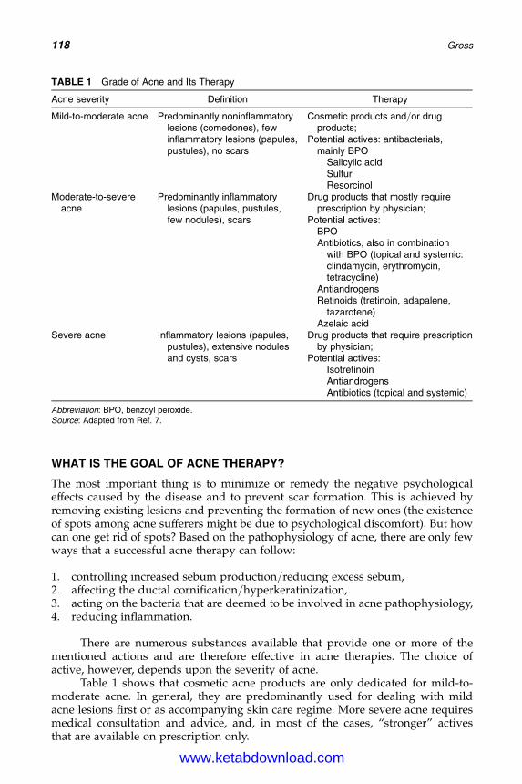
DK4962-Webster-ch11_R2_240307
WHAT IS THE GOAL OF ACNE THERAPY?
The most important thing is to minimize or remedy the negative psychologicaleffects caused by the disease and to prevent scar formation. This is achieved byremoving existing lesions and preventing the formation of new ones (the existenceof spots among acne sufferers might be due to psychological discomfort). But howcan one get rid of spots? Based on the pathophysiology of acne, there are only fewways that a successful acne therapy can follow:
1. controlling increased sebum production/reducing excess sebum,2. affecting the ductal cornification/hyperkeratinization,3. acting on the bacteria that are deemed to be involved in acne pathophysiology,4. reducing inflammation.
There are numerous substances available that provide one or more of thementioned actions and are therefore effective in acne therapies. The choice ofactive, however, depends upon the severity of acne.
Table 1 shows that cosmetic acne products are only dedicated for mild-to-moderate acne. In general, they are predominantly used for dealing with mildacne lesions first or as accompanying skin care regime. More severe acne requiresmedical consultation and advice, and, in most of the cases, “stronger” activesthat are available on prescription only.
TABLE 1 Grade of Acne and Its Therapy
Acne severity Definition Therapy
Mild-to-moderate acne Predominantly noninflammatorylesions (comedones), fewinflammatory lesions (papules,pustules), no scars
Cosmetic products and/or drugproducts;
Potential actives: antibacterials,mainly BPO
Salicylic acidSulfurResorcinol
Moderate-to-severeacne
Predominantly inflammatorylesions (papules, pustules,few nodules), scars
Drug products that mostly requireprescription by physician;
Potential actives:BPOAntibiotics, also in combination
with BPO (topical and systemic:clindamycin, erythromycin,tetracycline)
AntiandrogensRetinoids (tretinoin, adapalene,
tazarotene)Azelaic acid
Severe acne Inflammatory lesions (papules,pustules), extensive nodulesand cysts, scars
Drug products that require prescriptionby physician;
Potential actives:IsotretinoinAntiandrogensAntibiotics (topical and systemic)
Abbreviation: BPO, benzoyl peroxide.Source: Adapted from Ref. 7.
118 Gross
www.ketabdownload.com
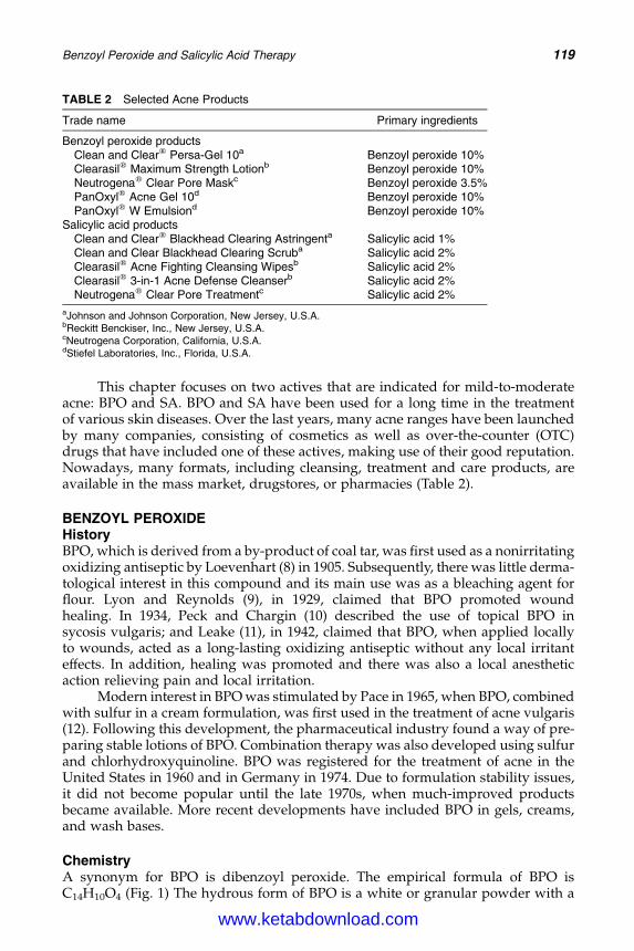
DK4962-Webster-ch11_R2_240307
This chapter focuses on two actives that are indicated for mild-to-moderateacne: BPO and SA. BPO and SA have been used for a long time in the treatmentof various skin diseases. Over the last years, many acne ranges have been launchedby many companies, consisting of cosmetics as well as over-the-counter (OTC)drugs that have included one of these actives, making use of their good reputation.Nowadays, many formats, including cleansing, treatment and care products, areavailable in the mass market, drugstores, or pharmacies (Table 2).
BENZOYL PEROXIDEHistoryBPO, which is derived from a by-product of coal tar, was first used as a nonirritatingoxidizing antiseptic by Loevenhart (8) in 1905. Subsequently, there was little derma-tological interest in this compound and its main use was as a bleaching agent forflour. Lyon and Reynolds (9), in 1929, claimed that BPO promoted woundhealing. In 1934, Peck and Chargin (10) described the use of topical BPO insycosis vulgaris; and Leake (11), in 1942, claimed that BPO, when applied locallyto wounds, acted as a long-lasting oxidizing antiseptic without any local irritanteffects. In addition, healing was promoted and there was also a local anestheticaction relieving pain and local irritation.
Modern interest in BPO was stimulated by Pace in 1965, when BPO, combinedwith sulfur in a cream formulation, was first used in the treatment of acne vulgaris(12). Following this development, the pharmaceutical industry found a way of pre-paring stable lotions of BPO. Combination therapy was also developed using sulfurand chlorhydroxyquinoline. BPO was registered for the treatment of acne in theUnited States in 1960 and in Germany in 1974. Due to formulation stability issues,it did not become popular until the late 1970s, when much-improved productsbecame available. More recent developments have included BPO in gels, creams,and wash bases.
ChemistryA synonym for BPO is dibenzoyl peroxide. The empirical formula of BPO isC14H10O4 (Fig. 1) The hydrous form of BPO is a white or granular powder with a
TABLE 2 Selected Acne Products
Trade name Primary ingredients
Benzoyl peroxide productsClean and Clearw Persa-Gel 10a Benzoyl peroxide 10%Clearasilw Maximum Strength Lotionb Benzoyl peroxide 10%Neutrogenaw Clear Pore Maskc Benzoyl peroxide 3.5%PanOxylw Acne Gel 10d Benzoyl peroxide 10%PanOxylw W Emulsiond Benzoyl peroxide 10%
Salicylic acid productsClean and Clearw Blackhead Clearing Astringenta Salicylic acid 1%Clean and Clear Blackhead Clearing Scruba Salicylic acid 2%Clearasilw Acne Fighting Cleansing Wipesb Salicylic acid 2%Clearasilw 3-in-1 Acne Defense Cleanserb Salicylic acid 2%Neutrogenaw Clear Pore Treatmentc Salicylic acid 2%
aJohnson and Johnson Corporation, New Jersey, U.S.A.bReckitt Benckiser, Inc., New Jersey, U.S.A.cNeutrogena Corporation, California, U.S.A.dStiefel Laboratories, Inc., Florida, U.S.A.
Benzoyl Peroxide and Salicylic Acid Therapy 119
www.ketabdownload.com
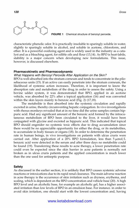
DK4962-Webster-ch11_R2_240307
characteristic phenolic odor. It is practically insoluble to sparingly soluble in water,slightly to sparingly soluble in alcohol, and soluble in acetone, chloroform, andether. It is a powerful oxidizing agent and is widely used in the industry as a cata-lyst and as a bleaching agent, for edible oils and flour (13,14). As BPO is heat-labile,stability is a major concern when developing new formulations. This issue,however, is discussed elsewhere.
Pharmacokinetic and PharmacodynamicWhat Happens with Benzoyl Peroxide After Application on the Skin?BPO is well-absorbed into the stratum corneum and tends to concentrate in the pilo-sebaceous units (15). If an active can easily penetrate into the stratum corneum, thelikelihood of systemic action increases. Therefore, it is important to know theabsorption rate and metabolism of the drug in order to assess the safety. Using abovine udder system, it was demonstrated that BPO, applied in an acetonevehicle, was absorbed by 22% after a topical application (16) and was convertedwithin the skin layers mainly to benzoic acid (Fig. 2) (17,18).
The metabolite is then absorbed into the systemic circulation and rapidlyexcreted in urine, thereby circumventing hepatic conjugation. In vivo investigationswith rhesus monkeys revealed that at in no time did the urine samples contain hip-puric acid. Had any significant amount of the benzoic acid formed by the intracu-taneous metabolism of BPO been circulated to the liver, it would have beenconjugated with glycine and excreted as hippuric acid. This indicated that topicalBPO should engender no systemic toxic effects due to drug accumulation sincethere would be no appreciable opportunity for either the drug, or its metabolites,to accumulate in body tissues or organs (18). In order to determine the penetrationrate in human beings, in vivo investigations on patients with ulcus cruris werecarried out. After application of a 20% BPO formulation, less than 5 mmol/Lbenzoic acid were detected in the serum and after three days no metabolite couldbe found (19). Transferring these results to acne therapy, a lower penetration ratecould even be expected since the skin barrier in acne patients is normally notbroken as in ulcus cruris patients and the applied concentration is much lowerthan the one used for antiseptic purpose.
SafetyAs discussed in the earlier section, it is unlikely that BPO causes systemic adversereactions or intoxications due to its rapid renal clearance. The main adverse reactionin acne therapy is the occurrence of skin irritation such as dryness, erythema, andscaling, which is dependent on the BPO concentration and vehicle base (20). A highBPO level and an alcoholic base, for example an alcoholic gel, has a higher associ-ated irritation than low levels of BPO in an emulsion base. For instance. in order toavoid skin irritation, one should start with the lowest concentration and, having
OO
O O
FIGURE 1 Chemical structure of benzoyl peroxide.
120 Gross
www.ketabdownload.com

DK4962-Webster-ch11_R2_240307
accustomed the skin to BPO, continue with increased strength formulations. Ingeneral, a lower strength or less frequent application of the preparation mayreduce irritation. A further possible way of reducing irritation is through usageof BPO washes, where the contact time with the skin is limited to seconds. Conse-quently, the irritation potential may also be lessened.
Another noteworthy side effect that needs to be considered is contact sensit-ization that has been observed occasionally among acne patients during treatmentwith topical preparations of BPO (21). Under the guinea pig maximization test,which is designed for testing the contact allergenic potential of chemicals, 76%contact sensitization was observed. A high sensitization potential is observedduring treatment of ulcus cruris; however, under the treatment of acne, the sensit-ization rate is disproportionally less. Numerous investigators have assessed thelikelihood of sensitization after use of BPO in acne treatment and have found anincidence of less than 1/500 (22), which is not significant. (14,23).
It is also important to mention the data on toxicity of the active. Investigationsregarding acute, subacute, and chronic toxicity demonstrated that toxicity couldbe regarded as harmless (24). Although BPO does not show toxicity, it is still con-sidered to be a carcinogen. Over the last 30 years, many studies were conductedto evaluate the safety of BPO (in general, the results of epidemiology studies andanimal carcinogenicity tests serve as important markers for cancer risk assessment).Epidemiology studies have provided evidence that its use to treat acne is not associ-ated with any increased risk of skin cancer (25–27).
FIGURE 2 Metabolism of benzoyl peroxide in keratinocytes of SENCAR-mice. Source: Adaptedfrom Ref. 18.
Benzoyl Peroxide and Salicylic Acid Therapy 121
www.ketabdownload.com
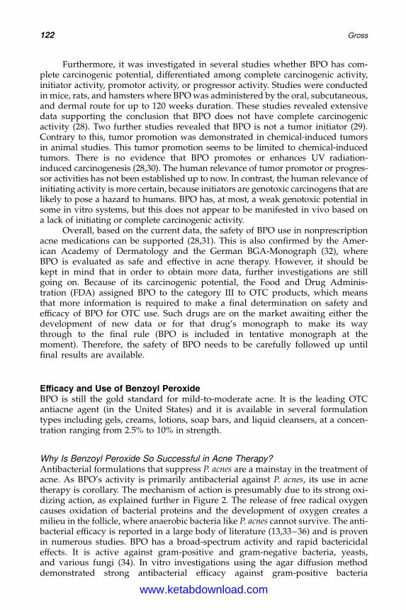
DK4962-Webster-ch11_R2_240307
Furthermore, it was investigated in several studies whether BPO has com-plete carcinogenic potential, differentiated among complete carcinogenic activity,initiator activity, promotor activity, or progressor activity. Studies were conductedin mice, rats, and hamsters where BPO was administered by the oral, subcutaneous,and dermal route for up to 120 weeks duration. These studies revealed extensivedata supporting the conclusion that BPO does not have complete carcinogenicactivity (28). Two further studies revealed that BPO is not a tumor initiator (29).Contrary to this, tumor promotion was demonstrated in chemical-induced tumorsin animal studies. This tumor promotion seems to be limited to chemical-inducedtumors. There is no evidence that BPO promotes or enhances UV radiation-induced carcinogenesis (28,30). The human relevance of tumor promotor or progres-sor activities has not been established up to now. In contrast, the human relevance ofinitiating activity is more certain, because initiators are genotoxic carcinogens that arelikely to pose a hazard to humans. BPO has, at most, a weak genotoxic potential insome in vitro systems, but this does not appear to be manifested in vivo based ona lack of initiating or complete carcinogenic activity.
Overall, based on the current data, the safety of BPO use in nonprescriptionacne medications can be supported (28,31). This is also confirmed by the Amer-ican Academy of Dermatology and the German BGA-Monograph (32), whereBPO is evaluated as safe and effective in acne therapy. However, it should bekept in mind that in order to obtain more data, further investigations are stillgoing on. Because of its carcinogenic potential, the Food and Drug Adminis-tration (FDA) assigned BPO to the category III to OTC products, which meansthat more information is required to make a final determination on safety andefficacy of BPO for OTC use. Such drugs are on the market awaiting either thedevelopment of new data or for that drug’s monograph to make its waythrough to the final rule (BPO is included in tentative monograph at themoment). Therefore, the safety of BPO needs to be carefully followed up untilfinal results are available.
Efficacy and Use of Benzoyl PeroxideBPO is still the gold standard for mild-to-moderate acne. It is the leading OTCantiacne agent (in the United States) and it is available in several formulationtypes including gels, creams, lotions, soap bars, and liquid cleansers, at a concen-tration ranging from 2.5% to 10% in strength.
Why Is Benzoyl Peroxide So Successful in Acne Therapy?Antibacterial formulations that suppress P. acnes are a mainstay in the treatment ofacne. As BPO’s activity is primarily antibacterial against P. acnes, its use in acnetherapy is corollary. The mechanism of action is presumably due to its strong oxi-dizing action, as explained further in Figure 2. The release of free radical oxygencauses oxidation of bacterial proteins and the development of oxygen creates amilieu in the follicle, where anaerobic bacteria like P. acnes cannot survive. The anti-bacterial efficacy is reported in a large body of literature (13,33–36) and is provenin numerous studies. BPO has a broad-spectrum activity and rapid bactericidaleffects. It is active against gram-positive and gram-negative bacteria, yeasts,and various fungi (34). In vitro investigations using the agar diffusion methoddemonstrated strong antibacterial efficacy against gram-positive bacteria
122 Gross
www.ketabdownload.com
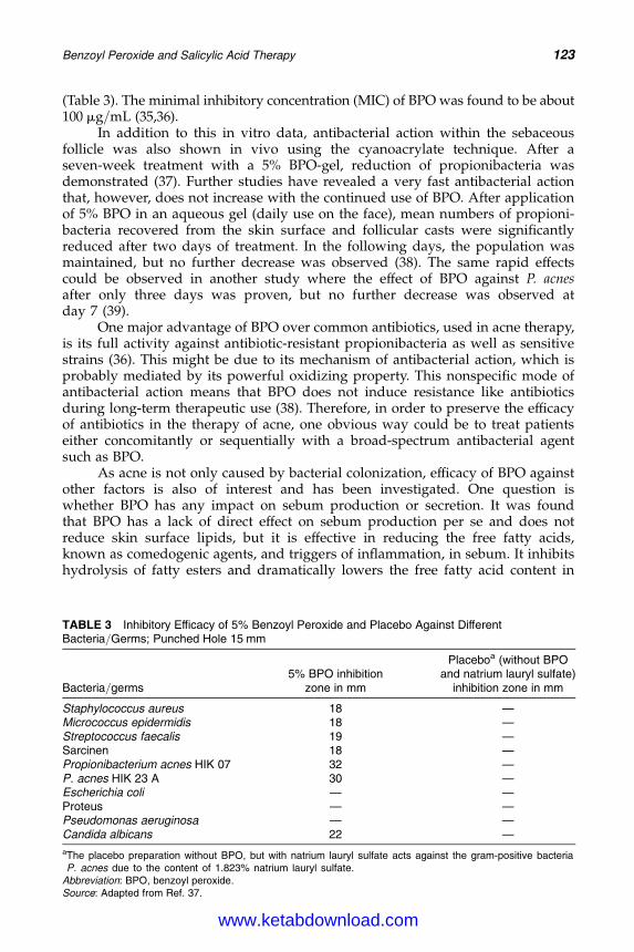
DK4962-Webster-ch11_R2_240307
(Table 3). The minimal inhibitory concentration (MIC) of BPO was found to be about100 mg/mL (35,36).
In addition to this in vitro data, antibacterial action within the sebaceousfollicle was also shown in vivo using the cyanoacrylate technique. After aseven-week treatment with a 5% BPO-gel, reduction of propionibacteria wasdemonstrated (37). Further studies have revealed a very fast antibacterial actionthat, however, does not increase with the continued use of BPO. After applicationof 5% BPO in an aqueous gel (daily use on the face), mean numbers of propioni-bacteria recovered from the skin surface and follicular casts were significantlyreduced after two days of treatment. In the following days, the population wasmaintained, but no further decrease was observed (38). The same rapid effectscould be observed in another study where the effect of BPO against P. acnesafter only three days was proven, but no further decrease was observed atday 7 (39).
One major advantage of BPO over common antibiotics, used in acne therapy,is its full activity against antibiotic-resistant propionibacteria as well as sensitivestrains (36). This might be due to its mechanism of antibacterial action, which isprobably mediated by its powerful oxidizing property. This nonspecific mode ofantibacterial action means that BPO does not induce resistance like antibioticsduring long-term therapeutic use (38). Therefore, in order to preserve the efficacyof antibiotics in the therapy of acne, one obvious way could be to treat patientseither concomitantly or sequentially with a broad-spectrum antibacterial agentsuch as BPO.
As acne is not only caused by bacterial colonization, efficacy of BPO againstother factors is also of interest and has been investigated. One question iswhether BPO has any impact on sebum production or secretion. It was foundthat BPO has a lack of direct effect on sebum production per se and does notreduce skin surface lipids, but it is effective in reducing the free fatty acids,known as comedogenic agents, and triggers of inflammation, in sebum. It inhibitshydrolysis of fatty esters and dramatically lowers the free fatty acid content in
TABLE 3 Inhibitory Efficacy of 5% Benzoyl Peroxide and Placebo Against DifferentBacteria/Germs; Punched Hole 15 mm
Bacteria/germs5% BPO inhibition
zone in mm
Placeboa (without BPOand natrium lauryl sulfate)
inhibition zone in mm
Staphylococcus aureus 18 —Micrococcus epidermidis 18 —Streptococcus faecalis 19 —Sarcinen 18 —Propionibacterium acnes HIK 07 32 —P. acnes HIK 23 A 30 —Escherichia coli — —Proteus — —Pseudomonas aeruginosa — —Candida albicans 22 —
aThe placebo preparation without BPO, but with natrium lauryl sulfate acts against the gram-positive bacteriaP. acnes due to the content of 1.823% natrium lauryl sulfate.
Abbreviation: BPO, benzoyl peroxide.Source: Adapted from Ref. 37.
Benzoyl Peroxide and Salicylic Acid Therapy 123
www.ketabdownload.com
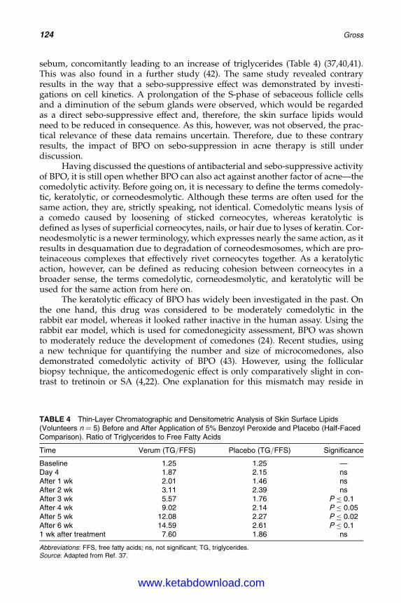
DK4962-Webster-ch11_R2_240307
sebum, concomitantly leading to an increase of triglycerides (Table 4) (37,40,41).This was also found in a further study (42). The same study revealed contraryresults in the way that a sebo-suppressive effect was demonstrated by investi-gations on cell kinetics. A prolongation of the S-phase of sebaceous follicle cellsand a diminution of the sebum glands were observed, which would be regardedas a direct sebo-suppressive effect and, therefore, the skin surface lipids wouldneed to be reduced in consequence. As this, however, was not observed, the prac-tical relevance of these data remains uncertain. Therefore, due to these contraryresults, the impact of BPO on sebo-suppression in acne therapy is still underdiscussion.
Having discussed the questions of antibacterial and sebo-suppressive activityof BPO, it is still open whether BPO can also act against another factor of acne—thecomedolytic activity. Before going on, it is necessary to define the terms comedoly-tic, keratolytic, or corneodesmolytic. Although these terms are often used for thesame action, they are, strictly speaking, not identical. Comedolytic means lysis ofa comedo caused by loosening of sticked corneocytes, whereas keratolytic isdefined as lyses of superficial corneocytes, nails, or hair due to lyses of keratin. Cor-neodesmolytic is a newer terminology, which expresses nearly the same action, as itresults in desquamation due to degradation of corneodesmosomes, which are pro-teinaceous complexes that effectively rivet corneocytes together. As a keratolyticaction, however, can be defined as reducing cohesion between corneocytes in abroader sense, the terms comedolytic, corneodesmolytic, and keratolytic will beused for the same action from here on.
The keratolytic efficacy of BPO has widely been investigated in the past. Onthe one hand, this drug was considered to be moderately comedolytic in therabbit ear model, whereas it looked rather inactive in the human assay. Using therabbit ear model, which is used for comedonegicity assessment, BPO was shownto moderately reduce the development of comedones (24). Recent studies, usinga new technique for quantifying the number and size of microcomedones, alsodemonstrated comedolytic activity of BPO (43). However, using the follicularbiopsy technique, the anticomedogenic effect is only comparatively slight in con-trast to tretinoin or SA (4,22). One explanation for this mismatch may reside in
TABLE 4 Thin-Layer Chromatographic and Densitometric Analysis of Skin Surface Lipids(Volunteers n ¼ 5) Before and After Application of 5% Benzoyl Peroxide and Placebo (Half-FacedComparison). Ratio of Triglycerides to Free Fatty Acids
Time Verum (TG/FFS) Placebo (TG/FFS) Significance
Baseline 1.25 1.25 —Day 4 1.87 2.15 nsAfter 1 wk 2.01 1.46 nsAfter 2 wk 3.11 2.39 nsAfter 3 wk 5.57 1.76 P � 0.1After 4 wk 9.02 2.14 P � 0.05After 5 wk 12.08 2.27 P � 0.02After 6 wk 14.59 2.61 P � 0.11 wk after treatment 7.60 1.86 ns
Abbreviations: FFS, free fatty acids; ns, not significant; TG, triglycerides.Source: Adapted from Ref. 37.
124 Gross
www.ketabdownload.com
DK4962-Webster-ch11_R2_240307
the nature of comedones. In the follicular biopsy technique, native microcomedoneswere studied, which may well be different from the tar- or azetulen-induced come-dones used in the rabbit studies (43). Overall, a weak comedolytic activity of BPOcan be stated.
Another supplementary benefit of BPO in acne therapy is its anti-inflammatory action. BPO, however, does not have significant direct in vivoanti-inflammatory potency. It had been suggested that protein kinase C mightserve as an additional pharmacological target of BPO. Therefore, the effects ofBPO were investigated on the release of reactive oxygen species, regulated byprotein kinase C and calmodulin, from human neutrophils, a potential importantstep in acne inflammation. Micromolar concentrations of BPO were found toinhibit the release of reactive oxygen species from human neutrophils but associ-ated with a marked drug-induced cytotoxicity. When, in cell-free assays, theeffects of BPO on protein kinase C and calmodulin as regulators of the release ofreactive oxygen species were investigated, there was only a marginal inhibitionof protein kinase C and no detectable inhibition of calmodulin. Thus, the inflamma-tory activity of BPO is unlikely to be mediated by protein kinase C or calmodulin(44,45). A potential explanation for the anti-inflammatory effects of BPO could bethe mediation by the antibacterial or oxidizing action of BPO. Following thedecrease in the population of P. acnes in the follicles and the reduction of freeoxygen radicals, one source of inflammation is cut and therefore inflammatory reac-tions are suppressed (4,22).
The anti-inflammatory benefit of BPO, regardless of being direct or indirect, isreflected in several studies (37,46,47). BPO products are indicated for mild-to-moderate acne with occurrence of predominantly inflamed lesions.
One study tested gel formulations with different concentrations of BPO (3, 5,or 10%) depending on the skin condition. After four weeks of treatment, the differ-ent BPO formulations were proven to be effective in the treatment of moderate acne.BPO was shown to decrease the number of papules and pustules and, to a smallerdegree, the number of comedones (48).
Interestingly, not only efficacy in mild-to-moderate acne but also a quickonset of action can be observed after BPO application, which accompanies thequick antibacterial action. A double-blind study showed that a 5% BPO lotionused topically for only five days significantly reduced the number of inflamedlesions (49). Since acne usually responds slowly to treatment, these results arevery surprising. Another study also demonstrated the effect of short-durationtreatment with a BPO aqueous gel. Compared to baseline, a statistically signifi-cant reduction of inflamed-lesions was determined after nine days and areduction of noninflamed lesions after four days of treatment. Over the wholestudy period (28 days), improvement in the reduction of inflamed lesions couldbe observed (50).
Is There a Difference in Efficacy Between Various Benzoyl Peroxide Levels?As mentioned earlier, BPO products are not only available in a variety of formu-lations such as gel, cleanser, or emulsion but also in a variety of concentrations.But is a higher level of active really more efficacious than a lower one? Ingeneral, one would expect superior efficacy from a product with the higher concen-tration. Surprisingly, a 2.5% gel formulation of BPO was proven to be equivalent in
Benzoyl Peroxide and Salicylic Acid Therapy 125
www.ketabdownload.com
DK4962-Webster-ch11_R2_240307
reducing the number of inflammatory lesions to 5% or 10% BPO gel preparations.With respect to the side effects, however, a difference could be observed. Desqua-mation, erythema, and symptoms of burning with the 2.5% gel were less frequentthan with the 10% preparation but equivalent to the 5% gel. The 2.5% formulationalso significantly reduced P. acnes and the percentage of free fatty acids in thesurface lipids after two weeks of topical application (51). So why are not thelowest strengths in favor? The answer is probably consumer psychology. Folklorehas established the belief that products with higher levels of active are generallybetter and more efficacious than those with a lower concentration (52). Which prep-aration should be used then? In practical use, people with delicate or irritable skinshould start with preparations of lower strength and, when this preparation istolerated, can continue with higher strength formulations.
SALICYLIC ACIDHistorySA can be regarded as an old-timer in acne therapy. Its benefits in treating skindisorders and other diseases have been known for generations. SA is a naturalingredient in numerous plants like the willow tree, sweet birch bark, wintergreenleaves, or chamomile flowers. The Romans knew of the efficacy of willow bark intreating ailments such as pain and fever, and the salicylic word in salicylic acidis derived from the Latin for willow, salix. For the first time, in 1838 the chemist Raf-faele Piria succeeded in synthesizing Salicin from willow bark components. In 1860,Kolbe synthesized pure SA and proposed its use as a preservative and drug. Due tobetter tolerability of stomach, (SA is highly irritating to gastric mucosa if ingestedorally, and therefore is used only as a topical agent), SA was enhanced and the deri-vate acetyl salicylic acid was born 30 years later (53). This well-established active,better known under the trade name of Aspirinw (Bayer Healthcare AG, Leverkusen,Germany), has been used systemically for pain relief and fever for over 100 years.For topical use, however, the pure acid is used for several skin disorders like acne,dandruff, psoriasis, or ichthyosis.
ChemistryThe systematic name for SA is a 2-hydroxybenzoic acid with the empirical formulaC7H6O3 (Fig. 3). It belongs to the group of hydroxyacids, which are carboxylic acids,classified into the a- and b-types according to their molecular structure (a-acids arethe so-called fruit acids, whereas SA represents the only b-type).
SA is a white, odorless, crystalline powder, or occurs as white or colorlessacicular crystals. It is slightly soluble in water and freely soluble in alcohol andether (54).
FIGURE 3 Chemical structure of salicylic acid.
126 Gross
www.ketabdownload.com
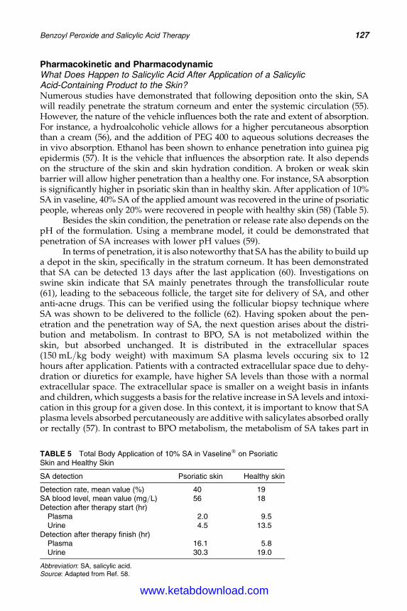
DK4962-Webster-ch11_R2_240307
Pharmacokinetic and PharmacodynamicWhat Does Happen to Salicylic Acid After Application of a SalicylicAcid-Containing Product to the Skin?Numerous studies have demonstrated that following deposition onto the skin, SAwill readily penetrate the stratum corneum and enter the systemic circulation (55).However, the nature of the vehicle influences both the rate and extent of absorption.For instance, a hydroalcoholic vehicle allows for a higher percutaneous absorptionthan a cream (56), and the addition of PEG 400 to aqueous solutions decreases thein vivo absorption. Ethanol has been shown to enhance penetration into guinea pigepidermis (57). It is the vehicle that influences the absorption rate. It also dependson the structure of the skin and skin hydration condition. A broken or weak skinbarrier will allow higher penetration than a healthy one. For instance, SA absorptionis significantly higher in psoriatic skin than in healthy skin. After application of 10%SA in vaseline, 40% SA of the applied amount was recovered in the urine of psoriaticpeople, whereas only 20% were recovered in people with healthy skin (58) (Table 5).
Besides the skin condition, the penetration or release rate also depends on thepH of the formulation. Using a membrane model, it could be demonstrated thatpenetration of SA increases with lower pH values (59).
In terms of penetration, it is also noteworthy that SA has the ability to build upa depot in the skin, specifically in the stratum corneum. It has been demonstratedthat SA can be detected 13 days after the last application (60). Investigations onswine skin indicate that SA mainly penetrates through the transfollicular route(61), leading to the sebaceous follicle, the target site for delivery of SA, and otheranti-acne drugs. This can be verified using the follicular biopsy technique whereSA was shown to be delivered to the follicle (62). Having spoken about the pen-etration and the penetration way of SA, the next question arises about the distri-bution and metabolism. In contrast to BPO, SA is not metabolized within theskin, but absorbed unchanged. It is distributed in the extracellular spaces(150 mL/kg body weight) with maximum SA plasma levels occuring six to 12hours after application. Patients with a contracted extracellular space due to dehy-dration or diuretics for example, have higher SA levels than those with a normalextracellular space. The extracellular space is smaller on a weight basis in infantsand children, which suggests a basis for the relative increase in SA levels and intoxi-cation in this group for a given dose. In this context, it is important to know that SAplasma levels absorbed percutaneously are additive with salicylates absorbed orallyor rectally (57). In contrast to BPO metabolism, the metabolism of SA takes part in
TABLE 5 Total Body Application of 10% SA in Vaselinew on PsoriaticSkin and Healthy Skin
SA detection Psoriatic skin Healthy skin
Detection rate, mean value (%) 40 19SA blood level, mean value (mg/L) 56 18Detection after therapy start (hr)
Plasma 2.0 9.5Urine 4.5 13.5
Detection after therapy finish (hr)Plasma 16.1 5.8Urine 30.3 19.0
Abbreviation: SA, salicylic acid.Source: Adapted from Ref. 58.
Benzoyl Peroxide and Salicylic Acid Therapy 127
www.ketabdownload.com
DK4962-Webster-ch11_R2_240307
the liver by conjugation with glycine to salicyluric acid, with glucuronic acid toetherglucuronide, and by hydroxylation to gentisinic acid and benzoic acid. Thehalf-life of SA is about two or three hours if a normal dosage is used. However, ifoverdosed, or due to limited liver metabolism capacity, half-life could be prolongedto 15 to 30 hours, which should be considered when using SA containing productswhile suffering with liver diseases. About 65% to 85% of a topically administeredSA dose is recoverable from the urine. Almost 95% of a single dose of SA is excretedwithin 24 hours of its entrance into the extracellular space.
SafetySA is likely to cause some degree of local skin peeling and discomfort, such asburning or skin reddening, as it is a mild irritant (54,63). Beside this topicaladverse event, one major adverse reaction is the potential intoxication caused byincreased penetration of SA. Intoxication has been shown in patients with adamaged skin barrier who received a treatment on the whole body over severaldays (such as psoriatic or ichthyotic patients). This can be explained due to highpenetration of SA through a damaged skin barrier. Clinically, the patients havethirst, tinnitus, headache, lethargy, confusion, nausea, vomiting, diaphoresis,depression, and disorientation (54,57). According to the opinion of the ScientificCommittee on Cosmetic Products and Non-Food Products (SCCNFP), however,this event is rare and depends on various factors, such as the age of patient, theintensity of the skin damage, the concentration of SA in the formulation, or thesurface of the application. Ointments containing 3% to 6% SA have causednausea, dyspnoea, hearing loss, confusion, and hallucinations in three patientswith extensive psoriasis. They had two soap and water baths daily combinedwith UV therapy and six ointment applications. Under these conditions, the symp-toms developed in four days and were associated with significant SA plasma levels(46–64 mg/100 mL). Fortunately, the symptoms disappeared rapidly after discon-tinuation of the ointment applications (63). The application of SA to extensiveareas, particularly in children, may involve a risk of toxicity from absorption. Asdiscussed earlier, children are particularly susceptible (63). Having a greater pro-portion of body surface and weight, they are exposed to a higher risk of systemiceffects during large surface treatment. However, the number of reported cases ofintoxication of children is quite lower than one would expect. This could be dueto the fact that the horny layer barrier of children over one year is nearly identicalto the one of adults and therefore their skin has nearly the same absorption rates, orbecause SA formulations were only rarely used on infants and children.
As salicylate plasma levels can be indicative of SA intoxication, plasma levelsshould be measured first in case of any intoxication suspicion. Symptoms occur at aplasma level of 35 mg/100 mL or higher (63,64).
Overall, the correlation between body salicylate and clinical severity of theintoxication is poor. Severe manifestations are linked with diseased skin and mul-tiple applications on large body areas of formulations containing high concen-trations of SA (63). Therefore, to prevent a toxic reaction, three factors should bekept in mind: the quantity of SA used should not be excessive (repeated, largeareas of the body), the available extracellular fluid volume should not be limited(smaller children, because their extracellular fluid volume is much smaller in com-parison to the potential surface area available for treatment), and the ability tometabolize and excrete the absorbed medication should not be impaired (64).
128 Gross
www.ketabdownload.com
DK4962-Webster-ch11_R2_240307
In terms of safety, the question of sensitization potential is as important as theadverse topic reaction. In the past, there have been numerous discussions on thesensitization potential of SA. Studies and literature research reveal that only inrare cases a sensitization potential was detected (65). According to the SCCNFP,SA is also not classified as a sensitizer. The results of human repeated insultpatch tests conducted with formulation up to 2% SA confirm that topical appli-cation does not cause skin sensitization. On the other hand, other scientists doubtthe available data. Although there are many examples of positive patch tests, noexample was found that fulfilled the operational definition of clinically relevantallergic contact dermatitis. Allergic contact dermatitis to SA in man probablyremains unproven in their opinion (66). However, based on data and current think-ing, SA can be regarded as not being a sensitizer, or at most a weak sensitizer.
Overall, the safety of SA can be taken as granted when considering the givenprecautions and contraindications. SCCNFP considers that SA is safe for “otheruses” than as a preservative, at a concentration up to 2% for the leave-on and rinse-off cosmetic products, and at a concentration up to 3% for rinse-off hair products (63).
Efficacy and Use of Salicylic AcidKeratolytics like SA are commonly used as first-line therapy in mild cases of acnebecause of their widespread availability OTC. SA is widely used as a topical thera-peutic agent for a variety of skin diseases, including acne, psoriasis, dandruff, andichthyosis. Concentrations of SA ranging from 0.5% to 10% have been rec-ommended for acne, but the maximum strength allowed in nonprescription acneproducts in the United States is up to 2% by the FDA Final Acne Monograph. Athigher concentrations up to 40%, SA is also used for wart and corn removal.
Why Does Salicylic Acid Have Such a Good Reputation in Acne?As the microcomedone is believed to be the precursor to all acne lesions, it is logicalto treat it with a comedolytic agent that will counteract the plugging of the follicles.SA is an effective drug for this due to its keratolytic activity. Although SA has longbeen used as a keratolytic agent, the mechanism of action remains unsolved. Theaction may occur due to the reduced cohesion between corneocytes, resulting inthe shedding of epidermal cells, rather than “lysing” of keratin. This was investi-gated in numerous studies. In 1943, it was first claimed that SA acts by producingepidermal irritation, but there has been no substantiation of this finding (67).Further studies suggested that the keratolytic effect of SA consists of a reductionof intercellular stickiness, thereby reducing corneocyte adhesion. The so-calledcementing substances are the site of action and not the stratum corneum itself(68–70). No damage of the living epidermis is caused by SA and it has beenshown to cause no epidermal change beneath the stratum corneum (68). Further-more, there is no evidence that it works by lysing keratin. Keratin is a toughfibrous protein that remains in the intracellular region and it is regarded as unlikelythat agents that promote desquamation do so by attacking this material (71). SA hasno effect on the mitotic activity of the normal epidermis (69) and therefore does notinfluence the disordered cornification itself.
While the mechanism of action of SA’s main property remains uncertain, anadditional action is well-established. Besides the keratolytic action of SA withinthe pilosebaceous unit, the value of the antibacterial action is often neglected.Since antibacterial colonization in the pore is deemed to be a main cause for spot
Benzoyl Peroxide and Salicylic Acid Therapy 129
www.ketabdownload.com
DK4962-Webster-ch11_R2_240307
formation, the use of antibacterials in acne treatment or prevention plays a majorrole, as discussed earlier.
The antibacterial action of SA against P. acnes has been demonstrated inboth in vitro and in vivo studies. MIC was determined at 0.25% (w/w) using theplate method. In vivo measurement also reveals the efficacy of SA within thepore, expected as penetration occurs through the transfollicular root to the follicle(72–75). Having penetrated into the pore, SA can start to work and inhibit bacteriagrowth.
SA also offers anti-inflammatory benefits in acne therapy, which havebeen reported in the literature (73,74,76). Anti-inflammatory activity was investi-gated using the UV model (76). Upon topical application to the skin of guineapigs with UV dermatitis, SA exerted a distinct anti-inflammatory effect, whichwas most pronounced at concentrations between 0.5% and 5%. Its integratedactivity over the range of concentrations tested, that is, from 0.05% to 5% andover a period from 4 to 26 hours after irradiation, equals that of the contact antiphlo-gistic agent bufexamac, which is a well-established active in clinical use (76).
The anti-inflammatory effect of SA might be adjuvant in acting not onlyagainst non-inflammatory but also against inflammatory lesions. Many clinicalstudies have proven the anti-acne efficacy of SA-containing products (77,78).
For instance, one study demonstrated that twice daily application of 2% SAalcohol-detergent lotion impregnated into pads was significantly better than theplacebo in the treatment of mild-to-moderate acne. Significant benefit from theactive therapy was evident at four, eight, and 12 weeks when total lesion countswere assessed. Regarding noninflamed lesions superiority over placebo could bedemonstrated after eight and 12 weeks and for inflamed lesions after 12 weeks.These results strengthen the suggestion that SA has a primary effect on comedogen-esis. The observation that inflamed lesions were reduced at a later date could beexplained by the fact that inflamed lesions arise from noninflamed lesions. There-fore, the efficacy against comedones will prevent the development of inflamedlesions, though this a lagged efficacy (77).
Another double-blind, controlled study evaluated the efficacy of 0.5% SA in analcohol-detergent solution and placebo (pads soaked in buffered water). SA wasshown to be more effective in reducing open and inflamed lesions than placebo (78).
Furthermore, efficacy has been proven in a comparative study with BPO,which will be discussed separately.
Is There Another Use for Salicylic Acid Other than in Anti-acne Cosmetics?SA can also be used as a peeling agent in high concentrations. However, at thesehigh levels of SA, irritation or other adverse events are very likely and thereforesuch peelings must be done under the supervision and strict control of a physician.
SA peels are available in 20% and 30% concentrations. In general, superficialchemical peels are an invaluable accoutrement for dermatologists when treatingfine wrinkles, photoageing, keratoses, acne, and dyspigmentation. It could beshown that SA peels facilitated the clearing of pustules, papules, and comedones.Moderate-to-significant clearing of acne vulgaris was also demonstrated (79). Afurther study using 20% to 30% SA in a hydroethanolic vehicle demonstratedthat SA is useful as a single or multiple peeling agent for acne as well as for photo-damage and hyperpigmentation. SA as a peeling agent was useful for both come-donal and papular acne. Areas of intense inflammation showed decreasederythema, and pustules were rapidly dried at one or two days postpeel. Many
130 Gross
www.ketabdownload.com

DK4962-Webster-ch11_R2_240307
closed and open comedones were extruded by several days postpeel (80). Whenapplied under strict control and indication, the SA peels can be regarded as safeand efficacious, and offer adjunctive benefit when treating acne vulgaris.
Comparison of Benzoyl Peroxide and Salicylic AcidBPO and SA are both indicated for use in mild-to-moderate acne, but their mainaction of mechanism is different. While BPO has a strong antibacterial action, SAacts through a keratolytic action. Does this different action have any impact onthe efficacy of the products? Is BPO more effective against inflamed lesions whileSA predominantly acts against noninflamed lesions? Comparative studies demon-strated similar efficacy of both products or even slight superior efficacy of SA.
In a four-week crossover study, the efficacy of a 2% SA acne cleanser wascompared with that of 10% BPO wash in the treatment of 30 patients with mild-to-moderate acne. The results demonstrated that only patients treated with theSA cleanser had a significant reduction in the number of comedones. However,when patients crossed over to BPO, acne worsened over the following two weeksand the number of comedones slightly increased. In contrast, the number of come-dones decreased in patients who first used BPO and then crossed over to SA. Theseresults suggest that SA is more effective than BPO for the treatment of comedonalacne (81).
In another, 12-week double-blind study with 180 subjects, 2% SA, the vehiclesolution, and a 5% BPO cream were compared. Surprisingly, SA was not onlyfound to produce a substantially greater reduction from baseline in the numberof comedones but also a reduction in inflammatory lesions when compared with5% BPO (Fig. 4) (82).
The observed superiority of SA might be due to its mode of action. Since theprimary lesion of acne is the microcomedo, a treatment that is effective in prevent-ing its formation will be effective in preventing inflammatory lesions into which themicrocomedo can progress. As the action of SA is mainly keratolytic, it interferes inan earlier stage than BPO, and in consequence it seems to be superior in actingagainst later steps, the inflamed lesions. When evaluating the results of these
0
10
20
Per
cent
red
uctio
n
30
40
50
60
70
Total Lesions InflammatoryLesions
OpenComedones
ClosedComedones
2% Salicylic acid
5% Benzoyl peroxide
Placebo
FIGURE 4 Percentage reductions in total number of lesions, inflammatory lesions, open come-dones, and closed comedones in patients treated with salicylic acid, placebo, or benzoyl peroxide.Source: Adapted from Ref. 82.
Benzoyl Peroxide and Salicylic Acid Therapy 131
www.ketabdownload.com
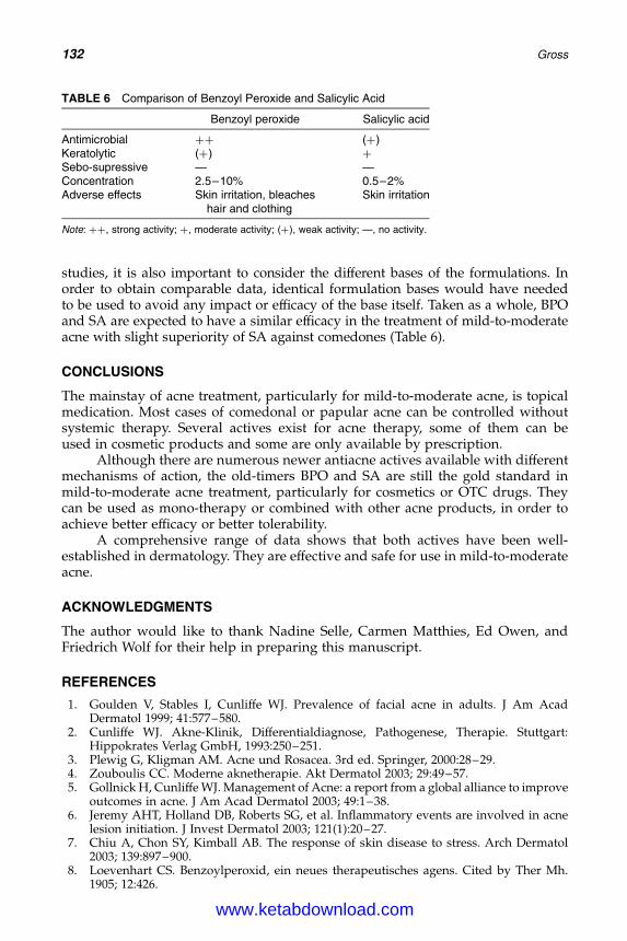
DK4962-Webster-ch11_R2_240307
studies, it is also important to consider the different bases of the formulations. Inorder to obtain comparable data, identical formulation bases would have neededto be used to avoid any impact or efficacy of the base itself. Taken as a whole, BPOand SA are expected to have a similar efficacy in the treatment of mild-to-moderateacne with slight superiority of SA against comedones (Table 6).
CONCLUSIONS
The mainstay of acne treatment, particularly for mild-to-moderate acne, is topicalmedication. Most cases of comedonal or papular acne can be controlled withoutsystemic therapy. Several actives exist for acne therapy, some of them can beused in cosmetic products and some are only available by prescription.
Although there are numerous newer antiacne actives available with differentmechanisms of action, the old-timers BPO and SA are still the gold standard inmild-to-moderate acne treatment, particularly for cosmetics or OTC drugs. Theycan be used as mono-therapy or combined with other acne products, in order toachieve better efficacy or better tolerability.
A comprehensive range of data shows that both actives have been well-established in dermatology. They are effective and safe for use in mild-to-moderateacne.
ACKNOWLEDGMENTS
The author would like to thank Nadine Selle, Carmen Matthies, Ed Owen, andFriedrich Wolf for their help in preparing this manuscript.
REFERENCES
1. Goulden V, Stables I, Cunliffe WJ. Prevalence of facial acne in adults. J Am AcadDermatol 1999; 41:577–580.
2. Cunliffe WJ. Akne-Klinik, Differentialdiagnose, Pathogenese, Therapie. Stuttgart:Hippokrates Verlag GmbH, 1993:250–251.
3. Plewig G, Kligman AM. Acne und Rosacea. 3rd ed. Springer, 2000:28–29.4. Zouboulis CC. Moderne aknetherapie. Akt Dermatol 2003; 29:49–57.5. Gollnick H, Cunliffe WJ. Management of Acne: a report from a global alliance to improve
outcomes in acne. J Am Acad Dermatol 2003; 49:1–38.6. Jeremy AHT, Holland DB, Roberts SG, et al. Inflammatory events are involved in acne
lesion initiation. J Invest Dermatol 2003; 121(1):20–27.7. Chiu A, Chon SY, Kimball AB. The response of skin disease to stress. Arch Dermatol
2003; 139:897–900.8. Loevenhart CS. Benzoylperoxid, ein neues therapeutisches agens. Cited by Ther Mh.
1905; 12:426.
TABLE 6 Comparison of Benzoyl Peroxide and Salicylic Acid
Benzoyl peroxide Salicylic acid
Antimicrobial þþ (þ)Keratolytic (þ) þ
Sebo-supressive — —Concentration 2.5–10% 0.5–2%Adverse effects Skin irritation, bleaches
hair and clothingSkin irritation
Note: þþ, strong activity; þ, moderate activity; (þ), weak activity; —, no activity.
132 Gross
www.ketabdownload.com
DK4962-Webster-ch11_R2_240307
9. Lyon RA, Reynolds TE. Promotion of healing by benzoyl peroxide and other agents. ProcSoc Exp Biol Med 1929; 27(2):122–124.
10. Peck SM, Chargin L. Sycosis vulgaris: a new method of treatment. Arch Dermatol Sysp1934; 29:456.
11. Leake CD. Advantages of benzoyl peroxide over zinc peroxide or sulfonamides intreating wounds or burns. JAMA 1942; 119:101.
12. Pace WE. A benzoyl peroxide-sulfur cream for acne vulgaris. Can Med Assoc J 1965;93:252–254.
13. Sweetman SC. Martindale: The Complete Drug Reference: Benzoyl Peroxide. London-Chicago: Pharmaceutical Press, 2002:1111.
14. Cotterill JA. Benzoyl peroxide. Acta Derm Venereol 1980; 89:57–62.15. Orth DS, Widjaja J, Wortzman MS. Benzoyl peroxide concentration in follicular casts.
Cosmetics & Toiletries Magazin 1997; 112:87.16. Kietzmann M, Loscher W, Arens D, et al. The isolated perfused bovine udder as an in
vitro model of percutaneous drug absorption—skin viability and percutaneous absorp-tion of dexamethasone, benzoyl peroxide and efonemate. J Pharmacol Toxicol Methods1993; 30:75–84.
17. Binder RL, Aardema MJ, Thompson ED. Benzoyl peroxide: review of experimental car-cinogenesis and human safety data. Prog Clin Biol Res 1995; 391:245–294.
18. Yeung D, Nacht S, Buchs D, et al. Benzoyl peroxide: percutaneous penetration and meta-bolic circulation. II Effect of concentration. J Am Acad Dermatol 1983; 6:920–924.
19. Holzmann H, Morsches R, Benes P. The absorption of benzoyl peroxide from leg ulcers.Arzneimittelforschung 1979; 29:1180–1183.
20. Brown SK, Shalita AR. Acne vulgaris. Lancet 1998; 351:1871–1876.21. Poole RL, Griffith JF, MacMillan FSK. Experimental contact sensitation with benzoyl
peroxide. Arch Dermatol 1970; 102:635–639.22. Gollnick H, Schramm M. Topical drug treatment in acne. Dermatology 1998; 196:119–125.23. Bandmann HJ, Agathos M. Die posttherapeutische benzoylperoxid-kontaktallergie bei
Ulcus cruris-patienten. Hautarzt 1985; 35:670–674.24. Gloor M. Benzoylperoxid in der dermatologischen Lokaltherapie. Zentralbl Hautkr
1990; 157:1010–1015.25. Hogan DJ, To T, Wilson ER, et al. A study of acne treatments as risk factors for skin
cancer of the head and neck. Br J Dermatol, 1991; 125:343–348.26. Hogan DJ, To T, Wilson ER. Drug and non-drug risk factors associated with facial skin
cancer. A report to theNon-prescriptionDrugManufacturers Association, Non-prescriptionDrug Manufacturers Association of Canada on the Saskatchewan Study, 1990.
27. To T, Hogan DJ, Wilson ER, et al. Benzoyl peroxide and facial skin cancer. Am JEpidermiol 1991; 134:772.
28. Binder RL, Aardema MJ, Thompson ED. Benzoyl peroxide: review of experimentalcarcinogenesis and human safety data. Prog Clin Biol Res 1995; 319:245–294.
29. Gloor M, Thoma K, Fluhr J. Benzoylperoxid (BPO) and Azelainsaure (AZ).In: Dermatologische Externatherapie. Berlin, Germany: Springer-Verlag, 1995:205.
30. King JK, Egner PA, Kensler TW. Generation of DNA base modification following treat-ment of cultured murine keratinocytes with benzoyl peroxide. Carcinogenesis 1996;17(2):317–320.
31. Zbinden G. Scientific opinion on the carcinogenic risk due to topical administration ofbenzoyl peroxide for the treatment of acne vulgaris. Pharmacol Toxicol 1988; 63:307–309.
32. Benzoylperoxid. In: Aufbereitungsmonographien Kommission B7, BGA. PZ 1990;27:1841–1842.
33. Beasley JN. Chemistry and metabolism of benzoyl peroxide and other antiacne drugs.Clin Res 1982; 30(3):698.
34. Kligman AM, Leyden JJ, Stewart R. New uses for benzoyl peroxide: a broad-spectrumantimicrobial agent. Int J Dermatol 1977; 16:413–417.
35. Patane AM, Pistillo M. Sull’azione antimicrobica del benzoilperossido. Ann Sclavo 1982;24(5):513–522.
36. Farmery MR, Jones CE, Eady EA, et al. In vitro activity of azaleic acid, benzoyl peroxideand zinc acetata against antibiotic resistant propionibacteria from acne patients.J Dermatol Treat 1994; 5:63–65.
Benzoyl Peroxide and Salicylic Acid Therapy 133
www.ketabdownload.com
DK4962-Webster-ch11_R2_240307
37. Puschmann M. Klinisch-experimentelle untersuchungen zum wirkungsnachweis vonbenzoylperoxid. Hautarzt 1982; 33:257–265.
38. Bojar RA, Holland KT, Cunliffe WJ. The in-vitro antimicrobial effects of azelaic acid uponPropionibacterium acnes strain P37. J Antimicrob Chemother 1991; 28(6):843–853.
39. Pagnoni A, Kligman AM, Kollias N, et al. Digital flourescence photography can assessthe suppressive effect of benzoyl peroxide on Propionibacterium acnes. J Am Acad Der-matol 1999; 41(4):710–716.
40. Fulton JE, Farzad-Bakshandeh A, Bradley S. Studies on the mechanism of action oftopical benzoyl peroxide and vitamin A acid in acne vulgaris. J Cutan Pathol 1974;1:191–200.
41. Gloor M, Hummel A, Friedrich HC. Experimentelle untersuchungen zur Benzoylperox-idtherapie der acne vulgaris. Z Hautkr 1975; 50(15):657–663.
42. Wirth H, Spurgel D, Gloor M. Untersuchungen zur Wirkung von Benzoylperoxid auf dieTalgdrusensekretion. Dermatol Monatsschr 1983; 169:289–293.
43. Pierard GE, Peirard-Franchimont C, Goffin V. Digital image analysis of microcome-dones. Dermatology 1995; 190:99–103.
44. Gollnick H, Krautheim A. Topical treatment in acne: current status and future aspects.Dermatology 2003; 206:29–36.
45. Hegemann L, Toso SM, Kitay K, et al. Anti-inflammatory actions of benzoyl peroxide:effects on the generation of reactive oxygen species by leucocytes and the activity ofprotein kinase C and calmodulin. Br J Dermatol 1994; 130(5):569–575.
46. Norris JFB, Hughes BR, Basey AJ, Cunliffe WJ. A comparison of the effectiveness oftopical tetracycline, benzoyl-peroxide gel and oral oxytetracycline in the treatment ofacne. Clin Exp Dermatol 1991; 16:31–33.
47. Lookingbill DP, Chalker DK, Lindholm JS, et al. Treatment of acne with a combinationclindamycin/benzoyl peroxide gel compared with clindamycin gel, Benzoyl peroxidegel and vehicle gel: combined results of two double blind investigations. J Am AcadDermatol 1997; 37:590–595.
48. Lassus A. Local treatment of acne. A clinical study and evaluation of the effect of differ-ent concentrations of benzoyl peroxide gel. Curr Med Res Opin 1981; 7:370–373.
49. Schutte H, Cunliffe WJ, Forster RA. The short-term effects of benzoyl peroxide lotion onthe resolution of inflamed acne lesions. Br J Dermatol 1981; 106:91–94.
50. Bojar RA, Cunliffe WJ, Holland KT. The short-term treatment of acne vulgaris withbenzoyl peroxide: effects on the surface and follicular cutaneous microflora. Br J Dermatol1995; 132:204–208.
51. Mills OH, Kligman AM, Pochi P, et al. Comparing 2.5%, 5%, and 10% benzoyl peroxideon inflammatory acne vulgaris. Int J Dermatol 1986; 25(10):664–667.
52. Kligman AM. Acne vulgaris: tricks and treatments. Part II: the benzoyl peroxide saga.CUTIS 1995; 56(5):260–261.
53. Schmid-Wendtner MH. Salicylsaure. In: Korting HC, Sterry W, eds. TherapeutischeVerfahren in der Dermatologie. Berlin: Blackwell Wissenschafts-Verlag Wien, 2001.
54. Sweetman SC. Martindale: The Complete Drug Reference: Salicylic Acid. London-Chicago: Pharmaceutical Press, 2002:1122–1123.
55. Taylor JR, Halprin KM. Percutaneous absorption of salicylic acid. Arch Dermatol 1975;111:740–743.
56. Davis AP, Kraus AL, Thompson GA, et al. Percutaneous absorption of salicylic acid afterrepeated (14-day) in vivo administration on normal, acnegenic or aged human skin.J Pharm Sci 1997; 86(8):896–899.
57. Goldsmith LA. Salicylic acid. Int J Dermatol 1979; 18:32–36.58. Arnold W,Trinnes F, Schroeder I. Zur hautresorption von salicylsaure bei psoriatikern
und hautgesunden. Beitr Gerichtl Med 1979; 37:325–328.59. Samuelov Y, Donbrow M, Friedman M. Effect of pH on salicylic acid permeation through
ethyl cellulose PEG 4000 films. J Pharm Pharmacol 1979; 31:120–121.60. Washitake M, Yajima T, Amno T, et al. Studies on percutaneous absorption of drugs. II Time
course of cutaneous reservoir of drugs. Chem Pharm Bull 1972; 20:2429–2435.61. Takahashi H, Ishii T, Tanabe K, et al. The percutaneous absorption of salicylic acid.
J Dermatol 1976; 3:135–138.
134 Gross
www.ketabdownload.com
DK4962-Webster-ch11_R2_240307
62. Mills OH, Berger RS, Cardin C, et al. Analysis of salicylic acid levels in the sebaceousfollicle. J Invest Dermatol 1995; 104(4):661–661.
63. Opinion of the scientific committee on cosmetic products and non-food productsintended for consumers concerning Salicylic acid adopted by the SCCNFP during the20th plenary meeting of 4 June 2002. SCCNFP/0522/01, final.
64. Lin AN, Nakatsui T. Salicylic acid revisited. Int J Dermatol 1998; 37:335–342.65. Goh CL, Ng SK. Contact allergy to salicylic acid. Contact Dermatitis1986; 14:114.66. Gallacher G, Hostynek JJ, Maibach HI. Salicylic acid: human allergic. Contact Dermatitis
1998; 46(3):129–130.67. StrakoschEA. Studies on ointments. II. Ointments containing salicylic acid. Arch
Dermatol Syphilol 1943; 47:16.68. Huber C, Christophers E. “Keratolytic” effect of salicylic acid. Arch Dermatol Res 1977;
257(3):293–297.69. Roberts DL, Marshall R, Marks R. Detection of the action of salicylic acid on the normal
stratum corneum. Br J Dermatol 1980; 103:191–196.70. Mills OH,Kligman AM. Assay of comedolytic activity in acne patients. Acta Derm
Venereol 1983; 63:68–71.71. Davies M, Marks R. Studies on the effect of salicylic acid on normal skin. Br J Dermatol
1976; 95:187–192.72. Gloor M, Steinbacher M, Franke M. Uber die antimikrobielle Wirkung der Salicylsaure
auf die Propionibakterien im Talgdruseninfundibulum bei der Lokalbehandlung derAcne vulgaris. Z Hautkr 1979; 54(19):856–860.
73. Gloor M, Wirth H, Schnyder UW. Pharmakologie der Salicylsaure bei topischerApplikation—eine Ubersicht uber die Literatur der letzten 20 Jahre. In: Hornmann OP,Schnyder UW, Schonfeld J, eds. Neue Entwicklungen in der Dermatologie. Berlin,Germany: Springer-Verlag, 1984:74–81.
74. Plewig G, Kligman AM. Exfoliants. In: Acne-Morphogenesis And Treatment. Berlin-Heidelberg-New York: Springer, 1975:277–279.
75. Hartmann AA, Hackel H, Elsner P, et al. Antibakterielle Wirkung der Salicylsaure—einezu wenig beachtete Wirkung. Zbl Haut-GeschlKrankh 1990; 157:983.
76. Weirich EG, Longauer JK, Kirkwood AH. Dermatopharmacology of salicylic acid.Dermatologica 1979; 152:87–99.
77. Eady EA, Burke BM, Pulling K, et al. The benefit of 2% salicylic acid lotion in acne—a placebo-controlled study. J Dermatol Treat 1996; 7:93–96.
78. Shalita AR. Treatment of mild and moderate acne vulgaris with salicylic acid in analcohol-detergent vehicle. CUTIS 1981; 28:556–561.
79. Grimes PE. The safety and efficacy of salicylic acid chemical peels in darker racial-ethnicgroups. Dermatol Surg 1999; 25:18–22.
80. Kligman D, Kligman AM. Salicylic acid as a peeling agent for the treatment of acne.Cosmetic Dermatol 1997; 10:44–47.
81. Shalita AR. Comparison of a salicylic acid cleanser and a benzoyl peroxide wash in thetreatment of acne vulgaris. Clin Ther 1989; 11(2):264–267.
82. Zander E, Weismann S. Treatment of acne vulgaris with salicylic acid pads. Clin Ther1992; 14(2):247–253.
Benzoyl Peroxide and Salicylic Acid Therapy 135
www.ketabdownload.com

DK4962-Webster-ch11_R2_240307
www.ketabdownload.com
DK4962-Webster-ch12_R2_020407
B12 Treating Acne with Octadecenedioic Acid:Mechanism of Action, Skin Delivery,and Clinical Results
Johann W. WiechersJW Solutions, Gouda, The Netherlands
Anthony V. RawlingsAVR Consulting Ltd., Northwich, Cheshire, U.K.
Nigel LindnerDepartment of Research and Development, Uniqema, Gouda, The Netherlands
William J. CunliffeDepartment of Dermatology, Leeds Foundation for Dermatological Research, LeedsGeneral Infirmary, Leeds, U.K.
INTRODUCTION
Acne is and will always remain a disturbing disease because it is highly visible aswell as manifesting itself at an age when young teenagers are very concerned abouttheir looks. However, to state that acne is only limited to adolescents (where malestend to show the more severe forms of the disease) is wrong, as especially postado-lescent women are also affected by the disease (1). The primary cause of acne isan obstruction of the pilosebaceous canal. This is due to at least four pathogenicfactors: (i) increased proliferation, cornification, and shedding of follicular epi-thelium (i.e., hyperkeratinization); (ii) increased sebum production (i.e., hyper-seborrhea); (iii) colonization of the follicle with Propionibacterium acnes andStaphylococcus aureus (i.e., microbes); (iv) induction of inflammatory responses bybacterial antigens and cell signals (i.e., inflammation) (2). Earlier chapters of thisbook will have dealt extensively with these etiological and pathogenetical aspectsof acne.
Topical treatments improving acne all affect one or more of these four maincauses of acne (3). For instance, topical retinoids, of which tretinoin, isotretinoin,and adapalene are probably the most well-known examples, cause an expulsionof mature comedones, an inhibition of the formation and number of microcome-dones, and an inhibition of inflammation. Benzoyl peroxide (BPO), erythromycin,clindamycin, and azelaic acid (AZA) are examples of topical antimicrobials andantibiotics, although due to the increasing development of bacterial resistance,the concentrations of the topical antibiotics clindamycin and erythromycinhave been raised from 1% to 4%. AZA is a C9:0 dicarboxylic acid with efficacyon follicular keratinization and on P. acnes. It seems to have some inflammatoryefficacy via effects on neutrophilic granulocytes. It is used at very high concen-trations (20%), although somewhat lower concentrations (15%) have recently
137
www.ketabdownload.com
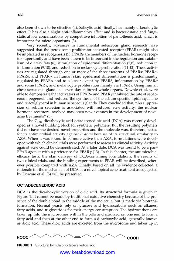
DK4962-Webster-ch12_R2_020407
also been shown to be effective (4). Salicylic acid, finally, has mainly a keratolyticeffect. It has also a slight anti-inflammatory effect and is bacteriostatic and fungi-static at low concentrations by competitive inhibition of pantothenic acid, which isimportant for micro-organisms (3).
Very recently, advances in fundamental sebaceous gland research havesuggested that the peroxisome proliferator-activated receptor (PPAR) might alsobe implicated in sebogenesis (5). PPARs are members of the nuclear hormone recep-tor superfamily and have been shown to be important in the regulation and catabo-lism of dietary fats (6), stimulation of epidermal differentiation (7,8), reduction ininflammation (9,10), and reduction in melanocyte proliferation (11,12). These activi-ties are regulated through one or more of the three isoforms of PPARs: PPARa,PPARd, and PPARg. In human skin, epidermal differentiation is predominantlyregulated by PPARa and to a lesser extent by PPARd, inflammation by PPARaand some PPARg, and melanocyte proliferation mainly via PPARg. Using humanchest sebaceous glands as seven-day cultured whole organs, Downie et al. wereable to demonstrate that activators of PPARa and PPARg inhibited the rate of sebac-eous lipogenesis and reduced the synthesis of the sebum-specific lipids squalaneand triacylglycerol in human sebaceous glands. They concluded that, “As suppres-sion of sebum secretion is associated with reduced acne activity, the nuclearhormone receptors involved may open new avenues in the development of novelacne treatments” (5).
The C18:1 dicarboxylic acid octadecenedioic acid (DCA) was recently devel-oped as a novel building block for synthetic polymers. But the resulting polymersdid not have the desired novel properties and the molecule was, therefore, testedfor its antimicrobial activity against P. acnes because of its structural similarity toAZA. When it was found to be more active than AZA, formulations were devel-oped with which clinical trials were performed to assess its clinical activity. Activityagainst acne could be demonstrated. At a later date, DCA was found to be a pan-PPAR agonist with a preference for PPARg (13). In this chapter, the antimicrobialefficacy tests, the skin delivery of DCA-containing formulations, the results oftwo clinical trials, and the binding experiments to PPAR will be described, wher-ever possible compared with AZA. Finally, based on all the evidence collected, arationale for the mechanism of DCA as a novel topical acne treatment as suggestedby Downie et al. (5) will be presented.
OCTADECENEDIOIC ACID
DCA is the dicarboxylic version of oleic acid. Its structural formula is given inFigure 1. It cannot be made by traditional oxidative chemistry because of the pre-sence of the double bond in the middle of the molecule, but is made via biotrans-formation. Normal yeasts rely on glucose and hydrocarbons such as alkanes,fatty acids, and triglycerides for their energy consumption. The hydrocarbons aretaken up into the microsomes within the cells and oxidized on one end to form afatty acid and then at the other end to form a dicarboxylic acid, generally knownas dioic acid. These dioic acids are excreted from the microsome and taken up in
FIGURE 1 Structural formula of octadecenedioic acid.
138 Wiechers et al.
www.ketabdownload.com
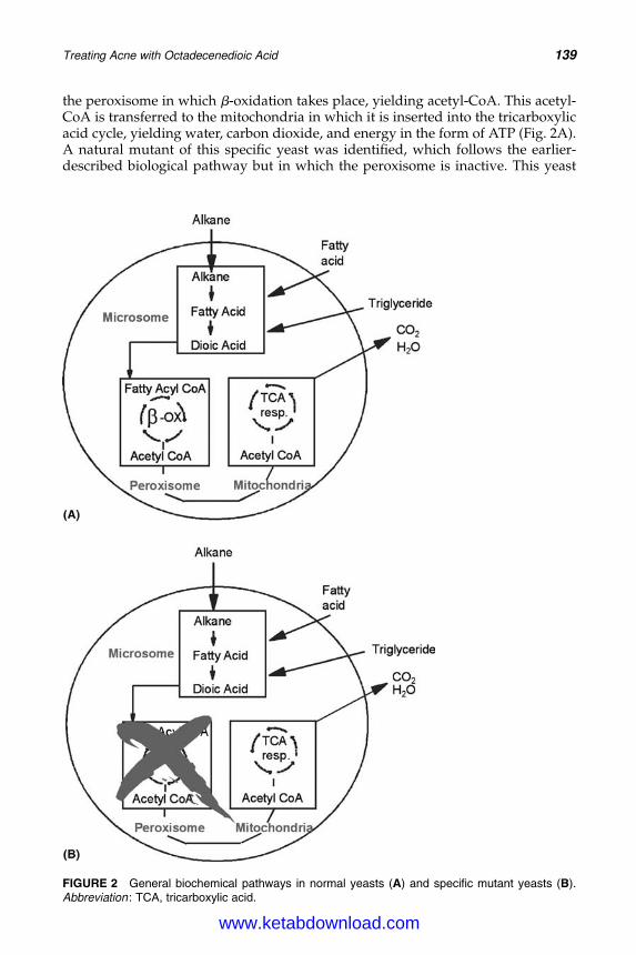
DK4962-Webster-ch12_R2_020407
the peroxisome in which b-oxidation takes place, yielding acetyl-CoA. This acetyl-CoA is transferred to the mitochondria in which it is inserted into the tricarboxylicacid cycle, yielding water, carbon dioxide, and energy in the form of ATP (Fig. 2A).A natural mutant of this specific yeast was identified, which follows the earlier-described biological pathway but in which the peroxisome is inactive. This yeast
FIGURE 2 General biochemical pathways in normal yeasts (A) and specific mutant yeasts (B).Abbreviation : TCA, tricarboxylic acid.
Treating Acne with Octadecenedioic Acid 139
www.ketabdownload.com
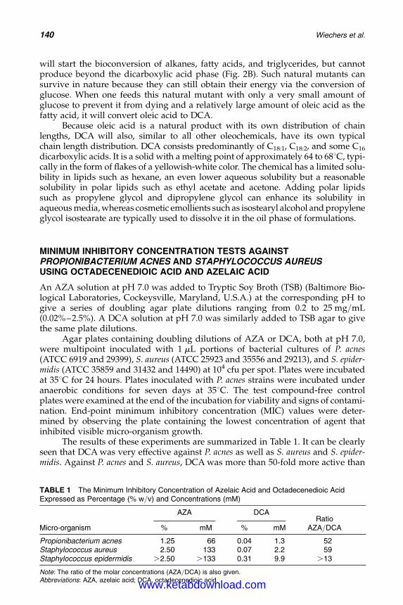
DK4962-Webster-ch12_R2_020407
will start the bioconversion of alkanes, fatty acids, and triglycerides, but cannotproduce beyond the dicarboxylic acid phase (Fig. 2B). Such natural mutants cansurvive in nature because they can still obtain their energy via the conversion ofglucose. When one feeds this natural mutant with only a very small amount ofglucose to prevent it from dying and a relatively large amount of oleic acid as thefatty acid, it will convert oleic acid to DCA.
Because oleic acid is a natural product with its own distribution of chainlengths, DCA will also, similar to all other oleochemicals, have its own typicalchain length distribution. DCA consists predominantly of C18:1, C18:2, and some C16
dicarboxylic acids. It is a solid with a melting point of approximately 64 to 688C, typi-cally in the form of flakes of a yellowish-white color. The chemical has a limited solu-bility in lipids such as hexane, an even lower aqueous solubility but a reasonablesolubility in polar lipids such as ethyl acetate and acetone. Adding polar lipidssuch as propylene glycol and dipropylene glycol can enhance its solubility inaqueous media, whereas cosmetic emollients such as isostearyl alcohol and propyleneglycol isostearate are typically used to dissolve it in the oil phase of formulations.
MINIMUM INHIBITORY CONCENTRATION TESTS AGAINSTPROPIONIBACTERIUM ACNES AND STAPHYLOCOCCUS AUREUSUSING OCTADECENEDIOIC ACID AND AZELAIC ACID
An AZA solution at pH 7.0 was added to Tryptic Soy Broth (TSB) (Baltimore Bio-logical Laboratories, Cockeysville, Maryland, U.S.A.) at the corresponding pH togive a series of doubling agar plate dilutions ranging from 0.2 to 25 mg/mL(0.02%–2.5%). A DCA solution at pH 7.0 was similarly added to TSB agar to givethe same plate dilutions.
Agar plates containing doubling dilutions of AZA or DCA, both at pH 7.0,were multipoint inoculated with 1 mL portions of bacterial cultures of P. acnes(ATCC 6919 and 29399), S. aureus (ATCC 25923 and 35556 and 29213), and S. epider-midis (ATCC 35859 and 31432 and 14490) at 104 cfu per spot. Plates were incubatedat 358C for 24 hours. Plates inoculated with P. acnes strains were incubated underanaerobic conditions for seven days at 358C. The test compound-free controlplates were examined at the end of the incubation for viability and signs of contami-nation. End-point minimum inhibitory concentration (MIC) values were deter-mined by observing the plate containing the lowest concentration of agent thatinhibited visible micro-organism growth.
The results of these experiments are summarized in Table 1. It can be clearlyseen that DCA was very effective against P. acnes as well as S. aureus and S. epider-midis. Against P. acnes and S. aureus, DCA was more than 50-fold more active than
TABLE 1 The Minimum Inhibitory Concentration of Azelaic Acid and Octadecenedioic AcidExpressed as Percentage (% w/v) and Concentrations (mM)
Micro-organism
AZA DCARatio
AZA/DCA% mM % mM
Propionibacterium acnes 1.25 66 0.04 1.3 52Staphylococcus aureus 2.50 133 0.07 2.2 59Staphylococcus epidermidis .2.50 .133 0.31 9.9 .13
Note: The ratio of the molar concentrations (AZA/DCA) is also given.Abbreviations: AZA, azelaic acid; DCA, octadecenedioic acid.
140 Wiechers et al.
www.ketabdownload.com
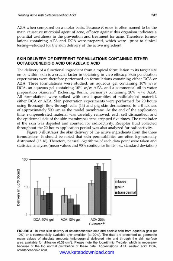
DK4962-Webster-ch12_R2_020407
AZA when compared on a molar basis. Because P. acnes is often named to be themain causative microbial agent of acne, efficacy against this organism indicates apotential usefulness in the prevention and treatment for acne. Therefore, formu-lations containing AZA and DCA were prepared, which were—prior to clinicaltesting—studied for the skin delivery of the active ingredient.
SKIN DELIVERY OF DIFFERENT FORMULATIONS CONTAINING EITHEROCTADECENEDIOIC ACID OR AZELAIC ACID
The delivery of a functional ingredient from a topical formulation to its target siteon or within skin is a crucial factor in obtaining in vivo efficacy. Skin penetrationexperiments were therefore performed on formulations containing either DCA orAZA. Three formulations were studied: an aqueous gel containing 10% w/wDCA, an aqueous gel containing 10% w/w AZA, and a commercial oil-in-waterpreparation Skinorenw (Schering, Berlin, Germany) containing 20% w/w AZA.All formulations were spiked with small quantities of radiolabeled material,either DCA or AZA. Skin penetration experiments were performed for 20 hoursusing Bronaugh flow-through cells (14) and pig skin dermatomed to a thicknessof approximately 500 mm as the model membrane. At the end of the applicationtime, nonpenetrated material was carefully removed, each cell dismantled, andthe epidermal side of the skin membranes tape-stripped five times. The remainderof the skin was digested and counted for radioactivity. Receptor fluid collectedthroughout the 20-hours application period was also analyzed for radioactivity.
Figure 3 illustrates the skin delivery of the active ingredients from the threeformulations. It should be noted that skin permeabilities are often log-normallydistributed (15,16). Therefore, natural logarithms of each data point were taken andstatistical analyses (mean values and 95% confidence limits, i.e., standard deviation)
FIGURE 3 In vitro skin delivery of octadecenedioic acid and azelaic acid from aqueous gels (at10%) or a commercially available o/w emulsion (at 20%). The data are presented as geometricmean values of absolute amounts (micrograms) delivered into and through the skin surfacearea available for diffusion (0.38 cm2). Please note the logarithmic Y-scale, which is necessarybecause of the log normal distribution of these data. Abbreviations: AZA, azelaic acid; DCA,octadecenedioic acid.
Treating Acne with Octadecenedioic Acid 141
www.ketabdownload.com
DK4962-Webster-ch12_R2_020407
performed on the transformed data. This is the reason why the data in Figure 3 arepresented on a logarithmic scale. Geometric mean values, which are listed inTable 2, were calculated by back transformation of the logarithmic mean values.
As was to be expected, the delivery of AZA from the marketed anti-acnepreparation was by far the best. Not only did it show the highest penetrationof the three formulations (absolute delivery expressed as mg/0.38 cm2), it alsodelivered more than half of the delivered AZA to the skin fraction, which isthe fraction in human skin where the sebaceous glands are located. Bothaqueous gels containing either DCA or AZA at 10% w/w showed a substantiallower absolute delivery. The relative distribution profiles of the two active ingre-dients from the three formulations were not that different (Table 2), although arelatively higher percentage of DCA penetrated transdermally (30.3% vs. 20.6%for DCA and AZA, respectively). The subdivision in deposition of deliveredmaterial in tape strips, skin, and transdermal, however, was not precise enoughto predict the in vivo efficacy of these gels. Therefore, additional autoradiographyexperiments were performed in which skin sections were cut perpendicular to theskin surface and placed onto microscopy slides coated with photographic emul-sions and left for 7 to 14 days before processing the skin sample for histologicalstaining.
The distribution of the radiolabeled DCA and AZA was only somewhatdifferent. For all formulations, the silver grains were concentrated at the skinsurface and the stratum corneum (Fig. 4), in line with skin-delivery data presentedin Table 2 and Figure 3. All three formulations showed active ingredient to bepresent in the upper part of some follicles, especially the infundibulum. Therewas no penetrant visible deep in the follicle in any of the sections viewed. Itshould be noted that the heavy graining adjacent to the stratum corneum, ratherthan directly over it, is the result of the cryostat sections which are not prefixedand shrink during staining. The only real difference in skin deposition betweenthe two penetrants was the fact that silver grains were more evident in the viableepidermis when using AZA (Fig. 4D and H) than when DCA was the penetrant(Fig. 4F). DCA appeared to be associated only with stratum corneum and hair fol-licles. This can be explained by the relative higher water solubility of AZA andhence a higher solubility in the viable epidermis/dermis.
Bojar et al. (17) found the transfollicular delivery of AZA from a 20% cream tobe comparable with the concentration required to inhibit the in vitro growth ofP. acnes and S. epidermidis (17). In the experiments described earlier, we couldconfirm the transfollicular delivery of AZA (Fig. 5). DCA was more active thanAZA in inhibiting the in vitro growth of P. acnes and S. epidermidis (see earlier),suggesting that lower concentrations of this lipophilic molecule would need to betransfollicularly delivered. In addition, recent transfollicular delivery research indi-cated that delivery to the hair follicle is improved by increasing the lipophilicity ofthe penetrating molecule (18,19). Relative to AZA, DCA combines a higher antimi-crobial activity with a preference for transfollicular delivery. The logical next step,therefore, was to perform in vivo clinical studies.
PHASE I CLINICAL TRIAL USING OCTADECENEDIOIC ACID,AZELAIC ACID, AND BENZOYL PEROXIDE
When new chemical entities are tested on human skin, it is advisable to firstconduct studies on healthy volunteers (so-called Phase I trials) prior to conducting
142 Wiechers et al.
www.ketabdownload.com
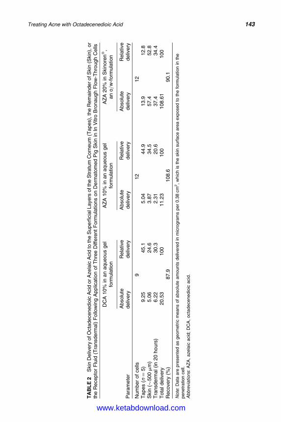
DK4962-Webster-ch12_R2_020407
TA
BL
E2
Skin
Deliv
ery
ofO
cta
decenedio
icA
cid
or
Azela
icA
cid
toth
eS
uperfi
cia
lLayers
ofth
eS
tratu
mC
orn
eum
(Tapes)
,th
eR
em
ain
der
ofS
kin
(Skin
),or
the
Recepto
rF
luid
(Tra
nsderm
al)
Follo
win
gA
pplic
ation
of
Thre
eD
iffere
nt
Form
ula
tions
on
Derm
ato
med
Pig
Skin
inIn
Vitro
Bro
naugh
Flo
w-T
hro
ugh
Cells
Para
mete
r
DC
A10%
inan
aqueous
gel
form
ula
tion
AZ
A10%
inan
aqueous
gel
form
ula
tion
AZ
A20%
inS
kin
ore
nw
,an
o/w
-form
ula
tion
Absolu
tedeliv
ery
Rela
tive
deliv
ery
Absolu
tedeliv
ery
Rela
tive
deliv
ery
Absolu
tedeliv
ery
Rela
tive
deliv
ery
Num
ber
of
cells
912
12
Tapes
(n¼
5)
9.2
545.1
5.0
444.9
13.9
12.8
Skin
(�500m
m)
5.0
624.6
3.8
734.5
57.4
52.8
Tra
nsd
erm
al(in
20
hours
)6.2
230.3
2.3
120.6
37.4
34.4
Tota
ldeliv
ery
20.5
3100
11.2
3100
108.6
1100
Recove
ry(%
)87.9
108.6
90.1
Note
:D
ata
are
pre
sente
das
geom
etr
icm
eans
of
absolu
team
ounts
deliv
ere
din
mic
rogra
ms
per
0.3
8cm
2,
whic
his
the
skin
surf
ace
are
aexposed
toth
efo
rmula
tion
inth
epenetr
ation
cell.
Abbre
via
tions:
AZ
A,
azela
icacid
;D
CA
,octa
decenedio
icacid
.
Treating Acne with Octadecenedioic Acid 143
www.ketabdownload.com
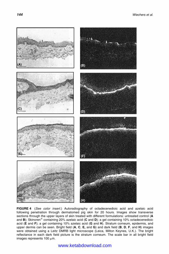
DK4962-Webster-ch12_R2_020407
FIGURE 4 (See color insert.) Autoradiography of octadecenedioic acid and azelaic acidfollowing penetration through dermatomed pig skin for 20 hours. Images show transversesections through the upper layers of skin treated with different formulations: untreated control (Aand B); Skinorenw containing 20% azelaic acid (C and D); a gel containing 10% octadecenedioicacid (E and F); a gel containing 10% azelaic acid (G and H). Stratum corneum, epidermis, andupper dermis can be seen. Bright field (A, C, E, and G) and dark field (B, D, F, and H) imageswere obtained using a Leitz DMRB light microscope (Leica, Milton Keynes, U.K.). The brightreflectance in each dark field picture is the stratum corneum. The scale bar in all bright fieldimages represents 100 mm.
144 Wiechers et al.
www.ketabdownload.com
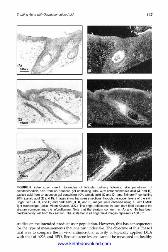
DK4962-Webster-ch12_R2_020407
studies on the intended product-user population. However, this has consequencesfor the type of measurements that one can undertake. The objective of this Phase Itrial was to compare the in vivo antimicrobial activity of topically applied DCAwith that of AZA and BPO. Because acne lesions cannot be measured on healthy
FIGURE 5 (See color insert.) Examples of follicular delivery following skin penetration ofoctadecenedioic acid from an aqueous gel containing 10% w/w octadecenedioic acid (A and B),azelaic acid from an aqueous gel containing 10% azelaic acid (C and D), and Skinorenw containing20% azelaic acid (E and F). Images show transverse sections through the upper layers of the skin.Bright field (A, C, and E) and dark field (B, D, and F) images were obtained using a Leitz DMRBlight microscope (Leica, Milton Keynes, U.K.). The bright reflectance in each dark field picture is thestratum corneum and the infundibulum. Note that the stratum corneum in (A) and (B) has beenpredominantly lost from this section. The scale bar in all bright field images represents 100 mm.
Treating Acne with Octadecenedioic Acid 145
www.ketabdownload.com

DK4962-Webster-ch12_R2_020407
volunteers, the basis of this trial was to measure changes in the skin surface micro-flora count (Micrococcaceae and Propionibacteria) and skin-surface free fatty acid(FFA) levels on the faces of healthy volunteers as an indicator of potential anti-acne activity. The trial was performed as a double-blind study.
A group of 59 volunteers (37 male, 22 female) aged 16 to 35 were recruited intothe study. All volunteers were medically screened to ensure their suitability toundertake the study. Their willingness to participate was documented throughthe completion of an informed consent form in the presence of medical staff.
Baseline levels of viable propionibacteria on the surface of the skin on theright cheek of each volunteer were determined seven days prior to commencementof the trial. The volunteers were assigned to one of five groups, as determined byprevisit numbers of skin surface propionibacteria, so that each group had asimilar statistical distribution of numbers of viable propionibacteria:
1. The first group (n ¼ 12) received the aqueous gel containing 10% w/w DCA,which was also investigated for skin delivery.
2. The second group (n ¼ 12) received the same aqueous gel formulation withoutthe DCA (placebo).
3. The formulation of the third group (n ¼ 12) was the same aqueous gel again,but this time with 10% w/w AZA.
4. The fourth group (n ¼ 12) received “Panoxyl Aquagel 5,” a commerciallyavailable product containing 5% BPO (Stiefel Laboratories, High Wycombe,Bucks, U.K.).
5. The fifth group (n ¼ 11), finally, received Skinorenw, the commercially availableproduct from Schering containing 20% AZA.
All volunteers were provided with a non-antimicrobial soap and instructed to washthe whole face twice daily prior to application of treatment products. Each volun-teer applied one of the products to the test site (right-hand side of the face includingforehead, just beyond the midline, cheek, and chin, avoiding the eyes, nostrils, andmouth) for the 21 days of duration of the trial.
The surface microbial populations (Micrococcaceae and Propionibacteria)were enumerated on the right cheek at baseline and after 3, 7, 14, and 21 days oftreatment. Sebum was collected from each volunteer at baseline and after 7, 14,and 21 days of treatment, and the FFA content was determined by gas–liquidchromatography. Volunteers were also asked to maintain a daily diary and noteany positive or negative effects of the treatment. Data were analyzed using para-metric and nonparametric statistical tests.
The main findings of the study were as follows:
1. Of the 59 volunteers recruited into the study, only six individuals failed to com-plete the study (10.2% drop-out rate). The dropouts were evenly spread overthe treatment groups, and there was no evidence that adverse reactions toany one treatment were responsible for noncompletion. Only those volunteerswho completed the treatment were included in the subsequent statistical analysisof the results.
2. All preparations containing dicarboxylic acids (both AZA and DCA) were welltolerated. In the BPO-treated group, 92% of volunteers reported adverse events(irritancy and drying of the skin). However, the volunteers in this group wereable to maintain the stipulated treatment regime throughout the study,although some were advised to use a moisturizing lotion.
3. None of the treatment regimes had any detectable effect on skin surface FFAs.
146 Wiechers et al.
www.ketabdownload.com
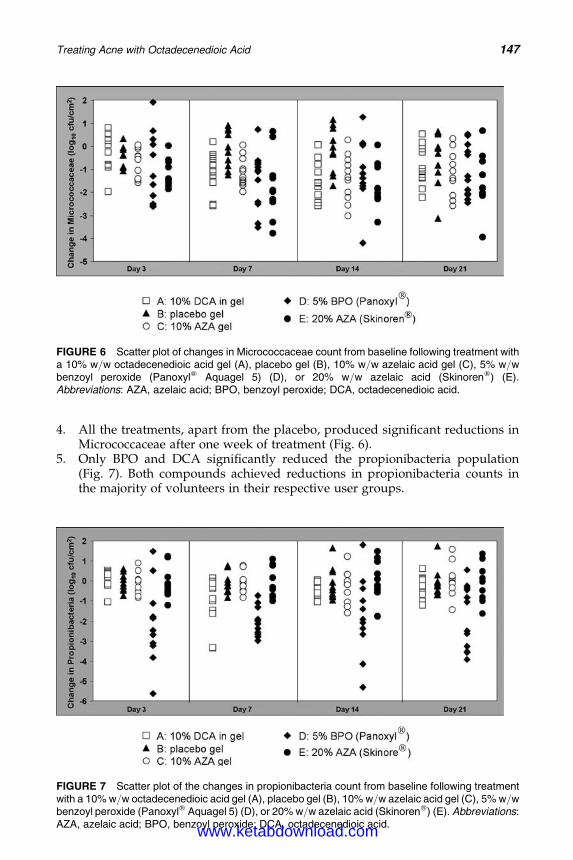
DK4962-Webster-ch12_R2_020407
4. All the treatments, apart from the placebo, produced significant reductions inMicrococcaceae after one week of treatment (Fig. 6).
5. Only BPO and DCA significantly reduced the propionibacteria population(Fig. 7). Both compounds achieved reductions in propionibacteria counts inthe majority of volunteers in their respective user groups.
FIGURE 6 Scatter plot of changes in Micrococcaceae count from baseline following treatment witha 10% w/w octadecenedioic acid gel (A), placebo gel (B), 10% w/w azelaic acid gel (C), 5% w/wbenzoyl peroxide (Panoxylw Aquagel 5) (D), or 20% w/w azelaic acid (Skinorenw) (E).Abbreviations: AZA, azelaic acid; BPO, benzoyl peroxide; DCA, octadecenedioic acid.
FIGURE 7 Scatter plot of the changes in propionibacteria count from baseline following treatmentwith a 10% w/w octadecenedioic acid gel (A), placebo gel (B), 10% w/w azelaic acid gel (C), 5% w/wbenzoyl peroxide (Panoxylw Aquagel 5) (D), or 20% w/w azelaic acid (Skinorenw) (E). Abbreviations:AZA, azelaic acid; BPO, benzoyl peroxide; DCA, octadecenedioic acid.
Treating Acne with Octadecenedioic Acid 147
www.ketabdownload.com
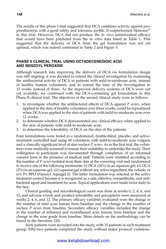
DK4962-Webster-ch12_R2_020407
The results of this phase I trial suggested that DCA combines activity against pro-pionibacteria with a good safety and tolerance profile. It outperformed Skinorenw
in this trial. However, DCA did not produce the in vivo antimicrobial efficacythat would have been predicted from the in vitro data listed in Table 1. Thissuggested that the delivery of DCA from the gel formulation was not yetoptimal, which was indeed confirmed in Table 2 and Figure 3.
PHASE II CLINICAL TRIAL USING OCTADECENEDIOIC ACIDAND BENZOYL PEROXIDE
Although research into improving the delivery of DCA via formulation designwas still ongoing, it was decided to extend the clinical investigation by examiningthe antibacterial activity of DCA in patients with mild-to-moderate acne, insteadof healthy human volunteers, and to extend the time of the investigation to12 weeks instead of three. As the improved delivery systems of DCA were notyet available, we continued with the DCA-containing gel formulation in thisPhase II clinical trial. The objectives of the second clinical study were as follows:
1. to investigate whether the antibacterial effects of DCA against P. acnes, whenapplied to the skin of healthy volunteers over three weeks, could be reproducedwhen DCA was applied to the skin of patients with mild-to-moderate acne over12 weeks;
2. to determine whether DCA demonstrated any clinical efficacy when applied tothe skin of patients with mild-to-moderate acne;
3. to determine the tolerability of DCA on the skin of the patients.
Four formulations were tested in a randomized, double-blind, placebo, and active-treatment controlled trial using 60 volunteers with mild-to-moderate acne vulgarisand a clinically significant level of skin surface P. acnes. As in the first trial, the volun-teers were medically screened to ensure their suitability to undertake the study. Theirwillingness to participate was documented through completion of an informedconsent form in the presence of medical staff. Patients were stratified according tothe number of P. acnes isolated from their skin at the screening visit and randomizedto receive one of the following treatments: (i) DCA (10%) in an aqueous gel; (ii) DCA(2%) in an aqueous gel; (iii) aqueous gel without any active ingredient, the vehicle; or(iv) 5% BPO (Panoxyl Aquagel 5). The latter formulation was selected as the activetreatment control because it is recognized as a safe, effective, nonantibiotic, and anti-bacterial agent and treatment for acne. Topical applications were made twice daily tothe face.
Clinical grading and microbiological count was done at weeks 0, 2, 4, 6, and12, and adverse events and product tolerability and perceptions were measured atweeks 2, 4, 6, and 12. The primary efficacy variables evaluated were the change inthe number of total acne lesions from baseline and the change in the number ofsurface P. acnes from baseline. Secondary efficacy variables included the changein the number of inflamed and noninflamed acne lesions from baseline and thechange in the acne grade from baseline. More details on the methodology can befound in the literature (20).
Sixty patients were recruited into the study, with 15 patients in each treatmentgroup. Fifty-two patients completed the study without major protocol violations.
148 Wiechers et al.
www.ketabdownload.com
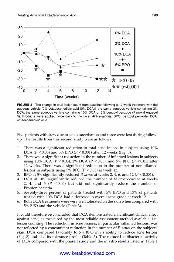
DK4962-Webster-ch12_R2_020407
Five patients withdrew due to acne exacerbation and three were lost during follow-up. The results from this second study were as follows:
1. There was a significant reduction in total acne lesions in subjects using 10%DCA (P ,0.05) and 5% BPO (P ,0.001) after 12 weeks (Fig. 8).
2. There was a significant reduction in the number of inflamed lesions in subjectsusing 10% DCA (P ,0.05), 2% DCA (P ,0.05), and 5% BPO (P ,0.01) after12 weeks. There was a significant reduction in the number of noninflamedlesions in subjects using 5% BPO (P ,0.05) at week 12.
3. BPO at 5% significantly reduced P. acnes at weeks 2, 4, 6, and 12 (P ,0.001).4. DCA at 10% significantly reduced the number of Micrococcaceae at weeks
2, 4, and 6 (P ,0.05) but did not significantly reduce the number ofPropionibacteria.
5. Seventy-three percent of patients treated with 5% BPO and 53% of patientstreated with 10% DCA had a decrease in overall acne grade at week 12.
6. Both DCA treatments were very well tolerated on the skin when compared with5% BPO and the vehicle (Table 3).
It could therefore be concluded that DCA demonstrated a significant clinical effectagainst acne, as measured by the most reliable assessment method available, i.e.,lesion counting. The reduction in acne lesions, in particular inflamed lesions, wasnot reflected by a concomitant reduction in the number of P. acnes on the subjects’skin. DCA compared favorably to 5% BPO in its ability to reduce acne lesions(Fig. 8) and also its tolerance profile (Table 3). The reduced antibacterial activityof DCA compared with the phase I study and the in vitro results listed in Table 1
FIGURE 8 The change in total lesion count from baseline following a 12-week treatment with theaqueous vehicle [0% octadecenedioic acid (0% DCA)], the same aqueous vehicle containing 2%DCA, the same aqueous vehicle containing 10% DCA or 5% benzoyl peroxide (Panoxyl Aquagel5). Products were applied twice daily to the face. Abbreviations: BPO, benzoyl peroxide; DCA,octadecenedioic acid.
Treating Acne with Octadecenedioic Acid 149
www.ketabdownload.com
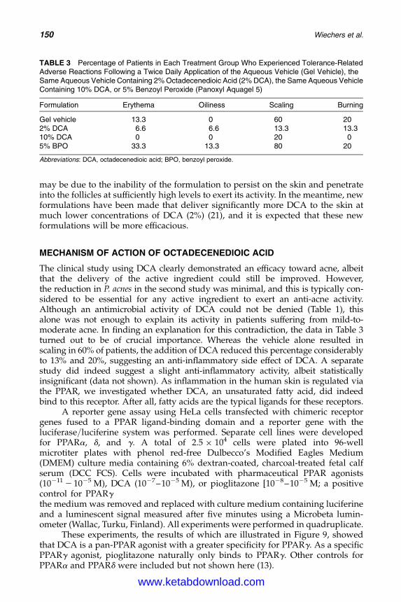
DK4962-Webster-ch12_R2_020407
may be due to the inability of the formulation to persist on the skin and penetrateinto the follicles at sufficiently high levels to exert its activity. In the meantime, newformulations have been made that deliver significantly more DCA to the skin atmuch lower concentrations of DCA (2%) (21), and it is expected that these newformulations will be more efficacious.
MECHANISM OF ACTION OF OCTADECENEDIOIC ACID
The clinical study using DCA clearly demonstrated an efficacy toward acne, albeitthat the delivery of the active ingredient could still be improved. However,the reduction in P. acnes in the second study was minimal, and this is typically con-sidered to be essential for any active ingredient to exert an anti-acne activity.Although an antimicrobial activity of DCA could not be denied (Table 1), thisalone was not enough to explain its activity in patients suffering from mild-to-moderate acne. In finding an explanation for this contradiction, the data in Table 3turned out to be of crucial importance. Whereas the vehicle alone resulted inscaling in 60% of patients, the addition of DCA reduced this percentage considerablyto 13% and 20%, suggesting an anti-inflammatory side effect of DCA. A separatestudy did indeed suggest a slight anti-inflammatory activity, albeit statisticallyinsignificant (data not shown). As inflammation in the human skin is regulated viathe PPAR, we investigated whether DCA, an unsaturated fatty acid, did indeedbind to this receptor. After all, fatty acids are the typical ligands for these receptors.
A reporter gene assay using HeLa cells transfected with chimeric receptorgenes fused to a PPAR ligand-binding domain and a reporter gene with theluciferase/luciferine system was performed. Separate cell lines were developedfor PPARa, d, and g. A total of 2.5 � 104 cells were plated into 96-wellmicrotiter plates with phenol red-free Dulbecco’s Modified Eagles Medium(DMEM) culture media containing 6% dextran-coated, charcoal-treated fetal calfserum (DCC FCS). Cells were incubated with pharmaceutical PPAR agonists(10211 2 1025 M), DCA (1027–1025 M), or pioglitazone [1028–1025 M; a positivecontrol for PPARg binding (22)] for 16 hours, and at the end of the incubation,the medium was removed and replaced with culture medium containing luciferineand a luminescent signal measured after five minutes using a Microbeta lumin-ometer (Wallac, Turku, Finland). All experiments were performed in quadruplicate.
These experiments, the results of which are illustrated in Figure 9, showedthat DCA is a pan-PPAR agonist with a greater specificity for PPARg. As a specificPPARg agonist, pioglitazone naturally only binds to PPARg. Other controls forPPARa and PPARd were included but not shown here (13).
TABLE 3 Percentage of Patients in Each Treatment Group Who Experienced Tolerance-RelatedAdverse Reactions Following a Twice Daily Application of the Aqueous Vehicle (Gel Vehicle), theSame Aqueous Vehicle Containing 2% Octadecenedioic Acid (2% DCA), the Same Aqueous VehicleContaining 10% DCA, or 5% Benzoyl Peroxide (Panoxyl Aquagel 5)
Formulation Erythema Oiliness Scaling Burning
Gel vehicle 13.3 0 60 202% DCA 6.6 6.6 13.3 13.310% DCA 0 0 20 05% BPO 33.3 13.3 80 20
Abbreviations: DCA, octadecenedioic acid; BPO, benzoyl peroxide.
150 Wiechers et al.
www.ketabdownload.com
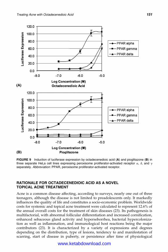
DK4962-Webster-ch12_R2_020407
RATIONALE FOR OCTADECENEDIOIC ACID AS A NOVELTOPICAL ACNE TREATMENT
Acne is a common disease affecting, according to surveys, nearly one out of threeteenagers, although the disease is not limited to preadolescents only. It markedlyinfluences the quality of life and constitutes a socio-economic problem. Worldwidecosts for systemic and topical acne treatment were calculated to represent 12.6% ofthe annual overall costs for the treatment of skin diseases (23). Its pathogenesis ismultifactorial, with abnormal follicular differentiation and increased cornification,enhanced sebaceous gland activity and hyperseborrhea, bacterial hypercoloniza-tion as well as inflammation, and immunological host reactions being the majorcontributors (23). It is characterized by a variety of expressions and degreesdepending on the distribution, type of lesions, tendency to and manifestation ofscarring, start of disease in puberty, or persistence after time of physiological
FIGURE 9 Induction of luciferase expression by octadecenedioic acid (A) and pioglitazone (B) inthree separate HeLa cell lines expressing peroxisome proliferator-activated receptor a, d, and g
separately. Abbreviation: PPAR, peroxisome proliferator-activated receptor.
Treating Acne with Octadecenedioic Acid 151
www.ketabdownload.com
DK4962-Webster-ch12_R2_020407
regression, each form requiring a different active ingredient or drug or combinationthereof (24).
There are basically four modes of action for treating acne in line with thepathogenesis described earlier: most anti-acne agents have an antimicrobial activityto counteract the hypercolonization (in particular, e.g., BPO), some agents interactwith the hyperkeratinization aspect of acne to fight the abnormal follicular differen-tiation and increased cornification (in particular, the retinoids such as tretinoin, iso-tretinoin, and adapalene), a few have only a moderate anti-inflammatory activity(e.g., adapalene, AZA, and BPO), whereas none have a strong sebo-suppressiveeffect to fight the hyperseborrhea (24). Because of the complexity of the disease, itis very difficult to differentiate between cause and effect, and as a consequence,relapses often occur because the fundamental underlying cause may not havebeen dealt with. As a consequence, combination therapies are popular, involvingboth the topical and the oral route of administration. Mammone et al. (25), forinstance, proposed a combination of chemicals with antimicrobial, anti-inflammatory,and antiandrogen properties and a desquamation enhancer, resulting in a statisticallysignificant higher reduction in noninflamed lesions relative to a 5% BPO gel, whereasthere was no statistically significant additional benefit toward inflamed lesions.The downside of combination therapy, however, is that this increases the chance ofside effects, as most of these products have some. AZA, for instance, causes a verystrong burning and BPO results in strong scaling and erythema (24), which was con-firmed in our studies (Table 3).
Here, we report the use of DCA as a novel topical treatment of acne thatinvolves all four modes of action within a single molecule. Similar to most otheranti-acne agents, DCA has an antimicrobial activity as indicated in Table 1. Allthree other modes of action may be produced via its binding to PPAR, as shownin Figure 9, which illustrates its capability to act as a pan-PPAR agonist. Bindingto PPARg is most pronounced, followed by that to PPARa and PPARd. As shownby Downie et al. (5), activators of PPARa and PPARg inhibit the rate of sebaceouslipogenesis and reduce the synthesis of the sebum-specific lipids squalane and tria-cylglycerol in human sebaceous glands. DCA, as a PPARg and PPARa agonist, istherefore likely to do the same, provided it is delivered well enough. The import-ance of PPAR in the regulation of lipid synthesis and metabolism in human sebo-cytes is recognized, albeit far from clear yet. Makrantonaki and Zouboulis (26),for instance, described the stimulatory interactions between androgens and PPARligands, similar to the natural PPARd/g ligand linoleic acid, on neutral and polarlipid synthesis. Although linoleic acid is stimulating lipid synthesis in vitro inthis communication (26), topical administration of 2.5% linoleic acid in a Carbopolgel formulation for one month resulted in a significant reduction (25%) in theoverall size of follicular casts and microcomedones, whereas no change wasfound at placebo-treated sites (27). Further research will be necessary to elucidatethe interrelationships between all factors involved.
DCA, however, is one of the very few molecules that has the potential tointeract at all levels with acne: enhanced differentiation to prevent the increasedcornification of the sebaceous duct (via PPARd), reduction in sebum productionto counteract hyperseborrhea (via PPARg), and reduction in inflammation(PPARa). These PPAR-related effects combined with its demonstrated antimicro-bial properties to overcome the hypercolonization with microbes such as P. acnesand S. aureus, make DCA a viable nonantibiotic alternative for the treatmentof acne.
152 Wiechers et al.
www.ketabdownload.com

DK4962-Webster-ch12_R2_020407
REFERENCES
1. Knaggs H, Wood EJ, Rizer RL, et al. Post-adolescent acne. Int J Cosmet Sci 2004;26(1):129–138.
2. Krautheim A, Gollnick H. Transdermal penetration of topical drugs used in the treat-ment of acne. Clin Pharmacokinet 2003; 42:1287–1304.
3. Gollnick HPM, Krautheim A. Topical treatment of acne: Current status and futureaspects. Dermatology 2003; 206:29–36.
4. Gollnick HPM, Graupe K, Zaumseil R-P.15% Azelainsauregel in der Behandlung derAkne. Zwei doppelblinde klinische Vergleichstudien. JDDG 2004; 2:841–847.
5. Downie MMT, Sanders DA, Maier LM, et al. Peroxisome proliferator-activatedreceptor and farnesoid X receptor ligands differentially regulate sebaceous differen-tiation in human sebaceous gland organ cultures in vitro. Br J Dermatol 2004;151:766–775.
6. Berger J, Moller DE. The mechanisms of action of PPARs. Annu Rev Med 2002;53:409–435.
7. Komuves LG, Hanley K, Lefebvre A-M, et al. Stimulation of PPARa promotes epidermalkeratinocyte differentiation in-vivo. J Invest Dermatol 2000; 115:353–360.
8. Westergaard M, Henningsen J, Svendsen ML, et al. Modulation of keratinocyte geneexpression and differentiation by PPAR—selective ligands and tetradecylthioaceticacid. J Invest Dermatol 2001; 116:702–712.
9. Kippenberger S, Loitsch SM, Grundmann-Kollmann M, et al. Activators of peroxisomeproliferator-activated receptors protect human skin from ultraviolet-B-light-inducedinflammation. J Invest Dermatol 2001; 117:1430–1436.
10. Sheu MY, Fowler AJ, Kao J, et al. Topical peroxisome proliferator activated receptor-aactivators reduce inflammation in irritant and allergic contact dermatitis models.J Invest Dermatol 2002; 118:94–101.
11. Mossner R, Schulz U, Kruger U, et al. Agonists of peroxisome proliferator-activatedreceptor gamma inhibit cell growth in malignant melanoma. J Invest Dermatol 2002;119:576–582.
12. Placha W, Gil D, Dembinska-Kiec A, et al. The effect of PPARg on the proliferation andapoptosis of human melanoma cells. Melanoma Res 2003; 13:447–456.
13. Wiechers JW, Rawlings AV, Garcia C, et al. A new mechanism of action for skin whiteningagents: Binding to the peroxisome proliferator-activated receptor (PPAR). Int J CosmetSci 2005; 27:123–132.
14. Bronaugh RL, Stewart RF. Methods for in vitro percutaneous absorption studies. IV. Theflow-through diffusion cell. J Pharm Sci 1985; 74:69–77.
15. Williams AC, Cornwell PA, Barry BW. On the non-Gaussian distribution of human skinpermeabilities. Int J Pharm 1992; 86:69–77.
16. Cornwell PA, Barry BW. Effect of penetration enhancers on the statistical distribution ofhuman skin permeabilities. Int J Pharm 1995; 117:101–112.
17. Bojar RA, Cutcliffe AG, Graupe K, et al. Follicular concentrations of azelaic acid after asingle topical application. Br J Dermatol 1993; 129:399–402.
18. Grams YY, Bouwstra JA. Penetration and distribution of three lipophilic probes in vitro inhuman skin focussing on the hair follicle. J Control Release 2002; 83:253–262.
19. Grams YY, Alaruikka S, Lashley L, et al. Permeant lipophilicity and vehicle compositioninfluence on accumulation of dyes in hair follicles of human skin. Eur J Pharm Sci 2003;18:329–336.
20. Burke BM, Cunliffe WJ. The assessment of acne vulgaris. The Leeds technique.Br J Dermatol 1984; 111:83–92.
21. Wiechers JW, Kelly CL, Blease TG, et al. Formulating for efficacy. Int J Cosmet Sci 2004;26:173–182.
22. Bhagavathula N, Nerusu KC, Lal A, et al. Rosiglitazone inhibits proliferation, motility,and matrix metalloproteinase production in keratinocytes. J Invest Dermatol 2004;122:130–139.
23. Zouboulis CC. Acne pathogenesis: New experimental findings elucidate the involve-ment of androgens, PPAR ligands and regulatory neuropeptides. J Invest Dermatol2005; 124:A 48, #284.
Treating Acne with Octadecenedioic Acid 153
www.ketabdownload.com

DK4962-Webster-ch12_R2_020407
24. Gollnick H, Schramm A. Topical drug treatment in acne. Dermatology 1998; 196:119–125.25. Mammone T, Maes D, Gan D, et al. A multi-prong approach to the treatment of acne.
J Invest Dermatol 2005; 124:A15, #086.26. Makrantonaki E, Zouboulis CC. The effect of androgens on lipid metabolism in human
sebocytes occurs in interaction with PPAR ligands. J Invest Dermatol 2005; 124:A109,#649.
27. Letawe C, Boone M, Pierard GE. Image analysis of the effect of topically applied linoleicacid on acne microcomedones. Clin Exp Dermatol 1998; 23:56–58.
154 Wiechers et al.
www.ketabdownload.com

DK4962-Webster-ch13_R2_020407
B13 The Effect of Sphingolipids as a NewTherapeutic Option for Acne Treatment
Saskia K. Klee, Mike Farwick, and Peter LerschDegussa, Goldschmidt Personal Care, Essen, Germany
INTRODUCTION
The skin is one of the largest organs of the human body. It is a highly specializedtissue that acts as a barrier against the influences of the environment. It plays acrucial role in the protection against dehydration and the control of body tempera-ture (1). The skin’s primary role is to protect our health, but the skin’s barrier isimperfect and it also has the ability to absorb external substances. Moreover,these external substances can access healthy skin via its pilosebaceous glands.Drugs have been detected in the blood stream after topical application, demonstrat-ing transdermal delivery through the skin (2). This is desired for the treatment ofmany skin diseases, but this characteristic can also be a contributing factor forcausing many diverse health risks.
The lipid environment of the stratum corneum is an essential factor for main-taining the skin’s equilibrium. Changes in the barrier lipid composition have beendirectly linked to skin barrier function impairments, such as pathologically dry andrough skin (3,4). Besides protective tasks, the skin is confronted with a tremendouschallenge during puberty, when the increase in circulating levels of testosteroneinfluences the function of pilosebaceous units (PSU) and increases sebogenesis.Increased sebum levels are a major contributory factor in acnegenesis. Duringpuberty, the pituitary gland initiates the sexual maturation process via the releaseof gonadotropins and the production of hormones called androgens. This processregulates the production of sebum in the sebaceous glands of both adolescentboys and girls (5). This is the time period in which most adolescents develop amore or less severe form of acne. Although superficial and non-life-threatening,acne is a disease that, if left untreated, can have serious physical and psychologicalconsequences (6,7).
Acne is one of the most common skin diseases of human kind, affecting 80%of adolescent boys and girls during puberty (8) that may persist throughout adult-hood, or it may even develop after puberty (commonly known as persistent or lateonset acne, respectively) (9). Sebum hypersecretion is the first problem associatedwith acne-prone skin and is caused by the enzymatic hyperactivity of 5-alpha-reductase—a key enzyme that converts testosterone into dihydrotestosterone(DHT) (5). By binding to its receptor in the skin, DHT stimulates the secretionof sebum (Fig. 1). Men are more severely affected because they produce higherconcentrations of androgens that regulate sebum production and enlargement ofsebaceous glands (10,11).
The earliest morphological change observed in acne patients is the aberrantfollicular epithelial proliferation and differentiation of the PSU that results in clini-cally invisible microcomedones (12–15). Due to the hormonal imbalance in
155
www.ketabdownload.com
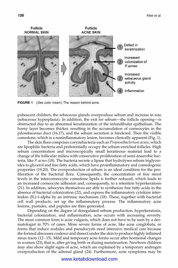
DK4962-Webster-ch13_R2_020407
pubescent children, the sebaceous glands overproduce sebum and increase in size(sebaceous hyperplasia). In addition, the exit for sebum—the follicle opening—isobstructed due to an abnormal keratinization of the infundibular epithelium. Thehorny layer becomes thicker, resulting in the accumulation of corneocytes in thepilosebaceous duct (16,17), and the sebum secretion is hindered. Then the visiblecomedone, which is a noninflammatory lesion, becomes clinically apparent (Fig. 1).
The skin flora comprises corynebacteria such as Propionibacterium acnes, whichare lipophilic bacteria and preferentially occupy the sebum-enriched follicles. Highsebum concentration and microscopically small keratinous material lead to achange of the follicular milieu with consecutive proliferation of semi-anaerobic bac-teria, like P. acnes (18). The bacteria secrete a lipase that hydrolyses sebum triglycer-ides to glycerol and free fatty acids, which have proinflammatory and comedogenicproperties (19,20). The overproduction of sebum is an ideal condition for the pro-liferation of the bacterial flora. Consequently, the concentration of free sterollevels in the intercorneocytic comedone lipids is further reduced, which leads toan increased corneocyte adhesion and, consequently, to a retention hyperkeratosis(21). In addition, sebocytes themselves are able to synthesize free fatty acids in theabsence of bacterial colonization (22), and express the inflammatory cytokine inter-leukin (IL)-1-alpha by an intrinsic mechanism (18). These, together with bacterialcell wall products, set up the inflammatory process. The inflammatory acnelesions, pustules, and papules are then generated.
Depending on the degree of deregulated sebum production, hyperkeratosis,bacterial colonization, and inflammation, acne occurs with increasing severity.The most common form is acne vulgaris, which does not have to be seen by a der-matologist in 70% of cases. More severe forms of acne, like acne conglobata, orforms that induce nodules and pseudocysts need intensive medical care becausethe formed abscesses coalesce and dissect under the skin to produce highly inflamedsinus tracts (12–15). Mild and temporary acne forms occur after hormonal changesin women (23), that is, after giving birth or during menstruation. Newborn childrenmay also show slight signs of acne, which are explained by a temporary androgenoverproduction of the adrenal gland (24). Furthermore, acne symptoms may be
FIGURE 1 (See color insert.) The reason behind acne.
156 Klee et al.
www.ketabdownload.com
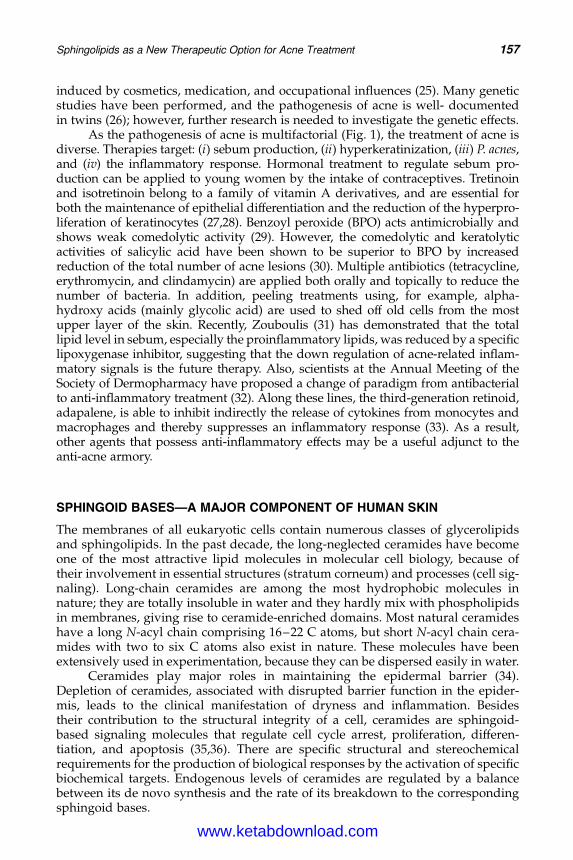
DK4962-Webster-ch13_R2_020407
induced by cosmetics, medication, and occupational influences (25). Many geneticstudies have been performed, and the pathogenesis of acne is well- documentedin twins (26); however, further research is needed to investigate the genetic effects.
As the pathogenesis of acne is multifactorial (Fig. 1), the treatment of acne isdiverse. Therapies target: (i) sebum production, (ii) hyperkeratinization, (iii) P. acnes,and (iv) the inflammatory response. Hormonal treatment to regulate sebum pro-duction can be applied to young women by the intake of contraceptives. Tretinoinand isotretinoin belong to a family of vitamin A derivatives, and are essential forboth the maintenance of epithelial differentiation and the reduction of the hyperpro-liferation of keratinocytes (27,28). Benzoyl peroxide (BPO) acts antimicrobially andshows weak comedolytic activity (29). However, the comedolytic and keratolyticactivities of salicylic acid have been shown to be superior to BPO by increasedreduction of the total number of acne lesions (30). Multiple antibiotics (tetracycline,erythromycin, and clindamycin) are applied both orally and topically to reduce thenumber of bacteria. In addition, peeling treatments using, for example, alpha-hydroxy acids (mainly glycolic acid) are used to shed off old cells from the mostupper layer of the skin. Recently, Zouboulis (31) has demonstrated that the totallipid level in sebum, especially the proinflammatory lipids, was reduced by a specificlipoxygenase inhibitor, suggesting that the down regulation of acne-related inflam-matory signals is the future therapy. Also, scientists at the Annual Meeting of theSociety of Dermopharmacy have proposed a change of paradigm from antibacterialto anti-inflammatory treatment (32). Along these lines, the third-generation retinoid,adapalene, is able to inhibit indirectly the release of cytokines from monocytes andmacrophages and thereby suppresses an inflammatory response (33). As a result,other agents that possess anti-inflammatory effects may be a useful adjunct to theanti-acne armory.
SPHINGOID BASES—A MAJOR COMPONENT OF HUMAN SKIN
The membranes of all eukaryotic cells contain numerous classes of glycerolipidsand sphingolipids. In the past decade, the long-neglected ceramides have becomeone of the most attractive lipid molecules in molecular cell biology, because oftheir involvement in essential structures (stratum corneum) and processes (cell sig-naling). Long-chain ceramides are among the most hydrophobic molecules innature; they are totally insoluble in water and they hardly mix with phospholipidsin membranes, giving rise to ceramide-enriched domains. Most natural ceramideshave a long N-acyl chain comprising 16–22 C atoms, but short N-acyl chain cera-mides with two to six C atoms also exist in nature. These molecules have beenextensively used in experimentation, because they can be dispersed easily in water.
Ceramides play major roles in maintaining the epidermal barrier (34).Depletion of ceramides, associated with disrupted barrier function in the epider-mis, leads to the clinical manifestation of dryness and inflammation. Besidestheir contribution to the structural integrity of a cell, ceramides are sphingoid-based signaling molecules that regulate cell cycle arrest, proliferation, differen-tiation, and apoptosis (35,36). There are specific structural and stereochemicalrequirements for the production of biological responses by the activation of specificbiochemical targets. Endogenous levels of ceramides are regulated by a balancebetween its de novo synthesis and the rate of its breakdown to the correspondingsphingoid bases.
Sphingolipids as a New Therapeutic Option for Acne Treatment 157
www.ketabdownload.com
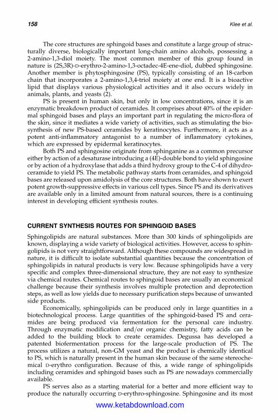
DK4962-Webster-ch13_R2_020407
The core structures are sphingoid bases and constitute a large group of struc-turally diverse, biologically important long-chain amino alcohols, possessing a2-amino-1,3-diol moiety. The most common member of this group found innature is (2S,3R)-D-erythro-2-amino-1,3-octadec-4E-ene-diol, dubbed sphingosine.Another member is phytosphingosine (PS), typically consisting of an 18-carbonchain that incorporates a 2-amino-1,3,4-triol moiety at one end. It is a bioactivelipid that displays various physiological activities and it also occurs widely inanimals, plants, and yeasts (2).
PS is present in human skin, but only in low concentrations, since it is anenzymatic breakdown product of ceramides. It comprises about 40% of the epider-mal sphingoid bases and plays an important part in regulating the micro-flora ofthe skin, since it mediates a wide variety of activities, such as stimulating the bio-synthesis of new PS-based ceramides by keratinocytes. Furthermore, it acts as apotent anti-inflammatory antagonist to a number of inflammatory cytokines,which are expressed by epidermal keratinocytes.
Both PS and sphingosine originate from sphinganine as a common precursoreither by action of a desaturase introducing a (4E)-double bond to yield sphingosineor by action of a hydroxylase that adds a third hydroxy group to the C-4 of dihydro-ceramide to yield PS. The metabolic pathway starts from ceramides, and sphingoidbases are released upon amidolysis of the core structures. Both have shown to exertpotent growth-suppressive effects in various cell types. Since PS and its derivativesare available only in a limited amount from natural sources, there is a continuinginterest in developing efficient synthesis routes.
CURRENT SYNTHESIS ROUTES FOR SPHINGOID BASES
Sphingolipids are natural substances. More than 300 kinds of sphingolipids areknown, displaying a wide variety of biological activities. However, access to sphin-golipids is not very straightforward. Although these compounds are widespread innature, it is difficult to isolate substantial quantities because the concentration ofsphingolipids in natural products is very low. Because sphingolipids have a veryspecific and complex three-dimensional structure, they are not easy to synthesizevia chemical routes. Chemical routes to sphingoid bases are usually an economicalchallenge because their synthesis involves multiple protection and deprotectionsteps, as well as low yields due to necessary purification steps because of unwantedside products.
Economically, sphingolipids can be produced only in large quantities in abiotechnological process. Large quantities of the sphingoid-based PS and cera-mides are being produced via fermentation for the personal care industry.Through enzymatic modification and/or organic chemistry, fatty acids can beadded to the building block to create ceramides. Degussa has developed apatented biofermentation process for the large-scale production of PS. Theprocess utilizes a natural, non-GM yeast and the product is chemically identicalto PS, which is naturally present in the human skin because of the same stereoche-mical D-erythro configuration. Because of this, a wide range of sphingolipidsincluding ceramides and sphingoid bases such as PS are nowadays commerciallyavailable.
PS serves also as a starting material for a better and more efficient way toproduce the naturally occurring D-erythro-sphingosine. Sphingosine and its most
158 Klee et al.
www.ketabdownload.com
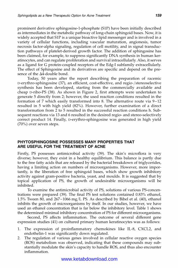
DK4962-Webster-ch13_R2_020407
prominent derivative sphingosine-1-phosphate (S1P) have been initially describedas intermediates in the metabolic pathway of long-chain sphingoid bases. Now, it iswidely accepted that S1P is a unique bioactive lipid messenger and is involved in avariety of cellular functions, including vascular maturation, angionesis, tumornecrosis factor-alpha signaling, regulation of cell motility, and in signal transduc-tion pathways of platelet-derived growth factor. The addition of sphingosine hasbeen claimed, for example, to suppress significantly DNA synthesis in human ker-atinocytes, and can regulate proliferation and survival intracellularly. Also, it servesas a ligand for G protein-coupled receptors of the Edg-I subfamily extracellularly.The effect of Sphingosine and its derivatives are specific and depend on the pre-sence of the D4-double bond.
Today, 50 years after the report describing the preparation of racemicD-erythro-sphingosine (37), an efficient, cost-effective, and regio-/stereoselectivesynthesis has been developed, starting from the commercially available andcheap D-ribo-PS (38). As shown in Figure 2, first attempts were undertaken togenerate 5 directly from 2; however, the used reaction conditions resulted in theformation of 7 which easily transformed into 8. The alternative route via 9–12resulted in 5 with high yield (82%). However, further examination of a directtransformation from 2 to 5 resulted in the successful reaction conditions h. Sub-sequent reactions via 13 and 6 resulted in the desired regio- and stereo-selectivelycorrect product 14. Finally, D-erythro-sphingosine was generated in high yield(70%) over seven steps.
PHYTOSPHINGOSINE POSSESSES MANY PROPERTIES THATARE USEFUL FOR THE TREATMENT OF ACNE
Firstly, PS possesses antimicrobial activity (39). The skin’s microflora is verydiverse; however, they exist in a healthy equilibrium. This balance is partly dueto the free fatty acids that are released by the bacterial breakdown of triglycerides,having a limiting action on numbers of microorganisms. However, more impor-tantly, is the liberation of free sphingoid bases, which show growth inhibitoryactivity against gram-positive bacteria, yeast, and moulds. It is suggested that bytopical application of PS, the growth of undesirable microorganisms will beinhibited.
To examine the antimicrobial activity of PS, solutions of various PS-concen-trations were prepared (39). The final PS test solutions contained 0.83% ethanol,1.5% Tween 80, and 267–1066 mg/L PS. As described by Bibel et al. (40), ethanolinhibits the growth of microorganisms by itself. In our studies, however, we haveused an ethanol concentration that is far below the inhibitory level. Table 1 showsthe determined minimal inhibitory concentration of PS for different microorganisms.
Second, PS affects inflammation. The outcome of several different geneexpression studies (41) on cultured primary human keratinocytes was as follows:
1. The expression of proinflammatory chemokines like IL-8, CXCL2, andendothelin-1 was significantly down regulated.
2. The regulation of various genes involved in cellular reactive oxygen species(ROS) metabolism was observed, indicating that these compounds may sub-stantially modulate the skin’s capacity to handle ROS, and thus also encounterinflammation.
Sphingolipids as a New Therapeutic Option for Acne Treatment 159
www.ketabdownload.com
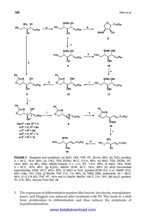
DK4962-Webster-ch13_R2_020407
3. The expression of differentiation markers like loricrin, involucrin, transglutami-nase1, and filaggrin was induced after treatment with PS. This leads to a shiftfrom proliferation to differentiation and thus reduces the symptoms ofhyperkeratinization.
FIGURE 2 Reagents and conditions: (a) BzCl, TEA, THF, RT, 30 min, 99%; (b) TsCl, pyridine,0! 208C, 18 hr, 80%; (c) TrtCl, TEA, EtOAc, 808C, 2.5 hr, 99%; (d) BzCl, TEA, EtOAc, RT,16 hr, 99%; (e) BF3, OEt2, MeOH/toluene (1:1, v/v), RT, 1.5 hr, 99%; (f) MsCl, TEA, DCM,0! 208C, 18 hr, 86%; (g) K2CO3, MeOH, DCM, 408C, 18 hr, 99%; (h) ethyl benzimidatehydrochloride, DCM, 408C, 48 hr, 95%; (i) MsCl or TsCl, pyridine/DCM (3:2, v/v), DMAP (5%),69% (13b), 75% (13d); (j) tBuOK, THF, 08C, 1 hr, 99%; (k) TMSI, DBN, acetonitrile, 40! 868C,94%; (l) (i ) 2 N HCl, THF, RT, 18 hr and (ii ) NaOH, MeOH, 1008C, 2 hr, 79%; (M) Ac2O, pyridine,RT, 2 hr, 99%. Source: From Ref. 38.
160 Klee et al.
www.ketabdownload.com
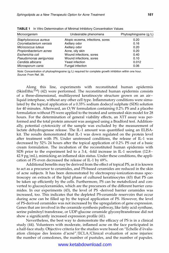
DK4962-Webster-ch13_R2_020407
Along this line, experiments with reconstituted human epidermis(SkinEthicTM) (42) were performed. The reconstituted human epidermis consistsof a three-dimensional, multilayered keratinocyte structure grown on an air–liquid interphase, without any other cell type. Inflammatory conditions were simu-lated by the topical application of a 0.35% sodium dodecyl sulphate (SDS) solutionfor 40 minutes. Afterward, an O/W formulation containing 0.2% PS and a placeboformulation without PS were applied to the treated and untreated skin model for 28hours. For the determination of general viability effects, an XTT assay was per-formed and the total protein amount was assigned using a Bradford test. Addition-ally, potential cytotoxicity of the sample was excluded by the measurement oflactate dehydrogenase release. The IL-1 amount was quantified using an ELISA-kit. The results demonstrated that IL-1 was down regulated on the protein levelafter treatment with PS. Under unstressed conditions, the release of IL-1 wasdecreased by 52% 24 hours after the topical application of 0.2% PS out of a basiccream formulation. The incubation of the reconstituted human epidermis withSDS prior to the experiment led to a 3.4, -fold increase in IL-1 secretion (12.8–42.9 pg/mL), mimicking an inflamed skin status. Under these conditions, the appli-cation of PS even decreased the release of IL-1 by 69%.
Additional benefits may be derived from the effect of topical PS, as it is knownto act as a precursor to ceramides, and PS-based ceramides are reduced in the skinof acne subjects. It has been demonstrated by electrospray-ionization-mass spec-troscopy on extracts of the lipid phase of cultured keratinocytes (43) that PS canbe taken up efficiently by the cells. Furthermore, PS can be metabolized and con-verted to glucosylceramides, which are the precursors of the different barrier cera-mides. In our experiments (43), the level of PS -derived barrier ceramides wasincreased, too. This indicates that the depleted PS-ceramide reservoirs that occurduring acne can be filled up by the topical application of PS. However, the levelof PS-derived ceramides was not increased by the upregulation of gene expression.Genes that are involved in the ceramide synthesis pathway, like fatty acid synthase,serine palmitoyl transferase, or UDP-glucose ceramide glycosyltransferase did notshow a significantly increased expression profile (41).
Nevertheless, the best way to demonstrate the efficacy of PS is in a clinicalstudy (44). Volunteers with moderate, inflamed acne on the face participated ina half-face study. Objective criteria for the studies were based on “Echelle d’evalu-ation clinique des lesions d’acne”/ECLA/Clinical evaluation of acne injuries:the number of comedones, the number of pustules, and the number of papules.
TABLE 1 In Vitro Determination of Minimal Inhibitory Concentration Values
Microorganism Undesirable phenomena Phytosphingosine (g/L)
Staphylococcus aureus Atopic eczema, infections, sores 0.20Corynebacterium xerosis Axillary odor 0.16Micrococcus luteus Axillary odor 0.20Propionibacterium acnes Acne, oily skin 0.20Escherichia coli Wound infections, sores 0.40Pseudomonas aerigunosa Wound infections, sores 0.10Candida albicans Yeast infection 0.012Microsporum canis Fungal infection 0.06
Note: Concentration of phytosphingosine (g/L) required for complete growth inhibition within one hour.Source: From Ref. 39.
Sphingolipids as a New Therapeutic Option for Acne Treatment 161
www.ketabdownload.com
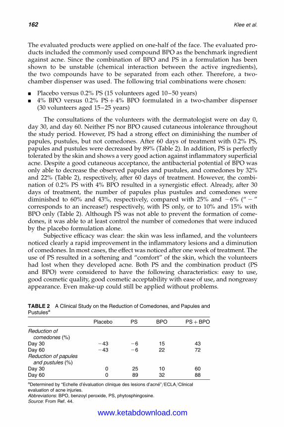
DK4962-Webster-ch13_R2_020407
The evaluated products were applied on one-half of the face. The evaluated pro-ducts included the commonly used compound BPO as the benchmark ingredientagainst acne. Since the combination of BPO and PS in a formulation has beenshown to be unstable (chemical interaction between the active ingredients),the two compounds have to be separated from each other. Therefore, a two-chamber dispenser was used. The following trial combinations were chosen:
B Placebo versus 0.2% PS (15 volunteers aged 10–50 years)B 4% BPO versus 0.2% PSþ 4% BPO formulated in a two-chamber dispenser
(30 volunteers aged 15–25 years)
The consultations of the volunteers with the dermatologist were on day 0,day 30, and day 60. Neither PS nor BPO caused cutaneous intolerance throughoutthe study period. However, PS had a strong effect on diminishing the number ofpapules, pustules, but not comedones. After 60 days of treatment with 0.2% PS,papules and pustules were decreased by 89% (Table 2). In addition, PS is perfectlytolerated by the skin and shows a very good action against inflammatory superficialacne. Despite a good cutaneous acceptance, the antibacterial potential of BPO wasonly able to decrease the observed papules and pustules, and comedones by 32%and 22% (Table 2), respectively, after 60 days of treatment. However, the combi-nation of 0.2% PS with 4% BPO resulted in a synergistic effect. Already, after 30days of treatment, the number of papules plus pustules and comedones werediminished to 60% and 43%, respectively, compared with 25% and 26% (“ 2 ”corresponds to an increase!) respectively, with PS only, or to 10% and 15% withBPO only (Table 2). Although PS was not able to prevent the formation of come-dones, it was able to at least control the number of comedones that were inducedby the placebo formulation alone.
Subjective efficacy was clear: the skin was less inflamed, and the volunteersnoticed clearly a rapid improvement in the inflammatory lesions and a diminutionof comedones. In most cases, the effect was noticed after one week of treatment. Theuse of PS resulted in a softening and “comfort” of the skin, which the volunteershad lost when they developed acne. Both PS and the combination product (PSand BPO) were considered to have the following characteristics: easy to use,good cosmetic quality, good cosmetic acceptability with ease of use, and nongreasyappearance. Even make-up could still be applied without problems.
TABLE 2 A Clinical Study on the Reduction of Comedones, and Papules andPustulesa
Placebo PS BPO PSþ BPO
Reduction ofcomedones (%)
Day 30 243 26 15 43Day 60 243 26 22 72Reduction of papules
and pustules (%)Day 30 0 25 10 60Day 60 0 89 32 88
aDetermined by “Echelle d’evaluation clinique des lesions d’acne”/ECLA/Clinicalevaluation of acne injuries.Abbreviations: BPO, benzoyl peroxide, PS, phytosphingosine.Source: From Ref. 44.
162 Klee et al.
www.ketabdownload.com
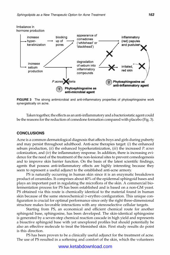
DK4962-Webster-ch13_R2_020407
Taken together, theeffectsas an anti-inflammatory and a bacteriostatic agent couldbe the reasons for the reduction of comedone formation compared with placebo (Fig. 3).
CONCLUSIONS
Acne is a common dermatological diagnosis that affects boys and girls during pubertyand may persist throughout adulthood. Anti-acne therapies target: (i) the enhancedsebum production, (ii) the enhanced hyperkeratinization, (iii) the increased P. acnescolonization, and (iv) the inflammatory response. In addition, there is increasing evi-dence for the need of the treatment of the non-lesional sites to prevent comedogenesisand to improve skin barrier function. On the basis of the latest scientific findings,agents that possess anti-inflammatory effects are highly interesting because theyseem to represent a useful adjunct to the established anti-acne armory.
PS is naturally occurring in human skin since it is an enzymatic breakdownproduct of ceramides. It comprises about 40% of the epidermal sphingoid bases andplays an important part in regulating the microflora of the skin. A commercial bio-fermentation process for PS has been established and is based on a non-GM yeast.PS obtained via this route is chemically identical to the material found in humanskin because of the same stereochemical D-erythro configuration. This unique con-figuration is crucial for optimal performance since only the right three-dimensionalstructure makes favorable interactions with any stereoselective cellular targets.
Starting from PS, an economical and efficient chemical route for anothersphingoid base, sphingosine, has been developed. The skin-identical sphingosineis generated by a seven-step chemical reaction cascade in high yield and representsa bioactive sphingoid base with yet unexplored profiles but should potentially bealso an effective molecule to treat the blemished skin. First study results do pointis this direction.
PS has been proven to be a clinically useful adjunct for the treatment of acne.The use of PS resulted in a softening and comfort of the skin, which the volunteers
FIGURE 3 The strong antimicrobial and anti-inflammatory properties of phytosphingosine worksynergistically on acne.
Sphingolipids as a New Therapeutic Option for Acne Treatment 163
www.ketabdownload.com
DK4962-Webster-ch13_R2_020407
had lost when they developed acne. Both PS and a combination product of PS andBPO were considered to be effective treatments and offer qualities such as good cos-metic acceptability with ease of use and non-greasy appearance.
Taken together, the effects of PS as an anti-inflammatory and a bacteriostaticagent could be considered a new effective way to treat acne-prone skin and mightfind its way into a number of skin products designed for this specific purpose.
ACKNOWLEDGMENT
The authors wish to thank Dr. Kees Korevaar for his helpful contribution to themanuscript.
REFERENCES
1. Rawlings AV, Harding CR. Moisturization and skin barrier function. Dermatol Ther2004; 17(suppl 1):43–48.
2. Van Hoogendalem EJ. Transdermal absorption of topical anti-acne agents in man; reviewof clinical pharmacokinetic data. J Eur Acad Dermatol Venereo 1998; 11(suppl 1):13–19;discussion 28–29.
3. Wertz PW, Swartzendruber DC, Madison KC, Downing DT. Composition and mor-phology of epidermal cyst lipids. J Invest Dermatol 1987; 89:419–425.
4. Williams ML, Elias PM. The extracellular matrix of stratum corneum: role of lipids innormal and pathological function. Rev Ther Drug Carrier Syst 1987; 3:95–122.
5. Thiboutot D, Knaggs H, Gilliland K, et al. Activity of 5-alpha-reductase and 17-beta-hydroxysteroid dehydrogenase in the infrainfundibulum of subjects with and withoutacne vulgaris. Dermatology 1998; 196:38–42.
6. Clark SM, Goulden V, Finlay AY, et al. The psychological and social impact of acne: acomparison study using three acne disability questionnaires (Abstract). Br J Dermatol1997; 137(suppl 50):41.
7. Gupta MA, Johnson AM, Gupta AK. The development of an acne quality of life scale:reliability, validity, and relation to subjective acne severity in mild to moderate acnevulgaris. Acta Derm Venereol 1998 (Stockhausen); 78:451–456.
8. Kligman AM. An overview of acne. J Invest Dermatol 1974; 62:268–287.9. White GM. Recent findings in the epidemiology, classification and subtypes of acne
vulgaris. J Am Acad Dermatol 1998; 39:534–537.10. Schmidt JB, Spona J, Huber J. Androgen receptor in hirsutism and acne. Gynecol Obstet
Invest 1986; 22:206–211.11. Henze C, Hinney B, Wuttke W. Incidence of increased androgen levels in patients suffer-
ing from acne. Dermatology 1998; 196:53–54.12. Cunliffe WJ, Gollnick H. Acne diagnosis and management. In: Martin Dunitz. London:
Taylor & Francis Group, 2001.13. Zouboulis CC. Human skin: an independent peripheral endocrine organ. Horm Res
2000; 54:230–242.14. Plewig G, Kligman AM. Acne and Rosacea. 3rd Completely Revised and Enlarged ed.
Berlin: Springer-Verlag, 2000:581–582.15. Gollnick HP, Zouboulis CC, Akamatsu H, et al. Pathogenesis and pathogenesis related
treatment of acne. J Dermatol 1991; 18:489–499.16. Knaggs HE, Holland DB, Morris C, et al. Quantification of cellular proliferation in acne
using the monoclonal antibody Ki-67. J Invest Dermatol 1994; 102:89–92.17. Thiboutot DM, Knaggs H, Gilliland K, et al. Activity of type I 5-alpha-reductase is
greater in the follicular infundibulum compared with the epidermis. Br J Dermatol1997; 136:166–171.
18. Zouboulis CC, Xia L, Akamatsu H, et al. The human sebocyte culture model providesnew insight into development and management of seborrhoea and acne. Dermatology1998; 196:21–31.
164 Klee et al.
www.ketabdownload.com
DK4962-Webster-ch13_R2_020407
19. Holland KT, Greenman J, Cunliffe WJ. Growth of cutaneous propionibacteria on syntheticmedium: growth yields and enzyme production. J Appl Bacteriol 1979; 47:383–394.
20. Shalita AR, Lee WL. Inflammatory acne. Dermatol Clin 1983; 1:361–364.21. Melnik B, Plewig G. New biochemical aspects of lipids in the pathogenesis of abnormal
follicular keratinisation in acne vulgaris (Article in German). Z Hautkr 1988; 63:591–596.22. Fujie T, Shikiji T, Uchida N, et al. Culture of cells derived from the human sebaceous
gland under serum-free conditions without a biological feeder layer or specificmatices. Arch Dermatol Res 1996; 288:703–708.
23. http://www.medizininfo.de/hautundhaar (accessed April 2005).24. Jansen T, Burgdorf W, Plewig G. Pathogenesis and treatment of acne in childhood.
Pediatr Dermatol 1997; 14:7–21.25. Humbert P. Induced Acne (Article in French). Rev Prat 2002; 52:838–840.26. Friedman GD. Twin studies of disease heritage based on medical records: application to
acne vulgaris. Acta Genet Med Gemellol 1984; 33:487–495.27. Gollnick H. Current concepts of the pathogenesis of acne. Drugs 2003; 63:1579–1596.28. Torma H. Interaction of isotretinoin with endogenous retinoids. J Am Acad Dermatol
2001; 45(suppl 5):143–149.29. White GM. Acne therapy. Dis Mon 1999; 45:301–330.30. Zander E, Weisman S. Treatment of acne vulgaris with salicylic acid. Clin Ther 1992;
14:247–253.31. Zouboulis CC. Exploration of retinoid activity and the role of inflammation in acne:
issues affecting the future directions for acne therapy. J Eur Acad Dermatol Venereol2001; 15(suppl 3):63–67.
32. Muller-Bohn T. Neue Chancen in der Dermatologie. Deutsche Apotheker Zeitung 2004;144:1967–1974.
33. Vega B, Ferret C, Jomard A, et al. Regulation of Toll-like receptor 2 expression by adapa-lene: implications for the treatment of inflammatory acne. Poster P0156, InternationalInvestigative Dermatology Meeting, Miami Beach, FL, April 30–May 4, 2003.
34. Elias PM. Structural lipids, barrier function, and desquamation. J Invest Dermatol 1983;80:44s–49s.
35. Harris IR, Farrell AM, Grunfeld C, Holleran WM, Elias PM, Feingold KR. Permeabilitybarrier disruption coordinately regulates mRNA levels for key enzymes of cholesterol,fatty acid, and ceramide synthesis in the epidermis. J Invest Dermatol 1997;109(6):783–787.
36. Geilen CC, Wieder T, Orfanos CE. Ceramide signalling: regulatory role in cell prolifer-ation, differentiation and apoptosis in human epidermis. Arch Dermatol Res 1997;289(10):559–566.
37. Shapiro D, Segal K. The synthesis of sphingosine. J Am Chem Soc 1954; 76: 5896–5895.38. van den Berg RJBH, Korevaar CGN, Overkleeft HS, et al. Effective, high-yield, and
stereospecific total synthesis of D-erythro-(2R,3S)-sphingosine from D-ribo-(2S,3S,4R)-phytosphingosine. J Org Chem 2004; 69:5699–5704.
39. Inhibition of bacterial growth by phytosphingosine: An in-vitro antimicrobial study,Degussa AG (unpublished data).
40. Bibel, DJ, Aly, R, Shienfield, HR. Antimicrobial activity of sphingosines. J Invest Dermatol2000; 98(3):269–273.
41. DNA-Chip analysis of changes in the gene expression patterns of keratinocytes afterincubation with phytosphingosine, Degussa AG (unpublished data).
42. Influence on the application of phytosphingosine on reconstituted epidermis, DegussaAG (unpublished data).
43. Analysis of changes in the lipid composition of keratinocytes after incubation with phy-tosphingosine, Degussa AG (unpublished data).
44. Phytosphingosine clinical study on acne: determination of the effectiveness of phyto-sphingosine to enhance or complement existing acne therapies, Degussa AG (unpub-lished data).
Sphingolipids as a New Therapeutic Option for Acne Treatment 165
www.ketabdownload.com

DK4962-Webster-ch13_R2_020407
www.ketabdownload.com
DK4962-Webster-ch14_R2_220307
B14 5a-Reductase and Its Inhibitors
Rainer VoegeliDepartment of Research and Development, Pentapharm Ltd., Basel, Switzerland
Christos C. ZouboulisDepartments of Dermatology and Immunology, Dessau Medical Center, Dessau, Germany
Peter ElsnerDepartment of Dermatology and Allergology, Friedrich-Schiller-University Jena,Jena, Germany
Thomas SchreierDepartment of Research and Development, Pentapharm Ltd., Basel, Switzerland
INTRODUCTION
5a-dihydrotestosterone (DHT) exerts a 2 to 10 times stronger androgen activity thantestosterone. Thus, this steroid hormone was supposed to play a more importantrole than testosterone in many androgen-dependent diseases such as acne, hirsut-ism, androgenetic alopecia, benign prostatic hyperplasia, and prostate carcinoma.DHT is converted from testosterone by the enzyme 5a-reductase. There are two iso-enzymes of 5a-reductase (types 1 and 2) that differ in their localization within thebody and even in the skin. Activity of the type 1 isoenzyme predominates in sebac-eous glands, where it may be involved in regulation of sebum production. There-fore, specific inhibition of type 1 5a-reductase may represent a therapeuticapproach to acne. This review refers to the features both of 5a-reductases and oftheir inhibitors.
ANDROGEN METABOLISM IN THE SKIN
The androgenic provoked stimulation of sebum production plays a crucial role inthe pathogenesis of acne. Although numerous factors contribute to the develop-ment of acne, the requirement for androgens is absolute (1). Thiboutot et al. (2)and Fritsch et al. (3) demonstrated that human skin is indeed a steroidogenictissue. The skin functions as an independent peripheral endocrine organ (4) expres-sing all the enzymes necessary for androgen synthesis and catabolism. It can syn-thesize cholesterol from acetate and can further metabolize steroids such asdehydroepiandrosterone sulfate into potent androgens such as testosterone andDHT. DHT itself further induces enzymes such as 3a-hydroxysteroid dehydrogen-ase and 17b-hydroxysteroid dehydrogenase (5).
The skin compartment responsible for both the initiation of steroidogenesisand the local production of potent androgens seems to be the sebaceous gland.Therefore, selective local interruption of this pathway can be of therapeutic benefitin androgen-mediated diseases of the skin and allows for effective treatments (3).
167
www.ketabdownload.com
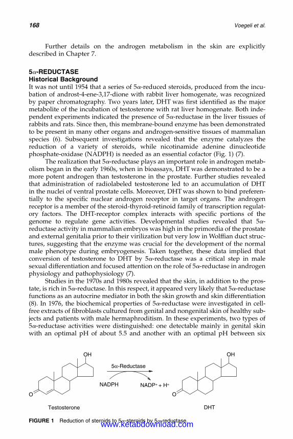
DK4962-Webster-ch14_R2_220307
Further details on the androgen metabolism in the skin are explicitlydescribed in Chapter 7.
5a-REDUCTASEHistorical BackgroundIt was not until 1954 that a series of 5a-reduced steroids, produced from the incu-bation of androst-4-ene-3,17-dione with rabbit liver homogenate, was recognizedby paper chromatography. Two years later, DHT was first identified as the majormetabolite of the incubation of testosterone with rat liver homogenate. Both inde-pendent experiments indicated the presence of 5a-reductase in the liver tissues ofrabbits and rats. Since then, this membrane-bound enzyme has been demonstratedto be present in many other organs and androgen-sensitive tissues of mammalianspecies (6). Subsequent investigations revealed that the enzyme catalyzes thereduction of a variety of steroids, while nicotinamide adenine dinucleotidephosphate-oxidase (NADPH) is needed as an essential cofactor (Fig. 1) (7).
The realization that 5a-reductase plays an important role in androgen metab-olism began in the early 1960s, when in bioassays, DHT was demonstrated to be amore potent androgen than testosterone in the prostate. Further studies revealedthat administration of radiolabeled testosterone led to an accumulation of DHTin the nuclei of ventral prostate cells. Moreover, DHT was shown to bind preferen-tially to the specific nuclear androgen receptor in target organs. The androgenreceptor is a member of the steroid-thyroid-retinoid family of transcription regulat-ory factors. The DHT-receptor complex interacts with specific portions of thegenome to regulate gene activities. Developmental studies revealed that 5a-reductase activity in mammalian embryos was high in the primordia of the prostateand external genitalia prior to their virilization but very low in Wolffian duct struc-tures, suggesting that the enzyme was crucial for the development of the normalmale phenotype during embryogenesis. Taken together, these data implied thatconversion of testosterone to DHT by 5a-reductase was a critical step in malesexual differentiation and focused attention on the role of 5a-reductase in androgenphysiology and pathophysiology (7).
Studies in the 1970s and 1980s revealed that the skin, in addition to the pros-tate, is rich in 5a-reductase. In this respect, it appeared very likely that 5a-reductasefunctions as an autocrine mediator in both the skin growth and skin differentiation(8). In 1976, the biochemical properties of 5a-reductase were investigated in cell-free extracts of fibroblasts cultured from genital and nongenital skin of healthy sub-jects and patients with male hermaphroditism. In these experiments, two types of5a-reductase activities were distinguished: one detectable mainly in genital skinwith an optimal pH of about 5.5 and another with an optimal pH between six
O
OH
Testosterone
O
OH
DHT
NADPH NADP+ + H+
5α-Reductase
FIGURE 1 Reduction of steroids to 5a-steroids by 5a-reductase.
168 Voegeli et al.
www.ketabdownload.com
DK4962-Webster-ch14_R2_220307
and nine in all fibroblasts, cultured from normal and mutant skin taken from bothgenital and nongenital areas. Patients with congenital 5a-reductase deficiencyseemed to lack the former enzyme only (9). In contrast to type-2 isoenzyme, nohuman pathology with a deficiency or mutation of the type 1 isoenzyme hasbeen identified so far (10).
In the following, attention has been focused on molecular biologicalapproaches to identifying and isolating 5a-reductase. In 1989, a cDNA encodinga rat liver 5a-reductase was isolated and used to identify a single mRNA speciesin both liver and prostate of the rat. When this rat 5a-reductase cDNA was usedto screen cDNA libraries constructed from human prostate mRNA, a correspondingcDNA was isolated in 1990. However, when the human cDNA was expressed inCOS cells, the 5a-reductase activity produced was highest at the physiologicalpH values, and not at 5.5 as deduced from earlier biochemical studies. Moreover,the enzyme showed different inhibitor specificity than expected. As the gene encod-ing this cDNA was also normal in 5a-reductase-deficient individuals, it was againsuggested in 1991 that there must be at least 2 isoforms of human 5a-reductase (11),as already proposed by Moore and Wilson (12) by biochemical means in 1976. Lateron, a second 5a-reductase gene was also isolated from a human prostate genomiclibrary. This gene encoded a protein with 50% homology to the former one. Accord-ingly, these enzymes were designated as type 1 and type 2 5a-reductase, respect-ively, in the order in which they were cloned (11).
Physiological Role5a-reductase is the key enzyme in androgen metabolism. Altered enzyme functionand/or regulation are responsible for numerous human pathologies. In humans,the normal activity of 5a-reductases routinely maintains testosterone-mediatedbiological functions, such as anabolic actions (muscle-mass increase, penis enlarge-ment, scrotum enlargement, and vocal cords enlargement), spermatogenesis (malesex drive and performance), and DHT-mediated effects, such as increased facial andbody hair, scalp hair recession, acne, and prostate enlargement (6).
The deficiency of 5a-reductase in males results in incomplete differentiationof the external genitalia at birth. At puberty, patients have normal-to-elevatedlevels of testosterone in plasma, and virilization occurs, but the prostate remainssmall and there is no acne. Male pseudohermaphroditism is accompanied by lowlevels of DHT. However, females with 5a-reductase deficiency appear to have noclinical signs, however, the enzyme also metabolizes other 4-ene-3-oxosteroids,such as progesterone and corticosterone. Abnormally, high 5a-reductase activityin humans results in excessively high DHT levels in peripheral tissues, which isimplicated in the pathogenesis of prostate carcinoma, benign prostatic hyperplasia,acne, hirsutism, and androgenetic alopecia (6).
The exact physiological role of type 2 5a-reductase in tissues other than pros-tate and accessories of the external genitalia remains unknown, since these organsdo not appear to be affected by the genetic absence of this isoenzyme or by itspharmacological inhibition and the women concerned have normal sexual develop-ment and are fertile. The physiological reasons and the factors that mediate thistemporal pattern of expression are not known. However, given the role of andro-gens in 5a-reductase regulation, it is intriguing to speculate that the expression ofone gene may influence the timing or expression level of the other: type 2 isoen-zyme had a priming effect on tissues, such as the prostate, skin and scalp, onwhich type 1 isoenzyme will act later in life (13).
5a-Reductase and Its Inhibitors 169
www.ketabdownload.com
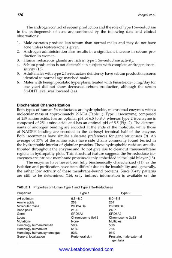
DK4962-Webster-ch14_R2_220307
The androgen control of sebum production and the role of type 1 5a-reductasein the pathogenesis of acne are confirmed by the following data and clinicalobservations:
1. Male castrates produce less sebum than normal males and they do not haveacne unless testosterone is given.
2. Androgen administration also results in a significant increase in sebum pro-duction in women.
3. Human sebaceous glands are rich in type 1 5a-reductase activity.4. Sebum production is not detectable in subjects with complete androgen insen-
sitivity (13).5. Adult males with type 2 5a-reductase deficiency have sebum production scores
identical to normal age-matched males.6. Males with benign prostatic hyperplasia treated with Finasteride (5 mg/day for
one year) did not show decreased sebum production, although the serum5a-DHT level was lowered (14).
Biochemical CharacterizationBoth types of human 5a-reductases are hydrophobic, microsomal enzymes with amolecular mass of approximately 29 kDa (Table 1). Type 1 isoenzyme, composedof 259 amino acids, has an optimal pH of 6.5 to 8.0, whereas type 2 isoenzyme iscomposed of 254 amino acids and has an optimal pH of 5.5 (Fig. 2). The determi-nants of androgen binding are encoded at the ends of the molecule, while thoseof NADPH binding are encoded in the carboxyl terminal half of the enzyme.Both isoenzymes have similar substrate preferences for gene structures (9). Anaverage of 37% of the amino acids have side chains commonly found buried inthe hydrophobic interior of globular proteins. These hydrophobic residues are dis-tributed throughout the enzyme and do not give rise to clear-cut transmembraneregions in hydropathy plots. This structural feature suggests the 5a-reductase iso-enzymes are intrinsic membrane proteins deeply embedded in the lipid bilayer (15).
The enzymes have never been fully biochemically characterized (11), as theisolation and purification have been difficult due to the insolubility and, generally,the rather low activity of these membrane-bound proteins. Since X-ray patternsare still to be determined (16), only indirect information is available on the
TABLE 1 Properties of Human Type 1 and Type 2 5a-Reductases
Properties Type 1 Type 2
pH optimum 6.5–8.0 5.0–5.5Amino acids 259 254Molecular mass 29,494 Da 28,389 DaBase pairs 2100 2437Gene SRD5A1 SRD5A2Locus Chromosome 5p15 Chromosome 2p23Mutations None MultiplesHomology human/human 50% 50%Homology human/rat 61% 75%Homology human/cynomolgus 93% 95%General localization Peripheral skin Prostate, male external
genitalia
170 Voegeli et al.
www.ketabdownload.com
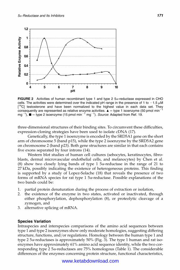
DK4962-Webster-ch14_R2_220307
three-dimensional structures of their binding sites. To circumvent these difficulties,expression-cloning strategies have been used to isolate cDNA (17).
Genetically, the type 1 isoenzyme is encoded by the SRD5A1 gene on the shortarm of chromosome 5 (band p15), while the type 2 isoenzyme by the SRD5A2 geneon chromosome 2 (band p23). Both gene structures are similar in that each containsfive exons separated by four introns (14).
Western blot studies of human cell cultures (sebocytes, keratinocytes, fibro-blasts, dermal microvascular endothelial cells, and melanocytes) by Chen et al.(8) show two closely lying bands of type 1 5a-reductase in the range of 21 to27 kDa, possibly indicating the existence of heterogeneous proteins. This findingis supported by a study of Lopez-Solache (18) that reveals the presence of twoforms of mRNA species for rat type 1 5a-reductase. Possible explanations of thetwo bands could be:
1. partial protein denaturation during the process of extraction or isolation,2. the existence of the enzyme in two states, activated or inactivated, through
either phosphorylation, dephosphorylation (8), or proteolytic cleavage of azymogen, and
3. alternative splicing of mRNA.
Species VariationIntraspecies and interspecies comparisons of the amino acid sequences betweentype 1 and type 2 isoenzymes show only moderate homologies, suggesting differingstructure, functions, and/or regulations. Homology between the human type 1 andtype 2 5a-reductases is approximately 50% (Fig. 3). The type 1 human and rat iso-enzymes have approximately 61% amino acid sequence identity, while the two cor-responding type 2 5a-reductases are 75% homologous (Table 1). The considerabledifferences of the enzymes concerning protein structure, functional characteristics,
0
2.0
4.0
6.0
8.0
1
2.1
01987654pH
Rel
ativ
e E
nzy
me
Act
ivit
y
FIGURE 2 Activities of human recombinant type 1 and type 2 5a-reductase expressed in CHOcells. The activities were determined over the indicated pH range in the presence of 1 to 21.5 mM[14C] testosterone and have been normalized to the highest value in each data set. Theyconsequently are represented as relative enzyme activities. O ¼ type 1 isoenzyme (50 pmol min21
mg21), B ¼ type 2 isoenzyme (15 pmol min21 mg21). Source: Adapted from Ref. 19.
5a-Reductase and Its Inhibitors 171
www.ketabdownload.com
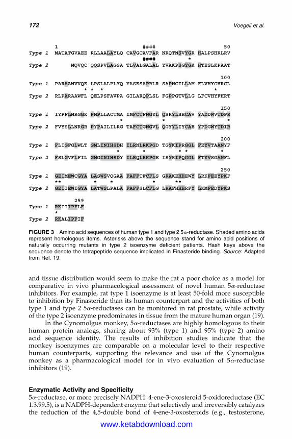
DK4962-Webster-ch14_R2_220307
and tissue distribution would seem to make the rat a poor choice as a model forcomparative in vivo pharmacological assessment of novel human 5a-reductaseinhibitors. For example, rat type 1 isoenzyme is at least 50-fold more susceptibleto inhibition by Finasteride than its human counterpart and the activities of bothtype 1 and type 2 5a-reductases can be monitored in rat prostate, while activityof the type 2 isoenzyme predominates in tissue from the mature human organ (19).
In the Cynomolgus monkey, 5a-reductases are highly homologous to theirhuman protein analogs, sharing about 93% (type 1) and 95% (type 2) aminoacid sequence identity. The results of inhibition studies indicate that themonkey isoenzymes are comparable on a molecular level to their respectivehuman counterparts, supporting the relevance and use of the Cynomolgusmonkey as a pharmacological model for in vivo evaluation of 5a-reductaseinhibitors (19).
Enzymatic Activity and Specificity5a-reductase, or more precisely NADPH: 4-ene-3-oxosteroid 5-oxidoreductase (EC1.3.99.5), is a NADPH-dependent enzyme that selectively and irreversibly catalyzesthe reduction of the 4,5-double bond of 4-ene-3-oxosteroids (e.g., testosterone,
1 #### 50 Type 1 MATATGVAEE RLLAALAYLQ CAVGCAVFAR NRQTNSVYGR HALPSHRLRV #### *
Type 2 MQVQC QQSPVLAGSA TLVALGALAL YVAKPSGYGK HTESLKPAAT
100 Type 1 PARAAWVVQE LPSLALPLYQ YASESAPRLR SAPNCILLAM FLVHYGHRCL * * * *
Type 2 RLPARAAWFL QELPSFAVPA GILARQPLSL FGPPGTVLLG LFCVHYFHRT
150 Type 1 IYPFLMRGGK PMPLLACTMA IMFCTFNGYL QSRYLSHCAV YADDWVTDPR * * *
Type 2 FVYSLLNRGR PYPAILILRG TAFCTGNGVL QGYYLIYCAE YPDGWYTDIR
200 Type 1 FLIGFGLWLT GMLINIHSDH ILRNLRKPGD TGYKIPRGGL FEYVTAANYF * * * * * Type 2 FSLGVFLFIL GMGINIHSDY ILRQLRKPGE ISYRIPQGGL FTYVSGANFL
250 Type 1 GEIMEWCGYA LASWSVQGAA FAFFTFCFLS GRAKEHHEWY LRKFEEYPKF ** * * * **
Type 2 GEIIEWIGYA LATWSLPALA FAFFSLCFLG LRAFHHHRFY LKMFEDYPKS
259 Type 1 RKIIIPFLF *
Type 2 RKALIPFIF
FIGURE 3 Amino acid sequences of human type 1 and type 2 5a-reductase. Shaded amino acidsrepresent homologous items. Asterisks above the sequence stand for amino acid positions ofnaturally occurring mutants in type 2 isoenzyme deficient patients. Hash keys above thesequence denote the tetrapeptide sequence implicated in Finasteride binding. Source: Adaptedfrom Ref. 19.
172 Voegeli et al.
www.ketabdownload.com
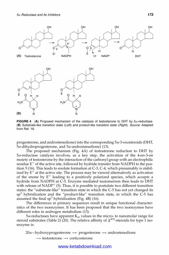
DK4962-Webster-ch14_R2_220307
progesterone, and androstenedione) into the corresponding 5a-3-oxosteroids (DHT,5a-dihydroprogesterone, and 5a-androstanedione) (13).
The proposed mechanism (Fig. 4A) of testosterone reduction to DHT by5a-reductase catalysis involves, as a key step, the activation of the 4-en-3-onmoiety of testosterone by the interaction of the carbonyl group with an electrophilicresidue Eþ of the active site, followed by hydride transfer from NADPH to the pos-ition 5 (16). This leads to enolate formation at C-3, C-4, which presumably is stabil-ized by Eþ at the active site. The process may be viewed alternatively as activationof the enone by Eþ leading to a positively polarized species, which accepts ahydride from NADPH at C-5. Enzyme mediated tautomerism then leads to DHTwith release of NADPþ (5). Thus, it is possible to postulate two different transitionstates: the “substrate-like” transition state in which the C-5 has not yet changed itssp2 hybridization and the “product-like” transition state, in which the C-5 hasassumed the final sp3 hybridization (Fig. 4B) (16).
The differences in primary sequences result in unique functional character-istics of the two isoenzymes. It has been proposed that the two isoenzymes havedifferent roles in androgen metabolism (13).
5a-reductases have apparent Km values in the micro- to nanomolar range forsteroid substrates (Table 2) (20). The relative affinity of D4(5)-steroids for type 1 iso-enzyme is:
20a�hydroxyprogesterone �! progesterone �! androstenedione
�! testosterone �! corticosterone
O
OH
Testosterone
O
OH
O
OH
EH
O
OH
H
DHTNADPH NADP+H+E+
δ–
δ+
(A)
N
R
H HO
NH2
O
OH
E
+O
OH
E
N
R
H HO
NH2
+
(B)
FIGURE 4 (A) Proposed mechanism of the catalysis of testosterone to DHT by 5a-reductase.(B) Substrate-like transition state (Left) and product-like transition state (Right). Source: Adaptedfrom Ref. 16.
5a-Reductase and Its Inhibitors 173
www.ketabdownload.com
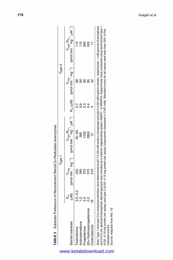
DK4962-Webster-ch14_R2_220307
TA
BL
E2
Substr
ate
Pre
fere
nce
of
Recom
bin
ant
Ste
roid
5a
-Reducta
se
Isoenzym
es
Type
1T
ype
2
Ste
roid
substr
ate
Km
(mM
)V
max
(pm
olm
in2
1m
g2
1)
Vm
ax/K
m
(pm
olm
in2
1m
g2
1m
M2
1)
Km
(mM
)V
max
(pm
olm
in2
1m
g2
1)
Vm
ax/K
m
(pm
olm
in2
1m
g2
1m
M2
1)
Test
oste
rone
3.5
–5.2
220
40
–60
0.7
88
115
Andro
stenedio
ne
1.0
395
390
0.8
90
110
Pro
geste
rone
0.3
370
1230
0.3
80
260
20a
-Hyd
roxypro
geste
rone
0.2
570
2850
0.4
85
240
Cort
icoste
rone
18
210
12
442
11
Note
:T
ype
1and
type
25a
-reducta
se
activitie
sw
ere
dete
rmin
ed
atpH
7.5
(50
mM
sodiu
mphosphate
)and
pH
5.0
(50
mM
sodiu
mcitra
te),
respectively
.1
mM
glu
cose-6
-phosphate
and
0.5
U/m
Lglu
cose-6
-phosphate
dehydro
genase
was
inclu
ded
as
acofa
cto
rre
genera
ting
syste
m(N
AD
Pþ
toN
AD
PH
).E
xperim
ents
were
conducte
dusin
gre
com
bin
antty
pe
1(0
.05
–0.1
0m
gpro
tein
per
assay)
and
type
2(0
.02
–0.1
5m
gpro
tein
per
assay)
isoenzym
es
expre
ssed
inC
HO
cells
.S
tandard
err
ors
for
all
valu
es
were
less
than
20%
of
the
indic
ate
dentr
ies.
Sourc
e:
Adapte
dfr
om
Ref.
19.
174 Voegeli et al.
www.ketabdownload.com

DK4962-Webster-ch14_R2_220307
and for the type 2 isoenzyme:
progesterone �! 20a�hydroxyprogesterone �! testosterone
�! androstenedione �! corticosterone
The conversion of testosterone to DHT amplifies the androgenic signalthrough four mechanisms:
1. DHT, unlike testosterone, cannot be aromatized to estrogen, thus its effectremains purely androgenic (14).
2. DHT binds to the human androgen receptor with a several-fold higher affinity thantestosterone does (14). It has also been suggested that the DHT/androgen receptorcomplex has a higher affinity for the acceptor site in the nuclear chromatin (5).
3. The DHT/androgen receptor complex appears to be more stable (14).4. Both the 5a-reductase genes are adjusted by DHT via a feed-forward
regulation (13).
Although testosterone and DHT share the same androgen receptor, it is not sur-prising that the two androgens exert varying physiological effects.
Localization, Distribution, and Cutaneous Enzymatic ActivityGeneralThe specific localization and activity of type 1 isoenzyme in sebaceous glandsmakes this isoenzyme a potential therapeutic target for the treatment of acne. Forthis reason, it becomes important to understand where and how the two isoen-zymes of 5a-reductase act in the skin (21).
Type 1 5a-reductase is present, above all, in the skin, whereas type 2 is predo-minantly located in the prostate and in genital skin (9). Both the isoenzymes areexpressed in the liver. In humans type 1 is not detectable in the fetus but is perma-nently expressed in skin and scalp from the time of puberty (13).
Cutaneous DistributionIn humans, the cutaneous distribution of type 1 isoenzyme was identified immuno-histochemically to be localized predominantly within sebaceous glands (8). Theenzymatic activity is significantly higher, on the one hand in sebaceous glands ofthe face and scalp compared to nonacne-prone areas, and on the other hand insebaceous glands in men than women (14). Type 1 isoenzyme was also found in epi-dermis, eccrine sweat glands, apocrine sweat glands (in normal ones as well as inpeople with osmidrosis), hair follicles (outer root sheath cells, dermal papilla cells,matrix cells), endothelial cells of small vessels, and in Schwann cells of cutaneousmyelinated nerves (8).
Within hair follicles, type 2 isoenzyme was localized by prominent immuno-staining in the inner layer of outer root sheaths, inner root sheaths, and the infun-dibulum, as well as in sebaceous ducts. Regional studies showed the type 2 mRNApresent in beard dermal papilla, but absent from occipital scalp and axillary dermalpapilla. The type 2 isoenzyme in beard dermal papilla has a three-times higheractivity than the type 1 5a-reductase present in the occipital scalp and axillarydermal papilla. The specific activity of 5a-reductase in the hair dermal papillaexceeded those in other hair follicle compartments (connective tissue sheaths andouter root sheaths) by a factor of at least 14 in the scalp and at least 80 in the
5a-Reductase and Its Inhibitors 175
www.ketabdownload.com
DK4962-Webster-ch14_R2_220307
beard. The beard dermal papilla cells appeared to generate more DHT than thosefrom nonbalding scalp hair follicles. However, the individual freshly isolatedintact dermal papilla was shown to possess considerably different levels of exvivo enzyme activities (14).
Taken together, while the type 1 5a-reductase has been definitely demon-strated in sebaceous glands, the isoenzyme distribution in hair follicle is less welldefined, probably due to
1. local and temporal variation of enzyme activity,2. utilization of different polyclonal/monoclonal antibodies, and3. inadequate assessment of enough specimens.
More evidence is needed to better define the existence of the type 1 isoenzymein hair follicles and the precise localization of the type 2 isoenzyme within the hairfollicles (in outer root sheaths or in dermal papilla) (14).
In the following sections we refer extensively to three eminent publicationsdemonstrating the localization of 5a-reductase in cells, in sebaceous follicles, andits enzymatic activity in epidermal and infrainfundibular keratinocytes.
Subcellular Localization and Expression of Type 1 5a-ReductaseChen et al. (8) examined cutaneous expression and subcellular localization of type 15a-reductase in vitro by using human cell cultures (sebocytes, keratinocytes, fibro-blasts, dermal microvascular endothelial cells, hair dermal papilla cells, and mela-nocytes) with immunocytochemistry, western and northern blot studies.
With immunocytochemistry it was shown that type 1 5a-reductase is locatedin the cytoplasm of all examined cultured skin cell types and not in the nucleus asdescribed by some authors. The expression could be broadly divided into threegroups, in the order of decreasing intensity (Fig. 5):
1. adult facial sebocytes and neonatal foreskin keratinocytes,2. adult nongenital keratinocytes (flank), occipital dermal papilla cells, occipital
fibroblasts, and3. beard dermal papilla cells, melanocytes, dermal microvascular endothelial
cells, and chin fibroblasts.
Moreover, among sebocytes, the smaller, less differentiated sebocytes weremore strongly stained than the larger, differentiated ones (Fig. 6).
Further findings of the study were:
1. The most abundant expression of type 1 5a-reductase is in neonatal foreskinkeratinocytes, followed by adult facial sebocytes.
2. The strongest expression of type 1 5a-reductase among skin cells from adultsubjects is in facial sebocytes.
3. The expression of type 1 5a-reductase in fibroblasts and keratinocytes is subjectto regional variance.
4. Type 1 5a-reductase is present in dermal papilla cells of both beard and occipi-tal hair cells, whereas it is expressed more strongly in the latter.
5. Type 1 5a-reductase is expressed in cultured melanocytes. The significance ofthis interesting finding remains unknown.
The strongest expression of type 1 5a-reductase in sebocytes among adult skincells examined confirmed the previous hypothesis of the authors that the sebaceous
176 Voegeli et al.
www.ketabdownload.com
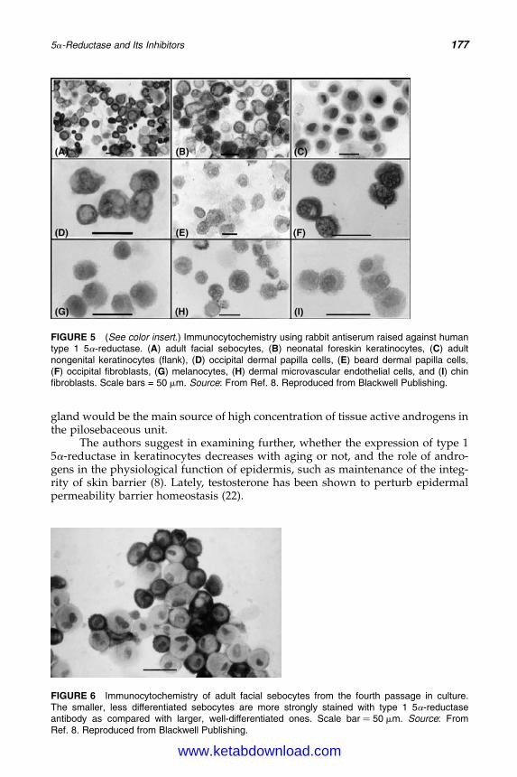
DK4962-Webster-ch14_R2_220307
gland would be the main source of high concentration of tissue active androgens inthe pilosebaceous unit.
The authors suggest in examining further, whether the expression of type 15a-reductase in keratinocytes decreases with aging or not, and the role of andro-gens in the physiological function of epidermis, such as maintenance of the integ-rity of skin barrier (8). Lately, testosterone has been shown to perturb epidermalpermeability barrier homeostasis (22).
FIGURE 5 (See color insert.) Immunocytochemistry using rabbit antiserum raised against humantype 1 5a-reductase. (A) adult facial sebocytes, (B) neonatal foreskin keratinocytes, (C) adultnongenital keratinocytes (flank), (D) occipital dermal papilla cells, (E) beard dermal papilla cells,(F) occipital fibroblasts, (G) melanocytes, (H) dermal microvascular endothelial cells, and (I) chinfibroblasts. Scale bars = 50 mm. Source: From Ref. 8. Reproduced from Blackwell Publishing.
FIGURE 6 Immunocytochemistry of adult facial sebocytes from the fourth passage in culture.The smaller, less differentiated sebocytes are more strongly stained with type 1 5a-reductaseantibody as compared with larger, well-differentiated ones. Scale bar ¼ 50 mm. Source: FromRef. 8. Reproduced from Blackwell Publishing.
5a-Reductase and Its Inhibitors 177
www.ketabdownload.com

DK4962-Webster-ch14_R2_220307
Immunolocalization of 5a-Reductases in Sebaceous FolliclesThiboutot et al. (21) determined immunohistochemically (hematoxylin-eosin staining)the distribution of 5a-reductase isoenzymes within sebaceous follicles and terminalhair follicles in acne lesions and normal skin. Higher levels of type 1 isoenzyme aredetected in sebaceous glands derived from acne-prone areas of skin, such as the face,compared to the glands from other areas, such as the legs or arms. This seems to corre-late with the finding that the stimulatory effect of DHTon cell proliferation is more pro-minent on facial than on nonfacial sebocytes, as shown by Akamatsu et al. (23).
5a-Reductase Localization in Sebaceous Follicles of Normal SkinIn subjects with clinically normal skin, the majority of follicles contained a milddegree of hyperkeratosis. Monoclonal antibodies to type 1 5a-reductase specificallylocalized to the sebaceous glands of sebaceous follicles from normal skin (Fig. 7A).Reactivity was most pronounced at the periphery of the gland, that is, at the area ofundifferentiated sebocytes in concomitance with the data by Chen et al. (8), but wasnoted throughout the sebaceous gland in most sections. No localization within hairfollicles was noted (21).
Antibodies to type 2 5a-reductase localized to the companion layer (inner-most layer of the outer root sheath) of sebaceous follicles and sebaceous ducts(Fig. 7B). Immunoreactivity was also noted in the granular layer of the epidermisand in the sheath of myelinated cutaneous nerves (not shown). No stainingwithin sebaceous glands was observed with type 2 antibody (21).
5a-Reductase Localization in Acne LesionsThe pattern of immunoreactivity in acne lesions was similar to that observed innormal sebaceous follicles. In sections of closed (Fig. 8A) and open comedones(Fig. 8B), type 1 immunoreactivity was noted only in remnants of sebaceous
FIGURE 7 Immunolocalization of 5a-reductase in sebaceous follicles from the back of a subjectwithout acne. (A) Skin incubated with antibody to type 1 5a-reductase reveals specific localizationwithin sebaceous glands (arrow) (original magnification �17). (B) Skin incubated with an antibodyto type 2 5a-reductase reveals localization within the follicle and sebaceous duct (arrow), but notwithin the sebaceous gland (original magnification �42). Source: From Ref. 21. Reproduced fromArchives of Dermatology.
178 Voegeli et al.
www.ketabdownload.com

DK4962-Webster-ch14_R2_220307
glands and not within follicles. Type 2 antibody localized within the walls of bothopen and closed comedones, but not in the adjacent sebaceous gland, and extendedinto keratinized cells contained within the lumen (Fig. 9). In inflammatory lesions,type 1 immunoreactivity was noted specifically in sebaceous glands and not in fol-licles or any other epidermal or dermal structures (Fig. 10A and B). Immunoreactiv-ity was most pronounced within basal sebocytes and least evident in highlydifferentiated sebocytes in proximity to sebaceous ducts (Fig. 10C). Localizationof type 2 antibody was observed within the granular layer of the epidermis, thecompanion layer of the follicle, and the sebaceous duct, but not in the sebaceousgland (Fig. 11A). Type 2 immunoreactivity was noted in endothelial cells adjacentto an inflammatory acne lesion (Figs. 11B and 12A) but not in endothelial cells incontrol sections of dermatitic skin (Fig. 12B).
An interesting observation is the localization of the type 2 isoenzyme of5a-reductase in the walls of both open and closed comedones and in endothelialcells surrounding inflammatory acne lesions. This localization parallels are seen insimilar regions of normal follicles. The significance of the localization of the type 2isoenzyme within the walls of the open and closed comedones or endothelial cellsin inflammatory acne lesions remains unknown. It has been suggested that andro-gens may play a role in the follicular hyperkeratinization seen in acne. Although acause-and-effect relationship between androgens and follicular hyperkeratinizationcannot be drawn from immunohistochemical findings, localization of 5a-reductasewithin the comedonal wall provides a rationale for research in this area (21).
Enzymatic Activity of 5a-Reductase in Epidermal andInfrainfundibular KeratinocytesThiboutot et al. (24) determined the activity of 5a-reductase in keratinocytes excisedand cultured from the infrainfundibulum and the epidermis of the forehead, using(1,2-3H) testosterone as substrate.
FIGURE 8 (See color insert.) Immunolocalization of type 1 5a-reductase in comedones(hematoxylin and eosin). (A) Sections of closed comedones and (B) of open comedones reveal nolocalization of type 1 antibody within the comedone (original magnification �17). Of note is type 1antibody localization in a sebaceous gland adjacent to an open comedone (arrow). Source: FromRef. 21. Reproduced from Archives of Dermatology.
5a-Reductase and Its Inhibitors 179
www.ketabdownload.com
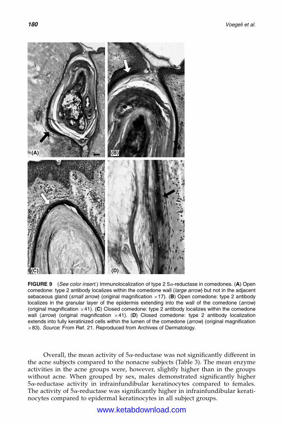
DK4962-Webster-ch14_R2_220307
Overall, the mean activity of 5a-reductase was not significantly different inthe acne subjects compared to the nonacne subjects (Table 3). The mean enzymeactivities in the acne groups were, however, slightly higher than in the groupswithout acne. When grouped by sex, males demonstrated significantly higher5a-reductase activity in infrainfundibular keratinocytes compared to females.The activity of 5a-reductase was significantly higher in infrainfundibular kerati-nocytes compared to epidermal keratinocytes in all subject groups.
FIGURE 9 (See color insert.) Immunolocalization of type 2 5a-reductase in comedones. (A) Opencomedone: type 2 antibody localizes within the comedone wall (large arrow) but not in the adjacentsebaceous gland (small arrow) (original magnification �17). (B) Open comedone: type 2 antibodylocalizes in the granular layer of the epidermis extending into the wall of the comedone (arrow)(original magnification �41). (C) Closed comedone: type 2 antibody localizes within the comedonewall (arrow) (original magnification �41). (D) Closed comedone: type 2 antibody localizationextends into fully keratinized cells within the lumen of the comedone (arrow) (original magnification�83). Source: From Ref. 21. Reproduced from Archives of Dermatology.
180 Voegeli et al.
www.ketabdownload.com
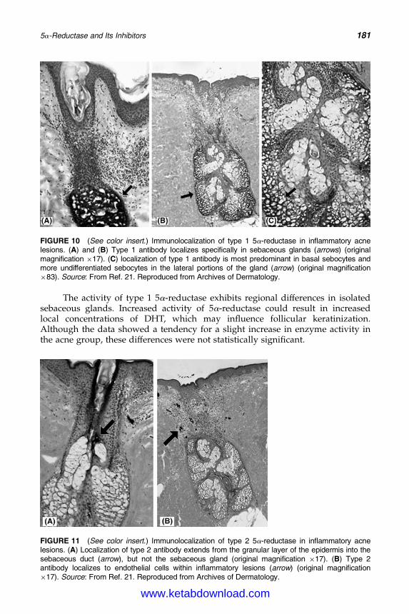
DK4962-Webster-ch14_R2_220307
The activity of type 1 5a-reductase exhibits regional differences in isolatedsebaceous glands. Increased activity of 5a-reductase could result in increasedlocal concentrations of DHT, which may influence follicular keratinization.Although the data showed a tendency for a slight increase in enzyme activity inthe acne group, these differences were not statistically significant.
FIGURE 10 (See color insert.) Immunolocalization of type 1 5a-reductase in inflammatory acnelesions. (A) and (B) Type 1 antibody localizes specifically in sebaceous glands (arrows) (originalmagnification �17). (C) localization of type 1 antibody is most predominant in basal sebocytes andmore undifferentiated sebocytes in the lateral portions of the gland (arrow) (original magnification�83). Source: From Ref. 21. Reproduced from Archives of Dermatology.
FIGURE 11 (See color insert.) Immunolocalization of type 2 5a-reductase in inflammatory acnelesions. (A) Localization of type 2 antibody extends from the granular layer of the epidermis into thesebaceous duct (arrow), but not the sebaceous gland (original magnification �17). (B) Type 2antibody localizes to endothelial cells within inflammatory lesions (arrow) (original magnification�17). Source: From Ref. 21. Reproduced from Archives of Dermatology.
5a-Reductase and Its Inhibitors 181
www.ketabdownload.com
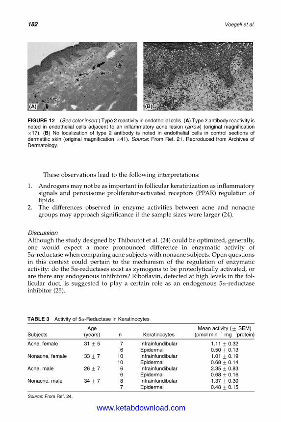
DK4962-Webster-ch14_R2_220307
These observations lead to the following interpretations:
1. Androgens may not be as important in follicular keratinization as inflammatorysignals and peroxisome proliferator-activated receptors (PPAR) regulation oflipids.
2. The differences observed in enzyme activities between acne and nonacnegroups may approach significance if the sample sizes were larger (24).
DiscussionAlthough the study designed by Thiboutot et al. (24) could be optimized, generally,one would expect a more pronounced difference in enzymatic activity of5a-reductase when comparing acne subjects with nonacne subjects. Open questionsin this context could pertain to the mechanism of the regulation of enzymaticactivity: do the 5a-reductases exist as zymogens to be proteolytically activated, orare there any endogenous inhibitors? Riboflavin, detected at high levels in the fol-licular duct, is suggested to play a certain role as an endogenous 5a-reductaseinhibitor (25).
FIGURE 12 (See color insert.) Type 2 reactivity in endothelial cells. (A) Type 2 antibody reactivity isnoted in endothelial cells adjacent to an inflammatory acne lesion (arrow) (original magnification�17). (B) No localization of type 2 antibody is noted in endothelial cells in control sections ofdermatitic skin (original magnification �41). Source: From Ref. 21. Reproduced from Archives ofDermatology.
TABLE 3 Activity of 5a-Reductase in Keratinocytes
SubjectsAge
(years) n KeratinocytesMean activity (+ SEM)
(pmol min21 mg21protein)
Acne, female 31+ 5 7 Infrainfundibular 1.11+ 0.326 Epidermal 0.50+ 0.13
Nonacne, female 33+ 7 10 Infrainfundibular 1.01+ 0.1910 Epidermal 0.68+ 0.14
Acne, male 26+ 7 6 Infrainfundibular 2.35+ 0.836 Epidermal 0.68+ 0.16
Nonacne, male 34+ 7 8 Infrainfundibular 1.37+ 0.307 Epidermal 0.48+ 0.15
Source: From Ref. 24.
182 Voegeli et al.
www.ketabdownload.com
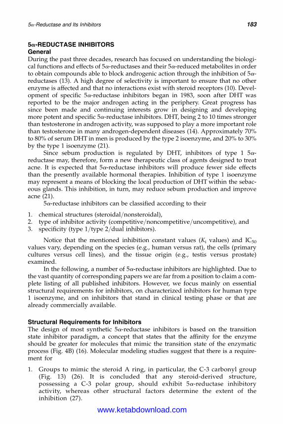
DK4962-Webster-ch14_R2_220307
5a-REDUCTASE INHIBITORSGeneralDuring the past three decades, research has focused on understanding the biologi-cal functions and effects of 5a-reductases and their 5a-reduced metabolites in orderto obtain compounds able to block androgenic action through the inhibition of 5a-reductases (13). A high degree of selectivity is important to ensure that no otherenzyme is affected and that no interactions exist with steroid receptors (10). Devel-opment of specific 5a-reductase inhibitors began in 1983, soon after DHT wasreported to be the major androgen acting in the periphery. Great progress hassince been made and continuing interests grow in designing and developingmore potent and specific 5a-reductase inhibitors. DHT, being 2 to 10 times strongerthan testosterone in androgen activity, was supposed to play a more important rolethan testosterone in many androgen-dependent diseases (14). Approximately 70%to 80% of serum DHT in men is produced by the type 2 isoenzyme, and 20% to 30%by the type 1 isoenzyme (21).
Since sebum production is regulated by DHT, inhibitors of type 1 5a-reductase may, therefore, form a new therapeutic class of agents designed to treatacne. It is expected that 5a-reductase inhibitors will produce fewer side effectsthan the presently available hormonal therapies. Inhibition of type 1 isoenzymemay represent a means of blocking the local production of DHT within the sebac-eous glands. This inhibition, in turn, may reduce sebum production and improveacne (21).
5a-reductase inhibitors can be classified according to their
1. chemical structures (steroidal/nonsteroidal),2. type of inhibitor activity (competitive/noncompetitive/uncompetitive), and3. specificity (type 1/type 2/dual inhibitors).
Notice that the mentioned inhibition constant values (Ki values) and IC50
values vary, depending on the species (e.g., human versus rat), the cells (primarycultures versus cell lines), and the tissue origin (e.g., testis versus prostate)examined.
In the following, a number of 5a-reductase inhibitors are highlighted. Due tothe vast quantity of corresponding papers we are far from a position to claim a com-plete listing of all published inhibitors. However, we focus mainly on essentialstructural requirements for inhibitors, on characterized inhibitors for human type1 isoenzyme, and on inhibitors that stand in clinical testing phase or that arealready commercially available.
Structural Requirements for InhibitorsThe design of most synthetic 5a-reductase inhibitors is based on the transitionstate inhibitor paradigm, a concept that states that the affinity for the enzymeshould be greater for molecules that mimic the transition state of the enzymaticprocess (Fig. 4B) (16). Molecular modeling studies suggest that there is a require-ment for
1. Groups to mimic the steroid A ring, in particular, the C-3 carbonyl group(Fig. 13) (26). It is concluded that any steroid-derived structure,possessing a C-3 polar group, should exhibit 5a-reductase inhibitoryactivity, whereas other structural factors determine the extent of theinhibition (27).
5a-Reductase and Its Inhibitors 183
www.ketabdownload.com
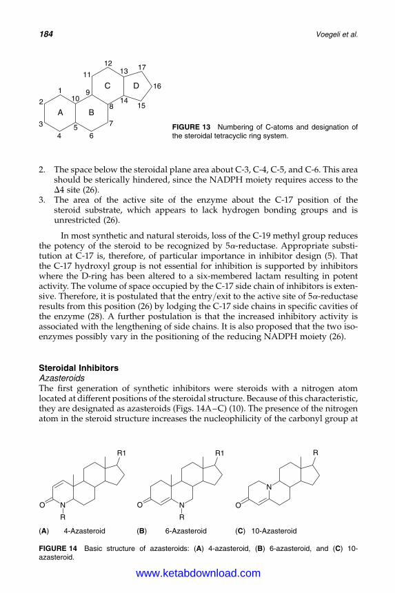
DK4962-Webster-ch14_R2_220307
2. The space below the steroidal plane area about C-3, C-4, C-5, and C-6. This areashould be sterically hindered, since the NADPH moiety requires access to theD4 site (26).
3. The area of the active site of the enzyme about the C-17 position of thesteroid substrate, which appears to lack hydrogen bonding groups and isunrestricted (26).
In most synthetic and natural steroids, loss of the C-19 methyl group reducesthe potency of the steroid to be recognized by 5a-reductase. Appropriate substi-tution at C-17 is, therefore, of particular importance in inhibitor design (5). Thatthe C-17 hydroxyl group is not essential for inhibition is supported by inhibitorswhere the D-ring has been altered to a six-membered lactam resulting in potentactivity. The volume of space occupied by the C-17 side chain of inhibitors is exten-sive. Therefore, it is postulated that the entry/exit to the active site of 5a-reductaseresults from this position (26) by lodging the C-17 side chains in specific cavities ofthe enzyme (28). A further postulation is that the increased inhibitory activity isassociated with the lengthening of side chains. It is also proposed that the two iso-enzymes possibly vary in the positioning of the reducing NADPH moiety (26).
Steroidal InhibitorsAzasteroidsThe first generation of synthetic inhibitors were steroids with a nitrogen atomlocated at different positions of the steroidal structure. Because of this characteristic,they are designated as azasteroids (Figs. 14A–C) (10). The presence of the nitrogenatom in the steroid structure increases the nucleophilicity of the carbonyl group at
A B
C D1
2
3
45
6
7
8
910
11
1213
1415
16
17
FIGURE 13 Numbering of C-atoms and designation ofthe steroidal tetracyclic ring system.
O N
R1
R
O N
R1
R
N
O
R
4-Azasteroid 6-Azasteroid 10-Azasteroid (A) (B) (C)
FIGURE 14 Basic structure of azasteroids: (A) 4-azasteroid, (B) 6-azasteroid, and (C) 10-azasteroid.
184 Voegeli et al.
www.ketabdownload.com
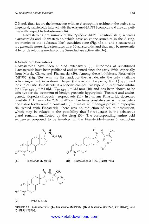
DK4962-Webster-ch14_R2_220307
C-3 and, thus, favors the interaction with an electrophilic residue in the active site.In general, azasteroids interact with the enzyme NADPH complex and are competi-tive with respect to testosterone (16).
4-Azasteroids are mimics of the “product-like” transition state, whereas6-azasteroids and 10-azasteroids, which have an enone structure in the A ring,are mimics of the “substrate-like” transition state (Fig. 4B). 4- and 6-azasteroidsare generally more rigid structures than 10-azasteroids, and thus may be more suit-able for developing models of the 5a-reductase active site (16).
4-Azasteroid Derivatives4-Azasteroids have been studied extensively (6). Hundreds of substituted4-azasteroids have been published and patented since the early 1980s, especiallyfrom Merck, Glaxo, and Pharmacia (29). Among these inhibitors, Finasteride(MK906) (Fig. 15A) was the first and, for the last decade, the only availableactive ingredient in systemic drugs, (Proscar and Propecia, Merck) approvedfor clinical use. Finasteride is a specific competitive type 2 5a-reductase inhibi-tor (IC50 type 2 ¼ 9.4 nM, IC50 type 1 ¼ 313 nm) (10) and has been shown to beeffective for the treatment of benign prostatic hyperplasia (Proscar) and andro-genetic alopecia (Propecia), respectively (14). In humans Finasteride decreasesprostatic DHT levels by 70% to 90% and reduces prostate size, while testoster-one tissue levels remain constant (5). In males with benign prostatic hyperpla-sia treated with Finasteride, there was no reduction of sebum production,which may be related to the possibility that 5a-reductase in the sebaceousgland remains unaffected by the drug (30). The corresponding amino acidsequences proposed to be involved in the Finasteride/human 5a-reductase
O NH
HN
O
C(CH3)3
O NH
HN
O CF3
F3C
Finasteride (MK906) Dutasteride (GG745, GI198745)
O NH
O HN
CF3
CF3
PNU 175706
(A)
(C)
(B)
FIGURE 15 4-Azasteroids: (A) finasteride (MK906), (B) dutasteride (GG745, GI198745), and(C) PNU 175706.
5a-Reductase and Its Inhibitors 185
www.ketabdownload.com
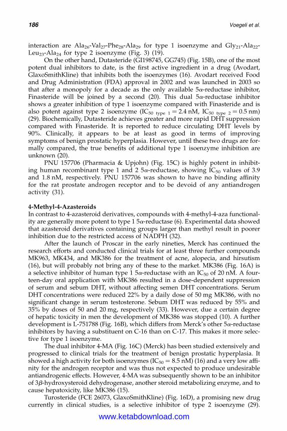
DK4962-Webster-ch14_R2_220307
interaction are Ala26-Val27-Phe28-Ala29 for type 1 isoenzyme and Gly21-Ala22-Leu23-Ala24 for type 2 isoenzyme (Fig. 3) (19).
On the other hand, Dutasteride (GI198745, GG745) (Fig. 15B), one of the mostpotent dual inhibitors to date, is the first active ingredient in a drug (Avodart,GlaxoSmithKline) that inhibits both the isoenzymes (16). Avodart received Foodand Drug Administration (FDA) approval in 2002 and was launched in 2003 sothat after a monopoly for a decade as the only available 5a-reductase inhibitor,Finasteride will be joined by a second (20). This dual 5a-reductase inhibitorshows a greater inhibition of type 1 isoenzyme compared with Finasteride and isalso potent against type 2 isoenzyme (IC50 type 1 ¼ 2.4 nM, IC50 type 2 ¼ 0.5 nm)(29). Biochemically, Dutasteride achieves greater and more rapid DHT suppressioncompared with Finasteride. It is reported to reduce circulating DHT levels by90%. Clinically, it appears to be at least as good in terms of improvingsymptoms of benign prostatic hyperplasia. However, until these two drugs are for-mally compared, the true benefits of additional type 1 isoenzyme inhibition areunknown (20).
PNU 157706 (Pharmacia & Upjohn) (Fig. 15C) is highly potent in inhibit-ing human recombinant type 1 and 2 5a-reductase, showing IC50 values of 3.9and 1.8 nM, respectively. PNU 157706 was shown to have no binding affinityfor the rat prostate androgen receptor and to be devoid of any antiandrogenactivity (31).
4-Methyl-4-AzasteroidsIn contrast to 4-azasteroid derivatives, compounds with 4-methyl-4-aza functional-ity are generally more potent to type 1 5a-reductase (6). Experimental data showedthat azasteroid derivatives containing groups larger than methyl result in poorerinhibition due to the restricted access of NADPH (32).
After the launch of Proscar in the early nineties, Merck has continued theresearch efforts and conducted clinical trials for at least three further compoundsMK963, MK434, and MK386 for the treatment of acne, alopecia, and hirsutism(16), but will probably not bring any of these to the market. MK386 (Fig. 16A) isa selective inhibitor of human type 1 5a-reductase with an IC50 of 20 nM. A four-teen-day oral application with MK386 resulted in a dose-dependent suppressionof serum and sebum DHT, without affecting semen DHT concentrations. SerumDHT concentrations were reduced 22% by a daily dose of 50 mg MK386, with nosignificant change in serum testosterone. Sebum DHT was reduced by 55% and35% by doses of 50 and 20 mg, respectively (33). However, due a certain degreeof hepatic toxicity in men the development of MK386 was stopped (10). A furtherdevelopment is L-751788 (Fig. 16B), which differs from Merck’s other 5a-reductaseinhibitors by having a substituent on C-16 than on C-17. This makes it more selec-tive for type 1 isoenzyme.
The dual inhibitor 4-MA (Fig. 16C) (Merck) has been studied extensively andprogressed to clinical trials for the treatment of benign prostatic hyperplasia. Itshowed a high activity for both isoenzymes (IC50 ¼ 8.5 nM) (16) and a very low affi-nity for the androgen receptor and was thus not expected to produce undesirableantiandrogenic effects. However, 4-MA was subsequently shown to be an inhibitorof 3b-hydroxysteroid dehydrogenase, another steroid metabolizing enzyme, and tocause hepatoxicity, like MK386 (15).
Turosteride (FCE 26073, GlaxoSmithKline) (Fig. 16D), a promising new drugcurrently in clinical studies, is a selective inhibitor of type 2 isoenzyme (29).
186 Voegeli et al.
www.ketabdownload.com
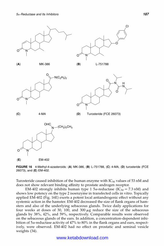
DK4962-Webster-ch14_R2_220307
Turosteride caused inhibition of the human enzyme with IC50 values of 53 nM anddoes not show relevant binding affinity to prostate androgen receptor.
EM-402 strongly inhibits human type 1 5a-reductase (IC50 ¼ 7.3 nM) andshows low potency on the type 2 isoenzyme in transfected cells in vitro. Topicallyapplied EM-402 (Fig. 16E) exerts a potent local antiandrogenic effect without anysystemic action in the hamster. EM-402 decreased the size of flank organs of ham-sters and also of the underlying sebaceous glands. Twice daily applications forfour weeks at doses of 30, 100, and 300 mg reduce the size of the sebaceousglands by 38%, 42%, and 59%, respectively. Comparable results were observedon the sebaceous glands of the ears. In addition, a concentration-dependent inhi-bition of 5a-reductase activity of 47% to 80% in the flank organs and ears, respect-ively, were observed. EM-402 had no effect on prostatic and seminal vesicleweights (34).
O N
NO
O
Cl
MK-386 L-751788
O
N(C2H5)2
O
N
O
NO
N
O
HN
4-MA Turosteride (FCE 26073)
EM-402
O
N (CH2)5CH3OHC
N
(A)
(C)
(E)
(D)
(B)
FIGURE 16 4-Methyl-4-azasteroids: (A) MK-386, (B) L-751788, (C) 4-MA, (D) turosteride (FCE26073), and (E) EM-402.
5a-Reductase and Its Inhibitors 187
www.ketabdownload.com
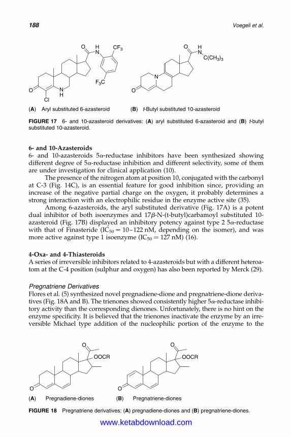
DK4962-Webster-ch14_R2_220307
6- and 10-Azasteroids6- and 10-azasteroids 5a-reductase inhibitors have been synthesized showingdifferent degree of 5a-reductase inhibition and different selectivity, some of themare under investigation for clinical application (10).
The presence of the nitrogen atom at position 10, conjugated with the carbonylat C-3 (Fig. 14C), is an essential feature for good inhibition since, providing anincrease of the negative partial charge on the oxygen, it probably determines astrong interaction with an electrophilic residue in the enzyme active site (35).
Among 6-azasteroids, the aryl substituted derivative (Fig. 17A) is a potentdual inhibitor of both isoenzymes and 17b-N-(t-butyl)carbamoyl substituted 10-azasteroid (Fig. 17B) displayed an inhibitory potency against type 2 5a-reductasewith that of Finasteride (IC50 ¼ 10–122 nM, depending on the isomer), and wasmore active against type 1 isoenzyme (IC50 ¼ 127 nM) (16).
4-Oxa- and 4-ThiasteroidsA series of irreversible inhibitors related to 4-azasteroids but with a different heteroa-tom at the C-4 position (sulphur and oxygen) has also been reported by Merck (29).
Pregnatriene DerivativesFlores et al. (5) synthesized novel pregnadiene-dione and pregnatriene-dione deriva-tives (Fig. 18A and B). The trienones showed consistently higher 5a-reductase inhibi-tory activity than the corresponding dienones. Unfortunately, there is no hint on theenzyme specificity. It is believed that the trienones inactivate the enzyme by an irre-versible Michael type addition of the nucleophilic portion of the enzyme to the
O NH
Cl
HN
O CF3
N
O
HN
O
Aryl substituted 6-azasteroid
F3C
(A) (B) t-Butyl substituted 10-azasteroid
C(CH3)3
FIGURE 17 6- and 10-azasteroid derivatives: (A) aryl substituted 6-azasteroid and (B) t-butylsubstituted 10-azasteroid.
O
O
OOCR
O
O
OOCR
Pregnadiene-diones Pregnatriene-diones (A) (B)
FIGURE 18 Pregnatriene derivatives: (A) pregnadiene-diones and (B) pregnatriene-diones.
188 Voegeli et al.
www.ketabdownload.com
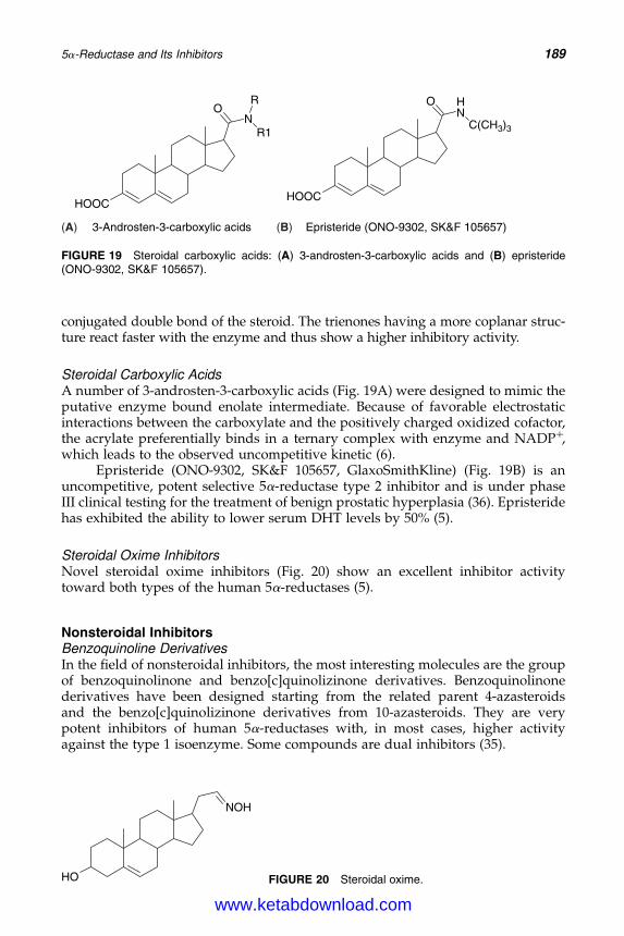
DK4962-Webster-ch14_R2_220307
conjugated double bond of the steroid. The trienones having a more coplanar struc-ture react faster with the enzyme and thus show a higher inhibitory activity.
Steroidal Carboxylic AcidsA number of 3-androsten-3-carboxylic acids (Fig. 19A) were designed to mimic theputative enzyme bound enolate intermediate. Because of favorable electrostaticinteractions between the carboxylate and the positively charged oxidized cofactor,the acrylate preferentially binds in a ternary complex with enzyme and NADPþ,which leads to the observed uncompetitive kinetic (6).
Epristeride (ONO-9302, SK&F 105657, GlaxoSmithKline) (Fig. 19B) is anuncompetitive, potent selective 5a-reductase type 2 inhibitor and is under phaseIII clinical testing for the treatment of benign prostatic hyperplasia (36). Epristeridehas exhibited the ability to lower serum DHT levels by 50% (5).
Steroidal Oxime InhibitorsNovel steroidal oxime inhibitors (Fig. 20) show an excellent inhibitor activitytoward both types of the human 5a-reductases (5).
Nonsteroidal InhibitorsBenzoquinoline DerivativesIn the field of nonsteroidal inhibitors, the most interesting molecules are the groupof benzoquinolinone and benzo[c]quinolizinone derivatives. Benzoquinolinonederivatives have been designed starting from the related parent 4-azasteroidsand the benzo[c]quinolizinone derivatives from 10-azasteroids. They are verypotent inhibitors of human 5a-reductases with, in most cases, higher activityagainst the type 1 isoenzyme. Some compounds are dual inhibitors (35).
O
HOOC
NR1
R HN
O
C(CH3)3
HOOC
3-Androsten-3-carboxylic acids Epristeride (ONO-9302, SK&F 105657)(A) (B)
FIGURE 19 Steroidal carboxylic acids: (A) 3-androsten-3-carboxylic acids and (B) epristeride(ONO-9302, SK&F 105657).
HO
NOH
FIGURE 20 Steroidal oxime.
5a-Reductase and Its Inhibitors 189
www.ketabdownload.com
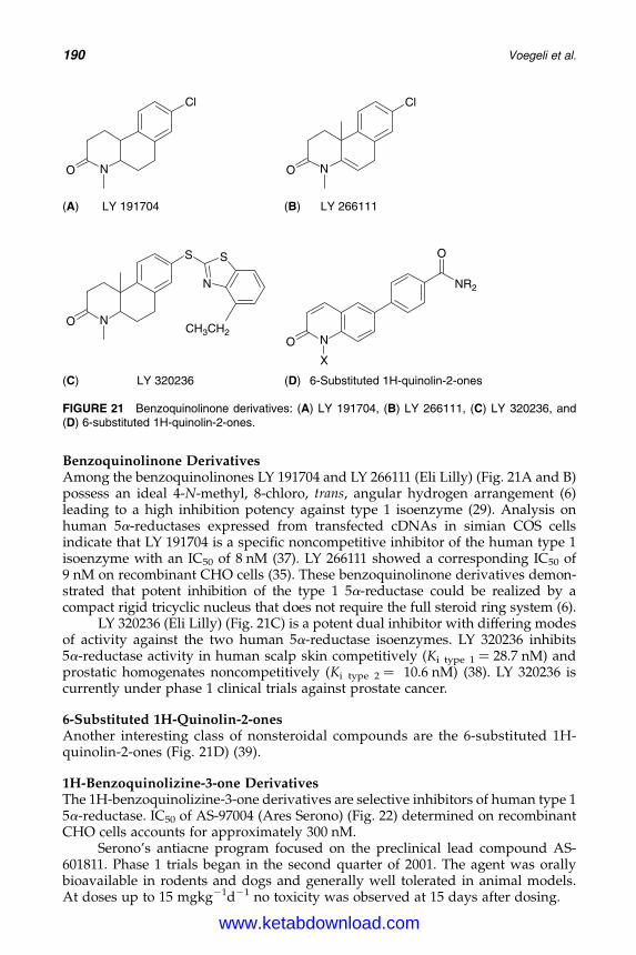
DK4962-Webster-ch14_R2_220307
Benzoquinolinone DerivativesAmong the benzoquinolinones LY 191704 and LY 266111 (Eli Lilly) (Fig. 21A and B)possess an ideal 4-N-methyl, 8-chloro, trans, angular hydrogen arrangement (6)leading to a high inhibition potency against type 1 isoenzyme (29). Analysis onhuman 5a-reductases expressed from transfected cDNAs in simian COS cellsindicate that LY 191704 is a specific noncompetitive inhibitor of the human type 1isoenzyme with an IC50 of 8 nM (37). LY 266111 showed a corresponding IC50 of9 nM on recombinant CHO cells (35). These benzoquinolinone derivatives demon-strated that potent inhibition of the type 1 5a-reductase could be realized by acompact rigid tricyclic nucleus that does not require the full steroid ring system (6).
LY 320236 (Eli Lilly) (Fig. 21C) is a potent dual inhibitor with differing modesof activity against the two human 5a-reductase isoenzymes. LY 320236 inhibits5a-reductase activity in human scalp skin competitively (Ki type 1 ¼ 28.7 nM) andprostatic homogenates noncompetitively (Ki type 2 ¼ 10.6 nM) (38). LY 320236 iscurrently under phase 1 clinical trials against prostate cancer.
6-Substituted 1H-Quinolin-2-onesAnother interesting class of nonsteroidal compounds are the 6-substituted 1H-quinolin-2-ones (Fig. 21D) (39).
1H-Benzoquinolizine-3-one DerivativesThe 1H-benzoquinolizine-3-one derivatives are selective inhibitors of human type 15a-reductase. IC50 of AS-97004 (Ares Serono) (Fig. 22) determined on recombinantCHO cells accounts for approximately 300 nM.
Serono’s antiacne program focused on the preclinical lead compound AS-601811. Phase 1 trials began in the second quarter of 2001. The agent was orallybioavailable in rodents and dogs and generally well tolerated in animal models.At doses up to 15 mgkg21d21 no toxicity was observed at 15 days after dosing.
NO
Cl
NO
Cl
LY 191704 LY 266111
NO
S
N
S
NO
X
NR2
O
LY 320236 6-Substituted 1H-quinolin-2-ones
CH3CH2
(A)
(C) (D)
(B)
FIGURE 21 Benzoquinolinone derivatives: (A) LY 191704, (B) LY 266111, (C) LY 320236, and(D) 6-substituted 1H-quinolin-2-ones.
190 Voegeli et al.
www.ketabdownload.com
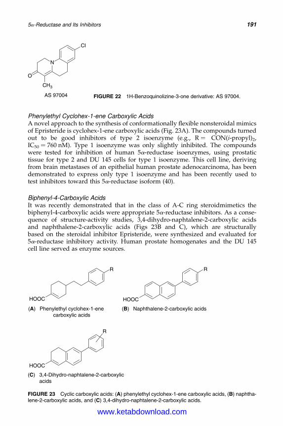
DK4962-Webster-ch14_R2_220307
Phenylethyl Cyclohex-1-ene Carboxylic AcidsA novel approach to the synthesis of conformationally flexible nonsteroidal mimicsof Epristeride is cyclohex-1-ene carboxylic acids (Fig. 23A). The compounds turnedout to be good inhibitors of type 2 isoenzyme (e.g., R ¼ CON(i-propyl)2,IC50 ¼ 760 nM). Type 1 isoenzyme was only slightly inhibited. The compoundswere tested for inhibition of human 5a-reductase isoenzymes, using prostatictissue for type 2 and DU 145 cells for type 1 isoenzyme. This cell line, derivingfrom brain metastases of an epithelial human prostate adenocarcinoma, has beendemonstrated to express only type 1 isoenzyme and has been recently used totest inhibitors toward this 5a-reductase isoform (40).
Biphenyl-4-Carboxylic AcidsIt was recently demonstrated that in the class of A-C ring steroidmimetics thebiphenyl-4-carboxylic acids were appropriate 5a-reductase inhibitors. As a conse-quence of structure-activity studies, 3,4-dihydro-naphtalene-2-carboxylic acidsand naphthalene-2-carboxylic acids (Figs 23B and C), which are structurallybased on the steroidal inhibitor Epristeride, were synthesized and evaluated for5a-reductase inhibitory activity. Human prostate homogenates and the DU 145cell line served as enzyme sources.
HOOC
(A) (B)
(C)
R
HOOC
R
Phenylethyl cyclohex-1-enecarboxylic acids
Naphthalene-2-carboxylic acids
HOOC
R
3,4-Dihydro-naphtalene-2-carboxylicacids
FIGURE 23 Cyclic carboxylic acids: (A) phenylethyl cyclohex-1-ene carboxylic acids, (B) naphtha-lene-2-carboxylic acids, and (C) 3,4-dihydro-naphtalene-2-carboxylic acids.
N
O
CH3
Cl
AS 97004 FIGURE 22 1H-Benzoquinolizine-3-one derivative: AS 97004.
5a-Reductase and Its Inhibitors 191
www.ketabdownload.com

DK4962-Webster-ch14_R2_220307
Apparently, the 3,4-dihydronaphtalene-2-carboxylic and naphthalene-2-carboxylic acid moiety mimicking the steroidal A-B ring is appropriate for gen-erating potent transition state analogs of the steroidal substrate testosterone andandrosterone. The most potent inhibitors of the human enzymes show IC50
values in the nM range. These naphthalene derivatives exceeded similarlysubstituted 1H-quinolin-2-ones in their inhibitory activities. This was to beexpected, since in the class of biphenyl type inhibitors, a benzoic acid moietyis superior to a 2-pyridone structure as a steroidal A ring mimetic.
The strong influence of the type and the position of the substituents at the6-phenyl group as well as the absence or presence of a double bond in 1 positionon the activity of the compounds are remarkable. The big difference in their inhibi-tory activities toward type 1 and type 2 isoenzyme in the case of single compoundsdemonstrates that they are appropriate to elaborate the structural requirements fora selective or a dual inhibition of type 1 or type 2 isoenzyme inhibition (39).
Indolizine- and Indol-Butanoic AcidsFR146687 (Fujisawa Pharmaceutical) (Fig. 24A) shows a noncompetitive inhibitionof both isoenzymes in vitro and has no inhibitory effects on other steroid oxido-reductases (41).
FK143 (Fujisawa Pharmaceutical) (Fig. 24B) was shown to inhibit both typesof human 5a-reductase equally. The mode of inhibition against both the isoenzymesis noncompetitive. The inhibition constants for the human type 1 and type 2 isoen-zymes were 27 and 20 nM, respectively. Species selectivity between human and raton the inhibitory activity of FK143 was not found for both the isoenzymes. No otherinhibitory effects were shown to other oxidoreductases (42). After a single oraladministration of FK143 (100–500 mg) on healthy volunteers, DHT plasma concen-trations decreased to about 65% of a predose value (43).
CationsZinc has long been used in dermatology and is reported to reduce sebum secretion.Metal ions have numerous roles in protein function, especially at the active site ofenzymes. They could be complexed to a substrate or modify the structure ofthe protein. The two types of human 5a-reductases were transfected into SW-13cells, a human adrenal carcinoma cell line, containing negligible endogeneous
N
HOOC
OO
i-Bu
N
HOOC
O
HN
i-Bu
i-Bu
FR 146687(A) (B) FK 143
(CH2)3 (CH2)3
FIGURE 24 Indolizine- and indol-butanoic acids: (A) FR 146687 and (B) FK 143.
192 Voegeli et al.
www.ketabdownload.com
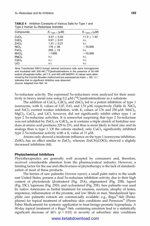
DK4962-Webster-ch14_R2_220307
5a-reductase activity. The expressed 5a-reductases were analyzed for their sensi-tivity to heavy metal ions using 0.2 mM (14C)androstenedione as a substrate.
The addition of CuCl2, CdCl2, and ZnCl2 led to a potent inhibition of type 1isoenzyme, with Ki values of 3.47, 0.61, and 1.51 mM, respectively (Table 4). NiCl2
and FeCl3 exerted weaker inhibition, with Ki values of 176 and 250 mM. MnCl2,MgCl2, CaCl2, and LiCl, however, did not significantly inhibit either type 1 ortype 2 5a-reductase activities. It is somewhat surprising that type 2 5a-reductasewas not inhibited by ZnCl2 or CdCl2, as it contains a triple stretch of histidine resi-dues at amino acid positions 229 to 231, and thus is more likely to bind zinc and itsanalogs than is type 1. Of the cations studied, only CuCl2 significantly inhibitedtype 2 5a-reductase activity with a Ki value of 11 mM.
Anions only showed a moderate influence on the type 1 isoenzyme inhibition.ZnSO4 has an effect similar to ZnCl2, whereas Zn(CH3COO)2 showed a slightlydecreased inhibition (44).
Phytochemical InhibitorsPhytotherapeutics are generally well accepted by consumers and, therefore,received considerable attention from the pharmaceutical industry. However, alimiting factor for the use and effectiveness of phytotherapy is the lack of standardi-zation of most of these products.
The berries of saw palmetto (Serenoa repens), a small palm native to the southeast United States, possess a dual 5a-reductase inhibition activity, due to their highcontent of phytosterols (b-sitosterol (Fig. 25A), stigmasterol (Fig. 25B), lupeol(Fig. 25C), lupenone (Fig. 25D), and cycloartenol (Fig. 25E). Saw palmetto was usedby native Americans as herbal treatment for enuresis, nocturia, atrophy of testes,impotence, inflammation of the prostate, and low libido in men. Standardized lipo-philic saw palmetto extracts are commercially available: e.g., Reguw-Seb (Penta-pharm) for topical treatment of seborrheic skin conditions and Permixonw (PierreFabre Medicament) for systemic application to treat benign prostatic hyperplasia. A90-day topical treatment of a Reguw-Seb, containing emulsion lead to a statisticallysignificant decrease of 46% (p , 0.01) in severity of seborrheic skin conditions
TABLE 4 Inhibition Constants of Various Salts for Type 1 andType 2 Human 5a-Reductase Activities
Compounds Ki Type 1 (mM) Ki Type 2 (mM)
CuCl2 3.47 + 0.43 11.0 + 1.42CdCl2 0.61 + 0.01 n.i.ZnCl2 1.51 + 0.21 n.i.NiCl2 176+ 26 .10,000FeCl3 250+ 15 n.i.MnCl2 .1000 .10,000MgCl2 n.i. n.i.CaCl2 n.i. n.i.LiCl n.i. n.i.
Note: Transfected SW13 human adrenal carcinoma cells were homogenizedand incubated with 200 nM [14C]androstenedione in the presence of 50 mMsodium phosphate buffer, pH 7.5, and 400 mM NADPH. Ki values were deter-mined by the Cornish-Bowden method and are expressed as mean + SD. “n.i.”indicates that no significant inhibition was observed.Source: Adapted from Ref. 44.
5a-Reductase and Its Inhibitors 193
www.ketabdownload.com
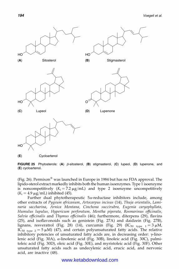
DK4962-Webster-ch14_R2_220307
(Fig. 26). Permixonw was launched in Europe in 1984 but has no FDA approval. Thelipido-sterol extract markedly inhibits both the human isoenzymes. Type 1 isoenzymeis noncompetitively (Ki ¼ 7.2 mg/mL) and type 2 isoenzyme uncompetitively(Ki ¼ 4.9 mg/mL) inhibited (45).
Further dual phytotherapeutic 5a-reductase inhibitors include, amongother extracts of Pygeum africanum, Artocarpus incisus (14), Thuja orientalis, Lami-naria saccharina, Arnica Montana, Cinchona succirubra, Eugenia caryophyllata,Humulus lupulus, Hypericum perforatum, Mentha piperata, Rosmarinus officinalis,Salvia officinalis and Thymus officinalis (46); furthermore, diterpens (29), flavins(25), and isoflavonoids such as genistein (Fig. 27A) and daidzein (Fig. 27B),lignans, resveratrol (Fig. 28) (14), curcumin (Fig. 29) (IC50 type 1 ¼ 3 mM,IC50 type 2 ¼ 5 mM) (47), and certain polyunsaturated fatty acids. The relativeinhibitory potencies of unsaturated fatty acids are, in decreasing order: g-lino-lenic acid (Fig. 30A), a-linolenic acid (Fig. 30B), linoleic acid (Fig. 30C), palmi-toleic acid (Fig. 30D), oleic acid (Fig. 30E), and myristoleic acid (Fig. 30F). Otherunsaturated fatty acids such as undecylenic acid, erucic acid, and nervonicacid, are inactive (48).
OH OH
Sitosterol(A) (B)
(C)
(E)
(D)
Stigmasterol
OH O
Lupeol Lupenone
OH
Cycloartenol
FIGURE 25 Phytosterols: (A) b-sitosterol, (B) stigmasterol, (C) lupeol, (D) lupenone, and(E) cycloartenol.
194 Voegeli et al.
www.ketabdownload.com

DK4962-Webster-ch14_R2_220307
0.0
0.5
1.0
1.5
2.0
2.5
3.0
day 01 day 30 day 91
Clin
ical
Sco
rin
g
FIGURE 26 Efficacy of topically applied Reguw-Seb on seborrheic skin conditions.Score: 0 ¼ no clinical relevant seborrheic skin,,3 comedones, 1 ¼ slightly seborrheic skin, 3–10comedones, 2 ¼ easily visible seborrheic skin, 10 –30 comedones, 3 ¼ obviously seborrheicskin, .30 comedones, 4 ¼ extensive seborrhoea covering the whole face, .30 comedones.
O
OOH
HO
(A) (B)
OH
O
O
HO
OH
Genistein Daidzein
FIGURE 27 Isoflavonoids: (A) genistein and (B) daidzein.
OH
OH
HO
FIGURE 28 Resveratrol.
O O
HO OH
OMeMeO
FIGURE 29 Curcumin.
5a-Reductase and Its Inhibitors 195
www.ketabdownload.com
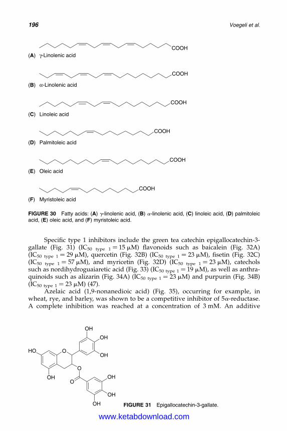
DK4962-Webster-ch14_R2_220307
Specific type 1 inhibitors include the green tea catechin epigallocatechin-3-gallate (Fig. 31) (IC50 type 1 ¼ 15 mM) flavonoids such as baicalein (Fig. 32A)(IC50 type 1 ¼ 29 mM), quercetin (Fig. 32B) (IC50 type 1 ¼ 23 mM), fisetin (Fig. 32C)(IC50 type 1 ¼ 57 mM), and myricetin (Fig. 32D) (IC50 type 1 ¼ 23 mM), catecholssuch as nordihydroguaiaretic acid (Fig. 33) (IC50 type 1 ¼ 19 mM), as well as anthra-quinoids such as alizarin (Fig. 34A) (IC50 type 1 ¼ 23 mM) and purpurin (Fig. 34B)(IC50 type 1 ¼ 23 mM) (47).
Azelaic acid (1,9-nonanedioic acid) (Fig. 35), occurring for example, inwheat, rye, and barley, was shown to be a competitive inhibitor of 5a-reductase.A complete inhibition was reached at a concentration of 3 mM. An additive
COOH(A)
(B)
(C)
(D)
γ-Linolenic acid
COOH
α-Linolenic acid
COOH
Linoleic acid
COOH
Palmitoleic acid
COOH
Oleic acid
COOH
Myristoleic acid
(E)
(F)
FIGURE 30 Fatty acids: (A) g-linolenic acid, (B) a-linolenic acid, (C) linoleic acid, (D) palmitoleicacid, (E) oleic acid, and (F) myristoleic acid.
HO
OH
O
O
OH
OH
OH
OOH
OH
OH FIGURE 31 Epigallocatechin-3-gallate.
196 Voegeli et al.
www.ketabdownload.com
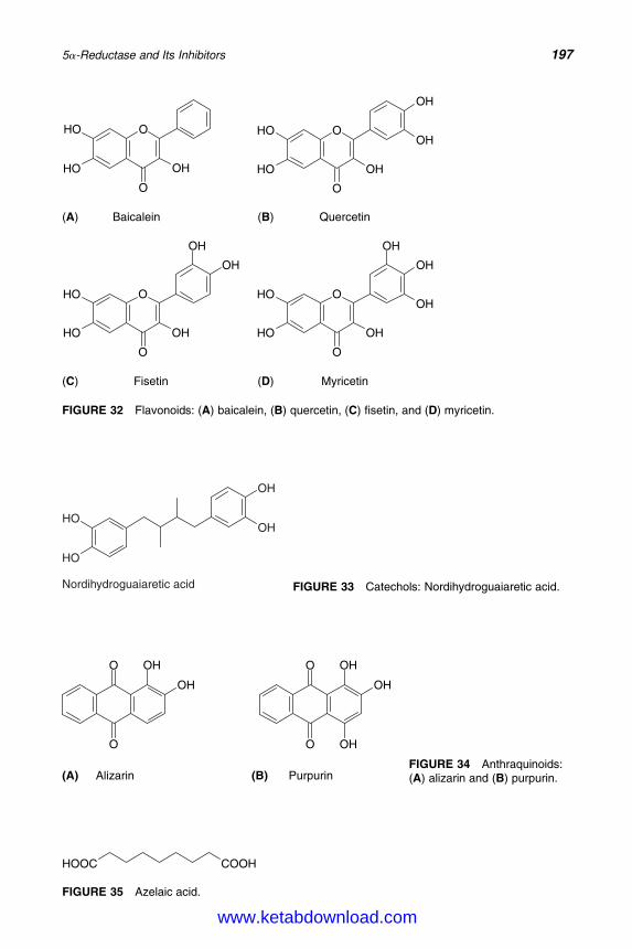
DK4962-Webster-ch14_R2_220307
O
O
HO
HO OH
(A)
(C) (D)
(B)
O
O
HO
HO OH
OH
OH
Baicalein Quercetin
O
O
HO
HO OH
OH
OH
O
O
HO
HO OH
OH
OH
OH
Fisetin Myricetin
FIGURE 32 Flavonoids: (A) baicalein, (B) quercetin, (C) fisetin, and (D) myricetin.
HO
HO
OH
OH
Nordihydroguaiaretic acid FIGURE 33 Catechols: Nordihydroguaiaretic acid.
O
O
OH
OH O
O
OH
OH
OH
Alizarin Purpurin(A) (B)FIGURE 34 Anthraquinoids:(A) alizarin and (B) purpurin.
HOOC COOH
FIGURE 35 Azelaic acid.
5a-Reductase and Its Inhibitors 197
www.ketabdownload.com
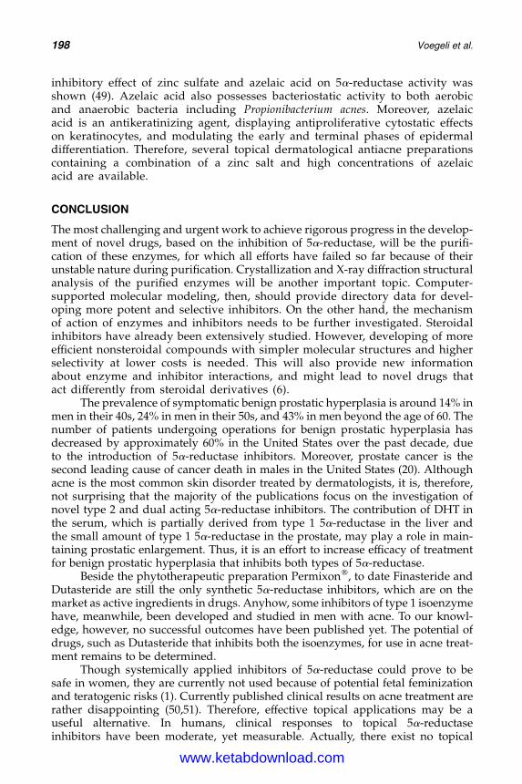
DK4962-Webster-ch14_R2_220307
inhibitory effect of zinc sulfate and azelaic acid on 5a-reductase activity wasshown (49). Azelaic acid also possesses bacteriostatic activity to both aerobicand anaerobic bacteria including Propionibacterium acnes. Moreover, azelaicacid is an antikeratinizing agent, displaying antiproliferative cytostatic effectson keratinocytes, and modulating the early and terminal phases of epidermaldifferentiation. Therefore, several topical dermatological antiacne preparationscontaining a combination of a zinc salt and high concentrations of azelaicacid are available.
CONCLUSION
The most challenging and urgent work to achieve rigorous progress in the develop-ment of novel drugs, based on the inhibition of 5a-reductase, will be the purifi-cation of these enzymes, for which all efforts have failed so far because of theirunstable nature during purification. Crystallization and X-ray diffraction structuralanalysis of the purified enzymes will be another important topic. Computer-supported molecular modeling, then, should provide directory data for devel-oping more potent and selective inhibitors. On the other hand, the mechanismof action of enzymes and inhibitors needs to be further investigated. Steroidalinhibitors have already been extensively studied. However, developing of moreefficient nonsteroidal compounds with simpler molecular structures and higherselectivity at lower costs is needed. This will also provide new informationabout enzyme and inhibitor interactions, and might lead to novel drugs thatact differently from steroidal derivatives (6).
The prevalence of symptomatic benign prostatic hyperplasia is around 14% inmen in their 40s, 24% in men in their 50s, and 43% in men beyond the age of 60. Thenumber of patients undergoing operations for benign prostatic hyperplasia hasdecreased by approximately 60% in the United States over the past decade, dueto the introduction of 5a-reductase inhibitors. Moreover, prostate cancer is thesecond leading cause of cancer death in males in the United States (20). Althoughacne is the most common skin disorder treated by dermatologists, it is, therefore,not surprising that the majority of the publications focus on the investigation ofnovel type 2 and dual acting 5a-reductase inhibitors. The contribution of DHT inthe serum, which is partially derived from type 1 5a-reductase in the liver andthe small amount of type 1 5a-reductase in the prostate, may play a role in main-taining prostatic enlargement. Thus, it is an effort to increase efficacy of treatmentfor benign prostatic hyperplasia that inhibits both types of 5a-reductase.
Beside the phytotherapeutic preparation Permixonw, to date Finasteride andDutasteride are still the only synthetic 5a-reductase inhibitors, which are on themarket as active ingredients in drugs. Anyhow, some inhibitors of type 1 isoenzymehave, meanwhile, been developed and studied in men with acne. To our knowl-edge, however, no successful outcomes have been published yet. The potential ofdrugs, such as Dutasteride that inhibits both the isoenzymes, for use in acne treat-ment remains to be determined.
Though systemically applied inhibitors of 5a-reductase could prove to besafe in women, they are currently not used because of potential fetal feminizationand teratogenic risks (1). Currently published clinical results on acne treatment arerather disappointing (50,51). Therefore, effective topical applications may be auseful alternative. In humans, clinical responses to topical 5a-reductaseinhibitors have been moderate, yet measurable. Actually, there exist no topical
198 Voegeli et al.
www.ketabdownload.com
DK4962-Webster-ch14_R2_220307
pharmaceuticals claiming exclusively 5a-reductase inhibition activity, althoughthere are antiacne products on the market containing, for example, zinc acetateand azelaic acid, respectively. Most of the synthetic or phytochemical 5a-reductaseinhibitors are still under in vitro investigation, animal studies, or clinical trials.Finally, it nevertheless remains to be pointed out that current research in acnetherapy favors investigations of topical androgen receptor blockers over topical5a-reductase inhibitors.
REFERENCES
1. Shaw JC. Hormonal therapies in acne. Expert Opin Pharmacother 2002; 3:865–874.2. Thiboutot D, Jabara S, McAllister JM, et al. Human skin is a steroidogenic tissue: steroi-
dogenic enzymes and cofactors are expressed in epidermis, normal sebocytes, and inimmortalized sebocyte cell line (SEB-1). J Invest Dermatol 2003; 120:905–913.
3. Fritsch M, Orfanos CE, Zouboulis CC. Sebaceous are the key regulators of androgenhomeostasis in human skin. J Invest Dermatol 2001; 116:793–800.
4. Zouboulis CC. Human skin: an independent peripheral endocrine organ. Horm Res2000; 54:230–242.
5. Flores E, Bratoeff E, Cabeza M, et al. Steroid 5a-reductase inhibitors. Mini Rev MedChem 2003; 3:225–237.
6. Li X, Chen C, Singh SM, et al. The enzyme and inhibitors of 4-ene-3-oxosteroid 5a-oxido-reductase. Steroids 1995; 60:430–441.
7. McConnel JD, Stoner E. 5a-reductase inhibitors. Adv Protein Chem 2001; 56:143–180.8. Chen W, Zouboulis CC, Fritsch M, et al. Evidence of heterogeneity and quantitative
differences of the type 1 5a-reductase expression in cultured human skin cells—evidenceof its presence in melanocytes. J Invest Dermatol 1998; 110:84–89.
9. Chen W, Zouboulis CC, Orfanos CE. The 5a-reductase system and its inhibitors. Derma-tology 1996; 193:177–184.
10. Cilotti A, Danza G, Serio M. Clinical application of 5a-reductase inhibitors. J EndocrinolInvest 2001; 24:199–203.
11. Randall VA. Role of 5a-reductase in health and disease. Bailliere’s Clin EndocrinolMetab 1994; 8:405–431.
12. Moore RJ, Wilson JD. Steroid 5a-reductase in cultured human fibroblasts. Biochemicaland genetic evidence for two distinct enzyme activities. J Biol Chem 1976; 251:5895–5900.
13. Spera G, Lubrano C. 5a-reductases and their inhibitors. Int J Immunopathol Pharm 1996;9:33–38.
14. Chen WC, Thiboutot D, Zouboulis CC. Cutaneous androgen metabolism: Basic researchand clinical perspectives. J Invest Dermatol 2002; 119:992–1007.
15. Lopez-Solache I. Characterization and expression of 5a-reductase isoenzymes. Ph.D.dissertation, Laval University, Quebec, Canada, 1998.
16. Guarna A, Occhiato EG, Danza G, et al. 5a-Reductase inhibitors, chemical and clinicalmodels. Steroids 1998; 63:355–361.
17. Thigpen AE, Cala KE, Russell DW. Characterization of Chinese hamster ovary cell linesexpressing human steroid 5a-reductase isoenzymes. J Biol Chem 1993; 268:17404–17412.
18. Lopez-Solache I, Luu-The V, Seralini GE, et al. Heterogeneity of rat type 1 5a-reductasecDNA. Cloning, expression and regulation by pituitary implants and dihydrotestoster-one. Biochem Biophys Acta 1996; 1305:139–144.
19. Levy MA, Brandt M, Sheedy KM, et al. Cloning, expression and functional characteriz-ation of type 1 and type 2 steroid 5a-reductases from Cynomolgus monkey: comparisonswith human and rat isoenzymes. J Steroid Biochem Mol Biol 1995; 52:307–319.
20. Foley CL, Kirby RS. 5a-Reductase inhibitors: what’s new ? Curr Opin Urol 2003; 13:31–37.21. Thiboutot D, Bayne E, Thorne J, et al. Immunolocalization of 5a-reductase isozymes in
acne lesions and normal skin. Arch Dermatol 2000; 136:1125–1129.22. Kao JS, Garg A, Mao-Qiang M, et al. Testosterone perturbs epidermal permeability
barrier homeostasis. J Invest Dermatol 2001; 116:443–451.
5a-Reductase and Its Inhibitors 199
www.ketabdownload.com

DK4962-Webster-ch14_R2_220307
23. Akamatsu H, Zouboulis CC, Orfanos CE. Control of human sebocyte proliferation invitro by testosterone and 5a-dihydrotestosterone is dependent on the localization ofthe sebaceous glands. J Invest Dermatol 1992; 99:509–511.
24. Thiboutot D, Knaggs H, Gilliland K, et al. Activity of 5a-reductase and 17b-hydroxysteroiddehydrogenase in the infrainfundibulum of subjects with and without Acne vulgaris.Dermatology 1998; 196:38–42.
25. Nakayama O, Yagi M, Kioyto S, et al. Riboflavin, a testosterone 5a-reductase inhibitor.J Antibiot 1990; 43:1615–1616.
26. Ahmed S. Molecular modelling of 5a-reductase inhibitors. Pharm Sci 1996; 2:251–253.27. Ahmed S, Denison S. Structure activity relationship study of known inhibitors of the
enzyme 5a-reductase. Bioorg Med Chem Lett 1998; 8:409–414.28. Grisenti P, Magni A, Olgiati V, et al. Substrate interaction with 5a-reductase enzyme:
Influence of the 17b-chain chirality in the mechanism of action of 4-azasteroid inhibitors.Steroids 2001; 66:803–810.
29. Macchetti F, Guarna A. Novel inhibitors of 5a-reductase. Expert Opin Ther Patents 2002;12:201–215.
30. Amichai B, Grunwald MH, Sobel R. 5a-reductase inhibitors—a new hope in dermatol-ogy? Int J Dermatol 1997; 36:182–184.
31. di Salle E, Guidici D, Radice A, et al. PNU 157706, a novel dual type 1 and 2 5a-reductaseinhibitor. J Steroid Biochem Mol Biol 1998; 64:179–186.
32. Ahmed S, Denison S. Mechanism based representation of the active site of 5a-reductase.Bioorg Med Chem Lett 1998; 8:2615–2620.
33. Schwartz JI, Tanaka WK, Wang DZ, et al. MK-386, an inhibitor of 5a-reductase type 1,reduces dihydrotestosterone concentrations in serum and sebum without affectingdihydrotestosterone concentrations in semen. J Clin Endocrinol Metab 1997; 82:1373–1377.
34. Chen C, Li X, Singh SM, et al. Activity of 17b-(N-alkyl/arylformamido) and 17b-[(N-alkyl/aryl) alkyl/arylamido]-4-methyl-4-aza-5a-androstan-3-ones as 5a-reductaseinhibitors in the hamster flank organ and ear. J Invest Dermatol 1998; 111:273–278.
35. Guarna A, Occhiato EG, Scarpi D, et al. Synthesis of benzo[c]quinolizin-3-ones: Selectivenonsteroidal inhibitors of steroid 5a-reductase 1. Bioorg Med Chem Lett 1998; 8:2871–2876.
36. Ishibashi K. 3-Oxo-4-aza-5a-androstane-17b-carboxamide derivatives with testosterone5a-reductase inhibitory activity. Annu Report Sankyo Res Lab 2000; 52:1–14.
37. Hirsch KS, Jones CD, Audia JE, et al. LY191704: A selective, nonsteroidal inhibitor ofhuman steroid 5a-reductase type 1. Proc Natl Acad Sci USA 1993; 90:5277–5281.
38. McNulty AM, Audia JE, Bemis KG, et al. Kinetic analysis of LY320236: competitiveinhibitor of type 1 and non-competitive inhibitor of type 2 human steroid 5a-reductase.J Steroid Biochem Mol Biol 2000; 72:13–21.
39. Baston E, Salem OIA, Hartmann RW. 6-Substituted 3,4-dihydro-naphtalene-2-carboxylicacids: synthesis and structure-activity studies in a novel class of 5-reductase inhibitors.J Enzyme Inhibition Med Chem 2002; 17:303–320.
40. Baston E, Salem OIA, Hartmann RW. Cyclohex-1-ene carboxylic acids: synthesis and bio-logical evaluation of novel inhibitors of human 5a-reductase. Arch Pharmacol Pharma-col Med Chem 2003; 1:31–38.
41. Nakayama O, Hirosumi J, Chida N, et al. FR146687, a novel steroid 5a-reductase inhibi-tor: In vitro and in vivo effects on prostates. Prostate 1997; 31:241–249.
42. Kojo H, Nakayama O, Hirosumi J, et al. Novel steroid 5a-reductase inhibitor FK143: Itsdual inhibition against the two isoenzymes and its effect on transcription of the isoen-zyme genes. Mol Pharmacol 1995; 48:401–406.
43. Katashima M, Irino T, Shimojo F, et al. Pharmacokinetics and pharmacodynamicsof FK143, a nonsteroidal inhibitor of steroid 5a-reductase, in healthy volunteers. ClinPharmacol Ther 1998; 63:354–366.
44. Sugimoto Y, Lopez-Solache I, Labrie F, et al. Cations inhibit specifically type 15a-reductase found in human skin. J Invest Dermatol 1995; 104:775–778.
45. Iehle C, Delos S, Guirou O, et al. Human prostatic steroid 5a-reductase isoforms—acomparative study of selective inhibitors. J Steroid Biochem Mol Biol 1995; 54:273–279.
200 Voegeli et al.
www.ketabdownload.com

DK4962-Webster-ch14_R2_220307
46. Picard-Lesboueyries E. Composition comprising 7-hydroxy DHEA and/or 7-keto DHEAand at least a 5a-reductase inhibitor. PCT Application WO 02/47644 A1.
47. Liao S, Hiipakka R. Method and compositions for regulation of 5a-reductase activity. USPatent Application Publication US 2003/0105030 A1.
48. Liang T, Liao S. Inhibition of steroid 5a-reductase by specific aliphatic unsaturated fattyacids. Biochem J 1992; 285:557–562.
49. Stamatiadis B, Bulteau-Portois MC, Mowszowicz I. Inhibition of 5a-reductase activity inhuman skin by zinc and azelaic acid. Br J Dermatol 1988; 119:627–632.
50. Leyden J, Bergfeld W, Drake L, et al. A systemic type I a-reductase inhibitor is ineffectivein the treatment of acne vuljans. J Am Acad Dermatol 2004; 50:443–447.
51. Seiffert K, Seltmann H, Fritsch M, et al. Inhibition of 5 a-reductase activity in 5Z95 sebo-cytes and HaCaT keratinocytes in vitro. Norm Metab Res 2007; 35:141–148.
5a-Reductase and Its Inhibitors 201
www.ketabdownload.com

DK4962-Webster-ch14_R2_220307
www.ketabdownload.com
DK4962-Webster-ch15_R2_020407
Part III: Active Delivery, Formulation, and Testing
B15 Sebum: Physical–Chemical Properties,Macromolecular Structure, andEffects of Ingredients
Linda D. RheinSchool of Natural Sciences, Fairleigh Dickinson University, Teaneck, New Jersey, U.S.A.
Joel L. Zatz and Monica R. MotwaniCollege of Pharmacy, Rutgers University, Piscataway, New Jersey, U.S.A.
INTRODUCTION
Sebum is a hallmark characteristic of oily skin, which typifies or is usually associ-ated with acne-prone skin. The goal of this chapter is to present recent findingsregarding the macromolecular structure of sebum and the influence of variationsin composition of sebum on that macromolecular structure. It is further intendedto discuss how these findings might relate to the pathogenesis of acne. In orderto introduce a basis for this research, a brief review of the current knowledge ofthe composition of sebum is in order, although some repetition of information inother chapters on this subject is likely. After this review, the results of thethermal analysis of model sebum using differential scanning calorimetry (DSC)are presented along with an attempt to provide an explanation of the phase beha-vior, as it relates to sebum composition and to variations in the composition thatmight occur as part of a disease state such as acne. Further studies of the influenceof vehicles on the phase behavior are presented, along with a discussion of how animbalance in typical phase behavior may be relevant to the pathogenesis of acne.
Toward that end, our findings show that a model of sebum is a mixture ofsolid and liquid phases. We speculate that the liquid phase helps dissolve or“soften” the solid phase and that these two phases exist in a delicate balance. Wefurther speculate that if this balance is altered, the consequences could be blockageof the sebaceous duct and of the infundibulum if the liquid part of sebum is too lowand can no longer dissolve the solid part. This could result from action of Propioni-bacterium acnes bacteria on the sebum or from metabolic alterations in sebum com-position in situ.
ORIGIN OF SEBUM
Sebum is the oily secretion of the sebaceous gland (Fig. 1). This “oil gland” islocated juxtaposed to the hair follicle. The large ducts of the hair follicles arefilled with this white pasty sebaceous material derived from the sebaceous gland.Sebaceous glands are generally found all over the body, except on the palms,soles, and dorsum of the feet (1). Most of the glands are associated with hair folliclesand hence are termed pilosebaceous glands. In humans, they are concentrated onthe scalp, forehead, and face, where there may be as many as 400 to 900 glands/cm2 and up to 1600 glands/cm2 on the nose (2). Sebaceous follicles are particularly
203
www.ketabdownload.com
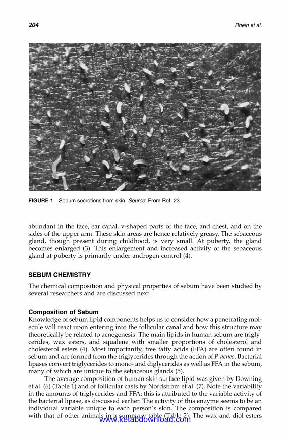
DK4962-Webster-ch15_R2_020407
abundant in the face, ear canal, v-shaped parts of the face, and chest, and on thesides of the upper arm. These skin areas are hence relatively greasy. The sebaceousgland, though present during childhood, is very small. At puberty, the glandbecomes enlarged (3). This enlargement and increased activity of the sebaceousgland at puberty is primarily under androgen control (4).
SEBUM CHEMISTRY
The chemical composition and physical properties of sebum have been studied byseveral researchers and are discussed next.
Composition of SebumKnowledge of sebum lipid components helps us to consider how a penetrating mol-ecule will react upon entering into the follicular canal and how this structure maytheoretically be related to acnegenesis. The main lipids in human sebum are trigly-cerides, wax esters, and squalene with smaller proportions of cholesterol andcholesterol esters (4). Most importantly, free fatty acids (FFA) are often found insebum and are formed from the triglycerides through the action of P. acnes. Bacteriallipases convert triglycerides to mono- and diglycerides as well as FFA in the sebum,many of which are unique to the sebaceous glands (5).
The average composition of human skin surface lipid was given by Downinget al. (6) (Table 1) and of follicular casts by Nordstrom et al. (7). Note the variabilityin the amounts of triglycerides and FFA; this is attributed to the variable activity ofthe bacterial lipase, as discussed earlier. The activity of this enzyme seems to be anindividual variable unique to each person’s skin. The composition is comparedwith that of other animals in a summary table (Table 2). The wax and diol esters
FIGURE 1 Sebum secretions from skin. Source: From Ref. 23.
204 Rhein et al.
www.ketabdownload.com
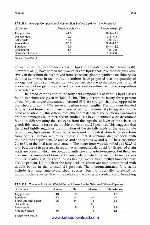
DK4962-Webster-ch15_R2_020407
appear to be the predominant class of lipid in animals other than humans (8).Stewart et al. (9) have shown that wax esters are lipids that have their origin exclu-sively in the sebum that is derived from sebaceous gland’s synthetic machinery viade nova synthesis. In fact, the same authors have proposed that the quantity ofendogenous lipids synthesized de nova per cell relative to the sebocytes’ originalendowment of exogenously derived lipids is a major influence on the compositionof secreted sebum.
The broad composition of the fatty acid components of various lipid classesfound in sebum are given in Table 3 (10). Thirty percent to fourty three percentof the fatty acids are unsaturated. Around 85% are straight chains as opposed tobranched and about 75% are even carbon chain lengths. The monounsaturatedfatty acids of human sebum are characterized by the unusual placing of a doublebond at position D6; this differs from other animals where the D9 monounsaturatesare predominant (4). In fact, recent studies (11) have identified a D6-desaturasefound in differentiating the sebocytes from the suprabasal layer of the sebaceousgland; this enzyme forms the double bonds at the D6 position. This suggests thatthe gland tightly regulates the formation of the D6 fatty acids at the appropriatetime during lipogenesis. These acids are found in greatest abundance in sebumfrom adults. Human sebum is unique in that it contains dienoic acids withdouble bonds at positions D5 and D8 and at positions D7 and D10. These constitute2% to 3% of the total fatty acid content. The major form was identified as 18:2D5, 8and, because of its presence in sebum, was named sebaleic acid (4). Branched chainacids are present, which are predominantly iso- and anteiso-isomers, but there arealso smaller amounts of branched chain acids, in which the methyl branch occursat other positions in the chain. Acids having two or three methyl branches mayalso be present. Up to half of the fatty acids of sebum are monounsaturated withdouble bonds in the unusual D6 position. The monounsaturated fatty acidsinclude iso- and anteiso-branched species, but no internally branched ormultibranched species. The fatty alcohols of the wax esters contain chain branching
TABLE 2 Classes of Lipids in Weight Percent Present in the Sebum of Different Species
Lipid class Human Rat Mouse Hamster ear
Triglycerides 43 8 6 20Squalene 12 — — —Sterol and wax esters 26 44 15 68Diol esters — — 66 —Free sterols 1 6 13 2.9Free fatty acids 16 1 1 5.5
Source: From Ref. 8.
TABLE 1 Average Composition of Human Skin Surface Lipid from the Forehead
Lipid class Mean, weight (%) Range, weight (%)
Triglycerides 41.0 19.5–49.4Diglycerides 2.2 2.3–4.3Fatty acids 16.4 7.9–39.0Wax esters 25.0 22.6–29.5Squalene 12.0 10.1–13.9Cholesterol 1.4 1.2–2.3Cholesterol esters 2.1 1.5–2.6
Source: From Ref. 6.
Sebum 205
www.ketabdownload.com
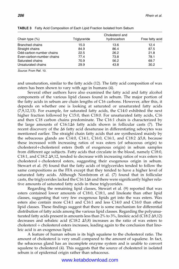
DK4962-Webster-ch15_R2_020407
and unsaturation, similar to the fatty acids (12). The fatty acid composition of waxesters has been shown to vary with age in humans (4).
Several other authors have also examined the fatty acid and fatty alcoholcomponents of the various lipid classes found in sebum. The major portion ofthe fatty acids in sebum are chain lengths of C16 carbons. However, after this, itdepends on whether one is looking at saturated or unsaturated fatty acids(7,9,12,13). For example, for saturated fatty acids, the C14:0 exhibited the nexthigher fraction followed by C15:0, then C18:0. For unsaturated fatty acids, C16and then C18 carbon chains predominate. The C16:1 chain is characterized bythe large amounts of C16:1D6 fatty acids shown in follicular casts (7). Therecent discovery of the D6 fatty acid desaturase in differentiating sebocytes wasmentioned earlier. The straight chain fatty acids that are synthesized mainly bythe sebaceous glands are C14:0, C14:1, C16:0, C16:1, and C18:2 D5,8, becausethese increased with increasing ratios of wax esters (of sebaceous origin) tocholesterolþcholesterol esters (both of exogenous origin) in sebum samplesfrom different age subjects. Fatty acids that circulate in the blood, namely C18:0,C18:1, and C18:2 D9,12, tended to decrease with increasing ratios of wax esters tocholesterolþ cholesterol esters, suggesting their exogenous origin in sebum.Stewart et al. (9) found that the fatty acids of triglycerides tended to follow thesame compositions as the FFA except that they tended to have a higher level ofsaturated fatty acids. Although Nordstrom et al. (7) found that in follicularcasts, the triglycerides lacked the C16:1D6 and there were significantly higher rela-tive amounts of saturated fatty acids in these triglycerides.
Regarding the remaining lipid classes, Stewart et al. (9) reported that waxesters contained lower amounts of C18:0, C18:1, and linoleate than other lipidclasses, suggesting that very few exogenous lipids get into the wax esters. Waxesters also contain more C14:1 and C16:1 and less C14:0 and C16:0 than otherlipid classes. These findings suggest that there is some mechanism for differentialdistribution of fatty acids among the various lipid classes. Regarding the polyunsa-turated fatty acids present in amounts less than 2% to 3%, linoleic acid (C18:2 D9,12)decreases and sebaleic acid (C18:2 D5,8) increases as the ratio of wax esters tocholesterolþ cholesterol esters increases, leading again to the conclusion that lino-leic acid is an exogenous lipid.
A feature of human sebum is its high squalene to the cholesterol ratio. Theamount of cholesterol is very small compared to the amount of squalene. In fact,the sebaceous gland has an incomplete enzyme system and is unable to convertsqualene to cholesterol (4). This suggests that the source of cholesterol in isolatedsebum is of epidermal origin rather than sebaceous.
TABLE 3 Fatty Acid Composition of Each Lipid Fraction Isolated from Sebum
Chain type (%) TriglycerideCholesterol and
hydrocarbon Free fatty acid
Branched chains 15.0 13.6 12.4Straight chains 84.9 86.4 87.5Odd-carbon-number chains 22.5 26.2 21.8Even-carbon-number chains 77.4 73.8 78.1Saturated chains 70.9 56.2 69.7Unsaturated chains 29.9 43.8 30.2
Source: From Ref. 10.
206 Rhein et al.
www.ketabdownload.com
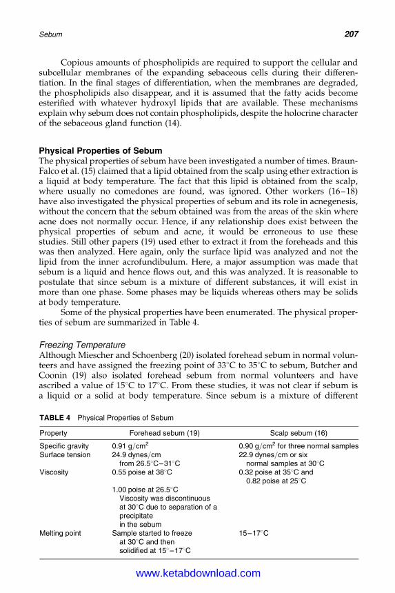
DK4962-Webster-ch15_R2_020407
Copious amounts of phospholipids are required to support the cellular andsubcellular membranes of the expanding sebaceous cells during their differen-tiation. In the final stages of differentiation, when the membranes are degraded,the phospholipids also disappear, and it is assumed that the fatty acids becomeesterified with whatever hydroxyl lipids that are available. These mechanismsexplain why sebum does not contain phospholipids, despite the holocrine characterof the sebaceous gland function (14).
Physical Properties of SebumThe physical properties of sebum have been investigated a number of times. Braun-Falco et al. (15) claimed that a lipid obtained from the scalp using ether extraction isa liquid at body temperature. The fact that this lipid is obtained from the scalp,where usually no comedones are found, was ignored. Other workers (16–18)have also investigated the physical properties of sebum and its role in acnegenesis,without the concern that the sebum obtained was from the areas of the skin whereacne does not normally occur. Hence, if any relationship does exist between thephysical properties of sebum and acne, it would be erroneous to use thesestudies. Still other papers (19) used ether to extract it from the foreheads and thiswas then analyzed. Here again, only the surface lipid was analyzed and not thelipid from the inner acrofundibulum. Here, a major assumption was made thatsebum is a liquid and hence flows out, and this was analyzed. It is reasonable topostulate that since sebum is a mixture of different substances, it will exist inmore than one phase. Some phases may be liquids whereas others may be solidsat body temperature.
Some of the physical properties have been enumerated. The physical proper-ties of sebum are summarized in Table 4.
Freezing TemperatureAlthough Miescher and Schoenberg (20) isolated forehead sebum in normal volun-teers and have assigned the freezing point of 338C to 358C to sebum, Butcher andCoonin (19) also isolated forehead sebum from normal volunteers and haveascribed a value of 158C to 178C. From these studies, it was not clear if sebum isa liquid or a solid at body temperature. Since sebum is a mixture of different
TABLE 4 Physical Properties of Sebum
Property Forehead sebum (19) Scalp sebum (16)
Specific gravity 0.91 g/cm2 0.90 g/cm2 for three normal samplesSurface tension 24.9 dynes/cm
from 26.58C–318C22.9 dynes/cm or six
normal samples at 308CViscosity 0.55 poise at 388C 0.32 poise at 358C and
0.82 poise at 258C1.00 poise at 26.58C
Viscosity was discontinuousat 308C due to separation of aprecipitatein the sebum
Melting point Sample started to freezeat 308C and thensolidified at 158–178C
15–178C
Sebum 207
www.ketabdownload.com
DK4962-Webster-ch15_R2_020407
lipidic substances, it is assumed that it will not have a single freezing point, butrather a range of temperatures in which transitions from a liquid to a solid takesplace. Butcher and Coonin (19) claim that the sebum sample started to freeze upto 308C, but the whole sample solidified at 158C to 178C and stopped flowing.
Specific GravityThe specific gravity of sebum given by Butcher and Coonin (19) from the foreheadsebum of normal individuals was 0.911 + 0.01 g/cm3 for all samples. Burton (16)confirmed this for three normal subjects when scalp sebum was used.
Surface TensionThe surface tension (Table 4) at temperatures varying from 26.58C to 318C was foundto be an average of 24.89 dynes/cm for forehead sebum of normal subjects. A veryclose value (22.9 + 0.9–24.2 + 1.1) was also obtained for scalp sebum of normaland acne patients.
ViscosityThe viscosity increased from 0.55 to 0.98 poise, as temperature decreased from 388Cto 28.58C. The sebum separated into various components when the temperaturewas lowered from 298C to 308C and ceased to flow at 158C to 178C (19). Burton(16) reported a viscosity of 0.32 + 0.03 poise at 358C, 0.82 + 0.22 poise at 258C,and 1.71 + 0.70 poise at 208C. In addition, it was found that the viscosity ofscalp sebum and forehead sebum was similar in normal individuals. Burton alsoreported that the mean viscosity of the sebum from 10 acne patients taking tetra-cycline was higher than with seven patients not taking tetracycline at 358C.
IMPORTANCE OF SEBUM TO THE PATHOGENESIS OF ACNE
The role of sebum in acnegenesis is poorly understood. Acne vulgaris, a multifac-torial disease of the skin, is found in areas rich in sebaceous follicles. It is character-ized by seborrhea, disturbed keratinization in the follicles with comedones, andsubsequent inflammatory papules, pustules and nodular abscesses, and scars(15). The pathology of acne is described in various other chapters in this bookbut a brief review here sets the stage for relevance of the studies of sebum describedlater in this chapter. There are three essential factors as a group that may cause acne.
1. There is an increase in androgen production in puberty, which induces enlarge-ment of sebaceous follicles and increased sebum production (21).
2. Follicular hyperkeratinization leads to retention hyperkeratosis, that is, obstruc-tion of the pilosebaceous duct by accumulation of an excessive amount of keratinas an important factor in the pathogenesis of acne (22).
3. P. acnes proliferate in the follicle, producing a variety of extracellular inflamma-tory products, which excite an inflammatory response (23).
The primary event in acne is faulty keratinization and the production of come-dones. In the secondary stage, inflammation can occur in the comedones. Twopathways define the subsequent development of the lesions of acne. In the nonin-flammatory pathway, the microcomedo proceeds to mature into closed and opencomedones through distention of the follicle wall and lumen. In the inflammatorypathway, the extracellular products of P. acnes incite inflammation. P. acnes
208 Rhein et al.
www.ketabdownload.com
DK4962-Webster-ch15_R2_020407
colonization occurs relatively early in acne, and the production of extracellular pro-ducts by this organism provides multiple potential mechanisms for the develop-ment of inflammation. Among the earliest findings, drugs that reduce fatty acids(FFA), such as tetracycline, are beneficial in inflammatory acne (24). It must benoted that skin surface-derived lipids produce an inflammatory papule or nodulewhen they are injected into the human skin (25). Furthermore, certain fractions ofthe sebaceous secretion, especially fatty acids and squalene, when applied torabbits’ ears promoted follicular hyperkeratosis and comedone formation (26–28).
In the absence of comedones, the large infundibulum channels are filled withthe white pasty sebaceous material. It is the normal content of a sebaceous follicle,not of an acne lesion. Sometimes, the follicles are filled with a cocoon-type skeletonof corneocytes, having 20 to 40 cells surrounding the central fine hair, with achannel left free in the sebum of which P. acnes and staphylococci may be found(15). This is called a follicular filament or follicular cast. A comedo can arise froma follicular filament. It is believed that the lipid composition of the follicular castsmay play a role in acne genesis. It was demonstrated by Nordstrom et al. (7) thatabout 29% of the net weight of follicular casts was lipid. In addition, the hydrolysisof the triglycerides in the follicular casts was much higher than on the skin surface.
The differences in the rate of sebum production in acne patients and normalcontrols have so far been the only consistent finding that links sebum secretion toacnegenesis (4,7). It has long been thought that acne is a result of an abnormalityof sebum or sebaceous gland function. Yet, the current concept of follicular kerati-nization in the pathogenesis of the disease makes this relationship difficult to justify.However, four major reasons based on circumstantial but compelling evidence thatlink sebum to acne include:
1. Acne develops at puberty, when sebaceous glands become very active (29).2. The rate of sebum secretion is generally higher in acne subjects than in normal
individuals, though not all persons having high sebum secretion suffer fromacne (30).
3. Some components of sebum are irritants and can be comedogenic in inflamma-tory acne.
4. Acne is improved by any modality that reduces sebaceous gland activity (31).
This indicates that sebum is probably an essential factor in the pathogenesis ofacne, but not necessarily the only one.
It is possible that the changes in the horny layer could be the result of analteration of the nature of the sebum, so that it causes keratinization of the ductand leads to comedone formation. The macromolecular structure of the sebumcould change to a more crystalline state that forms a blockage and impedes itsown flow through the sebaceous duct and infundibulum or alternatively cause irri-tation to the follicular epidermal lining. Gonzalez-Serva (32) and Abramovits andGonzalez-Serva (33) have speculated that acne involves the build up of an intra-ductal calculus termed sebolith, which results from crystallization of sebum.They proposed that the sebolith punctures and erodes the follicular lining,leading to rupture and inflammation. The proliferation of bacteria could be theresult of a change in sebum composition or structure, so that either it provides aricher, more bioavailable medium for bacterial growth or it no longer containsinhibitory factors. In such a manner, sebum can be involved. The authors believethat the characterization of the lipids of the sebaceous follicles and their physical
Sebum 209
www.ketabdownload.com
DK4962-Webster-ch15_R2_020407
behavior may provide more detailed information on the possible role of sebum inacne vulgaris.
Electron microscopy studies on comedones showed the presence of yeasts,bacteria, keratinized cells, sebum, and hairs. In these comedones, sebum was a ubi-quitous component and appeared as an osmiophilic granular material, whichvaried in amount from scanty to abundant between the keratinized cells (34).Depressions between the microridges of the keratinized cells had the greatest pro-pensity for sebum. Many yeast and bacteria occupied these sebum-rich areas andtheir surfaces were covered with fatty material.
The composition of the lipid components of comedones was also examined byNicolaides et al. (13) and compared with the skin surface lipids. Chapter 3 by Dr.Wertz covers this in more detail, but findings relative to the purposes of thischapter are summarized here. The lipids in comedones contain all the same lipidclasses as sebum, but some are in different proportions. Triglycerides representonly a small portion of the comedonal lipids (7%) and fatty acids represent 55%of the lipids. Wax esters are only 14% of the lipids compared with 24% in surfacesebum. Free cholesterol is also much higher in comedonal lipids. Another notabledifference is a larger amount of saturated fatty acids in comedonal lipids—about70% of the comedonal FFA are saturated compared with only 65% of the surfacesebum. The differences between comedonal and surface lipid reflect the contri-bution of epidermal lipids to the mix. Epidermal lipids do not contain any waxesters, and cholesterol is derived from epidermal lipid. Epidermal lipids alsoprovide saturated fatty acids to the mix. The reduced amounts of triglyceridesare due to nearly complete bacterial hydrolysis of this class to FFA.
Since sebum is a mixture of the different lipid components discussed earlier, itis important to understand how each component affects the macromolecular struc-ture and the physical–chemical behavior of sebum, and how this is altered in dis-eases of the pilosebaceous unit, for example, acne. It is, therefore, useful to knowhow variations in the components, for example, concentration and ratio of differentcomponents, their carbon chain length, and the ratios of unsaturation to saturation,affect the macromolecular structure of sebum and how this relates to the compo-sition of sebum in acne lesions in particular comedones. The ultimate goal is todetermine how this composition can be altered in the skin of acne patients tocorrect the defects in the infundibulum and the sebaceous duct, and to facilitatepenetration of anti-acne drugs into the pilosebaceous unit.
INVESTIGATIONS OF THE EFFECT OF VARIATIONS OF SEBUMCOMPONENTS ON THE PHASE BEHAVIOR OF SEBUM
The presence of sebaceous secretions seen as a white pasty substance in the infun-dibulum, and the relationship between sebaceous activity and acnegenesis dis-cussed earlier suggests that sebum macromolecular structure may play a role inthe pathogenesis of acne. The physical properties of sebum, melting point, and visc-osities in particular are important, as it would mediate the blockage of the sebac-eous follicle. To evaluate this theory, we have examined the physical propertiesof the sebum components and their mixtures using DSC. The purpose was toexamine parameters controlling the phase behavior of sebum (35). There are,however, physical limitations in collecting large amounts of sample from the skinsurface and it is not easy to get “pure” sebum since it is contaminated with thevarying amounts of epidermal lipids. Furthermore, sebum varies quantitatively
210 Rhein et al.
www.ketabdownload.com
DK4962-Webster-ch15_R2_020407
from person to person and would contribute to the variability of the data. We (35)hence decided to carry out our experiments on a model sebum based on compo-sitions given in the literature (Table 1). We have evaluated the effect of differentratios of lipid classes in a sebum model on phase behavior and on melting tempera-tures of these phases. In the models of sebum, the composition can be varied andthe effect on the phase behavior can be determined by DSC. The aim of this researchwas to determine the effect of component characteristics on the phase behavior ofthe model sebum. In particular, we investigated the effects of the following onthe melting behavior of the model sebum:
1. Carbon chain length of the triglycerides, fatty acids, and wax esters.2. Ratio of unsaturated to saturated components in triglycerides, fatty acids, and
wax esters.3. Ratio of triglyceride to fatty acid content.4. Effect of cosmetic additives.
Preparation of Lipid Samples and Differential ScanningCalorimetry ProceduresThe ingredients for a particular model were weighed out and dissolved in amixture of chloroform:methanol (3:1) (35). Small portions of the above modelsebum were withdrawn and put on to a preweighed DSC pan. The solvent wasevaporated and the weight of the pan was taken again. The difference gave usthe weight of the lipid mixture and this weight was entered on the DSC run bycomputer. In the absence of chloroform–methanol cosolvent, the samples with-drawn were not uniform since there was a separation of the different phases. TheDSC pan was then covered with an aluminum lid and run at a scan rate of 58C/min from 2508C to 1008C using a Perkin–Elmer DSC-7. Each of the compositionswas run in triplicates. For each individual component DSC runs were done in pureform, after dissolving in chloroform–methanol and evaporating the solvent. Thiswas done to facilitate the identification of peaks. Polymorphic changes mightoccur when the individual components are dissolved in the cosolvent mixture,which is consequently evaporated. This step identifies if any such changehas occurred.
The effect of the three different carbon chain lengths of sebum components,namely C14, C16, and C18, on phase behavior of sebum was examined. Majorfatty acids of sebum fall in the carbon chain length of C14, C16, and C18, respect-ively, with negligible amounts of other carbon chain lengths (13). To investigatethe phase transitions of sebum prepared with lipid classes with C14 carbonchains, the triglycerides, fatty acids, and wax esters in the sebum mixture wereall of carbon chain length 14 (Table 5). Similarly, a sebum mixture for carbonchain length 16 and separately C18 was prepared. According to Table 1, sebum con-tains amounts of 41% triglycerides, 25% wax esters, and 16.4% FFA, and we usedapproximately that composition; the remainder was squalene.
The lipid classes are, however, a mixture of unsaturated and saturated com-ponents; therefore, these were varied also for our second objective. In Table 5, wehave listed the ingredients used for the experiment. To study the effect of degreeof saturation, the carbon chain length of all the components was kept constant andvariation was done in the unsaturated and saturated portions. As an example, forthe fatty acids, which made up 16.4% of the total, the ratio of unsaturated to saturatedfatty acids was varied keeping the total to 16.4% of the sebum mixture.
Sebum 211
www.ketabdownload.com
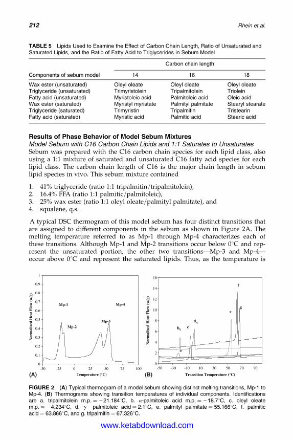
DK4962-Webster-ch15_R2_020407
Results of Phase Behavior of Model Sebum MixturesModel Sebum with C16 Carbon Chain Lipids and 1:1 Saturates to UnsaturatesSebum was prepared with the C16 carbon chain species for each lipid class, alsousing a 1:1 mixture of saturated and unsaturated C16 fatty acid species for eachlipid class. The carbon chain length of C16 is the major chain length in sebumlipid species in vivo. This sebum mixture contained
1. 41% triglyceride (ratio 1:1 tripalmitin/tripalmitolein),2. 16.4% FFA (ratio 1:1 palmitic/palmitoleic),3. 25% wax ester (ratio 1:1 oleyl oleate/palmityl palmitate), and4. squalene, q.s.
A typical DSC thermogram of this model sebum has four distinct transitions thatare assigned to different components in the sebum as shown in Figure 2A. Themelting temperature referred to as Mp-1 through Mp-4 characterizes each ofthese transitions. Although Mp-1 and Mp-2 transitions occur below 08C and rep-resent the unsaturated portion, the other two transitions—Mp-3 and Mp-4—occur above 08C and represent the saturated lipids. Thus, as the temperature is
TABLE 5 Lipids Used to Examine the Effect of Carbon Chain Length, Ratio of Unsaturated andSaturated Lipids, and the Ratio of Fatty Acid to Triglycerides in Sebum Model
Components of sebum model
Carbon chain length
14 16 18
Wax ester (unsaturated) Oleyl oleate Oleyl oleate Oleyl oleateTriglyceride (unsaturated) Trimyristolein Tripalmitolein TrioleinFatty acid (unsaturated) Myristoleic acid Palmitoleic acid Oleic acidWax ester (saturated) Myristyl myristate Palmityl palmitate Stearyl stearateTriglyceride (saturated) Trimyristin Tripalmitin TristearinFatty acid (saturated) Myristic acid Palmitic acid Stearic acid
0
0.1
0.2
0.3
0.4
0.5
0.6
0.7
0.8
0.9
1
-50 -25 0 25 50 75 100
Temperature (°C)
Nor
mal
ized
Hea
t F
low
(w
/g)
Mp-1
Mp-3Mp-2
Mp-4
(A)
0
2
4
6
8
10
12
14
16
-50 -30 -10 10 30 50 70 90
Transition Temperature (°C)
Nor
mal
ized
Hea
t F
low
(w
/g)
f
g e
bη
a
c
dη
(B)
FIGURE 2 (A) Typical thermogram of a model sebum showing distinct melting transitions, Mp-1 toMp-4. (B) Thermograms showing transition temperatures of individual components. Identificationsare a. tripalmitolein m.p. ¼ 221.1848C, b. a-palmitoleic acid m.p. ¼ 218.78C, c. oleyl oleatem.p. ¼ 24.2348C, d. g 2 palmitoleic acid ¼ 2.18C, e. palmityl palmitate ¼ 55.1668C, f. palmiticacid ¼ 63.8668C, and g. tripalmitin ¼ 67.3268C.
212 Rhein et al.
www.ketabdownload.com
DK4962-Webster-ch15_R2_020407
increased from 2508C to 08C, the unsaturated fractions of the lipid mixture melt(Fig. 2A). Standards were run and their transitions are shown in Figure 2B. Theunsaturated triglycerides and unsaturated fatty acids overlap at approximately2208C in transition Mp-1 and the unsaturated wax esters (also with a smalloverlap with fatty acids), Mp-2 at approximately 2158C. As the temperature isfurther increased, the two saturated fractions of the model sebum melt (above408C); one is designated as Mp-3 due to the saturated wax ester and then finallythe last solid fraction of the model sebum melts at approximately 558C and is desig-nated as Mp-4—a transition represented by the overlap of saturated tripalmitin andpalmitic acid. At 2508C, the sebum lipid mixture is still a solid, and by 1008C, allcomponents would have completely melted and the mixture is a liquid.
From the DSC profiles and melting endotherms, we can conclude that themodel sebum both at room temperature and at the temperature of the skin (328Cat the skin surface and closer to 408C in the lower epidermis) is crystalline innature. Generally, and as discussed by Motwani et al. (35), pure lipids or homo-geneous systems yield single sharp peaks, whereas heterogeneous systemsproduce broad multiple peaks. If sebum was a homogeneous mixture, therewould be one melting transition, but the presence of multiple melting transitionsin the model sebum suggests that natural sebum has multiple phases. From thetemperatures of these melting transitions, it appears that at skin temperature,some of the components of sebum are solids and some are liquids, which are notcompletely miscible with each other. The findings further suggest that the liquidphase probably helps solubilize the solid phase of the sebum. Therefore, it islikely that sebum exists in a macromolecular structure that is a delicate balancebetween the liquid and solid phases. This balance is determined in part by theratio of saturated (solids) to unsaturated (liquids) species.
Effect of Varying the Ratio of Saturates to UnsaturatesThe effect of degree of saturation on the phase transitions is shown in Figure 3A–C.The C16 lipid species were again used for these studies and each model sebum con-tained a different ratio of saturated to unsaturated fatty species. The transitiontemperatures are shown in relation to the percent of saturated components ineach sample of model sebum in Figure 3. As the saturation increases (or unsatura-tion decreases), the intensity of the transitions for Mp-3 and Mp-4 (transitions forthe saturated species) was greater and the intensities of Mp-1 and Mp-2 (transitionsfor the unsaturated species) was diminished, not surprising as their concentrationwas increasingly reduced. More interesting was that the temperatures where thephase transitions occurred decreased for the unsaturates and increased for thesaturated species. The transition temperature for the unsaturated components(Mp-1 and Mp-2) decreases with a concurrent increase in the Mp-3 and Mp-4 forthe saturates (Fig. 3B and C). This increase in transition temperature for the satu-rated compounds (Mp-3 and Mp-4) renders the sebum even more solid at theinner and outer temperature of skin.
As mentioned earlier, the presence of other substances decrease the meltingpoint of the pure compounds. When there is more unsaturation than saturation,by the time the temperature is reached for the melting of the saturated compounds,some of the saturated portion dissolves in the already melted unsaturatedportion and their melting point decreases. The more the unsaturation, the morethe dissolving and hence higher decreases in the melting point. In these cases,the unsaturated portion acts as the solvent. When the percent of the saturated
Sebum 213
www.ketabdownload.com
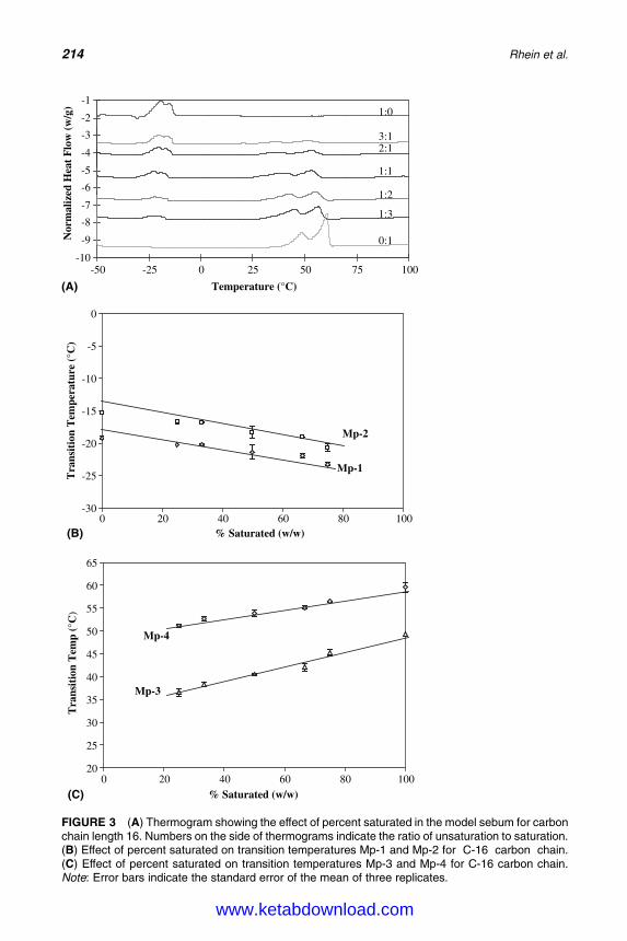
DK4962-Webster-ch15_R2_020407
-10
-9
-8
-7
-6
-5
-4
-3
-2
-1
-50 -25 0 25 50 75 100
Temperature (°C)(A)
Nor
mal
ized
Hea
t F
low
(w
/g)
0:1
1:3
1:2
1:1
2:13:1
1:0
-30
-25
-20
-15
-10
-5
0
(B)0 20 40 60 80 100
% Saturated (w/w)
Tra
nsit
ion
Tem
pera
ture
(°C
)
Mp-1
Mp-2
20
25
30
35
40
45
50
55
60
65
(C)0 20 40 60 80 100
% Saturated (w/w)
Tra
nsit
ion
Tem
p (°
C)
Mp-3
Mp-4
FIGURE 3 (A) Thermogram showing the effect of percent saturated in the model sebum for carbonchain length 16. Numbers on the side of thermograms indicate the ratio of unsaturation to saturation.(B) Effect of percent saturated on transition temperatures Mp-1 and Mp-2 for C-16 carbon chain.(C) Effect of percent saturated on transition temperatures Mp-3 and Mp-4 for C-16 carbon chain.Note: Error bars indicate the standard error of the mean of three replicates.
214 Rhein et al.
www.ketabdownload.com
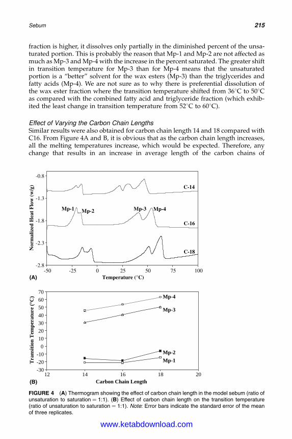
DK4962-Webster-ch15_R2_020407
fraction is higher, it dissolves only partially in the diminished percent of the unsa-turated portion. This is probably the reason that Mp-1 and Mp-2 are not affected asmuch as Mp-3 and Mp-4 with the increase in the percent saturated. The greater shiftin transition temperature for Mp-3 than for Mp-4 means that the unsaturatedportion is a “better” solvent for the wax esters (Mp-3) than the triglycerides andfatty acids (Mp-4). We are not sure as to why there is preferential dissolution ofthe wax ester fraction where the transition temperature shifted from 368C to 508Cas compared with the combined fatty acid and triglyceride fraction (which exhib-ited the least change in transition temperature from 528C to 608C).
Effect of Varying the Carbon Chain LengthsSimilar results were also obtained for carbon chain length 14 and 18 compared withC16. From Figure 4A and B, it is obvious that as the carbon chain length increases,all the melting temperatures increase, which would be expected. Therefore, anychange that results in an increase in average length of the carbon chains of
-2.8
-2.3
-1.8
-1.3
-0.8
(A)-50 -25 0 25 50 75 100
Temperature (°C)
Nor
mal
ized
Hea
t F
low
(w
/g)
Mp-1 Mp-3Mp-2 Mp-4
C-14
C-18
C-16
-30
-20
-10
0
10
20
30
40
50
60
70
(B)12 14 16 18 20
Carbon Chain Length
Tran
sit
ion
Tem
peratu
re (
°C
)
Mp-3
Mp-4
Mp-2
Mp-1
FIGURE 4 (A) Thermogram showing the effect of carbon chain length in the model sebum (ratio ofunsaturation to saturation ¼ 1:1). (B) Effect of carbon chain length on the transition temperature(ratio of unsaturation to saturation ¼ 1:1). Note: Error bars indicate the standard error of the meanof three replicates.
Sebum 215
www.ketabdownload.com
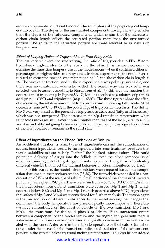
DK4962-Webster-ch15_R2_020407
sebum components could yield more of the solid phase at the physiological temp-erature of skin. The slopes of the unsaturated components are significantly smallerthan the slopes of the saturated components, which means that the increase incarbon chain length affects the saturated portion more than the unsaturatedportion. The shifts in the saturated portion are more relevant to in vivo skintemperatures.
Effect of Varying Ratios of Triglycerides to Free Fatty AcidsThe last variable examined was varying the ratio of triglycerides to FFA. P. acneshydrolyses triglycerides to fatty acids in the skin. It is hence necessary toexamine the transition temperature of the model sebum when it contained differentpercentages of triglycerides and fatty acids. In these experiments, the ratio of unsa-turated to saturated portion was maintained at 1:2 and the carbon chain length at16. The wax ester fraction used in these experiments was palmityl myristate, andthere was no unsaturated wax ester added. The reason why this wax ester wasselected was because, according to Nordstrom et al. (7), this was the fraction thatoccurred most frequently. In Figure 5A–C, Mp-4 is ascribed to a mixture of palmiticacid (m.p. ¼ 638C) and tripalmitin (m.p. ¼ 688C). The three figures show the effectof decreasing the relative amount of triglycerides and increasing fatty acids. MP-4decreases from 598C to 488C, as the percentage of triglyceride decreases. The shift inMp-3 was very small as the percent of triglycerides decreased (fatty acid increases),which was not unexpected. The decrease in the Mp-4 transition temperature whenfatty acids increases still leaves it much higher than that of the skin (328C to 408C),and it is probably not going to have a significant impact on physiological conditionsof the skin because it remains in the solid state.
Effect of Ingredients on the Phase Behavior of SebumAn additional question is what types of ingredients can aid the solubilization ofsebum. Such ingredients could be incorporated into acne treatment products thatwould solubilize sebum to help “soften” the blocked infundibulum. This couldpotentiate delivery of drugs into the follicle to treat the other components ofacne, for example, exfoliating drugs and antimicrobials. The goal was to identifydifferent vehicles that affect the thermal behavior of sebum monitored by DSC.
For this purpose, the model sebum mixture was prepared based on a compo-sition discussed in the previous section (35,36). The test vehicle was added in a con-centration of 15% of the weight of sebum. Small portions of the above mixture wereput on a preweighed DSC pan. These were run from 2508C to 1008C at 58C/min. Inthe model sebum, four distinct transitions were observed. Mp-1 and Mp-2 (whichoccurred below 08C) and Mp-3 and Mp-4 (which occurred above 308C), ingredientsthat affected Mp-3 and Mp-4 were considered for further analysis. The reason againis that on addition of different substances to the model sebum, the changes thatoccur near the body temperature are physiologically more important; therefore,we have concentrated on the effect of vehicle on the two transitions Mp-3 andMp-4—the transitions for the solid phases of sebum. If an interaction occursbetween a component of the model sebum and the ingredient, generally there isa decrease in the transition temperatures along with a decrease in the heat associ-ated with the same. A decrease in transition temperature or the heats of transition(area under the curve for the transition) indicates dissolution of the sebum com-ponent in the vehicle below its usual melting temperature. This can be considered
216 Rhein et al.
www.ketabdownload.com

DK4962-Webster-ch15_R2_020407
1.5
2.0
2.5
3.0
3.5
4.0
4.5
5.0
(A)-50 -25 0 25 50 75 100
Temperature (°C)
Nor
mal
ized
Hea
t F
low
(w
/g)
0 %
10 %
20 %
30 %50 %
60 %
-30
-25
-20
-15
-10
-5
0
0 10 20(B)
30 40 50 60 70% Triglyceride (w/w)
Tem
pera
ture
(°C
)
Mp-1
Mp-2
20
25
30
35
40
45
50
55
60
65
(C)
0 10 20 30 40 50 60 70
% Triglyceride (w/w)
Tem
pera
ture
(°C
)
Mp-3
Mp-4
FIGURE 5 (A) Representative thermogram showing the effect of percent triglyceride in the modelsebum. Numbers on the side of thermograms indicate the percent of triglycerides. (B) Effect ofpercent triglycerides on the transition temperatures Mp-1 and Mp-2 for C-16 (ratio of unsaturationto saturation ¼ 1:2). (C) Effect of percent triglycerides on the transition temperatures Mp-3 andMp-4 for C-16 (ratio of unsaturation to saturation ¼ 1:2). Note: Error bars indicate the standarderror of the mean of three replicates.
Sebum 217
www.ketabdownload.com
DK4962-Webster-ch15_R2_020407
as a measure of miscibility of the vehicle with the sebum component associatedwith that transition. Heats of transition were too variable to assess reliably.
The effect of vehicles on Mp-3 and Mp-4 transitions of model sebum (control)is plotted in Figure 6A and B, respectively. Several vehicles reduced the transitiontemperatures; however, there was only slight correlation between vehicles thataffect both, transitions for saturated wax esters and fatty acids/triglyceride. Thiswould be logical since Mp-3 is the transition for the wax ester, whereas Mp-4 is acombination peak for the wax fatty acid and triglyceride. If a vehicle interactswith one component of the model sebum, it is not necessary that it will also interactwith the other component.
Numerous vehicles have been examined and are reported elsewhere (36); afew are discussed here for demonstration purposes. In general, we can see fromFigure 6A that the hydrophobic materials [e.g., isopropyl myristate (IPM), oleic
34
35
36
37
38
39
40
41
42
(A)
IPM DMI Oleicacid
Sebum Glycerin PEG 400
Additive
EFFECT OF ADDITIVES ON MP-3
38
40
42
44
46
48
50
(B)
Tem
per
atu
re(°
c)T
emp
erat
ure
(°C
)
DMI TC PC Sebum Butanediol Glycolic A
Additive
EFFECT OF ADDITIVES ON MP-4
FIGURE 6 (A) Effect of cosmetic additives on Mp-3 for model sebum. (B) Effect of cosmeticadditives on Mp-4 for model sebum. Note: The model sebum without any additives is included asthe control (additive present at 15% w/w). Abbreviations: DMI, dimethyl isosorbide; IPM, isopropylmyristate; OA, oleic acid; PC, phosphatidyl choline; TC, transcutol.
218 Rhein et al.
www.ketabdownload.com
DK4962-Webster-ch15_R2_020407
acid (OA)] are more effective in lowering Mp-3, whereas the polar materials (glycer-ine and PEG-400) actually raise Mp-3 above sebum. This can be in part becausesebum is made up of a mixture of nonpolar lipids and the hydrophobic vehiclesare easily miscible. Similarly, another set of vehicles, which are miscible with thetriglyceride and fatty acid fraction of sebum (represented by a reduction in theMp-4 transition), are seen in Figure 6B [dimethyl isosorbide (DMI), transcutol,and phosphatidyl choline]. Other more polar vehicles are not miscible, such asbutanediol and glycolic acid. On examining the data (Fig. 6) closely, it is seenthat some of the vehicles show miscibility with sebum are also good skin per-meation enhancers like IPM (37), oleic acid (38), transcutol (39), and DMI (40),whereas some such as glycolic acid (41) are known to not be skin permeation enhan-cers. Similarly, glycerin is a humectant and does not act as a permeation enhancer.In our DSC studies too, glycerin did not show much effect on Mp-3 and Mp-4,which means it is not miscible with the model sebum (36). It is interesting to notethat the permeation enhancers also interact with sebum and are miscible withsome of its components, as shown by our experiments. Isopropyl myristate isknown to be a permeation enhancer and increases follicular delivery (42), and itspresence led to a considerable decrease in the Mp-3 peak. It is possible that theincrease in permeability is due to the increased miscibility with sebum and hencehigher follicular delivery. Many other vehicles tested (36) in our studies have notbeen tested earlier for follicular delivery. This method may help us to identifyother potential vehicles for follicular drug delivery.
Summary of FindingsWe conclude that under the experimental conditions, the unsaturated lipids helpdissolve the saturated lipids. As the proportion of the saturated, solid components(associated with the Mp-3 and Mp-4 peak) increase, it is more unlikely that theliquid components will be able to dissolve the solid under physiological conditionsof the skin. Hence, if P. acnes action preferentially increases the relative percent oflonger chains and more saturated constituents, producing more solids, these willnot be dissolved by the skins’ natural liquid oils. These solids may plug the pilose-baceous ducts and the infundibulum.
Nicolaides et al. (13) reported that comedonal material is relatively more satu-rated whereas sebum collected from the skin surface is more unsaturated.Additionally, it is well-published that there is a deficiency in polyunsaturatedfatty acids, such as linoleic and sebaleic acids, in sebum from acne patients (14);this would result in relatively more of the saturated components. The findingssuggest that conversion of triglycerides to FFA by the P. acnes may not have thelargest physiological consequences as far as the physical properties of sebum areconcerned, providing that the concentration of the saturated forms relative to theunsaturated species does not change. However, if relatively more of the longerchain, saturated fatty acid species are produced by the bacterial hydrolysiswithin the infundibulum, the balance between the liquid and solid phases willbe altered.
Actual sebum may lie somewhere in between all the model sebum that wehave investigated, and a portion of it may be solid at physiological temperaturesof 328C to 408C. Further, the presence of the solid portion depends in a large parton the presence of the unsaturated or liquid portion of the sebum. The fact thatsebum may exist in different phases has also been suggested by Burton (16).
Sebum 219
www.ketabdownload.com
DK4962-Webster-ch15_R2_020407
Butcher and Coonin (19) also showed that although some components of foreheadsebum started to solidify at 308C, it completely solidified at 158C to 178C. Thesestudies are in agreement with the present studies that sebum does not exist asone phase but rather as a mixture of a solid and a liquid at skin temperature.DSC of scalp sebum has been done by Bore et al. (18). Their results also confirmthe presence of multiple phases in sebum. Their samples were also mixtures ofsolids and liquids at body temperature. They showed that as the percentage ofunsaturated portion increased, the viscosity of the sample decreased. We showedthat as the unsaturated portion increases, the saturated portion’s melting tempera-ture decreased, which in turn may contribute to the decreased viscosity.
Vehicles were also identified, that can “soften” the solid components ofsebum. Some of these are known penetration enhancers such as IPM, OA, andtranscutol. These vehicles are miscible with the solid components (Mp-3 and Mp-4).The effect of these ingredients on delivery of polar and nonpolar anti-acne treat-ment drugs would be interesting and is the subject of future investigations.
The most important finding from this research is that the mixture of sebumlipids exists in multiple phases—liquids and solids—and that the average carbonchain length and degree of saturation controls the relative amounts of thesephases. Whether the presence of an excessive amount of the solid phase ofsebum in the follicle plays a role in the pathogenesis of acne still remains to beinvestigated. The presence of excessive amounts of the solid phase would renderit difficult for the liquid phase to dissolve the solid phase and could plausiblylead to altered sebum flow and blockage of the pilosebaceous duct, as is knownto occur in acne patients. Gonzalez and Kroumpouzos reported the presence ofpolarizable crystals in comedonal sebum (32), supporting this view.
CONCLUSIONS
In this chapter, the composition of sebum has been described and the relevance ofalterations in sebum composition to the pathogenesis of acne discussed. Excessivesebum production is generally associated with acne. Comedonal sebum appears tobe enriched in saturated fatty acids compared with skin surface lipids. Sebumappears to be a mixture of solid and liquid phases, as evidenced by the appearanceof the four transitions in the thermal analysis. The liquid phase of sebum compris-ing unsaturated lipid species facilitates dissolution of the solid phase of sebumcomprising saturated species. If this balance is suboptimal, it could lead to blockageof the pilosebaceous duct. These two appear to be in a delicate balance. In acne, itseems that this balance is disrupted probably due to the metabolic activity of P.acnes, producing relatively more of the saturated solid components found in thecomedones; these would presumably cause the blockages in the pilosebaceousunit. This thermal analysis method can be used to identify cosmetic ingredientsthat facilitate dissolution of sebum, thereby reducing the blockage and influencingthe penetration of certain types of acne medications to the pilosebaceous unit.
During the preparation of this chapter, others have proposed new strategies tosolubilize sebum components. The use of triblock surfactants and hydrophilic andhydrophobic linker molecules efficiently form single phase microemulsions duringemulsification. This new approach allows suppression of undesirable lamellarphases that usually dominate the aqueous behavior of surfactant-triglyceridemixtures (43,44).
220 Rhein et al.
www.ketabdownload.com
DK4962-Webster-ch15_R2_020407
REFERENCES
1. Strauss JS, Pochi PE. Histology, photochemistry and electron microscopy of sebaceousglands in man. In: Gans O, Steigleder GK, eds. Hanbuch der Haut-und Geschlechtskran-kheiten; Normale und Pathologische Anatomie der haut I. Berlin: Springer-Verlag,1968:184–223.
2. Pagnoni A, Kligman AM, Gammal S, Stoudemayer T. Determination of density of fol-licles on various regions of the face by cyanoacrylate biopsy: correlation with sebumoutput. Br J Dermatol 1994; 131:862–865.
3. Strauss JS, Downing DT, Ebling FJ. In: Goldsmith LA, ed. Biochemistry and Physiologyof Skin. Vol. 1, Oxford University Press, 1983:569–595.
4. Thody AJ, Shuster S. Control and function of sebaceous glands. Physiol Rev 1989;69(2):383–416.
5. Nicolaides N, Ansari MNA. The dienoic fatty acids of human surface lipid. Lipids 1969;4:79–81.
6. Downing DT, Strauss JS, Pochi PE. Variability of the chemical composition of humanskin surface lipids. J Invest Dermatol 1969; 53:322–327.
7. Nordstrom KM, Labows JN, McGinley KJ, Leyden JJ. Characterization of wax esters, tri-glycerides, and free fatty acids of follicular casts. J Invest Dermatol 1986; 86(6):700–705.
8. Lauer AC, Ramachandran C, Lieb LM, Niemiec S, Weiner ND. Targeted delivery to thepilosebaceous unit via liposomes. Adv Drug Deliv Rev 1996; 18:311–324.
9. Stewart ME, Steele WA, Downing DT. Changes in relative amounts of endogenous andexogenous fatty acids in sebaceous lipids during early adolescence. J Invest Dermatol1989; 92:371–378.
10. Kosugi H, Ueta N. The fatty acid composition of sebum. Jpn J Exp Med 1977; 47:335.11. Gordon GL, Hsuan JS, Stenn K, Prouty SM. The identification of delta-6 desaturase of
human sebaceous glands: expression and enzyme activity. J Invest Dermatol 2003;120(5):707–714, erratum in J Invest Dermatol 2003; 121(3):434.
12. Nicolaides N. The monoene and other wax alcohols of human skin surface lipid andtheir relation to the fatty acids of this lipid. Lipids 1969; 2:266–275.
13. Nicolaides N, Ansari MN, Fu HC, Lindsay DG. Lipid composition of comedones com-pared with that of human skin surface in acne patients. J Invest Dermatol 1970;54(6):487–495.
14. Downing DT, Stewart ME, Wertz PW, Strauss JS. Essential fatty acids and acne. J AmAcad Dermatol 1986; 14:221–225.
15. Braun-Falco O, Plewig G, Wolff HH, Winkelmann RK. Diseases of sebaceous follicles. In:Braun-Falco O, Plewig G, Wolff HH, Winkelmann RK, eds. Dermatology. Berlin:Springer-Verlag, 1991:716–743.
16. Burton JL. The physical properties of sebum in acne vulgaris. Clin Sci 1970; 39:757–767.17. Bore P, Goetz N, Caron JC. Differential thermal analysis of human sebum as a new
approach to rheological behavior. Int J Cosmet Sci 1980; 2:177–191.18. Bore P, Goetz N. A physical method for qualitative examination of human sebum. J Soc
Cos Chem 1977; 28:317–328.19. Butcher EO, Coonin A. The physical properties of human sebum. J Invest Dermatol 1949;
12:249–254.20. Miescher G, Schoenberg A. Unterchungen uber die funktion der talgdrusen. Bulletin der
Schweizerischen Akademie der Medizinischen Wissenschaften 1944; 1:101–107.21. Ebling JF. Effects of steroids on the skin. Biochem Soc Trans 1976; 4:597–602.22. Cunliffe WJ, Perera WDH, Tan SG, Williams M, Williams S. Pilosebaceous duct physi-
ology. Br J Dermatol 1976; 94:431–434.23. Plewig G, Kligman AM. Acne and Rosacea. Berlin: Springer-Verlag, 1975, p. 59.24. Freinkel RK, Strauss JS, Yip SY. Effect of tetracycline on the composition of sebum in acne
vulgaris. N Engl J Med 1965; 273(16):850–854.25. Strauss JS, Pochi PE. Intracutaneous injection of sebum and comedones. Arch Dermatol
1965; 92:443–456.26. Kligman AM, Katz AG. Pathogenesis of acne vulgaris I. Comedogenic properties of
human sebum in external ear canal of the rabbit. Arch Dermatol 1968; 98(1):53–57.
Sebum 221
www.ketabdownload.com
DK4962-Webster-ch15_R2_020407
27. Kligman AM. Pathogenesis of acne vulgaris II Histopathology of comedones induced inthe rabbit ear by human sebum. Arch Dermatol 1968; 98:58–66.
28. Kanaar P. Follicular-keratogenic properties of fatty acids in the external ear canal of therabbit. Dermatologica 1971; 142:14–22.
29. Jacobsen E, Billings JK, Frantz RA, Kinney CK, Stewart ME, Downing DT. Age relatedchanges in sebaceous wax ester secretion rates in men and women. J Invest Dermatol1985; 85(5):483–485.
30. Strauss JS, Pochi PE, Downing DT. Acne: perspectives. J Invest Dermatol 1974;62(3):321–325.
31. Cotterill JA, Cunliffe WJ, Williamson B. Sebum excretion rate and biochemistry inpatients with acne vulgaris treated by oral fenfluramine. Br J Dermatol 1971; 85:127–129.
32. Gonzalez-Serva A, Kroumpouzos G. Demonstration of polarizable crystals in freshcomedonal extracts. Acta Derm Venereol 2004; 84(6):418–421.
33. Abramovits W, Gonzalez-Serva A. Sebum, cosmetics and skin care. Dermatol AspectsCosmet 2000; 18:617–620.
34. Wilborn WH, Montes LF, Lyons RE, Battista GW. Ultrastructural basis for the assay oftopical acne treatments. transmission and scanning electron microscopy of untreatedcomedones. J Cutan Pathol 1978; 5:165–183.
35. Motwani M, Rhein L, Zatz J. Differential scanning calorimetry studies of sebum models.J Cosmet Sci 2001; 52:211–224.
36. Motwani M, Rhein L, Zatz J. Influence of vehicles on the phase transitions of modelsebum. J Cosmet Sci 2002; 53:35–42.
37. Gorukanti SR, Li L, Kim KH. Transdermal delivery of antiparkinsonian agent, benztro-pine.I. Effect of vehicles on skin permeation. Int J Pharm 1999; 192:159–172.
38. Tanojo H, Boelsma E, Junginger HE, Ponec M, Bodde HE. In vivo human skin per-meability enhancement by oleic acid: a laser Doppler velocimetry study. J ControlRelease 1999; 58:97–104.
39. Mura P, Faucci MT, Bramanti G, Corti P. Evaluation of transcutol as a clonazepam trans-dermal permeation enhancer from hydrophilic gel formulations. Eur J Pharm Sci 2000;9:365–372.
40. Squillante E, Needham T, Maniar A, Kislalioglu S, Hossein Z. Codiffusion of propyleneglycol and dimethyl isosorbide in hairless mouse skin. Eur J Pharm Biopharm 1998;46:265–271.
41. Hood HL, Kraeling ME, Robl MG, Bronaugh RL. The effects of an alpha hydroxy acid(glycolic acid) on hairless guinea pig skin permeability. Food Chem Toxicol 1999;37(11):1105–1111.
42. Kogan A, Garti H. Microemulsions and transdermal delivery vehicles. Adv ColloidInterface Sci 2006; 123–126:369–385.
43. Komesvarakul N, Sanders MD, Szekeves E, et al. Microemulsions of triglyceride-basedoils: the effect of co-oil and salinity on phase diagram. J Cosmet Sci 2006; 57(4):309–325.
44. Huang L, Lips A, Co CC. Microemulsions of triglyceride sebum and the role of inter-facial structure on bicontinuous behavior. Langmuir 2004; 20(9):3559–3563.
222 Rhein et al.
www.ketabdownload.com
DK4962-Webster-ch16_R2_020407
B16 Targeted Delivery of Actives from TopicalTreatment Products to the Pilosebaceous Unit
Linda D. RheinSchool of Natural Sciences, Fairleigh Dickinson University, Teaneck, New Jersey, U.S.A.
Joel L. Zatz and Monica R. MotwaniCollege of Pharmacy, Rutgers University, Piscataway, New Jersey, U.S.A.
INTRODUCTION
Delivery of topical acne medications has focused on two challenges during thepast decade. First, delivering the drug to the pilosebaceous unit (PSU) is requiredfor treating diseases such as acne that have their origin in that unit. The other chal-lenge is delivering medications such as exfoliating agents via controlled releasesystems that keep the irritation caused by these drugs in abeyance. Follicular deliv-ery involves depositing drugs in the hair follicle, hair shaft, sebaceous glands, andall components of the PSU. It is hypothesized that vehicles, which are miscible withsebum, may help the active ingredient concentrate preferentially in the PSU. Such apreferential accumulation can be advantageous for increasing the transport of thedrugs through the skin, as well as for targeting the drug to achieve therapeutic effi-cacy in the PSU itself. Knowledge of sebum lipid components helps us to considerhow a penetrating vehicle will react upon entrance into the follicular canal.Additionally, these systems must release drugs into the follicle slowly over time,as well as target their delivery into the epidermis in the follicle via partitioningfrom slow release agents and mild vehicles so that the system is non-irritating.The major goals of this chapter are:
1. To review methods/models used to study penetration of and actions of ingre-dients within the PSU,
2. To review the fundamental research on technologies that target drugs to thePSU,
3. To review investigations of different vehicle effects on the thermal behavior ofmodel sebum and their role in targeting delivery of acne medications to thePSU, and
4. To discuss recent advances in new drug delivery technologies in marketed ornewly developed topical acne treatments.
The overall goal of this chapter is to summarize the state-of-the-art knowledgeof targeting follicular delivery identifying strategies that could potentially provideeffective topical anti-acne products.
SEBACEOUS GLANDS AND THE PILOSEBACEOUS UNIT
Sebaceous glands are generally found all over the body, except on the palms, soles,and dorsum of the feet. Most of the glands are associated with hair follicles and
223
www.ketabdownload.com
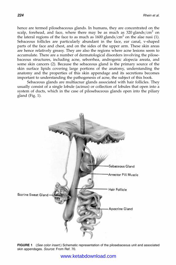
DK4962-Webster-ch16_R2_020407
hence are termed pilosebaceous glands. In humans, they are concentrated on thescalp, forehead, and face, where there may be as much as 320 glands/cm2 onthe lateral regions of the face to as much as 1600 glands/cm2 on the alae nasi (1).Sebaceous follicles are particularly abundant in the face, ear canal, v-shapedparts of the face and chest, and on the sides of the upper arm. These skin areasare hence relatively greasy. They are also the regions where acne lesions seem toaccumulate. There are a number of dermatological disorders involving the pilose-baceous structures, including acne, seborrhea, androgenic alopecia areata, andsome skin cancers (2). Because the sebaceous gland is the primary source of theskin surface lipids covering large portions of the anatomy, understanding theanatomy and the properties of this skin appendage and its secretions becomesimportant to understanding the pathogenesis of acne, the subject of this book.
Sebaceous glands are multiacnar glands associated with hair follicles. Theyusually consist of a single lobule (acinus) or collection of lobules that open into asystem of ducts, which in the case of pilosebaceous glands open into the piliarygland (Fig. 1).
FIGURE 1 (See color insert.) Schematic representation of the pilosebaceous unit and associatedskin appendages. Source: From Ref. 70.
224 Rhein et al.
www.ketabdownload.com
DK4962-Webster-ch16_R2_020407
A sebaceous follicle consists of four parts (3):
1. The infundibulum, which is coated with keratinized epithelium2. The large sebaceous gland acini (or lobes)3. The small vellus hair structure4. The sebaceous duct, via which the sebaceous gland lobes (sebaceous gland
acini) open into the infundibulum.
The large, cauliflower–like, lobed acini produce the sebum, and its chemicalcomposition and physical properties are also described in other chapters and byThody and Shuster (4). The secretory duct, the infundibulum is a long duct linedby keratinocytes. After differentiating, the keratinocytes produce corneocytes,which are ejected outward; that is, into the lumen. The infundibulum consists ofa distal part adjoining the epidermis, the acroinfundibulum. The infrainfundibu-lum is the lower part of the infundibulum and shows a keratinization differingfrom that of the epidermis. The corneocytes produced here are brittle and small.Human follicles can range in diameter from 10 to 70 mm (5).
There are two different types of sebaceous cells: the lipid-producing cells ofthe acinus and the stratified squamous epithelium of the duct, which is continuouswith the wall of the piliary canal and the surface epidermis (4). Sebaceous glandsare composed of undifferentiated, differentiated, and mature cells. The sebaceousglands are holocrine (self-destruct), and their secretion, sebum, is formed whenthe fully mature, lipid-rich cells die and disintegrate. This results because there isa large reserve of fully synthesized sebum contained in the follicular reservoir;that is, in the upper portions of the hair follicle and the orifice to the sebaceousgland. This source of sebum is not depleted, even by repeated solvent extractions.Thus, after careful cleansing of the skin, sebum contained in the follicular reservoirinitially appears to flow out on to the skin surface at a constant rate over a severalhour period, until the amount of sebum normally present on the skin surface isreached. After the normal level of sebum is reached, no further increase in thesebum concentration is observed (6). Once on the surface of the skin, the sebumhas already been chemically modified by microorganisms in the PSU andbecomes mixed with the lipids of epidermal origin (4). Hence, the term sebum ismore precisely reserved to describe the lipid content of the sebaceous glands,and the term skin surface lipid is used to describe the lipid mixture on the skinsurface. However, the composition of the sebum from the surface may not be thepart that, is involved in acne.
ACNE
While subsequent chapters cover the pathogenesis of acne in great detail, a briefoverview will be provided here to set the stage for the discussion of deliverydrug. Acne vulgaris, a multifactorial disease of the skin, is found in areas rich insebaceous follicles. It is characterized by seborrhea, disturbed keratinization inthe follicles with comedones, and subsequent inflammatory papules, pustules,and nodular abscesses and scars (3,7). In the absence of comedones, the large infun-dibulum channels are filled with white pasty material. It is the normal content of asebaceous follicle, not of an acne lesion. Sometimes, the follicles are filled with acocoon-type skeleton of corneocytes, with 20 to 40 cells surrounding the centralfine hair, with a channel left free in which sebum, Propionibacterium acnes, and
Targeted Delivery of Actives 225
www.ketabdownload.com
DK4962-Webster-ch16_R2_020407
staphylococci may be found (3). This is called a follicular filament or follicular cast.A comedo can arise from a follicular filament. It is believed that the lipid compositionof the follicular casts may play a role in acnegenesis and in targeting therapies (6,7).
The primary event in acne is faulty keratinization and the production ofmicrocomedones. The initial noninflammatory lesions of acne result from altera-tions in the follicular epithelium, as demonstrated both physiologically and bylight and electron microscopic level (7). The proliferation and retention-type hyper-keratosis develops in the infundibulum, which expands like a balloon. Two path-ways define the subsequent development of the lesions of acne. In thenoninflammatory pathway, the microcomedo proceeds to mature into closed andopen comedones through distention of the follicle wall and lumen. Accumulationof corneocytes continues with the conversion of the follicle epithelium to thecomedo epithelium. Open comedones arise from closed ones by continuousgrowth, sometimes directly from microcomedones without the intermediatestage. This plug consists of a very densely packed set of several hundred closelyadhering corneocytes, together with sebum and P. acnes. Exfoliating drugs, suchas salicylic acid (SA) and retinoic acid (RA), are often used to treat these types oflesions. In the inflammatory pathway, the extracellular products of P. acnes inciteinflammation. P. acnes colonization occurs relatively early in acne, and the pro-duction of extracellular products by this organism provides multiple potentialmechanisms for the development of inflammation. Among the earliest findingswas that drugs that reduce free fatty acids (FFA), such as tetracycline, are beneficialin inflammatory acne (8). It must be noted that skin surface-derived lipids producean inflammatory papule or nodule (9) and/or follicular hyperkeratosis and come-dones when they are injected into human skin or rabbit ears (10–12).
PRINCIPLES OF THERAPY AND TYPES OF DRUGS DELIVEREDIN TOPICAL ACNE TREATMENTS
Four principles have been used to treat acne on an individualized basis, dependingupon the clinical presentation (13).
1. Correcting the defect in keratinization, for example, SA, RA2. Decreasing sebaceous gland activity, for example, antiandrogens3. Reducing the population of P. acnes, for example, antibiotics, benzoyl peroxide4. Producing an anti-inflammatory response, for example, benzoyl peroxide
In mild acne that consists mainly of comedones, it is important to correct thedefect in keratinization using exfoliating agents. In inflammatory acne, it is import-ant to reduce the population of P. acnes in the follicle and the generation of extra-cellular products of the organism and reduce the inflammatory effects. In moresevere, inflammatory acne that has proven to be resistant to therapy, it is importantto consider adding a drug that decreases sebaceous gland activity. Since the comedois the initial noticeable lesion even in inflammatory acne, it is important to correctthe defect in keratinization in all cases of acne (13).
FOLLICULAR ROUTE
The follicular route is important for drug penetration as well as localized action totreat acne. Targeted delivery of active compounds to the PSU or its components can
226 Rhein et al.
www.ketabdownload.com
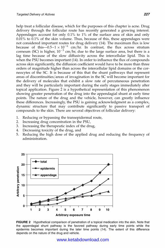
DK4962-Webster-ch16_R2_020407
help treat a follicular disease, which for the purposes of this chapter is acne. Drugdelivery through the follicular route has recently generated a growing interest.Appendages account for only 0.1% to 1% of the surface area of skin and only0.01% to 0.1% of the skin volume. Thus, because of this, these appendages werenot considered important routes for drug delivery (14). The maximum flux is lowbecause of this—0.5–1 � 1026 cm/hr. In contrast, the flux across stratumcorneum (SC) is higher, 1023 cm/hr, due to the large surface area, but there is alag time because of the slow diffusivity across the intercellular lipid. This iswhen the PSU becomes important (14). In order to influence the flux of compoundsacross skin significantly, the diffusion coefficient would have to be more than threeorders of magnitude higher than across the intercellular lipid domains or the cor-neocytes of the SC. It is because of this that the shunt pathways that representareas of discontinuities/areas of invagination in the SC will become important forthe delivery of molecules that exhibit a slow rate of percutaneous penetrationand they will be particularly important during the early stages immediately aftertopical application. Figure 2 is a hypothetical representation of this phenomenonshowing greater penetration of the drug into the appendagial shunt at early timepoints. The nature of the drug and the vehicle, however, can greatly influencethese differences. Increasingly, the PSU is gaining acknowledgment as a complex,dynamic structure that may contribute significantly to passive transport ofcompounds to the skin. There are several objectives of follicular delivery:
1. Reducing or bypassing the transepidermal route,2. Increasing drug concentration in the PSU,3. Increasing the therapeutic index of the drug,4. Decreasing toxicity of the drug, and5. Reducing the high dose of the applied drug and reducing the frequency of
administration.
0
5
10
15
20
25
1 2 3 4 5 6 7 8 9 10
Arbitrary exposure time
Arb
itra
ry p
enet
rati
on
total
epidermis
appendage
FIGURE 2 Hypothetical comparison of penetration of a topical medication into the skin. Note thatthe appendagial shunt pathway is the favored pathway during early time points while theepidermis becomes important during the later time points (14). The extent of this differencedepends on the nature of the drug and vehicle.
Targeted Delivery of Actives 227
www.ketabdownload.com
DK4962-Webster-ch16_R2_020407
BARRIERS AGAINST DRUG DELIVERY TO THE FOLLICLES
Several sites are present within the follicle that represent potentially significanttargets for drug delivery, but these may be limited by the structural aspects ofthe hair follicle and its chemical environment (14,15). Getting the drug to thesesites is also limited by the chemical nature of the drug, for example, hydrophobicor hydrophilic. Aspects of the hair follicle structure that may impede drug deliveryare the following.
1. The glass membrane surrounding the entire follicle and the keratinous layers ofthe inner and outer sheaths may physically restrict the passage of chemicalagents deep within the follicle.
2. Sebum flow into the hair follicle may represent a physical barrier to drugdelivery from some formulations. The effect of sebum composition on deliveryof compounds into the follicles is not known. It is, however, likely that in thesebum-filled follicle, efficient drug delivery, and pharmacological activitywould depend on the interaction of drug and sebum and the physicochemicalproperties of the vehicle.
3. Optimum particle size requirements may also be required to traverse the hairfollicle. It was found (16) that delivery of polystyrene beads into the hair folliclewas size-dependent. Beads between 5 and 7 mm in diameter were optimallydeposited deep within the follicle, whereas smaller or larger beads weremore likely to be localized in the SC and skin surface.
ROLE OF SEBUM IN THE FOLLICULAR ROUTE
Sebaceous glands connected to the hair follicle by ducts release sebum into theupper third of the follicular canal, creating an environment enriched in neutral,nonpolar lipids. Among mammals, sebum composition varies considerably. It hasbeen reported that the sebum-enriched environment of the hair follicle that invagi-nates the epidermis into the dermis could provide significant lipoidal pathway forthe transport of large lipophilic molecules. Chapter 15 shows a comparison ofsebum composition among different species commonly used in percutaneousabsorption studies (Table 2 in that chapter). Wide variations in sebum compositionamong species should be considered before extrapolating data from animalto human studies. However, knowledge of sebum lipid components and theirphysical chemical interactions helps us to consider how a penetrating vehiclewill react upon entrance into the follicular canal.
The use of vehicles that are compatible with sebum to selectively transportdrug via the transfollicular pathway is certainly not a new concept (17). The basisfor understanding a vehicle’s effect on the delivery into the sebum-rich areas,such as the hair follicle, appears to be fully explained by conventional solubilityproperties. The Hildebrand coefficients for model sebum compositions demon-strate that sebum is overall a nonpolar, oily material, with a Hildebrand coefficientof approximately 7.5 to 8.0 cal1/2/cm3/2. Some authors believe (6) that many polarvehicles used in topicals, vehicles such as water, propylene glycol (PG), andethanol would be too polar to be readily soluble in sebum. Hence, to deliver rela-tively polar drugs, if the vehicle is not specifically designed to solubilize sebum,there would be little chance to effectively deliver the drug to the deeper portionsof the hair follicle. Hydrophilic or marginally hydrophobic drugs would not be
228 Rhein et al.
www.ketabdownload.com

DK4962-Webster-ch16_R2_020407
expected to be highly compatible with the sebum. For hydrophobic drugs, thetransport of the drug is predominantly follicular and is brought about by cotran-sport of the drug dissolved in the oil phase into and across the PSUs (17). The effi-ciency of such a follicular enhancement in drug transport would depend on thesolubility of the drug in the vehicle and the compatibility of the vehicle, with thesebum-rich lipid environment within the follicles. The deposition of a drug intosebaceous glands follows the rank order suggested in Table 1 (17). Thus, sebumtherefore may represent a significant physical barrier to drug delivery from someformulations.
It has been reported that the highest pilosebaceous penetration occurred withvehicles that were mixtures of various solvents, interface active agents, couplingagents, and solubilizers (18). Studies identified isopropyl myristate, glyceryl dilau-rate (GDL), and polyoxyethylene-10-stearyl ether (POE-10) as being effective atdelivering drugs into the pilosebaceous duct because of their compatibility withsebum. Studies have suggested that the follicular delivery may be dependent onthe physicochemical properties of the drug and/or vehicle (19,20). The lipoidalenvironment of the follicular canal may favor certain drugs and manipulatingformulation factors, and thereby may enhance delivery of vehicles and drugs bythe follicular delivery route. Solvents, which interact with sebum, thus wereopening the passageway for drug deposition within the follicle.
Since sebum is a mixture of different components, it is important to character-ize it. It is useful to know how variations in the components, their carbon chainlength, and the ratios of unsaturation to saturation affect the physical but thermalbehavior of sebum. This will provide direction on how to manipulate these proper-ties with topical delivery systems. The thermal behavior of sebum in response tothese variables has been reviewed in Chapter 15. Vehicles that alter this behaviorhave also been reviewed in that chapter and in Motwani et al. (20). These are dis-cussed later in this chapter in relation to drug delivery.
MODELS USED TO STUDY FOLLICLES
A major limitation in elucidating the follicular pathway is the current lack of anadequate pharmacokinetic model that can clearly distinguish transfollicular fromtransepidermal percutaneous absorption. Animal models and human models canbe used to study follicular delivery. The experiments can be in vivo or in vitro.These models can further be divided into models for studies of diseases andmodels to investigate follicular delivery. To date, the following models (Table 2)have been used and details on each model are given after the table, along withstudies using these models in the next section.
TABLE 1 Deposition of Drugs into the Pilosebaceous Unit
Relative compatibility ofdrug with sebuma
Relative compatibility ofvehicle with sebumb
Amount insebaceous glands
Incompatible Incompatible 0Incompatible Compatible þ
Compatible Incompatible þ
Compatible Compatible þþþ
aCompatibility in terms of partitioning and solubility/miscibility of drug with components of sebum.bCompatibility in terms of miscibility of vehicle with components of sebum.
Targeted Delivery of Actives 229
www.ketabdownload.com

DK4962-Webster-ch16_R2_020407
Rabbit Ear ModelKligman and Kwong (21) proposed this model for studying the comedogenic poten-tial of substances commonly found in cosmetic formulations. This model is verywell-suited for assessing comedogenic compounds that are encountered in the etio-pathogenesis of acne vulgaris, acne cosmetica, chloracne, cutting oil acne, pitchacne, pomade acne, and tar acne (22). The procedure involves applying a comedo-lytic substance using a glass rod over the undersurface during a two-week period.After the animal is sacrificed, skin is sampled up to the level of the cartilage and isimmersed in 608C water for two minutes, after which the epidermis is peeled off,with the follicular extensions still attached. The comedones can be observedunder the microscope. For quantitative studies, the number of comedones can becounted and compared to comedones induced by other comedogenic substances.
Rhino Mouse ModelThis model was used extensively by Kligman and Kligman (23). The skin of therhino mouse, which is an allelic of the hairless mouse, contains deep dermalcysts and a huge number of utriculi filled with keratinous debris that resemblescomedones. The horny cells do not contain P. acnes, but like in human acne, thesebaceous glands shrink as the pseudocomedones enlarge. They concluded thatthe rhino mouse is a suitable model for assessing chemicals that effect epithelialdifferentiation (retinoids) or that promote loss of cohesion between horny cells (des-caling agents). In the procedure, formulations are applied to the dorsal trunk of themouse for a specified period and then the skin sections are histologically examined.The procedure involves application of the test substances to the entire dorsal trunkfor a specified time.
Macaque Monkey ModelThis model has been used to study drugs affecting hair growth. This speciesexhibits baldness due to hormonal and genetic factors, which is similar to andro-genic alopecia (19). The stumptailed macaque monkey model exhibits a species-specific frontal scalp baldness that coincides with puberty at a rate of nearly100% in both sexes.
TABLE 2 Animal and Human Models for Follicular Delivery
Model Comment
Rabbit ear model Used to assess comedogenicity of cosmetic chemicalsRhino mouse model Has large comedonal cysts; used to assess effectiveness of
exfoliatitng agentsMacaque monkey model Frontal scalp baldness; used to assess delivery of antiandrogens and
their effect on hair growthHamster ear model Has large sebaceous glands; similar to humans; used to assess drug
delivery by isolation of glandsRat model Similar to humans; heat treat to remove epidermis containing
sebaceous glandsGuinea pig model Hairy epidermis on back and nonhairy epidermis behind the ears used
to compare follicular and nonfollicular deliveryHuman model Best for studying anti-acne drug efficacy; cyanoacrylate glue or skin
biopsy used to study follicular delivery by isolating follicular casts
230 Rhein et al.
www.ketabdownload.com
DK4962-Webster-ch16_R2_020407
Uno and Kurata (24) investigated the effects of topically applied minoxidiland diazoxide on hair growth in bald macaque monkeys and they studied theactivity of 5-a reductase inhibitors in nonbald monkeys. Hair and folliculargrowth were evaluated by obtaining phototrichograms (gross photographs andimages of frontal scalps) after closely clipping the hairs and folliculograms (4 mmpunch biopsies embedded in paraffin, serially sectioned and stained). The rate ofDNA synthesis in the follicular cells was also determined. The phototrichogramsrevealed a conversion of short vellus hair to long terminal hairs, and the folliculo-grams showed enlarged follicles. The DNA synthesis studies indicated an increasedrate of follicular cell proliferation. This model therefore appears to have promise forfurther work related to human androgenic alopecia.
Hamster Ear ModelMatias and Orentreich (25) examined this model by planimetry of peeled off skininstead of routine vertical histological sections. This model has been proposed asa model system for human sebaceous glands because of the similarities in mor-phology and in turnover time (26). In this case, each sebaceous gland unit consistsof an infundibular canal, several large lobulated acini with 6 to 20 layers of sebo-cytes, and one centrally located piliary unit. Male hamsters are large, and the sizeof their sebaceous glands is similar to that of humans. Microscopic observation ofthe sebaceous glands from biopsies detects pilosebaceous drug deposition.
Rat ModelThis model has been used extensively by Illel et al. (27). The isolation procedureproduces both follicle-free and follicular skin. The hairless rat model is chosenbecause the sebaceous glands density and size are closely related to those ofhuman forehead skin. The process involves inducing anesthesia to the animals,after which the animals are treated by immersion of the back in 608C water forone minute. The epidermis is removed from this treated area. The subsequenthealing is monitored histologically and transepidermal water loss measurements(TEWLs) are done. After 9 to 10 weeks, the epidermis and horny layer regrow asa continuous layer, without the pilosebaceous structures, and the TEWL returnsto normal, thereby indicating normal barrier properties. In this model, the SClooks normal, but the epidermis looks hyperplastic. The permeation is done invitro to see the differences between follicle and follicle-free skin. However,aside from the pain and suffering inflicted on the animal and the time involvedin developing the follicle-skin, there is the question of structural compositionand permeability behavior of the newly regrown tissue compared with thenormal epidermis of hairless rat skin and whether the comparison between thetwo tissue types is valid. A hairless rat has also been used without the treatmentmentioned earlier, with autoradiography to give a visual impression of the follicu-lar route (28).
Guinea PigWahlberg (29) used hairy and nonhairy skin of guinea pigs to demonstrate thedelivery into the follicles. For the hairy skin, the back of the guinea pig was used,whereas for the nonhairy skin, the area behind the ears of guinea pigs, which iscompletely devoid of hair follicles and of sebaceous and sweat glands, was used.
Targeted Delivery of Actives 231
www.ketabdownload.com
DK4962-Webster-ch16_R2_020407
However, the physiological differences between the two areas make it difficult tojustify its use. Also, in some cases, different strains of guinea pigs (hairy and hair-less) (30) have been used to investigate the importance of follicles. However, directcomparison due to differences in species is difficult.
Human ModelThe human skin contains acne-prone areas with numerous huge sebaceousfollicles—an ideal site for acne vulgaris and other such follicular diseases.The human model can be used for both investigating follicular diseases as wellas follicular delivery. The human skin is generally investigated by the noninvasivecyanoacrylate technique or the skin biopsy technique, the details of which are givenin the next section.
METHODS TO INVESTIGATE DEPOSITION INTO THE FOLLICLES
The skin is a multilayered organ, complex in structure and function, composed ofouter epidermis and inner dermis. There are various potential pathways for per-meation through the SC, including the transepidermal (or “bulk”) and the trans-appendageal (or “shunt”) routes. The transepidermal route across the continuousSC and the epidermis comprises two routes of entry: the intercellular spaces, con-sisting of bilayers of lipids, and the intracellular or transcellular route through thekeratin of the dead cornified cells (corneocytes). The transappendageal route com-prises transport via both the sweat glands, hair follicles with their associatedsebaceous glands. Capillaries closely surround both the follicles and the sweatglands, and rapid absorption of permeating molecules is assumed once the mol-ecules have passed through the follicular walls. An important aspect to considerfor transdermal delivery is the mechanism of penetration, that is, the fractionalcontribution of the bulk strata versus the appendageal pathway to the totalabsorption. Some of the techniques, both qualitative and quantitative, are sum-marized next.
Skin Biopsy TechniquesNoninvasive Cyanoacrylate TechniqueMarks and Dawber (31) expanded the ancient technique to remove horny cell layersby presenting a very potent glue—cyanoacrylate—and termed it skin surfacebiopsy. This technique was used for excavation of follicular contents, analogousto the painful squeezing of sebaceous filaments. It involves placing a drop ofadhesive on a clean region of the skin and then gently pressing a slightly moistglass slide on top of the area. After 30 to 40 seconds, the slide is removed alongwith the superficial horny layer of SC and follicular casts. Lavker et al. (32) haveinvestigated the earlier events of comedo formation in relation to the bacterial con-tents using this technique. Mills and Kligman (33) used this model to assaycomedolytic substances. Microcomedones were induced on the back of adultmales by a two-week occlusive exposure to 10% crude coal tar. The test agentswere then applied for two weeks and the reduction in the density of microcome-dones is determined by the noninvasive cyanoacrylate technique. This methodwas used to screen anti-acne formulations. Mills et al. (34) analyzed SA levels insebaceous follicles after topically applying it in a 2% hydroalcoholic vehicle orgel base.
232 Rhein et al.
www.ketabdownload.com
DK4962-Webster-ch16_R2_020407
Azelaic acid (20%) (AzA) cream was used on human volunteers for the treat-ment of acne (35). Surface AzA was removed by washing with acetone and then col-lected the follicular casts using follicular biopsy method and analyzed by HPLCmethod. Initial samples were taken in five minutes and thereafter over a periodof five hours, five times with no fixed intervals.
Invasive TechniqueOften, a full thickness biopsy containing several sebaceous follicles is needed.Biopsies are inevitable if sebaceous glands are to be investigated. It has beenused for human tissues where the tissue is used within 24 hours of excision (36).After the permeation experiments in vitro, the SC is removed by stripping andthen the biopsies are taken from the formulation-treated skin. The biopsies areimmersed in CaCl2 for a few hours and then the epidermis and dermis is separated.Under microscopic observation, the epidermis with the attached PSU is removedfrom the dermis. These sebaceous glands are cut off from the under side of theepidermis and then analyzed by a suitable analytical technique.
Mechanical Separation of the FolliclesThis has been documented recently by Lieb (37). The procedure involved using theSyrian hamster ear as a model. The scheme involved mounting the ears, ventral sideup on the diffusion cells, which maintained the temperature at 328C. An aliquot ofthe test preparation was then applied to the skin surface. At predetermined timeintervals, the skin specimens were separated by gentle peeling or scraping intothree layers: epidermis, ventral dermis, and dorsal dermis. The sebaceous glandsin the ventral dermis were removed by gentle dragging of a dull scalpel acrossthe bottom surface of the layer. The pilosebaceous material appeared as a milkymaterial. The scraping process was considered complete when the skin areas pre-viously containing glands appear empty under the microscope. The scraped sebac-eous glands and skin layers were assayed separately by scintillation counting.
Follicle-Free ModelsThis has been explained earlier in the rat model. Illel and Schafer (38) reported thefirst systematic, quantitative in vivo studies of follicular delivery using this method.By employing the Franz diffusion cell method, it is possible to compare penetrationof drug across both follicle-free and normal skin (30).
Skin Autoradiography by Computerized Image AnalysisIn this technique, the spatial distribution of a radiolabeled substance within abiological specimen is detected by exposure of the specimen to radiation-sensitivefilm. The main drawback of qualitative autoradiography is that it merely enablesvisualization but not quantification of the penetrant in the skin. Furthermore, thesignals appearing on the images generally represent the tissue-bound residuesrather than the freely diffusable substance. The technique, therefore, onlyprovides an indication of drug transport through the integument.
Some workers have used quantitative autoradiography (39). With thismethod, it is possible to visualize and measure by means of a software programthe concentrations in the hair follicle, glands, and various skin layers without
Targeted Delivery of Actives 233
www.ketabdownload.com
DK4962-Webster-ch16_R2_020407
resorting to mechanical horizontal sectioning. The general procedure involvesfreezing the test skin sample at 21308C, and then vertically sectioning them by cryo-stat in order to yield 6-mm thick slices. These sections are then fixated on glassslides, placed in film cassettes, and covered by tritium-sensitive autoradiographyfilm. Following a four- to seven-week exposure period, the autoradiograms aredeveloped. An imaging system consisting of a video camera, high-resolutionlight microscope, IBM-compatible computer, and appropriate software transformsthe optical density readings of the experimental autoradiograms into drug concen-trations values and projects the resultant digitized images on a TV screen, wherethey are displayed in pseudocolor mode. This provides a more visually meaningfulpicture of drug localization. In addition to autoradiography, the original skin sec-tions are stained with hematoxylin and eosin dye. In the final step, the instrumen-tation is used to superimpose the autoradiography image with the histologicalimage. The computerized system is then used to quantify drug deposition densitiesinto drug concentration. These values require reference to calibration graph data,which are derived from a separate set of experiments. In these calibration experi-ments, good reproducibility was shown between the optical density readings ofstandard sections containing the same drug concentration (40).
Confocal Laser MicroscopyThis method depends upon the analysis of re-emitted light, with the measuringdepth dependent upon the beam wavelength. However, in fluorescence spec-troscopy, the remitted light is detected after passing a second monochromator,that is, only a specific emission wavelength is detected. The procedure involvesapplication of a self-fluorescent marker in a formulation on the skin. After a prede-termined time, the diffusion cells are dismantled and the area of the skin exposed totreatment is punched out. The peeling and scraping technique previously describedis used to mechanically isolate the sebaceous glands. The scraped glands are thensuspended in a buffer and sonicated in order to release the fluorescent marker.This solution could then be assayed for the marker with a fluorescent microscope.This technique has been used quantitatively by Weiner et al. (18).
Horizontal Sectioning of StrataThis technique involves the application of the test compound (typicallyradiolabeled) to a specific area of skin. After a defined exposure time, the excessdrug is removed. The horny layer is stripped off by the consecutive applicationof an adhesive tape. The underlying tissue can then be excised, and the epidermisand dermis are sectioned parallel to the skin surface with a freezing microtome. Thequantity of drug in each horny layer strip or tissue slice can be determined by usinga suitable analytical technique. From the derived data, it is possible to generate aprofile representing drug concentration versus depth for the investigated tissue.A disadvantage of this technique is the fact that unless sufficient data is availablefrom the literature about the thickness of each skin layer at the required regionalsite, preliminary experiments are needed to assess this. Another major limitationof this technique is that it does not yield any data about penetrant concentrationsin the PSUs. It usually has to be used in conjunction with other techniquesdescribed earlier (41).
234 Rhein et al.
www.ketabdownload.com
DK4962-Webster-ch16_R2_020407
REVIEW OF INVESTIGATIONS OF DELIVERY VIATHE FOLLICULAR PATHWAYMathematical ConsiderationsScheuplein in 1967 (42) did the mathematical modeling for follicular delivery ofsmall molecular weight electrolytes and nonelectrolytes. He used reported valuesof thickness, diffusion constant, area of each layer of the skin. According to him,during the initial part of the experiment, the shunt pathway (follicular and sweatglands) is dominant and that introduces the lag period. At steady state, the transe-pidermal route is dominant. The point at which the shunt pathway becomes equalto the bulk pathway is around 300 seconds, as per these calculations. According tothe paper, the concentration levels in the neighborhood of the ducts and the folliclesmay be very much higher than in the bulk of the epidermis in the early stages ofdiffusion, before steady-state diffusion is achieved for small molecules (ordinarilywithin the first one hour after application of an aqueous solution). The reason forthis behavior arises from the limited area of the skin surface occupied by theseappendages, their relatively large diffusion constants, and the nonlinear characterof diffusion prior to steady state. Usually, measurements of skin permeabilitydone in vitro are invariably measurements of steady state because it is quite difficultto detect concentrations within the first few minutes. However, this calculation wasonly for small hydrophilic molecules and has been shown to fail many timesbecause it does not take into consideration the nature of the vehicle.
This theory was then extended in another report (43), where it was found thatmore polar steroid molecules in aqueous solutions had a longer lag time than thenonpolar ones. The reason given for this was that the diffusion constant of polarmolecules is smaller and they tend to prefer the follicular route as comparedwith the epidermal route, and hence the long lag times and small fluxes (Fig. 2).
In still another paper (44), aqueous ibuprofen solution penetration wasmonitored through hydrated human skin. The parameters of the experimentobtained were fitted into the equation as given by the authors, accounting for theshunt and bulk pathway. It was found that the shunt accounted for almost 25%of the total permeation at steady state. Also, in the presence of shunt pathway,the lag time was eight minutes, which would have otherwise been 92 minutes inthe absence of the appendages.
Studies Done to Investigate Physicochemical Propertiesof Drug and Follicular DepositionThe importance of the appendages was shown by Kao et al. (45) in mouse skin.Three different strains of mice were used: the normal-haired mice, the nonhairedmice, and the intermediate fuzzy-haired mice. The actives, benzopyrene and testos-terone, in acetone under nonoccluded conditions were then applied to the exciseddorsal skin organ culture of the mice and the permeation monitored hourly byscintillation counting for 16 hours. Some of the preparations were also investigatedby fluorescence microscopy. It was concluded that the hair follicles could contributesignificantly to skin permeation, although there were differences in the differentstrains of the mice. Testosterone permeated to a higher degree than benzopyrene.It was found that for testosterone, permeation was rapid, peak absorption wasobserved, and peak absorption was higher and faster than in fuzzy-haired orhairless skin. Hence, it was concluded that testosterone that is absorbed extensively,the transappendageal route, played an important role in the beginning of the
Targeted Delivery of Actives 235
www.ketabdownload.com

DK4962-Webster-ch16_R2_020407
experiment, but later the percutaneous absorption was important. For benzopyr-ene, that was less well-absorbed and peak absorption was not seen during thecourse of the experiment, indicating that the transappendageal route is the domi-nant route. It is not explained how the permeation took place for 16 hours afteracetone had evaporated.
Skin permeation of two steroids, hydrocortisone (log P ¼ 1.61) andtestosterone (log P ¼ 3.32), in 95% ethanol in water was evaluated in vivo onnormal and artificially damaged hairless mouse skin in which sebaceous glandsdisappeared during healing (41). The test compounds were applied for 0.5, 2,and 6 hours. The results indicated that the permeation into the dermis and epider-mis was more through the normal skin than appendage-free skin. It was postulatedthat sebaceous glands probably contributed to the penetration of hydrocortisoneand testosterone. Testosterone, which is a more lipophilic molecule, was found ata higher concentration than hydrocortisone in the SC. It was also observed thatin the case of normal skin, the epidermal and dermal amounts of these drugsincreased with application time, but this increase was smaller in the case of scarskin. It was hypothesized that the sebaceous glands acted as reservoir for thesteroids. In these experiments, it was not mentioned whether they were carriedout under occluded conditions and if the vehicle evaporated before the end ofthe experiment.
In another study done by Hueber et al. (41), percutaneous absorption ofsteroids on human skin was examined in vitro. Percutaneous absorption studieson four steroids—progesterone (log P ¼ 3.87), testosterone (log P ¼ 3.32), estradiol(log P ¼ 2.49), and hydrocortisone (log P ¼ 1.61)—were done using freshly excisednormal and scar (obtained from abdominal and mammary plasties) human skin.The steroids were dissolved in 95% ethanol in water and applied. The experimentswere carried out for eight hours for progesterone and testosterone and up to 24hours for estradiol and hydrocortisone. It was found that permeation of the steroidswas significantly greater in normal skin as compared to scar skin. The fluxesobtained for progesterone and testosterone were significantly higher in normalskin than in scar skin. The fluxes of estradiol and hydrocortisone became significantafter four hours. From the percentage absorbed f[(Normal skin – scar skin)/normalskin] � 100g, it was also concluded from the study that transfollicular absorptionwas higher for progesterone and testosterone, the more lipophilic steroids, thanfor estradiol and hydrocortisone. It was not mentioned if the experiment wascarried out under occluded or nonoccluded conditions.
Viprostol is a synthetic PGE2, is a vasodilator, and its deposition wasmonitored in the skin, following application in different animals (mouse, rat,guinea pig, rabbit, and monkey) using scintillation counting and autoradiography(46). The active was incorporated in petrolatum base. Autoradiography was doneonly for mice and monkeys. In mice, the radioactivity was visible in SC and thehair shafts by 30 minutes. In two hours, radioactivity was also visible throughoutthe viable epidermis and only the hair shaft had considerable radioactivity. Skintaken after 12 hours indicated the presence of radioactivity well down in the follicleand, after 72 hours, radioactivity was visible only in the hair shaft and hair follicle.The monkey skin also showed a similar pattern, but the time was longer. By scintil-lation counting, it was concluded that the drug was present in the skin for a rela-tively long period, following removal of the drug from the application site. Theauthors indicated that there was formation of a significant drug depot in the skinand that viprostol penetrated through the follicular route.
236 Rhein et al.
www.ketabdownload.com
DK4962-Webster-ch16_R2_020407
Rutherford and Black (47) used autoradiography to study the localization ofgermicides in guinea pig skin. The deposition of two germicides, zinc pyride-2-thione 1 N oxide [zinc pyrithione (PTO)] and zirconium pyride-2-thione 1 Noxide (zirconium PTO), from shampoos in guinea pig skin was investigated. Appli-cation on the skin was rubbed for 10 minutes, after which the animals were killedand the skin excised. Both germicides were observed in the SC, hair follicles, andsebaceous glands. However, while zirconium PTO was detected in the upper epi-dermis, zinc PTO was not. It was concluded that zinc PTO’s solubility in sebummay allow it to become localized in the hair follicle, zirconium PTO is not solublein sebum, and this causes it to penetrate the dermis. This suggested that thephysicochemical properties of the drug influence delivery into the PSU.
Studies Done to Investigate Vehicle Effects on Follicular DepositionVehicles Containing Organic SolventsMacKee et al. (48) were among the first to investigate the nature of vehicle influen-cing the deposition of iron, bismuth, sulfonamide compounds, and dyes inboth guinea pigs and human skin. The penetration was monitored for 5, 30,and 60 minutes. The vehicles investigated were (i) ointment bases, whichincluded lanolin, (ii) organic solvents, which included PG, (iii) aqueous solutions,(iv) mixtures of water, surface active agents, and solubilizers, (v) mixtures oforganic solvents, surface active agents, solubilizers, and coupling agents, (vi) mix-tures of organic solvents, surface active agents, and solubilizers, (vii) mixtures oforganic solvents, water, surface active agents, and solubilizers, and (viii) mixturesof organic solvents, surface active agents, and water. None of the studies wereabove one hour. Histological examination of the skin biopsies was done to investi-gate the distribution of the “active.” There was little or no penetration seen by theointment bases, or by PG, better penetration with the aqueous solutions with thesurface-active agent and best penetration with the combination vehicles.The authors first suggested the follicular route as being important for delivery ofactives. In this particular study, the time in which it was carried out was veryshort, and it is possible that due to the viscosity of the oleaginous bases, theeffect was not visible until one hour.
Rutherford and Black (47) used autoradiography to study the localization ofgermicides in guinea pig skin. A trichlorocarbanilide (TCC) compound in soapvehicle resulted in predominantly transepidermal penetration through the skin,whereas follicular deposition occurred with a nonsoap detergent. Application onthe skin was rubbed for 10 minutes, after which the animals were killed and theskin excised. They concluded that the vehicle influenced the penetration route.
Montagna (49) studied penetration and local effect of vitamin A on the skin ofthe guinea pig. The effect of vehicles, linoleic acid, oleic acid, alcohol, and chloro-form and a paste made of petrolatum, zinc oxide, and talcum on penetration ofthe 0.5% vitamin A active was investigated. Specimens of skin were in contactwith these agents for 10 minutes, one hour, and two hours and, in some cases,four and eight hours, after which skin biopsies were taken. The penetration wastracked by the fluorescence of vitamin A. It was found that the penetration of theactive (dissolved in alcohol and chloroform) into the PSU was seen very quickly(in 10 minutes). In oleic acid, the penetration to the sebaceous ducts took almosttwo hours. With linoleic acid, it was almost four to eight hours before fluorescencecould be detected in the sebaceous ducts. With the paste, it was even slower. The
Targeted Delivery of Actives 237
www.ketabdownload.com
DK4962-Webster-ch16_R2_020407
author concluded that the speed of vitamin A depended on the vehicle used. It ispossible that viscosity effects played a role in delivery to the PSU.
Estradiol distribution and penetration was penetration of studies in rat skinafter topical application, by high-resolution autoradiography (50). Estradiol in differ-ent concentrations was applied in vehicles dimethyl sulfoxide (DMSO), ethyleneglycol, and sesame oil in vivo. It was observed that the rate of estradiol localizationin the sebaceous glands was dependent on the vehicle and dose. At the end of twohours, the rate of deposition of the drug into the sebaceous glands was more withDMSO as compared to ethylene glycol. The concentration of estradiol is found tobe the highest in the sebaceous glands at the end of two hours, after which it startsto decrease. It was observed that radioactivity was retained in the sebaceous glandsfor 24 hours or longer in low but significant amounts in all vehicles, suggestingthat a drug depot effect may occur within the PSU.
The importance of the appendageal pathway was also observed in the percu-taneous absorption of 5% pyridostigmine bromide (hydrophilic) through variousvehicles, which was evaluated using normal and appendage-free scar rat skin invitro during 72 hours (51). It was found that the drug absorbed was higher fromnerol 8% in ethanol, followed by azone 5% in ethanol–PG (90:10), followed byDMSO 10% in ethanol. PG 10% in ethanol inhibited pyridostigmine absorption ascompared to the control, which was an ethanolic solution. In all cases, the absorp-tion through the appendage-free skin was lower than the absorption throughcontrol skin. The percentage of appendageal pathway was calculated by theformula (1-Scar skin flux/normal skin flux) � 100. It was found that in the firstfour hours, the appendageal absorption was most for the DMSO solution, followedby the proplylene glycol in ethanol, followed by ethanol, then azone solution, andthen the nerol solution. At the end of 72 hours, proplylene glycol in ethanol had thehighest percentage of appendageal transport, followed by DMSO, and the otherswere not that significant. The authors suggested that the enhancers (Nerol,azone) affected the structure of the epidermis, whereas the other solvents(DMSO, ethanol, and PG) were incorporated in the sebum and dragged the druginto the sebaceous ducts. These experiments were done under occlusion. It was con-cluded that ethanol, DMSO, and PG in ethanol favored the transfollicular pathway,but the other vehicles did not. Since ethanol is primarily a lipid solvent, it can solu-bilize sebum and allow the migration of the active in the sebaceous glands, explain-ing why it is primarily transfollicular. It was concluded that by using the rightvehicle it was possible to favor the transfollicular pathway and target the drug.
Radiography was used quantitatively by Fabin et al. (39). Two drugs, tetra-hydrocannabinol (THC) and oleic acid, were evaluated for delivery into hairlessrat skin appendages with different vehicles in vivo. These vehicles included poly-ethylene glycol 400 (PEG 400), transcutol, and PG:ethanol (7:3). It was found thatafter two hours, THC had the highest penetration from transcutol and the lowestfrom PEG 400. After two hours, distribution of THC and oleic acid from transcutolwas not very different. At 24 hours, the transcutol system had delivered themaximum THC in the different skin layers and PEG 400 delivered the lowest. At24 hours more, THC had been delivered in the different layers of the skin comparedtwo hours. It appears that there is a time-dependent effect in the distribution andlocalization of the drug in the follicle. In the same experiments, when oleic acidwas added as a vehicle to the PG:ethanol system, the penetration of THC aftertwo hours was much higher with compared to when oleic acid was not added. Itwas concluded that the presence of oleic acid in the delivery system applied to
238 Rhein et al.
www.ketabdownload.com

DK4962-Webster-ch16_R2_020407
the skin could increase penetration to all the strata of the skin, including theappendages.
The percutaneous absorption of RA was monitored in haired and hairlessguinea pigs with the expectation that drug penetration would be lower in hairlessguinea pigs because they have fewer hair follicles (30). RA was formulated in a0.025% in an ethanolic gel formulation together with and without polyolprepoly-mer (PP-2) at 10%. In vitro permeation was monitored on haired and hairlessguinea pigs for 24 hours. It was observed from penetration profile thathairless guinea pig skin was more permeable to RA than haired skin despite thelower follicular density. This was attributed to the different strains having differentthickness of the SC and different structure rather than greater follicular density. Itwas suggested that a depot of RA was formed in the hair follicle of the hairguinea pig due to the PP-2. The addition of PP-2 to the formulation decreasedthe penetration of the drug. It is possible that by addition of a polymer, the viscosityof the formulation increased and hence there was decrease in the penetration.
In summary, from these studies it can be concluded that vehicles that interactwith sebum seem to be delivering the drug into the PSU. If sufficient time is givenfor the experiment to run, then the more viscous materials may help in deliveringthe drug. Viscosity of the vehicle may play a role in the delivery.
Vehicles Made of LiposomesLiposomes have become popular in the delivery of drugs to the PSU. In a studydone by Li et al. (52), liposomes made of phosphatidylcholine (PC)-containingcalcein dye were investigated for delivery to the PSU using mouse skinhistocultures by confocal microscopy. It was found that liposomes-entrapped dyebecame associated with the hair follicles in contrast to free dye without liposomesin 20 minutes. In a similar study (53) done by the same authors, liposome, made ofPC, mediated targeted delivery of melanin and into the hair follicles of histocul-tured mouse skin in 12 hours was reported. This was monitored by fluorescencemicroscopy. Similarly, DNA encapsulated liposomes were shown to target thehair follicle of histocultured mouse skin in 44 hours, as shown by autoradiography(54). For all these studies, the entrapped drug was separated from the freedrug using gel filtration. It could be concluded that it takes longer for larger mol-ecules to be targeted to the PSU.
Liposomes have been used by Lieb et al. (18) for delivery into the PSU of thehamster ear in vitro. Carboxyfluorescein (CF) encapsulated in multilamellar ves-icles was prepared. The multilamellar vesicles were made of PC:Cholesterol(CH):Phosphatidylserine (PS) in the ratio of 1:0.5:0.1, respectively. Other vehiclescontaining the same concentration of CF were also used, which included HEPESbuffer (pH ¼ 7.4), 5% PG in HEPES, 10% ethanol in HEPES, and 0.05% sodiumlauryl sulfate in HEPES. In vitro diffusion studies for 24 hours, quantitativefluorescence microscopy, and scraping technique was done to investigate the depo-sition into the follicles. It was found that the most intense fluorescence wasobserved with the liposomal formulations, whereas the other formulations werenot much different from each other.
Liposomes made of PC:CH and PS at a mole ratio of 1.0:0.5:0.1 were madecontaining 0.5% cimetidine (55) and were evaluated for deposition into the PSUof hamster ears. Nonionic liposomes made of GDL:CH:POE-10 at a weight ratioof 57:15:28 containing cimetidine were also evaluated in vitro and in vivo. Theseformulations were compared with (i) aqueous solution of pH ¼ 8.3, (ii) 50%
Targeted Delivery of Actives 239
www.ketabdownload.com
DK4962-Webster-ch16_R2_020407
alcohol solution of pH ¼ 7.4, (iii) aqueous solution of pH ¼ 5.5, (iv) phospholipidliposome pH ¼ 5.5, (v) phospholipid liposome pH ¼ 8.3, (vi) nonionic liposomeof pH ¼ 5.5, and (vii) nonionic liposome of pH ¼ 5.5. At pH ¼ 8.3, the drug is pre-dominately unionized. In vitro deposition studies were done on excised hamsterears when the formulation was applied for 24 hours, after which the cells were dis-mantled and the distribution of the drug in the various strata of the skin was deter-mined. In the in vitro studies, the aqueous solution showed significant depositioninto the sebum-rich PSU of the hamster ear. In the in vivo studies done similarly,after 12 hours, maximum deposition in the PSU was observed by the phospholipidliposome at pH ¼ 5.5 as compared to the 50% alcohol solution at pH ¼ 7.4 and thenonionic liposome at pH ¼ 5.5. Therefore, when bioassay was done and a decreasein size of the sebaceous glands was monitored, it was found that only the hydroal-coholic solution and the nonionic liposomes suppressed the growth of the glands.From these studies it was found that there were discrepancies in the in vivo and thein vitro data, and caution needs to be exercised about the activity of the drug inspecific tissue.
Influence of nonionic liposomal composition on topical delivery of alphainterferon (a-IFN) into PSUs was done using the hamster ear model (56). The depo-sition of hydrophilic protein, a-IFN into the PSU and other strata of the hamsterear 12 hours after topical in vivo application from three nonionic formulations,a phospholipid formulation, and an aqueous control was determined. The depo-sition of cyclosporin (CsA), a hydrophobic peptide into the PSUs and other strataof the ear using the same liposomal formulations and a hydroalcoholic control,was also done. The nonionic liposome (Non-1) was made with GDL, POE-10,and CH, Non-2 was made with GDS (glyceryl distearate) and POE-10 and CH,and Non-3 was made with POE-10 and CH. The liposomal formulation wasmade from PC:CH and PS at a mole ratio of 1.0:0.5:0.1. The analysis was done thescintillation counting. The deposition a-IFN of into the PSUs was in the order:Non-1 .. PC . Non-2 . Non-3 ¼ Aq. The deposition of CsA into the PSUs wasin the order: Non-1 .. Hydroalcoholic solution . PC . Non-2 ¼ Non-3. It wasconcluded that in spite of differences in the hydrophobicities of the peptide drugs,Non-1 liposome significantly enhanced the deposition into the PSUs. Hence, thevehicle carrying the drug made a major impact on the deposition irrespective ofwhether it was hydrophilic or hydrophobic. CsA was, however, deposited to agreater extent than a-IFN. It was explained that since GDL melted at 308C, it wasable to cause some fluidization of the liposomal bilayer and partial release of itscontents like POE-10 at body temperature, whereas GDS melted at 548C and hencedid not cause this fluidization.
Expression plasmid DNA for the human interleukin-1 receptor antagonist(IL-1ra) protein was formulated with nonionic:cationic (NC) liposome andPC:cationic (PC) liposomes and applied to auricular skin of hamsters (57). Confocalmicroscopy identified delivery of the plasmid DNA proximal to the perifollicularcells. In the second part of the study, the skin was treated for three days with theNC liposomes and had statistically significant levels of transgenic IL-1ra presentfor five days post-treatment. The results indicated that the NC liposomes coulddeliver expression plasmid DNA to perifollicular cells and mediate transient trans-fection in vivo. The control formulations were made with empty niosomes, that is,no DNA, and were done with infinite dosing. The control used should have beenempty niosomes with the plasmid DNA outside it and would have indicated ifdrug or niosome had the property of reaching the follicle.
240 Rhein et al.
www.ketabdownload.com
DK4962-Webster-ch16_R2_020407
The sebaceous gland deposition of isotretinoin after topical application onhuman facial skin in vitro was measured 16 hours after application by takingskin biopsies and separation of the skin compartments (58). Ethanolic gel was thecontrol and was compared with a liposomal formulation, Natipide II (made ofPC) formulation, and a mixed micelle formulation. All the experiments were per-formed with skin from the periauricular skin of women undergoing plasticsurgery. Autoradiography and fluorescence microscopy elucidated the penetrationpathway of the active. The results showed that neither the liposomes nor the mixedmicellar system revealed an improved sebaceous gland deposition of isotretenoinas compared with the ethanolic gel.
The significance of the sebaceous gland pathway in the cutaneous permeationof an antiandrogen, RU58841 in liposomes, was studied with normal and scar skinin hairless rat (59). RU 58841 was made into a liposomal formulation containinglipoid E 100-35 and a-tocopherol in a phosphate buffer of pH ¼ 7, whereas thecontrol solution was made of ethanol PG:water (40:10:50 w/w). The in vitrocutaneous permeation studies were carried out for 24 hours. The cumulative per-centage of RU 58841 absorbed was three-fold higher through normal skin after 24hours, whereas that of the liposomes was four-fold higher through normal skinas compared to scar skin. However, the permeation of the ethanolic solution wasmuch higher through normal skin as compared to the liposomal formulationthrough the skin. In in vivo studies carried out for 24 hours, it was foundthat the epidermis and dermis of normal skin contained more amounts of theactive than the scar tissue. An autoradiography showed that with the ethanolic sol-ution, the drug was mainly localized in the SC/epidermis and with the liposomes itwas localized in the sebaceous glands. The overall permeation was, however,higher through the normal skin for the ethanolic solutions as compared to the lipo-somal solutions. When the correction for targeting was applied, liposomes targetedbetter than the ethanolic solution.
Mechanism of Action of LiposomesWeiner et al. (60) have hypothesized the use of liposomes in the following way. Forhydrophobic drugs, a major fraction of the added drug would be encapsulated orintercalated within the bilayers of the liposomes. The transfer of drug from thelipid bilayers into the skin could occur as long as the bilayers were in liquid crystal-line state. If the liquid crystalline phase is altered to the gel state, transport of thedrug would cease or be negligibly low. Dehydration of liposomal suspensionshas been shown to reduce transitions from the liquid crystalline phase to the gelstate. If, however, dehydration was complete and the bilayers were transformedfrom the liquid crystalline state to a gel state, then the transfer of the drug wouldcease. If dehydration to an equilibrium stage occurs, wherein a constant amountof water is always retained, then the transport of the drug is steady and continuous.A second consequence of dehydration involves the formation of a strong adhesivepatch of the liposomal bilayers on the skin. The formation of such patches maxi-mizes the intimacy of contact between the drug-laden bilayers and the skin.
For hydrophilic drugs, too, a similar mode of action is explained. Liposomalsystems that undergo total dehydration, drug transport ceases, as the drug is nolonger in a dissolved state. For liposomal systems that retain a constant amountof water within the bilayers following dehydration to an equilibrium, drug trans-port would continue over extended periods. A major consequence of dehydrationfor hydrophilic drugs involves the enhancement or enrichment of drug
Targeted Delivery of Actives 241
www.ketabdownload.com
DK4962-Webster-ch16_R2_020407
concentration in the aqueous phase of the bilayers, leading to an enhancement influx of drug into and across skin. When a follicular pathway is present, upon dehy-dration, the liposomal bilayers can partition and can pack into the follicular ductscontaining the lipids. The filling of the follicular openings with the liposomalbilayers not only results in entrapped drugs being carried into the follicles, butalso allows partitioning of free drugs into the bilayer matrix within the follicles.
Vehicles Made of Lipid MeltsIn these studies, the melted form of the lipid components, which were used to makethe niosomes (namely, GDL:CH:POE-10), were evaluated as a vehicle for the trans-port of CsA and other agents into and through hairless mouse skin (58,60,61). Theexperiments were carried out under nonoccluded conditions. At predeterminedtimes of two, four, eight and 24 hours, the diffusion setup was dismantled and thedrug in various strata of the skin determined using stripping and digesting of theskin using scintillation counting. The profiles of the extent and uptake of CsAfrom the lipid melt formulations are similar to those of the liposomal formulationsof the same compositions. In these same experiments, the ratio of GDL to POE-10was varied. In all these formulation, CH was kept at a constant concentration of15% and GDL to POE-10 ratios examined were 0:85, 15:70, 45:40, and 57:28. It wasfound that the 45:40 lipid melt was more effective in delivery of the active into andthrough the mouse skin. The authors explained that that particular combination oflipid melt had the lowest melting point (�238C) and that was responsible for thebetter uptake. Nevertheless, the remaining melts (except 0% GDL) were alsoliquids at body temperature and the melting point does not seem to explain thepoint enough. The microautoradiographs and light microscope images alsoconfirm these results. It was thus concluded that the similarity of the kinetic profilesfor CsA transport into and across living skin strata of hairless mice from the lipo-somal and lipid melt formulations along with the microautoradiographic evidencefor localization within the PSUs implied that transport of CsA into the highly hydro-philic viable skin strata occurs mainly via the follicular route.
Similar studies were also done on the delivery of hydrocortisone (hydro-phobic) and mannitol (hydrophilic) through the same lipid-based formulations inand through hairless mouse skin in vitro (62). The results after 24 hours indicatedthat the extent of hydrocortisone uptake rose with increasing the GDL to POE-10ratio, whereas for mannitol, the uptake was the opposite decreasing with increasein this ratio in liposomes. Lipid melt formulations for mannitol were not madebecause it is a hydrophilic drug and niosomes of the above compositions alongwith water were made. From the autoradiographic studies, it was reported thatthe nonionic lipid-based formulations were predominantly transfollicular. It wasalso suggested that a hydrophilic molecule like mannitol could not partition intothe lipid environment of the sebum-filled follicles or into and across the SC andwas transported mainly via the transepidermal route. This suggested that thetwo macromolecules were transported through the skin via two different pathways.It was thus was possible to tailor formulations for specific and targeted deliveryacross a certain route.
Studies Done on Microbeads: Size Effects on Follicular DepositionFluorescent polystyrene (16) microbeads of different sizes were made in aqueousmedium and Miglyolw (hydrophobic vehicle) as suspensions and were appliedon live female hairless rat’s back and freshly excised human skin. Suspensionswere applied with massaging for five minutes. The experiment was carried on
242 Rhein et al.
www.ketabdownload.com
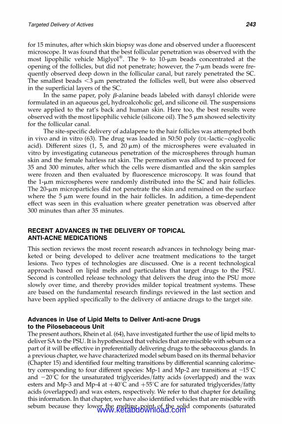
DK4962-Webster-ch16_R2_020407
for 15 minutes, after which skin biopsy was done and observed under a fluorescentmicroscope. It was found that the best follicular penetration was observed with themost lipophilic vehicle Miglyolw. The 9- to 10-mm beads concentrated at theopening of the follicles, but did not penetrate; however, the 7-mm beads were fre-quently observed deep down in the follicular canal, but rarely penetrated the SC.The smallest beads ,3 mm penetrated the follicles well, but were also observedin the superficial layers of the SC.
In the same paper, poly b-alanine beads labeled with dansyl chloride wereformulated in an aqueous gel, hydroalcoholic gel, and silicone oil. The suspensionswere applied to the rat’s back and human skin. Here too, the best results wereobserved with the most lipophilic vehicle (silicone oil). The 5 mm showed selectivityfor the follicular canal.
The site-specific delivery of adalapene to the hair follicles was attempted bothin vivo and in vitro (63). The drug was loaded in 50:50 poly (DL-lactic–coglycolicacid). Different sizes (1, 5, and 20 mm) of the microspheres were evaluated invitro by investigating cutaneous penetration of the microspheres through humanskin and the female hairless rat skin. The permeation was allowed to proceed for35 and 300 minutes, after which the cells were dismantled and the skin sampleswere frozen and then evaluated by fluorescence microscopy. It was found thatthe 1-mm microspheres were randomly distributed into the SC and hair follicles.The 20-mm microparticles did not penetrate the skin and remained on the surfacewhere the 5 mm were found in the hair follicles. In addition, a time-dependenteffect was seen in this evaluation where greater penetration was observed after300 minutes than after 35 minutes.
RECENT ADVANCES IN THE DELIVERY OF TOPICALANTI-ACNE MEDICATIONS
This section reviews the most recent research advances in technology being mar-keted or being developed to deliver acne treatment medications to the targetlesions. Two types of technologies are discussed. One is a recent technologicalapproach based on lipid melts and particulates that target drugs to the PSU.Second is controlled release technology that delivers the drug into the PSU moreslowly over time, and thereby provides milder topical treatment systems. Theseare based on the fundamental research findings reviewed in the last section andhave been applied specifically to the delivery of antiacne drugs to the target site.
Advances in Use of Lipid Melts to Deliver Anti-acne Drugsto the Pilosebaceous UnitThe present authors, Rhein et al. (64), have investigated further the use of lipid melts todeliver SA to the PSU. It is hypothesized that vehicles that are miscible with sebum or apart of it will be effective in preferentially delivering drugs to the sebaceous glands. Ina previous chapter, we have characterized model sebum based on its thermal behavior(Chapter 15) and identified four melting transitions by differential scanning calorime-try corresponding to four different species: Mp-1 and Mp-2 are transitions at 2158Cand 2208C for the unsaturated triglycerides/fatty acids (overlapped) and the waxesters and Mp-3 and Mp-4 at þ408C and þ558C are for saturated triglycerides/fattyacids (overlapped) and wax esters, respectively. We refer to that chapter for detailingthis information. In that chapter, we have also identified vehicles that are miscible withsebum because they lower the melting point of the solid components (saturated
Targeted Delivery of Actives 243
www.ketabdownload.com
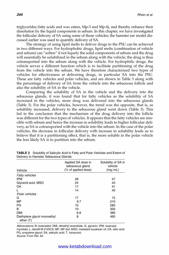
DK4962-Webster-ch16_R2_020407
triglycerides/fatty acids and wax esters, Mp-3 and Mp-4), and thereby enhance theirdissolution by the liquid components in sebum. In this chapter, we have investigatedthe follicular delivery of SA using some of those vehicles; the hamster ear model dis-cussed earlier was used to quantify delivery of SA.
The strategy of using lipid melts to deliver drugs to the PSU can be achievedin two different ways. For hydrophobic drugs, lipid melts (combination of vehicleand sebum) can “soften” if not liquefy the solid components of sebum and the drugwill essentially be solubilized in the sebum along with the vehicle; the drug is thuscotransported into the sebum along with the vehicle. For hydrophilic drugs, thevehicle serves a different function which is to facilitate partitioning of the drugfrom the vehicle into the sebum. We have therefore characterized two types ofvehicles for effectiveness at delivering drugs, in particular SA into the PSU.These are fatty vehicles and polar vehicles, and are shown in Table 3 along withthe percentage of delivery of SA from the vehicle into the sebaceous follicle andalso the solubility of SA in the vehicle.
Comparing the solubility of SA in the vehicle and the delivery into thesebaceous glands, it was found that for fatty vehicles as the solubility of SAincreased in the vehicles, more drug was delivered into the sebaceous glands(Table 3). For the polar vehicles, however, the trend was the opposite, that is, assolubility increased, delivery to the sebaceous gland went down (Table 3). Thisled to the conclusion that the mechanism of the drug delivery into the folliclewas different for the two types of vehicles. It appears that the fatty vehicles are mis-cible with sebum and hence the increase in solubility leads to higher follicular deli-very, as SA is cotransported with the vehicle into the sebum. In the case of the polarvehicles, the decrease in follicular delivery with increase in solubility leads us tobelieve that it is a partitioning effect, that is, the more soluble in the polar vehiclethe less likely SA is to partition into the sebum.
TABLE 3 Solubility of Salicylic Acid in Fatty and Polar Vehicles and Extent ofDelivery to Hamster Sebaceous Glands
Vehicle
Applied SA dose insebaceous gland
Solubility of SA invehicle
(% of applied dose) (mg/mL)
Fatty vehiclesIPM 28 47Glycerid acid, MSO 24 43OA 17 41L 14 40Polar vehiclesG 17 15MP 9.7 210PG 10 280B 10 300DMI 6.8 380Diethylene glycol monoethyl
ether (T)8 465
Abbreviations: B, butanediol; DMI, dimethyl isosorbide; G, glycerin; IPM, isopropylmyristate; L, labrafil M 2125CS; MP, MP diol; MSO, maleated soyabean oil; OA, oleic acid;PG, propylene glycol; SA, salicylic acid; T, transcutol.Source: From Ref. 64.
244 Rhein et al.
www.ketabdownload.com
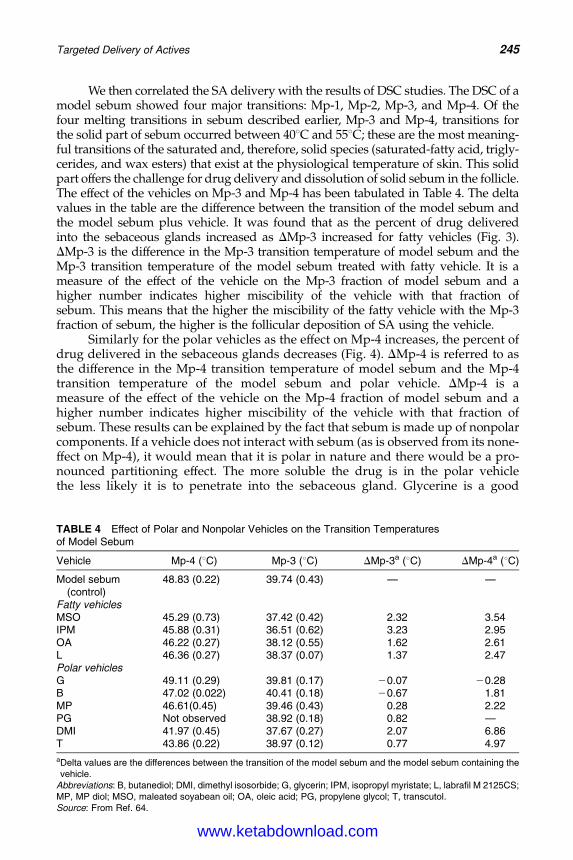
DK4962-Webster-ch16_R2_020407
We then correlated the SA delivery with the results of DSC studies. The DSC of amodel sebum showed four major transitions: Mp-1, Mp-2, Mp-3, and Mp-4. Of thefour melting transitions in sebum described earlier, Mp-3 and Mp-4, transitions forthe solid part of sebum occurred between 408C and 558C; these are the most meaning-ful transitions of the saturated and, therefore, solid species (saturated-fatty acid, trigly-cerides, and wax esters) that exist at the physiological temperature of skin. This solidpart offers the challenge for drug delivery and dissolution of solid sebum in the follicle.The effect of the vehicles on Mp-3 and Mp-4 has been tabulated in Table 4. The deltavalues in the table are the difference between the transition of the model sebum andthe model sebum plus vehicle. It was found that as the percent of drug deliveredinto the sebaceous glands increased as DMp-3 increased for fatty vehicles (Fig. 3).DMp-3 is the difference in the Mp-3 transition temperature of model sebum and theMp-3 transition temperature of the model sebum treated with fatty vehicle. It is ameasure of the effect of the vehicle on the Mp-3 fraction of model sebum and ahigher number indicates higher miscibility of the vehicle with that fraction ofsebum. This means that the higher the miscibility of the fatty vehicle with the Mp-3fraction of sebum, the higher is the follicular deposition of SA using the vehicle.
Similarly for the polar vehicles as the effect on Mp-4 increases, the percent ofdrug delivered in the sebaceous glands decreases (Fig. 4). DMp-4 is referred to asthe difference in the Mp-4 transition temperature of model sebum and the Mp-4transition temperature of the model sebum and polar vehicle. DMp-4 is ameasure of the effect of the vehicle on the Mp-4 fraction of model sebum and ahigher number indicates higher miscibility of the vehicle with that fraction ofsebum. These results can be explained by the fact that sebum is made up of nonpolarcomponents. If a vehicle does not interact with sebum (as is observed from its none-ffect on Mp-4), it would mean that it is polar in nature and there would be a pro-nounced partitioning effect. The more soluble the drug is in the polar vehiclethe less likely it is to penetrate into the sebaceous gland. Glycerine is a good
TABLE 4 Effect of Polar and Nonpolar Vehicles on the Transition Temperaturesof Model Sebum
Vehicle Mp-4 (8C) Mp-3 (8C) DMp-3a (8C) DMp-4a (8C)
Model sebum(control)
48.83 (0.22) 39.74 (0.43) — —
Fatty vehiclesMSO 45.29 (0.73) 37.42 (0.42) 2.32 3.54IPM 45.88 (0.31) 36.51 (0.62) 3.23 2.95OA 46.22 (0.27) 38.12 (0.55) 1.62 2.61L 46.36 (0.27) 38.37 (0.07) 1.37 2.47Polar vehiclesG 49.11 (0.29) 39.81 (0.17) 20.07 20.28B 47.02 (0.022) 40.41 (0.18) 20.67 1.81MP 46.61(0.45) 39.46 (0.43) 0.28 2.22PG Not observed 38.92 (0.18) 0.82 —DMI 41.97 (0.45) 37.67 (0.27) 2.07 6.86T 43.86 (0.22) 38.97 (0.12) 0.77 4.97
aDelta values are the differences between the transition of the model sebum and the model sebum containing thevehicle.
Abbreviations: B, butanediol; DMI, dimethyl isosorbide; G, glycerin; IPM, isopropyl myristate; L, labrafil M 2125CS;MP, MP diol; MSO, maleated soyabean oil; OA, oleic acid; PG, propylene glycol; T, transcutol.Source: From Ref. 64.
Targeted Delivery of Actives 245
www.ketabdownload.com
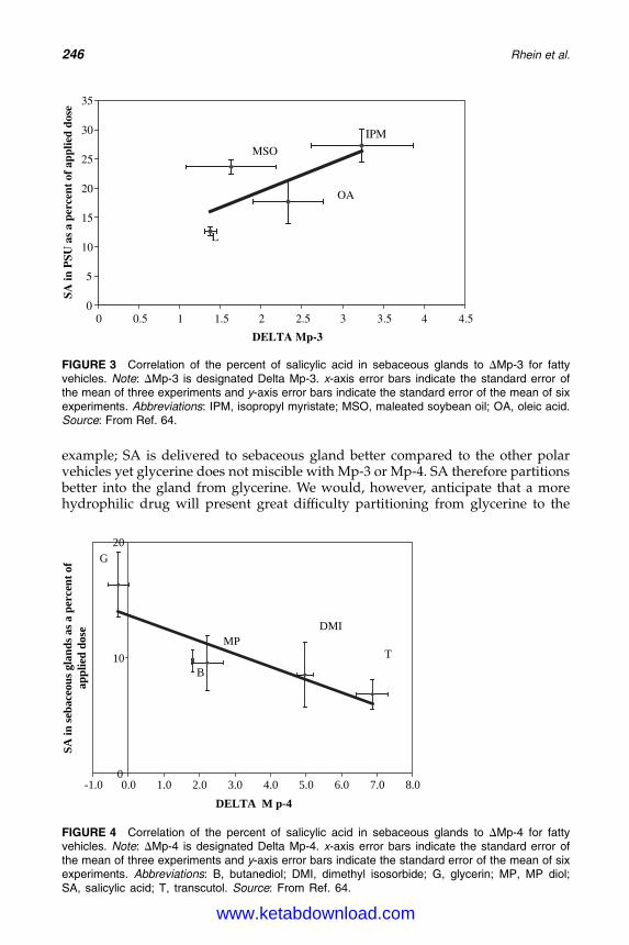
DK4962-Webster-ch16_R2_020407
example; SA is delivered to sebaceous gland better compared to the other polarvehicles yet glycerine does not miscible with Mp-3 or Mp-4. SA therefore partitionsbetter into the gland from glycerine. We would, however, anticipate that a morehydrophilic drug will present great difficulty partitioning from glycerine to the
0
5
10
15
20
25
30
35
0 0.5 1 1.5 2 2.5 3 3.5 4 4.5
DELTA Mp-3
SA in
PSU
as
a pe
rcen
t of
app
lied
dose
L
MSO
OA
IPM
FIGURE 3 Correlation of the percent of salicylic acid in sebaceous glands to DMp-3 for fattyvehicles. Note: DMp-3 is designated Delta Mp-3. x-axis error bars indicate the standard error ofthe mean of three experiments and y-axis error bars indicate the standard error of the mean of sixexperiments. Abbreviations: IPM, isopropyl myristate; MSO, maleated soybean oil; OA, oleic acid.Source: From Ref. 64.
0
10
20
-1.0 0.0 1.0 2.0 3.0 4.0 5.0 6.0 7.0 8.0
DELTA M p-4
SA
in
seb
aceou
s g
lan
ds a
s a
percen
t o
f
ap
pli
ed
dose
G
B
MP
DMI
T
FIGURE 4 Correlation of the percent of salicylic acid in sebaceous glands to DMp-4 for fattyvehicles. Note: DMp-4 is designated Delta Mp-4. x-axis error bars indicate the standard error ofthe mean of three experiments and y-axis error bars indicate the standard error of the mean of sixexperiments. Abbreviations: B, butanediol; DMI, dimethyl isosorbide; G, glycerin; MP, MP diol;SA, salicylic acid; T, transcutol. Source: From Ref. 64.
246 Rhein et al.
www.ketabdownload.com
DK4962-Webster-ch16_R2_020407
gland. These results further strengthen our theory of the miscibility of the fattyvehicles and the partitioning of the polar vehicles. Future research will investigatechallenges of delivery of other drugs of varying hydrophilicity.
Delivery of Adapalene to the Pilosebaceous UnitUsing Polymeric MicrospheresA classic execution of a targeted delivery system for the anti-acne medication,adapalene, was done by Galderma Researchers (63,65–68). Adapalene is the chemi-cally stable form of naphthoic acid. It binds in skin to the RA receptor subtypes RARgamma in the epidermis and RAR beta in the dermal fibroblasts activating differen-tiation specific genes and is a fast-acting anti-acne treatment. Researchers therehave investigated the use of particulate microspheres to deliver this anti-acne medi-cations to the PSU. The drug is entrapped inside the microspheres. Shroot et al. (65)and Allec et al. (66) have discovered that delivery of the microspheres to the PSUdepends on the size of the sphere. The composition of the microspheres is 50:50poly(DL-lactic–coglycolic acid) and they are impregnated with adapalene usingthe solvent evaporation technique. Depending on the stir rate and emulsificationtechnique, spheres of different diameter can be formulated, 1, 5, and 20 mm withcorresponding doses of adapalene of 0.01%, 0.05%, and 0.1%.
The hairless mouse model along with full thickness human skin describedearlier was used to assess site of penetration of the microspheres (63). Thespheres were applied topically and the location of the spheres was determinedusing fluorescence microscopy and scanning electron microscopy. What was dis-covered was that spheres of 5 mm primarily were localized within the PSU and pen-etration depth was time-dependent. On the other hand, larger spheres, .10 mm,remained on the skin surface and smaller spheres, ,3 mm, penetrated both thePSU and the epidermis.
One could naively argue that smaller spheres would more effectively treatacne because they penetrate both the PSU and the epidermal surface since PSUsoccupy only 0.1% of the epidermal surface. However, acne is a disease of thePSU and further studies showed that formulations of microspheres that target pen-etration to the PSU provide similar efficacy without irritation, a known side effect ofother anti-acne medications such as tretinoin (68). This suggests that penetrationthrough the epidermis rather than the PSU tends to deliver drugs into andthrough the living tissue and into the systemic circulation more readily, thuscausing irritation. These results also suggest that the target site for optimal treat-ment of the disease is the PSU where acne develops. However, more research isneeded to confirm these findings.
Controlled Release SystemsVarious controlled release formulations are now on the market that are designed todeliver the acne medication into the skin more slowly to avoid an initial burstduring the first few hours. Such an initial burst of drug and/or enhancers canlead to irritation that is frequently observed with acne medications and is thesubject of numerous consumer complaints in the marketplace. However it is notknown whether these technologies preferentially deliver the medicament to thePSU. Bertek and GlaxoSmithKline employ a polyurethane polymer technology—PP-2 that alters the activity of the free drug in the respective formulations. Bertekmarkets such a technology with RA and GlaxoSmithKline markets a similar
Targeted Delivery of Actives 247
www.ketabdownload.com
DK4962-Webster-ch16_R2_020407
technology with SA. Rhein et al. (69) published studies showing the benefit of thecontrolled release PP-2 in controlling delivery of SA with regard to irritationreduction; however, they failed to demonstrate the benefit of this technology tothe resolution of acne. Additionally, while studies were done to show targeteddelivery to the cutaneous epidermis, no penetration experiments were done onthe PSU versus epidermis.
CONCLUSIONS AND FUTURE DIRECTIONS
Numerous methods have been reviewed that enable the researcher to study tar-geted delivery of acne medications and enhancers to the PSU. The current statusof drug delivery to the PSU for the potential treatment of acne has been summar-ized in this chapter. Techniques to deliver anti-acne drugs have been summarized,along with methods to assess transfollicular delivery. What is eminently clear is thatthe biochemistry and physiology of the PSU is not well-understood fundamentallyand the pathogenesis of acne as it progresses within the PSU is also not well-understood. It appears to be a chance occurrence that a delivery vehicle isformulated that targets delivery of anti-acne medications to the PSU with somefundamental learning emerging form it. The knowledge is thus very much in therudimentary stages, given that the structure of sebum then manipulating thephase behavior by the appropriate chemical enhancers such as liposomes andfluidizing agents is feasible. It seems that by knowing the chemical structureof the active, that is, hydrophobic or hydrophilic in nature among otherthings, topical delivery vehicles can be developed that potentiate penetration intothe PSU.
Future strategies will likely embrace new delivery devices to target the PSUs(71). Some of these are microneedle-based delivery systems, microfluid devices,sonophoresis, iontophoresis, and even magnetic modulation. The future of suchtechniques should yield interesting developments in the treatment of acne via thePSU. The continuing issue is the unavailability of an appropriate in vitro model.Directions of future developments have been review by Meiden et al. (72). A newin vitro skin sandwich model that mimics transfollicular penetration is discussed.Confocal laser scanning microscopy is the most focused direction as a noninvasivemethodology for deriving high resolution images of the PSU the principal advan-tages is its capacity for in vivo application, good time-resolution and the abilityto visualize at multiple depths parallel to the sample surface without the needfor mechanical sectioning (73). The combination use with confocal Raman spec-troscopy is another promising tool for analyzing follicular drug delivery. Newdevelopments in innovative liposomes and microspheres continue to be on the fore-front (74,75). The porcine ear is emerging as an in vitro model that may better mimichuman skin (76). Latest techniques being studied for follicular delivery is to removethe sebum plug and then apply treatment which greatly potentiates delivery to thePSU (74). The key decision of the researcher is whether they are targeting the PSU oris the intent transfollicular penetration. This will dictate the strategy to a largeextent and the delivery systems required. The more formidable question is whatis the appropriate pharmacology that identifies more effective actives targetingthe appropriate site within the PSU? The delivery aspects will be tailored to thatactive depending on its chemical nature and the target site. Perhaps some ofthese questions are also answered in other chapters in this book.
248 Rhein et al.
www.ketabdownload.com

DK4962-Webster-ch16_R2_020407
REFERENCES
1. Pagnoni A, Kligman AM, Gammal S, Stoudemayer T. Determination of density of fol-licles on various regions of the face by cyanoacrylate biopsy: correlation with sebumoutput. Br J Dermatol 1994; 131:862–865.
2. Gupchup GV, Zatz JL. Targeted delivery to pilosebaceous structure. Cosmet Toileteres1997; 112:79–87.
3. Braun-Falco O, Plewig G, Wolff HH, Winkelmann RK. Diseases of sebaceous follicles.In: Braun-Falco O, Plewig G, Wolff HH, Winkelmann RK, eds. Dermatology. Berlin:Springer-Verlag, 1991:716–743.
4. Thody AJ, Shuster S. Control and function of sebaceous glands. Physiol Rev 1989;69(2):383–416.
5. Odland GF. Structure of the skin. In: Goldsmith LA, ed. Biochemistry and Physiology ofthe Skin. Oxford: Oxford University Press, 1985:3–63.
6. Osborne DW, Hatzenbuhler DA. Influence of skin surface lipids on topical formulations.In: Osborne DW, Amann AH, eds. Topical Drug Delivery Formulations. New York:Marcel Dekker, Inc., 1990:69–86.
7. Gollnick H. Current concepts in the pathogenesis of acne. Drugs 2003; 63(15):1579–1596.8. Freinkel RK, Strauss JS, Yip SY. Effect of tetracycline on the composition of sebum in acne
vulgaris. N Engl J Med 1965; 273(16):850–854.9. Strauss JS, Pochi PE. Intracutaneous injection of sebum and comedones. Arch Dermatol
1965; 92:443–456.10. Kligman AM, Katz AG. Pathogenesis of acne vulgaris I. comedogenic properties of
human sebum in external ear canal of the rabbit. Archiv Dermatol 1968; 98(1):53–57.11. Kligman AM. Pathogenesis of acne vulgaris II histopathology of comedones induced in
the rabbit ear by human Sebum. Archiv Dermatol 1968; 98:58–66.12. Kanaar K. Follicular-keratogenic properties of fatty acids in the external ear canal of the
rabbit. Dermatologica 1971; 142:14–22.13. Strauss JS. Sebaceous gland, acne and related disorders-an epilogue. Dermatology 1998;
196:182–184.14. Schaefer H, Redelmeie TE. Biological factors influencing percutaneous absorption.
Principles of Percutaneous Absorption. New York: Karager, 1996:189–212.15. Lauer AC, Ramachandran C, Lieb LM, Niemiec S, Weiner ND. Targeted delivery to the
pilosebaceous unit via liposomes. Adv Drug Deliv Rev 1996; 18:311–324.16. Schaefer H, Watts F, Brod J, Illel B. Follicular penetration. In: Scott RC, Guy RH, Hadgraft
J eds. Prediction of Percutaneous Penetration: Methods, Measurements and Modeling.London: IBC Technical Services, 1990:163–173.
17. Shyamla JC. Follicular delivery of erythromycin from nonionic liposomes and emulsion.Doctoral Dissertation, 1997.
18. Lieb L, Ramachandran C, Egbaria E, Weiner N. Topical delivery enhancement withmultilamellar liposomes into pilosebaceous units: I In vitro evaluation using flouresenttechniques with the hamster ear model. J Invest Dermatol 1992; 99(3):108–113.
19. Lauer AC, Lieb LM, Ramachandran C, Flynn GL, Weiner ND. Transfollicular drug deliv-ery. Pharm Res 1995; 12(2):179–186.
20. Motwani M, Rhein L, Zatz J. Influence of vehicles on the phase transitions of modelsebum. J Cosmet Sci 2002; 53:35–42.
21. Kligman LH, Kwong T. An improved rabbit ear model for assessing comedogenic sub-stances. Br J Dermatol 1979; 100:699–702.
22. Plewig G. Models to study follicular diseases. In: Plewig G, ed. Skin Models to StudyFunction of the Disease of the Skin. Berlin: Springer Verlag, 1986:13–23.
23. Kligman LH, Kligman AM. The effect on rhino mouse skin of agents which influencekeratinization and exfoliation. J Invest Dermatol 1979; 73:354–358.
24. Uno K, Kurata K. Chemical agents and peptides affect hair growth. J Invest Dermatol1993; 101(1):143S–147S.
25. Matias JR, Orentreich N. The hamster ear sebaceous glands I examination of regionalvariation by stripped skin planimetry. J Invest Dermatol 1983; 81:43–46.
26. Plewig G, Luderschmidt C. Hamster ear model for sebaceous glands. J Invest Dermatol1977; 68:171–176.
Targeted Delivery of Actives 249
www.ketabdownload.com
DK4962-Webster-ch16_R2_020407
27. Illel B, Schaefer H, Wepierre J, Doucet O. Follicles play an important role in percutaneousabsorption. J Pharm Sci 1991; 80(5):424–427.
28. Waranuch N, Ramachandran C, Weiner ND. Controlled topical delivery of cyclosporinA from nonionic liposomal formulations: mechanistic aspects. J Liposome Res 1998;8(2):225–238.
29. Wahlberg JE. Transepidermal or transfollicular absorption? Acta Derm Venereol 1968;48:336–344.
30. Hisoire G, Bucks D. An unexpected finding in percutaneous absorption observedbetween hairy and hairless guinea pig skin. J Pharm Sci 1997; 86(3):398–400.
31. Marks R, Dawber RPR. Skin surface biopsy: an improved technique for the examinationof the horny layer. Br J Dermatol 1971; 84:117–123.
32. Lavker RM, Leyden JJ, MCGinley KJ. The relationship between bacteria and the abnor-mal follicular keratinization in acne vulgaris. J Invest Dermatol 1981; 77:253–255.
33. Mills OH, Kligman AM. Is sulfur helpful or harmful in acne vulgaris? Br J Dermatol1972; 86:620–627.
34. Mills OH, Berger RS, Cardin CW, Erekuff GG, Smiles KA. Analysis of salicylic acid levelsin the sebaceous follicles. Poster Presented at the Annual Meeting of the Society of Inves-tigative Dermatology, 1995.
35. Bojar RA, Cutcliffe AG, Graupe K, Cunliffe WJ, Holland KT. Follicular concentrations ofazelaic acid after a single topical application. Br J Dermatol 1993; 129:399–402.
36. Tschan T, Steffen H, Supersaxo A. Sebaceous gland deposition of isotretinoin aftertopical application: an in vitro study using human facial skin. Skin Pharmacol 1997;10:135–143.
37. Lieb LM. Formulation factors affecting follicular (pilosebaceous route) drug delivery asevaluated with hamster ear model. College of Pharmacy, University of Michigan,Doctoral Dissertation, 1994.
38. Illel B, Schaefer H. Transfollicular percutaneous absorption: skin model for quantitativestudies. Acta Derm Venereol (Stockh) 1988; 68:427–430.
39. Touitou E, Fabin B. Localization of lipophilic molecules penetrating rat skin in vivo byquantitative autoradiography. Int J Pharm 1991; 74:59–65.
40. Touitou E, Meidan VM, Horwitz E. Methods for quantitative determination of drug loca-lized in the skin. J Control Release 1998; 567–572.
41. Hueber F, Wepierre J, Schaefer H. Role of transepidermal and transfollicular routes inpercutaneous absorption of hydrocortisone and testosterone: in vivo study in the hairlessrat. Skin Pharmacol 1992; 5:99–107.
42. Scheuplin RJ. Mechanism of percuatneous Absorption II. Transient diffusion and therelative importance of various routes of skin penetration. J Invest Dermatol 1967;48(1):79–88.
43. Scheuplin RJ, Blank IH, Brauner GI, MacFarlane DJ. Percutaneous absorption of steroids.J Invest Dermatol 1969; 52(1):63–70.
44. Keister JC, Kasting KB. The use of transient diffusion to investigate transport pathwaysthrough the skin. J Control Release 1986; 4:111–117.
45. Kao J, Hall J, Helman G. In vitro percutaneous absorption in mouse skin: influence ofskin appendages. Toxicol Appl Pharmacol 1988; 94:93–103.
46. Nicolau G, Baughman RA, Tonelli A, McWilliams W, Schiltz J, Yacobi A. Deposition ofviprostol in the skin following topical administration to laboratory animals. Xenobiotica1987; 17(9):1113–1120.
47. Rutherford T, Black JG. The use of autoradiography to study the localization of germi-cides in skin. Br J Dermatol 1954; 81(suppl 4):75–87.
48. MacKee MG, Sulzberger MB, Herrmann F, Baer RL. Histological studies on percuta-neous penetration with special reference to the effect of vehicles. J Invest Dermatol1945; 6:43–61.
49. Montagna W. Penetration and local effect of vitamin A on the skin of the guinea pig. ProcSoc Exp Biol Med 1969; 86:668–672.
50. Bidmon HJ, Pitts JD, Solomon HF, Bondi JV, Stumpf WE. Estradiol distribution and pen-etration in rat skin after topical application, studied by high resolution autoradiography.Histochemistry 1990; 95:43–54.
250 Rhein et al.
www.ketabdownload.com
DK4962-Webster-ch16_R2_020407
51. Bamba FL, Wepierre J. Role of appendageal pathway in the percutaneous absorption ofpyridostigmine bromide in various vehicles. Eur J Drug Metab Pharmacokinet 1993;18(4):349–353.
52. Li L, Margolis LB, Lishko VK, Hoffman RM. Product-delivering liposomes specificallytarget hair follicles in histocultured intact skin. In Vitro Cell Dev Biol 1992; 28A:679–681.
53. Li L, Lishko VK, Hoffman RM. Liposomes can specifically target entrapped melanin tohair follicles in histocultured skin. In Vitro Cell Dev Biol 1993; 29A:192–194.
54. Li L, Lishko VK, Hoffman RM. Liposome targeting of high molecular weight DNA to thehair follicles of histocultured skin:a model for gene therapy of the hair growth process. InVitro Cell Dev Biol 1993; 29A:258–260.
55. Lieb LM, Flynn GL, Weiner ND. Follicular (pilosebaceous unit) deposition and pharma-cological behavior of cimetidine as a function of formulation. Pharm Res 1994;11(10):1414–1423.
56. Niemiec S, Ramachandran C, Weiner ND. Influence of nonionic liposomal compositionon topical delivery of peptide drugs into pilosebaceous units: an in vivo study using thehamster ear model. Pharm Res 1995; 12(8):1184–1188.
57. Niemiec S, Latta JM, Ramachandran C, Weiner ND. Perifollicular transgenic expressionof human interleukin-1 receptor antagonist protein following topical application ofnovel liposome-plasmid DNA formulations in vivo. J Pharm Sci 1997; 86(6):701–708.
58. Tschan T, Steffen H, Supersaxo A. Sebaceous gland deposition of isotretinoin aftertopical application: an in vitro study using human facial skin. Skin Pharmacol 1997;10:135–143.
59. Bernard E, Dubois J-L, Wepierre J. Importance of sebaceous glands in cutaneouspenetration of an antiandrogen: target effect of liposomes. J Pharm Sci 1997; 86(5):573–578.
60. Weiner ND, Ramachandran C, Lieb LM, Egbaria K. Deposition of liposomally associatedsubstances in the skin strata and the pilosebaceous structures. J Pharm Sci 1994;83(9):1196–1199.
61. Weiner ND. Targeted delivery of macromolecules via liposomes. Int J Pharm 1998;162:29–38.
62. Waranuch N, Ramachandran C, Weiner ND. Controlled topical delivery of hydrocorti-sone and mannitol via select pathway. J Liposome Res 1999; 9(1):139–153.
63. Rolland A, Wagner N, Chatelus A, Shroot B, Schaefer H. Site specific drug delivery to thepilosebaceous structures using polymeric microspheres. Pharm Res 1993; 10(12):1738–1744.
64. Rhein L, Motwani M, Zatz J. Deposition of salicylic acid into hamster sebaceous glands.J Cosmet Sci 2004; 55(6):519–531.
65. Shroot B, Michel S, Allec J, Chatelus A, Wagner N. A new concept off drug delivery foracne. Dermatology 1998; 196:165–170.
66. Allec J, Chatelus A, Wagner N. Skin distribution and pharmaceutical aspects of adapa-lene. J Am Acad Dermatol 1997; 36S:119–125.
67. Rolland A, Wagner N, Chatelus A, Shroot B, Schaefer H. Site specific drug delivery topilosebaceous structures using polymeric microspheres. Pharm Res 1993; 10:1738–1744.
68. Waugh J, Scott IJ. Adapalene: a review of its use in the treatment of acne vulgaris. Drugs2004; 64:1465–1478.
69. Rhein L, Chaudhuri B, Jivani N. Targeted delivery of salicylic acid to from acne treat-ment products into and through the skin: role of solution and ingredient propertiesand relationships to irritation. J Cosmet Sci 2004; 55:65–80.
70. Montagna W, Kligman A, Carlisle K. Atlas of Normal Human Skin, Plate 107. New York,NY: Springer-Verlag, 1992:226.
71. Willie JJ. Skind delivery systems: transdermals, dermatologicals, and cosmetic actives.In: Willie J, ed. Blackwell Publishing Limited, 2006.
72. Meidan VM, Michael C, Bonner MC, Michniak BB. Transfollicular drug delivery—is it areality. Intl J Pharmaceutics 2005; 306(1–2):1–14.
73. R. Alvarez-Romam RA, Naik A, Kalia YN, Fessi H, Guy RH. Visualization of skinpenetration using confocal laser scanning microscopy. Eur J Pharmaceutics and Bio-pharmaceutics 2004; 58(2):301–316.
Targeted Delivery of Actives 251
www.ketabdownload.com

DK4962-Webster-ch16_R2_020407
74. Toll R, Jacobi U, Richter H, Lademann J, Schaefer H, Blume-Peytavi U. Penetrationprofile of microspheres in follicular targeting of terminal hair follicles. J Invest Dermatol2004; 123(1):168–176.
75. Jung S, Otberg N, Thiede G, et al. Innovative liposomes as a transfollicular drug deliverysystem: penetration into porcine hair follicles. J Invest Dermatol 2006; 128(8):1728–1732.
76. Jacobi U, Kaiser M, Toll R, et al. Porcine ear skin: an in vitro model for human skin. SkinRes Technol 2007; 13(1):19–24.
252 Rhein et al.
www.ketabdownload.com
DK4962-Webster-ch17_R2_240307
B17 Topical Therapy and Formulation Principles
Steve BoothroydDepartment of Research and Development, Reckitt Benckiser PLC, Hull, U.K.
INTRODUCTION
Other chapters have discussed the biology and medical consequences of acne, andwith it the types of actives required to be delivered to resolve them. This chapterfocuses on the actives and the delivery vehicle used, and considers the myriadtypes of formulation available.
This is not intended to be a truly comprehensive and in-depth study, sincespace does not permit it. Rather, it is designed to inform and, hopefully, excite inter-est and provide a starting platform for further investigation and experimentation.Some guideline formulations will be given, but there are other sources that offerthese in greater number. Suppliers of raw materials are always valuable sourcesof starting formulary, some of which may already have undergone some perform-ance testing. As with all published formulae, the principle of caveat emptor applies,and the formulator should always confirm that the product performs to therequired standards.
The sections within this chapter are intended to form the basis of the infor-mation that needs to be generated in developing the product. Although this maynot seem relevant in this context, the risks associated with the formulation aresuch that a wide range of proof should be established. Some of this is prescribedin legislation, and the requirements this imposes on formulations will be discussed.
Formulations are much more than a simple mix of chemicals with long andoften incomprehensible names. The formulation is the long-term success story ofa product in that:
B It carries the active and ultimately determines whether or not the active deliversthe desired benefits.
B It can influence a user’s desire to continue using the product to obtain the fullbenefit of the active.
B For commercial products, persuading consumers to make an initial purchaserequires good marketing; getting them to purchase it a second time (and hope-fully more) requires a good formulation.
It is important to note that the majority of topical acne treatment products arecommercial in nature, purchased directly from a store shelf or pharmacy counter.Consumer promotion and advertising are an integral part of this market place,and the formulator of such products must be aware of and take into account thedemands of these consumers.
Before a product can be considered fit for release to the consumer, consider-ation must be given to the following points:
B Does the product meet the brief set by the marketing team and will it satisfy theend consumer’s needs?
253
www.ketabdownload.com
DK4962-Webster-ch17_R2_240307
B Does it conform to the product description, advertising, and any claims thatmay be made?
B Is it economically viable?B Does it infringe others’ intellectual property rights?B Is it safe for its intended purpose?B Is it stable and will it remain fit for the purpose for a reasonable length of
time?B Can it be manufactured reproducibly and in reasonable quantities to satisfy
consumer demand?
Failure to satisfy these principles increases the risk of issues arising in the marketplace and, with it, potential harm to the user and commercial damage to the produ-cer. As with many things, preparation is the key and a clear strategy of how thesepoints will be addressed should be in place before the formulation is considered.Careful planning, sound scientific skills, and a robust process will ensure the ulti-mate end point—a successful product.
In treating acne, we are attempting to:
B Clear existing lesions and prevent the formation of new ones.B Relieve the discomfort from the biophysical process of acne formation and
therapy.B Reduce inflammation.B Minimize the negative psychological effects.
The physical/chemical activities that the product will be attempting to dealwith are:
B Reducing excess oil.B Controlling the bacteria associated with acne.B Reducing the effects of hyperkeratinization.B Unclogging pores.
The extent of the condition will dictate the type of active required:
B Moderate-to-severe drug products, some requiring a physician’s prescription.B Mild-to-moderate mixture of drug products that do not require a physician’s
prescription and more cosmetic products from a range of producers, forexample, Clearasilw (Reckitt Benckiser Inc., New Jersey, U.S.A.), Clean &Clearw (Johnson & Johnson Corporation, New Jersey, U.S.A.), and Neutrogenaw
(Neutrogena Corporation, California, U.S.A.).
The formulator must consider the technical characteristics of maintaining anddelivering the drug active alongside the need for an elegant product that the userwill enjoy using. Although this may sound incongruous, a product that is enjoyableto use is more likely to be used as required and, therefore, deliver the full benefit ofthe active.
Formulating the product, that is, putting a mixture of ingredients together, isonly the first stage and in doing so the formulator must always bear in mind thebasic principles outlined earlier. In order to do that, a clear understanding ofwhat is required allows a robust strategy to be developed, reducing time wastedon unnecessary formulation.
254 Boothroyd
www.ketabdownload.com
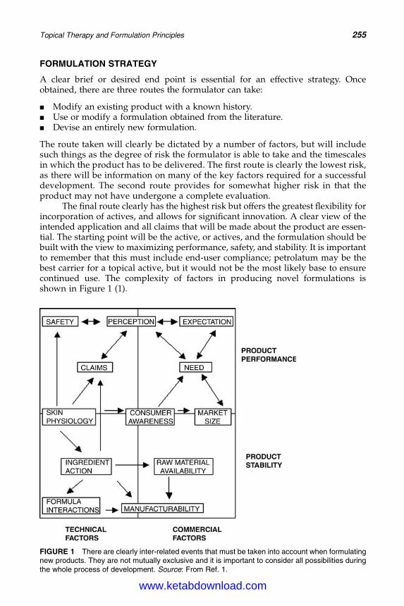
DK4962-Webster-ch17_R2_240307
FORMULATION STRATEGY
A clear brief or desired end point is essential for an effective strategy. Onceobtained, there are three routes the formulator can take:
B Modify an existing product with a known history.B Use or modify a formulation obtained from the literature.B Devise an entirely new formulation.
The route taken will clearly be dictated by a number of factors, but will includesuch things as the degree of risk the formulator is able to take and the timescalesin which the product has to be delivered. The first route is clearly the lowest risk,as there will be information on many of the key factors required for a successfuldevelopment. The second route provides for somewhat higher risk in that theproduct may not have undergone a complete evaluation.
The final route clearly has the highest risk but offers the greatest flexibility forincorporation of actives, and allows for significant innovation. A clear view of theintended application and all claims that will be made about the product are essen-tial. The starting point will be the active, or actives, and the formulation should bebuilt with the view to maximizing performance, safety, and stability. It is importantto remember that this must include end-user compliance; petrolatum may be thebest carrier for a topical active, but it would not be the most likely base to ensurecontinued use. The complexity of factors in producing novel formulations isshown in Figure 1 (1).
FIGURE 1 There are clearly inter-related events that must be taken into account when formulatingnew products. They are not mutually exclusive and it is important to consider all possibilities duringthe whole process of development. Source: From Ref. 1.
Topical Therapy and Formulation Principles 255
www.ketabdownload.com
DK4962-Webster-ch17_R2_240307
Acne treatments fall within three major regulatory areas:
B Licensed prescription or licensed over-the-counter (OTC) medicines.B Monographed OTC medicines.B Cosmetics.
Regulations exist in all countries concerning most, if not all, of these three areas.Although somewhat easier in the European Union (EU), where there is harmoniza-tion of regulations, formulators should confirm that the ingredients used and pro-ducts produced comply with local regulations, especially where they are likely to besold in different countries. While the International Conference on Harmonization ofTechnical Requirements for Registration of Pharmaceuticals for Human Use (ICH)has attempted to introduce a degree of harmonization in certain areas, it should beremembered that products considered drugs in one country are cosmetics inanother.
Even with increasing harmonization, formulators should be aware of the localregulatory status of their product as this may affect:
B The type of claims that can be made.B The status of certain ingredients such as sunscreens, colors, preservatives, and
herbal extracts in terms of ability to use and level.B The statutory warnings that require displaying for the product, actives, and
excipients.B The quality standards of the actives and excipients used in the formulation.B The type and extent of the testing required to bring the product to market.
Regulations constantly change and formulators require up-to-date knowledge onthe legal requirements and the implications that these may have on the formulationof topical acne products.
In the next section, the range of technologies available for use will bediscussed.
PRODUCT TYPES
There are six major technology areas that formulators may wish to utilize, as shownin Table 1.
EmulsionsAn emulsion is a dispersion of two immiscible liquids in the formof droplets. The majority of emulsions fall into one of two types, as shown inFigure 2.
The following general characteristics of emulsions are displayed in Table 2.Emulsions are thermodynamically unstable, which means they are always attempt-ing to return to their natural, single-phase states. This is overcome through theaddition of energy in two forms:
B Mechanical energy through the use of high shear mixers.B Chemical energy through the use of emulsifiers.
In practice, both forms of energy would be used to deliver a longer-term stableproduct.
256 Boothroyd
www.ketabdownload.com
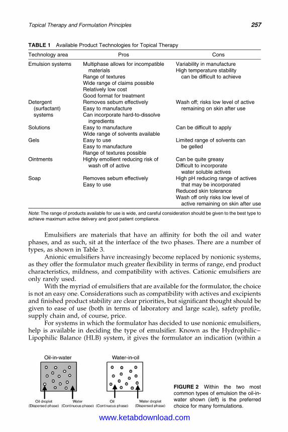
DK4962-Webster-ch17_R2_240307
Emulsifiers are materials that have an affinity for both the oil and waterphases, and as such, sit at the interface of the two phases. There are a number oftypes, as shown in Table 3.
Anionic emulsifiers have increasingly become replaced by nonionic systems,as they offer the formulator much greater flexibility in terms of range, end productcharacteristics, mildness, and compatibility with actives. Cationic emulsifiers areonly rarely used.
With the myriad of emulsifiers that are available for the formulator, the choiceis not an easy one. Considerations such as compatibility with actives and excipientsand finished product stability are clear priorities, but significant thought should begiven to ease of use (both in terms of laboratory and large scale), safety profile,supply chain and, of course, price.
For systems in which the formulator has decided to use nonionic emulsifiers,help is available in deciding the type of emulsifier. Known as the Hydrophilic–Lipophilic Balance (HLB) system, it gives the formulator an indication (within a
TABLE 1 Available Product Technologies for Topical Therapy
Technology area Pros Cons
Emulsion systems Multiphase allows for incompatiblematerials
Range of texturesWide range of claims possibleRelatively low costGood format for treatment
Variability in manufactureHigh temperature stability
can be difficult to achieve
Detergent(surfactant)systems
Removes sebum effectivelyEasy to manufactureCan incorporate hard-to-dissolve
ingredients
Wash off; risks low level of activeremaining on skin after use
Solutions Easy to manufactureWide range of solvents available
Can be difficult to apply
Gels Easy to useEasy to manufactureRange of textures possible
Limited range of solvents canbe gelled
Ointments Highly emollient reducing risk ofwash off of active
Can be quite greasyDifficult to incorporate
water soluble activesSoap Removes sebum effectively
Easy to useHigh pH reducing range of actives
that may be incorporatedReduced skin toleranceWash off only risks low level of
active remaining on skin after use
Note: The range of products available for use is wide, and careful consideration should be given to the best type toachieve maximum active delivery and good patient compliance.
FIGURE 2 Within the two mostcommon types of emulsion the oil-in-water shown (left) is the preferredchoice for many formulations.
Topical Therapy and Formulation Principles 257
www.ketabdownload.com
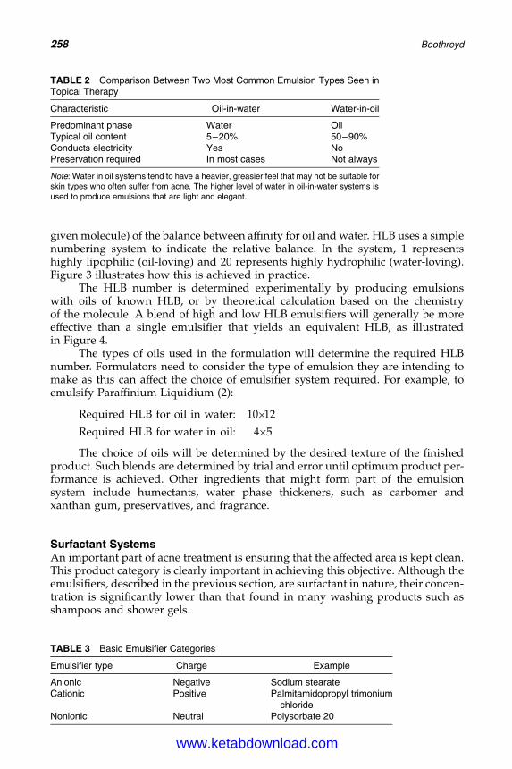
DK4962-Webster-ch17_R2_240307
given molecule) of the balance between affinity for oil and water. HLB uses a simplenumbering system to indicate the relative balance. In the system, 1 representshighly lipophilic (oil-loving) and 20 represents highly hydrophilic (water-loving).Figure 3 illustrates how this is achieved in practice.
The HLB number is determined experimentally by producing emulsionswith oils of known HLB, or by theoretical calculation based on the chemistryof the molecule. A blend of high and low HLB emulsifiers will generally be moreeffective than a single emulsifier that yields an equivalent HLB, as illustratedin Figure 4.
The types of oils used in the formulation will determine the required HLBnumber. Formulators need to consider the type of emulsion they are intending tomake as this can affect the choice of emulsifier system required. For example, toemulsify Paraffinium Liquidium (2):
Required HLB for oil in water: 10�12
Required HLB for water in oil: 4�5
The choice of oils will be determined by the desired texture of the finishedproduct. Such blends are determined by trial and error until optimum product per-formance is achieved. Other ingredients that might form part of the emulsionsystem include humectants, water phase thickeners, such as carbomer andxanthan gum, preservatives, and fragrance.
Surfactant SystemsAn important part of acne treatment is ensuring that the affected area is kept clean.This product category is clearly important in achieving this objective. Although theemulsifiers, described in the previous section, are surfactant in nature, their concen-tration is significantly lower than that found in many washing products such asshampoos and shower gels.
TABLE 2 Comparison Between Two Most Common Emulsion Types Seen inTopical Therapy
Characteristic Oil-in-water Water-in-oil
Predominant phase Water OilTypical oil content 5–20% 50–90%Conducts electricity Yes NoPreservation required In most cases Not always
Note: Water in oil systems tend to have a heavier, greasier feel that may not be suitable forskin types who often suffer from acne. The higher level of water in oil-in-water systems isused to produce emulsions that are light and elegant.
TABLE 3 Basic Emulsifier Categories
Emulsifier type Charge Example
Anionic Negative Sodium stearateCationic Positive Palmitamidopropyl trimonium
chlorideNonionic Neutral Polysorbate 20
258 Boothroyd
www.ketabdownload.com

DK4962-Webster-ch17_R2_240307
The classification given in Table 3 remains applicable here, although Table 4indicates the relative merits of these materials for this application.
The primary purpose of surfactant products in acne applications is theremoval of surface oils. While it is possible to add active ingredients to suchsystems, formulators must remember that they are wash-off products and thedegree of deposition of active can vary.
Formulating surfactant products is not simply a matter of maximizing thelevel of surfactant. Even in oily skin, excessive defatting remains a problem, andthe balance between cleanliness and drying/irritation should be taken intoaccount. Due to their nature, surfactants tend to form the micellular structureshown in Figure 5.
This is an important property as it helps to explain the mechanism by whichthe products remove oil and prevent re-deposition. The surfactant reduces thesurface tension of the oil on the skin, allowing dispersion of the unwanted oil inthe surfactant system. The lipophilic core of the micelle is where the oil is held.
Formulators must also be aware of the reverse happening with a detrimentaleffect on product performance. Such situations may occur where actives have signifi-cant affinity for the surfactant micelle, and the active ingredient becomes effectivelytrapped and is removed on rinsing. This is most likely to occur with lipophilicactives, or in anionic systems with small cationic materials due to charge coupling.
H OO
Dpn
n = 0:2
Hydrophilic – Glucoside Lipophillic – Cetearyl
O
OH
OHHO ( )
FIGURE 3 Nonionic emulsifier, Cetearyl Glucoside [Tegow Care CG90 (Goldschmidt GmbH,Essen, Germany)], illustrating the balance between hydrophilic and lipophilic entities. TheHydrophilic–Lipophilic Balance of this molecule is 11+1 and by manipulating the number of unitson either side, changes the balance and with it the likelihood of being able to produce differentemulsion types. Source: Diagram courtesy Degussa Goldschmidt Personal Care.
FIGURE 4 A greater degree of emulsifierpacking in an oil droplet is possible whenusing a mix of emulsifiers. In thisexample, emulsifier A is more lipophilic(lower hydrophilic–lipophilic balance) thanemulsifier B.
Topical Therapy and Formulation Principles 259
www.ketabdownload.com

DK4962-Webster-ch17_R2_240307
There is a range of anionic surfactants that the formulator may consider. Themost popular is based on alkyl ether sulfates, such as sodium or ammonium laurylether sulfate. The extent of ethylene oxide addition determines the overall proper-ties of the surfactant. Two or three units are the most common format combininggood surfactancy; ammonium salts produce richer creamy foam than the moreopen but equally copious sodium version. For the equivalent degree of ethoxyla-tion, magnesium salts show more mildness than their sodium counterpart andincreasing the number of ethoxylated units also increases mildness (3). Alkylether sulfates provide the backbone to many wash products, given their ease ofhandling, wide range of stability, and cost.
Other commonly used primary surfactants include those from groups such assarcosinates and sulfosuccinates. These ingredients offer greater mildness (3) and con-ditioning than alkyl ether sulfates, but their cost-performance ratio has not led to their
TABLE 4 Basic Surfactant Categories
Surfactant type Charge Properties
Anionic Negative High levels of flash foamOffer wide variety of foam qualityExcellent primary surfactantWide range of types availableChoice of counter ion important in
terms of irritancy potentialAmphoteric Overall neutral
due to positiveand negativecharges presentwithin one molecule
Substantive to skinGood secondary surfactant
Cationic Positive Substantive to the skin offering conditioning butincreased risk of irritation
Do not produce high levels of foamNot widely used
Nonionic Neutral Often used as secondary surfactants to stabilise foamOften used as primary surfactant in milder productsGenerally produce low levels of flash foam
Note the additional types available in surfactants to be used in this application compared to emulsifiers. Amphoterics,although possessing an overall neutral charge, have very different properties compared to the nonionics.
FIGURE 5 In micelles, the hydrophobic tails point intothe center, creating a void into which oil and particulatematter can be captured and retained.
260 Boothroyd
www.ketabdownload.com
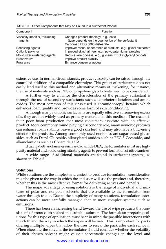
DK4962-Webster-ch17_R2_240307
extensive use. In normal circumstances, product viscosity can be raised through thecontrolled addition of a compatible electrolyte. This group of surfactants does noteasily lend itself to this method and alternative means of thickening, for instance,the use of materials such as PEG-55 propylene glycol oleate need to be considered.
A further way to enhance the characteristics of the primary surfactant isthrough the use of secondary surfactants such as amphoteric betaines and amineoxides. The most common of this class used is cocamidopropyl betaine, whichenhances foam quality and provides some form of skin conditioning.
Although many nonionic surfactants are equally effective at removing excessoils, they are not widely used as primary materials in this medium. The reason istheir poor foam production that most consumers associate with an effectiveproduct. More commonly found playing a secondary role, many nonionic productscan enhance foam stability, leave a good skin feel, and may also have a thickeningeffect for the products. Among commonly used nonionics are sugar-based gluco-sides such as Decyl Glucoside, alkoxylated amides such as PEG 5 Cocamide, andalkanolamides such as Cocamide DEA.
If using diethanolamines such as Cocamide DEA, the formulator must use high-purity material and avoid using nitrating agents to prevent formation of nitrosamines.
A wide range of additional materials are found in surfactant systems, asshown in Table 5.
SolutionsWhile solutions are the simplest and easiest to produce formulation, considerationmust be given to the way in which the end user will use the product and, therefore,whether this is the most effective format for delivering actives and benefits.
The major advantage of using solutions is the range of individual and mix-tures of polar and nonpolar solvents that are available to the formulator fromwater through to oils. Due to the simplicity of many solutions, formulation inter-actions can be more carefully managed than in more complex systems such asemulsions.
There has been an increasing trend toward the use of wipe products that con-sists of a fibrous cloth soaked in a suitable solution. The formulator preparing sol-utions for this type of application must bear in mind the possible interactions withthe cloth and the way in which the wipes will be used. This is important for packsoffering multiple wipes that allow the user to reseal the pack after each removal.When choosing the solvent, the formulator should consider whether the volatilityof their chosen solvent might cause unacceptable changes in the level and
TABLE 5 Other Components that May be Found In a Surfactant Product
Component Function
Viscosity modifier/thickeningagents
Changes product rheology, e.g., salts(type depends on the counter ion of the surfactant)
Carbomer, PEG 6000 distearatePearlizing agents Improves visual appearance of products, e.g., glycol distearateCationic polymer Improved skin/hair feel, e.g., polyquaterniums, proteinsMoisturizers/refatting agents Reduce skin dryness, e.g., glycerin, PEG 7 glyceryl cocoatePreservative Improve product stabilityFragrance Enhance consumer appeal
Topical Therapy and Formulation Principles 261
www.ketabdownload.com

DK4962-Webster-ch17_R2_240307
availability of the active. This may manifest itself in two ways. First, the loss ofsolvent may increase the concentration of active above the legally allowed level,or above the level at which it remains safe. Second, for crystalline solutes (whichmay or may not include the active), loss of solvent increases the risk of precipi-tation. This may result in injury to the end user, particularly if the product isintended for use around the eye area.
GelsGel formulations are particularly useful in acne treatments as they offer the possi-bility of delivering active ingredients in a totally oil-free manner. The majority offormulations will be water, alcohol, or mixtures of these materials, but formulatorsneed to be aware that it is possible to gel oils, although this is unlikely to be used inthis application. The exception to this is ointments, which it could be argued, arethickened oils, but the formulation of ointments is more complex and is dealtwith in a later section.
In its simplest terms, a gel is a thickened solution, although the differing rheo-logical qualities of gels from solutions offer formulators the possibility of improvingapplication and ensuring better control of coverage than for solutions.
Carbomers are acrylic acid polymers available in a variety of grades, offeringformulators a range of viscosities and textures with defined yield points that allowexcellent spreading and rapid absorption during use. Standard grade carbomerslose viscosity in the presence of electrolyte, although hydrophobically modifiedgrades with improved electrolyte tolerance are available. Due to the need to neu-tralize carbomers to achieve the gel structure, the range of actives that can beused is reduced compared with other gellants.
Xanthan gums are polysaccharides with high molecular weight that showmuch greater stability toward electrolytes. They have excellent suspending proper-ties due to their pseudoplastic rheology. They will not produce clear gels easily andthey are not as rapidly adsorbed onto the skin as carbomers.
Modified cellulose gums are similar in their chemistry to xanthan gums, butclear gels can be produced with certain grades. With a more lubricious feel than car-bomers, they can be used on their own or in combination with carbomers to extendrub-in time.
Silicate systems are available from both natural and synthetic sources. Thesynthetic material produces clear water white gels that can be stabilized toproduce products over a very wide pH range. Due to their anionic nature, theyare incompatible with cationic materials.
OintmentsAlthough consumers may classify these products and creams as the same for thepurpose of this section, an ointment is considered to be an oil base only with nowater present. While this may seem unusual for a condition where excess oil is amajor cause of the problem, as will be seen later, water-free conditions may berequired to provide the best environment for the active.
Ointments, due to their oily nature, can be difficult to remove and leave theskin feeling greasy. For this application, formulators may consider the ointmentto be the oil phase of an as yet unfinished emulsion, where the water used towash the product off forms the final piece. Careful choice of ingredients willimprove the rate of removal and with it the after use-skin feel. As always, there
262 Boothroyd
www.ketabdownload.com
DK4962-Webster-ch17_R2_240307
is a balance, in this case, between the desire for effective removal and the risk of irri-tation from the emulsifiers. There are a number of emulsifiers that offer a lowirritation potential and their ability to form emulsions at low temperatures, includ-ing the polysorbate group.
SoapSoap remains one of the most widely used personal washing products, and formu-lators should not discount this fact when considering topical treatments. Soap,while it has excellent cleansing properties, does not lend itself to efficient activedelivery. The two major drawbacks with soap are that it is washed off and so theactive concentration deposited may be small and its high pH can degrade certainactives, and cause a lower tolerability on more sensitive skin.
Alternatives to soap are Combi bars (combinations of soap and syntheticdetergents) or Syndet bars, based wholly on synthetic detergents. Syndet barsoffer the formulator more flexibility in that their pH is lower and can be modifiedby allowing a wider range of actives to be used. Soap, Combi, and Syndet formu-lations require specialist knowledge and equipment, and a number of companiesoffer contracted services in this area.
ACTIVE RAW MATERIALSRetinoidsIn this section, the range of materials being considered may be seen described asretinoids, retinoid types, or retinoid prodrugs. The appropriate description to usewill depend on the individual compound being considered, but all the materialsare used in topical treatments for acne, and are described in more detail elsewhere.
Retinoids are highly active, and from a formulator’s viewpoint, often unstablecompounds being affected variously by light, heat, and oxygen. There are a numberof compounds that fall within this classification, including:
B Tretinoin, [Retin Aw (Johnson & Johnson Corporation, New Jersey, U.S.A.)]B Isotretinoin, [Accutanew (Hoffman-La Roche Inc., New Jersey, U.S.A.)]B Adapalene, [Differinw (Galderma S.A., Lausanne, Switzerland)]B Tazarotene, [Tazoracw (Allergan Inc., California, U.S.A.)]
For many, use in actinic lighting conditions should be avoided, and the rawmaterial and finished product storage vessels should be flushed with nitrogenafter use. In emulsion products, care should be taken during manufacture, bothto avoid excess heat and to minimize incorporation of air that may lead tolonger-term stability problems.
Retinoids are generally insoluble in water, but have varying solubility inalcohol, other solvents, and oils.
Products should be packed for storage and consumers in protective materialssuch as amber glass, metal, or plastic laminated tubes.
Benzoyl PeroxideThe clinical effectiveness of benzoyl peroxide is well-established and is discussedelsewhere. Benzoyl peroxide is a highly effective oxidizing agent, and contactshould be avoided with reducing agents, metals (particularly transition metals),and organic materials to prevent the risk of fire and explosion. Peroxides break
Topical Therapy and Formulation Principles 263
www.ketabdownload.com
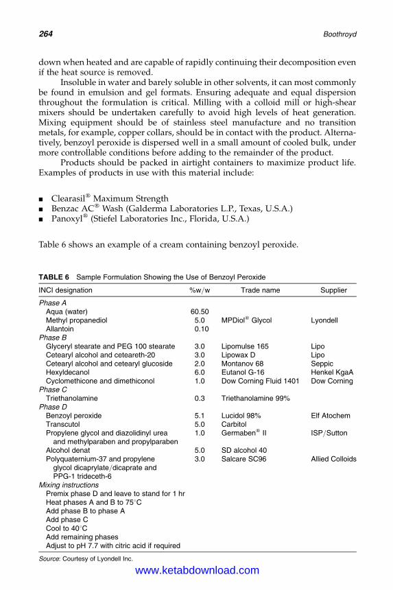
DK4962-Webster-ch17_R2_240307
down when heated and are capable of rapidly continuing their decomposition evenif the heat source is removed.
Insoluble in water and barely soluble in other solvents, it can most commonlybe found in emulsion and gel formats. Ensuring adequate and equal dispersionthroughout the formulation is critical. Milling with a colloid mill or high-shearmixers should be undertaken carefully to avoid high levels of heat generation.Mixing equipment should be of stainless steel manufacture and no transitionmetals, for example, copper collars, should be in contact with the product. Alterna-tively, benzoyl peroxide is dispersed well in a small amount of cooled bulk, undermore controllable conditions before adding to the remainder of the product.
Products should be packed in airtight containers to maximize product life.Examples of products in use with this material include:
B Clearasilw Maximum StrengthB Benzac ACw Wash (Galderma Laboratories L.P., Texas, U.S.A.)B Panoxylw (Stiefel Laboratories Inc., Florida, U.S.A.)
Table 6 shows an example of a cream containing benzoyl peroxide.
TABLE 6 Sample Formulation Showing the Use of Benzoyl Peroxide
INCI designation %w/w Trade name Supplier
Phase AAqua (water) 60.50Methyl propanediol 5.0 MPDiolw Glycol LyondellAllantoin 0.10
Phase BGlyceryl stearate and PEG 100 stearate 3.0 Lipomulse 165 LipoCetearyl alcohol and ceteareth-20 3.0 Lipowax D LipoCetearyl alcohol and cetearyl glucoside 2.0 Montanov 68 SeppicHexyldecanol 6.0 Eutanol G-16 Henkel KgaACyclomethicone and dimethiconol 1.0 Dow Corning Fluid 1401 Dow Corning
Phase CTriethanolamine 0.3 Triethanolamine 99%
Phase DBenzoyl peroxide 5.1 Lucidol 98% Elf AtochemTranscutol 5.0 CarbitolPropylene glycol and diazolidinyl urea
and methylparaben and propylparaben1.0 Germabenw II ISP/Sutton
Alcohol denat 5.0 SD alcohol 40Polyquaternium-37 and propylene
glycol dicaprylate/dicaprate andPPG-1 trideceth-6
3.0 Salcare SC96 Allied Colloids
Mixing instructionsPremix phase D and leave to stand for 1 hrHeat phases A and B to 758CAdd phase B to phase AAdd phase CCool to 408CAdd remaining phasesAdjust to pH 7.7 with citric acid if required
Source: Courtesy of Lyondell Inc.
264 Boothroyd
www.ketabdownload.com
DK4962-Webster-ch17_R2_240307
Salicylic AcidThe clinical effectiveness of salicylic acid is well-established and is discussed else-where. Salicylic acid is a white crystalline powder showing slight solubility in waterbut being freely soluble in alcohol. Salicylic acid is most active in its acid form, andformulators should use suitable buffer solutions in formulations to maintain the pHbetween 2 and 4.
Salicylic acid can be found in most of the formats described earlier under anumber of brands, for example, Clearasilw, Clean & Clearw, and Neutrogenaw.
AntibioticsThere are a variety of antibiotics used for topical applications and these includeTetracyclines and Macrolides such as Erythromycin. Although they each haveunique physical properties and solubility, a common feature is their stability inaqueous solutions. It has been suggested that antibiotics such as erythromycinshow increased loss of anti-microbial activity, over time, although this varies withproduct base (4). Varying pH can change this loss of activity, but, for products requir-ing extended shelf life of greater than a few weeks, alternative solvents or systemsshould be considered. Antibiotics are also adversely affected by heat and light.
For topical application gels, alcoholic solutions or ointments are the mostcommonly produced formats. Examples include:
B Akne-Mycinw [Erythromycin (Hermal Kurt Hermann GmbH, Reinbek, Germany)]B Cleocin Tw [Clindamycin (Pharmacia & Upjohn Company LLC, New Jersey,
U.S.A.)]B Topicyclinew [Tetracycline (Shire LLC, Pennsylvania, U.S.A.)].
ResorcinolResorcinol is colorless to slightly pink–grey and is very reactive to light and air,turning red when oxidized.
The effectiveness of resorcinol has been recognized and claims that can bemade through the use of this material are available (5).
Due to its good solubility in water and alcohol, resorcinol can be incorporatedinto a wide range of products, including emulsions, being stable to heat at normalemulsification temperatures and times. Manufacturing under vacuum reduces therisk of loss of activity, and products containing this material should be packed inmaterials with good oxygen barriers.
Examples of products containing resorcinol include Clearasilw Adult Carew
Acne Treatment Cream.
SulfacetamideSulfacetamide has been widely utilized for its antibacterial properties for a con-siderable period of time. In the sodium form, it is a white to off-white crystallinesubstance showing good solubility in water but reduced solubility in alcohol.
This material can be found in a 10% form in Klaronw (Aventis PharmaceuticalInc., New Jersey, U.S.A.). Sulfacetamide has also been shown to be effective in com-bination with sulfur (6).
Azelaic AcidAzelaic acid was first licensed for use in the United States in the late 1990s for mild-to-moderate inflammatory acne. While its exact mode of action remains unclear, it
Topical Therapy and Formulation Principles 265
www.ketabdownload.com
DK4962-Webster-ch17_R2_240307
has antimicrobial properties and is active in reducing hyper keratinization. It hasbeen reported to be active in a number of areas, including against hyper pigmenta-tion, so there may exist a risk of skin lightening (7). Studies have suggested that ithas similar activity to other active agents (8).
Azelaic acid is soluble in boiling water and alcohol and is, therefore, able to beformulated into a range of different product types.
Products containing this material include Azelexw (Allergan Inc., California,U.S.A.) and Finevinw (Schering Aktiengesellschaft, Berlin, Germany).
SulfurSulfur is a yellow powder with a characteristic odor and should be stored awayfrom light. It is practically insoluble in water, but shows some solubility in vegetableoils.
Due to its lack of solubility, sulfur must be well-dispersed. High-shear mixingappropriate for dispersing powders or making pastes prior to incorporation in theremainder of the formulation should be considered.
As it is a powder, the overall stability of the product will depend on the effec-tive maintenance of the dispersion. This may be achieved through the appropriatechoice of formulation and a good understanding of the rheology of the systemto prevent migration and agglomeration of the powder. Although it may be accep-table to allow the consumer to remix the formulation through the process ofshaking, the formulator should ensure that adequate dispersion is maintainedthroughout the shelf life of the product. Although not commonly used, products uti-lizing sulfur include Stiefelw Sulfur Soap (Stiefel Laboratories Inc., Florida, U.S.A.).
Sulfur is often used in combination with other ingredients, for example:
B With resorcinol in products such as Clearasilw Adult Carew Acne TreatmentCream
B With sulfacetamide in Sulfacet–Rw (Aventis Pharmaceuticals Inc., New Jersey,U.S.A.).
Table 7 shows an example formulation utilizing sulfur and resorcinol.
STABILITY
Stability trials are used to identify problems that may affect the long-term qualityand safety of the product. Due to the nature of the products under test, a numberof testing regimes are possible. In this context, a distinction will be drawnbetween licensed (whether via prescribing requirements, monograph status, orOTC) and nonlicensed, where the product claims and/or ingredients are suchthat the product is more cosmetic in nature.
Informal TestingDuring the normal course of product development, informal testing will be under-taken. This will normally be carried out at a limited number of locations not necess-arily used for formal stability testing, for example, freeze-thaw testing. Testing maybe undertaken in glass to allow easy visual assessment of formulation instability, forexample, separation, discoloration. Active analysis may be performed, but is notthe main focus of this testing.
266 Boothroyd
www.ketabdownload.com
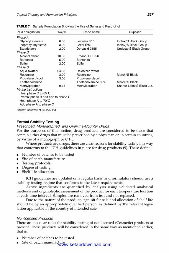
DK4962-Webster-ch17_R2_240307
Formal Stability TestingPrescribed, Monographed, and Over-the-Counter DrugsFor the purposes of this section, drug products are considered to be those thatcontain either drugs that must be prescribed by a physician or, in certain countries,by virtue of a monograph or OTC.
Where products are drugs, there are clear reasons for stability testing in a waythat conforms to the ICH guidelines in place for drug products (9). These define:
B Number of batches to be testedB Site of batch manufactureB Testing protocolsB Degree of testingB Shelf life allocation
ICH guidelines are updated on a regular basis, and formulators should use astability-testing regime that conforms to the latest requirements.
Active ingredients are quantified by analysis using validated analyticalmethods and organoleptic assessment of the product for each temperature locationat each time interval. Samples are removed from test and not replaced.
Due to the nature of the product, sign-off for sale and allocation of shelf lifeshould be by an appropriately qualified person, as defined by the relevant legis-lation applicable in the country of intended sale.
Nonlicensed ProductsThere are no clear rules for stability testing of nonlicensed (Cosmetic) products atpresent. These products will be considered in the same way as mentioned earlier,that is:
B Number of batches to be testedB Site of batch manufacture
TABLE 7 Sample Formulation Showing the Use of Sulfur and Resorcinol
INCI designation %w/w Trade name Supplier
Phase AGlyceryl stearate 6.00 Lexemul 515 Inolex/S Black GroupIsopropyl myristate 3.00 Lexol IPM Inolex/S Black GroupStearic acid 2.00 Dervacid 3155 Undesa/S Black Group
Phase BAlcohol denat. 10.00 Ethanol DEB 96Bentonite 5.00 BentoniteSulfur 2.00 Sulfur
Phase CAqua (water) 64.85 Deionized waterResorcinol 3.00 Resorcinol Merck/S BlackPropylene glycol 3.00 Propylene glycolTriethanolamine Triethanolamine 99% Merck/S BlackMethylparaben 0.15 Methylparaben Sharon Labs/S Black Ltd.
Mixing instructionsHeat phase C to 658CPremix phase B and add to phase CHeat phase A to 708CAdd phase A to phase C
Source: Courtesy of S Black Ltd.
Topical Therapy and Formulation Principles 267
www.ketabdownload.com
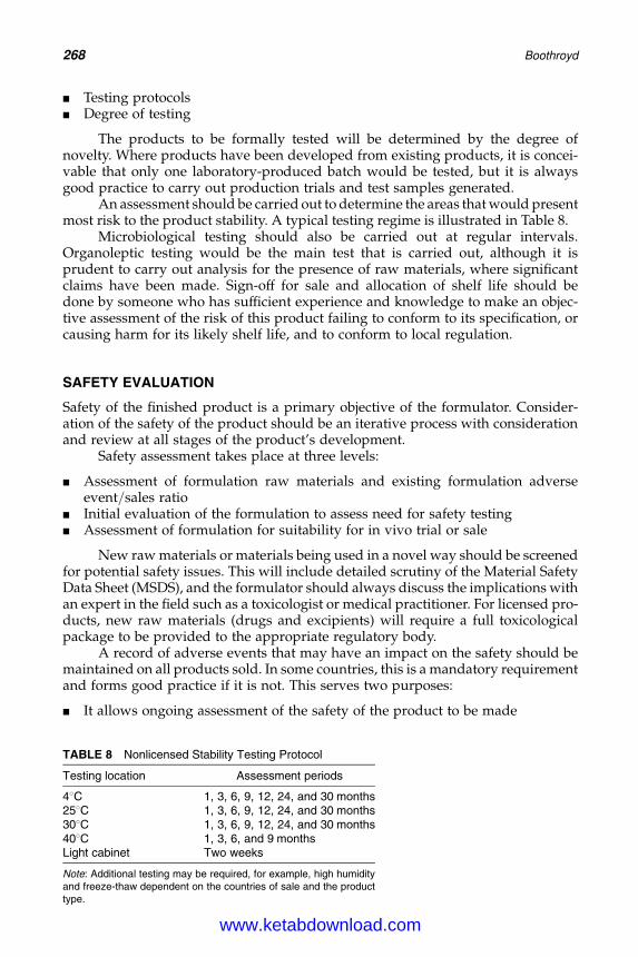
DK4962-Webster-ch17_R2_240307
B Testing protocolsB Degree of testing
The products to be formally tested will be determined by the degree ofnovelty. Where products have been developed from existing products, it is concei-vable that only one laboratory-produced batch would be tested, but it is alwaysgood practice to carry out production trials and test samples generated.
An assessment should be carried out to determine the areas that would presentmost risk to the product stability. A typical testing regime is illustrated in Table 8.
Microbiological testing should also be carried out at regular intervals.Organoleptic testing would be the main test that is carried out, although it isprudent to carry out analysis for the presence of raw materials, where significantclaims have been made. Sign-off for sale and allocation of shelf life should bedone by someone who has sufficient experience and knowledge to make an objec-tive assessment of the risk of this product failing to conform to its specification, orcausing harm for its likely shelf life, and to conform to local regulation.
SAFETY EVALUATION
Safety of the finished product is a primary objective of the formulator. Consider-ation of the safety of the product should be an iterative process with considerationand review at all stages of the product’s development.
Safety assessment takes place at three levels:
B Assessment of formulation raw materials and existing formulation adverseevent/sales ratio
B Initial evaluation of the formulation to assess need for safety testingB Assessment of formulation for suitability for in vivo trial or sale
New raw materials or materials being used in a novel way should be screenedfor potential safety issues. This will include detailed scrutiny of the Material SafetyData Sheet (MSDS), and the formulator should always discuss the implications withan expert in the field such as a toxicologist or medical practitioner. For licensed pro-ducts, new raw materials (drugs and excipients) will require a full toxicologicalpackage to be provided to the appropriate regulatory body.
A record of adverse events that may have an impact on the safety should bemaintained on all products sold. In some countries, this is a mandatory requirementand forms good practice if it is not. This serves two purposes:
B It allows ongoing assessment of the safety of the product to be made
TABLE 8 Nonlicensed Stability Testing Protocol
Testing location Assessment periods
48C 1, 3, 6, 9, 12, 24, and 30 months258C 1, 3, 6, 9, 12, 24, and 30 months308C 1, 3, 6, 9, 12, 24, and 30 months408C 1, 3, 6, and 9 monthsLight cabinet Two weeks
Note: Additional testing may be required, for example, high humidityand freeze-thaw dependent on the countries of sale and the producttype.
268 Boothroyd
www.ketabdownload.com
DK4962-Webster-ch17_R2_240307
B It provides a means by which existing formulations can be assessed for safetybefore further development
The figures stored within the database should be used with the number ofproducts sold to determine the degree of the potential problem and whether thebase could be used. Monitoring of existing formulations allows reformulation tobe rapidly undertaken, should adverse events rise to an unacceptable level. Allreports related to safety issues should be investigated further, as it is importantthat only true adverse reactions are considered.
If a formulation is judged to be different from known base formulations,safety testing should be carried out. These may take the following forms:
B In vitro tests for example:. 3-D human skin model to measure the effects on cell viability.. 3T3 NRU photo toxicity test to measure the number of cells that survive
after exposure to the test chemical and UV light, compared with thenumber that survive after exposure to the chemical in the absence of UVlight.
B Transcutaneous electrical resistance (TER) procedure to measure skin corrosiv-ity through electrical properties.
B Repeat Insult Patch testing on human volunteers to assess the irritancy potentialof the formulation. Such studies should always be undertaken under the super-vision of medical practitioner and with appropriate ethical approval.
B A user trial (controlled or uncontrolled) on a group of people.
Once the testing is complete, the product should be assessed to decidewhether it is safe for sale. This may involve submission to a regulatory authority.
For products where this is not the case, assessment should be made by a suit-ably qualified and experienced assessor. There are published guidelines andrequirements in certain countries as to the qualifications needed by these individ-uals (10,11). Formulators should always ensure that local regulations regardingthis important aspect of developing a new product are met.
REPRODUCIBLE MANUFACTURE
Considerable effort is expended in ensuring that any formulation developed is safe,efficacious, and stable. As the product enters the marketplace, the formulationmoves from the laboratory to larger production areas. The degree of controlthat the formulator can employ in the laboratory is significantly reduced, and con-siderations such as degree of mixing and heating and cooling rates become moresignificant.
Formulators should consider how the formulation will be manufactured atthe time they produce their first laboratory sample, and recognize the differencebetween laboratory and scaled production in terms of raw materials and finishedproduct.
Raw Material ConsiderationsA scale-up of formulation has two significant consequences for raw materials
B Health and safety risk of the materialB Handling of the material in bulk
Topical Therapy and Formulation Principles 269
www.ketabdownload.com
DK4962-Webster-ch17_R2_240307
In a laboratory setting, the risks posed by the majority of ingredients used intopical acne products are small and manageable. As scale-up occurs, the quantity ofmaterial increases and, as a result, the risk posed by materials that may be con-sidered essentially nonhazardous in a laboratory setting increases. Powders or sur-factants handled in larger quantities can pose a significantly higher risk. A riskassessment should always be undertaken and should take into account the quantityof material being handled, the environment in which it will be handled, and theextent to which negative effects could be ameliorated through training and theuse of personal protective equipment.
Normal laboratory production usually entails handling of small quantities ofraw materials in receptacles that allow easy removal. This may not be the case whenthe product is scaled up to full production size. Formulators should consider theway in which the material will be handled, the type of packaging in which itarrives, and whether any special handling equipment is required to enable it tobe used safely and efficiently. Early discussion with appropriate personnel is essen-tial if difficulties are to be avoided.
Product ConsiderationsManufacture of bulk in a laboratory allows much greater intervention from the for-mulator than is possible in a production environment. The formulator should con-sider the method of manufacturing on a larger scale from first laboratory sample,through understanding the type of equipment that will ultimately be used. Pro-duction processes that are achievable in the laboratory are often not reproducibleon a larger scale. These may include heating and cooling rates, and the type andamount of mixing experienced. Individual products will vary in the extent towhich they are affected by these considerations, but as a general rule emulsionsare likely to be the most susceptible.
The use of intermediate-sized mixing vessels in pilot facilities allows the for-mulator to assess the feasibility of manufacture and optimize the productionmethod. Texture, efficacy, and stability are all attributes that may be affectedduring scale-up, and formulators should consider what testing needs to becarried out on pilot and full production batches before releasing the product foruse by a wider population. As was seen earlier, there are clear requirements totest a range of pilot studies when considering stability and it is good practice,even when not legally required, to test batches produced in a production environ-ment. Robust pilot studies leading to a consistent, reproducible method of manufac-ture are essential for the long-term success of the formulation.
INTELLECTUAL PROPERTY
A patent is designed to protect the intellectual property of an individual or organ-ization, and can cover raw materials, product formulations, claims, and techniques.Patents may be filed in individual countries or as world patents, where a range ofcountries may be specified. Patents last up to 20 years, but the existence of pub-lished patent does not always mean that the patent is in force, as a fee must bepaid each year to maintain continued validity.
Formulations should be screened by an appropriate expert in the field toidentify potential patent infringements. This can save both considerable amountof time and money.
270 Boothroyd
www.ketabdownload.com
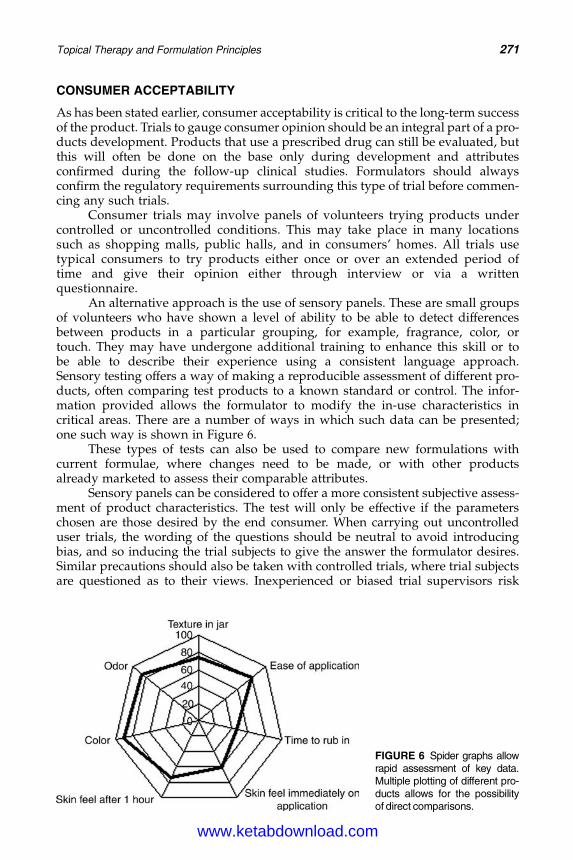
DK4962-Webster-ch17_R2_240307
CONSUMER ACCEPTABILITY
As has been stated earlier, consumer acceptability is critical to the long-term successof the product. Trials to gauge consumer opinion should be an integral part of a pro-ducts development. Products that use a prescribed drug can still be evaluated, butthis will often be done on the base only during development and attributesconfirmed during the follow-up clinical studies. Formulators should alwaysconfirm the regulatory requirements surrounding this type of trial before commen-cing any such trials.
Consumer trials may involve panels of volunteers trying products undercontrolled or uncontrolled conditions. This may take place in many locationssuch as shopping malls, public halls, and in consumers’ homes. All trials usetypical consumers to try products either once or over an extended period oftime and give their opinion either through interview or via a writtenquestionnaire.
An alternative approach is the use of sensory panels. These are small groupsof volunteers who have shown a level of ability to be able to detect differencesbetween products in a particular grouping, for example, fragrance, color, ortouch. They may have undergone additional training to enhance this skill or tobe able to describe their experience using a consistent language approach.Sensory testing offers a way of making a reproducible assessment of different pro-ducts, often comparing test products to a known standard or control. The infor-mation provided allows the formulator to modify the in-use characteristics incritical areas. There are a number of ways in which such data can be presented;one such way is shown in Figure 6.
These types of tests can also be used to compare new formulations withcurrent formulae, where changes need to be made, or with other productsalready marketed to assess their comparable attributes.
Sensory panels can be considered to offer a more consistent subjective assess-ment of product characteristics. The test will only be effective if the parameterschosen are those desired by the end consumer. When carrying out uncontrolleduser trials, the wording of the questions should be neutral to avoid introducingbias, and so inducing the trial subjects to give the answer the formulator desires.Similar precautions should also be taken with controlled trials, where trial subjectsare questioned as to their views. Inexperienced or biased trial supervisors risk
FIGURE 6 Spider graphs allowrapid assessment of key data.Multiple plotting of different pro-ducts allows for the possibilityof direct comparisons.
Topical Therapy and Formulation Principles 271
www.ketabdownload.com
DK4962-Webster-ch17_R2_240307
skewing the results in such a way that false positives are received that are notrepeated when the general consumer uses the product.
Consultation with market research experts on conducting trials is important,as understanding the dynamics of such trials is key to meaningful results.
CLAIMS SUPPORT
It is a requirement that any claims made in relation to products are truthful.Formulators need to be aware when commencing formulation of the claimsto be made and should consider the ways in which these could be delivered.
Care must be exercised in this area regarding the nature of the claim beingmade for the product. Formulators need to be clear of the local regulatory require-ments surrounding the difference between claims that are purely cosmetic andthose that may be considered to be medicinal.
Claims made for formulations fall into these broad categories:
B Claims made for specific or groups of raw materials.B Claims made based on monograph requirements.B Claims made based on the product function.B Claims based on product performance.
Raw MaterialsRaw material claims are made based either on the inclusion of an ingredient (“con-tains Chamomile”), its reported action (“contains Chamomile renowned forits soothing properties”), or testing (“contains alpha bisabalol, the active agent ofChamomile, which has been shown to reduce irritation”).
MonographThere are certain claims that are justified by recognition of the inclusion in legis-lation. Primary among these are the acne claims made in the U.S. monographfollowing the inclusion of a prescribed level of salicylic acid, sulfur, or resorcinol,or combinations of the latter two (5).
Product Function ClaimsSuch claims justify the function of a particular product, for example, shampoo andmoisturiser.
Product PerformanceCertain claims may only be attributable to the whole product rather than individualingredients. Such claims usually quantify the action of the product function, forexample, product X decreases sebum by 30%.
CLAIMS SUPPORT TESTING
With the exception of claims made for product function or based on ingredientsincluded in monograph, some form of claim substantiation should be carried out.These may be labeled as clinical or claims substantiation trials but it is best
272 Boothroyd
www.ketabdownload.com
DK4962-Webster-ch17_R2_240307
practice to conduct any in vivo trial in accordance with Good Clinical Practice(GCP) principles and any additional local regulatory requirement.
Careful thought should be given to the type and nature of the trial, and thisshould only be carried out if the benefits justify the potential risks. Any informationon the trial product should support the proposed trial and be able to be used toproduce a trial protocol. This protocol should be reviewed and approved by anappropriate body.
The safety of the trial subjects is of paramount importance at all times and anytrial should always be the responsibility of a qualified physician. Screening of anypotential trial subject should take into account their health and the impact the trialmay have on them. Informed consent should be obtained from every subject priorto participation in any trial.
Each individual involved in conducting a trial should be qualified and trainedappropriately. All trial information should be gathered and used in a way thatallows accurate analysis and conclusions to be drawn. Records should always bestored in a secure and confidential way and comply with any local regulatoryrequirement.
Trial products should be made in accordance with appropriate good manufac-turing practice and used as stated in the approved protocol. There should be inplace a method of auditing the trial to confirm that it was carried out accordingto the protocol.
ICH has produced guidelines on conducting clinical trials, and formulatorswill find important information that will allow them to perform high-qualitystudies (12). Although these guidelines are widely recognized, formulatorsshould confirm before commencing clinical trials that local regulatory requirementshave been satisfied.
For claims that are more cosmetic in nature, the European Cosmetic, Toile-tries, and Perfumery Association publish guidance on the evaluation of the efficacyof cosmetic products (13).
SUMMARY
Topical anti-acne products covers a wide range of technology, regulatory, andproduct types. Good understanding of the fundamental formulation buildingblocks is essential in order to maximize the effectiveness of the range of ingredientsavailable. A working knowledge of the activities that go to support the formulationenables the formulator to develop a clear strategy to deliver the desired stable, effi-cacious product with the minimum of problems.
This chapter has described many of the elements required to develop high-quality formulations that meet consumer’s needs and expectations. The formulatoris encouraged to use this only as a starting point and not to be afraid to carry outany experiment with the myriad of possible combinations of ingredients todelight new and existing users of topical acne products.
ACKNOWLEDGMENTS
The author would like to thank all those who helped in the preparation of thismanuscript, with special thanks to Patrick Love, Steve Barton, Nick Moody, andDiane Pavis.
Topical Therapy and Formulation Principles 273
www.ketabdownload.com
DK4962-Webster-ch17_R2_240307
REFERENCES
1. Barton S. Formulation of Skin Moisturizers. In: Leyton, Rawlings, ed. Skin Moisturiza-tion. New York: Marcel Dekker, 2002: 547–584.
2. Knowlton J, Pearce S. Emulsions. In: Knowlton J, Pearce S, eds. Handbook of CosmeticScience and Technology. 1st ed. Oxford: Elsevier Advanced Technology, 1993:108.
3. Searle J, Cognis (UK) Ltd. Do we need milder surfactants or is it a case of careful blend-ing? SCS Formulate, Birmingham, Nov 2001.
4. Vandenbossche GMR, Vanhaecke E, De Muynck C, Remon JP. Stability of topical ery-thromycin formulations. Int J Pharm 1991; 67(2):195–199.
5. Code of federal regulations Title 21 Food and Drugs Part 333—Topical Anti-microbialDrug products for over-the-counter human use.
6. Breneman DL, Ariano MC. Successful treatment of acne vulgaris in women with a newtopical sodium sulfacetamide/sulfur lotion. Int J Dermatol 1993; 32(5):365–367.
7. Thiboutot D. New treatments and therapeutic strategies for acne. Arch Fam Med 2000;9(2):179–187.
8. Mackrides PS, Shaugnessy AF. Azelaic acid therapy for acne. Am Fam Physician 1996;54(8):2457–2459.
9. Quality Guidelines on Stability Testing. International Conference on Harmonisation ofTechnical Requirements for Registration of Pharmaceuticals for Human Use. http://www.ich.org. Accessed April 2005.
10. European Community, The rules governing cosmetic products in the European Union.European Community 1999, Cosmetic Legislation Vol. 1.
11. Department of Trade and Industry, Cosmetic Products Regulations 1996, HMSO 1996.12. Efficacy Guidelines. International Conference on Harmonisation of Technical Require-
ments for Registration of Pharmaceuticals for Human Use http://www.ich.org.Accessed April 2005.
13. The European Cosmetic, Toiletries and Perfumery Association: Guidelines for the Evalu-ation of the Efficacy of Cosmetic Products. http://www.colipa.com/. Accessed April2005.
274 Boothroyd
www.ketabdownload.com
DK4962-Webster-ch18_R2_220307
B18 In Vitro Models for the Evaluationof Anti-acne Technologies
John BajorDepartment of Research and Development, Home and Personal Care Division,Unilever PLC, Trumbull, Connecticut, U.S.A.
INTRODUCTION
Acne is the disease manifesting from a multifactorial process involving the pilose-baceous appendages, and although the exact mechanism is unknown, four biologi-cal mechanisms are believed to play critical roles in the formation of a lesion:
1. increased sebum production,2. aberrant keratinocyte proliferation and differentiation,3. enhanced bacterial growth and metabolism, and4. irritation and inflammation.
Acne is unique to humans, and although animals can be induced to form come-dones, there is no representative in vivo model aside from humans, which incorpor-ates the above-mentioned factors. Furthermore, of the four types of pilosebaceousappendages described by Kligman (1), only the sebaceous pilosebaceous unit,which contains a large sebaceous gland and a small vellus hair, has the propensityto develop acne. Even so, acne is still a remarkably rare event, with only a small per-centage of follicles being diseased at any single time.
A detailed structural analysis of the follicular plug, or comedone, thatcan develop into an acne lesion reveals a compacted agglomeration of desquamatedfollicular keratinocytes, bacteria, sebaceous gland products/remnants, and hair fibers.Finally, the static condition of the comedone allows for a favorable growth environ-ment for bacteria, such as Proprionibacterium acnes and Staphylococcus epidermidis thatcan lead to inflammation.
Although no in vivo or in vitro model exists to examine the entire complexityof lesion formation, several in vitro models are available to evaluate technologiestargeting specific biological mechanisms occurring in acne. This review is aimedat disclosing the types of in vitro models available to evaluate technologies thatmay alleviate or prevent acne lesion formation.
EVALUATION OF TECHNOLOGIES TARGETING THE SEBACEOUS GLAND
The sebaceous gland of the skin is the part of the pilosebaceous unit, which secretesa unique lipid mixture of triglycerides, wax esters, and squalene during pro-grammed differentiation (2).
The sebaceous gland consists solely of one epithelial cell type, the sebocyte.Sebocyte migration out of the gland’s basal layer and full differentiation requiresapproximately two weeks, during which time the cells increase in volume 100- to150-fold and become lipid-filled (3–6). Lipid secretion into the follicular duct
275
www.ketabdownload.com
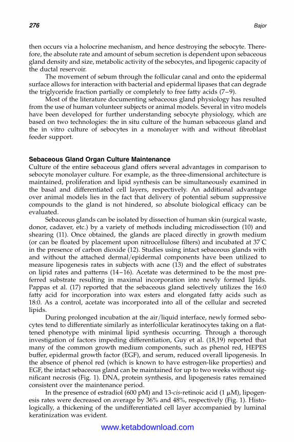
DK4962-Webster-ch18_R2_220307
then occurs via a holocrine mechanism, and hence destroying the sebocyte. There-fore, the absolute rate and amount of sebum secretion is dependent upon sebaceousgland density and size, metabolic activity of the sebocytes, and lipogenic capacity ofthe ductal reservoir.
The movement of sebum through the follicular canal and onto the epidermalsurface allows for interaction with bacterial and epidermal lipases that can degradethe triglyceride fraction partially or completely to free fatty acids (7–9).
Most of the literature documenting sebaceous gland physiology has resultedfrom the use of human volunteer subjects or animal models. Several in vitro modelshave been developed for further understanding sebocyte physiology, which arebased on two technologies: the in situ culture of the human sebaceous gland andthe in vitro culture of sebocytes in a monolayer with and without fibroblastfeeder support.
Sebaceous Gland Organ Culture MaintenanceCulture of the entire sebaceous gland offers several advantages in comparison tosebocyte monolayer culture. For example, as the three-dimensional architecture ismaintained, proliferation and lipid synthesis can be simultaneously examined inthe basal and differentiated cell layers, respectively. An additional advantageover animal models lies in the fact that delivery of potential sebum suppressivecompounds to the gland is not hindered, so absolute biological efficacy can beevaluated.
Sebaceous glands can be isolated by dissection of human skin (surgical waste,donor, cadaver, etc.) by a variety of methods including microdissection (10) andshearing (11). Once obtained, the glands are placed directly in growth medium(or can be floated by placement upon nitrocellulose filters) and incubated at 378Cin the presence of carbon dioxide (12). Studies using intact sebaceous glands withand without the attached dermal/epidermal components have been utilized tomeasure lipogenesis rates in subjects with acne (13) and the effect of substrateson lipid rates and patterns (14–16). Acetate was determined to be the most pre-ferred substrate resulting in maximal incorporation into newly formed lipids.Pappas et al. (17) reported that the sebaceous gland selectively utilizes the 16:0fatty acid for incorporation into wax esters and elongated fatty acids such as18:0. As a control, acetate was incorporated into all of the cellular and secretedlipids.
During prolonged incubation at the air/liquid interface, newly formed sebo-cytes tend to differentiate similarly as interfollicular keratinocytes taking on a flat-tened phenotype with minimal lipid synthesis occurring. Through a thoroughinvestigation of factors impeding differentiation, Guy et al. (18,19) reported thatmany of the common growth medium components, such as phenol red, HEPESbuffer, epidermal growth factor (EGF), and serum, reduced overall lipogenesis. Inthe absence of phenol red (which is known to have estrogen-like properties) andEGF, the intact sebaceous gland can be maintained for up to two weeks without sig-nificant necrosis (Fig. 1). DNA, protein synthesis, and lipogenesis rates remainedconsistent over the maintenance period.
In the presence of estradiol (600 pM) and 13-cis-retinoic acid (1 mM), lipogen-esis rates were decreased on average by 36% and 48%, respectively (Fig. 1). Histo-logically, a thickening of the undifferentiated cell layer accompanied by luminalkeratinization was evident.
276 Bajor
www.ketabdownload.com
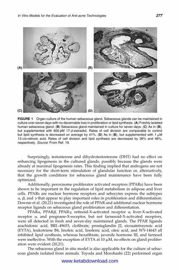
DK4962-Webster-ch18_R2_220307
Surprisingly, testosterone and dihydrotestosterone (DHT) had no effect onenhancing lipogenesis in the cultured glands, possibly because the glands werealready at maximal lipogenesis rates. This finding implied that androgens are notnecessary for the short-term stimulation of glandular function or, alternatively,that the growth conditions for sebaceous gland maintenance have been fullyoptimized.
Additionally, peroxisome proliferator activated receptors (PPARs) have beenshown to be important in the regulation of lipid metabolism in adipose and livercells. PPARs are nuclear hormone receptors and sebocytes express the subtypesa, b, and g that appear to play important roles in proliferation and differentiation.Downie et al. (20,21) investigated the role of PPAR and additional nuclear hormonereceptor ligands on sebaceous gland proliferation and differentiation.
PPARa, PPARb, PPARg, retinoid-X-activated receptor a, liver-X-activatedreceptor a, and pregnane-X-receptor, but not farnesoid-X-activated receptors,were all detected in fresh and seven-day maintained glands. The PPAR ligandsarachidonic acid, BRL-49653, clofibrate, prostaglandin J2, eicosatetraynoic acid(EYTA), leukotriene B4, linoleic acid, linolenic acid, oleic acid, and WY-14643 allinhibited lipid synthesis, whereas bezafibrate, juvenile hormone III, and farnesolwere ineffective. With the exception of EYTA at 10 mM, no effects on gland prolifer-ation were evident (20,21).
The sebaceous gland in situ model is also applicable for the culture of sebac-eous glands isolated from animals. Toyoda and Morohashi (22) performed organ
FIGURE 1 Organ culture of the human sebaceous gland. Sebaceous glands can be maintained inculture over seven days with no discernable loss in proliferation or lipid synthesis. (A) Freshly isolatedhuman sebaceous gland. (B) Sebaceous gland maintained in culture for seven days. (C) As in (B),but supplemented with 600 pM 17-b-estradiol. Rates of cell division are comparable to controlbut lipid synthesis is decreased on average by 41%. (D) As in (B), but supplemented with 1 mM13-cis-retinoic acid. Rates of cell division and lipid synthesis are decreased by 36% and 48%,respectively. Source: From Ref. 19.
In Vitro Models for the Evaluation of Anti-acne Technologies 277
www.ketabdownload.com
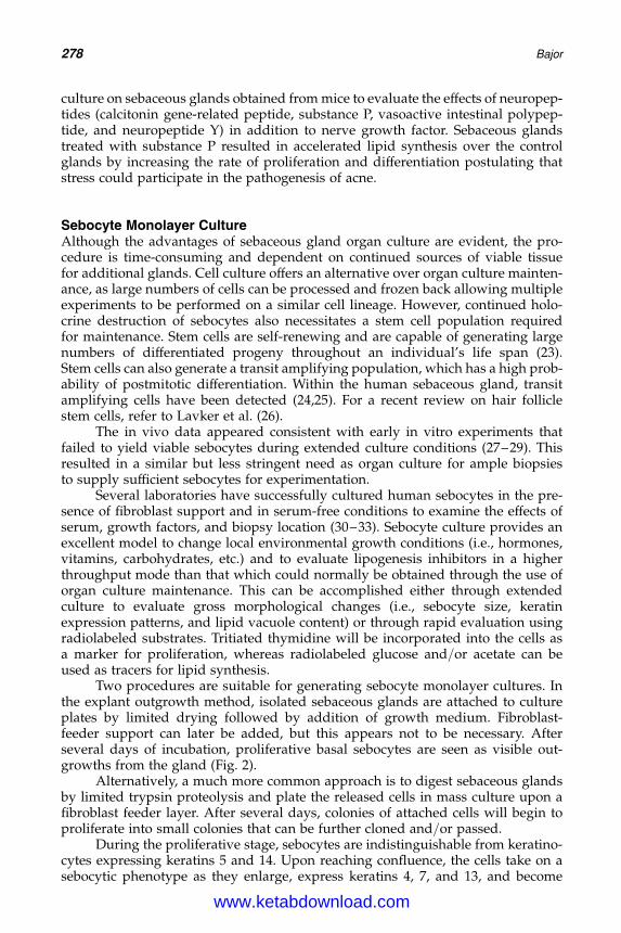
DK4962-Webster-ch18_R2_220307
culture on sebaceous glands obtained from mice to evaluate the effects of neuropep-tides (calcitonin gene-related peptide, substance P, vasoactive intestinal polypep-tide, and neuropeptide Y) in addition to nerve growth factor. Sebaceous glandstreated with substance P resulted in accelerated lipid synthesis over the controlglands by increasing the rate of proliferation and differentiation postulating thatstress could participate in the pathogenesis of acne.
Sebocyte Monolayer CultureAlthough the advantages of sebaceous gland organ culture are evident, the pro-cedure is time-consuming and dependent on continued sources of viable tissuefor additional glands. Cell culture offers an alternative over organ culture mainten-ance, as large numbers of cells can be processed and frozen back allowing multipleexperiments to be performed on a similar cell lineage. However, continued holo-crine destruction of sebocytes also necessitates a stem cell population requiredfor maintenance. Stem cells are self-renewing and are capable of generating largenumbers of differentiated progeny throughout an individual’s life span (23).Stem cells can also generate a transit amplifying population, which has a high prob-ability of postmitotic differentiation. Within the human sebaceous gland, transitamplifying cells have been detected (24,25). For a recent review on hair folliclestem cells, refer to Lavker et al. (26).
The in vivo data appeared consistent with early in vitro experiments thatfailed to yield viable sebocytes during extended culture conditions (27–29). Thisresulted in a similar but less stringent need as organ culture for ample biopsiesto supply sufficient sebocytes for experimentation.
Several laboratories have successfully cultured human sebocytes in the pre-sence of fibroblast support and in serum-free conditions to examine the effects ofserum, growth factors, and biopsy location (30–33). Sebocyte culture provides anexcellent model to change local environmental growth conditions (i.e., hormones,vitamins, carbohydrates, etc.) and to evaluate lipogenesis inhibitors in a higherthroughput mode than that which could normally be obtained through the use oforgan culture maintenance. This can be accomplished either through extendedculture to evaluate gross morphological changes (i.e., sebocyte size, keratinexpression patterns, and lipid vacuole content) or through rapid evaluation usingradiolabeled substrates. Tritiated thymidine will be incorporated into the cells asa marker for proliferation, whereas radiolabeled glucose and/or acetate can beused as tracers for lipid synthesis.
Two procedures are suitable for generating sebocyte monolayer cultures. Inthe explant outgrowth method, isolated sebaceous glands are attached to cultureplates by limited drying followed by addition of growth medium. Fibroblast-feeder support can later be added, but this appears not to be necessary. Afterseveral days of incubation, proliferative basal sebocytes are seen as visible out-growths from the gland (Fig. 2).
Alternatively, a much more common approach is to digest sebaceous glandsby limited trypsin proteolysis and plate the released cells in mass culture upon afibroblast feeder layer. After several days, colonies of attached cells will begin toproliferate into small colonies that can be further cloned and/or passed.
During the proliferative stage, sebocytes are indistinguishable from keratino-cytes expressing keratins 5 and 14. Upon reaching confluence, the cells take on asebocytic phenotype as they enlarge, express keratins 4, 7, and 13, and become
278 Bajor
www.ketabdownload.com
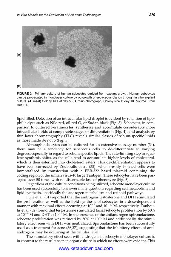
DK4962-Webster-ch18_R2_220307
lipid filled. Detection of an intracellular lipid droplet is evident by retention of lipo-philic dyes such as Nile red, oil red O, or Sudan black (Fig. 3). Sebocytes, in com-parison to cultured keratinocytes, synthesize and accumulate considerably moreintracellular lipids at comparable stages of differentiation (Fig. 4), and analysis bythin layer chromatography (TLC) reveals similar classes of sebum-specific lipidsas those made de novo (Fig. 5).
Although sebocytes can be cultured for an extensive passage number (34),there may be a tendency for sebaceous cells to de-differentiate to varyingdegrees, especially in regard to sebum specific lipids. The rate-limiting step in squa-lene synthesis shifts, as the cells tend to accumulate higher levels of cholesterol,which is then esterified into cholesterol esters. This de-differentiation appears tohave been corrected by Zouboulis et al. (35), when freshly isolated cells wereimmortalized by transfection with a PBR-322 based plasmid containing thecoding region of the simian virus-40 large Tantigen. These sebocytes have been pas-saged over 50 times with no discernable loss of phenotype (Fig. 6).
Regardless of the culture conditions being utilized, sebocyte monolayer culturehas been used successfully to answer many questions regarding cell metabolism andlipid synthesis, specifically the androgen metabolism and retinoid pathways.
Fujie et al. (31) reported that the androgens testosterone and DHT stimulatedthe proliferation as well as the lipid synthesis of sebocytes in a dose-dependentmanner with maximal effects occurring at 1029 and 10210 M, respectively. Zoubou-lis et al. (32) found that testosterone stimulated facial sebocyte proliferation by 50%at 1026 M and DHT at 1028 M. In the presence of the antiandrogen spironolactone,sebocyte proliferation was reduced by 50% at 1027 M and additionally, the stimu-latory effect seen with DHT was neutralized. Spironolactone has been successfullyused as a treatment for acne (36,37), suggesting that the inhibitory effects of anti-androgens may be occurring at the cellular level.
The stimulatory effect seen with androgens in sebocyte monolayer culture isin contrast to the results seen in organ culture in which no effects were evident. This
FIGURE 2 Primary culture of human sebocytes derived from explant growth. Human sebocytescan be propagated in monolayer culture by outgrowth of sebaceous glands through in vitro explantculture. (A, inset) Colony size at day 5. (B, main photograph) Colony size at day 10. Source: FromRef. 31.
In Vitro Models for the Evaluation of Anti-acne Technologies 279
www.ketabdownload.com
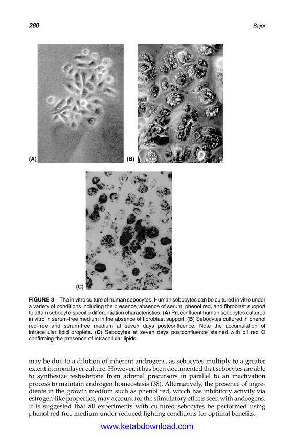
DK4962-Webster-ch18_R2_220307
may be due to a dilution of inherent androgens, as sebocytes multiply to a greaterextent in monolayer culture. However, it has been documented that sebocytes are ableto synthesize testosterone from adrenal precursors in parallel to an inactivationprocess to maintain androgen homeostasis (38). Alternatively, the presence of ingre-dients in the growth medium such as phenol red, which has inhibitory activity viaestrogen-like properties, may account for the stimulatory effects seen with androgens.It is suggested that all experiments with cultured sebocytes be performed usingphenol red-free medium under reduced lighting conditions for optimal benefits.
FIGURE 3 The in vitro culture of human sebocytes. Human sebocytes can be cultured in vitro undera variety of conditions including the presence/absence of serum, phenol red, and fibroblast supportto attain sebocyte-specific differentiation characteristics. (A) Preconfluent human sebocytes culturedin vitro in serum-free medium in the absence of fibroblast support. (B) Sebocytes cultured in phenolred-free and serum-free medium at seven days postconfluence. Note the accumulation ofintracellular lipid droplets. (C) Sebocytes at seven days postconfluence stained with oil red Oconfirming the presence of intracellular lipids.
280 Bajor
www.ketabdownload.com
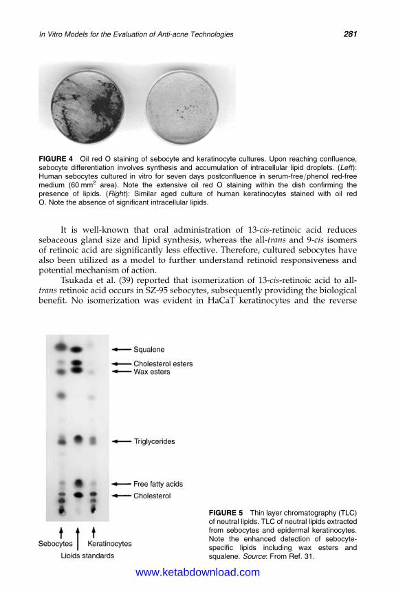
DK4962-Webster-ch18_R2_220307
It is well-known that oral administration of 13-cis-retinoic acid reducessebaceous gland size and lipid synthesis, whereas the all-trans and 9-cis isomersof retinoic acid are significantly less effective. Therefore, cultured sebocytes havealso been utilized as a model to further understand retinoid responsiveness andpotential mechanism of action.
Tsukada et al. (39) reported that isomerization of 13-cis-retinoic acid to all-trans retinoic acid occurs in SZ-95 sebocytes, subsequently providing the biologicalbenefit. No isomerization was evident in HaCaT keratinocytes and the reverse
FIGURE 4 Oil red O staining of sebocyte and keratinocyte cultures. Upon reaching confluence,sebocyte differentiation involves synthesis and accumulation of intracellular lipid droplets. (Left):Human sebocytes cultured in vitro for seven days postconfluence in serum-free/phenol red-freemedium (60 mm2 area). Note the extensive oil red O staining within the dish confirming thepresence of lipids. (Right): Similar aged culture of human keratinocytes stained with oil redO. Note the absence of significant intracellular lipids.
FIGURE 5 Thin layer chromatography (TLC)of neutral lipids. TLC of neutral lipids extractedfrom sebocytes and epidermal keratinocytes.Note the enhanced detection of sebocyte-specific lipids including wax esters andsqualene. Source: From Ref. 31.
In Vitro Models for the Evaluation of Anti-acne Technologies 281
www.ketabdownload.com

DK4962-Webster-ch18_R2_220307
conversion of all-trans to 13-cis-retinoic acid was not evident in sebocytes. Both13-cis and all-trans retinoic acid suppressed mRNA expression of cytochromeP450-1A2, and in addition, 13-cis, all-trans, and 9-cis-retinoic acid reduced sebo-cyte proliferation on average by 40% at 1027 M. Retinoids also do not appear toeffect the programmed cell death of human sebocytes (40).
In additional studies, Zouboulis et al. (41,42), using nontransfected sebocytes,reported that 13-cis-retinoic acid reduced radiolabeled acetate incorporation by48%, whereas all-trans and acitretin only reduced incorporation by 38% and 27%,respectively.
Additional reports using immortalized sebocytes have also been documentedto measure expression of steroidal enzymes (43), PPARs, and transcriptional factors(44), in addition to melanocortin receptors and the effects of select interleukins (ILs)(45). Finally, the patent databases are an excellent source of reviewing intellectualproperty on sebum suppressive technologies.
Rat Preputial Sebocyte Monolayer CultureThe rodent preputial gland has also been used as a model for the human sebaceousgland. These specialized glands open to the surface on either side of the urethralmeatus, and secretions are involved in territorial marking and mating behavior (46).
Using techniques similar to that used with human sebaceous glands, rat prepu-tial glands can be isolated, digested, and cultured upon a fibroblast feeder layer toproduce monolayer cultures (47,48) that can be manipulated and studied. Rosenfield(49) reported on the relationship of sebaceous cell stage with growth in culture in that
FIGURE 6 Culture of immortalized sebocytes. Human facial sebocytes can be immortalized toretain sebocyte-specific differentiation characteristics by transfection with simian virus-40 largeT antigen. The resulting cell line (SZ-95) was further cloned and extensively passaged with nodiscernible loss of features. (A) Preconfluent cultures of human sebocytes prior to transfection.(B) First passage of SZ-95 immortalized sebocytes. (C) SZ-95 immortalized sebocytes at 50thpassage. Note the appearance similar to sebocytes from first passage. Source: From Ref. 35.
282 Bajor
www.ketabdownload.com

DK4962-Webster-ch18_R2_220307
fully differentiated sebocytes are not capable of attachment and proliferation,whereas early differentiated cells are. Subsequent studies by Laurent et al. (48) docu-mented the requirement of serum and growth factors to enhance growth and thatpreputial sebocytes also expressed cytokeratin-4 and formed fewer cornifiedenvelopes than keratinocytes. The effects of estrogen on preputial cell behavior(50) and characterization of the cultured cells (51,52) along with the mechanism ofandrogen action (53) and effects of PPARs (54) have also been reported.
Preputial cells differentiate in a similar process, but to an overall lesser extentthan human sebocytes even in the presence of androgens (Fig. 7). Rosenfieldet al. (55) discovered that preputial cells cultured in vitro lack the presence ofPPAR ligands that will induce lipid droplet formation. The effect of PPARligands was found to be distinct and additive to the effect seen with androgensin increasing lipid droplet formation.
FIGURE 7 Differentiation of preputial sebocytes. Preputial sebocytes can be cultured invitro with 3T3-fibroblast support. Differentiated sebocytes containing lipids are positivelyidentified via staining with oil red O. (Top left): Oil red O staining of preputial sebocytescultured in vitro for nine days in which 11% of the culture has differentiated into lipid-formingcolonies. (Lower left): Oil red O staining of preputial sebocytes cultured in the presence of1026 M dihydrotestosterone (DHT) in which 25% of the culture has differentiated into lipid-forming colonies. (Top right): Oil red O staining of preputial sebocytes cultured in thepresence of 1026 M peroxisome proliferator activated receptor g ligand BRL-49653 in which66% of the culture has differentiated into lipid-forming colonies. (Lower right): Oil red Ostaining of preputial sebocytes cultured in the presence of 1026 M DHT and BRL-49653 inwhich 80% of the culture has differentiated into lipid-forming colonies. Abbreviation: DHT,dihydrotestosterone. Source: From Ref. 53.
In Vitro Models for the Evaluation of Anti-acne Technologies 283
www.ketabdownload.com
DK4962-Webster-ch18_R2_220307
Kim et al. (56), furthermore, investigated the effect of retinoids on prepu-tial cell growth and differentiation. All-trans retinoic acid and retinoic acidreceptor agonists resulted in a decrease in cell number, colony size, andnumber of lipid-forming colonies. Kim concluded that retinoic acid receptorsand retinoid-X-receptors play select roles in sebocyte proliferation anddifferentiation.
Other In Vitro Sebaceous-Cell-Based ModelsAnimal models such as the hamster flank organ and hamster ear have been devel-oped to study the effects of anti-acne and sebum-suppressive capabilities of com-pounds and formulations in vivo. Following application, the target areas areexcised, weighed, and processed for histological examination. In other animalmodels, monitoring of sebum secretions follows intravenous or oral administrationof sebum-suppressive agents.
The hamster auricle sebaceous gland has been successfully cultured andutilized as a model for human sebaceous glands. Glands are isolated andcells are grown on a 3T3 fibroblast-feeder layer using standard protocols. Itoet al. (57), Sato et al. (58), and Akimoto et al. (59) have reported that theamount of lipid produced increases following the peak of sebocyte proliferationand that androgens stimulate, whereas retinoic acid, EGF, and vitamin D3 sup-press overall activity.
EVALUATION OF TECHNOLOGIES TARGETINGTHE PILOSEBACEOUS DUCT
The pilosebaceous duct is lined by a stratified, squamous epithelium, and consistsof keratinocytes that undergo a similar, but distinct, terminal differentiation processcompared to the keratinocytes that comprise the interfollicular epidermis. Theupper fifth of the duct is identical to the interfollicular portion; however, theremaining portion, the infrainfundibulum, is histologically different. In the infrain-fundibulum, the granular layer is frequently absent and the keratinocytes contain atremendous amount of glycogen. In addition, the stratum corneum is only two tothree layers thick and will desquamate into the central part of the pilosebaceousduct where mixing with sebum and bacteria occurs (1).
Ductal hypercornification results initially in the formation of microcome-dones, eventually leading to the production of a comedonal lesion resulting fromthe accumulation of corneocytes in the pilosebaceous duct. This is believed to bethe result of enhanced keratinocyte proliferation, enhanced desquamation,inadequate squame separation, or a combination of factors thereof. Individualswith acne have more cornified material in their ducts when compared withcontrol subjects (60).
Organ Culture of the InfrainfundibulumKeratinocyte cell culture of the interfollicular epidermis is well-established, andprocedures are available to expand cell populations in serum containing andserum-free medium in the presence and absence of fibroblast-feeder support (61–63). In addition, three-dimensional models are also well-characterized to simulatea living skin equivalent. Using these models, it is possible to monitor the cultures
284 Bajor
www.ketabdownload.com
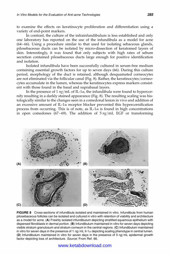
DK4962-Webster-ch18_R2_220307
to examine the effects on keratinocyte proliferation and differentiation using avariety of end-point markers.
In contrast, the culture of the infrainfundibulum is less established and onlyone laboratory has reported on the use of the infundibula as a model for acne(64–66). Using a procedure similar to that used for isolating sebaceous glands,pilosebaceous ducts can be isolated by micro-dissection of keratomed layers ofskin. Interestingly, it was found that only subjects with high rates of sebumsecretion contained pilosebaceous ducts large enough for positive identificationand isolation.
Isolated infundibula have been successfully cultured in serum-free mediumcontaining essential growth factors for up to seven days (66). During this cultureperiod, morphology of the duct is retained, although desquamated corneocytesare not eliminated via the follicular canal (Fig. 8). Rather, the keratinocytes/corneo-cytes accumulate in the lumen, whereas the keratinocytes express markers consist-ent with those found in the basal and suprabasal layers.
In the presence of 1 ng/mL of IL-1a, the infundibula were found to hypercor-nify resulting in a darkly stained appearance (Fig. 8). The resulting scaling was his-tologically similar to the changes seen in a comedonal lesion in vivo and addition ofan excessive amount of IL-1a receptor blocker prevented this hypercornificationprocess from occurring. This is of note, as IL-1a is found in high concentrationsin open comedones (67–69). The addition of 5 ng/mL EGF or transforming
FIGURE 8 Cross-sections of infundibula isolated and maintained in vitro. Infundibula from humanpilosebaceous follicles can be isolated and cultured in vitro with retention of viability and architectureas a model for acne. (A) Freshly isolated infundibulum depicting stratified squamous epithelium withdispersed fibroblasts in dermal portion. (B) Infundibulum maintained in vitro for seven days depictingvisible stratum granulosum and stratum corneum in the central regions. (C) Infundibulum maintainedin vitro for seven days in the presence of 1 ng/mL Il-1a depicting scaling phenotype in central lumen.(D) Infundibulum maintained in vitro for seven days in the presence of 5 ng/mL epidermal growthfactor depicting loss of architecture. Source: From Ref. 66.
In Vitro Models for the Evaluation of Anti-acne Technologies 285
www.ketabdownload.com
DK4962-Webster-ch18_R2_220307
growth factor-a resulted in a disorganized appearance of infundibular morphology,although individual cellular integrity was retained. Looking at the retinoids, 13-cis-retinoic acid (1 mM) supplemented into the medium resulted in a significant fall inthe rate of DNA synthesis after seven days maintenance which can be interpreted asan inhibition of new ductal cell growth.
In a subsequent report, the effects of inflammatory cytokines on the infundi-bulum were evaluated. The addition of 100 U of interferon-g, 10 ng/mL of tumornecrosis factor-a or 10 ng/mL IL-6 resulted in the expression of intercellularadhesion molecule-1 which was not seen with IL-1a treatment.
Although the organ culture of the infundibulum suffers from the same con-straints as sebaceous gland organ culture, the advantages over keratinocyte mono-layer culture lend support as an additional model for the understanding andtreatment of acne.
As previously mentioned, keratinocyte cell culture procedures are well-established and it is beyond the scope of this chapter to disclose the numerous refer-ences related to epidermal biology. However, the use of keratinocyte monolayermodels in further understanding acne is reported in subsequent areas of thisreview.
EVALUATION OF TECHNOLOGIES TARGETING ENZYMES INVOLVEDIN ANDROGEN METABOLISM AND LIPID SYNTHESISEnzyme Assays for In Vitro Androgen MetabolismHuman skin is a collection of androgen-responsive tissues, which have been recog-nized to contain a wide variety of enzymes capable of metabolizing topicallyapplied drugs and endogenous substrates supplied by the body.
One of the most important classes of hormones affecting the pilosebaceousappendage, and more specifically the sebaceous glands, is the sex steroids secretedby the testes and ovaries in response to pituitary gonadotrophic hormones (70). Ingeneral, androgens stimulate sebaceous gland activity, whereas estrogens have asuppressive function (70,71). Androgens are the best-known stimulators of thesebaceous glands, and are responsible for the development and enlargement ofthe glands that occur at puberty (72). Castration results in sebaceous glandatrophy accompanied by an overall decrease in sebum production (73). The mosteffective androgens possess a 17b-hydroxyl group that includes testosterone,5a-DHT, 5a-androstene-3b, and 17b-diol.
The enzymes involved in androgen metabolism in follicles that have beenidentified using biochemical and histological techniques include 3b-hydroxysteroiddehydrogenase (3b-HSD), 17b-HSD, and 5a-reductase. A schematic diagram of thepathways of cutaneous androgen metabolism and the converting enzymes involvedhas been effectively conveyed by Chen et al. (74) in Figure 9.
The reduction of testosterone to DHT by the enzyme 5a-reductase is one ofthe most important reactions involved in androgen metabolism. DHT is a morepotent androgen than testosterone due to its greater affinity for the androgen recep-tor and enhanced stability. DHT is generally considered responsible for increasedsebum production, as increased local formation of DHT has been documented inacne (75) and by increasing the size of the sebaceous gland (73). Two subtypes of5a-reductase have been identified.
The Type I isozyme has an optimal pH of 6 to 9 and has been found localizedin skin from the scalp, chest, dermal papilla, and sebaceous gland regions (76).
286 Bajor
www.ketabdownload.com
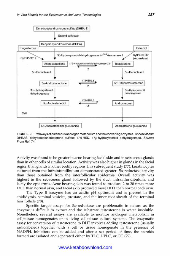
DK4962-Webster-ch18_R2_220307
Activity was found to be greater in acne-bearing facial skin and in sebaceous glandsthan in other cells of similar location. Activity was also higher in glands in the facialregion than glands in other bodily regions. In a subsequent study (77), keratinocytescultured from the infrainfundibulum demonstrated greater 5a-reductase activitythan those obtained from the interfollicular epidermis. Overall activity washighest in the sebaceous gland followed by the duct, infrainfundibulum, andlastly the epidermis. Acne-bearing skin was found to produce 2 to 20 times moreDHT than normal skin, and facial skin produced more DHT than normal back skin.
The Type II isozyme has an acidic pH optimum and is present in theepididymis, seminal vesicles, prostate, and the inner root sheath of the terminalhair follicle (78).
Specific target assays for 5a-reductase are problematic in nature as theenzyme is difficult to extract and the substrate testosterone is water insoluble.Nonetheless, several assays are available to monitor androgen metabolism incell/tissue homogenates or in living cell/tissue culture systems. The enzymaticassay for conversion of testosterone to DHT involves adding testosterone (usuallyradiolabeled) together with a cell or tissue homogenate in the presence ofNADPH. Inhibitors can be added and after a set period of time, the steroidsformed are isolated and separated either by TLC, HPLC, or GC (79).
FIGURE 9 Pathways of cutaneousandrogen metabolism and the convertingenzymes.Abbreviations:DHEAS, dehydroepiandrosterone sulfate; 17b-HSD, 17b-hydroxysteroid dehydrogenase. Source:From Ref. 74.
In Vitro Models for the Evaluation of Anti-acne Technologies 287
www.ketabdownload.com
DK4962-Webster-ch18_R2_220307
Complementary to using skin homogenates, activity of enzymes involved inandrogen modulation can be assessed using cell monolayer cultures and living skinequivalents. Although keratinocyte methods for cultivation of each are well-established, cell culture may be of limited use for evaluating water-insoluble com-pounds or formulations. For the latter, the use of a reconstituted three-dimensionalliving skin model has been reported by Bernard et al. (80) and Slivka (81).
Harris et al. (82) reported on the expression of 5a-reductase in COS cells,whereas Sugimoto et al. (83) constructed 5a-reductase expression vectors thatwere transfected into a human adrenal carcinoma cell line. Type I 5a-reductasewas strongly inhibited by the chloride salts of cadmium, copper, and zinc (inhi-bition constant or Ki less than 5 mM), moderately by nickel and iron (Ki less than250 mM), and no inhibition by manganese, magnesium, calcium, or lithium. Onlythe Type II isozyme was inhibited by copper at 11 mM. Additionally, minimaleffects were seen when the chloride counter ion of zinc was replaced withsulfate, chloride, or acetate, with acetate being the least effective.
In general, 5a-reductase inhibitors are classified into two categories: steroidaland nonsteroidal, and there is very little overlap in efficacy between the two differentisozymes. In fact, many of the Type II inhibitors have little to no effect on sebum pro-duction in vivo. This is possibly the result because the two enzymes have only 50%homology in amino acid sequences. The gene for Type I isozyme is located on chromo-some 5, whereas the gene for Type II isozyme is located on chromosome 2 (84).
Using foreskin keratinocytes (85), testosterone was utilized as a substrate inthe production of 5a-reduced metabolites including DHT (5a-reductase), androste-nedione (17b-HSD Type II), androsterone, and androstanediol (3a-HSD activity). Asimilar pattern was reported with fibroblasts, although scalp skin fibroblasts metab-olized testosterone only to a limited extent. Using cultured keratinocytes obtainedfrom the infrainfundibulum and epidermis (86), Thiboutot et al. found that meanenzyme activities were slightly higher in the acne group, but not statistically differ-ent from the control group. Overall activity was greatest in the infrainfundibulum.
Altenburger and Kissel (87) cultured the human keratinocyte cell line HaCaTas a model to express the enzyme systems involved in testosterone metabolism,which were subsequently identified by LC/MS. HaCaT cells were found to predo-minantly express the Subtype I isoform of 5a-reductase. Similar studies have beenreported utilizing keratinocyte cultures derived from breast skin (88), in culturedfibroblasts (89), and living skin equivalents (90).
Assays for the Rate-Limiting Enzymes Involved in Cholesteroland Fatty Acid SynthesisIn the formation of squalene and cholesterol, 3-hydroxy-3-methylglutaryl-coenzyme A (HMG-CoA) is the rate-limiting enzyme involved in the cholesterolsynthesis pathway. As reported by Shapiro et al. (91) and Smythe et al. (92), radio-labeled HMG-CoA is added to skin homogenates containing NADP (as a necessarycofactor) along with glucose-6-phosphate and glucose-6-phosphate dehydrogenaseto form radiolabeled mevalonate. The reaction then involves a spontaneous conver-sion of mevalonate to mevalonolactone, which can then be separated by TLC andquantified.
Using this assay with isolated sebaceous gland homogenates, Smythe et al.(92) reported that HMG-CoA activity decreased with subject age, and enzymeactivity was inactivated via phosphorylation.
288 Bajor
www.ketabdownload.com

DK4962-Webster-ch18_R2_220307
In the study of fatty acid synthesis, acetyl-coenzyme A carboxylase (acetyl-CoA) has been identified as a rate-limiting step. This assay involves adding radio-labeled bicarbonate to skin homogenates in the presence of ATP and acetyl-CoA.Total radioactivity incorporated is then quantified via direct scintillation counting.
All of the enzyme assays described are amenable for high throughput screen-ing to assess compounds that may modulate lipid synthesis.
EVALUATION OF TECHNOLOGIES TARGETING THE RESIDENTIALMICROBIAL FLORA
It is not clear why P. acnes, S. epidermidis, and other microbes colonize sebaceousfollicles and why no correlation is evident between the P. acnes population andthe acne grade of the subjects in question (93), but antimicrobial therapy providesexcellent clinical relief, especially for inflamed lesions.
Proprionibacterium acnes is an anaerobic, pleomorphic diphtheroid bacillusthat produces propionic and acetic acids during normal metabolism. These andother bacteria also produce biologically active extracellular products includingphosphatases, hyaluronidase, proteases, and neuraminidase, all of which mayincrease the permeability of the follicular epithelium. Bacteria can also producelow molecular weight chemotactic factors that can recruit polymorphonuclear leu-kocytes. In the process of phagocytosis of bacteria, hydrolases are liberated, whichfurther disrupts the integrity of the wall (94). Proprionibacterium acnes can alsostimulate both the classic and alternative pathways of complement activation,leading to the production of neutrophil chemotactic factors, which attractadditional leukocytes resulting in further inflammation (95).
An antimicrobial agent is a chemical that kills or inhibits the growth of bac-teria and other microbes. Agents that kill bacteria are bactericidal, whereas thosethat do not kill but only inhibit further growth are known as bacteriostatic. Manycompounds are both bacteriostatic and bactericidal depending on the concentrationbeing tested. Additionally, compounds may be bacteriolytic in which agents inducedeath by cellular lysis. An example of a bacteriolytic agent is the antibiotic penicil-lin. In general, antimicrobial agents should be selected on the basis of targetingspecific cell types such as bacteria instead of mammalian cells that can be testedin vitro by confirmation in keratinocyte or fibroblast monolayer cultures.
Antimicrobial activity is measured by determining the smallest amount of anagent that will inhibit the growth of a specific organism otherwise known as theminimum inhibitory concentration (MIC). Two methods are commonly utilizedto determine the MIC for a particular agent: the tube dilution technique and theagar diffusion method.
Test Tube Method (Tube Dilution Technique)A series of culture tubes containing microbial growth medium with varying levelsof the test compound are inoculated and incubated. A culture tube in which growthdoes not occur is evident by an absence of turbidity. This is an estimate as variablessuch as pH, oxygen level, temperature, and inoculum size can modulate the effect.
Agar Diffusion MethodPetri plates containing an agar medium are evenly inoculated with the antimicro-bial agent. Known amounts of the antimicrobial agents are added to filter
In Vitro Models for the Evaluation of Anti-acne Technologies 289
www.ketabdownload.com
DK4962-Webster-ch18_R2_220307
paper discs, which are then placed in contact with the surface of the agar. Duringincubation, the test compound will diffuse from the filter paper into the agar anda zone of inhibition will be created indicating the overall potency of the testcompound.
EVALUATION OF TECHNOLOGIES TARGETING IRRITATIONAND INFLAMMATION
Skin irritation is the culmination of a complex inflammatory process involving epi-dermal keratinocytes, dermal fibroblasts, endothelial cells, and invading leukocytesin which early symptoms may consist of swelling, redness, and itching continuingto scaling and erythema (96). Histologically, irritation involves both the epidermisand dermis with inflammatory infiltrates entering from the microvasculature andchemical mediators being released by a variety of cell types inhabiting the area.
The major function of the keratinocytes is not only to provide a barrier andstructural integrity of the pilosebaceous follicle, but also to play a pivotal role inthe inflammatory process through the release/response to surrounding cytokines.On the other hand, the central role of bacteria in acne is to incite inflammation(97). Cytotoxins are compounds that stimulate chemotactic activity and are pro-duced by P. acnes and other microbes within the follicle, which diffuse throughthe follicular wall and attract neutrophils (98). These chemical mediators canthen go on to activate macrophages, neutrophils, natural killer cells, and othercells inducing the production of additional cytokines. Important mediators ofinflammation include histamine, leukotrienes, interleukins, prostaglandins, andneuropeptides. Neutrophils then produce enzymes and generate free oxygen rad-icals that will result in disruption of the follicle. Inflamed papules, pustules, andnodules develop when comedones rupture and extrude their contents into thedermis rather than onto the surface of the skin.
The standard approach for assessing skin irritation is based on the Draizemethod (99), which involves applying a single dose of test material to shavedalbino rabbits. The formation of an erythema response is determined by visualinspection of the skin over a set time course after removal of the patch. Over thelast 60 years, many laboratories have developed in vitro models for a substituteof the Draize method with variable success. These models are based on invitro monolayer culture of fibroblasts or keratinocytes or construction of a three-dimensional living skin equivalent.
Cell Cytotoxicity AssaysMonolayer cultures of epithelial cells are usually used as the first in vitro screen forthe evaluation of toxicity and inflammatory potential of agents. There are severaltypes of assays available that can be used with keratinocyte and fibroblastmonolayer cultures, which differ in their end-point measurement. In general,cells are treated with test compounds during the active proliferation stage inwhich cytotoxicity can be readily detected.
Neutral Red Uptake AssayNeutral red is a nontoxic dye selectively retained by lysosomes of living cells. Agood correlation has been found between the amount of neutral red retained and
290 Bajor
www.ketabdownload.com
DK4962-Webster-ch18_R2_220307
the overall viability of the culture. The assay is limited to a neutral pH but canmeasure inhibitors over a wide range of concentrations (100,101).
Trypan Blue AssayCells with intact membranes exclude the dye trypan blue and remain uncolored,whereas cells with damaged membranes allow the dye to penetrate and be retained.Trypan blue is normally an “all or none” response in that partially damaged cellsmay not retain the blue color (101,102).
MTT AssayThe dye MTT [3-(4,5-dimethylthiazol-2-yl)-2,5-diphenyl tetrazolium bromide] isconverted from the oxidized to the reduced form by an NADP-dependent reactioncatalyzed by succinate dehydrogenase. In the oxidized form, MTT is yellow andturns blue/black upon reduction (103,104). This reaction is dependent uponoverall mitochondrial activity and agents that increase activity will also increaseMTT reduction.
Alamar BlueTM
The Alamar BlueTM assay involves the addition of a fluorogenic redox indicator togrowing cells in culture. The oxidized form of Alamar BlueTM has minimal fluor-escence; however, upon entering living cells, the dye becomes reduced and turnsbright red (105). Fluorescence is measured at 530/590 nm (excitation/emission)and is a direct reflection of cell viability.
Release of Cytosolic EnzymesLactate dehydrogenase is released into the culture medium in the later stages of celldeath. The reaction is based on the conversion of pyruvate to lactate and ismeasured at 340 nm (101).
Three-Dimensional Living Skin EquivalentsCurrent living skin equivalents are the most complex in vitro models available;however, they are also limited in their composition. Although most contain themajor skin structural cell types (keratinocytes and fibroblasts) along with dermalmatrix components (collagen), they do not contain the other cell types and vascularcomponents which are also important contributors to the skin inflammatorymechanism.
However limited, these models do reproduce many of the characteristics ofnormal human epidermis. Ponec et al. (106) performed a detailed study on thevariability among commercial living skin equivalents. Tissue architecture wasexamined using light and electron microscopy along with immunohistochemistryfor specific markers. High-performance TLC was additionally used to assess lipidcomposition. Of the three commercially available models examined along withan in-house prepared model, all generated a stratified epidermis, keratohyalingranules, and the formation of a stratum corneum (Fig. 10).
Faller and Bracher (107) determined the reproducibility of irritation responsesusing three commercially available three-dimensional models along with an in-house model using MTT and measurement of IL-1a as markers. IL-1a is a majorinflammatory mediator released by both corneocytes and keratinocytes upon irri-tant challenge and potentiates further effects in macrophages, neutrophils, and
In Vitro Models for the Evaluation of Anti-acne Technologies 291
www.ketabdownload.com
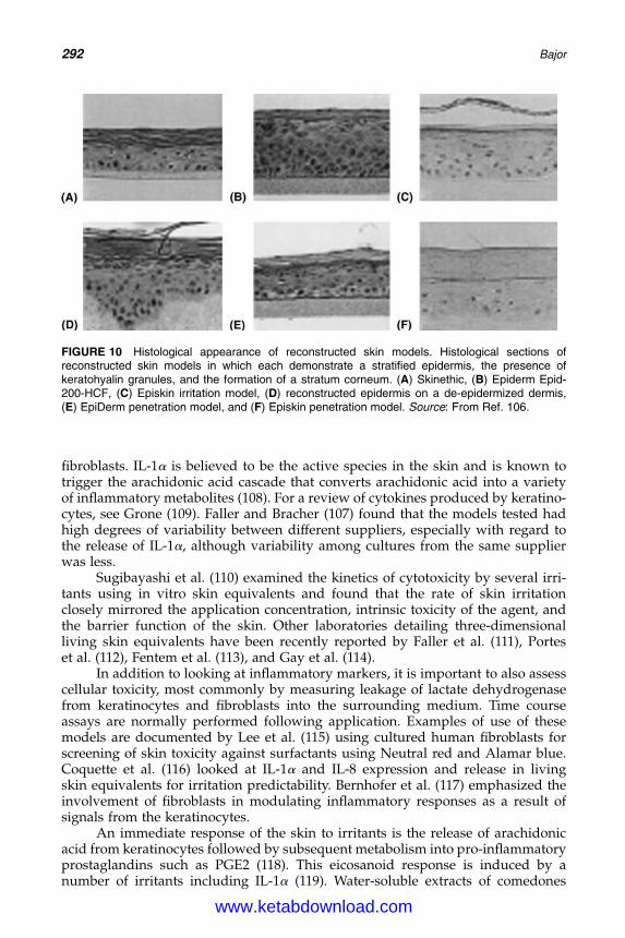
DK4962-Webster-ch18_R2_220307
fibroblasts. IL-1a is believed to be the active species in the skin and is known totrigger the arachidonic acid cascade that converts arachidonic acid into a varietyof inflammatory metabolites (108). For a review of cytokines produced by keratino-cytes, see Grone (109). Faller and Bracher (107) found that the models tested hadhigh degrees of variability between different suppliers, especially with regard tothe release of IL-1a, although variability among cultures from the same supplierwas less.
Sugibayashi et al. (110) examined the kinetics of cytotoxicity by several irri-tants using in vitro skin equivalents and found that the rate of skin irritationclosely mirrored the application concentration, intrinsic toxicity of the agent, andthe barrier function of the skin. Other laboratories detailing three-dimensionalliving skin equivalents have been recently reported by Faller et al. (111), Porteset al. (112), Fentem et al. (113), and Gay et al. (114).
In addition to looking at inflammatory markers, it is important to also assesscellular toxicity, most commonly by measuring leakage of lactate dehydrogenasefrom keratinocytes and fibroblasts into the surrounding medium. Time courseassays are normally performed following application. Examples of use of thesemodels are documented by Lee et al. (115) using cultured human fibroblasts forscreening of skin toxicity against surfactants using Neutral red and Alamar blue.Coquette et al. (116) looked at IL-1a and IL-8 expression and release in livingskin equivalents for irritation predictability. Bernhofer et al. (117) emphasized theinvolvement of fibroblasts in modulating inflammatory responses as a result ofsignals from the keratinocytes.
An immediate response of the skin to irritants is the release of arachidonicacid from keratinocytes followed by subsequent metabolism into pro-inflammatoryprostaglandins such as PGE2 (118). This eicosanoid response is induced by anumber of irritants including IL-1a (119). Water-soluble extracts of comedones
FIGURE 10 Histological appearance of reconstructed skin models. Histological sections ofreconstructed skin models in which each demonstrate a stratified epidermis, the presence ofkeratohyalin granules, and the formation of a stratum corneum. (A) Skinethic, (B) Epiderm Epid-200-HCF, (C) Episkin irritation model, (D) reconstructed epidermis on a de-epidermized dermis,(E) EpiDerm penetration model, and (F) Episkin penetration model. Source: From Ref. 106.
292 Bajor
www.ketabdownload.com

DK4962-Webster-ch18_R2_220307
have the ability to induce inflammation in most subjects, whereas insoluble kera-tinous material will promote inflammation in all subjects (120). Also reported byPuhvel and Sakamoto (121) was that lipids extracted from pooled comedones hadchemotactic activity for neutrophils in vitro using an assay monitoring cellularmigration on agarose plates. Allaker et al. (122) examined culture supernatantsfrom three Propionibacterium species and S. epidermidis for smooth muscle con-tracting compounds as an indicator of inflammatory activity. Webster et al.(123) reported that P. acnes stimulated lysosomal release in the presence, but notin the absence, of serum. This stimulatory activity has been shown to be theresult of P. acnes’ ability to activate and induce cytokine production by cells ofthe mononuclear phagocyte system (124).
SUMMARY AND FURTHER THOUGHTS
The factors involved in the pathogenesis of acne are depicted in Figure 11 (125), andalthough the remission of acne remains a mystery (125,126), several technologieshave been developed for the topical control of acne identified through single invitro assays or a combination of different models.
Benzoyl peroxide has been reported to have multiple effects including bacter-iostatic activity, mild comedolytic activity, enhancement of local blood flow, and therelease of free radical oxygen. Benzoyl peroxide is an organic peroxide that isbactericidal against P. acnes along with additional anti-inflammatory activity(127). There is a wide range of antimicrobial activity against multiple speciesand resistance does not occur. Hegemann et al. (127) investigated the release ofreactive oxygen species regulated by protein kinase C (PKC) and calmodulinfrom human neutrophils; although benzoyl peroxide inhibited PKC activity(IC50 of 1.35 mM), there was no activity against calmodulin, suggesting that the
HORMONESa) Adrenal/ovarian androgen production 1
IL-1α ,TNF α , HLA-DREarly lymphocytic infiltrateCD4+ > CD8+Langerhans cells HLA-DR+Basal kerantinocytes HLA-DR+Comedones contain IL-1 αCell wall substancespeptidoglycan polysaccharidefragmentes
Recruitment ofneutrophils/leukos into epidermalwall of sebaceous follicle
Complement activation Neutrophilic hydrolases:disruption ofsebaceous follicle
Heat shock proteins
Superantiges / Milogen
b) Sebocyte hormone production
AndrogensTestosterone < DHT
Increased Sebum
Comedones
proteases
Cytokine releaseKeratinocyte damagefact
hypothesis
P. acnes
lipas
eslip
ases
Other bacterial products
Proinflammatory lipids
DecreasedLinoleic acid
Ductalhypercornification
Influx ofwater
Impairedepidermal
barrier
Corticotropinreleasing hormone
Corticotropin releasingreceptors 1,2
INFLAMMATION
Steroid-5α-reductase
Androgens receptorsMacrophage activationIL-β , TNFα , ICAM-t
Immune granuloma typereactionhumoral Immune response
FIGURE 11 Synopsis of factors involved in the pathogenesis of acne. Factors involved in thepathogenesis of acne are increased sebum production, ductal cornification, bacterial colonization,and inflammation. Abbreviations: HLA-DR, human leukocyte antigen-D related; ICAM, intercellularadhesion molecule; IL, interleukin; TNF, tumor necrosis factor. Source: From Ref. 125.
In Vitro Models for the Evaluation of Anti-acne Technologies 293
www.ketabdownload.com
DK4962-Webster-ch18_R2_220307
anti-inflammatory activity of benzoyl peroxide was not mediated by PKC orcalmodulin.
Babich et al. (128) used a human keratinocyte cell line to evaluate the cytotox-icity of benzoyl peroxide. As determined by uptake of Neutral red, irreversible celldeath was evident after a one-hour exposure to 0.15 mM and greater concentrationsof benzoyl peroxide, whereas reductions in proliferation were seen at concen-trations from 0.02 to 0.08 mM. Stress and damage as evident by vacuolizationand LDH release were seen after four hours at 0.05 mM levels. As a control, theIC50 of benzoic acid was 29.5 mM.
Similar degrees of cytotoxicity were found with other organic peroxidessuggesting that the cytotoxicity of benzoyl peroxide may be related to the gener-ation of reactive oxidative free radicals. King et al. (129) found that benzoyl per-oxide, as a free radical generating compound, acts as a tumor promoter in mouseskin by causing DNA strand breaks and base modifications in cultured murinekeratinocytes. Matsui et al. (130) also found that benzoyl peroxide inhibited bothhuman and murine PKC in a cell-free system, although there was no translocationevident from the cytosol to the membrane when tested in cultured human keratino-cytes. Other reports investigating the mechanism of action of benzoyl peroxideinclude Valacchi et al. (131) who examined the depletion of vitamin E and IL-1agene expression in HaCaT keratinocytes, Burkhart et al. (132) who reportedincreases in free radical activity in combination with antibiotics, and Lawrenceet al. (133) in that benzoyl peroxide interferes with metabolic cooperation withcultured keratinocytes.
Another compound that has been utilized for the topical control of acne is sal-icylic acid. Salicylic acid reduces inflammation by inhibiting the synthesis of pros-taglandins that are generated in inflamed tissues. Salicylic acid also inhibits theconversion of arachidonic acid to PGE2, which is catalyzed by the enzyme cyclo-oxygenase. Salicylic acid also inhibits comedogenesis by promoting desquamationof the follicular epithelium. Additional agents that have been used to treat acneinclude sulfur for the treatment of inflammatory acne lesions through keratolysisactivity and azelaic acid, a dicarboxylic acid that has both antibacterial and antiker-atinizing activity (134,135).
In addition to the models and assays described in this review, other in vitromodels have been developed around the hair follicle. Although these modelswere not developed to look at acne per se, they may be of interest for specific celltypes. Blume et al. (136) reported on the monolayer culture of keratinocytes isolatedfrom terminal hair follicles that express glycoproteins during proliferation (gp38)and differentiation (gp80). These glycoproteins are absent in normal interfollicularkeratinocytes. Limat and Noser (137) also reported on the serial cultivation ofkeratinocytes from the outer root sheath of hair follicles.
Lenoir-Viale et al. (138) documented the effects of retinoic acid on an in vitromodel reconstructed from the outer root sheath of human hair follicles, whereasHoeller et al. (139) divulged an improved method on the construction of in vitroskin equivalents from human hair follicles and fibroblasts. Additionally, Michelet al. (140) has developed a tissue-engineered human skin equivalent containinghair follicles.
The in situ organ culture of the entire pilosebaceous appendage has furtherbeen developed by Kealey’s laboratory to monitor hair growth. This modelappears to be the most complex, as it contains the entire follicle including thesebaceous gland (141–143).
294 Bajor
www.ketabdownload.com
DK4962-Webster-ch18_R2_220307
Katz and Taichman (144) reported on the development of a two-chamberculture model in which proteins secreted by keratinocytes could be isolated andcharacterized. In this model, a fully differentiated keratinocyte epithelium isgrown on plastic inserts apart from fibroblasts in the lower chamber.
In conclusion, multiple in vitro models are available for the identification oftechnologies to alleviate or prevent the formation of acne lesions. By using thesemodels, it is possible to determine the mechanism of action of lead candidates.
REFERENCES
1. Kligman AM. An overview of acne. J Invest Dermatol 1974; 62:268–287.2. Stewart ME, Downing DT. Chemistry and function of mammalian sebaceous lipids.
Adv Lipid Res 1991; 24:263–301.3. Epstein EH, Epstein WL. New cell formation in human sebaceous glands. J Invest
Dermatol 1966; 46:453–458.4. Plewig G, Christophers E, Braun-Falco O. Proliferative cells in the human sebaceous
gland: labeling index and regional variations. Acta Derm 1971; 51:413–422.5. Tosti A. A comparison of the histodynamics of sebaceous glands and epidermis in man:
a microanatomic and morphometric study. J Invest Dermatol 1974; 62:147–152.6. Downing DT, Strauss JS. On the mechanism of sebaceous secretion. Arch Dermatol Res
1982; 272: 343–349.7. Nicolaides N, Wells GC. On the biogenesis of the free fatty acids in human skin surface
fat. J Invest Dermatol 1957; 29: 423–433.8. Freinkel RK. Origins of free fatty acids in sebum I: the role of coagulase negative
staphylococci. J Invest Dermatol 1968; 50:186–188.9. Marples RR, Kligman AM, Lantis LR, et al. The role of the aerobic microflora in
the genesis of fatty acids in human surface lipids. J Invest Dermatol 1970;55:173–178.
10. Hay JB, Hodgkins MN. Distribution of androgen metabolizing enzymes in isolatedtissues of human forehead and axillary skin. J Endocrinol 1978; 79:29–39.
11. Kealey T, Lee CM, Thody AJ, et al. The isolation of human sebaceous glands and apoc-rine sweat glands by shearing. Br J Dermatol 1986; 114:181–188.
12. Ridden J, Ferguson D, Kealey T. Organ maintenance of human sebaceous glands: invitro effects of 13-cis retinoic acid and testosterone. J Cell Sci 1990; 95:125–136.
13. Cooper MF, McGrath H, Shuster, S. Sebaceous lipogenesis in human skin. Br J Dermatol1976; 94:165–172.
14. Downie MMT, Kealey T. Lipogenesis in the human sebaceous gland: glycogen andglycerophosphate are substrates for the synthesis of sebum lipids. J Invest Dermatol1998; 111:199–205.
15. Middleton B, Birdi I, Heffron M, et al. The substrate determines the rate and patternsof neutral lipid synthesized by isolated human sebaceous glands. FEBS Lett 1988;231:59–61.
16. Cassidy DM, Lee CM, Laker MF, et al. Lipogenesis in isolated human sebaceous glands.FEBS Lett 1986; 200:173–176.
17. Pappas A, Anthonavage M, Gordon JS. Metabolic fate and selective utilization of majorfatty acids in human sebaceous gland. J Invest Dermatol 2002; 118:164–171.
18. Guy R, Ridden C, Kealey T. The improved organ maintenance of the humansebaceous gland: modeling in vitro the effects of epidermal growth factor,androgens, estrogens, 13-cis retinoic acid, and phenol red. J Invest Dermatol 1996;106:454–460.
19. Guy R, Kealey T. The organ-maintained human sebaceous gland. Dermatology 1998;196:16–20.
20. Downie MMT, Sanders DA, Maier LM, et al. Do PPARs have a Regulatory Function inthe Human Sebaceous Gland? Keystone Symposia: The PPARs—A TranscriptionOdyssey, 2001.
In Vitro Models for the Evaluation of Anti-acne Technologies 295
www.ketabdownload.com
DK4962-Webster-ch18_R2_220307
21. Downie MMT, Sanders DA, Stock DM, et al. The PPAR ligands ETYA, WY 14643, LY171883, clofibrate, BRL 49653 and prostaglandin J2 inhibit sebaceous lipogenesiswhereas the PPAR ligand bezafibrate is ineffective (abstr). J Invest Dermatol 2000;115:552.
22. Toyoda M, Morohashi M. Pathogenesis of acne. Med Electron Microsc 2001; 34:29–40.23. Lajtha LG. Stem cell concepts. Differentiation 1979; 14:23–24.24. Cotsarelis G, Sun TT, Lavker RL. Label retaining cells reside in the bulge area of pilo-
sebaceous unit: implications for follicular stem cells, hair cycle and skin carcinogenesis.Cell 1990; 61:1329–1337.
25. Lavker RM, Miller S, Wilson C, et al. Hair follicle stem cells: their location, role in haircycle, and involvement in skin tumor formation. J Invest Dermatol 1993; 101:16s–26s.
26. Lavker RM, Sun TT, Oshima H, et al. Hair follicle stem cells. J Invest Dermatol SympProc 2003; 8:28–38.
27. Xia L, Zouboulis C, Detmar M, et al. Isolation of human sebaceous glands and cultiva-tion of sebaceous gland-derived cells as an in vitro model. J Invest Dermatol 1989;93:315–321.
28. Zouboulis CC, Xia L, Detmar M, et al. Culture of human sebocytes and markers of sebo-cytic differentiation in vitro. Skin Pharmacol 1991; 4:74–83.
29. Yang JS, Lavker RM, Sun TT. Upper human hair follicle contains a subpopulation ofkeratinocytes with superior in vitro proliferative potential. J Invest Dermatol 1993;101:652–659.
30. Doran TI, Baff R, Jacobs P, et al. Characterization of human sebaceous cells in vitro.J Invest Dermatol 1991; 96:341–348.
31. Fujie T, Shikji T, Uchida N, et al. Culture of cells derived from the human sebaceousgland under serum-free conditions without a biological feeder layer or specificmatrices. Arch Dermatol Res 1995; 288:703–708.
32. Zouboulis CC, Akamatsu H, Stephanek K, et al. Androgens affect the activity of humansebocytes in culture in a manner dependent on the localization of the sebaceous glandsand their effect is antagonized by spironolactone. Skin Pharmacol 1994; 7:33–40.
33. Akamatsu H, Zouboulis CC, Orfanos CE. Control of human sebocyte proliferation invitro by testosterone and 5-alpha-dihydrotestosterone is dependent on the localizationof the sebaceous glands. J Invest Dermatol 1992; 99:509–511.
34. Bajor JS, Siegel DM, Kleinberg I, et al. The in vitro proliferative potential of the humansebocyte (abstr). J Invest Dermatol 1994; 102:564.
35. Zouboulis CC, Seltmann H, Neitzel H, et al. Establishment and characteristics ofan immortalized human sebaceous cell line (SZ95). J Invest Dermatol 1999; 113:1011–1020.
36. Messina M, Manieri C, Rizzi G, et al. A new therapeutic approach to acne: an anti-androgen percutaneous treatment with spironolactone. Curr Ther Res 1983;34:319–324.
37. Muhlemann MF, Carter GD, Cream JJ, et al. Oral spironolactone: an effective treatmentfor acne vulgaris in women. Br J Dermatol 1986; 115:227–232.
38. Fritsch M, Orfanos CE, Zouboulis CC. Sebocytes are the key regulators of androgenhomeostasis in human skin. J Invest Dermatol 2001; 116:793–800.
39. Tsukada M, Schroder M, Roos TC, et al. 13-cis retinoic acid exerts its specific activity onhuman sebocytes through selective intracellular isomerization to all-trans retinoic acidand binding to retinoid acid receptors. J Invest Dermatol 2000; 115:321–327.
40. Wrobel A, Seltmann H, Fimmel S, et al. Differentiation and apoptosis in human immor-talized sebocytes. J Invest Dermatol 2003; 120:175–181.
41. Zouboulis CC, Korge B, Akamatsu H, et al. Effects of 13-cis-retinoic acid, all-trans-retinoic acid, and acitretin on the proliferation, lipid synthesis and keratin expressionof cultured human sebocytes in vitro. J Invest Dermatol 1991; 96:792–797.
42. Zouboulis CC, Xia L, Korge B, et al. Cultivation of Human Sebocytes In Vitro: CellCharacterization and Influence of Synthetic Retinoids. Retinoids: 10 Years on. Basel:Karger. 1991:254–273.
43. Thiboutot D, Jabara S, McAllister JM, et al. Human skin is a steroidogenic tissue:steroidogenic enzymes and cofactors are expressed in epidermis, normal
296 Bajor
www.ketabdownload.com
DK4962-Webster-ch18_R2_220307
sebocytes, and an immortalized sebocyte cell line (SEB-1). J Invest Dermatol 2003;120:905–914.
44. Chen W, Yang CC, Sheu HM, et al. Expression of peroxisome proliferator-activatedreceptor and CCAAT/enhancer binding protein transcription factors in culturedhuman sebocytes. J Invest Dermatol 2003; 121:441–447.
45. Bohm M, Schiller M, Stander S, et al. Evidence for expression of melanocortin-1 recep-tor in human sebocytes in vitro and in situ. J Invest Dermatol 2002; 118:533–539.
46. Deplewski D, Rosenfield R. Role of hormones in pilosebaceous unit development.Endocr Rev 2000; 21:363–392.
47. Potter JER, Prutkin L, Wheatley VR. Sebaceous gland differentiation. I: separation, mor-phology and lipogenesis of isolated cells from the mouse preputial gland tumor.J Invest Dermatol 1979; 72:120–127.
48. Laurent SJ, Mednieks MI, Rosenfield RL. Growth of sebaceous cells in monolayerculture. In Vitro Cell Dev Biol 1992; 28A:83–89.
49. Rosenfield RL. Relationship of sebaceous cell stage to growth in culture. J InvestDermatol 1989; 92:751–754.
50. Alves AM, Thody AJ, Fisher C, et al. Measurement of lipogenesis in isolated preputialgland cells of the rat and the effect of oestrogen. J Endocrinol 1986; 109:1–7.
51. Wheatley VR, Potter JER, Lew G. Sebaceous gland differentiation. II: the isolation, sep-aration and characterization of cells from the mouse preputial gland tumor. J InvestDermatol 1979; 73:291–296.
52. Wheatley VR, Brind JL. Sebaceous gland differentiation. III: the uses and limitations offreshly isolated mouse preputial gland cells for the in vitro study of hormone and drugaction. J Invest Dermatol 1981; 76:293–296.
53. Rosenfield RL, Deplewski D, Kentsis A, et al. Mechanism of androgen induction ofsebocyte differentiation. Dermatology 1998; 196:43–46.
54. Rosenfield RL, Deplewski D, Greene ME. Peroxisome proliferator-activated receptorsand skin development. Horm Res 2000; 54:269–274.
55. Rosenfield R, Kentsis A, Deplewski D, et al. Rat preputial sebocyte differentiationinvolves peroxisome proliferator-activated receptors. J Invest Dermatol 1999;112:226–232.
56. Kim MJ, Ciletti N, Michel S, et al. The role of specific retinoid receptors in sebocytegrowth and differentiation in culture. J Invest Dermatol 2000; 114:349–353.
57. Ito A, Sakiguchi T, Kitamura K, et al. Establishment of a tissue culture system forhamster sebaceous gland cells. Dermatology 1998; 197:238–244.
58. Sato T, Imai N, Akimoto N, et al. Epidermal growth factor and 1a,25-dihydroxyvitaminD3 suppress lipogenesis in hamster sebaceous gland cells in vitro. J Invest Dermatol2001; 117:965–970.
59. Akimoto N, Sato T, Sakiguchi T, et al. Cell proliferation and lipid formation in hamstersebaceous gland cells. Dermatology 2002; 204:118–123.
60. Holmes RL, Williams M, Cunliffe WJ. Pilosebaceous duct obstruction and acne. Br JDermatol 1972; 87:327–332.
61. Rheinwald JG, Green H. Serial cultivation of strains of human epidermal keratinocytes:the formation of keratinizing colonies from single cells. Cell 1975; 6:331–334.
62. Randolph RK, Simon M. Characterization of retinol metabolism in cultured human epi-dermal keratinocytes. J Biol Chem 1993; 268:9198–9205.
63. Leigh IM, Lane EB, Watt FM. The culture of human epidermal keratinocytes. In: LeighIM, Lane EB, Watt FM, eds. The Keratinocyte Handbook. Cambridge: Cambridge Uni-versity Press, 1994:43–51.
64. Guy R, Ridden C, Barth J, et al. Isolation and maintenance of the human pilosebaceousduct: 13-cis retinoic acid acts directly on the duct in vitro. Br J Dermatol 1993; 128:242–248.
65. Guy R, Kealey T. The effects of inflammatory cytokines on the isolated human sebac-eous infundibulum. J Invest Dermatol 1998; 110:410–415.
66. Guy R, Kealey T. Modeling the infundibulum in acne. Dermatology 1998; 196:32–37.67. Ingham E, Eady A, Goodwin CE, et al. Pro-inflammatory levels of interleukin-1a like
bioactivity are present in the majority of open comedones in acne vulgaris. J InvestDermatol 1992; 98:895–901.
In Vitro Models for the Evaluation of Anti-acne Technologies 297
www.ketabdownload.com
DK4962-Webster-ch18_R2_220307
68. Antilla HIS, Reitamo S, Saurant JH. Interleukin-1 immunoreactivty in sebaceousglands. Br J Dermatol 1992; 127:585–588.
69. Hauser C, Surat JH, Schmitt A, et al. Interleukin-1 is present in normal human epider-mis. J Immunol 1986; 136:3317–3221.
70. Strauss JS, Pochi PE. The hormonal control of human sebaceous gland. In: Montagna W,Ellis R, Silver A, eds. Advances in the Biology of the Skin: The Sebaceous Glands.Oxford: Pergamon Press, 1963:220–254.
71. Rony HR, Zakon SJ. Effects of androgens in the sebaceous glands of human skin. ArchDermatol Syphilol 1943; 48:601–604.
72. Thody AJ, Shuster S. Control and function of sebaceous glands. Phys Rev 1989; 69:383–416.73. Ebling FJ, Ebling E, McCaffery V, et al. The response of the sebaceous glands of the
hypophysectomized-castrated male rat to 5a-dihydrotestosterone, androstenedione,dehydroepiandrosterone and androsterone. J Endocrinol 1971; 51:181–190.
74. Chen WC, Thiboutot D, Zouboulis CC. Cutaneous androgen metabolism: basicresearch and clinical perspectives. J Invest Dermatol 2002; 119:992–1007.
75. Sansone G, Reisner RM. Differential rates of conversion of testosterone to dihydrotes-tosterone in acne and in normal human skin: a possible pathogenic factor in acne.J Invest Dermatol 1971; 56:366–372.
76. Thiboutot DM, Harris G, Iles V, et al. Activity of the type I 5a-reductase exhibitsregional differences in isolated sebaceous glands and whole skin. J Invest Dermatol1995; 105:209–214.
77. Thiboutot DM, Knaggs H, Gilliland K, et al. Activity of type I 5a-reductase is greater inthe follicular infrainfundibulum compared with the epidermis. Br J Derm 1997;136:166–171.
78. Itami S, Kurata S, Sonoda T, et al. Characteristics of 5a-reductase in cultured humandermal papilla cells from beard and occipital scalp hair. J Invest Dermatol 1991;96:57–60.
79. Takayasu S, Wakimoto H, Itami S, et al. Activity of testosterone 5a-reductase in varioustissues of human skin. J Invest Dermatol 1980; 74:187–191.
80. Bernard F, Barrault C, DeGuercy A, et al. Expression of type I 5a-reductase andmetabolism of testosterone in reconstructed human epidermis (skinethicw): a newmodel for screening skin-targeted androgen modulators. Int J Cosmet Sci 2000;22:397–407.
81. Slivka SR. Testosterone metabolism in an in vitro skin model. Cell Biol Toxicol 1992;8:267–276.
82. Harris G, Azzolina B, Baginsky W, et al. Identification and selective inhibition of anisozyme of steroid 5a-reductase in human scalp. Proc Natl Acad Sci USA 1992;89:10787–10791.
83. Sugimoto Y, Lopez-Solache I, Labrie F, et al. Cations inhibit specifically type I5a-reductase found in human skin. J Invest Dermatol 1995; 104:775–778.
84. Amichai B, Grunwald MH, Sobel R. 5a-Reductase inhibitors—a new hope in dermatol-ogy? Int J Dermatol 1997; 36:182–184.
85. Munster U, Hammer S, Blume-Peytavi U, et al. Testosterone metabolism in human skincells in vitro and its interaction with estradiol and dutasteride. Skin Pharmacol ApplSkin Physiol 2003; 16:356–366.
86. Thiboutot D, Knaggs H, Gilliland K, et al. Activity of 5-alpha-reductase and 17-beta-hydroxysteroid dehydrogenase in the infrainfundibulum of subjects with andwithout acne vulgaris. Dermatology 1998; 196:38–42.
87. Altenburger R, Kissel T. The human keratinocyte cell line HaCaT: an in vitro cell culturemodel for keratinocyte testosterone metabolism. Pharm Res 1999; 16:766–771.
88. Milewich L, Kaimal V, Shaw CB, et al. Epidermal keratinocytes: a source of 5a-dihydrotestosterone production in human skin. J Clin Endocrinol Metab 1986;62:739–746.
89. Pinsky L, Kaufman M, Straisfeld C, et al. Lack of difference in testosterone metabolismbetween cultured skin fibroblasts of human adult males and females. J Clin EndocrinolMetab 1974; 39:395–398.
298 Bajor
www.ketabdownload.com
DK4962-Webster-ch18_R2_220307
90. Ernesti AM, Swiderek M, Gay R. Absorption and metabolism of topically appliedtestosterone in an organotypic skin culture. Skin Pharmacol 1992; 5:146–153.
91. Shapiro DJ, Imblum RL, Rodwell VW. Thin layer chromatographic assay for HMG-CoA reductase and mevalonic acid. Anal Biochem 1969; 31:383–390.
92. Smythe CDW, Greenall M, Kealey T. The activity of HMG-CoA reductase and acetyl-CoA carboxylase in human apocrine sweat glands, sebaceous glands, and hair folliclesis regulated by phosphorylation and by exogenous cholesterol. J Invest Dermatol 1998;111:139–148.
93. Leyden JJ, McGinley KJ, Mills OH, et al. Proprionibacterium levels in patients with andwithout acne vulgaris. J Invest Dermatol 1975; 65:382–384.
94. Winston MH. Acne vulgaris: pathogenesis and treatment. Pediatr Dermatol 1991;38;889–903.
95. Shalita AR, Wei LL. Inflammatory acne. Dermatol Clin 1983; 1;361–364.96. DeLeo VA. Cutaneous irritancy. Frazier JM, ed. In Vitro Testing: Applications to Safety
Evaluation. New York: Marcel Dekker 1992:191–203.97. Holland KT, Ingam E, Cunliffe WJ. A review: the microbiology of acne. J App Bacteriol
1981; 51:195–215.98. Ingham E. The immunology of Propionibacterium acnes and acne. Curr Opin Infect Dis
1999; 12:191–197.99. Draize JH, Woodard G, Calvery HO. Methods for the study of irritation and toxicity of
substances applied topically to the skin and mucous membranes. J Pharmacol Exp Ther1944; 82:377–390.
100. Filman DJ, Brawn RJ, Dandliker WB. Intracellular supravital stain delocalization as anassay for antibody-dependent complement-mediated cell damage. J Immunol Methods1975; 6:189–207.
101. Eun HC, Suh DH. Comprehensive outlook of in vitro tests for assessing skin irritancy asalternates to draize tests. J Dermatol Sci 2000; 24:77—91.
102. De Haan P, Heemskerk AE, Gerritsen A, et al. Comparison of toxicity tests on humanskin and epidermoid (A431) cells using free fatty acids as test substances. Clin ExpDermatol 1993; 18:428–433.
103. Mosmann T. Rapid colorimetric assay for cellular growth and survival: application toproliferation and cytotoxicity assays. J Immunol Methods 1983; 65:55–63.
104. Denizot F, Lang R. Rapid colorimetric assay for cell growth and survival. J ImmunolMethods 1986; 89:271–277.
105. Nakayama GR, Caton MC, Nova MP, et al. Assessment of the alamar blue assay forcellular growth and viability in vitro. J Immunol Methods 1997; 204:205–208.
106. Ponec M, Boelsma E, Gibbs S, et al. Characterization of reconstructed skin models. SkinPharmacol Appl Skin Physiol 2002; 15(suppl):4–17.
107. Faller C, Bracher M. Reconstructed skin kits: reproducibility of cutaneous irritancytesting. Skin Pharmacol Appl Skin Physiol 2002; 15(suppl):74–91.
108. Peplow PV. Actions of cytokines in relation to arachidonic acid metabolism and eicosa-noid production. Prostaglandins Leukot Essent Fatty Acids 1996; 54:303–317.
109. Grone A. Keratinocytes and cytokines. Vet Immunol Immunopathol 2002; 88:1–12.110. Sugibayashi K, Watanabe T, Hasegawa T, et al. Kinetic analysis on the in vitro cytotox-
icity using living skin equivalent for ranking the toxic potential of dermal irritants.Toxicol In Vitro 2002; 16:759–763.
111. Faller C, Bracher M, Dami N, et al. Predictive ability of reconstructed human epidermisequivalents for the assessment of skin irritation of cosmetics. Toxicol In Vitro. 2002;16:557–572.
112. Portes P, Grandidier MH, Cohen C, et al. Refinement of the Episkinw protocol for theassessment of acute skin irritation of chemicals: follow-up to the ECVAM prevalidationstudy. Toxicol In Vitro 2002; 16:765–770.
113. Fentem JH, Briggs D, Chesne C, et al. A prevalidation study on in vitro tests for acute skinirritation: results and evaluation by the management team. Toxicol In Vitro 2001; 13:57–93.
114. Gay R, Swierek M, Nelson D. The living sin equivalent as a model in vitro for rankingthe toxic potential of dermal irritants. Toxicol In Vitro 1992; 6:303–315.
In Vitro Models for the Evaluation of Anti-acne Technologies 299
www.ketabdownload.com
DK4962-Webster-ch18_R2_220307
115. Lee JK, Kim DB, Kim JI, et al. In vitro cytotoxicity tests on cultured human skin fibroblaststo predict skin irritation potential of surfactants. Toxicol In Vitro 2000; 14:345–349.
116. Coquette A, Berna N, Vandenbosch A, et al. Analysis of interleukin-1a and interleu-kin–8 expression and release in in vitro reconstructed human epidermis for the predic-tion of in vitro skin irritation and/or sensitization. Toxicol In Vitro 2003; 17:311–321.
117. Bernhofer L, Seiberg M, Martin K. The influence of the response of skin equivalentsystems to topically applied consumer products by epithelial-mesenchymal inter-actions. Toxicol In Vitro 1999; 13:219–229.
118. Needleman P, Turk J, Jakschik B, et al. Arachidonic acid metabolism. Ann Rev Biochem1986; 55:69–102.
119. Pentland A, Mahoney M. Keratinocyte prostaglandin synthesis is enhanced by IL-1.J Invest Dermatol 1990; 94:43–46.
120. Puhvel SM, Sakamoto M. An in vivo evaluation of the inflammatory effect of purifiedcomedonal components in human skin. J Invest Dermatol 1977; 69:401–406.
121. Puhvel SM, Sakamoto M. The chemoattractant properties of comedonal components.J Invest Dermatol 1978; 71:324–329.
122. Allaker RP, Greenman J, Osbourne RH. The production of inflammatory compounds byPropionibacterium acnes and other skin organisms. Br J Dermatol 1987; 117:175–183.
123. Webster GF, Leyden JJ, Tsai CC, et al. Polymorphonuclear leukocyte lysosomal releasein response to Propionibacterium acnes in vitro and its enhancement by sera from inflam-matory acne patients. J Invest Dermatol 1980; 74:398–401.
124. Ingham E, Walters CE, Eady EA, et al. Inflammation in acne vulgaris: failure of skinmicro-organisms to modulate keratinocyte interleukin-1a production in vitro. Derma-tology 1998; 196:86–88.
125. Jappe U. Pathological mechanisms of acne with special emphasis on Propionibacteriumacnes and related therapy. Acta Derm Venereol 2003; 83:241–248.
126. Downie MMT, Sanders DA, Kealey T. Modeling the remission of individual acnelesions in vitro. Br J Derm 2002; 147:869–878.
127. Hegemann L, Toso SM, Kitay K, et al. Anti-inflammatory actions of benzoyl peroxide:effects on the generation of reactive oxygen species by leukocytes and the activity ofprotein kinase C and calmodulin. Br J Dermatol 1994; 130:569–575.
128. Babich H, Zuckerbraun HL, Wurzburger BJ, et al. Benzoyl peroxide cytotoxicity evalu-ated in vitro with the human keratinocyte cell line, RHEK-1. Toxicology 1996; 106:187–196.
129. King FK, Egner PA, Kensler TW. Generation of DNA base modification following treat-ment of cultured murine keratinocytes with benzoyl peroxide. Carcinogenesis 1995;17:317–330.
130. Matsui MS, Mintz E, DeLeo VA. Effect of benzoyl peroxide on protein kinase C in cul-tured human epidermal keratinocytes. Skin Pharmacol 1995; 8:130–138.
131. Valacchi G, Rimbach G, Saliou C, et al. Effect of benzoyl peroxide on antioxidant status,NF-kB activity and interleukin-1a gene expression in human keratinocytes. Toxicology2001; 165:225–234.
132. Burkhart CN, Specht K, Neckers D. Synergistic activity of benzoyl peroxide anderythromycin. Skin Pharmacol Appl Skin Physiol 2000; 13:292–296.
133. Lawrence NJ, Parkinson EK, Emerson E. Benzoyl peroxide interferes with metabolicco-operation between cultured human epidermal keratinocytes. Carcinogenesis 1989;5:419–421.
134. Russell JJ. Topical therapy for acne. Am Fam Physician 2000; 61:357–366.135. Matsuoka LY. Acne and related disorders. Clin Plast Surg 1993; 20:35–41.136. Blume U, Schon M, Zouboulis CC, et al. Vellus hair follicle-derived keratinocyte
culture: a new experimental model in human hair research. Skin Pharmacol 1994;2:27–32.
137. Limat A, Noser FK. Serial cultivation of single keratinocytes from the outer root sheathof human scalp hair follicles. J Invest Dermatol 1986; 87:45–48.
138. Lenoir-Viale MC, Galup C, Darmon M, et al. Epidermis reconstructed from the outerroot sheath of human hair follicle: effect of retinoic acid. Arch Dermatol Res 1993;285:197–204.
300 Bajor
www.ketabdownload.com

DK4962-Webster-ch18_R2_220307
139. Hoeller D, Huppertz B, Roos TC, et al. An improved and rapid method to constructskin equivalents from human hair follicles and fibroblasts. Exp Dermatol 2001;10:264–271.
140. Michel M, Dheureux N, Pouliot R, et al. Characterization of a new tissue-engineeredhuman skin equivalent with hair. In Vitro Cell Dev Biol 1999; 35:318–326.
141. Philpott MP, Green MR, Kealey T. Human hair growth in vitro. J Cell Sci 1990; 97:436–471.
142. Sanders DA, Philpott MP, Nicolle FV, et al. The isolation and maintenance of the humanpilosebaceous unit. Br J Derm 1994; 131:166–176.
143. Kealey T, Philpott M, Guy R. Human pilosebaceous culture. Methods Mol Biol 1997;75:101–115.
144. Katz AB, Taichman LB. Epidermis as a secretory tissue: an in vitro tissue model to studykeratinocyte secretion. J Invest Dermatol 1994; 102:55–60.
In Vitro Models for the Evaluation of Anti-acne Technologies 301
www.ketabdownload.com

DK4962-Webster-ch18_R2_220307
www.ketabdownload.com
DK4962-Webster-Index_R2_220307
B Index
Accutane, 51Acne, 26, 63–66
androgens, 83antimicrobial peptides, 75–80antimicrobial therapy, 97–100bacterial factors, 2–3changes, 26–27development factors, 3–4DHT, 176–178endocrine disorder, 86–87estrogens, 85growth hormone, 85immunity, 4–5inflammation, 3–4
factors, 4response, 5
insulin-like growth factors, 85lesions
formation factors, 28healing, 4resolution, 4
pathogenesis, 208–210pathogenesis overview, 1–5phototherapy and laser therapy, 113–115phytosphingosine, 158–163pilosebaceous unit, 26–27potential targets for light, 113–115Propionibacterium acnes, 5
populations, 3role, 4–5
puberty, 2sebaceous secretion, 3sebum secretion, 37–41severity, 5treatment regimens, 99–100
Acne vulgaris, 45, 109, 225–226adult female, 91benzoyl peroxide, 117–126grades, 118hormonal influences, 83–92hormonal therapy, 87–88oral contraceptives, 90–92pathogenesis, 293reason behind, 156sphingolipids, 155–164
[Acne vulgaris]therapy, 118–119women, 87–88
Acroinfundibulum, 12Acrylic acid polymers, 262ACTH. See Adrenocorticotrophic hormone
(ACTH)Active raw materials, 263–266Acylceramide
ester-linked fatty acids, 41Adapalene (Differin), 106, 263
pilosebaceous unit, 247site-specific delivery, 243
Adrenal androgen productioninhibitors, 88–89
Adrenocorticotrophic hormone (ACTH), 24–25Adrenomedullin, 77Adult female. See also Women
acne clinical presentation, 85–86acne vulgaris, 91
Agar diffusion methodresidential microbial flora, 289–290
Agesebum secretion, 40–41
Akne-Mycin (erythromycin), 265ALA. See Aminolevulinic acid (ALA)Alamar blue, 291Alesse, 89, 91Alpha dihydrotestosterone, 167–199Alpha-hydroxyprogesterone, 174Alpha interferon (alpha-IFN)
nonionic liposomal composition, 240Alpha reductase, 167–199
inhibitors, 183–192steroidal, 184–185structure, 183–184
Alternate response elements (ARE), 104Amino acid sequences
DHT, 172Aminolevulinic acid (ALA), 111Androgen, 20, 86–87
acne, 83metabolism within skin, 83–85
Androgen metabolismskin, 167
303
www.ketabdownload.com
DK4962-Webster-Index_R2_220307
Androgen receptoragents blocking, 87–89antagonists, 21
Androstenedione, 174Anionic emulsifiers, 257Anti-acne technologies
in vitro models, 275–295Antibiotics, 91–92, 265
anti-inflammatory activity, 98overuse, 100
Anti-inflammatory activityantibiotics, 98
Antimicrobial peptidesacne, 78–80
Antimicrobialsproduction, 37
Antimicrobial therapyacne, 97–100
Appendageal pathwaypercutaneous absorption, 238
ARE. See Alternate response elements (ARE)AZA. See Azelaic acid (AZA)Azasteroids, 184–187
derivatives, 183Azelaic acid (AZA), 137, 265–267
MICPropionibacterium acnes, 140Staphylococcus aureus, 140
phase I clinical trial, 142–148skin delivery, 141–142
Bacterial colonizationlumen, 15
Bacterial lipolysis, 2Bentonite method, 38–39Benzac AC Wash, 264Benzopyrene, 235Benzoquinoline derivatives, 189Benzoquinolinone derivatives, 189–190Benzoquinolizine-3-one derivatives, 190–191Benzoyl peroxide, 97, 157, 263–264, 293
acne vulgaris, 117–126chemistry, 119cytotoxicity, 294efficacy, 122–126formulation, 264history, 119pharmacokinetic and pharmacodynamic, 120phase I clinical trial, 142–148phase II clinical trial, 148–150vs. SA, 131–132safety, 120–122
Biphenyl-4-carboxylic acids, 191Blackheads, 117Blue-light treatment, 114
Calmodulin, 293Carbomers, 262Carbon dating
petroleum, 37Catalysis
DHT, 173Cations, 193Cell cytotoxicity assays, 290–291Cephalosporines, 98Ceramides, 157Cetearyl Glucoside (Tego), 259Cholesterol
forehead, 205lipid, 206rate-limiting enzyme assays, 288–289synthesis, 18
Cholesterol estersforehead, 205
Cigarette paper method, 40Ciprofloxacin, 98, 99Claims support, 272
monograph, 272performance, 272product function claims, 272raw materials, 272testing, 272–273
Clearasil Maximum Strength, 264Cleocin T (clindamycin), 265Clindamycin, 109, 265Combi bars, 263Combination therapy, 109Comedo, 117, 209
formation, 1–2Comedogenesis, 2, 38Comedogenic potential, 230Comedones
electron microscopy, 210Confocal laser microscopy, 234Consumer acceptability, 271–272Contraceptives
newer forms, 92Controlled release systems, 247–248Corneocytes, 104Corticosterone, 174Corticotrophic-releasing hormone
(CRH), 25Corynebacterium parvum, 3CRABP. See Cytosolic retinoid-activating
binding proteins (CRABP)CRH. See Corticotrophic-releasing hormone
(CRH)Cyproterone acetate, 87–88, 92Cytokines
sebaceous glands, 25–26Cytosolic enzymes, 291
304 Index
www.ketabdownload.com
DK4962-Webster-Index_R2_220307
Cytosolic retinoid-activating binding proteins(CRABP), 105–107
Cytotoxinsinflammation, 290
DCA. See Dicarboxylic acid octadecenedioicacid (DCA)
Defensins, 75–77Dehydroepiandrosterone (DHEA), 83Dehydroepiandrosterone sulfate (DHEAS), 83
serum levels, 85Dermal papilla
hair growth, 13Desogestrel (Mircette), 88DHEA. See Dehydroepiandrosterone (DHEA)DHEAS. See Dehydroepiandrosterone sulfate
(DHEAS)DHT. See Dihydrotestosterone (DHT)Diane-35, 91Dicarboxylic acid octadecenedioic acid (DCA),
138–140structural formula, 138
Diethanolamines, 261Differin, 51, 263
pilosebaceous unit, 247site-specific delivery, 243
Diglyceridesforehead, 205
Dihydrotestosterone (DHT), 83, 155, 167–199acne, 178–179amino acid sequences, 172biochemical characterization, 170–171catalysis, 173cutaneous enzymatic activity, 175–176distribution, 175–176enzymatic activity and specificity, 172–173,
179history, 168–169immunolocalization, 178lipogenesis, 277localization, 175–176nonsteroidal inhibitors, 189–190normal skin, 178physiological role, 169–170sebaceous follicles, 178species variation, 171–172subcellular localization, 176–177substrates, 174
Dimethyl sulfoxide (DMSO), 238Diol esters
sebum, 205DMSO. See Dimethyl sulfoxide (DMSO)Dose-dependent epidermal acanthosis, 105Doxycycline, 91–92, 98
adverse effects, 99
Drospirenone, 90Drug and follicular deposition
physicochemical properties, 235–237Dutasteride, 185
Emulsifiers, 257categories, 258packing, 259
Emulsion, 256–258defined, 256types, 257
Endocrine disorderacne, 86–87
Epidermiscommitment to proliferation and
differentiation, 107interaction with sebaceous lipids, 41PPAR, 64–65
Erythromycin, 265Ester-linked fatty acids
acylceramide, 41Estradiol
lipogenesis, 276penetration, 238
Estranes, 89, 90Estrogen, 22
acne, 85oral contraceptives, 89
Estrostep, 89, 91Ethinyl estradiol cyproterone acetate
(Diane-35), 91Ethinyl estradiol desogestrel (Mircette), 89Ethinyl estradiol drospirenone (Yasmin), 91Ethinyl estradiol levonorgestrel
(Alesse), 89, 91Ethinyl estradiol norethindrone
(Estrostep), 89, 91Ethinyl estradiol norethindrone acetate
(Lo-Estrin), 89, 91Ethinyl estradiol norgestimate (Ortho
Tri-Cyclen), 91Ethynodiol diacetate, 90Ethynylestradiol
sebum secretion, 22Explant outgrowth method
sebocyte monolayer culture, 278
Fatty acidsforehead, 205rate-limiting enzyme assays, 288–289sebum, 18, 205synthesis, 19
Faulty desquamation, 1Faulty keratinization, 226Favre-Racouchaut syndrome, 2
Index 305
www.ketabdownload.com
DK4962-Webster-Index_R2_220307
Female acne. See also Womenhormonal therapy, 92
Finasteride, 1855alpha reductase, 167–199
inhibitors, 183–192Fluorescent polystyrene, 242Flutamide, 87–88, 92Follicle-free models, 233Follicles
deposition, 232–234mechanical separation, 233models, 229–231
Follicular deliveryanimal and human models, 230mathematical modeling, 235
Follicular depositionmicrobeads, 242–243vehicle effects, 237–243
Follicular filament, 209Follicular keratinization, 209Follicular pathway
delivery, 235–243Follicular route, 226–227
barriers, 228objectives, 227sebum, 228–234
Formulation. See also Product,formulating new
new productsinterrelated events, 255regulations, 256
principles, 253–273strategy, 254–256surfactant products, 259
4-methyl-4-azasteroids, 186–1874-oxa-thiasteroids, 1874-thiasteroids, 187Free fatty acids, 18
lipid, 206sebum, 205
Free sterolssebum, 205
FXR ligandsPPAR, 64–65
GDL. See Glyceryl dilaurate (GDL)Gels, 262Germicides, 237
autoradiography, 237Gestodene, 90Glucocorticoids, 88–89, 92Glycerin, 219Glyceryl dilaurate (GDL), 229Glycoproteins, 294Gonadotropin-releasing agonists, 89
Gonanes, 90Growth hormone
acne, 85Guinea pig, 231–232
Hair cyclecharacteristics, 13
Hair growth, 230–231Hamster ear model, 231hBD1. See Human beta-defensin 1 (hBD1)1H-benzoquinolizine-3-one derivatives,
190–191HDAC. See Histone deacetylase enzymes
(HDAC)Hildebrand coefficients
sebum, 228Histone deacetylase enzymes (HDAC), 49
manipulation, 52Histone deacetylase proteins
schematic overview, 49HLB. See Hydrophilic-lipophilic balance
(HLB) systemHormonal control, 40Hormonal therapy
female acne, 92HSD. See Hydroxysteroid dehydrogenase (HSD)Human beta-defensin 1 (hBD1), 75–77Human model, 232Hydrocarbon
lipid, 206Hydrocortisone
delivery, 242skin permeation, 236
Hydrophilic-lipophilic balance (HLB) system,257
Hydrophobic tailsmicelles, 260
Hydroxyprogesterone, 174Hydroxysteroid dehydrogenase (HSD), 84
IFN. See Interferon (IFN)Immortalized sebocytes
culture, 282Immune responsiveness
modulation, 114–115Incubation
sebaceous glands, 25Indolizine butanoic acids, 192Inflammation
acne, 3–4factors, 4response, 5
cytotoxins, 290neutrophils, 290technology, 290–291
306 Index
www.ketabdownload.com
DK4962-Webster-Index_R2_220307
Infrainfundibulumcornified envelopes, 14organ culture, 284–286
Infundibulum, 12, 225biochemical changes, 27–28immunohistochemical labeling, 21
Instrumental methods, 39Insulin-like growth factors
acne, 85Intellectual property, 270Interferon (IFN)
nonionic liposomal composition, 240Invasive technique, 233In vitro androgen metabolism
enzyme assays, 286–288Irritation
technology, 290–291Isopropyl myristate, 229Isotretinoin (Roaccutane), 263
mechanism of action, 51sebaceous gland deposition, 241
Keratinizationfaulty, 226follicular, 209modulation, 114
Keratinocyte, 14, 107DHT enzymatic activity and specificity,
179–182Keratinocyte growth factor (KGF), 25KGF. See Keratinocyte growth factor (KGF)
Laser therapyacne, 113–116
Levonorgestrel, 90Ligand-binding pockets
nuclear receptors, 47Light-based treatments, 113Lipid
biosynthesisgenes, 22sebocytes, 19
cholesterol, 206human skin surface, 204–205sebum, 204, 205TLC, 279triglycerides, 204, 205
Lipid meltsdelivering topical acne medications, 243–248vehicles, 242
Lipogenesis, 17estradiol, 276PPAR, 277retinoic acid, 276testosterone, 277
Liposomesmechanism of action, 241–242vehicles, 239–241
Liver-X-activated receptor (LXR), 57Living skin equivalents
three-dimensional, 291–293Lo-Estrin, 89Long-pulsed 585-nm pulsed-dye
laser, 115LXR. See Liver-X-activated receptor (LXR)
Macaque monkey model, 230–231Macrolides, 97–98Mathematical modeling
follicular delivery, 235Matrix metalloprotease, 98Melanocortins, 24Methyl aminolevulinic PDT, 1154-methyl-4-azasteroids, 186–187MIC. See Minimal inhibitory concentration
(MIC)Micelles
hydrophobic tails, 260Microbeads
size effects, 242–243Microcomedones
production, 226Microflora, 2Minimal inhibitory concentration (MIC)
AZAPropionibacterium acnes, 140Staphylococcus aureus, 140
Minocycline, 98adverse effects, 99
Mircette, 89, 90Model sebum with C16 carbon chain lipids,
212–213Modified cellulose gums, 262Monographed drugs
formal stability testing, 267MTT assay, 291
Neonatal pilosebaceous units, 10Neuropeptides
growth factors, 25Neutral red uptake assay, 290–291Neutrophils
inflammation, 290Niosomes, 242Noninvasive cyanoacrylate
technique, 232–233Nonionic emulsifier, 259Nonionic liposomal composition
alpha interferon (alpha-IFN), 240Nonionic surfactants, 261
Index 307
www.ketabdownload.com
DK4962-Webster-Index_R2_220307
Nonlicensed productsformal stability testing, 268–269stability testing protocol, 268
Nonsteroidal inhibitorsDHT, 189–190
Norethindrone, 90Norethindrone acetate, 90Norgestimate, 90Norgestrel, 90Nortestosterone derivatives, 90Nuclear hormone receptors
retinoid receptors, 45–49Nuclear receptors
ligand-binding pockets, 47
Octadecenedioic acid, 137–152mechanism of action, 150novel acne treatment, 151–152phase I clinical trial, 141–148phase II clinical trial, 148–150skin delivery, 140–141
Ointments, 262–263Oleic acid, 2381H-benzoquinolizine-3-one derivatives,
190–191Oral contraceptives, 89, 91–92
acne vulgaris, 90–91low doses of estrogen, 89
Oral cyproterone acetate, 21Oral retinoid therapy
systemic absorption and teratogenicity, 110Organic solvents, 237–238Ortho Tri-Cyclen, 91Ovarian androgen production, 89Over-the-counter drugs
formal stability testing, 2684-oxa-thiasteroids, 188
Panoxyl, 264Percutaneous absorption
retinoic acid, 239steroids, 236
Peroxisome proliferator-activated receptor(PPAR), 23–25, 138, 152
activation, 60–61differential expression, 58–59dimerization, 57–58DNA binding, 57–58epidermis, 62–63FXR ligands, 64–65lipogenesis, 277molecular biology, 55–67physiological functions, 66pilosebaceous unit, 62–63structure and localization, 56–57
[Peroxisome proliferator-activatedreceptor (PPAR)]
target genes, 61transcriptional activity, 57–58
Peroxisome proliferator response elements(PPRE), 57
Petroleumcarbon dating, 37
Phenylethyl cyclohex-1-ene carboxylic acids,191
Phototherapyacne, 113–116future, 115
Phytochemical inhibitors, 193–196Phytosphingosine
acne, 158–163Pilosebaceous duct
cells, 14–15environmental scanning electron
micrograph, 27structure, 12–14technology, 284–285
Pilosebaceous glandstructure, 15
Pilosebaceous unit (PSU), 155acne changes, 26–27activity regulation, 20–23adapalene (Differin), 247cell biology, 9–28disorders affecting, 26distribution, 11–12drug deposition, 229neonatal, 10polymeric microspheres, 247PPAR, 62–63schematic representation, 224sebaceous gland, 223–225topical treatment products, 223–248types, 9
PKC. See Protein kinase C (PKC)PMN. See Polymorphonuclear lymphocytes
(PMN)POE. See Polyoxyethylene-10-stearyl ether
(POE-10)Polymorphonuclear lymphocytes
(PMN), 104Polyolprepolymer-2, 105Polyoxyethylene-10-stearyl ether
(POE-10), 229Porphyrin phototherapy, 113PPAR. See Peroxisome proliferator-activated
receptor (PPAR)PPRE. See Peroxisome proliferator response
elements (PPRE)Pregnatriene derivatives, 188–189
308 Index
www.ketabdownload.com
DK4962-Webster-Index_R2_220307
Preputial sebocytessebocyte differentiation, 51
Prescribed drugsformal stability testing, 268
Primary surfactantenhance characteristics, 261
Productformulating new, 254, 255
active type, 254chemical activities, 254claims support, 272–273consumer acceptability, 271–272physical activities, 254range, 257raw materials, 270regulations, 256safety evaluation, 268–269testing, 272–273types, 256–263
Productionantimicrobials, 37
Progesterone, 174Progestin, 89–90Propionibacterium acnes, 2, 113–115, 156–157,
203, 275, 289, 293acne immunity, 4–5acne severity, 5AZA, 140MIC, 140populations, 3
Proposed hybrid method, 39–40Protein kinase C (PKC), 293PSU. See Pilosebaceous
unit (PSU)Puberty
acne, 2sebum secretion, 16
Pulsed-dye laser, 115
Quinolin-2-ones, 190
Rabbit ear model, 230RAMBA. See Retinoic acid metabolism
breakdown agents (RAMBAs)RAR. See Retinoic acid receptors (RARs)RARE. See Retinoic acid response element
(RARE)Rat model, 231Raw materials
products, 270reproducible manufacture, 269–270
Reductase, 167–199inhibitors, 183–192
structure, 183–184Reproducible manufacture, 269–270
Residential microbial floraagar diffusion method, 289–290technology, 289–290test tube method (tube dilution
technique), 289Resorcinol, 265
formulation, 268Retin-A, 51, 105, 109, 110, 263Retinoic acid, 294
lipogenesis, 276percutaneous absorption, 239
Retinoic acid metabolism breakdown agents(RAMBAs), 50
Retinoic acid receptors (RARs), 45schematic overview, 47
Retinoic acid response element(RARE), 46
Retinoids, 22–23, 263absorption, 103classification, 103effects, 50–52mechanism of action, 103–104metabolism binding proteins, 49–51molecular biology, 45–52pleiotropic effects, 46receptors, 104
nuclear hormone receptors, 45–49sebaceous glands, 23secondary effects, 107structures, 51types, 105–107
Retinol metabolismschematic representation, 50
Rhino mouse model, 230Roaccutane, 263
mechanism of action, 51sebaceous gland deposition, 241
SA. See Salicylic acid (SA)Safety evaluation, 268–269Salicylic acid (SA), 265, 294
acnes vulgaris, 126–132vs. benzoyl peroxide, 131–132chemistry, 126efficacy, 129–132pharmacokinetic and pharmacodynamic,
127–128safety, 128–129sebaceous glands, 246solubility, 244
Sebaceous cellsmodels, 284types, 225
Sebaceous folliclesDHT, 178
Index 309
www.ketabdownload.com
DK4962-Webster-Index_R2_220307
Sebaceous gland, 11, 231culture maintenance, 276cytokines, 25–26defined, 224density, 231deposition, 241digestion, 278incubation, 25isotretinoin, 241pilosebaceous unit, 223–225release of sebum, 15retinoids, 23SA, 246steroidogenic tissue, 83technology evaluation, 275–277
Sebaceous lipidsepidermis interaction, 41
Sebaceous secretionacne, 3
Sebocyte, 15, 275–276biosynthetic capacity, 21differentiation, 51lipid biosynthesis, 19monolayer culture, 278
explant outgrowth method, 278rat preputial, 282–284
preputial, 51proliferation, 25schematic representation, 20terminal differentiation, 19vitamin D, 23
Sebum, 9, 225acne pathogenesis, 208–209carbon chain lengths, 215–216composition, 16, 204–206differential scanning calorimetry
procedures, 211diol esters, 205diunsaturated fatty acids, 19fatty acids, 18follicular route, 228–234freezing temperature, 207–208function, 17Hildebrand coefficients, 228inhibition, 114origin, 203–204phase behavior, 210–220
ingredients, 216–219physical-chemical properties, 203–220physical properties, 207–208production, 17saturates to unsaturates, 213–215secretion, 37–41
age, 40–41ethynylestradiol, 22
[Sebumsecretion]
measurement, 39puberty, 16
specific gravity, 208sterol esters, 205surface tension, 208synthesis, 16triglycerides to free fatty acids, 216vehicles, 245viscosity, 208
Sebutape, 39Silicate systems, 262Single low-dose nonpurpuric treatment, 1156-substituted 1H-quinolin-2-ones, 190Skin
androgen metabolism, 167Skin autoradiography
computerized image analysis,233–234
Skin biopsy techniques, 232–233Skin delivery
AZA, 140–141octadecenedioic acid, 140–141
Skin permeationhydrocortisone, 236steroids, 236testosterone, 236
Soap, 263Solutions, 261–262Sphingoid bases, 157–158
current synthesis routes, 157–158Sphingolipids
acne vulgaris, 155–164Spider graphs, 271Spironolactone, 87–88, 92Squalene
biosynthesis, 17–18forehead, 205sebum, 205
Stability, 266–268formal testing, 267–268informal testing, 266–267
Staphylococcus aureusAZA, 140MIC, 140
Staphylococcus epidermidis, 275, 289, 293StAR. See Steroidogenic acute regulatory
protein (StAR)Steroidal oxime inhibitors, 189Steroidogenic acute regulatory protein
(StAR), 84Steroidogenic pathway, 84Steroidogenic tissue
sebaceous gland, 85
310 Index
www.ketabdownload.com

DK4962-Webster-Index_R2_220307
Steroidspercutaneous absorption, 236skin permeation, 236
Sterol esterssebum, 205
Stratahorizontal sectioning, 234
6-substituted 1H-quinolin-2-ones, 190Sulfacetamide, 265Sulfur, 266
formulation, 268Surfactant categories, 260Surfactant product
components, 261Surfactant systems, 258–261
Tazarotene (Tazorac), 106, 110, 263Tazarotenic acid, 110Tazorac, 106, 110, 263Tego, 259Terminal differentiation
sebocytes, 19Terminal pilosebaceous unit
structure, 10Testosterone, 83, 84, 174, 235, 281
lipogenesis, 277skin permeation, 234
Test tube method (tube dilution technique)residential microbial flora, 289
Tetracycline, 91–92, 98–99, 265Tetrahydrocannabinol (THC)
radiography, 238TEWL. See Transepidermal water loss
measurements (TEWL)THC. See Tetrahydrocannabinol (THC)Thiasteroids, 187Thin layer chromatography (TLC), 279
lipids, 281Thyroid hormones, 24TLC. See Thin layer chromatography (TLC)TLR. See Toll-like receptors (TLR)Toll-like receptors (TLR), 114Topical acne medications
delivery, 223, 243–244lipid melts delivering, 243–247penetration, 227principles, 226
Topical acne treatment productsconsumer demands, 253
Topical medications, 103
Topical retinoids, 103–110efficacy, 109minimizing side effects, 108receptor action, 104–105use and side effects, 107–108vehicles, 109
Topical therapy, 253–273Topicycline (tetracycline), 265Transepidermal water loss measurements
(TEWL), 231Transglutaminase
immunohistochemical staining, 15Tretinoin (Retin-A), 105, 109, 110, 263Triglycerides
forehead, 205lipid, 206sebum, 205
Trimethoprim-sulfamethoxazole, 99Trypan blue assay, 291Tube dilution technique
residential microbial flora, 289
Vehicleslipid melts, 242liposomes, 239–241sebum, 245topical retinoids, 109
Velac, 109Viprostol, 236Vitamin A, 110
penetration, 237Vitamin D
sebocytes, 23
Water in oil-in-water system, 258Wax esters
forehead, 205sebum, 205
Whiteheads, 117Women
acne, 92clinical presentation, 85–86
acne vulgaris, 87–88, 91hormonal therapy, 87–88, 92
Xanthan gums, 262
Yasmin, 91Yeasts
biochemical pathways, 139
Index 311
www.ketabdownload.com

DK4962-Webster-Index_R2_220307
www.ketabdownload.com
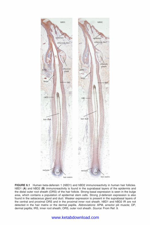
DK4962-Webster-ColorInsert_R2_020407
DP DPmelanin melanin
hair matrixhair matrix
hair shaft
epidermisepidermis
sebaceous duct sebaceous duct
sebaceouglands
sebaceousgland
IRSIRS
ORS ORS
hBD1 hBD2
bulge
bulge
APMAPM
hair shaft
FIGURE 6.1 Human beta-defensin 1 (hBD1) and hBD2 immunoreactivity in human hair follicles.hBD1 (A) and hBD2 (B) immunoreactivity is found in the suprabasal layers of the epidermis andthe distal outer root sheath (ORS) of the hair follicle. Strong basal expression is seen in the bulgearea, which contains a population of epidermal stem cells. Strong b-defensin expression is alsofound in the sebaceous gland and duct. Weaker expression is present in the suprabasal layers ofthe central and proximal ORS and in the proximal inner root sheath. hBD1 and hBD2 IR are notdetected in the hair matrix or the dermal papilla. Abbreviations: APM, arrector pili muscle; DP,dermal papilla; IRS, inner root sheath; ORS, outer root sheath. Source: From Ref. 9.
www.ketabdownload.com
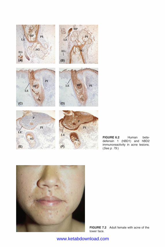
DK4962-Webster-ColorInsert_R2_020407
(A) (B)
(C)
(E)
SG SG
HP
HP
HP HP
PP
PI
PI
PI PI
PI PILE LE
LE
LE
LELE
SG SG
EG EG
(D)
(F)
FIGURE 6.2 Human beta-defensin 1 (hBD1) and hBD2immunoreactivity in acne lesions.(See p. 79.)
FIGURE 7.2 Adult female with acne of thelower face.
www.ketabdownload.com
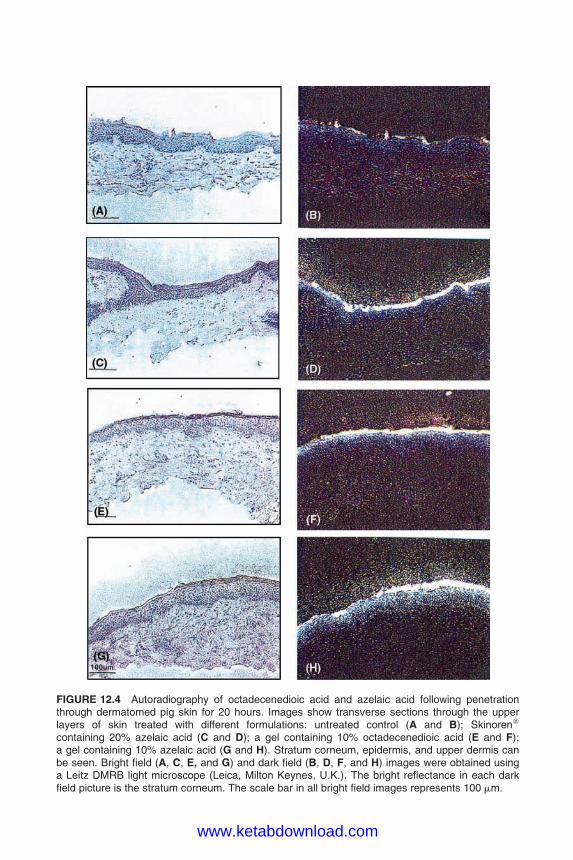
DK4962-Webster-ColorInsert_R2_020407
FIGURE 12.4 Autoradiography of octadecenedioic acid and azelaic acid following penetrationthrough dermatomed pig skin for 20 hours. Images show transverse sections through the upperlayers of skin treated with different formulations: untreated control (A and B); Skinorenw
containing 20% azelaic acid (C and D); a gel containing 10% octadecenedioic acid (E and F);a gel containing 10% azelaic acid (G and H). Stratum corneum, epidermis, and upper dermis canbe seen. Bright field (A, C, E, and G) and dark field (B, D, F, and H) images were obtained usinga Leitz DMRB light microscope (Leica, Milton Keynes, U.K.). The bright reflectance in each darkfield picture is the stratum corneum. The scale bar in all bright field images represents 100 mm.
www.ketabdownload.com
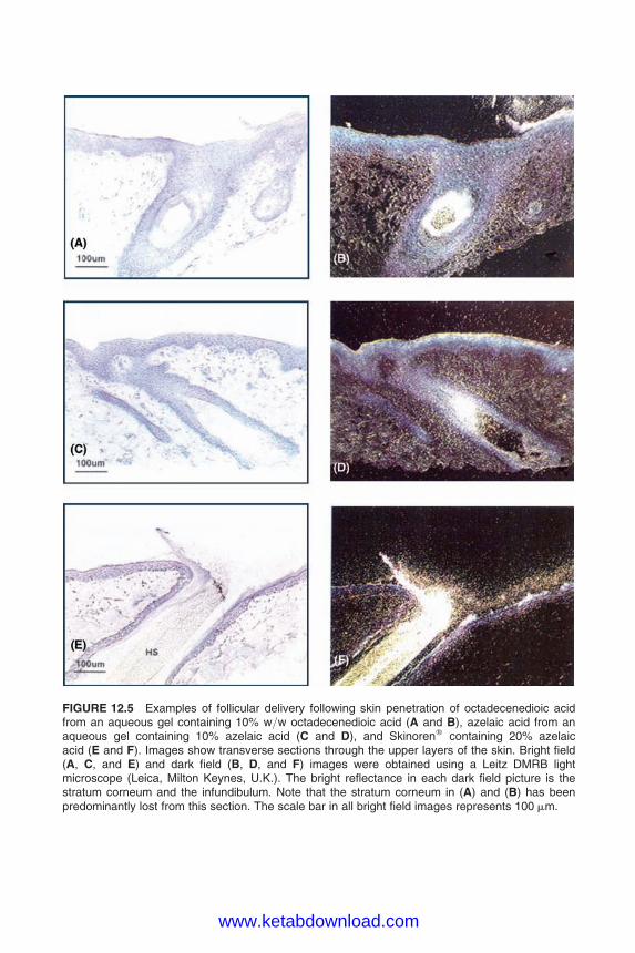
DK4962-Webster-ColorInsert_R2_020407
FIGURE 12.5 Examples of follicular delivery following skin penetration of octadecenedioic acidfrom an aqueous gel containing 10% w/w octadecenedioic acid (A and B), azelaic acid from anaqueous gel containing 10% azelaic acid (C and D), and Skinorenw containing 20% azelaicacid (E and F). Images show transverse sections through the upper layers of the skin. Bright field(A, C, and E) and dark field (B, D, and F) images were obtained using a Leitz DMRB lightmicroscope (Leica, Milton Keynes, U.K.). The bright reflectance in each dark field picture is thestratum corneum and the infundibulum. Note that the stratum corneum in (A) and (B) has beenpredominantly lost from this section. The scale bar in all bright field images represents 100 mm.
www.ketabdownload.com
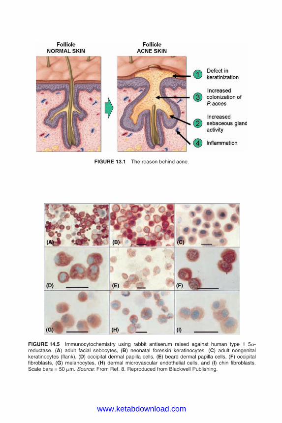
DK4962-Webster-ColorInsert_R2_020407
FIGURE 14.5 Immunocytochemistry using rabbit antiserum raised against human type 1 5a-reductase. (A) adult facial sebocytes, (B) neonatal foreskin keratinocytes, (C) adult nongenitalkeratinocytes (flank), (D) occipital dermal papilla cells, (E) beard dermal papilla cells, (F) occipitalfibroblasts, (G) melanocytes, (H) dermal microvascular endothelial cells, and (I) chin fibroblasts.Scale bars = 50 mm. Source: From Ref. 8. Reproduced from Blackwell Publishing.
FIGURE 13.1 The reason behind acne.
www.ketabdownload.com

DK4962-Webster-ColorInsert_R2_020407
FIGURE 14.8 Immunolocalization oftype 1 5a-reductase in comedones(hematoxylin and eosin). (See p. 179.)
FIGURE 14.9 Immunolocalization oftype 2 5a-reductase in comedones.(See p. 180.)
www.ketabdownload.com
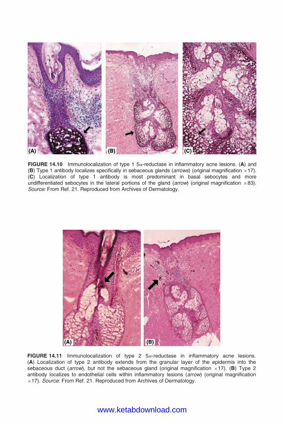
DK4962-Webster-ColorInsert_R2_020407
FIGURE 14.11 Immunolocalization of type 2 5a-reductase in inflammatory acne lesions.(A) Localization of type 2 antibody extends from the granular layer of the epidermis into thesebaceous duct (arrow), but not the sebaceous gland (original magnification �17). (B) Type 2antibody localizes to endothelial cells within inflammatory lesions (arrow) (original magnification�17). Source: From Ref. 21. Reproduced from Archives of Dermatology.
FIGURE 14.10 Immunolocalization of type 1 5a-reductase in inflammatory acne lesions. (A) and(B) Type 1 antibody localizes specifically in sebaceous glands (arrows) (original magnification �17).(C) Localization of type 1 antibody is most predominant in basal sebocytes and moreundifferentiated sebocytes in the lateral portions of the gland (arrow) (original magnification �83).Source: From Ref. 21. Reproduced from Archives of Dermatology.
www.ketabdownload.com
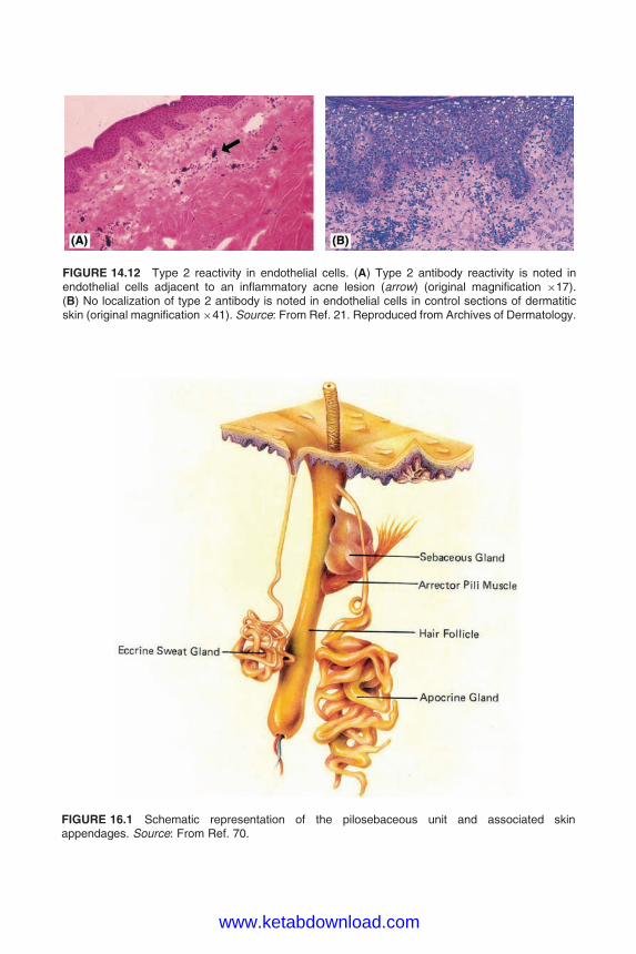
DK4962-Webster-ColorInsert_R2_020407
FIGURE 14.12 Type 2 reactivity in endothelial cells. (A) Type 2 antibody reactivity is noted inendothelial cells adjacent to an inflammatory acne lesion (arrow) (original magnification �17).(B) No localization of type 2 antibody is noted in endothelial cells in control sections of dermatiticskin (original magnification�41). Source: From Ref. 21. Reproduced from Archives of Dermatology.
FIGURE 16.1 Schematic representation of the pilosebaceous unit and associated skinappendages. Source: From Ref. 70.
www.ketabdownload.com
www.ketabdownload.com