Embed Size (px)
Citation preview

This article was downloaded by [Tulane University]On 10 September 2013 At 1234Publisher Taylor amp FrancisInforma Ltd Registered in England and Wales Registered Number 1072954 Registered office Mortimer House37-41 Mortimer Street London W1T 3JH UK
Journal of Vertebrate PaleontologyPublication details including instructions for authors and subscription informationhttpwwwtandfonlinecomloiujvp20
Acoustic transformer function of the postdentary bonesand quadrate of a nonmammalian cynodontTom S Kemp aa University Museum of Natural History and St Johns College Oxford University OxfordOX1 3PW UK E-mailPublished online 02 Aug 2010
To cite this article Tom S Kemp (2007) Acoustic transformer function of the postdentary bonesand quadrate of a nonmammalian cynodont Journal of Vertebrate Paleontology 272 431-441 DOI1016710272-4634(2007)27[431ATFOTP]20CO2
To link to this article httpdxdoiorg1016710272-4634(2007)27[431ATFOTP]20CO2
PLEASE SCROLL DOWN FOR ARTICLE
Taylor amp Francis makes every effort to ensure the accuracy of all the information (the ldquoContentrdquo) containedin the publications on our platform However Taylor amp Francis our agents and our licensors make norepresentations or warranties whatsoever as to the accuracy completeness or suitability for any purpose of theContent Any opinions and views expressed in this publication are the opinions and views of the authors andare not the views of or endorsed by Taylor amp Francis The accuracy of the Content should not be relied upon andshould be independently verified with primary sources of information Taylor and Francis shall not be liable forany losses actions claims proceedings demands costs expenses damages and other liabilities whatsoeveror howsoever caused arising directly or indirectly in connection with in relation to or arising out of the use ofthe Content
This article may be used for research teaching and private study purposes Any substantial or systematicreproduction redistribution reselling loan sub-licensing systematic supply or distribution in anyform to anyone is expressly forbidden Terms amp Conditions of access and use can be found at httpwwwtandfonlinecompageterms-and-conditions
ARTICLE
ACOUSTIC TRANSFORMER FUNCTION OF THE POSTDENTARY BONES AND QUADRATEOF A NONMAMMALIAN CYNODONT
TOM S KEMPUniversity Museum of Natural History and St Johnrsquos College Oxford University Oxford OX1 3PW UK
tomkempoumoxacuk
ABSTRACTmdashThe theory that the reduced postdentary bones and quadrate of non-mammalian cyndonts were not onlythe morphological homologues but also the functional equivalents of the mammalian tympanic bone and ear ossicles istested on the basis of detailed new information of a specimen of Chiniquodon The anatomy is shown to be a compromisebetween the respective requirements for a persistent though reduced stress transmission function of a jaw articulationand an acoustic transformation function of a middle ear There was a sound pressure level transformer ratio of about 30but the mass and compliances of the elements restricted sensitivity to low frequencies up to perhaps 2 kHz Neither anair-filled tympanic cavity nor a dedicated tympanic membrane were present and snakes and other modern reptileslacking a tympanic cavity offer a better mechanical analogy than mammals for the ear function of a cynodont The fullymammalian acoustic transformer system with tympanic cavity and tympanic membrane could only have evolved after theorigin of the dentary-squamosal jaw articulation and was correlated with miniaturisation in the lineage leading to basalmammaliaforms
INTRODUCTIONThe most celebrated case of the discovery of a wholly unex-
pected homology is surely that of the accessory ear ossicles ofmammals with the jaw hinge bones of reptiles a theory con-ceived by Reichert in 1837 and subsequently confirmed andelaborated upon by Gaupp (1913 see Maier 1990 for a briefreview) With the exception of Jarvik (1980) no serious workerhas since doubted the relationship
With the discovery of reduced jaw hinge bones in the non-mammalian cynodonts attention turned to how the implied evo-lutionary transition occurred Early work by Westoll (1945) Par-rington (1946 1949) Watson (1953 1956) and Tumarkin (1955)defined the basic question of whether cynodonts lacked a tym-panic membrane possessed one in the modern reptilianpostquadrate position behind the jaw articulation or possessedone in the homologue of the mammalian position attached to thepostdentary bones of the lower jaw The culmination of the viewthat they possessed a postquadrate tympanic membrane wasreached by Hopson (1966) who showed how such a tympanicmembrane might have grown forward and become associatedwith the posterior jaw bones
Allin (1975) presented the culmination of the alternative viewthat cynodonts had a tympanic cavity and tympanic membraneassociated with the lower jaw and that the hearing mechanismwas essentially mammalian already with sound conducted fromthis tympanic membrane via the articular and quadrate to thestapes and fenestra ovalis He proposed that the transition to themodern mammalian condition consisted only of improving theperformance of the system by reducing the mass of the bones andeventually freeing them of the damping effect of their attach-ment to the dentary Allinrsquos theory was soon accepted (egCrompton and Parker 1978 Kemp 1979 Kermack et al 1981)although there was a lingering suspicion on the part of some thata postquadrate tympanic membrane may have existed in addi-tion to the mandibular tympanic membrane (Kemp 1979 Allin1986 Allin and Hopson 1992)
The earlier demonstration by Parrington (1971) and Kermack
and colleagues (1973) that the mammaliaform Morganucodonpossessed a cynodont-like arrangement of the postdentary bonesand quadrate had not only added considerably to the argumentin favor of Allinrsquos (1975) theory on comparative anatomicalgrounds but also offered an opportunity for a functional analysisof how such a sound conducting system might have operatedKermack and colleagues (1981) concluded that Morganucodondid indeed hear by means of a mandibular tympanic membranecoupled to articular quadrate and stapes but that reception waslimited to low frequencies up to about 1kHz In contrast Ro-sowski and Graybeal (1991 Rosowski 1992) estimated valuesfor area of the tympanic membrane area of the stapes footplateand length of the basilar membrane of the cochlea in Morganu-codon from which they concluded that its hearing resembledthat of small modern mammals with sensitivity to frequencies ofover 10 kHz
A simple extrapolation of the mechanism of hearing in mam-mals even Morganucodon to a non-mammalian cynodont is ofdoubtful validity because the latter did not possess the secondaryjaw articulation between the dentary and squamosal Thereforethe postdentary bones and quadrate must have retained theirhinge function and at least a reduced stress transmission func-tion whether or not an auditory function was present The pre-sent paper considers the possible role of the postdentary bonesand quadrate as an acoustic impedance matching system in thenon-mammalian eucynodont genus Chiniquodon based on newinformation from a particularly well-preserved and completelyprepared specimen
Institutional AbbreviationsmdashGPIT Institut und Museum fuumlrGeologie und Palaumlontologie der Universitaumlt Tuumlbingen MCZThe Museum of Comparative Zoology Harvard NHM TheNatural History Museum London
MATERIAL
The main subject of this study is the almost complete skullNHM R8430 It was prepared by bisecting it just to the side of
Journal of Vertebrate Paleontology 27(2)431ndash441 June 2007copy 2007 by the Society of Vertebrate Paleontology
431
Dow
nloa
ded
by [
Tul
ane
Uni
vers
ity]
at 1
234
10
Sept
embe
r 20
13
the sagittal plane with a lsquoLastecrsquo diamond wire saw and thematrix then completely removed mechanically from the internaland external regions The posterior half of both lower jaws weredetached the right side was separated cleanly between articularand quadrate and the postdentary rod posterior to the dentarysubsequently detached The left side separated equally cleanlybetween quadrate and squamosal Neither quadratojugal nor sta-pes is present but the middle ear region of the left side is verywell preserved
A second specimen is an isolated fragment consisting of theleft postdentary bones lacking the angular MCZ 4002 Littlefurther preparation was necessary
SYSTEMATIC PALEONTOLOGY
CYNODONTIA Owen 1861EUCYNODONTIA Kemp 1982
PROBAINOGNATHIA Hopson 1990CHINIQUODONTIDAE von Huene 1935-42
CHINIQUODON THEOTENICUS von Huene 1935-42
HolotypemdashPartial skull GPIT 1050Type Locality and AgemdashChiniqua Rio Grande do Sul State
Brazil Ladinian Middle TriassicReferred MaterialmdashA complete skull NMH R8430 from the
Chantildeares River Campo de Talapaya La Rioja Province Argen-tina This specimen was originally referred to as Probelesodonlewisi Romer but Abdala and Giannini (2002) have revised thefamily Chiniquodontidae and shown that P lewisi is a juniorsynonym of Chiniquodon theotenicus A second specimen is anisolated fragment consisting of the left postdentary bones lackingthe angular It is part of a disarticulated skeleton of Chiniquodontheotenicus No MCZ 4002 Romer and Lewis (1973) describedthe postcranial skeleton but did not mention this fragment of thejaw
DESCRIPTION
The postdentary rod (Fig 1) consists of the surangular angu-lar prearticular and articular forming an integral unit with noevidence of possible movement between any of the individualbones Although sutures are present and therefore the bones arenot fused to one another there has not been any post-mortemdisarticulation at all This is in marked contrast to the relation-ship of the postdentary rod as a whole to the dentary where theappearance of a gap indicates that the connection had only beenby soft tissues rather than by tight suture and where the post-dentary rod has indeed been somewhat displaced after deathThe isolated fragment MCZ 4002 confirms this interpretationbecause loss of the angular has revealed the ridged and groovedsutural surfaces on the more medial bones that are characteristicof immoveable sutural connections (Fig 1I)
SurangularmdashThe surangular constitutes the dorsal surface ofthe postdentary rod At the posterior end it expands dorsally andlaterally like an asymmetrical trumpet horn The saddle-shapedsurface so created (Fig 1CndashH bear) fits closely against the un-derside of the hindmost 05-1 cm of the postdentary trough of the
dentary The lateral part of the expansion is the surangular boss(sa boss) of Crompton (1972) In posterior view (Fig 1G and H)the surangular is seen to consist of three surfaces The dorsal oneis concave faces posteriorly and slightly dorsally and bears finestriations radiating toward the margins The second surface(salig) is on the posterolateral side of the bone and has a finelyrugose surface indicating a ligamentous or tendinous attachmentIt is positioned close to but not in direct contact with the squa-mosal and as discussed later is interpreted as a region of con-nective tissue attachment of the surangular to the squamosalThe third surangular surface is ventral (sagl) and is a lateralcontinuation of the glenoid of the articular bone Like the latterit lacks a periosteal finish and has the fine texturing characteristicof bone covered by synovial joint cartilage
The medial surface of the surangular (Fig 1B and C) is ex-posed above the prearticular in the form of a horizontal troughthat is limited ventrally by a sharp edge that presumably marksthe upper limit of Meckelrsquos cartilage It ends anteriorly as a smallforwardly directed point The isolated postdentary rod of MCZ4002 (Fig1I) is missing the angular so the lateral face of thesurangular is exposed as a thin vertical sheet extending as far asthe ventral margin of the postdentary rod It is slightly concavefrom top to bottom and as evident from NMH R8430 its lateralface was tightly sutured to the thin sheet-like angular bone
As it extends forward internal to the dentary the surangularbecomes a narrow rod free of any direct contact with otherbones and shortly terminates Its dorsal surface (Fig 1D) ante-rior to the bearing surface is finely sculptured into striationscharacteristic of bone covered only by tight periosteum Thesecommence along the posterolateral edge of the bone run an-teromedially then turn to run longitudinally for the remaininglength of the bone
AngularmdashThe angular is a thin vertical sheet of bone form-ing most of the lateral surface of the postdentary rod and con-tinued forward as a ventrally positioned rod within the postden-tary trough of the dentary A fine very sharp ridge (Fig 1Eangri) lies close to the upper margin and turns ventrally parallelto the hind edge of the dentary The lateral surface of the angu-lar as bounded by this ridge above and by the rounded ventralmargin of the bone below is perfectly smooth and lacks stria-tions or rugosities The limited exposure of the medial face be-tween the surangular and the prearticular is also smooth andfeatureless
As it extends forward medial to the dentary the angulargradually changes in cross-sectional shape from a vertical sheetto a hemicylindrical rod with a broad trough occupying the uppersurface This is seen in the transverse section (Fig 1A) where itlies in the semicircular dentary trough The latter is much greaterin diameter than the angular rod and so the two must have beenseparated by a significant space occupied in life by soft tissues
Only the damaged root of the small reflected lamina of theangular (refllam) is preserved (Fig 1B) It consists of three orfour short finger-like fragments of bone suggesting that origi-nally it was corrugated
PrearticularmdashThe ventral surface of the prearticular (Fig 1F)expands posteriorly and sheaths the medial and ventral faces of
rarr
FIGURE 1 The right postdentary rod of Chiniquodon theotonicus NHM R8430 A transverse section as exposed in the posterior section of theright jaw B stereophotograph of the posterior section of the right jaw in medial view showing the postdentary rod as slightly displaced from thedentary trough C the right postdentary rod in medial view D stereophotograph and interpretive diagram of the right postdentary rod in dorsal viewE the same in lateral view F the same in ventral view G stereophotograph of the right postdentary rod and dentary in posterior view Hinterpretive diagram of the right postdentary rod and dentary trough in posterior view I Fragment of left postdentary rod of MCZ 4004 in lateralview Abbreviations ANG angular angri angular ridge angsu sutural surface for angular ART articular artgl glenoid surface of articularbear bearing surface of surangular against the dentary COR coronoid D dentary PRART prearticular refllam reflected lamina of the angularrartpr retroarticular process SA surangular saboss surangular boss sagl glenoid surface of surangular salig ligamentous attachment ofsurangular to squamosal sari surangular ridge SPL splenial Scale bars on the photographs equal 1 cm
JOURNAL OF VERTEBRATE PALEONTOLOGY VOL 27 NO 2 2007432
Dow
nloa
ded
by [
Tul
ane
Uni
vers
ity]
at 1
234
10
Sept
embe
r 20
13
KEMPmdashCYNODONT SOUND RECEPTION 433
Dow
nloa
ded
by [
Tul
ane
Uni
vers
ity]
at 1
234
10
Sept
embe
r 20
13
the articular bone The surface of the bone lacks any markingsthat might indicate the attachment of muscle or tendon Anteri-orly the prearticular attenuates to a slender rod extending for-ward and getting finer and finer By the time it is exposed in thetransverse section (Fig 1A) the diameter is less than 1 mm andit is well separated from the angular
Specimen MCZ 4002 reveals that the hind part of the prear-ticular curves ventrally and contributes much of the retroarticu-lar process ((Fig 1I rartpr) This specimen also shows thegrooved sutural surface (angsut) to which the angular wasfirmly attached
ArticularmdashThe articulating surface (artgl) of the articularbone is continuous with that of the surangular (sagl) Togetherthey form an approximately transversely aligned trough with amid-dorsal extension the whole facing backward (Fig 1G andH) The capping surangular boss (saboss) contributes a small butdefinite lip bounding the dorsal edge of the glenoid Due to astrip of damaged bone the ventral edge of the glenoid of speci-men NMH R8430 is not visible but MCZ 4002 is undamaged inthis region and possesses a slight lip separating the articulatingsurface of the articular from the retroarticular process below
The retroarticular process (rartpr) is also best preserved inthe latter specimen (Fig 1I) It is transversely compressed andcurves ventrally forward in the plane of the angular bone Theventral edge is thickened creating a curving edge while theanterior edge is fairly sharp
The only part of the articular bone anterior to the articulatingsurface that is exposed is on the medial side of the postdentaryrod (Fig 1B and C) Here it is seen to be tightly wedged betweenthe surangular above and the prearticular below It lacks a peri-osteal finish and was presumably continued forward as a persis-tent Meckelrsquos cartilage
Postdentary Trough of the DentarymdashAt the level of thetransverse section (Fig 1A) the postdentary trough of the den-tary is semicircular in cross-section and within it lies the anteriorextension of the angular bone as described above As it extendsbackward the lower edge of the trough remains horizontal allthe way to the hind border of the dentary The upper edgehowever rises gradually and then more steeply By its posteri-ormost region the trough has become effectively a transverselyconcave roof over the postdentary rod (Fig 1B) The diameter ofthe concavity matches the transverse convexity of the pos-terodorsal surface of the surangular (Fig 1H bear) and it isonly at this point that the two bones appear to have made directosteological contact with one another in life
QuadratemdashThe quadrate (Fig 2) is generally similar to that ofMassetognathus and Probainognathus as described by Luo andCrompton (1994)
The front face of the quadrate is virtually flat and featureless(Fig 2E and F) In shape it attenuates asymmetrically dorsallyending in a blunt apex toward the lateral side There is a promi-nent posterior process (Fig 2C and D pprq) whose lateral face(lsq) abuts against the delicate vertical process of the squamosalthat separates the quadrate recess from the narrow parasagittalslit (Fig 2E and F qjresq) that housed the missing quadrato-jugal The medial face of the posterior process of the quadrate(msq) fits conformably against the quadrate recess of the squa-mosal plus its antero-medial extension so that the quadrate isstrongly supported postero-medially There is a fine verticalgroove (Fig 2E and F qracon) close to the antero-medial edgewhich marks the point where the quadrate ramus of the ptery-goid made contact (Fig 2C) The actual contact is visible on theleft side of the skull (Fig 3D) The contact surface of the quad-rate for the stapes is not well-preserved its position is assumedto be a damaged roughened area close to the medial terminationof the condyle (Fig 2D sta)
The form of the condylar surface of the quadrate (Fig 2C)closely matches that of the articular-surangular glenoid surface
It faces anteriorly to anteroventrally and is hemicylindrical inform its central region is extended dorsally above the level of themedial and lateral parts The condyle extends laterally to thebody of the quadrate where its dorsal surface would have sup-ported the quadratojugal
SquamosalmdashThe left quadrate is slightly displaced from itsrecess in the squamosal (Fig 2E and F) revealing the latter tocorrespond in shape to the posterior face of the body of thequadrate The roof of the recess is demarcated by a sharp edgethat would approximate the dorsal margin of the quadrate if thelatter were in its natural position The recess is extended antero-medially as a vertical wing that caps the end of the paroccipitalprocess and is overlapped laterally by the slender quadrate ra-mus of the pterygoid (Fig 3C and D qrapt)
Lateral to the narrow recess for the quadratojugal (qjresq)the squamosal curves anterolaterally and extends ventrally (Fig2E and F) This region forms the articular flange of the squamo-sal (artflsq) which lies anterolateral to and at a somewhathigher level than the quadrate condyles It faces toward the sur-angular boss but the two are separated by a few millimeters Thesurface of the articular flange is marked by fine striations thatindicate the attachment of collagenous tissue such as ligament ortendon and not articular cartilage
Fenestra OvalismdashThe fenestra ovalis is perfectly preserved onthe left side of the skull (Fig 3C) where it faces directly laterallytoward the quadrate In shape the fenestra is indeed oval withthe major axis inclined anterodorsally to posterior-ventrally Itsapproximate dimensions are 5 mm by 3 mm giving a surface areaof about 12 mm2 A thickened rim around the fenestra presum-ably supported an annular ligament for attachment of the foot-plate of the stapes Neither stapes is preserved in the specimen
FUNCTIONAL INTERPRETATION
Notwithstanding the seductive morphological homologies andthe eventual evolution of the mammalian acoustic transformersystem from them the postdentary bones and quadrate of aeucynodont cannot have functioned in a manner closely similarto that found in modern mammals In the absence of a second-ary dentary-squamosal articulation these bones were substan-tially larger both relatively and absolutely because they hadnecessarily retained a significant mandibular function of stresstransmission and control of jaw movements The question to beaddressed is the extent to which the structure can be interpretedas a compromise between the requirements for the two respec-tive functions stress transmission and acoustic transformation
Mandibular Function
The demonstration that the geometrical reorganization of theadductor musculature of eucynodonts greatly reduced the reac-tion forces at the jaw articulation is of critical importance forunderstanding the function of the postdentary rod and quadrate(Crompton 1963 Kemp 1972 Bramble 1978 Kemp 1980Crompton and Hylander 1986) However in the absence of adirect contact between the dentary and the squamosal it is in-evitable that at times stresses would have been generated be-tween the dentary and cranium that had to be transmitted via thejaw articulation
By inference this would have been particularly the case duringjaw opening although the anatomy of the jaw opening muscu-lature of cynodonts is not well understood The retroarticularprocess of the articular (Fig 3A and B rartpr) is far too smalland feebly attached via the postdentary rod to the dentary for itto be considered the point of insertion of a large standard am-niote depressor mandibuli muscle Kemp (1979 1980) inter-preted narrow grooves on the medial side of the angular processof the dentary as the major site of insertion of jaw openingmusculature in both the basal cynodont Procynosuchus and the
JOURNAL OF VERTEBRATE PALEONTOLOGY VOL 27 NO 2 2007434
Dow
nloa
ded
by [
Tul
ane
Uni
vers
ity]
at 1
234
10
Sept
embe
r 20
13
eucynodont Luangwa This presumed muscle complex can onlyhave been associated with the fascia of the intermandibular andthroat regions perhaps acting via a hyoid To open the jaws theforce generated would have had a posteriorly directed compo-
nent that would have generated an anteroposterior compressivereaction at the dentary-postdentary articular-quadrate andquadrate-squamosal contacts At times of vigorous jaw activitythe magnitude of this stress would necessarily have been signifi-
FIGURE 2 The left quadrate and adjacent structures of Chiniquodon theotonicus NMH R8430 A stereophotograph of the left quadrate and hindend of the jaw in dorsal view B interpretive diagram of A C stereophotograph of the left quadrate and hind end of the lower jaw in posterior viewD interpretive diagram of C E stereophotograph of the right quadrate and squamosal in anterior view F interpretive diagram of E AbbreviationsART articular artflsq articular flange of the squamosal D dentary lsq lateral contact surface with squamosal msq medial contact surface withsquamosal pprq posterior process of quadrate PT pterygoid Q quadrate qcond quadrate condyle qjresq quadratojugal recess of thesquamosal rartpr retroarticular process SA surangular saboss surangular boss salig area of attachment of surangular-squamosal ligament SQsquamosal sta contact point for stapes Scale bars on photographs equal 1 cm
KEMPmdashCYNODONT SOUND RECEPTION 435
Dow
nloa
ded
by [
Tul
ane
Uni
vers
ity]
at 1
234
10
Sept
embe
r 20
13
cant Other circumstances generating a hinge reaction of somemagnitude must have included sudden changes in the stress pat-tern due to rapid and unexpected changes in the resistance to jawmovements when food between the teeth yielded and in the caseof a presumed predator such as Chiniquodon struggling preytending to disarticulate the mandible
The nature of the contacts between the dentary and the post-dentary rod supports the interpretation that modest compressionwas the main stress to be resisted here At the front there is themanifestly loose association between the rodlike anterior exten-sion of the angular and the much broader dentary trough Thesoft tissue filling the considerable space between them can have
provided little of the strength or stiffness required to resiststresses of any kind However the saddle-shaped posterior con-tact between the bearing surface of the surangular and the hindend of the dentary trough (Fig 3D bear) would have providedresistance to a longitudinal compressive force between them andalso perhaps a degree of dorso-ventral compression Little resis-tance would have been offered to any longitudinal tension tend-ing to pull the dentary forward The orientation of the articulat-ing surfaces of the mandibular glenoid and the quadrate condylein an approximately transverse plane is also clearly designed fortransmission of a mainly longitudinally oriented compressivestress Similarly the direct osteological support of the quadrate
FIGURE 3 Reconstruction of the lower jaw of Chiniquodon in A lateral view showing reconstructed putative tympanic membrane B medial viewshowing the postdentary rod cross-hatched and the position of the hypothetical axis of vibration C stereophotograph of the left half of the skull inventral and slightly lateral view D reconstruction of the left ear region in ventral view Abbreviations angprd angular process of the dentary BSPbasisphenoid COR coronoid extaume alleged groove for external auditory meatus fenov fenestra ovalis jugf jugular foramen Q quadrateqrapt quadrate ramus of the pterygoid rartpr retroarticular process refllam reflected lamina of the angular saboss surangular boss saligarea of attachment of surangular-squamosal ligament SQ squamosal SPL splenial STA stapes tympm tympanic membrane Scale bars equal2 cm
JOURNAL OF VERTEBRATE PALEONTOLOGY VOL 27 NO 2 2007436
Dow
nloa
ded
by [
Tul
ane
Uni
vers
ity]
at 1
234
10
Sept
embe
r 20
13
behind and medially by the squamosal recess appears to be de-signed primarily for resistance to such compression betweenthese elements
The relationship between the surangular boss and the articularflange of the squamosal is problematic Crompton (1972 Luoand Crompton 1994) ascribed to it the function of reducing theloading on the quadrate and Allin (1975) specifically suggestedthat it reduced the compressive load on that bone An importantpart of the reasoning was that it is the forerunner of the dentary-squamosal jaw articulation of mammals which in primitive formsdemonstrably does have a significant compression resisting func-tion (Crompton and Hylander 1986) In Chiniquodon howeverthere is no direct contact between the two bones the squamosalflange lies several millimeters posterolateral to the surangularboss Furthermore the surfaces of both are marked by fine ru-gosities indicating connective tissue attachment rather than thecartilage covering expected of a synovial joint It must have beeneither elastic ligament or muscle that spanned the gap becausethe distance between the flange and the boss would have variedwith angle of gape The only stress that might have been effec-tively transmitted by this arrangement is tension an interpreta-tion incongruent with the evidence for lack of significant tensileresistance between the postdentary rod and the dentary An-other function must be sought as discussed later
Thus it is concluded that the postdentary rod and quadrate hadgreatly reduced stress resistance as predicted from their reducedsize and the familiar reorganization of the cynodont adductorjaw musculature Nevertheless they retained a mechanical func-tion associated with mandibular action primarily in the form ofoffering significant resistance to a longitudinally oriented com-pression stress particularly in relation to the forces generated injaw opening but also at other times when brief stresses weregenerated at the hinge
Acoustic Function
For the postdentary rod and quadrate to function in essence asa mammalian acoustic transformer it would require a combina-tion of acoustic isolation which is defined as a low percentage oftransfer of energy from a vibrating element to an attached ele-ment and acoustic linkage which is defined as a high percentagetransfer of energy from a vibrating element to an attached ele-ment in the following pattern (i) acoustic isolation of the post-dentary rod from the dentary (ii) acoustic linkage between thearticular (malleus) and the quadrate (incus) (iii) acoustic isola-tion of the quadrate (incus) from the cranium (iv) acoustic link-age between the quadrate (incus) and the stapes There must alsobe (v) significant impedance matching by means of a transformerpressure ratio between the air-borne sound receiving surface andthe fenestra ovalis (vi) masses and compliances of the elementswithin acceptable limits
(i) Acoustic Isolation of the Postdentary RodmdashThe anteriorsupport of the postdentary rod consists of the rodlike anteriorsection of the angular lying within the larger diameter hemicy-lindrical dentary trough The presumed loose connective tissuebetween them would have offered insignificant constraint to ro-tational vibration about a longitudinal axis (Fig3Baxis) Theonly other attachment of the postdentary rod to the dentary isthe contact between the posterior end of the dorsal surface of thesurangular and the posteriormost part of the roof of the dentarytrough (Fig 3A bear) Both are approximately semicircular intransverse section and their surfaces are slightly rugose indicat-ing a syndesmotic attachment Assuming a non-restraining col-lagenous connection between them this arrangement would alsohave permitted angular vibration of the postdentary rod about alongitudinal axis at a level immediately below the contact
The loose syndesmotic connection between the surangularboss and the articular flange of the squamosal lies approximately
along the line of the axis as defined by the two attachment pointsof the postdentary rod to the dentary It is therefore positionedin the predictable place to act as a non-rigid suspension point forthe vibrating postdentary rod that is independent of the dentary(Fig 3Asalig) This is likely to have been the primary if not thesole function of the otherwise difficult to explain relationshipbetween the surangular and the squamosal in cynodonts
These three contacts therefore define a potentially vibratingpostdentary rod that is acoustically isolated from the jaw andcranium
(ii) Acoustic Linkage Between Articular and QuadratemdashWhen the lower jaw is placed in the closed position the poste-rior-facing glenoid receives the matching hemicylindrical quad-rate condyle along its transverse width and the slight supragle-noid and subglenoid ridges tighten the contact Even under theconstraint that the articular and quadrate acted as a jaw articu-lation with this geometrical arrangement vibration of the articu-lar about a longitudinal axis would have been efficiently trans-mitted as vibration of the quadrate also about a longitudinal axis
(iii) Acoustic Isolation of the QuadratemdashKemp (1979) forthe more basal cynodont Procynosuchus and Luo and Cromp-ton (1994) for the more progressive cynodont Probainognathusconcluded that the quadrate was syndesmotically rather thansuturally attached to the squamosal and that rotatational vibra-tion about a longitudinal axis running through the point of con-tact between the medial edge of the bone and the quadrate ra-mus of the pterygoid would have been relatively unconstrainedThe same is evidently the case here for Chiniquodon Neitherquadratojugal is preserved in this specimen but if it were similarto that of Probainognathus it would have been free to movedorso-ventrally in its narrow recess in the squamosal (Fig 2Fqjresq) and therefore would not have restricted the proposedvibration of the quadrate to which it was attached
(iv) Acoustic Linkage Between the Quadrate and theStapesmdashNeither stapes is preserved in this specimen but itsform is fairly constant among cynodonts (Fig 3D) It would havecontacted the medial side of the quadrate at or slightly above thelevel of the condyle a more dorsal contact would not have beenpossible because of the limit imposed by the horizontal level ofthe quadrate ramus of the pterygoid This proposed orientationof the stapes is supported by the fact that the fenestra ovalis facesdirectly toward the lower part of the quadrate The vibration ofthe quadrate about a longitudinal axis was transformed into apiston action of the stapes which vibrated directly in and out ofthe fenestra ovalis
(v) Impedance MatchingmdashThe amniote middle ear acts as animpedance matching system by transforming the low sound pres-sure of the airborne sound waves meeting the tympanic mem-brane (PT) into higher sound pressure waves (PFO) at the stapesfootplate where they enter the aqueous medium of the cochlea(for description of middle ear function see for example Moslashller1974 Relkin 1988 Rosowski 1994 1996) The principal meansof achieving this in modern mammals is referred to as the lsquoidealtransformerrsquo which consists of two linked acoustic transformersthe ratio of tympanic membrane area (AT) to fenestra ovalisarea (AFO) and the ratio of the effective lever arm of the ma-nubrium of the malleus (LM) to the length of the crus brevis ofthe incus (LI) such that
PFOPT = ATAFO LMLI gtgt 1
Following Allin (1975) the reconstruction of airborne sound re-ceiving soft tissue covering the lateral surface of the postdentaryrod and acting as a tympanic membrane (Fig 3A) assumes thatits boundary is marked by the ridge along the anterior and dorsalparts of the exposed angular the curved edge of the retroarticu-lar process and a ventral margin supported at the front by thereduced reflected lamina of the angular As such it has a surfacearea of about 240 mm2 The area of the fenestra ovalis is about
KEMPmdashCYNODONT SOUND RECEPTION 437
Dow
nloa
ded
by [
Tul
ane
Uni
vers
ity]
at 1
234
10
Sept
embe
r 20
13
12 mm2 This gives an area ratio ATAFO of about 20 which iswell within the modern mammalian range of 10ndash40 (Rosowskiand Graybeal 1991 Rosowski 1992) The length of the leverarm of the postdentary rod is taken to be the distance between itsinferred axis and a point close to the ventral margin (Fig 3B)This gives a figure for LM of about 12 mm The lever arm of thequadrate LI is taken to be the distance from the presumed axisof rotation of the postdentary rod to the point of attachment ofthe stapes It has a value of about 8 mm The lever arm ratioLMLI is therefore around 15 which again is comfortably withinthe modern mammal range Rosowski (1996) quotes figures of25 for the cat and 12 for the guinea pig and human middle earsTaken together the two ratios therefore represent an ideal trans-former ratio of 30
Most modern amniotes have at least one additional device toincrease the transformer ratio The catenary effect in modernmammals results from different amplitudes of vibration andtherefore different pressures at different parts of the tympanicmembrane and increases the ratio approximately twofold(Khanna and Tonndorf 1972 Rosowski 1996) However it canonly apply to a flexible tympanic membrane not a stiff unit likethe postdentary rod Most mammals also possess an externalpinna and external auditory meatus which in addition to pro-viding directional sensitivity can act as an acoustic horn (Ro-sowski 1996) However this effect is only significant at frequen-cies higher than what is argued shortly to have been the probablemaximum to which the cynodont system was sensitive In birdsand lizards the stapes is pivoted on the edge of the fenestraovalis and it has a rocking rather than a linear motion whichcreates a pressure transformation (Saunders et al 2000) In thecynodont the orientation of the quadrate stapes and fenestraovalis indicates that the movement of the stapes must have beenpiston-like as in mammals and that no such rocking action waspossible
(vi) Masses and CompliancesmdashThe performance of an acous-tic transformer depends on the physical attributes of its constitu-ent parts particularly the masses in so far as they are related tothe moment of inertia of a vibrating system and the compliances(reciprocals of stiffness) that affect the amplitude of movementunder a given force The relative effect of both is also related tothe frequency of the sound being received (eg Relkin 1988Rosowski 1996) The greater the moment of inertia the less theangular acceleration of a vibrating element under a given soundpressure level and therefore the higher the impedance this re-duction in performance becomes more significant as frequencyincreases The lower the compliance (the stiffer) the lower theamplitude of vibration under a given sound pressure level thisreduction in sensitivity becomes more significant as frequencydecreases Qualitative consideration of these properties gives anindication of the likely performance of the system
The separated part of the postdentary rod of the Chiniquodonspecimen has a measured volume of 1660 mm3 which whenmultiplied by the density of normal bone of approximately 2000kgmminus3 gives an estimated mass of about 033 g To this must beadded the mass of the anterior unexposed part of the postden-tary rod the quadrate and the stapes These add roughly an-other 50 bringing the overall mass of the postdentary rodquadrate and stapes to round about 05 g Even ignoring addi-tional mass due to unpreserved soft tissues this figure is far inexcess of the mass of modern mammalian ear ossicles for ex-ample Wever and Lawrence (1954) quote a mass of only 006 gfor the malleus and incus and a further 0003 g for the stapes inhumans Given the greater effect of high inertia on higher fre-quency sound an acoustic transformer of this mass and pre-sumed inertia in the cynodont can only have been sensitive tolow frequency sound perhaps up to a maximum of 1ndash2 kHz
There are several parts of the proposed cynodont acoustictransformer system where the value of the compliance would
have been particularly important the tympanic cavity if presentwhere the larger the volume of air the greater the compliancethe tympanic membrane or equivalent airborne sound receivingarea where the stiffer the material the lower the compliance andthe stiffness of the ligamentous or muscular attachments of thevibrating elements to the fixed points of the rest of the cranium
Allinrsquos (1975) model of the auditory function of the postden-tary rod assumed that there was an air-filled cavity occupying thelateral face of the body of the angular and that the tissuesbounding the cavity externally including the reduced reflectedlamina of the angular constituted a tympanic membrane Acous-tic vibrations of the latter were transmitted by the postdentaryrod via the quadrate to the stapes However there is a flaw inthis proposed mechanism because the reflected lamina and thebody of the angular bone must have vibrated as part of a singleunit To act as a tympanic membrane the whole postdentary rodas a unit would have to bound a tympanic cavity that lay entirelymedially to not contained within itself Allin (1975) does notdiscuss whether there was indeed an air cavity internal to thewhole postdentary rod Allin and Hopsonrsquos (1992) reconstruc-tion of the head of Thrinaxodon illustrates an air-filled lsquoman-dibular recessrsquo medial to the postdentary rod but which alsoextends over the lateral surface of the angular The authors donot comment on why this extension should have existed
An air-filled tympanic cavity having the same topological re-lationships as in a mammal would lie internal to the entire post-dentary rod and quadrate and enclose the stapes (cf Fig 3D)and would therefore be relatively huge in volume It would beunsupported ventrally and there would be a large distortion toits shape as the jaw opened Anatomically such a structure isentirely unrealistic It would also be functionally superfluousGiven the limitations to hearing unavoidably imposed by thehigh mass of the bony elements of Chiniquodon it is very doubt-ful that a large tympanic cavity would add much to the sensitiv-ity Among living amniotes Sphenodon several lizard taxa andsnakes lack or have only a vestigeal tympanic cavity (Henson1974 Wever 1978 Saunders et al 2000) Nevertheless all canhear airborne sound surprisingly well although only in the lowfrequency range up to 1ndash2 kHz (Gans and Wever 1976 Wever1978 Dooling et al 2000) The details differ among the groupsand are not well understood but the basic mechanism consists ofreception of airborne sound by acoustically partially isolated su-perficial tissues overlying the jaw or quadrate the vibrations ofwhich are transmitted to the stapes Instead of occupying anair-filled tympanic cavity the stapes is enclosed in diffuse cel-lular connective tissue It is assumed on phylogenetic groundsthat in these modern forms the tympanic cavity has been sec-ondarily lost but an analogous mechanism in an unrelated groupsuch as cynodonts in whose ancestry a tympanic cavity neednever have existed is perfectly feasible
Virtually by definition a tympanic membrane bounds a tym-panic cavity In the absence of the latter the former is simply anarea of tissue that acts as an air-borne sound receiver In thepresent case it is inferred that the area of the postdentary rodexposed laterally behind the dentary constituted the acousticallyisolated tissue (Fig 3A) As such its compliance would havebeen relatively low because of the stiffness of soft tissues lyingmedially to it in the animalrsquos throat region and surrounding thestapes below the paroccipital process The threshold of sensitiv-ity of Chiniquodon would therefore have been high but again byanalogy with those modern reptiles lacking a tympanic cavitywell within a biologically useful range
Allinrsquos (1975 see also Luo and Crompton 1994) argumentsagainst a postquadrate tympanic membrane in cynodonts eitheralone (Hopson 1966) or in addition to a mandibular one (Kemp1979 Allin and Hopson 1992) hold a fortiori in the presentinterpretation of the Chiniquodon ear The maximum realisticdiameter of a postquadrate tympanic membrane would be about
JOURNAL OF VERTEBRATE PALEONTOLOGY VOL 27 NO 2 2007438
Dow
nloa
ded
by [
Tul
ane
Uni
vers
ity]
at 1
234
10
Sept
embe
r 20
13
6mm giving an area of less than 30 mm2 compared to the fenes-tra ovalis area of 12 mm2 This would have provided a triviallysmall transformer ratio One argument offered in favor of thepresence of a postquadrate tympanic membrane in cynodontswas the claim that the groove or trough on the posterior face ofthe squamosal housed an air-filled external auditory meatusleading to it (Fig 3C and D extaume) First proposed byGregory (1910) the view was endorsed by many authors includ-ing Watson (1953 1956) Parrington (1949) and Hopson (1966)However Allin (1975) and Kermack and colleagues (1981) re-jected this interpretation of the function of the groove on thegrounds that such a meatus could not realistically have reachedthe postdentary tympanic membrane which they believed tohave been present Furthermore it may be noted that the distaltermination of the stapes of cynodonts lies some distance antero-medially from the end of the presumed external auditory meatusand therefore there would need to have been a long cartilaginousextrastapes with an orientation making it difficult to see how therelatively massive stapes could have an effective sound pressuretransformer function An alternative explanation for the squa-mosal trough is that it reflects the cranial architecture of theskull as related to the reorganization of the adductor muscula-ture within the temporal fenestra The basal cynodont Procyno-suchus lacks the trough In this form the squamosal forms arelatively high almost vertical posterior wall of the adductorfossa which must have provided an area of origin for much of thetemporalis muscle (Kemp 1979) The muscle ran forward toinsert on the relatively small and anteriorly situated coronoidprocess In later cynodonts the coronoid process became largerand more posteriorly placed For the temporalis muscle to re-main of adequate length there had to be a corresponding pos-terior shift in its origin It may have been this requirement thatled to the reflection of the dorsal edge of the fossa backwardthereby creating incidentally the trough below it
Another aspect of the proposed acoustic transformer system inwhich compliance is critical concerns the nature of the attach-ment of the postdentary rod Both its anterior and posteriorattachments to the dentary were evidently by relatively uncon-straining connective tissue as also was the inferred ligamentousconnection between the surangular boss and the articular flangeof the squamosal Such a mode of suspension of the postdentaryrod relative to the dentary and the cranium would have in-creased the compliance of the system and therefore the ampli-tude of vibration of the postdentary rod under the influence ofincoming air-borne sound waves As noted earlier this effect isparticularly important for lower frequencies and is thereforefunctionally consistent with the inference already drawn thatonly low frequency sound could be detected The similarly looseattachment of the quadrate to the squamosal despite its stresstransmission function would have resulted in increased compli-ance at this joint too
CONCLUSIONS
Anatomical and Functional Conclusions
The anatomy of the postdentary rod and quadrate of Chini-quodon indicates a compromise structure between that requiredof a reduced but still significant stress transmission function onthe one hand and that required of an acoustic transformer func-tion a good deal less sophisticated than in mammals on the otherThe principal stress at the jaw articulation consisted of longitu-dinal compression It was resisted successively by the posteriorpart of the surangular forming a concave bearing against theconvex posterior end of the dentary by the near-vertical orien-tation of the articular glenoid and quadrate condyle surfaces andby the nature of the quadrate-squamosal articulation The acous-tic transformation function was enhanced by the compliant na-
ture of the anterior and posterior attachments of the postdentaryrod to the dentary the latter again being a result of the special-ized bearing surface of the surangular In contrast to earlier pro-posals the association between the surangular boss and the ar-ticular flange of the squamosal is interpreted as a ligamentousconnection between the postdentary rod and cranium that con-stituted a compliant support allowing the postdentary rod tovibrate in acoustic isolation of the dentary
There is no anatomic evidence for a tympanic cavity boundedby a specialized tympanic membrane and furthermore suchstructures would not have significantly compensated for the con-straint imposed on hearing performance imposed by the substan-tial mass of the elements involved The inferred physical prop-erties of the system therefore point to an ability to hear onlyrelatively low frequencies of up to perhaps 1ndash2 kHz a conclusionthat is consistent with the small size of the cochlea recess ofcynodonts (Luo et al 1995)
Allinrsquos (1975) theory is therefore supported to the extent thatsound reception in cynodonts involved the homologues of themammalian ectotympanic bone (angular) and ear ossicles (ar-ticular and quadrate) while they were still associated with thedentary rather than there having been a modern reptile-likepostquadrate tympanic membrane However it is also concludedthat a significant modification to his theory is necessary becauseof the inferred absence at this stage of a tympanic cavity andspecialized tympanic membrane
Evolutionary Implications
When reviewed by Allin (1975) three decades ago no satis-factory understanding of hearing in pre-cynodont therapsids hadbeen reached and this is still the case In particular argumentsconcerning the possible acoustic function of their large reflectedlamina of the angular and associated angular recess bounded byit remain unresolved One of the possibilities supported by sev-eral authors including Allin himself is that the recess containedan air-filled chamber bounded by the reflected lamina and thatboth were implicated in sound reception even before the cy-nodont grade However it is implicit in the present study that nosuch air-filled recessus mandibularis was present in therapsidsand therefore that the main alternative view of the function ofthe reflected lamina and its recess as an area for insertion ofventral jaw musculature is more feasible and should be recon-sidered
The present analysis also has implications for the transitionfrom acoustic reception in the eucynodont stage to that found inmammals The critical step was removal of the stress transmis-sion function of the jaw articulation which could only happenwith the evolution of the direct dentary-squamosal articulationThis made possible the reduction of the mass of the postdentaryrod and quadrate which increased their sensitivity to higher fre-quency sound The reduction in size of these bones would alsohave compacted them within a small enough volume to render anair-filled tympanic cavity bounded by a tympanic membraneanatomically feasible This would have further increased the ef-ficiency of sound transmission by increasing the compliance ofthe system
There is a functional paradox here as long as the jaw hingeretained its stress transmission function the postdentary rod andquadrate could not become smaller yet for the acoustic abilitiesto have improved as they did in the transition to mammals theymust have become smaller A possible resolution may be foundin the miniaturisation that evidently occurred in the particularlineage leading from a eucynodont ancestral grade to the basalmammals (Kemp 2005 page 135) While by no means all eucy-nodont lineages experienced phylogenetic size reduction (Sidor2001) the one culminating in mammals undoubtedly did Notonly are all the early Jurassic mammals relatively small in size
KEMPmdashCYNODONT SOUND RECEPTION 439
Dow
nloa
ded
by [
Tul
ane
Uni
vers
ity]
at 1
234
10
Sept
embe
r 20
13
with skull lengths of 2 to 6 cm but also the tritheledontids whichare widely interpreted as constituting the sister group of Mam-maliaformes are too The largest known tritheledontid is in factElliotherium with an estimated skull length of 55 cm (Sidor andHancox 2006) the largest known of the early mammaliaforms isSinoconodon with skull length ranging from 23 to about 62 cm(Crompton and Sun 1985 Crompton and Luo 1993 Luo et al2001) As both these forms are at the extreme end of the sizerange for their taxa the ancestral mammalian condition may beinferred to have been substantially smaller Predictable conse-quences of the evolution of greatly reduced body size in a eucy-nodont include a proportionate reduction in the absolute massof the postdentary rod and quadrate a relative allometric re-duction in the mass of the hinge bones because of the allometricdecrease in the magnitude of muscle forces with body size arelative increase in brain volume to skull volume because of theallometric relationship between body and brain sizes and there-fore a reduction in the distance between quadrate and fenestraovalis needing to be spanned by the stapes This series of ana-tomic transitions alone would tend to bring the hind end of thedentary into close apposition to the squamosal while simulta-neously reducing the mass of the elements of the eucynodontacoustic transformer system The moment a direct contact be-tween dentary and squamosal occurred the postdentary rod andquadrate were freed of the functional constraint of stress trans-mission and therefore their function of acoustic transformationcould be optimized
ACKNOWLEDGMENTS
I should like to thank Ms Denise Blagden for help with thepreparation and photography and the Natural History MuseumLondon and the Museum of Comparative Zoology Harvard forthe loan of the specimens This work was financially supportedby St Johnrsquos College Oxford to which I am grateful
LITERATURE CITED
Abdala F and N P Giannini 2002 Chiniquodontid cynodonts system-atic and morphometric considerations Palaeontology 451151ndash1170
Allin E F 1975 Evolution of the mammalian middle ear Journal ofMorphology 147403ndash438
Allin E F 1986 The auditory apparatus of advanced mammal-like rep-tiles and early mammals pp 283ndash294 in N Hotton P D MacLeanJ J Roth and E C Roth (eds) The Ecology and Biology ofMammal-like Reptiles Smithsonian Institution Press Washington
Allin E F and J A Hopson 1992 Evolution of the auditory system inSynapsida (rsquomammal-like reptilesrsquo and primitive mammals) as seenin the fossil record pp 587ndash614 in D B Webster R R Fay andA N Popper (eds) The Evolutionary Biology of HearingSpringer-Verlag New York
Bramble D M 1978 Origin of the mammalian feeding complex modelsand mechanisms Paleobiology 4271ndash301
Crompton A W 1963 On the lower jaw of Diarthrognathus and theorigin of the mammalian lower jaw Proceedings of the ZoologicalSociety of London 140697ndash753
Crompton A W 1972 The evolution of the jaw articulation in cy-nodonts pp 231ndash251 in K A Joysey and T S Kemp (eds) Studiesin Vertebrate Evolution Oliver and Boyd Edinburgh
Crompton A W and Z-XLuo 1993 Relationships of the Liassic mam-mals Sinoconodon Morganucodon oehleri and Dinnetherium pp30ndash44 in F S Szalay M J Novacek and M C McKenna (eds)Mammal Phylogeny Mesozoic Differentiation MultituberculatesMonotremes Early Therians and Marsupials Springer Verlag NewYork
Crompton A W and P Parker 1978 Evolution of the mammalianmasticatory apparatus American Scientist 66192ndash201
Crompton A W and A-L Sun 1985 Cranial structure and relation-ships of the Liassic mammal Sinoconodon Zoological Journal of theLinnean Society 8599ndash119
Crompton A W and W L Hylander 1986 Changes in mandibularfunction following the acquisition of a dentary-squamosal jaw ar-
ticulation pp 263ndash282 in N Hotton P D MacLean J J Roth andE C Roth (eds) The Ecology and Biology of Mammal-like Rep-tiles Smithsonian Institution Press Washington
Dooling R J B Lohr and M L Dent 2000 Hearing in birds andreptiles pp 308ndash359 in R J Dooling R R Fay and A N Popper(eds) Comparative Hearing Reptiles and Birds Springer-VerlagNew York
Gans C and E G Wever 1976 The ear and hearing in Sphenodonpunctatus Proceedings of the National Academy of Sciences 734244ndash4246
Gaupp E 1913 Die Reichertsche Theorie Archiv fuumlr Anatomie undEntwicklungsgeschichte 1912 (Suppl)1ndash416
Gregory W K 1910 The orders of mammals Bulletin of the AmericanMuseum of Natural History 271ndash524
Henson O W 1974 Comparative anatomy of the middle ear pp 39ndash110in W D Keidel and W D Neff (eds) Handbook of Sensory Physi-ology Auditory System Anatomy and Physiology (ear) Springer-Verlag Berlin
Hopson J A 1966 The origin of the mammalian middle ear AmericanZoologist 6437ndash450
Jarvik E 1980 Basic Structure and Evolution of Vertebrates Volume 2Academic Press London 337 pp
Kemp T S 1972 Whaitsiid Therocephalia and the origin of cynodontsPhilosophical Transactions of the Royal Society SeriesB 2641ndash54
Kemp T S 1979 The primitive cyndont Procynosuchus functionalanatomy of the skull and relationships Philosophical Transactionsof the Royal Society Series B 28573ndash122
Kemp T S 1980 Aspects of the structure and functional anatomy of theMiddle Permian cynodont Luangwa Journal of Zoology London191193ndash239
Kemp T S 2005 The Origin and Evolution of Mammals Oxford Uni-versity Press Oxford 331 pp
Kermack K A F Mussett and H W Rigney 1973 The lower jaw ofMorganucodon Zoological Journal of the Linnean Society 5387ndash175
Kermack K A F Mussett and H W Rigney 1981 The skull of Mor-ganucodon Zoological Journal of the Linnean Society 711ndash158
Khanna S M and J Tonndorf 1972 Tympanic membrane vibrations incats studied by time-averaged holography Journal of the AcousticalSociety of America 72108ndash130
Luo Z-X and A W Crompton 1994 Transformation of the quadrate(incus) through the transition from non-mammalian cynodonts tomammals Journal of Vertebrate Paleontology 14341ndash374
Luo Z-X A W Crompton and S G Lucas 1995 Evolutionary originsof the mammalian promontorium and cochlea Journal of Verte-brate Paleontology 15113ndash121
Luo Z-X A W Crompton and A-L Sun 2001 A new mammaliaformfrom the Early Jurassic of China and evolution of mammalian char-acteristics Science 2921535ndash1540
Maier W 1990 Phylogeny and ontogeny of mammalian middle earstructures Netherlands Journal of Zoology 4155ndash74
Moslashller A R 1974 Function of the middle ear pp 491ndash517 in W DKeidel and W D Neff (eds) Handbook of Sensory PhysiologyAuditory System Anatomy Physiology (Ear) Springer-Verlag Ber-lin
Parrington F R 1946 On the cranial anatomy of cynodonts Proceed-ings of the Zoological Society of London 116181ndash197
Parrington F R 1949 Remarks on a theory of evolution of the tetrapodmiddle ear Journal of Laryngology and Otology 63580ndash595
Parrington F R 1971 On the Upper Triassic mammals PhilosophicalTransactions of the Royal Society Series B 261231ndash272
Relkin E M 1988 Introduction to middle ear function pp 103ndash123 inA F Jahn and J Santos-Sacchi (eds) Physiology of the Ear RavenPress New York
Romer A S and A D Lewis 1973 The Chanares (Argentina) Triassicreptile fauna XIX Postcranial material of the cynodonts Probele-sodon and Probainognathus Breviora 4071ndash26
Rosowski J J 1992 Hearing in transitional mammals predictions fromthe middle-ear anatomy and hearing capabilities of extant mammalspp 615ndash631 in D B Webster R R Fay and A N Popper (eds)The Evolutionary Biology of Hearing Springer New York
Rosowski J J 1994 Outer and middle ears pp 172ndash247 in R R Fay andA N Popper (eds) Comparative Hearing Mammals Springer-Verlag New York
Rosowski J J 1996 Models of external- and middle-ear function pp
JOURNAL OF VERTEBRATE PALEONTOLOGY VOL 27 NO 2 2007440
Dow
nloa
ded
by [
Tul
ane
Uni
vers
ity]
at 1
234
10
Sept
embe
r 20
13
15ndash61 in H L Hawkins T A McMullen A N Popper and R RFay (eds) Auditory Computation Springer New York
Rosowski J J and A Graybeal 1991 What did Morganucodon hearZoological Journal of the Linnean Society 101131ndash168
Saunders J C R K Duncan D E Doan and Y L Werner 2000 Themiddle ear of reptiles and birds pp 13ndash359 in R J Dooling R RFay and A N Popper (eds) Comparative Hearing Reptiles andBirds Springer Verlag New York
Sidor C A 2001 Simplification as a trend in synapsid cranial evolutionEvolution 551419ndash1442
Sidor C A and P J Hancox 2006 Elliotherium kersteni a new tri-theledontid from the Lower Elliot Formation (Upper Triassic) ofSouth Africa Journal of Paleontology 80333ndash342
Tumarkin A 1955 On the evolution of the auditory conducting appa-ratus Evolution 9221ndash242
Watson D M S 1953 The evolution of the mammalian middle earEvolution 7159ndash177
Watson D M S 1956 Paleontology and Modern Biology Yale Univer-sity Press New Haven
Westoll T S 1945 The mammalian middle ear Nature 155114ndash115Wever E G 1978 The Reptile Ear Princeton University Press Prince-
tonWever F M and M Lawrence 1954 Physiological Acoustics Princeton
University Press) Princeton
Submitted September 26 2006 accepted October 2 2006
KEMPmdashCYNODONT SOUND RECEPTION 441
Dow
nloa
ded
by [
Tul
ane
Uni
vers
ity]
at 1
234
10
Sept
embe
r 20
13
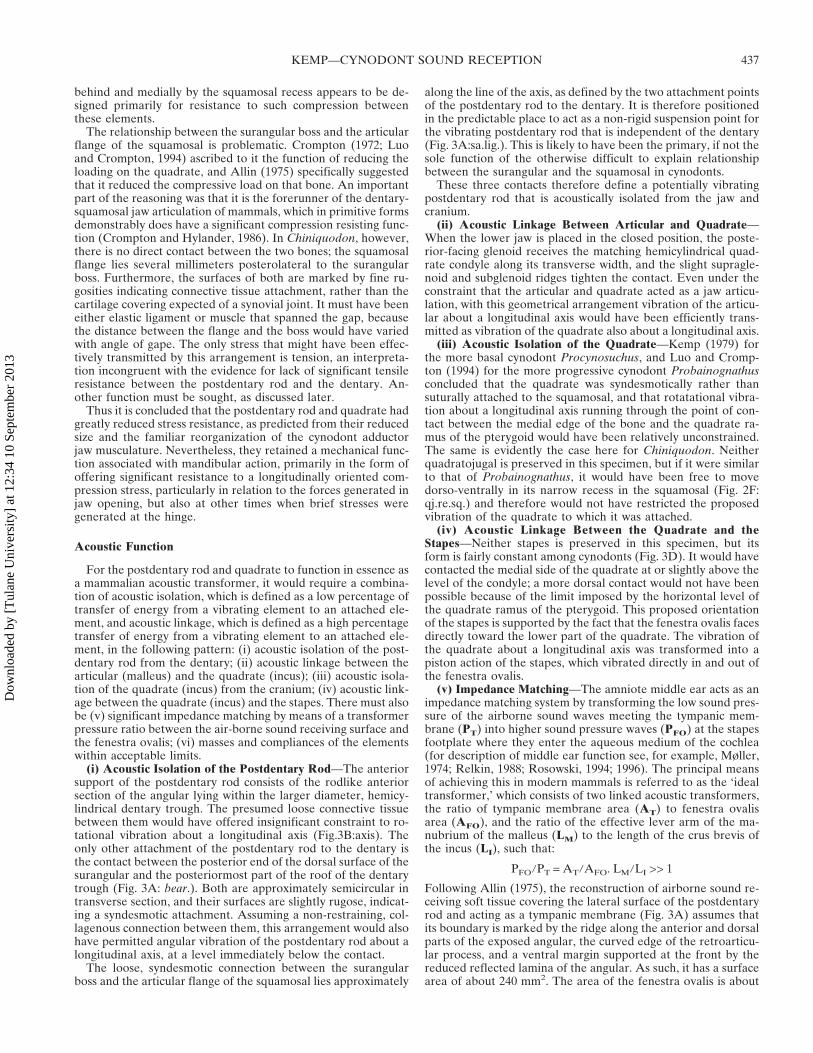