Embed Size (px)
Citation preview

3
Acquired Demyelinating Disorders of the CNS in Children
R. Govender1, Jo M. Wilmshurst2 and Nicky Wieselthaler2 1University of Kwa-Zulu Natal, Durban
2University of Cape Town, Cape Town South Africa
1. Introduction
Acquired Demyelinating disorders of the central nervous system in children span a wide spectrum. These conditions may be mono-phasic and self limiting or multi-phasic. Children may present with mono-focal (optic neuritis) or multi-focal (Acquired demyelinating encephalomyelitis) clinical findings. These demyelinating disorders also share many common clinical, radiological and laboratory features. Early classification of whether the disease is either mono- or multi-phasic has diagnostic and therapeutic implications. Identification of patients who present with a first demyelinating event and are at risk for evolution to multiple sclerosis, allows disease modifying therapeutic agents to be initiated early and thus preserve brain function. The aetiology of acquired demyelinating conditions is multi-factorial namely – genetic, post-infectious, post-immunization and possibly due to a T-cell mediated auto-immune response to myelin basic protein triggered by an infection or immunization. This chapter will cover the aetiologies, consensus definitions, clinical presentation, neuro-imaging, evolution and therapeutic advances in acquired demyelinating disorders in children. The pivotal role of neuro-imaging in unraveling the pathology, aetiology and diagnosis of these disorders is also highlighted. Clinical and neuro-imaging features of other acquired white matter lesions (via infections,
toxins, nutritional deficiencies, and osmotic myelinolysis) disease are also discussed.
2. Definitions
The International Paediatric Multiple Sclerosis Study group in 2007 proposed consensus
definitions for the demyelinating disorders in children (Krupp et al., 2007). This group was
convened to define an operational classification system for the demyelinating disorders.
Consensus definitions aid in standardization of diagnosis, investigation, management and
further research of these conditions.
3. Imaging techniques for white matter disorders
Magnetic Resonance Imaging (MRI) is the diagnostic modality of choice for evaluating
white matter disorders. Sophisticated applications of magnetic resonance technology, such
www.intechopen.com

Neuroimaging – Clinical Applications
62
Definition
Acquired Demyelinating Encephalomyelitis (ADEM)
A first clinical event with a presumed inflammatory or demyelinating cause, with acute or sub-acute onset that
affects multifocal areas of the CNS and must include encephalopathy
Recurrent ADEM New event of ADEM with a recurrence of the initial symptoms and signs, 3 or more months after the first
ADEM event, without involvement of new clinical areas by history, examination, or neuro-imaging
Multi-phasic Demyelinating Encephalomyelitis (MDEM)
ADEM followed by a new clinical event also meeting criteria for ADEM, but involving new anatomic areas
of the CNS as confirmed by history, neurologic examination, and neuro-imaging. The event must
develop within 3 months of the initial event.
Neuromyelitis Optica (NMO) Must have optic neuritis and acute myelitis as major criteria and a spinal MRI lesion extending over three or more segments or be NMO positive on antibody testing
Acute Transverse Myelitis (ATM)
A focal inflammatory disorder of the spinal cord resulting in motor, sensory and autonomic dysfunction
Schilder’s Disease A sub-acute demyelinating disorder characterized by bilateral large and vaguely symmetrical lesions
Paediatric Multiple Sclerosis (MS)
A clinical syndrome of multiple clinical demyelinating events involving
more than one area of the central nervous system with dissemination in time and space on imaging*
Clinically Isolated Syndromes (CIS)
A first acute clinical episode of CNS symptoms (without encephalopathy) with a presumed inflammatory demyelinating cause; for which there is no prior history of a demyelinating
event
*Important caveats in the definition of Paediatric MS put forward by the study group include:
1. The combination of an abnormal CSF (presence of Oligoclonal bands or an elevated IgG index) and
two lesions on the MRI, of which one must be in the brain, can also meet dissemination in space
criteria.
2. The MRI can meet the dissemination in space criteria if it shows 3 of the following 4 criteria (1)
nine or more white matter lesions or one gadolinium enhancing lesion, 2) three or more
periventricular lesions, 3) one juxta-cortical lesion, 4) an infra-tentorial lesion.
3. MRI can be used to satisfy criteria for dissemination in time following the initial clinical event,
even in the absence of a new clinical event if new T2 or gadolinium enhancing lesions develop
within 3 months of the initial clinical event.
4. A second non-ADEM event in a patient is insufficient to make the diagnosis of paediatric MS if the
first event meets the criteria for ADEM. MS can only be diagnosed if there is further evidence of
dissemination in time on the MRI (new T2 lesions > 3months since the second event) or a new
clinical event (> 3months since the second event).
Table 1. Definitions of Acquired Demyelinating Disorders (Adapted from Krupp et al 2007)
www.intechopen.com

Acquired Demyelinating Disorders of the CNS in Children
63
as magnetization transfer imaging, magnetic resonance spectroscopy, and diffusion tensor
imaging, provide quantitative information about the extent of damage that occurs in the
white matter.
1H magnetic resonance spectroscopy (MRS) is a valuable technique to non-invasively
acquire in-vivo information about biochemical processes in patients with neurologic
disorders. MRS measures N-acetyl-aspartate (NAA), total creatine, choline-containing
compounds, and lactate. NAA has been considered a marker of neuronal integrity,
whereas the levels of choline and lactate are indicative of cell membrane turnover and
anaerobic glycolysis (Bizzi et al., 2001). NAA is located almost exclusively in neurons and
neuronal processes and thus provides information about neuronal integrity (De Stefano et
al., 1995).
MRS studies on patients with white matter disorders have shown reduction in NAA in areas
that appear normal on conventional MRI studies suggesting that a significant amount of
axonal damage is present in these patients (Arnold et al., 1990; van Der Knaap et al., 1992).
Abnormalities in NAA on MRS have also shown a correlation with long term functional
outcomes. The author suggests that the extent of axonal damage rather than demyelination
may be more reliable in monitoring disease evolution in primary white matter disorders (De
Stefano et al., 2000).
Diffusion-weighted MR imaging (DWI) provides further information that may not be
apparent on conventional MR images. Engelbrecht et al showed that diffusion restriction
precedes brain myelination and is further increased during myelination (Engelbrecht et
al., 2002).
4. Acquired Demyelinating Encephalomyelitis (ADEM)
ADEM initially described by Lucas (1790) is characterized by acute onset of diffuse
neurological signs with multi-focal white matter involvement. In 1931 a series of case
studies was reported in The Lancet. McAlpine described 3 sets of patients with ADEM:
-post-vaccination, post-infectious, and those with spontaneous occurring disease. The
International Paediatric MS study group further defined ADEM (see Table 1).
4.1 Pathogenesis The aetiology of ADEM is not completely understood. The pathogenesis is thought to be
auto-immune mediated. The seasonal distribution and high rate of antecedent infections
(Dale et al., 2000; Murthy et al., 2002; Govender et al., 2010) reported in ADEM suggest a
link to an infectious aetiology. Infectious diseases are common in childhood however the
rate of preceding infection reported in these series exceed the rate of childhood infectious
diseases described (30-50%). The auto-immune reaction is thought to be on the basis of
molecular mimicry. The offending infection serves as an antigenic trigger and shares
epitopes with various autoantigens of myelin such as myelin basic protein, proteolipid
protein, and myelin oligodendrocyte protein (Alvord et al., 1987). This theory shares
many similarities with experimental allergic encephalitis. A second theory is that the
antigenic trigger activates T cells which cross the blood-brain barrier and react against
similar myelin epitopes. ADEM was associated with the class II alleles HLA-DRB1*01 and
HLA-DRB*03 in a Russian study (Idrissova et al., 2003). Pathological studies of children
www.intechopen.com

Neuroimaging – Clinical Applications
64
with ADEM showed that peripheral and cerebrospinal lymphocytes have increased
reactivity to myelin basic protein (Lisak and Zweiman, 1977). Viral infections described in
the context of ADEM include Herpes Simplex Virus-1, Cytomegalovirus (CMV), HIV
(Human Immunodeficiency virus), Measles, Mumps, Rubella, Ebsteinne Barre Virus
(EBV) and Varicella Zoster Virus (VZV). ADEM occurs after one of every 1000 cases of
measles, with a fatality rate of 20% (Tselis and Lisak, 1998). Bacterial antigens implicated
include Mycoplasma, Campylobacter, Streptococci and Borrelia Burgdofferi. Dale et al.
described a subgroup of ADEM associated with Group A β hemolytic streptococcus,
abnormal basal ganglia imaging and elevated antibasal ganglia antibodies (Dale et al.,
2001). Some studies fail to identify the agent responsible for the pre-demyelinating
infection (Murthy et al., 2002). The authors postulate that the inciting agents are
uncommon or unusual organisms that can not be identified by routine laboratory testing.
Vaccines, specifically the influenza, rabies and smallpox vaccines have also been reported
to precipitate ADEM (Saito et al., 1998). Post-vaccination ADEM is thought to be the result
of immune mediated mechanisms rather than the cyto-pathic effects of the virus. ADEM is
reported after the administration of drugs such as sulfonamides and streptomycin, further
supporting an immunological basis of the pathogenesis.
4.2 Epidemiology There are few epidemiological studies of ADEM in children. Prevalence studies are also complicated by the use of inconsistent case-definitions of ADEM. The estimated incidence in California is 0.4/100,000 population per year (Leake et al., 2004) and in Canada is 0.2/100,000 per year (Banwell et al., 2009). The mean age of presentation of ADEM in children ranges from 5-8 years (Hynson et al., 2001; Tenembaum et al., 2007). There is no specific ethnic distribution (Leake et al., 2004). Some studies indicate a slight male predominance (Murthy et al., 2002; Tenembaum et al., 2007). Prevalence in resource poor countries would be expected to be higher because of the significant frequency of childhood infections. However it is probably under-estimated because of limited access to health care facilities and MRI facilities.
4.3 Clinical presentation ADEM has a wide clinical spectrum of presentation. The hallmark of the disease is an
acute presentation of multifocal neurological signs with encephalopathy consistent with
diffuse brain involvement usually following a viral infection or immunization.
However, events may range from sub-clinical episodes diagnosed by MRI showing
mult-ifocal white matter lesions, to a more fulminant presentation with seizures and
encephalopathy. Seizures are reported to be more common in children compared to
adults with ADEM (Tenembaum et al., 2007). Fever and meningism are also common in
ADEM prompting treatment for meningo-encephalitis in the initial management (Dale
et al., 2000). Multi-focal neurological signs are pathognomic for ADEM and include
hemiparesis, paraparesis, cranial nerve involvement and ataxia. Atypical presentations
include concomitant peripheral nervous system involvement (Kinoshita et al., 1996),
presentation as an isolated acute psychotic episode (Moscovich et al., 1995) or with
an extra-pyramidal syndrome (dystonia and behaviour disturbances) (Dale et al.,
2001).
www.intechopen.com

Acquired Demyelinating Disorders of the CNS in Children
65
4.4 Laboratory investigations In the absence of specific biological markers diagnosis is based on a combination of
historical features, clinical and MRI characteristics. Other investigations are usually done
to exclude other differential diagnosis (e.g. meningitis, metabolic encephalopathies). In
resource poor settings where the burden of disease is predominantly infectious illnesses
and because of the overlap of symptoms, infectious aetiologies must be excluded first.
Peripheral blood leucocytosis is documented in ADEM (Jacobs et al., 1994). CSF studies in
ADEM are usually abnormal (in > 67% of cases), typically showing a moderate pleocytosis
with an elevated protein content (Miller et al., 1956; Govender et al., 2010). CSF
Oligoclonal bands synthesis may occur in ADEM; however this tends to disappear when
the patient recovers.
Electrophysiological studies have limited value in ADEM. Slow-wave abnormalities on
electro-encephalogram are compatible with an encephalopathic state (Dale et al., 2000). The
spindle coma pattern has been described in a child with post measles ADEM (Bortone et al.,
1996). Visual evoked potentials though are useful in detecting asymptomatic optic tract
lesions (Dale et al., 2000).
4.5 Neuro-imaging MRI is the investigation of choice. Since CT is often non-diagnostic for white matter lesions in patients with ADEM, this study is often normal or shows non-specific hypo-densities in the white matter.
Lesions are most easily recognized on T2 weighted (T2WI) and FLAIR MRI sequences. T2WI are more sensitive than T1 weighted images (T1WI) in detecting lesions (Sheldon et al., 1985). T1WI shows hypo-intense lesions. The lesions of ADEM are multi-focal and often do not correlate with clinical signs. Lesions tend to involve the cerebellum, the cerebral cortex and brainstem (Figure 1 a-g). They usually involve the sub-cortical, central and periventricular white matter. Lesions are typically hyper-intense, patchy, asymmetric and ill-defined. Diffusion-weighted imaging (DWI) and apparent diffusion co-efficient (ADC) maps may be helpful to prognosticate outcome. Low ADC values and restricted diffusion on DWI may suggest a worse outcome as this may indicate permanent tissue damage (Barkovich., 2007). A case study of ADEM with MRS reported reduced NAA and an elevation of choline and lactate (Gabis et al., 2004). These authors suggest a place for H MRS studies in longitudinal follow-up studies of ADEM to assess the response to immunomodulating therapies. Deep grey matter lesions in the thalami and basal ganglia have also been described (Baum et al., 1994; Govender et al., 2010) (Figure 2a-e). Lesions in the corpus callosum are uncommon and considered atypical for ADEM (Figure 3). Contrast enhancement of lesions post gadolinium administration indicates activity of the lesions (Figure 4). This correlates with the pathological finding of inflammation and demyelination in experimental allergic encephalitis. Non-enhancing and partially enhancing lesions in the presence of enhancing lesions have been described in ADEM and are thought to be because of lesions of differing ages and the evolution of the disease over several weeks (Schwaz et al., 2001;, Govender et al., 2010). Concomitant spinal cord lesions have been described in ADEM (Hynson et al., 2001;, Murthy et al., 2002;, Govender et al., 2010) (Figure 5a,b). Spinal cord lesions in ADEM typically have ill-defined margins, extend over multiple vertebral segments, are thoracic in location and result in mild cord expansion (Singh et al., 2002).
www.intechopen.com

Neuroimaging – Clinical Applications
66
A B
C D
E F G
Fig. 1. A - Flair axial MRI demonstrating bilateral asymmetrical cortex and sub-cortical white matter high signal intensities consistent with ADEM. B- T2 axial MRI demonstrating right ill-defined peritrigonal white matter high signal intensity lesion consistent with ADEM. C- Flair axial MRI demonstrating bilateral, fairly symmetrical high signal intensities in the posterior white matter consistent with ADEM. D - Flair axial MRI demonstrating bilateral asymmetrical high signal intensity lesions in the sub-cortical and deep white matter consistent with ADEM. E- Flair axial MRI demonstrating bilateral asymmetrical high signal intensity lesions in the cortex and sub-cortical and deep white matter consistent with ADEM. F - Flair axial MRI demonstrating hyper-intense lesions in the brachium ponti consistent with ADEM. G - T2 axial MRI of the brain demonstrating large hyper-intense pontine lesion with surrounding oedema
www.intechopen.com

Acquired Demyelinating Disorders of the CNS in Children
67
A B C
D E
Fig. 2. A- Flair axial MRI of the brain demonstrating bilateral symmetrical hyper-intense thalamic lesions. B- T2 coronal MRI of the brain demonstrating bilateral symmetrical rounded hyper-intense thalamic lesions as well as a large hyper-intense pontine lesion. C- Flair axial MRI demonstrating bilateral high signal intensity lesions in the basal ganglia and deep white matter on the left consistent with ADEM. D- Flair axial MRI demonstrating asymmetrical basal ganglia and white matter high signal intensities consistent with ADEM. E- DWI of 2D shows no evidence of restricted diffusion.
Fig. 3. T2 sagittal midline brain MRI demonstrating a well-defined rounded lesion in the splenium of the corpus callosum, which is unusual for ADEM.
www.intechopen.com

Neuroimaging – Clinical Applications
68
Fig. 4. T1 with contrast axial MRI demonstrating multiple hypo-intense white matter lesions of varying sizes, some with ring enhancement. This patient was diagnosed with ADEM.
A B Fig. 5. A- T2 sagittal MRI of the cord showing diffuse abnormal high signal intensity throughout the cord with cord expansion in the cervical region. There was no associated enhancement. This together with the brain lesions seen in 5B is consistent with ADEM. B- Flair axial MRI demonstrating bilateral high signal intensity lesions in the basal ganglia and deep white matter on the left consistent with ADEM.
www.intechopen.com

Acquired Demyelinating Disorders of the CNS in Children
69
4.6 Treatment There are no standard treatment protocols as there are insufficient large scale studies to form
consensus for optimal management. Supportive care (e.g. respiratory support for patients with
brainstem involvement, anti-epileptics for seizure control) in the acute phase is vital.
Therapies recommended are mainly immunomodulating agents targeting the immune-based mechanism of the disease. Corticosteroids are considered the mainstay of treatment based on the rapid improvement in symptoms following therapy (Straub et al., 1997;, Tenembaum et al., 2007). However widely varying doses, formulations, duration of therapy and tapering have been reported with corticosteroid use (Hynson et al., 2001; Murthy et al., 2002; Tenembaum et al., 2007; Govender et al., 2010). A single study reported worse outcomes in patients who received corticosteroids (Boe et al., 1965). Methylprednisone, dexamethasone and ACTH are used. Most reports in paediatric patients have used IV methylprednisolone (10 to 30 mg/kg/day) or dexamethasone (1 mg/kg) for 3 to 5 days (Dale et al., 2000; Hyson et al 2001; Tenembaum et al 2002; Govender et al., 2010) followed by a taper for 4 to 6 weeks with full recovery reported in 50 to 80% of patients. In resource poor countries high dose corticosteroids must be used with caution and only commenced once commonly occurring infections like tuberculosis and cytomegalovirus (CMV) are excluded. Outcomes on efficacy of corticosteroid treatment are mainly compared to historical controls. Worse outcomes are linked to shorter duration of treatment (Tenembaum et al., 2007). Other treatment modalities suggested include intravenous immunoglobulin, plasmapheresis and glatiramer acetate (Abramsky et al., 1977; Stricker et al., 1992; Finsterer et al., 1998). There is some evidence to suggest that patients may respond to a combination of methylprednisolone and immunoglobulin if they fail to respond to either separately (Straussberg et al., 2001).
4.7 Prognosis ADEM is by definition a monophasic illness (variants are discussed in Section 4.8). Mortality during the post-measles ADEM period in the 1950’s was reported as 10-30% (Johnson et al., 1985). At follow-up, approximately 60-80% of children have no neurologic deficits (Menge et al., 2007). This study also reports a mortality rate of 5%. The extent and site of lesions on the initial MRI do not predict the clinical outcome. Motor deficits persist in 8-30% (Dale et al., 2000; Tenembaum et al., 2007) of patients and include paraparesis, hemiparesis and ataxia. Neuro-cognitive deficits are also documented post ADEM (Hahn et al., 2003; Jacobs et al., 2004). These include deficits in short term memory, verbal processing skills and complex attention. Patient with early onset ADEM (<5year of age) were also more likely to have cognitive deficits and behaviour problems (Kumar et al., 1998). Follow-up MRI’s showed complete or partial resolution of abnormalities in the majority of cases (Kesslering et al., 1990; Dale et al., 2000; Tenembaum et al 2007; Govender et al., 2010). However, residual gliosis and demyelination persist in some patients (Kesselring et al., 1990). Clinical as well as imaging follow-up (at least 3 months later) (Figure 6) is important to monitor for evolution to MS. Risk factors for relapse are discussed in greater detail in Section 10.
4.8 Variants of ADEM Definitions for Recurrent and Multi-phasic ADEM are described in Table 1.
Acute Hemorrhagic Leukoencephalitis is a rare, hyper-acute form of ADEM with a mortality
rate of about 70% (Davies et al., 2006). Pathological studies show a necrotizing vasculitis
www.intechopen.com

Neuroimaging – Clinical Applications
70
with haemorrhage, oedema and a neutrophilic infiltration (Stone and Hawkins, 2007).
Seventy percent of survivors have neurological deficits (Stone and Hawkins, 2007).
A B Fig. 6. A- T2 axial MRI demonstrating asymmetrical basal ganglia and white matter high signal intensities consistent with ADEM. B- T2 axial MRI in same patient at 3 months follow-up showing resolution of lesions.
5. Transverse myelitis
5.1 Epidemiology Acute Transverse myelitis (ATM) is a focal inflammatory disorder of the spinal cord resulting in motor, sensory and autonomic dysfunction with evidence of inflammation on CSF or MRI studies. The initial definition was proposed by the Transverse Myelitis Consortium Working Group in 2002 and refined by the more recent consensus definitions for paediatric demyelinating disease (Krupp et al., 2007). The incidence is reported as 1- 8 per million people per year (Berman et al., 1981). There are no gender or ethnic differences in the prevalence of ATM (Berman et al., 1981). ATM is often difficult to distinguish clinically from ischaemic cord lesions, fibro-cartilagenous emboli or traumatic spinal cord lesions. ATM is also an important differential diagnosis for acute flaccid paralysis in childhood.
5.2 Pathogenesis
The aetiology of ATM is thought to be immune-mediated. In 30-60% of patients ATM is para-infectious (Jeffrey et al., 1993; Kalra et al., 2009). Molecular mimicry and super-antigen mediated mechanisms have been postulated (Kaplin et al., 2005). Positive anti-GM1 antibodies following Campylobacter and CMV infections have been implicated in the aetio-pathogenesis. Neuromyelitis optica—immunoglobulin G is an aquaporin-4–specific water channel antibody, which has been associated with neuromyelitis optica and longitudinally extensive transverse myelitis in adults (Lennon et al., 2004). This is discussed further in Section 6.
www.intechopen.com
Acquired Demyelinating Disorders of the CNS in Children
71
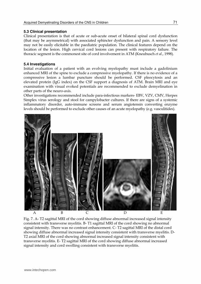
5.3 Clinical presentation Clinical presentation is that of acute or sub-acute onset of bilateral spinal cord dysfunction (that may be asymmetrical) with associated sphincter dysfunction and pain. A sensory level may not be easily elicitable in the paediatric population. The clinical features depend on the location of the lesion. High cervical cord lesions can present with respiratory failure. The thoracic segment is the commonest site of cord involvement in ATM (Kneubusch et al., 1998).
5.4 Investigations Initial evaluation of a patient with an evolving myelopathy must include a gadolinium enhanced MRI of the spine to exclude a compressive myelopathy. If there is no evidence of a compressive lesion a lumbar puncture should be performed. CSF pleocytosis and an elevated protein (IgG index) on the CSF support a diagnosis of ATM. Brain MRI and eye examination with visual evoked potentials are recommended to exclude demyelination in other parts of the neuro-axis. Other investigations recommended include para-infectious markers- EBV, VZV, CMV, Herpes Simplex virus serology and stool for campylobacter cultures. If there are signs of a systemic inflammatory disorder, auto-immune screens and serum angiotensin converting enzyme levels should be performed to exclude other causes of an acute myelopathy (e.g. vasculitides).
A B C D E
Fig. 7. A- T2 sagittal MRI of the cord showing diffuse abnormal increased signal intensity consistent with transverse myelitis. B- T1 sagittal MRI of the cord showing no abnormal signal intensity. There was no contrast enhancement. C- T2 sagittal MRI of the distal cord showing diffuse abnormal increased signal intensity consistent with transverse myelitis. D- T2 axial MRI of the cord showing abnormal increased signal intensity consistent with transverse myelitis. E- T2 sagittal MRI of the cord showing diffuse abnormal increased signal intensity and cord swelling consistent with transverse myelitis.
www.intechopen.com

Neuroimaging – Clinical Applications
72
5.5 Neuro-imaging MRI of the spinal cord usually shows nonspecific localized hyper-intense signal on T2WI
sequences with, in some cases, segmental cord enlargement and/or focal enhancement
(Figure 7 a-e). Acute partial transverse myelitis described in adults is characterized by MRI
lesions that are asymmetrically placed and spanning fewer than 2 vertebral segments in
length; these patients have been found to have a greater risk of progression to multiple
sclerosis (Ford et al., 1992). Longitudinally extensive myelitis (spanning > 3 vertebral
segments) is shown to have a lower risk of progression to MS in adults (Pittock et al, 2006).
Lesions of ATM in children are typically longitudinal, demonstrate rim enhancement and
are centrally located.
5.6 Treatment Previous case series did not demonstrate any benefit from the use of low dose
corticosteroids (Dunn et al., 1986; Adams et al., 1990). Recent reports demonstrate the benefit
of high dose corticosteroids (10-30mg/kg per day for 3-5 days) on recovery (Defrense et al.,
2001; Sebire et al., 1997). Compared to historical controls patients treated with steroids
walked independently sooner. If there is no clinical response to steroids within 5-7 days
plasma exchange was used as adjunctive therapy in isolated case reports. Supportive
measures include respiratory support and early management of a neuropathic bladder.
5.7 Prognosis Various studies have looked at prognostic indicators for ATM. Jain et al. (1983) described
backache at onset, acute course (within hours), spinal shock and a cervical sensory level as
poor prognostic features. Other studies did not demonstrate this (Govender et al., 2010).
Early recovery (within one week of presentation), age less than 10 years at presentation and
lumbosacral spinal level on clinical assessment were significant predictors of a good
outcome (De Goede et al., 2010). The extent of lesions on MRI has not shown consistent
correlation with outcome (Pradhan et al., 1997; Adronikou et al., 2003). Berman et al’s series
(1987) described more than one-third of the patients with ATM making a good recovery; in
one-third of patients recovery was only fair; 14 patients failed to improve and 3 demised.
In the series by Dunne et al (1986) that assessed the risk of progression to multiple sclerosis in children with ATM, definite evidence of multiple sclerosis did not develop in any of the patients. In the series by Pidcock et al of the 47 children with acute transverse myelitis, 2 experienced recurrent transverse myelitis, 1 was diagnosed with neuromyelitis optica, and 1 developed multiple sclerosis on follow-up (Pidcock et al., 2007).
6. Neuromyelitis Optica (NMO)
The association between myelitis and optic problems was first described in 1870 by Thomas Clifford Allbutt (Murray, 2005). In 1894 Eugene Devic described 16 patients with visual impairment who developed paraparesis, sensory deficits and sphincter dysfunction within weeks. They recognized that these symptoms were the result of inflammation of the optic nerve and spinal cord. NMO is a recurrent demyelinating disorder affecting the optic nerves and the spinal cord. Modifications to the definition of NMO in 2005 incorporated the inclusion of patients with brain lesions, and included the NMO-IgG antibody as a confirmatory test.
www.intechopen.com

Acquired Demyelinating Disorders of the CNS in Children
73
The Mayo Clinic proposed a revised set of criteria in 2006. The new guidelines for diagnosis requires both absolute criteria and two of the three supportive criteria to be present to make a diagnosis of NMO (Wingerchuck et al., 2006). Absolute criteria: 1. Optic neuritis 2. Acute myelitis Supportive criteria: 1. Brain MRI not meeting criteria for MS at disease onset 2. Spinal cord MRI showing contiguous T2-weighted signal abnormality extending over 3
or more vertebral segments, indicating a relatively large lesion in the spinal cord 3. NMO-IgG seropositive status. The association of NMO with the serum autoantibody marker NMO-IgG was reported in 2004 (Lennon et al., 2004).
NMO-IgG is 73% sensitive and 91% specific for distinguishing
NMO from classical MS. The new diagnostic criteria allows for the diagnosis of NMO in patients who are NMO-IgG antibody negative. NMO antibodies play a key role in the pathogenesis. These antibodies are directed against the aquaporin-4- receptors located in the cell membrane of astrocytes (Pearce, 2005). Aquaporin-4 is the most abundant channel facilitating water transport across membranes in the brain. NMO-IgG is also detected more commonly in patients with NMO symptoms who have clinical or serological evidence for SLE than in those who do not (McAdam et al., 2002).
6.1 Clinical characteristics Clinical characteristics include painful visual loss, weakness, sphincter dysfunction and sensory deficits. Loss of red color vision, a relative afferent pupillary defect and visual field defects are other features of optic neuritis in children. Other complications such as ataxia and respiratory failure result from extension of cervical cord lesions into the brainstem.
6.2 Investigations Diagnostic evaluation includes an MRI of the brain. During acute optic neuritis attacks, an orbital MRI may identify optic nerve gadolinium-enhancement. MRI of the brain is usually normal. However, brain lesions located in the hypothalamus, brainstem, and periventricular areas have been described in children who have typical features of NMO (Pittock et al., 2005). These are considered to be the aquaporin-4 rich areas of the brain. Patients with signs of myelitis should have a spinal MRI with contrast. The lesions are typically longitudinally extensive, centrally based in the cord and extend over three or more vertebral segments. All patients should have a serological test for the NMO-IgG antibody. A negative test however does not exclude the diagnosis. CSF pleocytosis also supports the diagnosis. In patients with longitudinal myelitis and no visual symptoms, visual evoked potentials can sometimes detect asymptomatic visual pathway dysfunction.
6.3 Treatment The recommended treatment for acute attacks of myelitis or optic neuritis is high dose methylprednisone. Prophylactic long-term immunosuppression is recommended for established NMO and patients who have a single attack of myelitis and are NMO-IgG positive (Wingerchuck et al., 2005). There are no efficacious preventative therapies demonstrated by controlled trials in NMO. Intravenous immunoglobulin is an alternative for patients who do not respond to corticosteroids.
www.intechopen.com

Neuroimaging – Clinical Applications
74
Characteristics of NMO that help to distinguish it from classical MS include:
Prominent CSF pleocytosis (more than 50 WBC) with a polymorphonuclear cell predominance (Mandler et al., 1993; O Riordan et al., 1996)
Lower frequency of CSF oligoclonal banding (15-30% in NMO compared to 85% in MS)
Bilateral symmetrical optic neuritis
At disease onset, the brain MRI scan is normal or reveals nonspecific white matter lesions
MRI of the spinal cord showing longitudinally extensive, central lesions (MS lesions are more peripherally located in the cord and extend over one to two segments in length)
7. Schilder’s disease/myelinoclastic diffuse sclerosis
This disorder was initially described by Schilder in 1912 and later clarified by Poser (1992).
There are further reported cases of solitary, large plaque like lesions, which were
histologically confirmed to be foci of demyelination (Kumar et al., 1998; Gutling and Landis,
1998). The aetiology is unclear; however an association with tuberculosis was described in 3
South African children (Pretorius et al., 1998). Schilder’s Disease occurs predominantly in
children (peak age 5-14 years) (Afifi et al., 1994).
The Poser criteria (1992) for diagnosis are:
one or two roughly symmetrical large plaques (greater than 2 cm diameter)
pathological analysis is consistent with sub-acute or chronic myelinoclastic diffuse
sclerosis
adrenoleukodystrophy and peripheral nervous system involvement must be excluded.
7.1 Clinical presentation The clinical presentation is non-specific and includes neuroregression, seizures, ataxia or
signs of raised intra-cranial pressure.
7.2 Neuro-imaging The lesions of Schilder’s Disease are typically large and plaque-like and have also been
termed tumefactive demyelination. MRI is the most accurate modality of delineating the
lesions that are often confused with brain neoplasms or abscesses. Making the distinction
between demyelination and infection/malignancies early is important to prevent
unnecessary surgical procedures and toxic therapies like radiation and chemotherapy
(McAdam et al., 2002). MRI studies demonstrate 1 or 2 large confluent lesions in the deep
white matter, usually the centrum semiovale (Figure 8 a-f). Lesions are at least 2 cm in
size in 2 of 3 dimensions. No additional lesions should be observed on imaging of the
brain or spinal cord- this would suggest MS or ADEM. On T1WI, tumefactive
demyelination lesions reveal a hypo-intense central area with a thick surrounding band of
moderately increased intensity. Lesions are centrally hyper-intense on T2WI.
Enhancement, when present, is incomplete. The lesions are characterized by enhancement
limited to one side of the lesion; usually the rim facing the lateral ventricles (McAdam et
al., 2007). Demyelination can be distinguished from other ring enhancing lesions (brain
abscesses, tumors, parasitic infections) by the presence of other demyelinating plaques
elsewhere in the central nervous system.
www.intechopen.com

Acquired Demyelinating Disorders of the CNS in Children
75
A B C
D E F
Fig. 8. A & B – T2 axial MRI demonstrating multiple well-defined hyper-intense white
matter foci surrounded by more ill-defined areas of increased white matter signal intensity
consistent with Schilder’s Disease. C- Flair axial MRI demonstrating multiple hyper-intense
lesions of varying sizes within the white matter with some areas of suppression within the
plaques consistent with Schilder’s Disease. D- Flair parasagittal MRI demonstrating large
flame- shaped white matter plaque with some areas of suppression within the lesions
consistent with Schilder’s Disease. E & F- T1 with contrast axial MRI demonstrating multiple
non- enhancing hypo-intense lesions of varying shapes and sizes within the white matter.
Other supportive diagnostic tests include an elevated CSF protein and an elevation of CSF
IgG in 50-60% of patients with Schilder’s Disease. Many patients with a large ring enhancing
lesion will have a brain biopsy mainly to exclude other disorders.
7.3 Treatment The treatment of choice is high dose intravenous corticosteroids. A rapid clinical and
radiological response to high dose corticosteroids favors the diagnosis of demyelination.
www.intechopen.com

Neuroimaging – Clinical Applications
76
8. Multiple Sclerosis (MS)
MS is defined in Table 1 (Krupp et al., 2007). MS in children is likely an under-recognized phenomenon that poses a unique set of challenges in terms of diagnosis and management. Early accurate diagnosis of MS is vital to facilitate early institution of disease modifying agents.
8.1 Epidemiology Childhood onset MS is an uncommon entity however, an estimated 2- 5% of patients with MS have onset of symptoms of MS before 16 years of age (Duquette et al., 1987; Boiko et al., 2002). The youngest reported patient with MS presented at 10 months of age. This was an indigenous African child who died at 6 years of age after 11 episodes of relapsing neurological symptoms (Shaw and Alvord, 1987). Similar to adult studies, a female preponderance is reported for MS in adolescence (Duquette et al., 1987; Govender et al., 2010). However there is no gender predilection in children presenting with MS under 6 years of age (Banwell et al., 2007). A crude period prevalence for patients of European ancestry was 25.63 per 100 000 and for patients of indigenous African descent was 0.22 per 100 000 (Bhigjee et al., 2007). Adult studies have described a more severe clinical and radiological phenotype in patients of African indigenous ancestry compared to patients of European ancestry (Kaufmann et al., 2003; Bhigjee et al., 2007). A retrospective single centre analysis showed a significantly higher relapse-rate in African-American children, compared with whites, suggesting a more aggressive disease course in the former group (Boster et al., 2009).
8.2 Pathogenesis Genetic and environmental factors are implicated in the aetiology of MS. Twin studies show a 20-30% higher risk of disease in monozygotic twins compared to dizygotic twins. Allelic variation in the MHC class II region exerts the single strongest effect on genetic risk (Ramgopalan SV et al., 2009). The HLA DR1B is the gene marker associated with higher risk of MS (Ness et al., 2007). Alleles of IL2RA, IL7RA (Hafler et al., 2007), the ecotropic viral integration site 5 (EVI5) (Hoppenbrouwers et al., 2008) and kinesin family member 1B (KIF1B) genes (Aulchenko et al., 2008) have recently been shown to increase susceptibility to MS. Epidemiological studies implicate environmental factors such as geographical variations (Kurtzke and Hyllested, 1979), season of birth (Sadovnick et al., 2007) and migration patterns (Pugliatti et al., 2006) in the aetiology of MS. Emerging evidence supports sunlight or vitamin D as an important environmental factor in aetiology (Ramgopalan SV et al., 2009). Children exposed to parental smoking also have a higher risk of MS (Mikaeloff et al., 2007).
8.3 Sub-types of MS The National MS Society in the US in 1996 categorized MS into four internationally recognized forms (Lublin and Reingold, 1996). Relapsing-remitting: refers to MS that has exacerbations/relapses followed by symptom-free periods of remission. This is the commonest form of MS in children (Ruggierri et al., 2004). Primary Progressive: It is characterized by gradual clinical decline from the time of disease
onset with no distinct periods of remission or relapses. There maybe plateau periods during
the disease but no periods of being symptom free. This entity, though rare in children, is
reported (Duquette et al., 1987; Govender et al., 2010).
Secondary Progressive: This type begins with a relapsing remitting course which may last
several years before the onset of the secondary progressive stage. Secondary progressive
www.intechopen.com

Acquired Demyelinating Disorders of the CNS in Children
77
multiple sclerosis is a second-stage, chronic, progressive form of the disease where there are
no periods of remission, only breaks in attack duration with no recovery from symptoms.
Relapsing progressive: have a steady neurologic decline but also suffer clear superimposed attacks. This is the least common of all subtypes. An acute/ Malignant MS (Marburg variant) form presenting with a fulminant, rapidly fatal disease has also been described.
8.4 Clinical presentation Children present with a wide variety of clinical symptomatology including motor, sensory, visual, cerebellar and brainstem dysfunction (Shaw and Alvord., 1987; Sindern et al., 1992; Ghezzi et al., 1997; Dale et al., 2000; Boiko et al., 2002; Pohl et al., 2006; Govender et al., 2010).
Motor manifestations are described as the most common clinical presentation (Duquette et al., 1987; Sindern et al., 1992; Pohl et al., 2006). Polysymptomatic presentation is reported to be more frequent in childhood onset MS compared to adults (Ghezzi et al., 1997; Dale et al., 2000; Boiko et al., 2002). However monosymptomatic presentation is also reported in children (Duquette et al., 1987). Encephalopathy and seizures also occur in MS (Gusev et al., 2002). Eye involvement is described in up to 50 % of children with MS (Pohl et al., 2006). Optic neuritis in MS is more likely to be unilateral (Dale et al., 2000). Optic tract involvement may be asymptomatic and diagnosed only by abnormal visual evoked potentials (Pohl et al., 2006). Fatigue in children is more frequent compared to adults with MS (Gusev et al., 2002). Cognitive decline is reported in 30-66% of children with MS (Banwell and Anderson 2005; Banwell et al., 2007a).
8.5 Laboratory evaluation Diagnostic evaluation is to exclude other conditions affecting predominantly the white matter and to look for supportive evidence for MS. The workup should also include CSF studies (including cell count, total protein, IgG index, evidence of oligoclonal bands, and cytology) (Hahn et al., 2007). CSF Oligoclonal bands are reported in 72-84% of children with MS (Sindern et al., 1992; Dale et al., 2000). Oligoclonal bands may be absent initially and only develop during the course of the illness. Leucocytosis in the peripheral blood, though described in MS (Dale et al., 2000; Govender et al., 2010), is uncommon and non-specific. Neuro-physiological testing such as visual and auditory evoked potentials are also of diagnostic importance in detecting sub-clinical evidence of demyelination.
8.6 Imaging Lesions of MS are typically multiple, discrete, plaque-like and involve predominantly the white matter (Mikaeloff et al., 2004). Commonly involved areas in MS include the corpus callosum, periventricular and sub-cortical white matter (Fig 9a-g). Lesions of MS are typically iso- or hypo-intense on T1WI, and hyper-intense on T2W1 and FLAIR sequences. Enhancement of active lesions post-gadolinium may be solid, ring-like or arc-like (Fig 10a-c). Children tend to have fewer lesions and less enhancement (Banwell et al., 2007b). However, some children lack typical MRI findings of MS and have either large tumefactive lesions with peri-lesional oedema (Hahn et al., 2004) or deep grey matter involvement. Basal ganglia affectation in MS, though described, is uncommon (Figure 11 a,b). Younger children with MS may also have more diffuse, bilateral ill defined lesions (Mikaeloff et al., 2004). The International Pediatric MS Study Group strongly recommended additional imaging of the entire spinal cord to identify other sites of demyelination (Figure 12 a,b). The cervical spinal cord is the commonest region involved in MS.
www.intechopen.com

Neuroimaging – Clinical Applications
78
A B
C D
E F G
Fig. 9. A- T2 parasagittal MRI of the brain demonstrating flame-shaped hyper-intense lesion perpendicular to lateral ventricle consistent with MS. B- Flair axial MRI demonstrating multiple asymmetrical hyper-intense plaque-like lesions in the centrum-semiovale. Features are consistent with MS. C -Flair axial MRI of brain demonstrating 2 periventricular hyper-intense white matter lesions consistent with MS. D- Flair axial at level of lateral ventricles demonstrating asymmetrical hyper-intense white matter lesions consistent with demyelination and MS. E- Flair axial of brain demonstrating hyper-intense right parietal white matter plaque-like lesion and left subtle white matter hyperintensity consistent with MS. F- Flair axial of brainstem demonstrating pontine and brachium pontis high signal intensity lesions consistent with demyelination. G-Flair axial MRI demonstrating rounded hyper-intense lesion in left brachium pontis consistent with demyelination.
www.intechopen.com

Acquired Demyelinating Disorders of the CNS in Children
79
A B C Fig. 10. A- T1 with contrast parasagittal MRI of brain demonstrating rim-enhancing plaque-
like lesion typical of active demyelination in a patient with MS. B- T1 with contrast axial
MRI demonstrating ring enhancement of left brachium pontis lesion consistent with active
demyelination in a patient with MS. C- T1 with contrast axial MRI demonstrating ill-defined
irregular marginal enhancement of the plaque-like lesions consistent with active
demyelination in a patient with MS.
A B
Fig. 11. A- Flair axial MRI of brain demonstrating 2 lesions in the right basal ganglia. This is
unusual for MS. B- Flair axial MRI demonstrating multiple hyper-intense lesions in the
periventricular white matter as well as left basal ganglia(atypical) consistent with MS.
MRS reveals a reduction in NAA and an elevation in choline, lipids and lactate in active
lesions (Smith AB, 2009). Volumetric MRI studies reveal progressive loss of tissue in white
matter tracts early in the course of the disease (Miller et al., 2002). A single study of
Magnetization transfer imaging and Diffusion tensor imaging in children with MS
suggested that there was no evidence of white matter degeneration in normal appearing
white matter areas (Tintore et al., 2000).
www.intechopen.com

Neuroimaging – Clinical Applications
80
A B
Fig. 12. A- T2 sagittal MRI of cervical spine in a patient with MS demonstrating an ill-defined
expansile hyper-intense lesion in the proximal cord consistent with demyelination. B- T1
with contrast sagittal MRI of cervical spine in a patient with MS demonstrating ill-defined
contrast enhancement of the lesion in 12A consistent with active demyelination.
8.7 Treatment MS is a chronic condition with significant impact on all aspects of the family’s life.
Management should be trans-disciplinary involving psychologists, physiotherapists,
occupational therapists and school teachers.
8.7.1 Management of relapses The mainstay of managing relapses is high dose corticosteroids. High dose IV
corticosteroids (10-30mg/kg/day) for 3-5 days, is usually used with an optional oral
tapering dose. High dose oral steroids were found to be efficacious in adults (Morrow et al.,
2004). Plasmapharesis and IVIG are alternatives to be considered if steroids are not effective
(Hahn et al., 1996; Duzova and Bakkaloglu, 2008).
8.7.2 Disease modifying therapy These therapies are known to alter the disease course and outcomes. They reduce the
frequency and severity of relapses (Mikaeloff et al., 2001; Kornek et al., 2003; Tenembaum
and Segura, 2006;). Patients on therapy are shown to have better outcomes compared to
untreated patients (Mikaeloff et al., 2008). First line agents include Interferon beta 1a, 1b and
Glatimer acetate. Case reports of second line therapies used include Natalizumab,
Cyclophosphamide and Mitoxantrone.
www.intechopen.com

Acquired Demyelinating Disorders of the CNS in Children
81
Study No. of patients Treatment Outcomes
Ghezzi et al, 2007 52 Interferon beta 1a Reduction in relapse rate Reduction in *EDSS score
Tenembaum and Segura, 2006
24 Interferon beta 1a Reduction in relapse rate
Kornek et al, 2003 7 Glatimer Acetate Reduction in relapse-2/7 Stable EDSS- 3/7
Huppke et al, 2008 3 Natalizumab Induction of remission in all
Makhani et al, 2009 17 Cyclophosphamide Reduction in relapse rate Stabilization of EDSS
* EDSS: Extended Disability Status Scale
Table 2. Studies of specific treatment interventions in MS
8.8 Prognosis Most children with MS follow a relapsing, remitting course with increasing neuro-
disability (Boiko et al., 2002). A slower rate of progression of disease compared to adults
suggests more plasticity and potential for recovery in the developing CNS (Simone et al.,
2002). Children tend to have more relapses in the first 2 years of the disease (Simone et al.,
2002; Mikaeloff et al., 2006). Patients with childhood-onset MS also take longer to reach the
stage of severe disability but reach irreversible neurological disability at a younger age
compared to patients with adult onset disease (Renoux et al., 2007). More severe disease
was noted in girls; when the time between the first and second attacks was <1 year; for
childhood-onset multiple sclerosis fulfilling MRI diagnostic criteria at onset; for an
absence of severe mental state changes at onset; and for a progressive course (Mikaeloff et
al., 2006).
9. Clinically isolated syndromes
These episodes may be mono-focal (the clinical features can be attributed to a single CNS site) or multi-focal if the clinical features can not be explained by a single lesion. These include isolated optic neuritis, transverse myelitis, brainstem (Fig 13 a-d) or cortical lesions. Typically in contrast to ADEM there is no associated fever or encephalopathy. A CIS often poses a diagnostic and therapeutic challenge. Multiple lesions (> 4 lesions) (Morissey et al., 1993) on the MRI increase the risk of evolution to MS. In adult studies up to 80% of patients with a CIS evolve MS (Brex et al., 2002). Brainstem lesions in CIS are associated with a worse prognosis (Tintore et al., 2010). Children with CIS tend to have more infra-tentorial lesions
www.intechopen.com

Neuroimaging – Clinical Applications
82
A B
C D
Fig. 13. (B&C same patient) Clinically Isolated Syndrome. A- T2 axial MRI demonstrating
abnormal increased signal in the brainstem which was the only abnormal lesion. Clinically
this patient had a CIS. B- T2 fat saturation axial MRI demonstrating a left swollen hyper-
intense optic nerve with resultant proptosis. Features consistent with a unilateral Optic
Neuritis. Remainder of brain and spine were normal. C- T2 fat saturation sagittal oblique
MRI demonstrating a left swollen hyper-intense optic nerve. Features consistent with a
unilateral Optic Neuritis. Remainder of brain and spine were normal. D- T2 fat saturation
axial MRI of the optic nerves demonstrating abnormal high signal intensity within the
proximal portions of the nerves and swelling of the nerves. Features consistent with Optic
Neuritis (worse on the right). Remainder of brain and spine were normal.
www.intechopen.com

Acquired Demyelinating Disorders of the CNS in Children
83
Characteristic ADEM 1st MS event
Demographics Age of presentation Younger Onset >10yrs
Sex Slight male predominance
Female predominance
History of Pre-demyelinating event
More frequent Less frequent
Seasonal distribution More frequent Less frequent
Family history of demyelinating disease
Not present More frequent
Clinical Presentation
Seizures More frequent Less frequent
Encephalopathy More frequent Less frequent
Headache, fever More frequent Less frequent
Optic Neuritis Bilateral More frequent
Unilateral
Mono-focal vs. Poly-focal signs
Polysymptomatic Mono-focal
Laboratory features
Elevated CSF protein Less frequent More frequent
Leucocytosis in CSF More frequent Less frequent
Oligoclonal bands in CSF
Less/ Usually transient
More frequent
Persistent
Serum Leucocytosis More frequent Less frequent
MRI
Characteristics
Lesion definition Ill Defined Well-defined
Lesion load Greater Lower
Periventricular Lesions Less frequent More frequent
Juxta-cortical Lesions More frequent Less frequent
Cortical Lesions More frequent Less frequent
Corpus Callosum
Involvement
Less frequent More frequent
Brainstem/ Cerebellum More frequent Less frequent
Spinal Cord
Involvement
More frequent Less frequent
Deep Grey matter
involvement
More frequent Less frequent
Contrast enhancement Less frequent More frequent
Outcome Cognitive deficits Less frequent More frequent
No neuro deficit after 1st
event
Less likely More likely
Table 3. Markers comparing ADEM to a first episode of MS
www.intechopen.com

Neuroimaging – Clinical Applications
84
Disorder Example Clinical/ Laboratory Radiological
Infections HIV
Encephalopathy
Developmental delay,
pyramidal tract signs,
microcephaly
-Confluent bilateral
symmetrical white
matter changes
-cerebral atrophy
-basal ganglia
calcification
Progressive Multi-
focal
Leucoencephalopa
thy (JC Virus)
Immunocompromised -Multi-focal ↑T2 WI
lesions
-Propensity for
frontal/ parieto-
occipital areas
-Subcortical U
fibres involved
Other infections Sub-acute Sclerosing Panencephalitis, Lymes Disease,
Neurosyphillus, HTLV1, Borreliosis
Auto-
immune/Vasculitides Systemic Lupus
Erythematosus
Multi-system auto-
immune disorder
Anti-nuclear factor
positive
-Multi-focal
↑T2WI/FLAIR
-Infarcts
-Contrast
enhancement
of active
lesions
Isolated CNS angiitis, CADASIL (adult disorder-rare in children)
Tumor CNS Lymphoma Insiduous onset, CSF
cytospin –malignant cells
-↓ T1WI
-↑ T2WI
-GM involved more
frequently
-MRS may help
Medulloblastoma, Astrocytoma
Leukodystrophies Adrenoleukody-
strophy
Boys with
hyperpigmentation of
skin, behaviour and
learning problems,
Abnormal very long chain
fatty acids
-Symmetrical,
confluent
-predominantly
posterior
involvement
-Splenium and
cortico-spinal tracts
involved
-Leading edge
enhancement in
peri-trigonal
area
www.intechopen.com
Acquired Demyelinating Disorders of the CNS in Children
85
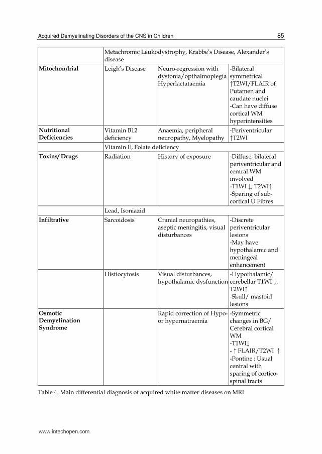
Metachromic Leukodystrophy, Krabbe’s Disease, Alexander’s disease
Mitochondrial Leigh’s Disease Neuro-regression with dystonia/opthalmoplegiaHyperlactataemia
-Bilateral symmetrical ↑T2WI/FLAIR of Putamen and caudate nuclei -Can have diffuse cortical WM hyperintensities
Nutritional Deficiencies
Vitamin B12 deficiency
Anaemia, peripheral neuropathy, Myelopathy
-Periventricular ↑T2WI
Vitamin E, Folate deficiency
Toxins/ Drugs Radiation History of exposure -Diffuse, bilateral periventricular and central WM involved -T1WI ↓, T2WI↑ -Sparing of sub-cortical U Fibres
Lead, Isoniazid
Infiltrative Sarcoidosis Cranial neuropathies, aseptic meningitis, visual disturbances
-Discrete periventricular lesions -May have hypothalamic and meningeal enhancement
Histiocytosis Visual disturbances, hypothalamic dysfunction
-Hypothalamic/ cerebellar T1WI ↓, T2WI↑ -Skull/ mastoid lesions
Osmotic Demyelination Syndrome
Rapid correction of Hypo-or hypernatraemia
-Symmetric changes in BG/ Cerebral cortical WM -T1WI↓ -↑FLAIR/T2WI ↑
-Pontine : Usual central with sparing of cortico-spinal tracts
Table 4. Main differential diagnosis of acquired white matter diseases on MRI
www.intechopen.com

Neuroimaging – Clinical Applications
86
(Ghassemi et al., 2008). This may be related to the differences in myelination patterns and maturation in children compared to adults. A radiologically isolated syndrome (RIS) is defined by incidental MRI findings suggestive of MS in an asymptomatic patient lacking any history, symptoms, or signs of MS (Okuda et al., 2009).
10. Risk of recurrence after a first demyelinating event
Predicting the risk of a first episode of demyelination evolving on to MS is important as new immunomodulating therapies become available. Early initiation of disease modifying therapy reduces the risk of relapse and long-term disability (Jacobs et al., 2000). Patients with “ADEM” progressing to MS vary from 0-29% (Belman et al., 2007). Multiple historical, clinical, laboratory and radiological criteria are used to predict the risk of recurrence/ progression to MS (Table 3). A seasonal pattern, a history of a precipitant, seizures, bilateral optic neuritis and encephalopathy are considered more likely in ADEM compared to MS (Dale et al., 2005).
Inflammatory markers, a high cerebrospinal fluid protein and leucocytosis are also more common in ADEM (Kesselring et al., 1990). MRI characteristics that are predictive of evolution to MS include well defined lesions that are peri-aqueductal or perpendicular to the corpus callosum (Dale et al., 2000), deep grey matter involvement and lesions that enhance post contrast (Govender et al., 2010).
11. Differential diagnosis of white matter disease in children
The differential diagnosis for a child who presents with a neurological symptom and white matter lesions on neuro-imaging is vast and includes infectious diseases, leukodystrophies, tumors, vasculitides, toxins and vitamin deficiencies. In resource poor countries, CNS infections must be excluded first as they are common and have acute therapeutic implications. CNS infections must be excluded in children presenting acutely especially with fever and encephalopathy. CNS infections that may present with multifocal white matter lesions include HTLV-1, Borreliosis and Subacute Sclerosing Panencephalitis. In resource poor settings HIV Encephalopathy (Figure 14 a,b) is common and is also characterized by confluent white matter lesions. Progressive Multi-focal Leukoencephalopathy (Figure 14 c),
is also common in immuno-compromised patients. Neurometabolic disorders, such as Adrenoleukodystrophy (Figure 14 d,e), presents with
primary white matter disease.
Osmotic Myelinolysis (Figure 14 f,g) is thought to be related to osmotic shifts associated with rapid correction of fluid and electrolyte abnormalities (especially sodium abnormalities). Malnourished children are at greater risk for developing myelinolysis. Lesions typically occur in the pons but have also been reported in extra-pontine sites such as the basal ganglia, cerebral cortex and cerebellar peduncles. Nutritional deficiencies such as vitamins B12, E and folate deficiencies may also cause
white matter lesions.
Drugs and toxins implicated in demyelination include tin, lead, isoniazid and radiation. Collagen Vascular Diseases refer to a group of auto-immune mediated disorders. The neurological manifestations are diverse. Neuro-imaging may show multi-focal white matter lesions- involving the cortex, cerebellum or spinal cord.
www.intechopen.com

Acquired Demyelinating Disorders of the CNS in Children
87
A B
C D
E F G
Fig. 14. A & B- T2 axial MRI demonstrating diffuse brain shrinkage/atrophy with ex-vacuo dilatation of the ventricles and abnormal increased signal intensity in the deep white matter bilaterally. This patient has features of HIV encephalopathy. C- Flair axial MRI demonstrating bilateral symmetrical increased signal intensity in the frontal white matter. This patient was diagnosed with PML. D- T2 axial MRI demonstrating bilateral symmetrical confluent abnormal increased white matter signal intensity in the posterior white matter consistent with Adrenoleukodystrophy. E- T1 with contrast axial MRI of same patient as 14D demonstrating peripheral enhancement of the white matter lesions. F- T2 axial MRI demonstrating well-defined rounded hyper-intense lesion in the pons. Note peripheral sparing of the pons. (Compared to figure 14G). Features consistent with Osmotic/ Pontine Myelinolysis. G- T1 with contrast sagittal MRI of the same pontine lesion as 14F showing no contrast enhancement. Features consistent with Pontine Myelinolysis.
www.intechopen.com

Neuroimaging – Clinical Applications
88
12. Conclusion
Acquired demyelinating disorders in children are a diverse, challenging group of conditions that are probably under-diagnosed. Early recognition is essential for optimal patient management as some of these disorders cause significant long-term sequelae. Advances in the last decade include establishing consensus definitions and improvement in neuro-imaging techniques. These advances set the stage for international collaborative studies to better define other areas such as understanding the aetio-pathogenesis, identifying biomarkers and standardizing treatment protocols of this diverse group of conditions.
13. References
Abramsky O, Teitelbaum D, Arnon R (1977). Effect of a synthetic polypeptide (cop1) on
patients with multiple sclerosis and with acute disseminated encephalomyelitis. J
Neurol Sci 31: 433-8.
Adams C, Armstrong D (1990). Acute transverse myelopathy in children. Can J Neurol Sci
17: 40–5.
Afifi AK, Bell WE, Menezes AH, Moore SA (1994). Myelinoclastic diffuse sclerosis
(Schilder's disease): report of a case and review of the literature. J. Child Neurol. 9
(4): 398–403
Alvord EC Jr, Jahnke U, Fischer EH, Kies MW, Driscoll BF, Compston DA (1987). The
multiple Causes of Multiple sclerosis: the importance of age of infections in
childhood. J Child Neurol. 2: 313–21
Andronikou S, Albuquerque-Jonathan G, Wilmshurst J, Hewlett R (2003). MRI findings in
acute idiopathic transverse myelopathy in children. Pediatr Radiol 33:624-629
Arnold DL, Matthews PM, Francis G, Antel J (1990). Proton magnetic resonance
spectroscopy of human brain in vivo in the evaluation of multiple sclerosis:
assessment of the load of disease. Magnet Reson Med 14: 154 -159.
Aulchenko YS, Hoppenbrouwers IA, Ramagopalan SV, Broer L, Jafari N, Hillert J, Link J,
Lundström W, Greiner E, Dessa Sadovnick A, Goossens D, Van Broeckhoven C,
Del-Favero J, Ebers GC, Oostra BA, van Duijn CM, Hintzen RQ (2008). Genetic
variation in the KIF1B locus influences susceptibility to multiple sclerosis. Nat
Genetics 40(12): 1402-3
Banwell B, Kennedy J, Sadovnick D, Magalhaes S et al (2009). Incidence of acquired
demyelination of the CNS in Canadian children. Neurology 72(3): 232-239
Banwell B, Ghezzi A, Bar-Or A, Mikaeloff Y, Tardieu M (2007a). Multiple sclerosis in
children: Clinical diagnosis, therapeutic strategies, and future directions. Lancet
Neurol. 6: 887-902.
Banwell BL, Anderson PE (2005). The cognitive burden of multiple sclerosis in children.
Neurology 64: 891-4.
Banwell B, Shroff M, Ness JM, Jeffery D, Schwid S, Weinstock-Guttman, B; for the
International Pediatric MS Study Group (2007b). MRI features of pediatric multiple
sclerosis. Neurology 68(16) Suppl 2, pp S46-S53
Barkovich JA, Moore KR, Grant E, Jones BV, Vezina G, Koch BL, Raybaud C, Blaser S,
Hedlund GB, Illner A (2007). Diagnostic Imaging: Pediatric Neuroradiology (1st
edition), Amirsys-Elsevier, ISBN 1-4160-4918-5 Salt Lake City, Utah
www.intechopen.com

Acquired Demyelinating Disorders of the CNS in Children
89
Baum PA, James Barkovich A, Koch TK (1994). Deep gray matter involvement in children
with acute disseminated encephalomyelitis. AJNR 15: 1275-1283
Belman A, Tanuja C, Renoux C, Waubant E for the International Pediatric MS Study Group
(2007). Challenges in the classification of Paediatric MS and future directions.
Neurology 68(16): pp S70-74
Berman M, Feldman S, Alter M, Zilber N, Kahana E (1981). Acute transverse myelitis:
incidence and etiological considerations. Neurology 31: 966-971
Bhigjee AI, Moodley K, Ramkissoon K (2007). Multiple sclerosis in KwaZulu Natal, South
Africa: an Epidemiological and clinical study. Multiple Sclerosis 13(9): 1095-1099
Boe J, Solberg CO, Saeter T. Corticosteroid treatment of acute meningo encephalitis:
Retrospective study of 346 cases (1965). BMJ 1: 1094-5
Boiko A, Vorobeychik G, Paty D, Devonshire V, Sadovnick V (2002). Early onset multiple
sclerosis: A Longitudinal study. Neurology 59: 1006-1010
Bortone E, Bettoni L, Buzio S, Delsoldato S, Giorgi C, Mancia D (1996). Spindle coma and
alternating pattern in the course of measles encephalitis. Clin Electroencephalogr 27:
210-4.
Boster AL, Endress CF, Hreha SA, Caon C, Perumal JS, Khan OA (2009). Pediatric-onset
multiple sclerosis in African- American black and European-origin white patients.
Pediatr Neurol 40: 31-33.
Bizzi A, Ulug AM, Crawford TO, Passe T, Bugiani M, Bryan RN, Barker PB (2001).
Quantitative proton MR spectroscopic imaging in acute disseminated
encephalomyelitis. Am J Neuroradiol 22: 1125-30
Brex PA, Ciccarelli O, O'Riordan JI, Sailer M, Thomson AJ, Miller DH (2002). A longitudinal
study of abnormalities on MRI and disability from multiple sclerosis. N Engl J Med
346: 158-164
Dale RC, Branson JA (2005). ADEM vs. MS Can initial presentation help in establishing a
correct diagnosis? Archive of Disease in Childhood 90: 636-639
Dale RC, de Sousa C, Chong WK, Cox TC, Harding B, Neville BG (2000). Acute
Disseminated Encephalomyelitis, Multiphasic disseminated encephalomyelitis and
Multiple Sclerosis in Children. Brain 123: 2407-2422
Dale RC, Church AJ, Cardoso F, Goddard E, Cox TC, Kling Chong WK, Williams A, Klein
NJ, Neville BG, Thomson EJ, Giovannoni G (2001). Post streptococcal acute
disseminated encephalomyelitis with basal ganglia involvement and auto-reactive
antibasal ganglia antibodies. Ann Neurol 50(5): 588–595.
Davies NW, Sharief MK, Howard RS ((2006). Infection-associated encephalopathies: their
investigation, diagnosis, and treatment. J. Neurol. 253 (7): 833–45
Defresne P, Meyer L, Tardieu M, Scalais E, Nuttin C, De Bont B, Loftus G, Landrieu P,
Kadhim H, Sébire G (2001). Efficacy of high dose steroid therapy in children with
severe acute transverse myelitis. J Neurol Neurosurg Psychiatr 71: 272–4
De Goede CG, Holmes EM, Pike MG (2010). Acquired transverse myelopathy in children in
the United Kingdom – A 2 year prospective study. European Journal of Paediatric
Neurology 14: 479-487
De Stefano N, Matthews PM, Arnold DL (1995). Reversible decreases in N-acetylaspartate
after acute brain injury. Magnet Reson Med 34: 721 ± 727
www.intechopen.com

Neuroimaging – Clinical Applications
90
De Stefano N, Narayanan S, Matthews PM, Mortilla M, Dotti MD, Federico A, Arnold DL
(2000). Proton MR spectroscopy to assess axonal damage in multiple sclerosis and
other white matter disorders. Journal of NeuroVirology 6 (2): S121- S129
De Stefano N, Narayanan S, Matthews PM, Mortilla M, Dotti MD, Federico A, Arnold DL
(2000). Proton MR spectroscopy to assess axonal damage in multiple sclerosis and
other white matter disorders. Journal of NeuroVirology 6 (2): S121- S129
Dunne K, Hopkins IJ, Shield LK (1986). Acute transverse myelopathy in childhood. Dev Med
Child Neurol 28: 198–204.
Duquette P, Murray TJ, Pleines J, Ebers GC, Sadovnick D, Weldon P, Warren S, Paty DW,
Upton A, Hader W (1987). Multiple sclerosis in childhood: clinical profile in 125
patients. J Pediatr 111(3): 359–63.
Duzova A, Bakkaloglu A (2008). Central nervous system involvement in pediatric rheumatic
diseases: current concepts in treatment. Curr Pharm Des 14: 1295-301
Engelbrecht V, Scherer A, Rassek M, Witsack HJ, Mödder U (2002). Diffusion-weighted MR
Imaging in the Brain in Children: Findings in the Normal Brain and in the Brain
with White Matter Diseases. Radiology 222: 410-418
Finsterer J, Grass R, Stollberger C, Mamoli B (1998). Immunoglobulins in acute,
parainfectious, disseminated encephalo-myelitis. Clin Neuropharmacol 21: 258-61.
Ford B, Tampori D, Francis G (1992). Long term follow up of acute partial transverse
myelopathy. Neurology 42: 250-252
Gabis LV, Panasci DJ, Andriola MR, Huang W (2004). Acute disseminated
encephalomyelitis: An MRI/MRS longitudinal study. Pediatr Neurol 30: 324-329.
Ghassemi R, Antel SB, Narayanan S, Francis SJ, Bar-Or A, Sadovnick AD, Banwell B, Arnold
DL; Canadian Pediatric Demyelinating Disease Study Group (2008). Lesion
distribution in children with clinically isolated syndromes. Ann Neurol. 63(3): 401-5
Ghezzi A, Deplano V, Faroni J, Grasso MG, Liguori M, Marrosu G, Pozzilli C, Simone IL,
Zaffaroni M (1997). Multiple sclerosis in childhood: clinical features of 149 cases.
Mult Scler 3: 43-46.
Ghezzi A, Amato MP, Capobianco M, Gallo P, Marrosu MG, Martinelli V, Milanese C,
Moiola L, Milani N, La Mantia L, Patti F, Pozzilli C, Trojano M, Comi G, Zaffaroni
M; Immunomodulatory Treatment of Early-onset MS (ITEMS) Group (2007).
Treatment of early-onset multiple sclerosis with intramuscular interferon beta-1a:
long-term results. Neurol Sci. 28(3): 127-32
Govender R, Wieselthaler N, Ndondo AP, Wilmshurst JM (2010). Acquired Demyelinating
Disorders of Childhood in the Western Cape, South Africa. J Child Neurol 25: 48-56
Gusev E, Boiko A, Bikova O, Maslova O, Guseva M, Boiko S, Vorobeichik G, Paty D (2002).
The natural history of early onset multiple sclerosis: Comparison of data from
Moscow and Vancouver. Clin Neurol Neurosurg 104: 203-7.
Gutling E, Landis T (1989). CT ring sign imitating tumour, disclosed as multiple sclerosis by
MRI: A case report. J Neurol Neurosurg Psychiatry 52: 903–6.
Hafler DA, Compston A, Sawcer S, Lander ES, Daly MJ, De Jager PL, de Bakker PI, Gabriel
SB, Mirel DB, Ivinson AJ, Pericak-Vance MA, Gregory SG, Rioux JD, McCauley JL,
Haines JL, Barcellos LF, Cree B, Oksenberg JR, Hauser SL International Multiple
Sclerosis Genetics Consortium (2007) Risk Alleles for Multiple Sclerosis Identified
by a Genomewide Study. N Engl J Med 357(9): 851-62.
www.intechopen.com

Acquired Demyelinating Disorders of the CNS in Children
91
Hahn CD, Miles BS, MacGregor DL, Blaser SI, Banwell BL, Hetherington CR (2003).
Neurocognitive outcome after acute disseminated encephalomyelitis. Pediatr.
Neurol. 29 (2): 117–23.
Hahn J S, Pohl, D, Rensel, M, Sanjai DO for the International Pediatric MS Study Group
(2007). Differential diagnosis and evaluation in pediatric multiple sclerosis.
Neurology 68(16) Suppl 2: pp S13-S22
Hahn CD, Shroff MM, Blaser SI, Banwell BL (2004). MRI criteria for multiple sclerosis:
Evaluation in a pediatric cohort. Neurology 62: 806-808
Hahn JS, Siegler DJ, Enzmann D (1996). Intravenous gammaglobulin therapy in recurrent
acute disseminated encephalomyelitis. Neurology 46: 1173-4.
Hung KL, Liao HT, Tsai ML (2000). Postinfectious encephalomyelitis: etiologic and
diagnostic trends. J Child Neurol. 15(10): 666 –670
Hoppenbrouwers IA, Aulchenko YS, Ebers GC, Ramagopalan SV, Oostra BA, van Duijn
CM, Hintzen RQ (2008). EVI5 is a risk gene for multiple sclerosis. Genes Immun 9(4):
334-7
Huppke P., Stark W., Zurcher C., Huppke B., Bruck W. and Gartner J. (2008). Natalizumab
use in pediatric multiple sclerosis. Arch Neurol 65: 1655–1658.
Hynson JL, Kornberg AJ, Coleman LT, Shield L, Harvey AS, Kean MJ (2001). Clinical and
neuroradiologic features of acute disseminated encephalomyelitis in children.
Neurology 56: 1308–1312
Idrissova ZR., Boldyreva MN, Dekonenko EP, Malishev NA, Leontyeva IY, Martinenko IN
(2003). Acute disseminated encephalomyelitis in children: clinical features and
HLA-DR linkage. Eur J Neurol 10: 537-546.
Jacobs RK, Anderson VA, Neale JL, Shield LK, Kornberg AJ (2004). Neuropsychological
outcome after acute disseminated encephalomyelitis: impact of age at illness onset.
Pediatr. Neurol. 31 (3): 191–7.
Jacobs LD, Beck RW, Simon JH, Kinkel RP, Brownscheidle CM, Murray TJ, Simonian NA,
Slasor PJ, Sandrock AW, and the CHAMPS Study Group (2000). Intramuscular
interferon beta-1-a therapy initiated during a first demyelinating event in multiple
sclerosis. N Engl J Med 343: 898-904
Jain AP, Gupta OP, Jajoo UN (1983). A study of some prognostic factors in acute transverse
Myelitis. Journal Assoc Physicians India. 31(8): 497-9.
Jeffery DR, Mandler RN, Davis LE (1993). Transverse myelitis: retrospective analysis of 33
cases with differentiation of cases associated with multiple sclerosis and
parainfectious agents. Arch Neurol. 50: 532-535
Johnson, R. T. (1994), The virology of demyelinating diseases. Annals of Neurology 36: S54–
S60
Johnson RT, Griffin DE, Gendelman HE (1985). Postinfectious encephalomyelitis. Semin
Neurol 5: 180–90.
Kalra V, Sharma S, Sahu J, Sankhyan N, Chaudry R, Dhawan B, Mridula B (2009).
Childhood acute transverse myelitis: clinical profile, outcome, and association with
antiganglioside antibodies. J Child Neurol. 24: 466-471.
Kaplin AI, Krishnan C, Deshpande DM, Pardo CA, Kerr DA(2005). Diagnosis and
management of acute myelopathies. Neurologist 11: 2-18
www.intechopen.com

Neuroimaging – Clinical Applications
92
Kaufman MD, Johnson SK, Moyer D, Bivens J, Norton HJ (2003). Multiple sclerosis: severity
and progression rate in African Americans compared with whites. Am J Phys Med
Rehabil 82: 582-90.
Kesselring J, Miller DH, Robb SA, Kendall BE, Moseley IF, Kingsley D, du Boulay EP,
McDonald WI (1990). Acute disseminated encephalomyelitis. MRI findings and the
distinction from multiple sclerosis. Brain 113: 291–302.
Kinoshita A, Hayashi M, Miyamoto K, Oda M, Tanabe H (1996). Inflammatory
demyelinating polyradiculitis in a patient with acute disseminated
encephalomyelitis (ADEM). J Neurol Neurosurg Psychiatry 60: 87-90.
Knebusch M, Strassburg HM, Reiners K (1998). ATM in Childhood: 9 cases and a review of
the literature. Dev Med Child. Neurol 40: 631-639
Kornek B, Bernert G, Balassy C, Geldner J, Prayer D, Feucht M (2003). Glatiramer acetate
treatment in patients with childhood and juvenile onset multiple sclerosis.
Neuropediatrics 34(3): 120-6.
Krupp Lauren B, Banwell B, and Tenembaum S for the International Pediatric MS Study
Group (2007). Neurology. Vol 68(16): pp S7–S12
Kumar K, Toth C, Jay V (1998). Focal plaque of demyelination mimicking cerebral tumor in
a pediatric patient. Pediatr Neurosurg 29: 60–3.
Kurtzke, J.F. and Hyllested, K (1979). Multiple sclerosis in the Faroe Islands: I. Clinical and
epidemiological features. Ann Neurol 5(1): 6-21.
Leake JA, Albani S, Kao AS, Senac MO, Billman GF, Nespeca MP (2004). Acute disseminated
encephalomyelitis in childhood: Epidemiologic clinical and laboratory features.
Pediatr Infect Dis J 23: 756-64.
Lennon VA, Wingerchuk DM, Kryzer TJ, Pittock SJ, Lucchinetti CF, Fujihara K, Nakashima
I, Weinshenker BGL (2004). A serum autoantibody marker of neuromyelitis optica:
distinction from multiple sclerosis. Lancet 364: 2106-2112.
Lisak RP, Zweiman B (1977). In vitro cell-mediated immunity of cerebrospinal-fluid
lymphocytes to myelin basic protein in primary demyelinating diseases. N Engl J
Med 297(16): 850–3.
Lublin FD, Reingold SC (1996). Defining the clinical course of multiple sclerosis: results of
an international survey. National Multiple Sclerosis Society (USA) Advisory
Committee on Clinical Trials of New Agents in Multiple Sclerosis. Neurology 46 (4):
907–11
Lucas J (1790). An account of uncommon symptoms succeeding the measles with additional
remarks on the infection of measles and smallpox. London Med J. 11: 325 –331
Makhani N, Gorman MP, Branson HM, Stazzone L, Banwell BL, Chitnis T (2009).
Cyclophosphamide use in pediatric multiple sclerosis. Neurology 72(24): 2064-5.
Mandler RN, Davis LE, Jeffery DR, Kornfeld M.(1993). Devic's neuromyelitis optica: a
clinicopathological study of 8 patients. Ann Neurol 34(2): 162-8.
McAdam LC, Blaser SI, Banwell BL (2002). Pediatric tumefactive demyelination: Case series
and review of the literature. Pediatr Neurol 26: 18-25
McAlpine D (1931). Acute disseminated encephalomyelitis: its sequelay and its relationship
to disseminated sclerosis. Lancet 846 –852
www.intechopen.com

Acquired Demyelinating Disorders of the CNS in Children
93
Menge T, Kieseier BC, Nessler S, Hemmer B, Hartung HP, Stüve O (2007). Acute
disseminated encephalomyelitis: an acute hit against the brain. Curr. Opin. Neurol.
20 (3): 247–54.
Mikaeloff Y, Adamsbaum C, Husson B, Vallée L, Ponsot G, Confavreux C, Tardieu M,
Suissa S; KIDMUS Study Group on Radiology (2004). MRI prognostic factors for
relapse after acute CNS inflammatory demyelination in childhood. Brain 127(9):
1942-7
Mikaeloff Y, Caridade G, Rossier M, Suissa S, Tardieu M (2007). Parental smoking at home
and the risk of childhood onset multiple sclerosis in children. Brain 130: 2589-95
Mikaeloff Y, Moreau T, Debouverie M, Pelletier J, Lebrun C, Gout O, Pedespan JM, Van
Hulle C, Vermersch P, Ponsot G (2001) Interferon-beta treatment in patients with
childhood-onset multiple sclerosis. J Pediatr 139: 443-6.
Mikaeloff Y, Caridade G, Tardieu M, Suissa S; KIDSEP study group of the French
Neuropediatric Society (2008). Effectiveness of early beta interferon on the first
attack after confirmed multiple sclerosis: A comparative cohort study. Eur J Paediatr
Neurol 12: 205-9.
Mikaeloff Y, Caridade G, Assi S, Suissa S, Tardieu M (2006). Prognostic factors for early
severity in a childhood multiple sclerosis cohort. Pediatrics 118: 1133-9.
Miller HG, Stanton JB, Gibbons JL (1956). Parainfectious encephalomyelitis and related
syndromes. QJM 25: 427-505
Miller DH, Barkhof F, Frank JA, Parker GJ, Thompson AJ (2002). Measurement of atrophy in
multiple sclerosis: pathological basis, methodological aspects and clinical relevance.
Brain 125: 1676-1695
Morrow SA, Stoian CA, Dmitrovic J, Chan SC, Metz LM (2004). The bioavailability of IV
methylprednisolone and oral prednisone in multiple sclerosis. Neurology 63: 1079-
80.
Moscovich DG, Singh MB, Eva FJ, Puri BK (1995). Acute disseminated encephalomyelitis
presenting as an acute psychotic state. J Nerv Ment Dis 183: 116-117
Morissey SP, Miller DH, Kendall BE, Kingsley DPE,Kelly MA, Francis DA, Macmanus DG,
Mcdonald WI. The significance of brain magnetic resonance imaging abnormalities
at presentation with clinically isolated syndromes suggestive of multiple sclerosis.
Brain (1993) 116 (1): 135-146
Murray TJ (2005). Multiple Sclerosis: The History of a Disease, Demos Medical Publishing,
ISBN 1-888799-80-3, New York
Murthy SN, Faden HS, Cohen ME, Bakshi R (2002). Acute disseminated Encephalomyelitis
in children. Paediatrics 110: e21
Ness, J.M., Chabas, D., Sadovnick, A. Pohl D, Banwell B, Weinstock-Guttman B;
International Pediatric MS Study Group (2007). Clinical features of children and
adolescents with mulptiple sclerosis. Neurology 68: S37-45.
O'Riordan JI, Gallagher HL, Thompson AJ, Howard RS, Kingsley DP, Thompson EJ,
McDonald WI, Miller DH (1996). Clinical, CSF, and MRI findings in Devic's
neuromyelitis optica. J Neurol Neurosurg Psychiatry 60(4): 382-7.
Okuda DT, Mowry EM, Beheshtian A, Waubant E, Baranzini SE, Goodin DS, Hauser SL,
Pelletier D (2009). Incidental MRI anomalies suggestive of multiple sclerosis: the
radiologically isolated syndrome. Neurology 72(9): 800-805
www.intechopen.com

Neuroimaging – Clinical Applications
94
Pearce JM (2005). "Neuromyelitis optica" (abstract). Spinal Cord 43 (11): 631–634.
Pidcock FS, Krishnan C, Crawford TO, Salorio CF, Trovato M, Kerr DA (2007). Acute
transverse myelitis in childhood: center-based analysis of 47 cases. Neurology 68:
1474-1480.
Pittock SJ, Lucchinetti CF (2006). Inflammatory transverse myelitis: evolving concepts. Curr
Opin Neurol. 19: 362-368
Pittock SJ, Wingerchuk DM, Krecke K, Lennon VA, Lucchinetti CF, Weinshenker BG (2005).
Brain abnormalities in patients with neuromyelitis optica (NMO). Neurology
2005;64: A39.
Pohl D, Rostasy K, Treiber-Held S Brockmann K, Gärtner J, Hanefeld F (2006). Pediatric
multiple sclerosis: detection of clinically silent lesions by multimodal evoked
potentials. J Pediatr 149: 125-127.
Poser S, Luer W, Bruhn H, Frahm J, Briick Y, Felgenhauer K (1992). Classification and Non-
invasive diagnosis. Acta Neurologica Scand. 86: 579-585
Pradhan S, Gupta RK, Ghosh D (1997). Parainfectious myelitis: three distinct clinico-
imagiological patterns with prognostic implications. Acta Neurol Scand 5: 241–7.
Pretorius ML, Loock DB, Ravenscroft A, Schoeman JF (1998). Demyelinating Disease of the
Schilder Type in 3 young SA children: dramatic response to Steroids. J Child. Neurol
13: 197– 201
Pugliatti M, Riise T, Sotgiu MA, Satta WM, Sotgiu S, Pirastru MI, Rosati G (2006). Evidence
of early childhood in the susceptibility period in multiple sclerosis: space-time
cluster analysis in a Sardinian population. Am J Epidemiol 164. 326–33
Ramagopalan SV, Maugeri NJ, Handunnetthi L, Lincoln MR, Orton S, Dyment DA, DeLuca
G, Herrera BM, Chao MJ, Sadovnick DA, Ebers GC, Knight JC (2009). Expression of
the Multiple Sclerosis-Associated MHC Class II Allele HLA-DRB1*1501 Is
Regulated by Vitamin D. PLoS Genet 5(2): e1000369
Renoux C, Vukusic S, Mikaeloff Y, Edan G, Clanet M, for the Adult Neurology Departments
KIDMUS Study Group (2007). Natural History of Multiple Sclerosis with
Childhood Onset. NEJM 356 (5): 2603-2613
Ruggieri M, Iannetti P, Polizzi A, Pavone L, Grimaldi LM (2004). Multiple sclerosis in
children under 10 years of age. Neurol Sci 25 (4): S326-S335.
Sadovnick AD, Duquette, P, Herrera B, Yee IM, Ebers GC (2007). A timing-of-birth effect on
multiple sclerosis clinical phenotype. Neurology 69. 60-2.
Saito H, Endo M, Takase S, Itahara K (1998). Acute disseminated encephalomyelitis after
influenza vaccination. Arch Neurol 37: 564–6
Schwaz S, Mohar A, Knauth M (2001). Acute disseminated encephalomyelitis: a follow-up
study of 40 patients. Neurology 56: 1383-1318
Sebire G, Hollenberg H, Meyer L, Huault G, Landrieu P, Tardieu M (1997). High dose
methylprednisolone in severe acute transverse myelopathy. Arch Dis Child 76:
167–8.
Shaw CM, Alvord EC (1987). Multiple sclerosis beginning in infancy. Journal of Child
Neurology 2: 252-256
Sheldon JJ, Siddharthan R, Tobias J, Sherenata WA, Soila K, Viamonte M (1985). MR
imaging of multiple sclerosis: Comparison with clinical and CT examinations in 74
patients. Am J Roentgenol 145 (5): 957-64.
www.intechopen.com

Acquired Demyelinating Disorders of the CNS in Children
95
Simone IL, Carrara D, Tortorella C, Liguori M, Lepore V, Pellegrini F, Bellacosa A, Ceccarelli
A, Pavone I, Livrea (2002). Course and prognosis in early-onset MS: comparison
with adult-onset forms. Neurology 24; 59(12): 1922-8.
Sindern E, Haas J, Stark E, Wurster U (1992). Early onset MS under the age of 16: clinical and
paraclinical features. Acta Neurol Scand 86: 280–4.
Singh S, Prabhakar S, Korah IP, Warade SS, Alexander M (2002). Acute disseminated
encephalomyelitis and multiple sclerosis: Magnetic resonance imaging
differentiation. Australas Radiol 44: 404–411
Smith AB (2009). Imaging of Demyelinating diseases, In: Radiological Pathology, 8th edition,
Craig DW, Chung EM, Galvin JR, Glassman LM, Lewis RB, Murphey MD, Smith
AB (eds), pp1103-9, American Registry of Pathology,ISBN 1-933477-28-8,
Washington DC
Stone MJ, Hawkins CP (2007). A medical overview of encephalitis. Neuropsychol Rehabil 17
(4-5): 429–49
Straub J, Chofflon M, Delavelle J (1997). Early high dose intravenous methylprednisolone in
acute disseminated encephalomyelitis: A successful recovery. Neurology 49: 1145-7.
Straussberg R, Schonfeld T, Weitz R, Karmazyn B, Harel L (2001). Improvement of atypical
acute disseminated encephalomyelitis with steroids and intravenous
immunoglobulins. Pediatr. Neurol. 24 (2): 139–43
Stricker RB, Miller RG, Kiprov DD (1992). Role of plasmapharesis in acute disseminated
(postinfectious) encephalomyelitis. J Clin Apheresis 7: 173-9.
Tenembaum S, Chitnis Tanuja MD, Ness Jayne, Hahn Jin S for the International Pediatric
MS Study Group (2007). Acute Disseminated Encephalomyelitis. Neurology 68(16):
s23-s26
Tenembaum S, Chamoles N, Fejerman N (2002). Acute disseminated encephalomyelitis: a
long-term follow-up study of 84 pediatric patients. Neurology 59: 1224-1231.
Tenembaum SN, Segura MJ (2006). Interferon beta-1a treatment in childhood and juvenile-
onset multiple sclerosis. Neurology 67: 511-13
Tintore M, Rovira A, Arrambide G, Mitjana R, Río J, Auger C, Nos C, Edo MC, Castillo J,
Horga A, Perez-Miralles F, Huerga E, Comabella M, Sastre-Garriga J, Montalban X
(2010). Brainstem lesions in clinically isolated syndromes. Neurology 75 (21): 1933-
1938
Tintoré M, Rovira A, Martínez MJ, Rio J, Díaz-Villoslada P, Brieva L, Borrás C, Grivé E,
Capellades J, Montalban X (2000). Isolated demyelinating syndromes: comparison
of different MR imaging criteria to predict conversion to clinically definite multiple
sclerosis. AJNR Am J Neuroradiol 21: 702-706.
Transverse Myelitis Consortium Working Group (2002). Proposed diagnostic criteria and
nosology of acute transverse myelitis. Neurology 59: 499–505
Tselis AC, Lisak RP (1998). Acute disseminated encephalomyelitis. In: Clinical
neuroimmunology. Antel J, Birnbaum G, Hartung HP, eds. pp116-47, Blackwell
Science,, Boston
Van der Knaap MS, van der Grond J, Luyten PR, den Hollander JA, Nauta JJP, Valk J (1992).
1H and 31P magnetic resonance spectroscopy of the brain in degenerative cerebral
disorders. Ann Neurol 31: 202 ± 211
www.intechopen.com

Neuroimaging – Clinical Applications
96
Wingerchuk DM, Lennon VA, Pittock SJ, Lucchinetti CF, Weinshenker BG (2006).Revised
diagnostic criteria for neuromyelitis optica. Neurology 66 (10): 1485–1489.
Wingerchuk DM, Weinshenker BG (2005). Neuromyelitis Optica. Curr Treat Options Neurol
7(3): 173-82
www.intechopen.com

Neuroimaging - Clinical ApplicationsEdited by Prof. Peter Bright
ISBN 978-953-51-0200-7Hard cover, 576 pagesPublisher InTechPublished online 09, March, 2012Published in print edition March, 2012
InTech EuropeUniversity Campus STeP Ri Slavka Krautzeka 83/A 51000 Rijeka, Croatia Phone: +385 (51) 770 447 Fax: +385 (51) 686 166www.intechopen.com
InTech ChinaUnit 405, Office Block, Hotel Equatorial Shanghai No.65, Yan An Road (West), Shanghai, 200040, China
Phone: +86-21-62489820 Fax: +86-21-62489821
Modern neuroimaging tools allow unprecedented opportunities for understanding brain neuroanatomy andfunction in health and disease. Each available technique carries with it a particular balance of strengths andlimitations, such that converging evidence based on multiple methods provides the most powerful approach foradvancing our knowledge in the fields of clinical and cognitive neuroscience. The scope of this book is not toprovide a comprehensive overview of methods and their clinical applications but to provide a "snapshot" ofcurrent approaches using well established and newly emerging techniques.
How to referenceIn order to correctly reference this scholarly work, feel free to copy and paste the following:
R. Govender, Jo M. Wilmshurst and Nicky Wieselthaler (2012). Acquired Demyelinating Disorders of the CNSin Children, Neuroimaging - Clinical Applications, Prof. Peter Bright (Ed.), ISBN: 978-953-51-0200-7, InTech,Available from: http://www.intechopen.com/books/neuroimaging-clinical-applications/acquired-demyelinating-disorders-of-the-cns-in-children

© 2012 The Author(s). Licensee IntechOpen. This is an open access articledistributed under the terms of the Creative Commons Attribution 3.0License, which permits unrestricted use, distribution, and reproduction inany medium, provided the original work is properly cited.