Embed Size (px)
Citation preview
From The Department of Cell and Molecular Biology Karolinska Institutet, Stockholm, Sweden
ACTIN AND MYOSIN IN TRANSCRIPTION AND CHROMATIN REGULATION
Bader Salem Almuzzaini
Stockholm 2016
CORE Metadata, citation and similar papers at core.ac.uk
Provided by Publications from Karolinska Institutet

All previously published papers were reproduced with permission from the publisher. Published by Karolinska Institutet. Printed by E-Print AB 2016 ©Bader Almuzzaini, 2016
ISBN 978-91-7676-277-6

Actin and Myosin in Transcription and Chromatin Regulation THESIS FOR DOCTORAL DEGREE (Ph.D.)
By
Bader Salem Almuzzaini
Principal Supervisor: Associate Professor Piergiorgio Percipalle Biology Program New York University Abu Dhabi And Stockholm University Department of Molecular Biosciences The Wenner-Gren Institute Co-supervisor(s): Professor Neus Visa Stockholm University Department of Molecular Biosciences The Wenner-Gren Institute Dr. Ibrahim Abdulkarym King Abdullah International Medical Research Center Department of Medical Genomics
Opponent: Professor Renate Voit Heidelberg University German Cancer Research Center Examination Board: Professor Mattias Mannervik Stockholm University Department of Molecular Biosciences The Wenner-Gren Institute Dr. Ana Teixeira Karolinska Institute Department of Medical Biochemistry and Biophysics (MBB) Associate Professor Lisa Westerberg Karolinska Institute Department of Microbiology Tumor and Cell Biology (MTC)


To my Mom and Dad


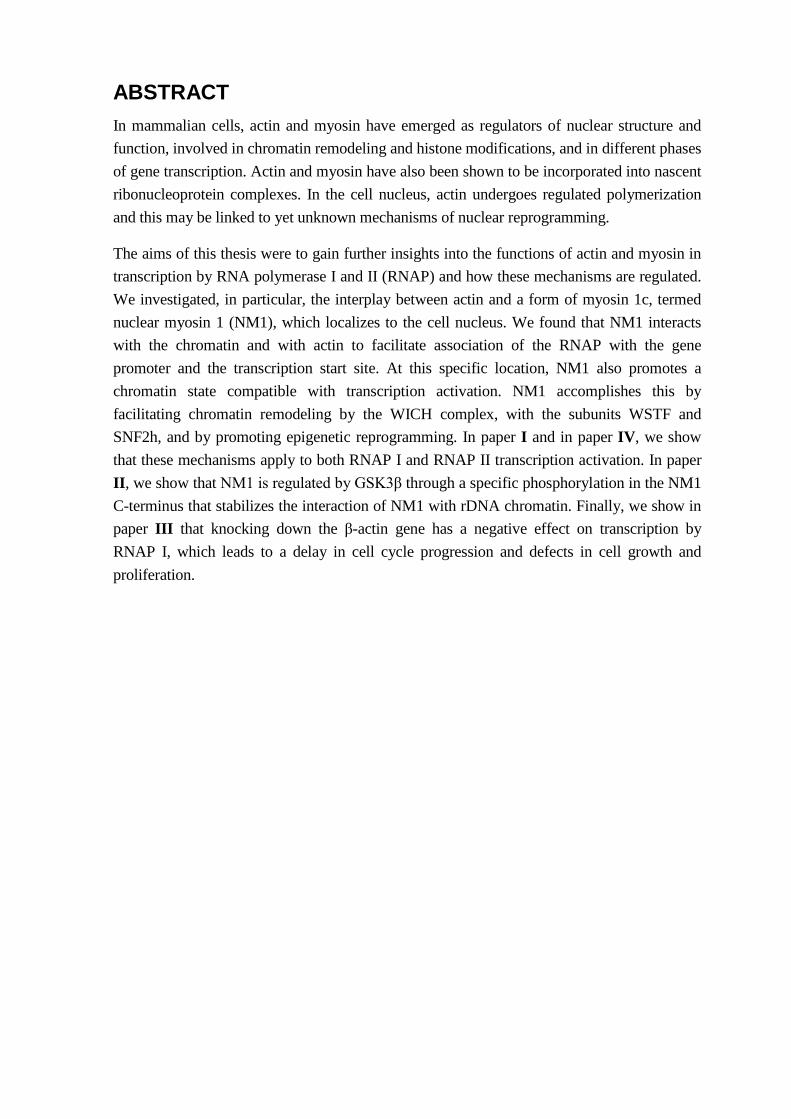
ABSTRACT In mammalian cells, actin and myosin have emerged as regulators of nuclear structure and function, involved in chromatin remodeling and histone modifications, and in different phases of gene transcription. Actin and myosin have also been shown to be incorporated into nascent ribonucleoprotein complexes. In the cell nucleus, actin undergoes regulated polymerization and this may be linked to yet unknown mechanisms of nuclear reprogramming.
The aims of this thesis were to gain further insights into the functions of actin and myosin in transcription by RNA polymerase I and II (RNAP) and how these mechanisms are regulated. We investigated, in particular, the interplay between actin and a form of myosin 1c, termed nuclear myosin 1 (NM1), which localizes to the cell nucleus. We found that NM1 interacts with the chromatin and with actin to facilitate association of the RNAP with the gene promoter and the transcription start site. At this specific location, NM1 also promotes a chromatin state compatible with transcription activation. NM1 accomplishes this by facilitating chromatin remodeling by the WICH complex, with the subunits WSTF and SNF2h, and by promoting epigenetic reprogramming. In paper I and in paper IV, we show that these mechanisms apply to both RNAP I and RNAP II transcription activation. In paper II, we show that NM1 is regulated by GSK3β through a specific phosphorylation in the NM1 C-terminus that stabilizes the interaction of NM1 with rDNA chromatin. Finally, we show in paper III that knocking down the β-actin gene has a negative effect on transcription by RNAP I, which leads to a delay in cell cycle progression and defects in cell growth and proliferation.
LIST OF SCIENTIFIC PAPERS
I. Sarshad, A., Sadeghifar, F., Louvet, E., Mori, R., Böhm, S., Al-Muzzaini, B., Vintermist, A., Fomproix, N., Östlund, A. K. &Percipalle, P. (2013) Nuclear myosin 1c facilitates the chromatin modifications required to activate rRNA gene transcription and cell cycle progression, PLoS Genet, 9(3), e1003397.
II. Sarshad, A. A., Corcoran, M., Al-Muzzaini, B., Borgonovo-Brandter, L., Von Euler, A., Lamont, D., Visa, N. &Percipalle, P. (2014) Glycogen synthase kinase (GSK) 3β phosphorylates and protects nuclear myosin 1c from proteasome-mediated degradation to activate rDNA transcription in early G1 cells, PLoS Genet, 10(6), e1004390.
III. Almuzzaini, B., Sarshad, A. A., Rahmanto, A.S., Hansson, M.L., Von Euler, A., Sangfelt, O., Visa, N., Östlund Farrants, A. K. &Percipalle, P. (2016) In β-actin Knockouts epigenetic reprogramming leads to rDNA transcription inactivation, growth and proliferation defects (Accepted for publication in the FASEB Journal 18-4-2016 ).
IV. Almuzzaini, B., Sarshad, A. A., Östlund Farrants, A. K. &Percipalle, P. (2015) Nuclear myosin 1 contributes to a chromatin landscape compatible with RNA polymerase II transcription activation, BMC Biol, 13(1), 35.
CONTENTS 1 Gene expression in eukaryotic cells ................................................................................ 1 2 RNA Polymerase I transcription ..................................................................................... 2
2.1 RNAP I transcription process................................................................................ 2 2.2 Regulation .............................................................................................................. 4
3 RNA polymerase II transcription .................................................................................... 5 3.1 Pre-initiation complex formation (PIC) ................................................................ 5 3.2 Initiation ................................................................................................................. 6 3.3 Elongation and termination ................................................................................... 7
4 Chromatin and transcription ............................................................................................ 8 4.1 Epigenetic regulation ............................................................................................. 9 4.2 Chromatin remodelling ......................................................................................... 9
5 Actin and myosin in eukaryotic cells ............................................................................ 11 5.1 Actin and its regulated polymerization states ..................................................... 11 5.2 Actin-based myosin motors................................................................................. 12 5.3 Actin and myosin in nucleus ............................................................................... 13 5.4 Actin and myosin in transcription ....................................................................... 15 5.5 Nuclear actin dynamics and impact on genome organization ............................ 18
6 Chromatin immunoprecipitation and genome-wide analysis ...................................... 20 6.1 Introduction to the technology ............................................................................ 20 6.2 Chromatin immunoprecipitation and deep sequencing ...................................... 20 6.3 Advantages and disadvantages ............................................................................ 24
7 Aim of the thesis ............................................................................................................ 25 8 Results and summary .................................................................................................... 26
8.1 Paper I .................................................................................................................. 26 Nuclear Myosin 1c Facilitates the Chromatin Modifications Required to
Activate rRNA Gene transcription and cell cycle progression .......................... 26 8.2 Paper II ................................................................................................................. 28 Glycogen Synthase Kinase (GSK) 3β Phosphorylates and Protects Nuclear
Myosin 1c from Proteasome-Mediated Degradation to Activate rDNA Transcription in Early G1 Cells .......................................................................... 28
8.3 Paper III ............................................................................................................... 30 In β-actin Knockouts epigenetic reprogramming leads to rDNA transcription
inactivation, growth and proliferation defects .................................................... 30 8.4 Paper IV ............................................................................................................... 31 Nuclear myosin 1 contributes to a chromatin landscape compatible with RNA
polymerase II transcriptional activation.............................................................. 31 9 General conclusion and future prespectives .............................................................. 33 10 Acknowledgements ....................................................................................................... 35 11 References ..................................................................................................................... 37

LIST OF ABBREVIATIONS
ABPS
ADP
ARP
Actin binding proteins
Adenosine diphosphate
Actin related proteins
ATP Adenosine triphosphate
BAF BRG-associated factor
BDM Butane dione monoxime
CB Cajal body
CDK Cyclin-dependent kinase
ChIP Chromatin immunoprecipitation
ChIP-seq ChIP combined with next generation sequencing
CORE Core promoter element
CP Core promoter
CPSF Cleavage and polyadenylation specificity factor
CR Chromatin remodeler
CRC Chromatin remodeling complexes
CT Chromosome territories
CTD C-Terminal domain
DFC
ETS
Dense fibrillar component
External transcribed spacer
F-actin Filamentous actin
FC Fibrillar center
G-actin Monomeric actin
GSK3β Glycogen synthase kinase 3beta
GTFs General transcription factors
HAT Histone acetyltransferase
HMG High mobility group
HMT Histone methyltransferase
hnRNP Heterogeneous nuclear ribonucleoprotein
IGS Intergenic sequence
Myb-bp 1a Myb-binding protein 1a
ncRNAs Non-coding RNAs
NM1 Nuclear myosin I
NORs Nucleolar organization regions
NPC Nuclear pore complex
PIC Pre-initiation complex
rDNA Ribosomal DNA
RIP RNA immunoprecipitation
RNAPI RNA polymerase I
RNAPII RNA polymerase II
RNAPIII RNA polymerase III
RNPs
rRNA
Ribonucleoproteins
Ribosomal RNA
SNF2h Sucrose non fermented 2h
TAFᴵS Transcription associated factor
TBP
tRNA
TATA-binding protein
Transfer RNA
TSS Transcription start site
TTF Transcription termination factor
UBF Upstream binding factor
UCE Upstream control element
WSTF Williams Syndrome Transcription Factor
1
1 GENE EXPRESSION IN EUKARYOTIC CELLS Gene expression is a multistep process, which is partly regulated at the chromatin level. The structure of chromatin is modified by different factors that either reposition nucleosomes with respect to each other or covalently modify histones (Voss and Hager, 2014, Valen and Sandelin, 2011). These mechanisms result in establishment of permissive chromatin where the DNA is more accessible to the RNA polymerases (RNAP). This mechanism therefore facilitates the process of transcription when the DNA is transcribed into messenger RNA (mRNA) molecules, which are subsequently translated into functional proteins (Alberts et al., 2008).
In eukaryotic cells, there are three RNA polymerase enzymes which are highly specialized: RNA polymerase II (RNAP II) transcribes protein-coding genes into mRNAs and also transcribes other non-coding RNAs (ncRNAs) (Goodfellow and Zomerdijk, 2013); RNA polymerase I (RNAP I) transcribes ribosomal DNA (rDNA) into ribosomal RNA (rRNA); RNA polymerase III (RNAP III) synthesizes transfer RNA (tRNA) and 5S rRNA (White, 2008).
The assembly of RNAPs at the gene promoter in order to begin the transcription process requires many factors, including general transcription factors (GTFs), activators and mediators, which altogether play vital roles in coordination and orchestration of the different transcription phases. Nuclear actin and myosin are among some of these factors that have recently been identified to have key roles in transcription as well as in other nuclear functions and are therefore believed to be key regulators of gene expression at multiple levels.
Actin is known to be part of chromatin remodeling complexes (CRC), to bind to all three eukaryotic nuclear RNAPs and to specifically interact with the RNAP II at the C-Terminal Domain (CTD). Actin is also known to associate with ribonucleoprotein complexes (RNPs) co-transcriptionally and to accompany the mature RNPs all the way to cytoplasm. In addition, soluble actin molecules shuttle between nuclear and cytoplasmic compartments in a regulated manner and this shuttling is dependent on transcriptional rates. Importantly, recent studies have also demonstrated that actin works as a complex with nuclear myosin 1 (NM1) to regulate gene expression (Percipalle, 2013).
The work presented in this thesis focuses on the importance of actin and NM1 in nuclear functions and highlights their roles in the transcription process of both protein-coding and rRNA genes.
In the next chapter, I will introduce the factors and primary mechanisms in the process of transcription by RNAP I and RNAP II, highlighting the efficiency of the process in eukaryotic cells.
2
2 RNA POLYMERASE I TRANSCRIPTION Ribosomal RNA (rRNA) synthesis is the result of the most abundant transcriptional activity in eukaryotic cells. It is assumed to comprise around 50%-60% of all transcriptional activity in the interphase nucleus throughout the cell cycle with peaks during S-phase (Schlesinger et al., 2009, Goodfellow and Zomerdijk, 2013). rRNA genes are arranged as tandem repeats separated from each other by long stretches of intergenic sequences (IGSs). The tandem repeats are further organized into arrays, termed nucleolar organization regions (NORs), around which the nucleolus is assembled at the exit of mitosis. Not all rRNA genes are active; it is believed that only 20-30% of all rRNA genes are actively transcribed (Moss and Stefanovsky, 2002, McStay and Grummt, 2008). These genes are found at the junction between the fibrillar center (FC) and the dense fibrillar component (DFC), two sub compartments within nucleoli (Raska, 2003, Bártová et al., 2010). rDNA transcribed by RNAP I, a large multiprotein complex with an average molecular size of 590-kDa, comprising 14 subunits. Five of these subunits including , Rbp5, 6, 8, 10, and 12 are shared between all three RNAPs (Vannini and Cramer, 2012).
RNAP I transcribes rDNA into a long pre (precursor) rRNA, the 47S pre-RNA, which is then gradually processed into 18S, 28S, and 5S rRNAs (Moore and Steitz, 2002, Goodfellow and Zomerdijk, 2013), the mature forms which are then incorporated into ribosomal subunits (Moore and Steitz, 2002). Because of its importance in global protein levels and therefore cellular growth and proliferation, rRNA synthesis is directly influenced by numerous signaling pathways and nutrients (White, 2008, Goodfellow and Zomerdijk, 2013, Drygin et al., 2010).
In the next paragraph I will highlight the different phases of RNAP I transcription
2.1 RNAP I TRANSCRIPTION PROCESS
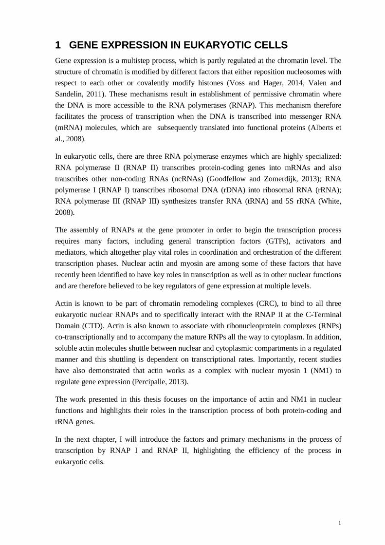
The initiation step takes place at the gene promoter and, in particular, at the upstream control element (UCE) and the core promoter element (CORE) where RNAP I and other transcription factors are recruited to the gene promoter to form the pre-initiation complex (PIC) (Grummt, 2003). In humans, RNAP I assembly is mediated by the upstream binding factor (UBF) and selectivity factor 1 (SL1). In mice SL1 is termed TIFIB: in either case, it is a protein complex which comprises the TATA Box Binding factor (TBP) and the transcription factors TAFI110, 63, and 48 (Goodfellow and Zomerdijk, 2013) (Figure 1-A) .
UBF is a high mobility group (HMG) protein and it is an essential factor for maximal activity of RNAP I (Voit and Grummt, 2001, Drygin et al., 2010). UBF binds to UCE and the core promoter (CP) to recruit TIF1B/SL1. Next, phosphorylated TIF1A (RNAP I-specific transcription initiation factor) binds to the RNAP I subunit RRN3, the RNAP I-associated RRN3 subsequently interacts with UBF and TIF1B/SL1 via its PAF53 subunit (Panov et al., 2006, Drygin et al., 2010, Goodfellow and Zomerdijk, 2013) and as a result, the polymerase becomes recruited to the promoter. These steps lead to the formation of the Pre-Initiation Complex (PIC) in a manner that is dependent on a local opening of the chromatin

3
(Goodfellow and Zomerdijk, 2013). Transcription activation requires some components of the sirtuin family of proteins. The deacetylase SIRT7 has recently been shown to be crucial for RNAP I transcription through the de-acetylation of the RNAP I subunit PAF53, to promote RNAP I association with rDNA and activate rDNA transcription (Chen, 2008). Together with UBF, SIRT7 interaction with chromatin remodeling complexes (CRC) and its association with the rDNA transcription unit are crucial to regulate rRNA synthesis.
Once the polymerase is assembled, it becomes engaged in the elongation of pre-rRNA. UBF is also involved at this stage of transcription. There is evidence that UBF occupies both the gene promoter and the coding region of rRNA genes (O'Sullivan et al., 2002, Schneider, 2012). In addition UBF is also phosphorylated by ERK, and this mechanism is required for RNAP I elongation of nascent pre-rRNA (Moss et al., 2006) (Figure 1-B). Finally, UBF competes for DNA binding with linker histone H1 to rDNA. This mechanism ensures efficient transcription elongation since depletion of UBF in mouse cells leads to H1 binding to rDNA followed by chromatin compaction (Sanij and Hannan, 2009).
Figure 1: Schematic diagram of the RNAP I transcription process. (A) The RNAP I is assembled at the gene promoter to form the initiation complex. (B) Dissociation of GTFs from gene promoter to allow RNAP I to engaged in elongation stage of RNAP I transcription.
The final step of RNAP I transcription process is termination, which takes place at the IGS, downstream of the rDNA sequence. The hall marks for transcription termination is a region termed Sal box that contains 10 termination sequences T1-T10. Termination is triggered by TTF (transcription termination factor) which binds to the T1-T10 sequences and leads to RNAP I cleavage. The cleaved RNAP I is rapidly reassembled at the promoter to start a new transcription cycle (Gerber et al., 1997, Goodfellow and Zomerdijk, 2013).
4
2.2 REGULATION
rDNA transcription is also regulated by other micro environmental factors, such as nutrient availability and cellular stress, in both negative and positive ways. For example, under sub-optimal cellular level of nutrients, RNAP I transcription is down-regulated in contrast to enhanced transcription observed upon increased amounts of cellular nutrients (Grummt, 2003, Moss, 2004, Grummt and Voit, 2010). In addition, transcriptional activity responds in a different way upon application of specific forms of cellular stresses (Russell and Zomerdijk, 2005, Boulon et al., 2010).
In eukaryotic cells, many oncogenes and tumor suppressors have been identified (Drygin et al., 2010). These factors work as key regulators of transcription, and generally speaking, they both target the RNAP I, impacting different phases transcription. Oncogenes and tumor suppressors interfere with each other’s function. For instance, inactivation of a tumor suppressor can lead to hyper activation of one or more oncogenes, which eventually leads to elevated transcriptional activity. The opposite happens inactivation of specific oncogenic factors. So oncogenes and tumor suppressors can affect transcription levels in either positive or negative ways (Drygin et al., 2010).
c-MYC is one of the best characterized oncogenes (Dang, 1999). Under normal conditions, c-MYC regulates transcription, it is known to be directly affect the amount of UBF that binds to rDNA, so that transcription rate is elevated (Hannan et al., 2013) Moreover, c-MYC induces transcription through a chromatin-based mechanism: it interacts directly with chromatin and facilitates recruitment of histone acetyl transferases (HAT) to contribute to a more open chromatin state compatible with transcription (Hannan et al., 2013). Mutations in the c-MYC gene lead to increased activity of the oncogene and enhanced rRNA transcriptional levels. This phenomenon has been correlated with many forms of cancer, including leukemic cases such as lymphoma (Drygin et al., 2010, Hannan et al., 2013, Campbell and White, 2014). On the other hand, tumor suppressors maintain transcription and cell cycle progression in the normal cells, and a loss of function mutation eventually leads to enhanced transcription and proliferation activity. The retinoblastoma (pRb) is among the best characterized tumor suppressors. In the normal cell, pRb targets UBF and interferes with UBF binding to the DNA, which leads to down-regulation in RNAP I transcriptional activity (Drygin et al., 2010).
5
3 RNA POLYMERASE II TRANSCRIPTION RNAP II transcribes protein-coding genes also referred to as class II genes into mRNA. In eukaryotic cells the newly synthesized mRNA is exported and translated into functional proteins within polyribosomes (Alberts et al., 2008).Transcription by RNAP II consists of a number of steps, including formation of the pre-initiation complex (PIC), initiation, elongation, and termination. Overall, these mechanisms require a cohort of many factors that ensure efficient and correct transcription. The RNAP II enzyme comprises 12 subunits, some of which are shared by all three classes of RNAPs as mentioned earlier. A peculiarity of the largest RNAP II subunit is the highly conserved CTD which comprises tens of heptapeptide repeats (Y1S2P3T4S5P6S7) with conserved serine residues in positions 2 (S2) and 5 (S5). During transcription, S2 and S5 are reversibly phosphorylated and this reflects the stage of the transcription process (Hsin and Manley, 2012).
3.1 PRE-INITIATION COMPLEX FORMATION (PIC)
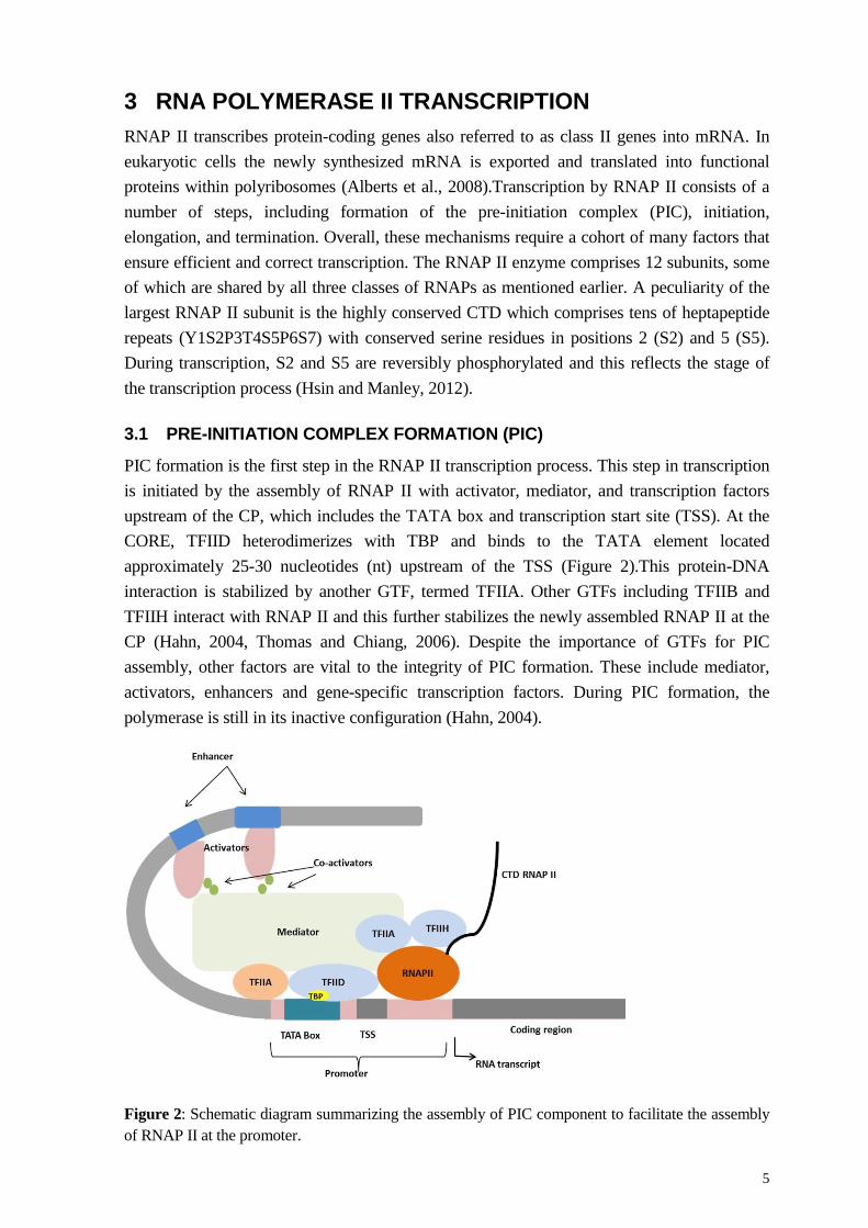
PIC formation is the first step in the RNAP II transcription process. This step in transcription is initiated by the assembly of RNAP II with activator, mediator, and transcription factors upstream of the CP, which includes the TATA box and transcription start site (TSS). At the CORE, TFIID heterodimerizes with TBP and binds to the TATA element located approximately 25-30 nucleotides (nt) upstream of the TSS (Figure 2).This protein-DNA interaction is stabilized by another GTF, termed TFIIA. Other GTFs including TFIIB and TFIIH interact with RNAP II and this further stabilizes the newly assembled RNAP II at the CP (Hahn, 2004, Thomas and Chiang, 2006). Despite the importance of GTFs for PIC assembly, other factors are vital to the integrity of PIC formation. These include mediator, activators, enhancers and gene-specific transcription factors. During PIC formation, the polymerase is still in its inactive configuration (Hahn, 2004).
Figure 2: Schematic diagram summarizing the assembly of PIC component to facilitate the assembly of RNAP II at the promoter.
6
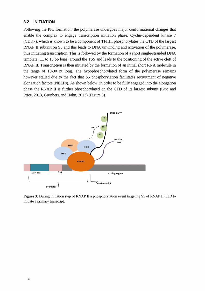
3.2 INITIATION
Following the PIC formation, the polymerase undergoes major conformational changes that enable the complex to engage transcription initiation phase. Cyclin-dependent kinase 7 (CDK7), which is known to be a component of TFIIH, phosphorylates the CTD of the largest RNAP II subunit on S5 and this leads to DNA unwinding and activation of the polymerase, thus initiating transcription. This is followed by the formation of a short single-stranded DNA template (11 to 15 bp long) around the TSS and leads to the positioning of the active cleft of RNAP II. Transcription is then initiated by the formation of an initial short RNA molecule in the range of 10-30 nt long. The hypophosphorylated form of the polymerase remains however stalled due to the fact that S5 phosphorylation facilitates recruitment of negative elongation factors (NELFs). As shown below, in order to be fully engaged into the elongation phase the RNAP II is further phosphorylated on the CTD of its largest subunit (Guo and Price, 2013, Grünberg and Hahn, 2013) (Figure 3).
Figure 3: During initiation step of RNAP II a phosphorylation event targeting S5 of RNAP II CTD to initiate a primary transcript.
7
3.3 ELONGATION AND TERMINATION
Commitment of RNAP II to the elongation process mainly occurs through phosphorylation of S2 within the heptapeptide repeats of the CTD by the cyclin-dependent kinase 9 (CDK9) to form a hyperphosphorylated polymerase. Hyperphosphorylation, in turn, facilitates recruitment of the positive elongation factor (p-ELF) that engages the polymerase into the elongation process, during which the nascent mRNA is synthesized (Peterlin and Price, 2006).
As the polymerase approaches the 3’ end of the active gene, the level of S5 phosphorylation drops whereas S2 phosphorylation levels are maintained. Therefore S2 phosphorylation is considered a hallmark for the termination process. During termination the 3’ end of the mRNA is processed: this involves transcript cleavage and addition of the polyadenylation signal AAUAAA, which is recognized by the cleavage and polyadenylation specificity factor (CPSF) (Richard and Manley, 2009). S2 phosphorylation plays an important role because it facilitates recruitment of polyadenylation factors (such as Pcf11) to form the cleavage and polyadenylation complex (Richard and Manley, 2009). The newly formed mRNA is nicked 20 nt downstream, leading to the end of mRNA synthesis. At the end of this process RNAP II disassociates from the DNA and the mRNA is completely cleaved (Richard and Manley, 2009).
8
4 CHROMATIN AND TRANSCRIPTION In eukaryotic cells, the DNA is in the form of chromatin. While this compaction is required in order to ensure that the DNA is stored in the cell nucleus, it also causes considerable hindrance to the transcription process as the DNA itself is not accessible to the polymerase machinery.
The structure unit of chromatin is termed nucleosome. The nucleosome is made of 147 bp of DNA wrapped around histone octamers consisting of H3/H4 tetramers, and a H2A/H4 dimer (Kornberg, 1974, Negri et al., 2000, Luger et al., 1997). The main consequence of this organization of chromatin is that the DNA is normally buried and must be made accessible to transcription factors and RNAP to allow for transcription (Gilbert and Ramsahoye, 2005). This is normally achieved by remodeling the chromatin, a dynamic event that requires dedicated machineries such as CRC and histone modifying enzymes. These specialized factors alter chromatin prior to transcription and allow transcription factors to access and bind to DNA and to start the transcription process.
Histone modifications target histone tails in different ways to perform several functions, including, acetylation (Bernstein et al., 2005, Haberland et al., 2009, Mathis et al., 1978, Grunstein, 1997), methylation (Bernstein et al., 2005, Schübeler et al., 2000), phosphorylation (Rossetto et al., 2012, DesJarlais and Tummino, 2016), ubiquitination (Bannister and Kouzarides, 2011) and, sumoylation (Bannister and Kouzarides, 2011). Such modifications leads to activation or repression of the transcription process, for example, euchromatin modification leads to transcriptional activation by acetylation of specific residues using HAT and/or methylation by histone methyltransferase (HMT) (Li et al., 2007). On the other hand, heterochromatin modification leads to inactivation or repression of transcription by methylation of a specific residue (Li et al., 2007).
In transcription, histone modifying enzymes are key players at each step from PIC assembly to termination, such that by altering the level of histone acetylation and/or methylation at the promoter leads to transcriptional activation or repression (Percipalle et al., 2006, Drygin et al., 2010, Strohner et al., 2004). One of the interesting aspect that is still under investigation is how histone modifying enzymes and chromatin remodeling complexes are recruited and maintained on the chromatin during the transcription process.
As discussed further in the next chapters, actin and myosin play an important role in the coordination of the different chromatin modifications that are required during transcriptional activity.
9
4.1 EPIGENETIC REGULATION
As mentioned earlier, the chromatin state influences gene activity. The chromatin, in turn, is epigenetically regulated through covalent histone modifications that are mediated by specialized histone modifying enzymes and allow for transcription to be either activated or repressed (Li et al., 2007). Modifications, such as acetylation and methylation, target the histone tails and determine the transcriptional state of the chromatin. For instance, acetylation of histone H3 on lysine 9 (H3K9ac) is enriched at the promoter and TSS of active genes and it is thus considered as an epigenetic mark for active transcription. In contrast, other histone modifications represent marks for inactive chromatin. An example is methylation of H3 on lysine 9 (H3k9m) (Li et al., 2007). Numerous HATs and HMTs are involved in this process, such as the HMT Set/Ash1 and the HATs PCAF and p300 (Li et al., 2007). Because these histone modifications are required for transcription activation, the HATs PCAF and p300 are believed to function as co-activators for RNAPII transcription (Li et al., 2007).
4.2 CHROMATIN REMODELLING
As mentioned earlier, chromatin is made up of nucleosomes and it is a key player in transcriptional activation. Alterations in nucleosome position, such as nucleosome remodeling, sliding and displacement, all lead to changes in DNA accessibility, rendering the DNA more or less available for interactions with transcription factors and RNAP. ATP-dependent chromatin remodelers (CR) play an essential role: they alter chromatin structure by repositioning nucleosomes with respect to each other and this leads to DNA loop formation and transcriptional activation. In mammalian cells, there are four major chromatin remodeling complexes: SWI2/SNF2h, Mi2/CHD, ISWI/ATPase, and Ino80 (Mohrmann and Verrijzer, 2005, Percipalle and Farrants, 2006). The SWI/SNF family of remodelers comprises an ATPase domain and a so-called bromodomain (Percipalle and Farrants, 2006). They have been recently shown to be localized at gene promoters and around TSS (Morris et al., 2014), suggesting these CR are positive regulators of many genes. These remodelers use their bromodomain modules to interact with acetylated lysine residues; once bound to the chromatin, ATP hydrolysis leads to disrupt the interaction between DNA and chromatin (Zeng and Zhou, 2002, Sanchez and Zhou, 2009). These mechanisms are believed to be important both at the promoter and across the active gene. Therefore, SWI/SNF remodelers are likely to play an important role in both transcription activation and elongation. In support of these hypothesis, SWI/SNF remodelers have been shown to occupy gene promoter and coding sequences (Percipalle et al., 2006, Morris et al., 2014) and in yeast cells, there is evidence that these remodelers also accompany the RNAP II machinery during the elongation phase either in a direct or indirect way (Schwabish and Struhl, 2007).
The Ino80 family of remodelers also plays a major role in the regulation of transcriptional activation and in DNA repair. The structure of Ino80 remodelers comprises two split ATPase domains (Conaway and Conaway, 2009). However, the precise mechanisms of how they work are not fully understood. In contrast to SWI/SNF and Ino80, the ISWI remodeler is known to be a negative regulator of transcription (Venters and Pugh, 2009).
10
The last CR family is CHD; DNA array experiments have shown that a mutant form of CHD1 leads to dysregulation of expression in a limited number of genes (Tran et al., 2000, Venters and Pugh, 2009). However, despite this evidence suggesting a regulatory role of CHD in gene pathways, it is remains unclear how it functions from a mechanistic point of view.
In transcription CRs are known to play both positive and negative roles (Zhou et al., 2002). The NORC complex, for example, which consists of TIP5 and the ATPase subunit Sucrose Non Fermented 2h (SNF2h) works as negative regulator by silencing transcription through the recruitment of HMTs. This eventually leads to repression of transcription (Zhou et al., 2002). In addition, another chromatin remodeler, termed WICH, belongs to both the SWI/SNF and SWI families. This complex comprises of two proteins, the Williams Syndrome Transcription Factor (WSTF) and the SNF2h. Within the WICH complex, WSTF binds to the chromatin and recruits the remodeler SNF2h and this is important during transcription (Bozhenok et al., 2002, Percipalle et al., 2003). Moreover, mass spectrometry data demonstrated that WSTF interacts with the Myb-binding protein 1a (Myb-bp 1a) and proto-oncogene DEK (Kappes et al., 2001). Additionally, the WICH complex interacts with NM1C to form the multiprotein complex B-WICH that plays major roles in RNAP I transcription (Percipalle et al., 2006).
11
5 ACTIN AND MYOSIN IN EUKARYOTIC CELLS
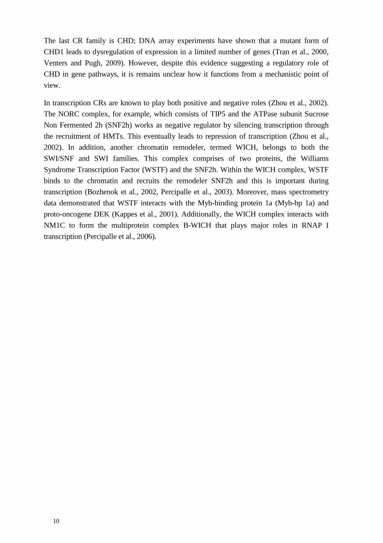
5.1 ACTIN AND ITS REGULATED POLYMERIZATION STATES
Actin is a 43-kDa protein and is comprised of three isoforms: α, β and γ (Herman, 1993). Actin, in particular, the β-actin isoform, is currently known to be the most abundant and conserved protein in eukaryotic cells (Dominguez and Holmes, 2011). Actin is present in cell cytoplasm in equilibrium between the monomeric (G) and filamentous (F) forms; this equilibrium between G and F actin plays major roles in numerous cellular processes, for example, cell motility and signal transduction (Clark and Rosenbaum, 1979, Pederson and Aebi, 2002). The transition between the G and F form of actin is an ATP-based process where actin constantly polymerizes and depolymerizes. This dynamic mechanism is referred to ‘treadmilling’ and is known to produce power force for movement (Paavilainen et al., 2004, Lee and Dominguez, 2010, Percipalle, 2013). In the treadmilling process, G actin joins the barbed end or (+) end in an ATP-bound state (Figure 4). At the other end of the filament, the pointed end or (-) end, G actin is released in an ADP-bound state. Addition and release of actin monomer is highly regulated by actin binding proteins (ABPs), including profilin and cofilin. Cofilin binds to the ADP-bound form of actin and mediates severing of the filament and release of ADP-bound actin monomers. Cofilin is in turn released from actin monomers and through a nucleotide exchange mechanism actin binds to ATP. The ATP-bound form of actin is recognized by profilin which in turn facilitates loading of ATP-bound actin monomers to the barbed ends of the growing filament. In this way, cofilin and profilin control the dynamic of depolymerization and polymerization of actin during treadmilling (Paavilainen et al., 2004, Lee and Dominguez, 2010, Percipalle, 2013) (Figure 4).
Figure 4: The mechanism of actin polymerization and depolymerization by treadmilling, adapted from (Percipalle, 2013).
12
Cofilin and profilin are not the only proteins regulating actin polymerization and depolymerization. The dynamic changes in actin polymerization are also controlled by co-factors such as the Arp2/3 complex and the N-WASP (neuronal Wiskott–Aldrich Syndrome protein), which required for nucleation and branching of actin filaments respectively (Vartiainen, 2008). Actin polymerization can also be controlled by formins which facilitates the elongation of actin filaments at the barbed end (Zigmond, 2004). Regulating the polymerization state of actin is important to ensure interactions with myosin motors that require actin filaments to ensure motion. The interaction between actin and myosin is dependent on ATP hydrolysis. Only ADP-bound myosin interacts with actin. In the presence of inorganic phosphate when the ATP is reconstituted, ATP-bound myosin leaves actin and generates short movement along the actin filament on which it resides. This step is called ‘power stroke’. Upon ATP hydrolysis, when the myosin is in an ADP-bound form, the interaction with actin is reconstituted and a new cycle starts again. This general mechanism is fundamental for cytoplasmic processes (Pollard and Korn, 1973).
Emerging evidence suggests that this mechanism is also important in the eukaryotic cell nucleus, where it is involved in transcription (Sarshad and Percipalle, 2014). There is evidence that actin also undergoes regulated polymerization (Baarlink et al., 2013, Belin et al., 2013). Possibly this may happen across active genes. Indirect evidence supporting this possibility is the discovery that the N-WASP (Wu et al., 2006) and the Arp2/3 (Yoo et al., 2007) are involved in transcription across the active gene, that profilin binds to active transcription sites (Söderberg et al., 2012) and that cofilin targets RNAP II in an actin dependent manner and is required for transcription elongation (Obrdlik and Percipalle, 2011).
5.2 ACTIN-BASED MYOSIN MOTORS
In eukaryotic cells, myosin proteins are clustered into a large family of proteins that includes 18 different types. Despite of this complexity, myosin has a conserved function, working as an actin-dependent motor that generates force to drive movement through establishment of a power stroke. Myosin is abundant in the cytoplasmic compartment and interacts with actin to generate movement in a number of processes. The main structure of myosin is generally conserved in all isoform and comprises a C-terminus or tail domain that differs in length on the basis of the type of myosin and the N-terminus or head domain, which includes the actin binding site and ATP-binding pocket. This explains the functional specificity of each type of myosin. The first non-canonical myosin was discovered 40 years ago and given the name Myosin 1 (Pollard and Korn, 1973).
Myosin processivity, defined as the time spent interacting with the actin filament, and force generation that is created by ATP hydrolysis are crucial parameters for the classification of different myosin types. Both parameters have implications on the type of movement generated by the individual myosin motor. For instance, myosin V is a highly processive motor, which makes it suitable for intracellular transport of vesicles. In contrast, myosin I has low processivity and therefore it is not suitable for long-range movement, mostly because it
13
does not spend enough time bound to the actin filament (Coluccio, 2008, Sarshad and Percipalle, 2014). Cytoplasmic Myosin 1 is involved in many cellular processes and it is ubiquitous. It is, for instance, enriched at microvilli of small intestine epithelial cells where it crosslinks acting bundles with the cell membrane (Nambiar et al., 2010). Myosin1 is also present in hair cells of the inner ear and regulates ion channel (Cyr et al., 2002). Myosin 1 is also enriched in neurons where it is needed together with actin for the formation of membrane extensions, a process important for cone motility (Wang et al., 2003).
In conclusion, myosin 1 is able to generate short movement but due to its low processivity, it cannot move cargos over long distances; it is therefore believed to function as crosslinker of actin filaments and not as cargo transporter (Coluccio, 2008).
5.3 ACTIN AND MYOSIN IN NUCLEUS
Despite many independent observations supporting the presence of actin in the cell nucleus of amphibians and animals (Clark and Rosenbaum, 1979, Nakayasu and Ueda, 1983), this hypothesis has faced much skepticism over the past 40 years. Many scientists claimed that the evidence of actin in the cell nucleus was a result of cytoplasmic contamination partly due to the sticky nature of actin itself (Pederson and Aebi, 2002, Pederson and Aebi, 2005, Louvet and Percipalle, 2009). Moreover, F-actin is known to be visualized in the cytoplasm using a drug called phalloidin (Wehland et al., 1977) which is not able to detect actin in the cell nucleus; therefore, many have adhered to the notion that actin is strictly cytoplasmic protein that has no role in the cell nucleus (Percipalle and Visa, 2006).
Since recent decades, the presence of actin in the cell nucleus has become a consolidated notion. In the cell nucleus, actin appears to be involved in numerous functions. Nuclear actin is part of CRCs; actin is required for transcription by all nuclear eukaryotic RNA polymerases and it is co-transcriptionally assembled into nascent ribonucleoprotein complexes (RNPs) (Louvet and Percipalle, 2009, Visa and Percipalle, 2010, Percipalle, 2013). Furthermore, regulated nuclear actin polymerization has now been observed and these mechanisms may be implicated in nuclear reprogramming and DNA repair (Miyamoto et al., 2011, Baarlink et al., 2013, Belin et al., 2015).
Many actin binding proteins have also been found in the eukaryotic cell nucleus, including several myosin types (see below). Among these myosin forms, the best characterized is a form of myosin 1c. Initial evidence for its presence in the cell nucleus dates back to the late 90s when electron microscopy analysis using a general antibody against myosin 1 demonstrated that myosin 1c is present in the cell nucleus and even in the nucleolus (Nowak et al., 1997). However, the formal discovery of the first myosin type localizing to the cell nucleus came from the laboratory of Primal de Lanerolle. This group found that a novel myosin 1C isoform entirely localized to the cell nucleus. This isoform, generally termed as nuclear myosin 1 C (NM1), is an alternatively spliced form of the canonical myosin 1c (Pestic-Dragovich et al., 2000).
14
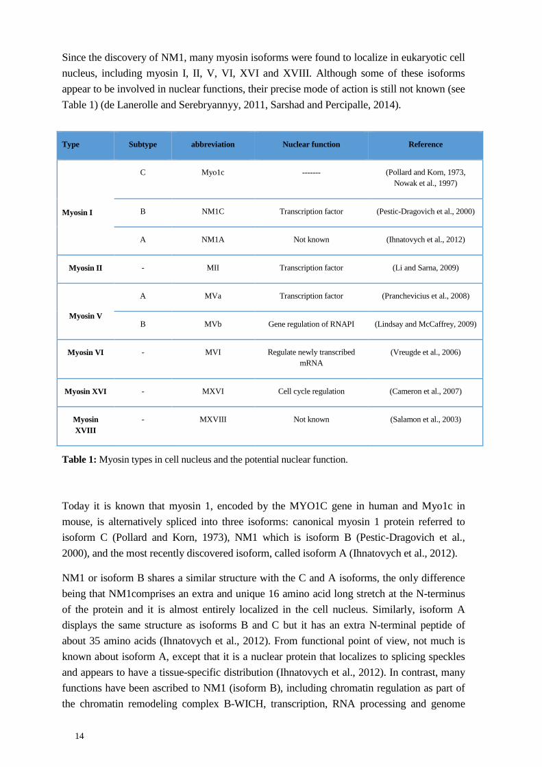
Since the discovery of NM1, many myosin isoforms were found to localize in eukaryotic cell nucleus, including myosin I, II, V, VI, XVI and XVIII. Although some of these isoforms appear to be involved in nuclear functions, their precise mode of action is still not known (see Table 1) (de Lanerolle and Serebryannyy, 2011, Sarshad and Percipalle, 2014).
Table 1: Myosin types in cell nucleus and the potential nuclear function.
Today it is known that myosin 1, encoded by the MYO1C gene in human and Myo1c in mouse, is alternatively spliced into three isoforms: canonical myosin 1 protein referred to isoform C (Pollard and Korn, 1973), NM1 which is isoform B (Pestic-Dragovich et al., 2000), and the most recently discovered isoform, called isoform A (Ihnatovych et al., 2012).
NM1 or isoform B shares a similar structure with the C and A isoforms, the only difference being that NM1comprises an extra and unique 16 amino acid long stretch at the N-terminus of the protein and it is almost entirely localized in the cell nucleus. Similarly, isoform A displays the same structure as isoforms B and C but it has an extra N-terminal peptide of about 35 amino acids (Ihnatovych et al., 2012). From functional point of view, not much is known about isoform A, except that it is a nuclear protein that localizes to splicing speckles and appears to have a tissue-specific distribution (Ihnatovych et al., 2012). In contrast, many functions have been ascribed to NM1 (isoform B), including chromatin regulation as part of the chromatin remodeling complex B-WICH, transcription, RNA processing and genome
Type Subtype abbreviation Nuclear function Reference
Myosin I
C Myo1c ------- (Pollard and Korn, 1973, Nowak et al., 1997)
B NM1C Transcription factor (Pestic-Dragovich et al., 2000)
A NM1A Not known (Ihnatovych et al., 2012)
Myosin II - MII Transcription factor (Li and Sarna, 2009)
Myosin V
A MVa Transcription factor (Pranchevicius et al., 2008)
B MVb Gene regulation of RNAPI (Lindsay and McCaffrey, 2009)
Myosin VI - MVI Regulate newly transcribed mRNA
(Vreugde et al., 2006)
Myosin XVI - MXVI Cell cycle regulation (Cameron et al., 2007)
Myosin XVIII
- MXVIII Not known (Salamon et al., 2003)
15
organization (Percipalle et al., 2006, Percipalle and Farrants, 2006, Visa and Percipalle, 2010, Obrdlik et al., 2010, Sarshad et al., 2013, Almuzzaini et al., 2015, Mehta et al., 2010). Furthermore, as outlined in the next sections, NM1 acts synergistically with actin in numerous nuclear functions as part of an actomyosin complex (Sarshad and Percipalle, 2014).
5.4 ACTIN AND MYOSIN IN TRANSCRIPTION
Actin is not only a component of CRCs but it is also required for ATP-dependent remodeling activity (Rando et al., 2002). In mammals, nuclear actin is directly associated with BRG-associated factor (BAF) to form a complex (Zhao et al., 1998). Although the functional significance is yet to be understood, in certain CRCs such as Ino80, actin interacts with specific actin-related proteins (ARPs) (Kapoor et al., 2013). As component of CRCs, actin probably plays a role at the gene level to ensure that the chromatin is in a permissive state prior to polymerase assembly and transcription initiation. Actin as CRCs is likely to have a global role that affects the general organization of the genome.
Although the precise role of actin in CRCs is still under investigation, at the transcriptional level we are beginning to have mechanistic insights into how actin functions. Actin is found to be associated with RNAP I, II, and III to form a complex (Philimonenko et al., 2004, Hofmann et al., 2004, Hu et al., 2004). The interaction with the polymerase machineries seems to occur in a conserved manner since actin interacts with the polymerase subunits Rbp6 and Rbp8 which are shared among all three nuclear RNAP (Hu et al., 2004). This observation suggests that actin has a conserved function in complex with the all three RNAPs and this might be in the context of their initial assembly at the gene promoter.
In the case of RNAP II, Hofmann et al (2004) demonstrated that formation of the PIC complex requires β-actin. At the early transcriptional stages, actin also plays an important role in escape from pausing, promoter clearance and later on, commitment of RNAP II to elongation (Percipalle, 2013). Indeed, there is evidence showing that actin interacts with the transcriptional co-activator PSF to facilitate recruitment of the p-TEFB complex required to phosphorylate the RNAP II CTD, a mechanism necessary for escape from pausing (Qi et al., 2011). In mammalian cells, our lab showed that actin interacts with the heterogeneous nuclear ribonucleoprotein (hnRNPU), and with the phosphorylated form of the RNAP II CTD (Kukalev et al., 2005).
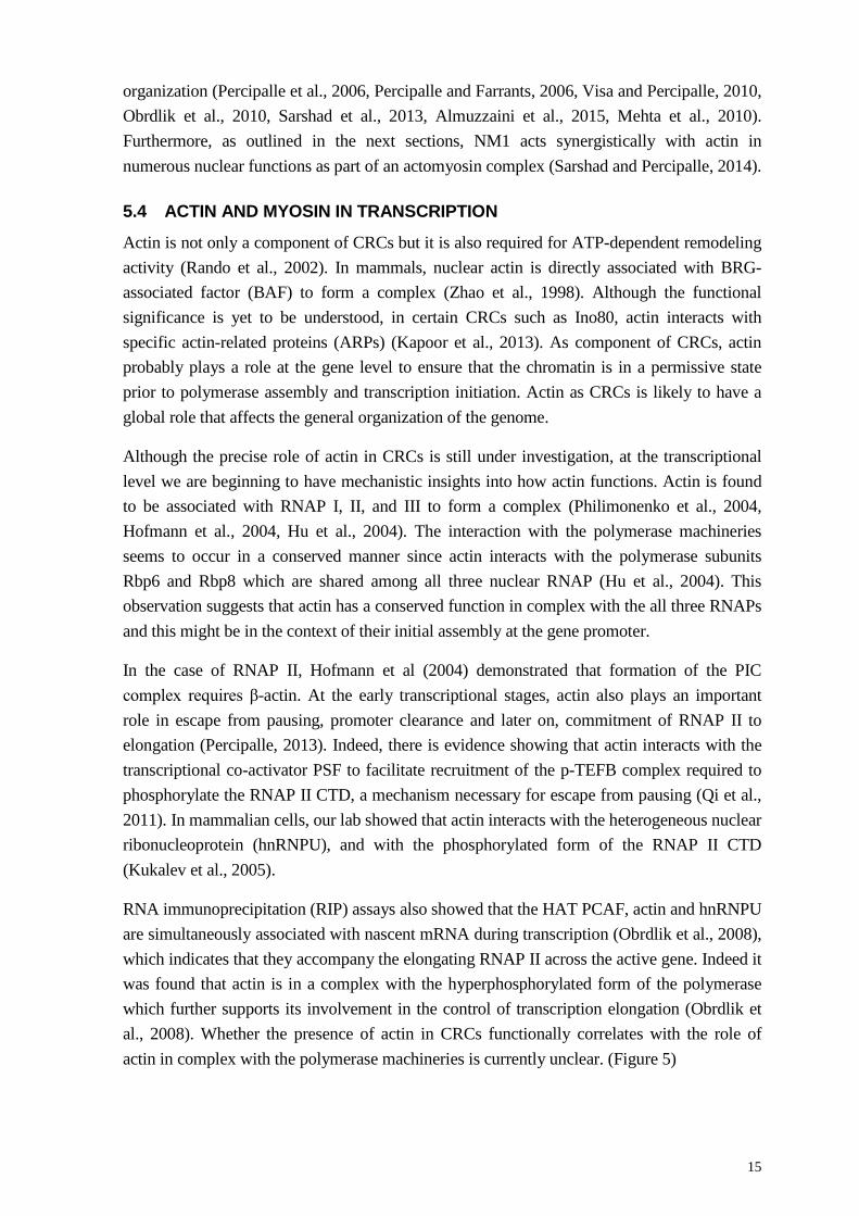
RNA immunoprecipitation (RIP) assays also showed that the HAT PCAF, actin and hnRNPU are simultaneously associated with nascent mRNA during transcription (Obrdlik et al., 2008), which indicates that they accompany the elongating RNAP II across the active gene. Indeed it was found that actin is in a complex with the hyperphosphorylated form of the polymerase which further supports its involvement in the control of transcription elongation (Obrdlik et al., 2008). Whether the presence of actin in CRCs functionally correlates with the role of actin in complex with the polymerase machineries is currently unclear. (Figure 5)
16
Figure 5: Role of actin in RNAP II transcription process, (Percipalle, 2013).
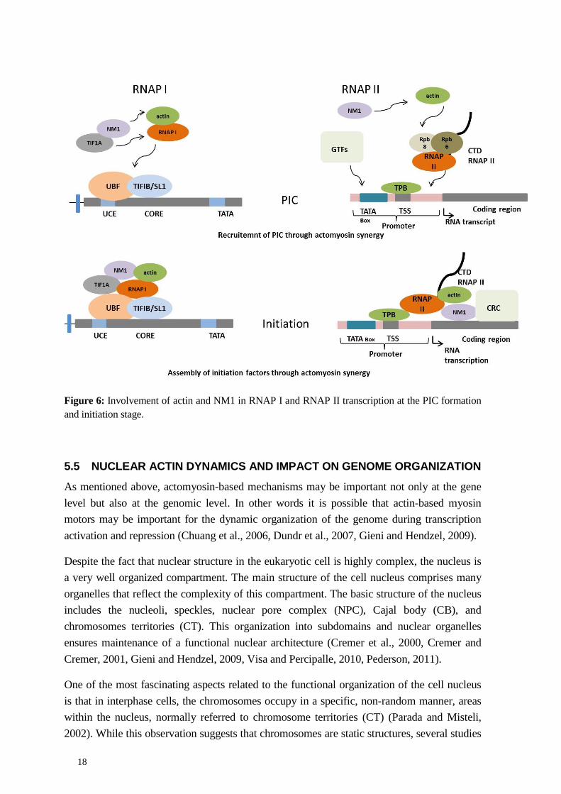
One of the main questions is whether actin functions with NM1 to facilitate the transcription process. This question is highly relevant as NM1 has also been shown to associate with the transcription machinery by several independent studies (Sarshad and Percipalle, 2014) . In situ run-on assays showed that NM1 is associated with nascent transcription foci both in the nucleus and in the nucleolus (Fomproix and Percipalle, 2004). The same study provided the first evidence that NM1 and actin are in a complex with the largest RNAP I subunit, suggesting that actin and NM1 may have a concerted function in transcription of rRNA genes (Fomproix and Percipalle, 2004). A follow up study supported this hypothesis. It was shown that NM1 binds the phosphorylated form of TIF-IA whereas actin directly interacts with the polymerase machinery (Philimonenko et al., 2004). These results were taken as initial evidence that actin and NM1 may facilitate assembly of RNAP I at the gene promoter (Philimonenko et al., 2004).
Indeed, using chromatin immunoprecipitation (ChIP) assays, our lab showed that NM1 associates with the rRNA gene promoter and it is also found across the entire rRNA gene (Percipalle et al., 2006, Ye et al., 2008, Obrdlik et al., 2010). Furthermore, in vitro transcription assays antibodies against NM1 impaired RNAP I transcription (Percipalle et al., 2006). Finally, NM1 gene knockdown by siRNA led to drops in rRNA synthesis supporting a direct role for NM1 in rRNA gene transcription in living cells (Sarshad et al., 2013).
NM1 has also been connected to transcription of protein-coding genes by RNAP II. In vitro, incubation of a nuclear lysate with an anti-NM1 antibody led to inhibition of RNAP II transcription (Pestic-Dragovich et al., 2000, Hofmann et al., 2006). Although the mechanisms
17
are not understood, a follow up in vitro study provided evidence that NM1 is important for the formation of the first phosphodiester bond by RNAP II (Hofmann et al., 2006).
It is now believed that NM1 has a more general role in RNAP II transcription. A recent genome-wide analysis performed by ChIP and deep sequencing (see below for a detailed explanation) showed that NM1 broadly associates with a large proportion of RNAP II promoters and is required for mRNA synthesis (Almuzzaini et al., 2015).
Whether NM1 cooperates with actin during the transcription process has been a key question to address. Initial insights came from a study by Fomproix and Percipalle (2004) where actin and NM1 co-precipitated with RNAP I from nuclear extracts. In this study the motor function of NM1 was suggested to be involved in RNAP I transcription. Further mechanistic details came from work by the Grummt lab (Ye et al., 2008). In this study, transcriptional analysis performed on cells constitutively expressing NM1 mutants showed that ablation of the actin- and ATP-binding sites on NM1 negatively regulated transcription of rRNA genes, supporting not only the requirement for the myosin motor domain but also a synergistic function of NM1 and actin in transcription.
In a recent study from our laboratory, the precise mechanisms through which NM1 and actin synergize to activate RNAP I transcription were further dissected (Sarshad et al., 2013). Here, NM1 was shown to directly bind the chromatin via its C-terminal domain. When bound to the chromatin, NM1 interacts with the RNAP I machinery via a direct interaction with the polymerase-associated actin. The interaction between actin and NM1 was found to be dependent on the NM1 motor function. Initial evidence indicated that when the chromatin-bound NM1 does not interact with actin, it associates with the chromatin remodeling complex WICH and in particular with the ATPase subunit SNF2h. This interaction stabilizes the B-WICH complex and its chromatin remodeling activity. Therefore, it is postulated that NM1 connects the RNA polymerase with a permissive chromatin compatible with transcription (Sarshad et al., 2013, Sarshad and Percipalle, 2014). Interestingly, binding of NM1 with the chromatin is dependent on a phosphorylation event directly mediated by the glycogen synthase kinase (GSK) 3β that phosphorylates Ser1020 within the C-terminal domain (Sarshad and Percipalle, 2014, Sarshad et al., 2014). These findings support the idea that the actin-NM1 complex involved in transcription may be regulated by multiple intracellular signaling pathways with GSK3β as downstream effector.
Recent evidence from our lab indicates that the above mechanisms were conserved at the promoters of RNA polymerase II genes where NM1 binds to the chromatin via its C-terminal domain and interacts with actin bound to the RNAP II subunits Rpb6 and Rpb8. Again the actin-NM1 interaction depends on the motor domain of NM1. Similarly to RNAP I transcription, when NM1 does not interact with actin, NM1 ensures correct assembly and WICH-dependent remodeling activity, followed by recruitment of HATs and HMTs that provide a chromatin state which is compatible with transcription (Almuzzaini et al., 2015) (Figure 6).
18
Figure 6: Involvement of actin and NM1 in RNAP I and RNAP II transcription at the PIC formation and initiation stage.
5.5 NUCLEAR ACTIN DYNAMICS AND IMPACT ON GENOME ORGANIZATION
As mentioned above, actomyosin-based mechanisms may be important not only at the gene level but also at the genomic level. In other words it is possible that actin-based myosin motors may be important for the dynamic organization of the genome during transcription activation and repression (Chuang et al., 2006, Dundr et al., 2007, Gieni and Hendzel, 2009).
Despite the fact that nuclear structure in the eukaryotic cell is highly complex, the nucleus is a very well organized compartment. The main structure of the cell nucleus comprises many organelles that reflect the complexity of this compartment. The basic structure of the nucleus includes the nucleoli, speckles, nuclear pore complex (NPC), Cajal body (CB), and chromosomes territories (CT). This organization into subdomains and nuclear organelles ensures maintenance of a functional nuclear architecture (Cremer et al., 2000, Cremer and Cremer, 2001, Gieni and Hendzel, 2009, Visa and Percipalle, 2010, Pederson, 2011).
One of the most fascinating aspects related to the functional organization of the cell nucleus is that in interphase cells, the chromosomes occupy in a specific, non-random manner, areas within the nucleus, normally referred to chromosome territories (CT) (Parada and Misteli, 2002). While this observation suggests that chromosomes are static structures, several studies
19
point towards the dynamic features of chromosomes since gene-rich chromosome regions have a tendency to localize in the interior of the cell nucleus, independently of the territory they belong to (Parada and Misteli, 2002, Foster and Bridger, 2005, Dundr et al., 2007). These apparent paradoxes have been explained by the fact that gene rich chromosomes loops are mostly located in the perichromatin region of chromosomes, located at the boundary between chromosome territories. Gene rich loops project outward into the nuclear interior upon transcription activation (Chubb et al., 2002, Gasser, 2002, Dundr et al., 2007). This ensures that loops originating from different chromosomes and therefore containing different genes mingle and define neighborhoods of active transcription where genes are simultaneously transcribed. Furthermore, organelles such as speckles and CB are known to be present in the nuclear interior and therefore the above mechanisms ensure that gene-rich chromosomes loops interact with these organelles as well.
Importantly, it has been suggested that directional movement of chromosomes loops upon transcription activation is partly dependent on actin polymerization and on possible interactions with myosin. Indeed, in the past few years, several lines of evidence have underscored the importance of actin and myosin in genomic organization. First, expression of mutated actin forms where polymerization is either negatively or positively affected lead to increases in the speed of long-range gene movement (Chuang et al., 2006). Second, transient expression of myosin or treatment with butane dione monoxime (BDM), that impairs the motor function of myosin, led to reduced gene positioning (Chuang et al., 2006, Gieni and Hendzel, 2009). More direct evidence about the involvement of actin in long-range chromosome movement came from a study by Dundr et al (2007). Here, stable cell lines that contain a tandem array of 16 tetracycline U2 snRNA genes were found to move away from their own territory located in the nuclear periphery into the nuclear interior to interact with CB. Movement of this gene-rich locus was negatively affected by expression of a mutated form of actin that cannot polymerize (Dundr et al., 2007, Carmo-Fonseca, 2007).
Although we still do not understand the mechanisms, the above studies and recent evidence that regulated actin polymerization occurs in the cell nucleus indicate that actin and myosin are likely to have an important role in the functional architecture of the cell nucleus (Belin et al., 2013). To address some of the open questions and gain mechanistic insights into the importance of nuclear actin as a major player in the context of genomic organization, high throughput genome wide technologies are likely to be important.
20
6 CHROMATIN IMMUNOPRECIPITATION AND GENOME-WIDE ANALYSIS
6.1 INTRODUCTION TO THE TECHNOLOGY
In 1985, Solomon and Varshavesky applied the first cross-linking method for protein and DNA in vivo (Solomon and Varshavsky, 1985). Since that time, protein-DNA interaction, or what is currently known as chromatin immunoprecipitation (ChIP), became a key method to investigate molecular mechanisms of regulatory proteins that interact and regulate DNA at the transcriptional level (Gilchrist et al., 2009).
The basic principle of the ChIP assay is based on cross-linking proteins with chromatin in living cells. Reversible cross-linking is generally performed with formaldehyde. Following the cross-linking, chromatin is isolated and digested or sheared into small fragments, which range in size between 200 to 500bp, using chemical or physical methods, such as enzymes or sonication, respectively. Sheared chromatin is then cleared to get rid of non-specific binding; it is then incubated with a specific antibody against the protein of interest which interacts with the chromatin. The chromatin-protein complexes are captured by affinity resins and released by reversing cross-linking that finally breaks down the interaction between protein and chromatin. The DNA is then extracted and the resulting material is analyzed by PCR, real time PCR (qPCR), or hybridization to a specific targeted locus in the genome (Gilbert et al., 2005, Mahony and Pugh, 2015) (Figure 7).
Although the ChIP method provided important information at the gene level, for many years it was not possible to apply it to a more global analysis to study genomic associations of proteins of interests. This dilemma was sorted out in the late 1990s after many advanced methods were introduced to study gene-protein interactions on a higher scale using comparative genomic hybridization (CGH) microarray and then genome-wide analysis, which allows scientists to improve upon the resolution of genomic analysis. In addition to that, combining ChIP with a genome-wide sequencing method, such as ChIP-on-Chip or ChIP combined with next generation sequencing (ChIP-seq) allow protein interactions with millions of base pairs of the genomic sequence to be studied.
In the next chapter, I will focus on how the ChIP method has gained advantage from the development of genome-wide analysis and next generation sequencing.
6.2 CHROMATIN IMMUNOPRECIPITATION AND DEEP SEQUENCING
During last decade, several high throughput data analysis methods introduced to the scientific community have allowed scientists to conduct more global studies, including genomic, epigenomic, and proteomic approaches, by applying high throughput methods in order to study different biological questions. Next generation sequencing is among the technologies developed extensively during the decades since DNA sequencing was invented by Sanger and Gilbert’s group during the 1970’s (Sanger et al., 1977, Maxam and Gilbert, 1977). Moreover, since the human genome map was completed in 2004 (Lander et al., 2001,
21
Consortium, 2004), the field of next generation sequencing had developed extensively and the mapping of chromatin states has become more feasible, leading to the discovery of the mysterious role of chromatin in many biological processes in health and disease.
ChIP-seq is one of the methods that have gained a lot of advantages from the development of next generation sequencing. It was introduced at the end of the last decade (Barski et al., 2007) and since that time, several chemical reagents and computational analysis software have been developed extensively to assist the achievement of high-quality sample preparation and more precise and informative downstream data analysis.
ChIP-seq, like the conventional ChIP method (see figure 5), is used to study transcription factors and epigenetic modifications, such as histone modifications and DNA methylation. However, ChIP-seq provides a more global overview of the role of TF association with the entire genome, which makes it a more robust method. This method is becoming very common, necessitating the importance of having a clear pipeline to be able to achieve a high-quality data set and also very user-friendly software to be used for large-volume data processing and downstream analysis.

22
Figure 7: Pipeline of a typical ChIP experiment followed by different types of analyses. A) Cells are fixed using formaldehyde to cross-link and preserve protein-DNA interaction, following by chromatin isolation and chromatin shearing into small fragments normally achieved by sonication. B) Chromatin fragments are subjected to immunoprecipitation with an antibody specific to the protein of the interest; generally, about 10% of chromatin is kept aside to be used at a later stage as “Input” reference for normalization. C) The crosslinking is reversed to break DNA-protein interactions and the DNA is isolated for analysis by PCR, q-PCR or deep sequencing to determine which DNA fragments of DNA interact with the protein of interest.
Next, I will summarize and highlight the most critical steps and conditions for ChIP-seq and the main pipeline of analysis.
Chromatin preparation. Even though ChIP is a standard method for both the conventional ChIP method and ChIP-seq, chromatin preparation is a very critical step. Many factors involved in this step including cell type, cross-linking method, cross-linking time, and shearing method, should be optimized to achieve high-quality chromatin in the size range of 200-500 bp. To test the efficiency of this step, several validation steps should be conducted
23
using different conditions to achieve the optimal chromatin fragment size. High-quality samples comprising 200-500bp fragments and DNA concentrations of 10ng/µl are recommended for library construction and subsequent sequencing (Landt et al., 2012).
Antibody selection. This is one of the most important factors. It is very important to select a high quality antibody with low cross reactivity. This could be investigated either by checking previous publications, if they are available, or by following the recommendations of the manufacturer if the antibody has already been tested and approved for use in the ChIP assay. To minimize the risk of having a false positive or false negative, it is important to validate every single batch at different concentrations and to use both positive and negative controls. Then, it should be checked using a known primer that gave a positive and negative result (Landt et al., 2012, Park, 2009).
Control. In addition to the previous considerations, using a negative control helps to avoid a false positive result, which may occur mainly as a result of antibody stickiness or cross reactivity. The most commonly used negative control is the input sample, which has total DNA from the host sample, IP IgG, which is subjected to the IgG antibody to detect nonspecific binding, or IP MOCK, which has no antibody to exclude nonspecific binding with beads (Ma and Wong, 2011).
Sequencing quality metric assessment. Output material from library construction and PCR amplification is sequenced using one of the commercially available sequencers, such as the Illumina Solexa, ABI SOLID, and Roche 454 platforms. As short reads tend to generate raw data, due to the complexity of the data generated by ChIP-seq, proper selection of a commercial platform and the accompanying computational data analysis software is important. The most favorable platforms for ChIP-seq are the Illumina and Roche 454 given the ability of these platforms to provide a high number of reads with a low error (Ma and Wong, 2011). Quality control of sequencing data is a very crucial step before proceeding to data processing and analysis. Many tools can be used to check data quality and one of the most common is the FASTX tool kit, which allows user to trim bases that do not match the quality score. This step will allow the elimination and trimming of undesired reads that result during sample preparation and library construction (Kaspi et al., 2012, Bailey et al., 2013).
Mapping. Short reads that pass the QC step then need to be mapped and aligned against a reference genome. The main idea of mapping is to identify where exactly those reads are allocated in the target genome of the same species and also to be sure that a bias is not present. Many software applications are used for this process, such as BOWTIE, MAC, BWA, and SOAP (Kaspi et al., 2012, Ma and Wong, 2011).
Peak calling. After mapping, the peaks that are generated by the sequencer software need to be defined as to where exactly those reads bind to the genome. This could be achieved through several processes, such as read shifting, background estimation, and peak enrichment at the site. Different peak callers software could be used, such as MAC, SPP and SICER (Bailey et al., 2013).
24
Annotation and differential analysis. To study the biological implication of the TF that interact with DNA, it is important to identify the genes in which those peaks localize; for this, the peaks are annotated according to the nearest gene that is localized either upstream or downstream of the gene. To do that, peaks that are generated from a peak caller can be compared directly to a genome browser, such as UCSC, using an appropriate folder format, such as WIG and BED. The output from this step could be used to study specific biological processes using certain software, such as DAVID or GREAT (Bailey et al., 2013).
ChIP-seq data could be taken further into motif analysis to find out if the TF have affinity for binding a specific DNA motif. Available tools for this study include MEME and others.
Currently, many commercial and free software applications are available to perform genomic analysis of ChIPed material. In addition, other packages have been introduced to perform the analysis, such as GALAXY (Blankenberg et al., 2010) and ChIPseek (Chen et al., 2014), web-based packages that are freely available.
6.3 ADVANTAGES AND DISADVANTAGES
Earlier studies of TF relied on ChIP-on-chip which was mainly based on hybridization methods. This technique has many drawbacks, mainly because of noise that results from hybridization; however, the main advantage of ChIP-seq compared with another high throughput technology and, in particular, ChIP-on-chip, is that ChIP-seq does not rely on hybridization, minimizing the amount of noise that affects result interpretation. In addition, ChIP-seq could be used to detect the binding of TF to any part of the genome with very high resolution, since binding is not ambiguous, moreover, it needs less material to start up the method compared with ChIP-on-chip (Park, 2009). However, ChIP-seq has many technical and logistic limitations and disadvantages that affect the power of this method. As discussed earlier, the quality of antibody to the protein of interest has a major effect on the final outcome of the result. Another limitation is the high cost of library construction preparation, sequencers, the cost, lack of free access to data processing software and the lack of accessibility to such software (Park, 2009).
Yet, ChIP-seq is becoming the dominant method to study transcription at the chromatin level and has great potential for technological improvements. Improvements in sequencing resolution and the lowering cost for both sequencer and computational software will definitely enhance the quality of the final result by applying more replicates of each sample. This will lead us to uncover many mysterious mechanisms in the transcriptional regulation process.
25
7 AIM OF THE THESIS The overall goal of the work presented in this thesis was to find out how actin and NM1 cooperate to regulate gene transcription.
Specifically, I was interested in the following aspects:
1- Find out how actin and NM1 synergize for transcription of rRNA genes 2- Identify how the actomyosin complex is regulated in rRNA gene transcription 3- Determine the specific contribution of actin in rRNA gene transcription, cell growth
and proliferation using a loss-of-function model, and 4- Evaluate the direct contribution of NM1 in transcription of protein coding genes using
a genome-wide approach

26
8 RESULTS AND SUMMARY
8.1 PAPER I
NUCLEAR MYOSIN 1C FACILITATES THE CHROMATIN MODIFICATIONS REQUIRED TO ACTIVATE rRNA GENE TRANSCRIPTION AND CELL CYCLE PROGRESSION
Aim
Based on evidence that NM1 interacts with RNAP I and NM1 is part of the chromatin remodeling complex B-WICH, in paper I we investigated how NM1 coordinated transcriptional activity by RNA polymerase I and chromatin modifications leading to efficient rRNA synthesis.
Results
Firstly, we performed ChIP assay in growing cells, and showed that NM1, actin, SNF2h, and WSTF bind to rDNA including the promoter, External Transcribed Spacer (ETS), 18S, and 28S, but not the IGS. However, in mitotic cells when RNAP I transcription is repressed, we observed a considerable reduced in NM1, SNF2h and actin occupancy levels across the gene whereas UBF and WSTF levels were not affected.
We therefore investigated how B-WICH is assembled on rRNA gene. By combining co-immunoprecipitation assay from nuclear extracts of WSTF-silenced cells, we found that WSTF is necessary for assembly of the B-WICH complex. WSTF regulates B-WICH assembly through rounds of phosphorylation and dephosphorylation events. In mitotic cells we found that WSTF becomes heavily phosphorylated and under these conditions it does not interact with either NM1 or SNF2h. Although the mechanisms are not known, we concluded that WSTF phosphorylation is reversible because in interphase cells, the three proteins were found to co-precipitate.
Next, we studied how NM1 associates with the WICH complex (with the WSTF and SNF2h subunits) to form B-WICH. To do this, we used HEK293T cells which constitutively expressing wild type and different mutated forms of NM1 tagged in the N-terminus with a V5 epitope. The mutated NM1 constructs include the ΔRK605AA NM1 lacking the actin binding site, the ΔC NM1 lacking the C-terminal domain, and the ΔIQ NM1 lacking the calmodulin site with IQ motifs. Co-immunoprecipitation from total nuclear lysates with anti V5 antibodies showed that ΔRK605AA NM1, as expected, does not interact with actin but avidly interacts with endogenous SNF2h. We therefore suggested that when NM1 cannot interact with actin, NM1 tends to interact with SNF2h. In ChIP experiments, we also discovered that the ΔC NM1 mutant lacking the C-terminal domain does not interact with the rDNA.
Analysis by micrococcal nuclease (MNase) followed by qPCR performed on chromatin isolated from the cells expressing NM1 mutants showed that NM1 is essential to stabilize the B-WICH complex and the chromatin remodeling activity of SNF2h on the rDNA.
27
Furthermore, assembly of the B-WICH complex is a requirement for recruitment of the HAT PCAF and H3K9 acetylation.
Since in the absence of NM1 or upon expression of mutated NM1 constructs we revealed significant reduction in the levels of rRNA synthesis, we concluded that NM1 facilitates transcription by mediating establishment of permissive chromatin state that is compatible with transcription. Using flow cytometry, measurements on cells lacking NM1, we also demonstrated that transcriptional activation at the exit of mitosis by NM1 is important to maintain effective cell cycle progression.
In conclusion, we thus suggest that NM1 interacts with rDNA via the C-terminal domain, and that NM1 interacts with RNAP I-associated actin. If NM1 cannot interact with RNAP I- associated actin, NM1 interacts with WSTF and SNF2h to form the B-WICH complex. This interaction leads to the recruitment of the HAT PCAF and acetylation at the Lys residue in position 9. This two-step mechanism based on the motor function of NM1 allows transcriptional activation, permissive chromatin and cell cycle progression.
28
8.2 PAPER II
GLYCOGEN SYNTHASE KINASE (GSK) 3β PHOSPHORYLATES AND PROTECTS NUCLEAR MYOSIN 1C FROM PROTEASOME-MEDIATED DEGRADATION TO ACTIVATE rDNA TRANSCRIPTION IN EARLY G1 CELLS
Aim
Here, we study how NM1 is regulated during RNAP I transcription activation at the exit of mitosis.
Result
NM1 is one of those proteins that is potentially targeted by the glycogen synthase kinase (GSK)3β (Taelman et al., 2010). Previous work from our lab highlighted the importance of GSK3β in RNAP I transcription (Vincent et al., 2008). In this study, we therefore investigated whether the function of NM1 during RNAP I transcription activation is directly regulated by GSK3β. We started addressing this question by studying the possible association of GSK3β with the mammalian genome using ChIP-seq with a custom-made antibody against the active form of GSK3β. We found that GSK3β occupies the rRNA gene promoter, it is present across the entire rDNA transcription unit and it is mostly excluded for the intergenic regions. In cells from an embryonic lethal GSK3β knockout mouse, we found that rRNA synthesis is repressed, indicating a direct role for GSK3β in RNA polymerase I transcription.
To find out how GSK3β functions, we investigated gene occupancies of RNAP I, UBF, actin, NM1, SNF2h, WSTF in GSK3β wild-type (GSK3β+/+MEFs) and knockout mouse embryonic fibroblasts (GSK3β-/-MEFs). ChIP followed by q-PCR analysis showed that in the absence of GSK3β, the levels of actin, NM1, and SNF2h at the promoter and across the rRNA gene were decreased considerably but not WSTF. This result suggested that GSK3β is important to control the levels of the above components across the rDNA unit and this affects RNAP I transcription. Indeed we found that in the absence of GSK3β the levels of epigenetic marks for active transcription, such as H3K9 acetylation and the corresponding HAT PCAF considerably dropped. Consistent with the transcriptionally repressed state of the cell, the morphology of nucleoli in the absence of GSK3β were altered.
Since we detected direct interactions between GSK3β and NM1 but not with actin or other components of the B-WICH complex (SNF2h or WSTF), we next tested whether NM1 is a substrate for GSK3β. Using a combination of in vitro phosphorylation assay and mass spectrometry, we discovered that GSK3β phosphorylates NM1 on the Ser residue located in position 1020, right within the chromatin binding C-terminal domain. We discovered that in the absence of GSK3β or upon GSK3β pharmacological inhibition NM1 cannot bind to the chromatin and it is degraded. This leads to inhibition of rRNA synthesis and delays cell cycle progression.
29
Analysis of the nuclear proteome associated with NM1 by mass spectrometry revealed that NM1 interacts with the E3 ligase UBR5.Ubiquitination assays we confirmed that UBR5 polyubiquitinate NM1 in the absence of GSK3β, and this leads NM1 to be degraded by the proteasome at the exit of mitosis.
In conclusion, we found that GSK3β is selectively associated with rDNA. The absence of GSK3β results in a considerable drop in RNAP I transcription level and reduction of the H3K9ac level. This regulation is primarily exerted by phosphorylating NM1 on the specific serine residue 1020, a requirement to stabilize NM1 binding to rDNA and protecting it from proteasome-dependent degradation by the E3 ligase UBR5.
30
8.3 PAPER III
IN β-ACTIN KNOCKOUTS EPIGENETIC REPROGRAMMING LEADS TO RDNA TRANSCRIPTION INACTIVATION, GROWTH AND PROLIFERATION DEFECTS
Aim
Herein, we set out to investigate the direct role of actin in RNAP I transcription in a loss-of-function background. To this end, we used mouse embryonic fibroblasts derived from a recently established embryonic lethal β-actin knockout mouse model (Tondeleir et al., 2012).
Result
Genome wide analysis by ChIP-Seq performed in β-actin wild-type mouse embryonic fibroblasts (β-actin+/+MEFs) showed that β actin is present across the rDNA transcription unit, occupying the gene promoter and the coding regions. Interestingly we found specific enrichments at the enhancer sequence T0 and within the T1-T10 Sal boxes. Although slightly different, similar patterns of enrichment were obtained for NM1, WSTF and SNF2h.
The distribution of actin across the rDNA transcription unit in wild type cells correlated with a decreased level of rRNA synthesis in the β-actin knockout MEF (β-actin-/-MEFs), as revealed by qPCR measurements and analysis of nucleoli morphology by electron microscopy. Remarkably, expression of wild-type β-actin in the β-actin-/-MEFs rescued the rRNA levels. However, full rescue was not obtained when expressing mutated forms of β-actin with altered polymerization activities. These results indicated a direct function of actin in RNAP I transcription.
We next performed ChIP analysis to study occupancies of RNAP I, UBF, NM1, WSTF and SNF2h. The main results obtained by qPCR analysis were that the RNAP I machinery and NM1 level of occupancy were significantly reduced in the absence of β-actin. Since actin directly binds to the RNAP I and to NM1, we concluded that actin bound to the polymerase machinery tethers NM1 to the gene which, in turn, interacts with the chromatin to facilitate WICH assembly. Indeed, in the absence of β-actin, MNase assay revealed that the chromatin was more compact at the T0 sequence and in the Sal Boxes (T1-T10 sequences). ChIP experiments specifically showed that at T0 sequence, NM1 and RNAP I were not recruited, similarly to the transcription termination factor TTF1, whose recruitment to the T0 sequence is necessary for transcription activation. Consistently with this picture, the levels of epigenetic marks for active transcription were considerably reduced and the chromatin was found in a repressed state with increased levels of H3K4 monomethylation.
In our working model, actin bound to the RNAP I machinery is therefore required to recruit NM1 to the rDNA. At T0, binding of NM1 is important for B-WICH assembly, nucleosome repositioning and epigenetic modifications that locally prepare the chromatin for TTF1 binding and transcription activation.
31
8.4 PAPER IV
NUCLEAR MYOSIN 1 CONTRIBUTES TO A CHROMATIN LANDSCAPE COMPATIBLE WITH RNA POLYMERASE II TRANSCRIPTIONAL ACTIVATION
Aim
In this study, we investigated the genomic distribution of NM1 to determine whether NM1 interacts with protein-coding genes and whether NM1 is required for RNAP II transcription activation. To test this hypothesis, we performed high-throughput analysis and applied numerous molecular and biochemical methods.
Results
The results from our ChIP-seq analysis performed on chromatin isolated from MEF using an antibody against NM1 showed that NM1 is associated with the entire mouse genome, including intergenic regions, introns and genes promoters. Promoter binding included the TSS, whereas exon regions showed a lesser degree of binding. These findings were validated by ChIP-qPCR in selected genes where NM1 was found to be associated with and are involved in different biological functions. Using the genome browser (UCSC) we found that NM1 distribution at the transcription start sites of protein coding or class II correlated with RNAP II, H3K9ac, H2K4m3, and H3K27ac, but not with H3K4m1, which marks active enhancers and it is also found enriched across repressive chromatin.
We next knocked down NM1 expression by RNAi gene silencing and we found that indeed transcription of class II genes was down regulated in the absence of NM1. Under these conditions using ChIP and qPCR analysis, we found that occupancy levels of active RNAP II, β-actin and the β-actin binding polymerase subunits Rbp6 and Rbp8 at transcription start sites of selected genes were significantly down-regulated. These results indicated a role for NM1 in maintaining the active polymerase at the transcription site together with the polymerase-associated actin.
In view of the above results, we investigated whether NM1 facilitates the association of RNAP II at the transcription start site by ensuring the correct chromatin configuration for transcription activation. To answer this question, we studied whether SNF2ha and WSTF occupancies at the transcription start sites were affected in the absence of NM1. Indeed, SNF2h level significantly dropped and this resulted in a more compacted chromatin organization, as revealed by micrococcal nuclease and qPCR analysis performed with primers amplifying regions within the transcription start site of selected genes. These results indicated that in the absence of NM1, the WICH complex is assembled on the chromatin but it is not active and local remodeling of nucleosomes does not take place. Furthermore, we found that, upon NM1 gene silencing, there is a massive drop in H3K9ac, H3K4m3, and H3K27ac, but not H3K4m1. This was accompanied by a reduction in the levels of the HMT Set/Ash1 and the HAT PCAF responsible for tri-methylation of H3K4 and acetylation of H3K9, respectively.
32
In conclusion, we have shown in this paper the importance of NM1 in the RNAP II transcription process. NM1 is associated with the mammalian genome, is needed to facilitate the assembly of RNAP II, and is associated with actin by direct protein-protein interaction. When NM1 is not interacting with actin, NM1 promotes a local chromatin configuration compatible with transcriptional activation by facilitating SNF2h-dependent remodeling and the establishment of epigenetic marks for active transcription.
33
9 GENERAL CONCLUSION AND FUTURE PRESPECTIVES
The overall outcome of the research projects in my PhD thesis underscores the importance of actin and NM1 in the transcription process. We demonstrated in papers I and IV that actin and NM1 interact to activate transcription and allow for permissive chromatin in both RNAP I and II transcription processes. In paper II, we showed GSK3β regulates NM1 by phosphorylating a specific Ser amino acid residue in the C–terminal domain. This posttranslational modification stabilizes NM1 by protecting it from proteasome-mediated degradation. GSK3β-dependent NM1 phosphorylation is also important for NM1 binding to the rDNA at the onset of RNAP I transcription activation. By regulating NM1 activity, this mechanism also controls the actomyosin complex. In paper III, a loss of function study on cells lacking the β-actin gene showed that the specific function of the polymerase-associated actin is to tether NM1 thus ensuring that the B-WICH complex is stably associated and active. This, in turn, facilitates binding of TTF1 with the T0 enhancer sequence and activates transcription. An interesting question for further studies is to determine whether this mechanism promotes loop formation, enhancing the topology of the rDNA tandem repeats prior to transcription initiation.
Mechanistically we would like to propose that NM1 functions as a molecular switch. Bound to the chromatin, NM1 swings between the polymerase-associated actin and the chromatin remodeler SNF2h. This mechanism depends on the motor function of NM1 and ultimately ensures that (1) the polymerase is firmly associated with the chromatin at the transcription start site and (2) by facilitating SNF2h-dependnet remodeling, the chromatin is in a permissive state compatible with transcription.
Although many studies by our group and others have uncovered how actin and myosin are likely to cooperate in transcription, there are plenty of questions which are still open for further investigations in the future. Today we know that there are at least 6 different species of myosin in the nucleus (Sarshad and Percipalle, 2014). It would be really interesting to find out whether these forms of myosin cooperate with actin on different genes or whether their synergies with actin depends on yet to be identified signaling pathways that affect nuclear function. In this context high throughput analysis at the genome level might be the way to address these questions. The results from these experiments have potential to give us insights as to whether these mechanisms are general and whether they also have a tissue-specific relevance. In the specific case of NM1, this question could be answered by generating an animal model where NM1 is not expressed. An attempt has already been made but it was not very successful mainly due to the fact that the other isoforms of myosin 1c probably took over the function of the ablated NM1 gene (Venit et al., 2013).However, finding a new strategy to generate an NM1 knockout model is likely to be rewarding as it might also give us insights into the suggested NM1 roles as proliferative factor (Sarshad and Percipalle, 2014) .
34
In addition, analysis of the transcriptome in the absence of actin might be an important step forward to evaluate the possibility that nuclear actin is important in nuclear reprogramming (Miyamoto et al., 2011, Miyamoto and Gurdon, 2013).
Working with transcriptional mechanisms is very exciting and promising. The discoveries that were made regarding actin and NM1 not only add to the general knowledge of transcription, but to the entire fields of nuclear organization and gene expression regulation. The main take home message from the work discussed in this thesis is that actin and NM1 are general transcription factors for both RNAP I and RNAP II.
35
10 ACKNOWLEDGEMENTS I would like express my sincere gratitude and thank people who are contributed to the creation of this thesis and supported me during my PhD period
My main supervisor Piergiorgio Percipalle I want to thank you very much for accepting me in your group as a PhD student. I still remember the first time when we had a SKYPE interview; I was so nervous and wanted this position so much. I would like to thank you so much for all of the support that you have provided to me during the whole of the period and for sharing many scientific ideas. I have learned many things from you.
My co-supervisor Neus Visa I would like to thank you so much for your input on my PhD project and thank you for all of the support that you have provided to me.
My co- supervisor Ibrahim your support for whole of the period is highly appreciated.
Aishe former PhD candidate in our group, you took away my concerned about experimental challenge, this work wouldn’t be done without your support, many thanks for your great ideas, scientific suggestions and friendship. I wish you all the best.
I would like also to thank our collaborator from Stockholm University Anki Ӧstlund-Farrants for the major input in our project.
My mentor Lena, thank you for being available whenever I need you.
The head of education at CMB Department Matti Nikkola, I would like to thank you for all of the support, many thanks for giving me an opportunity to get involved in teaching activities.
Magnus Hansson I would like to thank you for all of the support and assistant.
Previous lab member Nanaho, Laura, Mizan, Galina, Ghasem and Elena thank you so much guys.
Olle Sangfelt lab members especially Aldwin I would like to thank you for your input in our project and for the friendship.
All of my friends in Stockholm, Australia and in Saudi Arabia thank you so much guys for being a great friends, you are always available when I need your help.
Saudi embassy staff, you facilitate my life here in Sweden. Thank you for keeping in touch with me whenever I need your assistant.
Saudi culture bureau office in Berlin especially Mr.Abdulrahman your prompt reply and action during the whole period is highly appreciated.
My Scholar NGHA KAIMRC many thanks for the trust and believe on me.

36
My sibling in Saudi Arabia I would like to thank you all for cheering me up and giving me the strength to finish my PhD.
My Mom and Dad thank you so much for all of scarifies you did for me, without your help and support I wouldn’t have become this successful.
Finally the most important credit of my life (my Wife and my daughters)
My beautiful girls Joud, Danah and Joana you are the credit of my life, life without you is tasteless and colorless, I love you so much and I will be always there for you whenever you want and wherever you are.
Finally the most important person in my life my wife Muraifah , my life without you will not be completed , I want to thank you for all of sacrifices that you made for me, this work will not be accomplished without your strong support and encourage, I love you so much .

37
11 REFERENCES
Alberts, B., Wilson, J. H. and Hunt, T. (2008) Molecular biology of the cell. 5th edn. New York: Garland Science.
Almuzzaini, B., Sarshad, A. A., Farrants, A. K. and Percipalle, P. (2015) 'Nuclear myosin 1 contributes to a chromatin landscape compatible with RNA polymerase II transcription activation', BMC Biol, 13, pp. 35.
Baarlink, C., Wang, H. and Grosse, R. (2013) 'Nuclear actin network assembly by formins regulates the SRF coactivator MAL', Science, 340(6134), pp. 864-7.
Bailey, T., Krajewski, P., Ladunga, I., Lefebvre, C., Li, Q., Liu, T., Madrigal, P., Taslim, C. and Zhang, J. (2013) 'Practical guidelines for the comprehensive analysis of ChIP-seq data', PLoS Comput Biol, 9(11), pp. e1003326.
Bannister, A. J. and Kouzarides, T. (2011) 'Regulation of chromatin by histone modifications', Cell Res, 21(3), pp. 381-95.
Barski, A., Cuddapah, S., Cui, K., Roh, T. Y., Schones, D. E., Wang, Z., Wei, G., Chepelev, I. and Zhao, K. (2007) 'High-resolution profiling of histone methylations in the human genome', Cell, 129(4), pp. 823-37.
Belin, B. J., Cimini, B. A., Blackburn, E. H. and Mullins, R. D. (2013) 'Visualization of actin filaments and monomers in somatic cell nuclei', Mol Biol Cell, 24(7), pp. 982-94.
Belin, B. J., Lee, T. and Mullins, R. D. (2015) 'DNA damage induces nuclear actin filament assembly by Formin -2 and Spire-½ that promotes efficient DNA repair. [corrected]', Elife, 4, pp. e07735.
Bernstein, B. E., Kamal, M., Lindblad-Toh, K., Bekiranov, S., Bailey, D. K., Huebert, D. J., McMahon, S., Karlsson, E. K., Kulbokas, E. J., Gingeras, T. R., Schreiber, S. L. and Lander, E. S. (2005) 'Genomic maps and comparative analysis of histone modifications in human and mouse', Cell, 120(2), pp. 169-81.
Blankenberg, D., Von Kuster, G., Coraor, N., Ananda, G., Lazarus, R., Mangan, M., Nekrutenko, A. and Taylor, J. (2010) 'Galaxy: a web-based genome analysis tool for experimentalists', Curr Protoc Mol Biol, Chapter 19, pp. Unit 19.10.1-21.
Boulon, S., Westman, B. J., Hutten, S., Boisvert, F. M. and Lamond, A. I. (2010) 'The nucleolus under stress', Mol Cell, 40(2), pp. 216-27.
Bozhenok, L., Wade, P. A. and Varga-Weisz, P. (2002) 'WSTF-ISWI chromatin remodeling complex targets heterochromatic replication foci', EMBO J, 21(9), pp. 2231-41.
Bártová, E., Horáková, A. H., Uhlírová, R., Raska, I., Galiová, G., Orlova, D. and Kozubek, S. (2010) 'Structure and epigenetics of nucleoli in comparison with non-nucleolar compartments', J Histochem Cytochem, 58(5), pp. 391-403.
Cameron, R. S., Liu, C., Mixon, A. S., Pihkala, J. P., Rahn, R. J. and Cameron, P. L. (2007) 'Myosin16b: The COOH-tail region directs localization to the nucleus and overexpression delays S-phase progression', Cell Motil Cytoskeleton, 64(1), pp. 19-48.
Campbell, K. J. and White, R. J. (2014) 'MYC regulation of cell growth through control of transcription by RNA polymerases I and III', Cold Spring Harb Perspect Med, 4(5).
38
Carmo-Fonseca, M. (2007) 'How genes find their way inside the cell nucleus', J Cell Biol, 179(6), pp. 1093-4.
Chen, C. (2008) 'Prenatal diagnosis, fetal surgery, recurrence risk and differential diagnosis of neural tube defects.', Taiwan J Obstet Gynecol, 47(3), pp. 283-90.
Chen, T. W., Li, H. P., Lee, C. C., Gan, R. C., Huang, P. J., Wu, T. H., Lee, C. Y., Chang, Y. F. and Tang, P. (2014) 'ChIPseek, a web-based analysis tool for ChIP data', BMC Genomics, 15, pp. 539.
Chuang, C. H., Carpenter, A. E., Fuchsova, B., Johnson, T., de Lanerolle, P. and Belmont, A. S. (2006) 'Long-range directional movement of an interphase chromosome site', Curr Biol, 16(8), pp. 825-31.
Chubb, J. R., Boyle, S., Perry, P. and Bickmore, W. A. (2002) 'Chromatin motion is constrained by association with nuclear compartments in human cells', Curr Biol, 12(6), pp. 439-45.
Clark, T. G. and Rosenbaum, J. L. (1979) 'An actin filament matrix in hand-isolated nuclei of X. laevis oocytes', Cell, 18(4), pp. 1101-8.
Coluccio, L. M. (2008) Myosins : a superfamily of molecular motors. Dordrecht: Springer.
Conaway, R. C. and Conaway, J. W. (2009) 'The INO80 chromatin remodeling complex in transcription, replication and repair', Trends Biochem Sci, 34(2), pp. 71-7.
Consortium, I. H. G. S. (2004) 'Finishing the euchromatic sequence of the human genome', Nature, 431(7011), pp. 931-45.
Cremer, T. and Cremer, C. (2001) 'Chromosome territories, nuclear architecture and gene regulation in mammalian cells', Nat Rev Genet, 2(4), pp. 292-301.
Cremer, T., Kreth, G., Koester, H., Fink, R. H., Heintzmann, R., Cremer, M., Solovei, I., Zink, D. and Cremer, C. (2000) 'Chromosome territories, interchromatin domain compartment, and nuclear matrix: an integrated view of the functional nuclear architecture', Crit Rev Eukaryot Gene Expr, 10(2), pp. 179-212.
Cyr, J. L., Dumont, R. A. and Gillespie, P. G. (2002) 'Myosin-1c interacts with hair-cell receptors through its calmodulin-binding IQ domains', J Neurosci, 22(7), pp. 2487-95.
Dang, C. V. (1999) 'c-Myc target genes involved in cell growth, apoptosis, and metabolism', Mol Cell Biol, 19(1), pp. 1-11.
de Lanerolle, P. and Serebryannyy, L. (2011) 'Nuclear actin and myosins: life without filaments', Nat Cell Biol, 13(11), pp. 1282-8.
DesJarlais, R. and Tummino, P. J. (2016) 'The Role of Histone-modifying Enzymes and their Complexes in Regulation of Chromatin Biology', Biochemistry.
Dominguez, R. and Holmes, K. C. (2011) 'Actin structure and function', Annu Rev Biophys, 40, pp. 169-86.
Drygin, D., Rice, W. G. and Grummt, I. (2010) 'The RNA polymerase I transcription machinery: an emerging target for the treatment of cancer', Annu Rev Pharmacol Toxicol, 50, pp. 131-56.
Dundr, M., Ospina, J. K., Sung, M. H., John, S., Upender, M., Ried, T., Hager, G. L. and Matera, A. G. (2007) 'Actin-dependent intranuclear repositioning of an active gene locus in vivo', J Cell Biol, 179(6), pp. 1095-103.
39
Fomproix, N. and Percipalle, P. (2004) 'An actin-myosin complex on actively transcribing genes', Exp Cell Res, 294(1), pp. 140-8.
Foster, H. A. and Bridger, J. M. (2005) 'The genome and the nucleus: a marriage made by evolution. Genome organisation and nuclear architecture', Chromosoma, 114(4), pp. 212-29.
Gasser, S. M. (2002) 'Visualizing chromatin dynamics in interphase nuclei', Science, 296(5572), pp. 1412-6.
Gerber, J. K., Gögel, E., Berger, C., Wallisch, M., Müller, F., Grummt, I. and Grummt, F. (1997) 'Termination of mammalian rDNA replication: polar arrest of replication fork movement by transcription termination factor TTF-I', Cell, 90(3), pp. 559-67.
Gieni, R. S. and Hendzel, M. J. (2009) 'Actin dynamics and functions in the interphase nucleus: moving toward an understanding of nuclear polymeric actin', Biochem Cell Biol, 87(1), pp. 283-306.
Gilbert, N., Gilchrist, S. and Bickmore, W. A. (2005) 'Chromatin organization in the mammalian nucleus', Int Rev Cytol, 242, pp. 283-336.
Gilbert, N. and Ramsahoye, B. (2005) 'The relationship between chromatin structure and transcriptional activity in mammalian genomes', Brief Funct Genomic Proteomic, 4(2), pp. 129-42.
Gilchrist, D. A., Fargo, D. C. and Adelman, K. (2009) 'Using ChIP-chip and ChIP-seq to study the regulation of gene expression: genome-wide localization studies reveal widespread regulation of transcription elongation', Methods, 48(4), pp. 398-408.
Goodfellow, S. J. and Zomerdijk, J. C. (2013) 'Basic mechanisms in RNA polymerase I transcription of the ribosomal RNA genes', Subcell Biochem, 61, pp. 211-36.
Grummt, I. (2003) 'Life on a planet of its own: regulation of RNA polymerase I transcription in the nucleolus', Genes Dev, 17(14), pp. 1691-702.
Grummt, I. and Voit, R. (2010) 'Linking rDNA transcription to the cellular energy supply', Cell Cycle, 9(2), pp. 225-6.
Grunstein, M. (1997) 'Histone acetylation in chromatin structure and transcription', Nature, 389(6649), pp. 349-52.
Grünberg, S. and Hahn, S. (2013) 'Structural insights into transcription initiation by RNA polymerase II', Trends Biochem Sci, 38(12), pp. 603-11.
Guo, J. and Price, D. H. (2013) 'RNA polymerase II transcription elongation control', Chem Rev, 113(11), pp. 8583-603.
Haberland, M., Montgomery, R. L. and Olson, E. N. (2009) 'The many roles of histone deacetylases in development and physiology: implications for disease and therapy', Nat Rev Genet, 10(1), pp. 32-42.
Hahn, S. (2004) 'Structure and mechanism of the RNA polymerase II transcription machinery', Nat Struct Mol Biol, 11(5), pp. 394-403.
Hannan, K. M., Sanij, E., Rothblum, L. I., Hannan, R. D. and Pearson, R. B. (2013) 'Dysregulation of RNA polymerase I transcription during disease', Biochim Biophys Acta, 1829(3-4), pp. 342-60.
Herman, I. M. (1993) 'Actin isoforms', Curr Opin Cell Biol, 5(1), pp. 48-55.
40
Hofmann, W. A., Stojiljkovic, L., Fuchsova, B., Vargas, G. M., Mavrommatis, E., Philimonenko, V., Kysela, K., Goodrich, J. A., Lessard, J. L., Hope, T. J., Hozak, P. and de Lanerolle, P. (2004) 'Actin is part of pre-initiation complexes and is necessary for transcription by RNA polymerase II', Nat Cell Biol, 6(11), pp. 1094-101.
Hofmann, W. A., Vargas, G. M., Ramchandran, R., Stojiljkovic, L., Goodrich, J. A. and de Lanerolle, P. (2006) 'Nuclear myosin I is necessary for the formation of the first phosphodiester bond during transcription initiation by RNA polymerase II', J Cell Biochem, 99(4), pp. 1001-9.
Hsin, J. P. and Manley, J. L. (2012) 'The RNA polymerase II CTD coordinates transcription and RNA processing', Genes Dev, 26(19), pp. 2119-37.
Hu, P., Wu, S. and Hernandez, N. (2004) 'A role for beta-actin in RNA polymerase III transcription', Genes Dev, 18(24), pp. 3010-5.
Ihnatovych, I., Migocka-Patrzalek, M., Dukh, M. and Hofmann, W. A. (2012) 'Identification and characterization of a novel myosin Ic isoform that localizes to the nucleus', Cytoskeleton (Hoboken), 69(8), pp. 555-65.
Kapoor, P., Chen, M., Winkler, D. D., Luger, K. and Shen, X. (2013) 'Evidence for monomeric actin function in INO80 chromatin remodeling', Nat Struct Mol Biol, 20(4), pp. 426-32.
Kappes, F., Burger, K., Baack, M., Fackelmayer, F. O. and Gruss, C. (2001) 'Subcellular localization of the human proto-oncogene protein DEK', J Biol Chem, 276(28), pp. 26317-23.
Kaspi, A., Ziemann, M., Rafehi, H., Lazarus, R. and El-Osta, A. (2012) 'A pipeline for the identification and characterization of chromatin modifications derived from ChIP-Seq datasets', Biochimie, 94(11), pp. 2353-9.
Kornberg, R. D. (1974) 'Chromatin structure: a repeating unit of histones and DNA', Science, 184(4139), pp. 868-71.
Kukalev, A., Nord, Y., Palmberg, C., Bergman, T. and Percipalle, P. (2005) 'Actin and hnRNP U cooperate for productive transcription by RNA polymerase II', Nat Struct Mol Biol, 12(3), pp. 238-44.
Lander, E. S. and Linton, L. M. and Birren, B. and Nusbaum, C. and Zody, M. C. and Baldwin, J. and Devon, K. and Dewar, K. and Doyle, M. and FitzHugh, W. and Funke, R. and Gage, D. and Harris, K. and Heaford, A. and Howland, J. and Kann, L. and Lehoczky, J. and LeVine, R. and McEwan, P. and McKernan, K. and Meldrim, J. and Mesirov, J. P. and Miranda, C. and Morris, W. and Naylor, J. and Raymond, C. and Rosetti, M. and Santos, R. and Sheridan, A. and Sougnez, C. and Stange-Thomann, Y. and Stojanovic, N. and Subramanian, A. and Wyman, D. and Rogers, J. and Sulston, J. and Ainscough, R. and Beck, S. and Bentley, D. and Burton, J. and Clee, C. and Carter, N. and Coulson, A. and Deadman, R. and Deloukas, P. and Dunham, A. and Dunham, I. and Durbin, R. and French, L. and Grafham, D. and Gregory, S. and Hubbard, T. and Humphray, S. and Hunt, A. and Jones, M. and Lloyd, C. and McMurray, A. and Matthews, L. and Mercer, S. and Milne, S. and Mullikin, J. C. and Mungall, A. and Plumb, R. and Ross, M. and Shownkeen, R. and Sims, S. and Waterston, R. H. and Wilson, R. K. and Hillier, L. W. and McPherson, J. D. and Marra, M. A. and Mardis, E. R. and Fulton, L. A. and Chinwalla, A. T. and Pepin, K. H. and Gish, W. R. and Chissoe, S. L. and Wendl, M. C. and Delehaunty, K. D. and Miner, T. L. and Delehaunty, A. and Kramer, J. B. and Cook, L. L. and Fulton, R. S. and Johnson, D. L. and Minx, P. J. and Clifton, S. W. and Hawkins, T.
41
and Branscomb, E. and Predki, P. and Richardson, P. and Wenning, S. and Slezak, T. and Doggett, N. and Cheng, J. F. and Olsen, A. and Lucas, S. and Elkin, C. and Uberbacher, E. and Frazier, M. and Gibbs, R. A. and Muzny, D. M. and Scherer, S. E. and Bouck, J. B. and Sodergren, E. J. and Worley, K. C. and Rives, C. M. and Gorrell, J. H. and Metzker, M. L. and Naylor, S. L. and Kucherlapati, R. S. and Nelson, D. L. and Weinstock, G. M. and Sakaki, Y. and Fujiyama, A. and Hattori, M. and Yada, T. and Toyoda, A. and Itoh, T. and Kawagoe, C. and Watanabe, H. and Totoki, Y. and Taylor, T. and Weissenbach, J. and Heilig, R. and Saurin, W. and Artiguenave, F. and Brottier, P. and Bruls, T. and Pelletier, E. and Robert, C. and Wincker, P. and Smith, D. R. and Doucette-Stamm, L. and Rubenfield, M. and Weinstock, K. and Lee, H. M. and Dubois, J. and Rosenthal, A. and Platzer, M. and Nyakatura, G. and Taudien, S. and Rump, A. and Yang, H. and Yu, J. and Wang, J. and Huang, G. and Gu, J. and Hood, L. and Rowen, L. and Madan, A. and Qin, S. and Davis, R. W. and Federspiel, N. A. and Abola, A. P. and Proctor, M. J. and Myers, R. M. and Schmutz, J. and Dickson, M. and Grimwood, J. and Cox, D. R. and Olson, M. V. and Kaul, R. and Shimizu, N. and Kawasaki, K. and Minoshima, S. and Evans, G. A. and Athanasiou, M. and Schultz, R. and Roe, B. A. and Chen, F. and Pan, H. and Ramser, J. and Lehrach, H. and Reinhardt, R. and McCombie, W. R. and de la Bastide, M. and Dedhia, N. and Blöcker, H. and Hornischer, K. and Nordsiek, G. and Agarwala, R. and Aravind, L. and Bailey, J. A. and Bateman, A. and Batzoglou, S. and Birney, E. and Bork, P. and Brown, D. G. and Burge, C. B. and Cerutti, L. and Chen, H. C. and Church, D. and Clamp, M. and Copley, R. R. and Doerks, T. and Eddy, S. R. and Eichler, E. E. and Furey, T. S. and Galagan, J. and Gilbert, J. G. and Harmon, C. and Hayashizaki, Y. and Haussler, D. and Hermjakob, H. and Hokamp, K. and Jang, W. and Johnson, L. S. and Jones, T. A. and Kasif, S. and Kaspryzk, A. and Kennedy, S. and Kent, W. J. and Kitts, P. and Koonin, E. V. and Korf, I. and Kulp, D. and Lancet, D. and Lowe, T. M. and McLysaght, A. and Mikkelsen, T. and Moran, J. V. and Mulder, N. and Pollara, V. J. and Ponting, C. P. and Schuler, G. and Schultz, J. and Slater, G. and Smit, A. F. and Stupka, E. and Szustakowki, J. and Thierry-Mieg, D. and Thierry-Mieg, J. and Wagner, L. and Wallis, J. and Wheeler, R. and Williams, A. and Wolf, Y. I. and Wolfe, K. H. and Yang, S. P. and Yeh, R. F. and Collins, F. and Guyer, M. S. and Peterson, J. and Felsenfeld, A. and Wetterstrand, K. A. and Patrinos, A. and Morgan, M. J. and de Jong, P. and Catanese, J. J. and Osoegawa, K. and Shizuya, H. and Choi, S. and Chen, Y. J. and Consortium, I. H. G. S. (2001) 'Initial sequencing and analysis of the human genome', Nature, 409(6822), pp. 860-921.
Landt, S. G., Marinov, G. K., Kundaje, A., Kheradpour, P., Pauli, F., Batzoglou, S., Bernstein, B. E., Bickel, P., Brown, J. B., Cayting, P., Chen, Y., DeSalvo, G., Epstein, C., Fisher-Aylor, K. I., Euskirchen, G., Gerstein, M., Gertz, J., Hartemink, A. J., Hoffman, M. M., Iyer, V. R., Jung, Y. L., Karmakar, S., Kellis, M., Kharchenko, P. V., Li, Q., Liu, T., Liu, X. S., Ma, L., Milosavljevic, A., Myers, R. M., Park, P. J., Pazin, M. J., Perry, M. D., Raha, D., Reddy, T. E., Rozowsky, J., Shoresh, N., Sidow, A., Slattery, M., Stamatoyannopoulos, J. A., Tolstorukov, M. Y., White, K. P., Xi, S., Farnham, P. J., Lieb, J. D., Wold, B. J. and Snyder, M. (2012) 'ChIP-seq guidelines and practices of the ENCODE and modENCODE consortia', Genome Res, 22(9), pp. 1813-31.
Lee, S. H. and Dominguez, R. (2010) 'Regulation of actin cytoskeleton dynamics in cells', Mol Cells, 29(4), pp. 311-25.
Li, B., Carey, M. and Workman, J. L. (2007) 'The role of chromatin during transcription', Cell, 128(4), pp. 707-19.
42
Li, Q. and Sarna, S. K. (2009) 'Nuclear myosin II regulates the assembly of preinitiation complex for ICAM-1 gene transcription', Gastroenterology, 137(3), pp. 1051-60, 1060.e1-3.
Lindsay, A. J. and McCaffrey, M. W. (2009) 'Myosin Vb localises to nucleoli and associates with the RNA polymerase I transcription complex', Cell Motil Cytoskeleton, 66(12), pp. 1057-72.
Louvet, E. and Percipalle, P. (2009) 'Transcriptional control of gene expression by actin and myosin', Int Rev Cell Mol Biol, 272, pp. 107-47.
Luger, K., Mäder, A. W., Richmond, R. K., Sargent, D. F. and Richmond, T. J. (1997) 'Crystal structure of the nucleosome core particle at 2.8 A resolution', Nature, 389(6648), pp. 251-60.
Ma, W. and Wong, W. H. (2011) 'The analysis of ChIP-Seq data', Methods Enzymol, 497, pp. 51-73.
Mahony, S. and Pugh, B. F. (2015) 'Protein-DNA binding in high-resolution', Crit Rev Biochem Mol Biol, 50(4), pp. 269-83.
Mathis, D. J., Oudet, P., Wasylyk, B. and Chambon, P. (1978) 'Effect of histone acetylation on structure and in vitro transcription of chromatin', Nucleic Acids Res, 5(10), pp. 3523-47.
Maxam, A. M. and Gilbert, W. (1977) 'A new method for sequencing DNA', Proc Natl Acad Sci U S A, 74(2), pp. 560-4.
McStay, B. and Grummt, I. (2008) 'The epigenetics of rRNA genes: from molecular to chromosome biology', Annu Rev Cell Dev Biol, 24, pp. 131-57.
Mehta, I. S., Amira, M., Harvey, A. J. and Bridger, J. M. (2010) 'Rapid chromosome territory relocation by nuclear motor activity in response to serum removal in primary human fibroblasts', Genome Biol, 11(1), pp. R5.
Miyamoto, K. and Gurdon, J. B. (2013) 'Transcriptional regulation and nuclear reprogramming: roles of nuclear actin and actin-binding proteins', Cell Mol Life Sci, 70(18), pp. 3289-302.
Miyamoto, K., Pasque, V., Jullien, J. and Gurdon, J. B. (2011) 'Nuclear actin polymerization is required for transcriptional reprogramming of Oct4 by oocytes', Genes Dev, 25(9), pp. 946-58.
Mohrmann, L. and Verrijzer, C. P. (2005) 'Composition and functional specificity of SWI2/SNF2 class chromatin remodeling complexes', Biochim Biophys Acta, 1681(2-3), pp. 59-73.
Moore, P. B. and Steitz, T. A. (2002) 'The involvement of RNA in ribosome function', Nature, 418(6894), pp. 229-35.
Morris, S. A., Baek, S., Sung, M. H., John, S., Wiench, M., Johnson, T. A., Schiltz, R. L. and Hager, G. L. (2014) 'Overlapping chromatin-remodeling systems collaborate genome wide at dynamic chromatin transitions', Nat Struct Mol Biol, 21(1), pp. 73-81.
Moss, T. (2004) 'At the crossroads of growth control; making ribosomal RNA', Curr Opin Genet Dev, 14(2), pp. 210-7.
43
Moss, T., Stefanovsky, V., Langlois, F. and Gagnon-Kugler, T. (2006) 'A new paradigm for the regulation of the mammalian ribosomal RNA genes', Biochem Soc Trans, 34(Pt 6), pp. 1079-81.
Moss, T. and Stefanovsky, V. Y. (2002) 'At the center of eukaryotic life', Cell, 109(5), pp. 545-8.
Nakayasu, H. and Ueda, K. (1983) 'Association of actin with the nuclear matrix from bovine lymphocytes', Exp Cell Res, 143(1), pp. 55-62.
Nambiar, R., McConnell, R. E. and Tyska, M. J. (2010) 'Myosin motor function: the ins and outs of actin-based membrane protrusions', Cell Mol Life Sci, 67(8), pp. 1239-54.
Negri, R., Buttinelli, M., Panetta, G., De Arcangelis, V., Di Mauro, E. and Travers, A. (2000) 'Helical repeat of DNA in the nucleosome core particle', Biochem Soc Trans, 28(4), pp. 373-6.
Nowak, G., Pestic-Dragovich, L., Hozák, P., Philimonenko, A., Simerly, C., Schatten, G. and de Lanerolle, P. (1997) 'Evidence for the presence of myosin I in the nucleus', J Biol Chem, 272(27), pp. 17176-81.
O'Sullivan, A. C., Sullivan, G. J. and McStay, B. (2002) 'UBF binding in vivo is not restricted to regulatory sequences within the vertebrate ribosomal DNA repeat', Mol Cell Biol, 22(2), pp. 657-68.
Obrdlik, A., Kukalev, A., Louvet, E., Farrants, A. K., Caputo, L. and Percipalle, P. (2008) 'The histone acetyltransferase PCAF associates with actin and hnRNP U for RNA polymerase II transcription', Mol Cell Biol, 28(20), pp. 6342-57.
Obrdlik, A., Louvet, E., Kukalev, A., Naschekin, D., Kiseleva, E., Fahrenkrog, B. and Percipalle, P. (2010) 'Nuclear myosin 1 is in complex with mature rRNA transcripts and associates with the nuclear pore basket', FASEB J, 24(1), pp. 146-57.
Obrdlik, A. and Percipalle, P. (2011) 'The F-actin severing protein cofilin-1 is required for RNA polymerase II transcription elongation', Nucleus, 2(1), pp. 72-9.
Paavilainen, V. O., Bertling, E., Falck, S. and Lappalainen, P. (2004) 'Regulation of cytoskeletal dynamics by actin-monomer-binding proteins', Trends Cell Biol, 14(7), pp. 386-94.
Panov, K. I., Friedrich, J. K., Russell, J. and Zomerdijk, J. C. (2006) 'UBF activates RNA polymerase I transcription by stimulating promoter escape', EMBO J, 25(14), pp. 3310-22.
Parada, L. and Misteli, T. (2002) 'Chromosome positioning in the interphase nucleus', Trends Cell Biol, 12(9), pp. 425-32.
Park, P. J. (2009) 'ChIP-seq: advantages and challenges of a maturing technology', Nat Rev Genet, 10(10), pp. 669-80.
Pederson, T. (2011) 'The nucleus introduced', Cold Spring Harb Perspect Biol, 3(5).
Pederson, T. and Aebi, U. (2002) 'Actin in the nucleus: what form and what for?', J Struct Biol, 140(1-3), pp. 3-9.
Pederson, T. and Aebi, U. (2005) 'Nuclear actin extends, with no contraction in sight', Mol Biol Cell, 16(11), pp. 5055-60.
Percipalle, P. (2013) 'Co-transcriptional nuclear actin dynamics', Nucleus, 4(1), pp. 43-52.

44
Percipalle, P. and Farrants, A. K. (2006) 'Chromatin remodelling and transcription: be-WICHed by nuclear myosin 1', Curr Opin Cell Biol, 18(3), pp. 267-74.
Percipalle, P., Fomproix, N., Cavellán, E., Voit, R., Reimer, G., Krüger, T., Thyberg, J., Scheer, U., Grummt, I. and Farrants, A. K. (2006) 'The chromatin remodelling complex WSTF-SNF2h interacts with nuclear myosin 1 and has a role in RNA polymerase I transcription', EMBO Rep, 7(5), pp. 525-30.
Percipalle, P., Fomproix, N., Kylberg, K., Miralles, F., Bjorkroth, B., Daneholt, B. and Visa, N. (2003) 'An actin-ribonucleoprotein interaction is involved in transcription by RNA polymerase II', Proc Natl Acad Sci U S A, 100(11), pp. 6475-80.
Percipalle, P. and Visa, N. (2006) 'Molecular functions of nuclear actin in transcription', J Cell Biol, 172(7), pp. 967-71.
Pestic-Dragovich, L., Stojiljkovic, L., Philimonenko, A. A., Nowak, G., Ke, Y., Settlage, R. E., Shabanowitz, J., Hunt, D. F., Hozak, P. and de Lanerolle, P. (2000) 'A myosin I isoform in the nucleus', Science, 290(5490), pp. 337-41.
Peterlin, B. M. and Price, D. H. (2006) 'Controlling the elongation phase of transcription with P-TEFb', Mol Cell, 23(3), pp. 297-305.
Philimonenko, V. V., Zhao, J., Iben, S., Dingová, H., Kyselá, K., Kahle, M., Zentgraf, H., Hofmann, W. A., de Lanerolle, P., Hozák, P. and Grummt, I. (2004) 'Nuclear actin and myosin I are required for RNA polymerase I transcription', Nat Cell Biol, 6(12), pp. 1165-72.
Pollard, T. D. and Korn, E. D. (1973) 'Acanthamoeba myosin. I. Isolation from Acanthamoeba castellanii of an enzyme similar to muscle myosin', J Biol Chem, 248(13), pp. 4682-90.
Pranchevicius, M. C., Baqui, M. M., Ishikawa-Ankerhold, H. C., Lourenço, E. V., Leão, R. M., Banzi, S. R., dos Santos, C. T., Roque-Barreira, M. C., Barreira, M. C., Espreafico, E. M. and Larson, R. E. (2008) 'Myosin Va phosphorylated on Ser1650 is found in nuclear speckles and redistributes to nucleoli upon inhibition of transcription', Cell Motil Cytoskeleton, 65(6), pp. 441-56.
Qi, T., Tang, W., Wang, L., Zhai, L., Guo, L. and Zeng, X. (2011) 'G-actin participates in RNA polymerase II-dependent transcription elongation by recruiting positive transcription elongation factor b (P-TEFb)', J Biol Chem, 286(17), pp. 15171-81.
Rando, O. J., Zhao, K., Janmey, P. and Crabtree, G. R. (2002) 'Phosphatidylinositol-dependent actin filament binding by the SWI/SNF-like BAF chromatin remodeling complex', Proc Natl Acad Sci U S A, 99(5), pp. 2824-9.
Raska, I. (2003) 'Oldies but goldies: searching for Christmas trees within the nucleolar architecture', Trends Cell Biol, 13(10), pp. 517-25.
Richard, P. and Manley, J. L. (2009) 'Transcription termination by nuclear RNA polymerases', Genes Dev, 23(11), pp. 1247-69.
Rossetto, D., Avvakumov, N. and Côté, J. (2012) 'Histone phosphorylation: a chromatin modification involved in diverse nuclear events', Epigenetics, 7(10), pp. 1098-108.
Russell, J. and Zomerdijk, J. C. (2005) 'RNA-polymerase-I-directed rDNA transcription, life and works', Trends Biochem Sci, 30(2), pp. 87-96.
Salamon, M., Millino, C., Raffaello, A., Mongillo, M., Sandri, C., Bean, C., Negrisolo, E., Pallavicini, A., Valle, G., Zaccolo, M., Schiaffino, S. and Lanfranchi, G. (2003)
45
'Human MYO18B, a novel unconventional myosin heavy chain expressed in striated muscles moves into the myonuclei upon differentiation', J Mol Biol, 326(1), pp. 137-49.
Sanchez, R. and Zhou, M. M. (2009) 'The role of human bromodomains in chromatin biology and gene transcription', Curr Opin Drug Discov Devel, 12(5), pp. 659-65.
Sanger, F., Nicklen, S. and Coulson, A. R. (1977) 'DNA sequencing with chain-terminating inhibitors', Proc Natl Acad Sci U S A, 74(12), pp. 5463-7.
Sanij, E. and Hannan, R. D. (2009) 'The role of UBF in regulating the structure and dynamics of transcriptionally active rDNA chromatin', Epigenetics, 4(6), pp. 374-82.
Sarshad, A., Sadeghifar, F., Louvet, E., Mori, R., Böhm, S., Al-Muzzaini, B., Vintermist, A., Fomproix, N., Östlund, A. K. and Percipalle, P. (2013) 'Nuclear myosin 1c facilitates the chromatin modifications required to activate rRNA gene transcription and cell cycle progression', PLoS Genet, 9(3), pp. e1003397.
Sarshad, A. A., Corcoran, M., Al-Muzzaini, B., Borgonovo-Brandter, L., Von Euler, A., Lamont, D., Visa, N. and Percipalle, P. (2014) 'Glycogen synthase kinase (GSK) 3β phosphorylates and protects nuclear myosin 1c from proteasome-mediated degradation to activate rDNA transcription in early G1 cells', PLoS Genet, 10(6), pp. e1004390.
Sarshad, A. A. and Percipalle, P. (2014) 'New insight into role of myosin motors for activation of RNA polymerases', Int Rev Cell Mol Biol, 311, pp. 183-230.
Schlesinger, S., Selig, S., Bergman, Y. and Cedar, H. (2009) 'Allelic inactivation of rDNA loci', Genes Dev, 23(20), pp. 2437-47.
Schneider, D. A. (2012) 'RNA polymerase I activity is regulated at multiple steps in the transcription cycle: recent insights into factors that influence transcription elongation', Gene, 493(2), pp. 176-84.
Schwabish, M. A. and Struhl, K. (2007) 'The Swi/Snf complex is important for histone eviction during transcriptional activation and RNA polymerase II elongation in vivo', Mol Cell Biol, 27(20), pp. 6987-95.
Schübeler, D., Lorincz, M. C., Cimbora, D. M., Telling, A., Feng, Y. Q., Bouhassira, E. E. and Groudine, M. (2000) 'Genomic targeting of methylated DNA: influence of methylation on transcription, replication, chromatin structure, and histone acetylation', Mol Cell Biol, 20(24), pp. 9103-12.
Solomon, M. J. and Varshavsky, A. (1985) 'Formaldehyde-mediated DNA-protein crosslinking: a probe for in vivo chromatin structures', Proc Natl Acad Sci U S A, 82(19), pp. 6470-4.
Strohner, R., Németh, A., Nightingale, K. P., Grummt, I., Becker, P. B. and Längst, G. (2004) 'Recruitment of the nucleolar remodeling complex NoRC establishes ribosomal DNA silencing in chromatin', Mol Cell Biol, 24(4), pp. 1791-8.
Söderberg, E., Hessle, V., von Euler, A. and Visa, N. (2012) 'Profilin is associated with transcriptionally active genes', Nucleus, 3(3), pp. 290-9.
Taelman, V. F., Dobrowolski, R., Plouhinec, J. L., Fuentealba, L. C., Vorwald, P. P., Gumper, I., Sabatini, D. D. and De Robertis, E. M. (2010) 'Wnt signaling requires sequestration of glycogen synthase kinase 3 inside multivesicular endosomes', Cell, 143(7), pp. 1136-48.
46
Thomas, M. C. and Chiang, C. M. (2006) 'The general transcription machinery and general cofactors', Crit Rev Biochem Mol Biol, 41(3), pp. 105-78.
Tondeleir, D., Lambrechts, A., Müller, M., Jonckheere, V., Doll, T., Vandamme, D., Bakkali, K., Waterschoot, D., Lemaistre, M., Debeir, O., Decaestecker, C., Hinz, B., Staes, A., Timmerman, E., Colaert, N., Gevaert, K., Vandekerckhove, J. and Ampe, C. (2012) 'Cells lacking β-actin are genetically reprogrammed and maintain conditional migratory capacity', Mol Cell Proteomics, 11(8), pp. 255-71.
Tran, H. G., Steger, D. J., Iyer, V. R. and Johnson, A. D. (2000) 'The chromo domain protein chd1p from budding yeast is an ATP-dependent chromatin-modifying factor', EMBO J, 19(10), pp. 2323-31.
Valen, E. and Sandelin, A. (2011) 'Genomic and chromatin signals underlying transcription start-site selection', Trends Genet, 27(11), pp. 475-85.
Vannini, A. and Cramer, P. (2012) 'Conservation between the RNA polymerase I, II, and III transcription initiation machineries', Mol Cell, 45(4), pp. 439-46.
Vartiainen, M. K. (2008) 'Nuclear actin dynamics--from form to function', FEBS Lett, 582(14), pp. 2033-40.
Venit, T., Dzijak, R., Kalendová, A., Kahle, M., Rohožková, J., Schmidt, V., Rülicke, T., Rathkolb, B., Hans, W., Bohla, A., Eickelberg, O., Stoeger, T., Wolf, E., Yildirim, A., Gailus-Durner, V., Fuchs, H., de Angelis, M. H. and Hozák, P. (2013) 'Mouse nuclear myosin I knock-out shows interchangeability and redundancy of myosin isoforms in the cell nucleus', PLoS One, 8(4), pp. e61406.
Venters, B. J. and Pugh, B. F. (2009) 'How eukaryotic genes are transcribed', Crit Rev Biochem Mol Biol, 44(2-3), pp. 117-41.
Vincent, T., Kukalev, A., Andäng, M., Pettersson, R. and Percipalle, P. (2008) 'The glycogen synthase kinase (GSK) 3beta represses RNA polymerase I transcription', Oncogene, 27(39), pp. 5254-9.
Visa, N. and Percipalle, P. (2010) 'Nuclear functions of actin', Cold Spring Harb Perspect Biol, 2(4), pp. a000620.
Voit, R. and Grummt, I. (2001) 'Phosphorylation of UBF at serine 388 is required for interaction with RNA polymerase I and activation of rDNA transcription', Proc Natl Acad Sci U S A, 98(24), pp. 13631-6.
Voss, T. C. and Hager, G. L. (2014) 'Dynamic regulation of transcriptional states by chromatin and transcription factors', Nat Rev Genet, 15(2), pp. 69-81.
Vreugde, S., Ferrai, C., Miluzio, A., Hauben, E., Marchisio, P. C., Crippa, M. P., Bussi, M. and Biffo, S. (2006) 'Nuclear myosin VI enhances RNA polymerase II-dependent transcription', Mol Cell, 23(5), pp. 749-55.
Wang, F. S., Liu, C. W., Diefenbach, T. J. and Jay, D. G. (2003) 'Modeling the role of myosin 1c in neuronal growth cone turning', Biophys J, 85(5), pp. 3319-28.
Wehland, J., Osborn, M. and Weber, K. (1977) 'Phalloidin-induced actin polymerization in the cytoplasm of cultured cells interferes with cell locomotion and growth', Proc Natl Acad Sci U S A, 74(12), pp. 5613-7.
White, R. J. (2008) 'RNA polymerases I and III, non-coding RNAs and cancer', Trends Genet, 24(12), pp. 622-9.

47
Wu, X., Yoo, Y., Okuhama, N. N., Tucker, P. W., Liu, G. and Guan, J. L. (2006) 'Regulation of RNA-polymerase-II-dependent transcription by N-WASP and its nuclear-binding partners', Nat Cell Biol, 8(7), pp. 756-63.
Ye, J., Zhao, J., Hoffmann-Rohrer, U. and Grummt, I. (2008) 'Nuclear myosin I acts in concert with polymeric actin to drive RNA polymerase I transcription', Genes Dev, 22(3), pp. 322-30.
Yoo, Y., Wu, X. and Guan, J. L. (2007) 'A novel role of the actin-nucleating Arp2/3 complex in the regulation of RNA polymerase II-dependent transcription', J Biol Chem, 282(10), pp. 7616-23.
Zeng, L. and Zhou, M. M. (2002) 'Bromodomain: an acetyl-lysine binding domain', FEBS Lett, 513(1), pp. 124-8.
Zhao, K., Wang, W., Rando, O. J., Xue, Y., Swiderek, K., Kuo, A. and Crabtree, G. R. (1998) 'Rapid and phosphoinositol-dependent binding of the SWI/SNF-like BAF complex to chromatin after T lymphocyte receptor signaling', Cell, 95(5), pp. 625-36.
Zhou, Y., Santoro, R. and Grummt, I. (2002) 'The chromatin remodeling complex NoRC targets HDAC1 to the ribosomal gene promoter and represses RNA polymerase I transcription', EMBO J, 21(17), pp. 4632-40.
Zigmond, S. H. (2004) 'Formin-induced nucleation of actin filaments', Curr Opin Cell Biol, 16(1), pp. 99-105.