Embed Size (px)
Citation preview

© 2005 Nature Publishing Group
Activity of striatal neurons reflects dynamicencoding and recoding of procedural memoriesTerra D. Barnes1*, Yasuo Kubota1*, Dan Hu1, Dezhe Z. Jin1,2 & Ann M. Graybiel1
Learning to perform a behavioural procedure as a well-ingrainedhabit requires extensive repetition of the behavioural sequence,and learning not to perform such behaviours is notoriouslydifficult. Yet regaining a habit can occur quickly, with even oneor a few exposures to cues previously triggering the behaviour1–3.To identify neural mechanisms that might underlie such learningdynamics, we made long-term recordings from multiple neuronsin the sensorimotor striatum, a basal ganglia structure implicatedin habit formation4–8, in rats successively trained on a reward-based procedural task, given extinction training and then givenreacquisition training. The spike activity of striatal output neur-ons, nodal points in cortico-basal ganglia circuits, changed mark-edly across multiple dimensions during each of these phases oflearning. First, new patterns of task-related ensemble firingsuccessively formed, reversed and then re-emerged. Second,task-irrelevant firing was suppressed, then rebounded, and thenwas suppressed again. These changing spike activity patterns werehighly correlated with changes in behavioural performance. Wepropose that these changes in task representation in cortico-basalganglia circuits represent neural equivalents of the explore–exploitbehaviour characteristic of habit learning.
The ability to establish habits, procedures and stereotyped beha-viours brings great biological advantages to active organisms, andmuch evidence indicates that cortico-basal ganglia loops are criticalfor such learning4–10. If this view were correct, changes in the activityof basal ganglia neurons should accompany changes in behaviour notonly as habits and procedures are initially acquired, but also as theyare changed in response to altered behavioural contexts. To test forsuch restructuring of basal ganglia activity, we recorded chronicallywith multiple tetrodes for up to 63 sessions from the sensorimotorstriatum of rats undergoing consecutive acquisition, over-training,extinction and reacquisition training on a conditional T-maze task(Fig. 1, Supplementary Fig. 1 and Supplementary Table 1). The ratsnavigated the T-maze and turned right or left in response to auditorycues indicating whether a chocolate reward was at the left or rightchoice-arm of the maze (Fig. 1c). This task requires trial-and-errorlearning, in which initial ‘exploration’ of the environment oversuccessive trials leads, with successful learning, to ‘exploitation’, inwhich correct choices are made consistently11. Performance accuracyincreased during acquisition and was at or near asymptote duringover-training (Fig. 1d). Accuracy then steadily deterioratedduring extinction training, when reward was reduced (n ¼ 4) orwithheld entirely (n ¼ 3), but recovered rapidly during retrainingafter extinction. Running times similarly fell, rose and fell (Fig. 1e, f).
As these behavioural changes occurred, the spiking of striatalneurons became redistributed across task time (Fig. 2, Supplemen-tary Figs 2 and 3). We focused on the spike activity of neuronsclassified as striatal projection neurons, which directly participate in
cortico-basal ganglia loop processing12 (Fig. 1a, Supplementary Fig. 1and Supplementary Methods). At the start of acquisition training,the spike responses of the task-responsive projection neurons, as agroup, occurred throughout the maze runs (Fig. 2a). By the time thatthe learning criterion had been met, however, the strongest per-unitfiring occurred near the start and end of the runs. This progressiveconcentration of spike activity continued during the over-trainingperiod, even though behavioural performance had reached near-asymptotic values. In addition, early activity advanced from the timeof locomotion onset towards the waiting period after the warningcue, and late activity shifted from around goal-reaching to aroundthe end of turning (Fig. 2a, c, and Supplementary Figs 2 and 3).
These acquired spiking patterns were largely reversed during theextinction period (Fig. 2a). Mid-trial firing increased, and thetemporal shifts, particularly for the early activity, reversed. When
LETTERS
Figure 1 | T-maze task and behavioural learning. a, Simplified cortico-basalganglia circuit, indicating recording of striatal projection neurons.N, neocortex; S, striatum; T, thalamus; SN, substantia nigra. b, Trainingstages (acquisition (Acq, black), 1–5; over-training (OT, grey), 6–15;extinction (Ext, blue), 1–6; reacquisition (Rea, red), 1–6; described inSupplementary Methods). c, Run trajectories for one over-training session.d, e, Average percentages of correct responses (d) and average per-trialrunning times (e). Error bars represent s.e.m.. f, Trial-by-trial running timesfor the interval between tone onset and turn onset by a rat during successivetraining. Each dot represents performance in one trial.
1Department of Brain and Cognitive Sciences and the McGovern Institute for Brain Research, Massachusetts Institute of Technology, 43 Vassar Street, 46-6133, Cambridge,Massachusetts 02139, USA. 2Department of Physics, Pennsylvania State University, 0104 Davey Laboratory, University Park, Pennsylvania 16802, USA.*These authors contributed equally to this work.
Vol 437|20 October 2005|doi:10.1038/nature04053
1158
© 2005 Nature Publishing Group
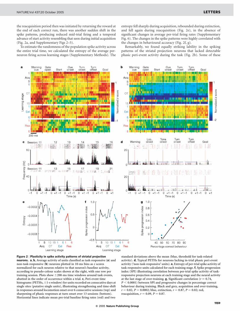
the reacquisition period then was initiated by returning the reward atthe end of each correct run, there was another sudden shift in thespike patterns, producing reduced mid-trial firing and a temporaladvance of start activity resembling that seen during initial acquisition(Fig. 2a, and Supplementary Figs 2–5).
To estimate the randomness of the population spike activity acrossthe entire trial time, we calculated the entropy of the average per-neuron firing across learning stages (Supplementary Methods). The
entropy fell sharply during acquisition, rebounded during extinction,and fell again during reacquisition (Fig. 2e), in the absence ofsignificant changes in average per-trial firing rates (SupplementaryFig. 6). The changes in the spike patterns were highly correlated withthe changes in behavioural accuracy (Fig. 2f, g).
Remarkably, we found equally striking lability in the spikingpatterns of the striatal projection neurons that lacked detectablephasic peri-event activity during the task (Fig. 2b). Some of these
Figure 2 | Plasticity in spike activity patterns of striatal projectionneurons. a, b, Average activity of units classified as task-responsive (a) andnon-task-responsive (b) neurons plotted in 10-ms bins as z scoresnormalized for each neuron relative to that neuron’s baseline activity,according to pseudo-colour scales shown at the right, with one row pertraining session. Plots show ^200-ms time windows around task events,abutted in the order of occurrence within a trial. c, Peri-event timehistograms (PETHs,^1-s window) for units recorded on consecutive days atsingle sites (putative single units), illustrating strengthening and time-shiftin responses around locomotion onset over 6 consecutive sessions (top) andsharpening of phasic responses at turn onset over 13 sessions (bottom).Horizontal lines indicate mean pre-trial baseline firing rates (red) and two
standard deviations above the mean (blue, threshold for task-relatedactivity). d, Typical PETHs for neurons lacking in-trial phasic peri-eventactivity (‘non-task-responsive’ units). e, Entropy of per-trial spike activity oftask-responsive units calculated for each training stage. f, Spike progressionindex (SPI) illustrating correlation between per-trial spike activity of task-responsive projection neurons at each training stage and the neural activityat the last stage of over-training. g, Significant correlation (r ¼ 0.74,P , 0.0001) between SPI and progressive changes in percentage correctbehaviour during training. Black and grey, acquisition and over-training,r ¼ 0.82, P ¼ 0.0002; blue, extinction, r ¼ 0.87, P ¼ 0.02; red,reacquisition, r ¼ 0.09, P ¼ 0.87.
NATURE|Vol 437|20 October 2005 LETTERS
1159

© 2005 Nature Publishing Group
non-task-responsive neurons fired at low rates both in task and outof task, and some fired more out of task than in task (Fig. 2d). Thein-task activity of these neurons dwindled during acquisition andthen nearly ceased. Yet, on the first day of extinction, the average per-neuron firing of these neurons returned to pre-training levels andremained elevated. Then, when reacquisition training began, theiractivity declined sharply. These abrupt shifts were evident whetherthe activity of the neurons during the task was classified with respectto pre-trial baseline firing (Fig. 2b) or relative to in-trial activity(Supplementary Fig. 2)
To determine whether the tuning of task-related responseschanged during learning, we measured multiple parameters (forexample, height, width, peri-event peak timing) of the phasic spikeresponses to specific task events detected by a slope threshold(Supplementary Methods). None of these was altered during learn-ing. By contrast, we found large-scale changes in the proportionof spikes per entire trial run that occurred within phasic responses(Fig. 3a, b). This proportion tripled during acquisition, fell abruptlyduring extinction and abruptly rose again during reacquisition(Fig. 3b). The number of phasic responses also changed successively(Fig. 3c). Reinforcing these redistributions of spike activity, theproportions of task-responsive projection neurons responding todifferent task events also progressively emphasized7, de-emphasizedand re-emphasized the beginning and end of the task (Fig. 3d, andSupplementary Fig. 6). Notably, the sharpening of phasic responsesduring acquisition held not only for those occurring in the early andlate parts of the task runs in which overall spiking increased, but alsofor responses occurring in the middle parts of the runs in which spikeactivity decreased (Supplementary Fig. 6). This result indicates thateven when fewer neurons responded, some ‘expert’ responders withsharpened responses developed in the striatum as the task wasacquired. This property, too, was subject to reversal and reappearanceduring subsequent extinction and reacquisition training.
Both the increase in concentration of spikes within phasic peaksduring acquisition and the redistribution of spikes across run timeshad the effect of reducing the spread of spiking across trial perform-ance time as the rats learned the task. We looked for, but did not
observe, significant changes in the variability of firing rates withinperi-event or phasic-response windows across learning. However, wefound major changes in the entropy (Fig. 2e) and in the variance(Supplementary Fig. 6) of spiking activity across the entire mazeruns. Changes in spike distribution within the time frame of theentire procedural performance thus represented the key modulationof spike variability that we detected during learning.
Taken together, our findings show that per-trial spike distri-butions, response tuning and task selectivity were dynamicallyreconfigured as the procedural behaviour was acquired, extinguishedand reacquired. Composite neural activity scores based on thesefactors were highly correlated with both behavioural accuracy andrunning times, especially during acquisition and extinction (Fig. 4,Supplementary Fig. 7 and Supplementary Methods). Restructuringof the day-by-day neural activity patterns in the ‘fast learners’ (n ¼ 5)but not in the ‘slow learners’ (n ¼ 2) early during acquisition(Supplementary Fig. 8) favoured a primary correspondence betweenthe evolution of the neural restructuring over time and associativelearning. The acquired patterns were detectable in both correct andincorrect trials (Supplementary Fig. 9), however, so that the ensemblepatterns were not tied to performance in individual trials.
It has been proposed that the basal ganglia promote variability inbehaviour during trial-and-error learning (exploration) and serve toevaluate behavioural changes to promote the acquisition of optimalbehaviour (exploitation)9,10,13,14. Our findings suggest that theremight be a direct neural analogue to this explore–exploit behaviourin the firing patterns of projection neurons in the sensorimotorstriatum. We demonstrate two fundamental changes in the spikeactivity of striatal projection neurons during procedural learning.
First, there was a global modulation of the firing of projectionneurons. Early in training, the spike activity of the task-responsivepopulation was spread throughout task time, as though all taskevents were salient (neural exploration). Even neurons withoutdetectable phasic task-responsive activity fired at low rates duringthe task. Then, with continued training, this widespread spiking ofthe task-responsive population diminished, and their spike activitybecame focused (neural exploitation). At the same time, the non-task-responsive population fell silent, further reducing the task-irrelevant firing of the total projection neuron population. Thesechanges in firing thus altered the distribution of striatal outputneuron firing across the actual time-frame of the behaviour to belearned (the entire task run time). The reversal of the acquired task-related patterning during extinction and its reappearance in reacqui-sition fits the idea of increased neural exploration in the newextinction context and then a return to neural exploitation in thereinstated original context during reacquisition15–17. The vivid
Figure 3 | Multiple changes in projection neuron activity in thesensorimotor striatum during acquisition, extinction and reacquisitiontraining. a, Unit activity at a single site recorded over 13 sessions. Each rowrepresents a session. b, Proportions of spikes concentrated in phasicresponses in each trial, averaged for each session. Error bars indicate s.e.m.c, Average numbers of phasic responses per unit. d, Percentages of task-responsive projection neurons with responses at warning cue (solid blueline), goal reaching (dotted green line), locomotion onset (solid purple line)or turning (dotted magenta line). Values are plotted relative to the firsttraining stage.
Figure 4 | Striatal neural activity predictive of behavioural performance.a, Composite neural scores based on weighted neural measures at trial start(normalized per-neuron firing rates during the ^200-ms interval aroundthe warning cue, proportions of warning cue-responsive neurons, andproportions of spikes within phasic warning cue responses). b, Significantcorrelation between the composite scores (shown in a) and actualbehavioural accuracy for each training stage (colour-coded as in a; r ¼ 0.69,P , 0.001). c, Plot as in b, showing significant correlation between thecomposite neural scores and actual running times for each training stage(r ¼ 20.69, P , 0.001).
LETTERS NATURE|Vol 437|20 October 2005
1160
© 2005 Nature Publishing Group
modulation of the spiking of striatal projection neurons withoutdetectable task-related activity also accords with this interpretation.
Second, in the exploitation phases of learning, ensemble firing atthe start and end of the learned procedure strengthened, and sharplytuned responses of ‘expert neurons’ appeared. These changes indicatethat early in training many candidate neurons fired, but that, withtraining and presumably competitive selection18–20, neurons withsharply tuned responses appeared and, as a population, were tunedpreferentially to respond near the start and end of the entireprocedure performed. Our experiments leave open the question ofwhere within the sensorimotor cortico-basal ganglia loop suchchanges were initiated. Because we recorded from striatal projectionneurons, however, our findings show that such learning-relatedchanges in neuronal firing occur as part of cortico-basal ganglialoop processing. The learning-related reduction in firing during themiddle of the task time could indicate that striatal activity during thistime was no longer needed for task execution, but could reflect themarking of behavioural boundaries in the process of chunking of theentire task performance5. These changing patterns could, in turn,reflect ongoing reorganization of cortico-basal ganglia activity20–23. Ifso, the patterns could reflect neural representations related to theready release of the learned behaviours by the appropriate context5.
Cortico-basal ganglia circuits probably act in determining,through reinforcement-based evaluation, which actions to enhanceor diminish as learning proceeds4–6,9,10,19,20,24–30. Viewed in the contextof such selection functions, our findings indicate that dynamic neuralrepresentations in the striatum could adjust the encoded salience oftask events and behavioural responses as habits are formed, lost andregained.
METHODSThe spike activity of neurons in the sensorimotor striatum was recordedchronically during behavioural training on a conditional reward-based T-mazetask for 24–63 daily sessions from seven rats in which seven tetrode headstageassemblies had been implanted. Recordings began on the first day that the ratsreceived training (about 40 trials per day) on the task, and were continuedthrough successive acquisition training (stages 1–5), over-training (stages 6–15),extinction (stages 1–6) and reacquisition training (stages 1–6; Fig. 1b, Sup-plementary Table 2 and Supplementary Methods). In this task, rats learned torun down the maze and to turn right or left as instructed by auditory cues inorder to receive reward. Behavioural data were acquired by means of photo-beams and a CCD camera. Neural data (32 kHz sampling) were collected bymeans of a Cheetah Data Acquisition System (Neuralynx Inc.). Well-isolatedunits accepted after cluster cutting were classified as striatal projection neuronsor interneurons (Supplementary Fig. 1b–d). Behavioural and neural data werealigned by time stamps and were analysed by in-house software. The propertiesof both task-responsive and non-task-responsive projection neurons wereanalysed. Task-related responses of putative projection neurons were identifiedwith respect to activity during a pre-trial 500-ms baseline period (threshold:2 s.d. above baseline mean) and used to define task-responsive and non-task-responsive populations (Supplementary Methods). Unit data were analysed perneuron and per neuronal population across task events (Fig. 1c). To analysepopulation activity, normalized firing rates were averaged for each learningstage, and indices of spike firing patterns across learning stages were computed.The proportions of neurons with different task-related response types, theproportions of spikes that occurred within peri-event phasic responses persession, and trial-to-trial spike variability were also calculated, along withcomposite neural scores and measures of the entropy of neural firing. Changesin these measures were compared to changes in per cent correct performance andrunning times of the rats across stages of training.
Received 2 June; accepted 21 July 2005.
1. James, W. The Principles of Psychology 104–-127 (Dover, New York, 1890).2. Dickinson, A. Actions and habits: the development of behavioural autonomy.
Phil. Trans. R. Soc. Lond. B 308, 67–-78 (1985).
3. Pavlov, I. P. Conditioned Reflexes: An Investigation of the Physiological Activity ofthe Cerebral Cortex (ed. and transl. Anrep, G. V.) (Oxford Univ. Press, London,1927).
4. Packard, M. G. & Knowlton, B. J. Learning and memory functions of the basalganglia. Annu. Rev. Neurosci. 25, 563–-593 (2002).
5. Graybiel, A. M. The basal ganglia and chunking of action repertoires. Neurobiol.Learn. Mem. 70, 119–-136 (1998).
6. Poldrack, R. A. et al. Interactive memory systems in the human brain. Nature414, 546–-550 (2001).
7. Jog, M., Kubota, Y., Connolly, C. I., Hillegaart, V. & Graybiel, A. M. Buildingneural representations of habits. Science 286, 1745–-1749 (1999).
8. Yin, H. H., Knowlton, B. J. & Balleine, B. W. Lesions of dorsolateral striatumpreserve outcome expectancy but disrupt habit formation in instrumentallearning. Eur. J. Neurosci. 19, 181–-189 (2004).
9. Olveczky, B. P., Andalman, A. S. & Fee, M. S. Vocal experimentation in thejuvenile songbird requires a basal ganglia circuit. PLoS Biol. 3, e153 (2005).
10. Kao, M. H., Doupe, A. J. & Brainard, M. S. Contributions of an avian basalganglia-forebrain circuit to real-time modulation of song. Nature 433, 638–-643(2005).
11. Sutton, R. S. & Barto, A. G. Reinforcement Learning: An Introduction (MIT Press,Cambridge, Massachusetts, 1998).
12. Wilson, C. J. in The Synaptic Organization of the Brain (ed. Shepherd, G. M.)361–-413 (Oxford Univ. Press, New York, 2004).
13. Doya, K. & Sejnowski, T. J. in Advances in Neural Information Processing SystemsVol. 7 (eds Tesauro, G., Touretzky, D. S. & Leen, T. K.) 101–-108 (MIT Press,Cambridge, Massachusetts, 1995).
14. Doya, K. & Sejnowski, T. in The New Cognitive Neurosciences (ed. Gazzaniga,M. S.) 469–-482 (MIT Press, Cambridge, Massachusetts, 2000).
15. Bouton, M. E. Context and behavioural processes in extinction. Learn. Mem. 11,485–-494 (2004).
16. Routtenberg, A. & Kim, H.-J. in Cholinergic–-Monoaminergic Interactions in theBrain (ed. Butcher, L. L.) 305–-331 (Academic, New York, 1978).
17. Myers, K. M. & Davis, M. Behavioral and neural analysis of extinction. Neuron36, 567–-584 (2002).
18. Gurney, K., Prescott, T. J. & Redgrave, P. A computational model of actionselection in the basal ganglia. I. A new functional anatomy. Biol. Cybern. 84,401–-410 (2001).
19. Graybiel, A. M., Aosaki, T., Flaherty, A. W. & Kimura, M. The basal ganglia andadaptive motor control. Science 265, 1826–-1831 (1994).
20. Djurfeldt, M., Ekeberg, O. & Graybiel, A. M. Cortex-basal ganglia interactionand attractor states. Neurocomputing 38–-40, 573–-579 (2001).
21. Frank, M. J., Loughry, B. & O’Reilly, R. C. Interactions between frontal cortexand basal ganglia in working memory: a computational model. Cogn. Affect.Behav. Neurosci. 1, 137–-160 (2001).
22. Houk, J. C. & Wise, S. P. Distributed modular architectures linking basalganglia, cerebellum, and cerebral cortex: their role in planning and controllingaction. Cereb. Cortex 5, 95–-110 (1995).
23. Doya, K. Metalearning and neuromodulation. Neural Netw. 15, 495–-506(2002).
24. Montague, P. R., Hyman, S. E. & Cohen, J. D. Computational roles for dopaminein behavioural control. Nature 431, 760–-767 (2004).
25. Tanaka, S. C. et al. Prediction of immediate and future rewards differentiallyrecruits cortico-basal ganglia loops. Nature Neurosci. 7, 887–-893 (2004).
26. Reynolds, J. N. J., Hyland, B. I. & Wickens, J. R. A cellular mechanism ofreward-related learning. Nature 413, 67–-70 (2001).
27. Mink, J. W. The basal ganglia: focused selection and inhibition of competingmotor programs. Prog. Neurobiol. 50, 381–-425 (1996).
28. McClure, S. M., Berns, G. S. & Montague, P. R. Temporal prediction errors in apassive learning task activate human striatum. Neuron 38, 339–-346 (2003).
29. Barto, A. G. in Models of Information Processing in the Basal Ganglia (eds Houk,J., Davis, J. & Beiser, D.) 215–-232 (MIT Press, Cambridge, Massachusetts,1995).
30. Dickinson, A. & Balleine, B. W. Motivational control of goal-directed action.Anim. Learn. Behav. 22, 1–-18 (1994).
Supplementary Information is linked to the online version of the paper atwww.nature.com/nature.
Acknowledgements We thank H. F. Hall, P. A. Harlan and C. Thorn for theirhelp. This work was funded by the National Institutes of Health and the Officeof Naval Research.
Author Information Reprints and permissions information is available atnpg.nature.com/reprintsandpermissions. The authors declare no competingfinancial interests. Correspondence and requests for materials should beaddressed to A.M.G. ([email protected]).
NATURE|Vol 437|20 October 2005 LETTERS
1161