Embed Size (px)
Citation preview
AN. VET. (MURCIA) 20: 87-94 (2004). ACUPOINT RENZHONG (JENCHUNG GV-26) IN THE HORSE. ZILBERSCHTEIN,J., et al. 87
ACUPOINT RENZHONG (JENCHUNG GV-26) IN THE HORSE.ANATOMICAL AND HISTOLOGICAL STUDY
Acupunto Renzhong en el caballo. Estudio anatómico e histológico.
Zilberschtein, J.1*, Gil Cano, F.2, Sánchez Valverde, M. A.1, Laredo, F.1, Vásquez, F.2
1 Departamento de Cirugía y Medicina Animal.2 Departamento de Anatomía y Anatomía Patológica Comparadas.Facultad de Veterinaria. Universidad de Murcia. Campus de Espinardo. Murcia (30071). España.* Autor de referencia: [email protected].
ABSTRACT
The purpose of this study was to document the anatomic and histologic characteristics of acupointRenzhong GV-26 in ten Spanish horses. First, the electrical skin resistance was measured to locate theacupoint, and the needles were inserted to a depth of 1cm into the acupoint. Then, the tissue surrounding theacupoint was removed, sectioned serially, and processed by routine histologic techniques. The results showthat the acupoint Renzhong GV-26 is situated in the intermediate area of the upper lip, represented by severalstructures of dense connective tissue, skeletal muscle tissue, nerve fibers and vascular structures. Therefore,numerous structures conform part of the acupoint Renzhong GV-26 in the horse.
Keywords: Acupuncture, Acupoint Renzhong GV-26, Horse, Histology, Anatomy.
RESUMEN
El propósito de este estudio es documentar las características histológicas y anatómicas del acupuntoRenzhong GV-26 en diez caballos de raza española. Primero, se midió la resistencia eléctrica para la localizacióndel acupunto, y posteriormente se insertó una aguja de acupuntura a una profundidad de 1 cm dentro delacupunto. Luego se obtuvieron muestras del acupunto y fueron procesadas por técnicas histológicas de rutina.Los resultados muestran que el acupunto Renzhong GV-26 está situado en el estrato intermedio del labiosuperior, representado por diferentes estructuras, como tejido conectivo denso, músculo estriado esquelético,fibras nerviosas y estructuras vasculares. Es decir, numerosas estructuras conforman el acupunto RenzhongGV-26 en el caballo.
Palabras clave: Acupunctura, Acupunto Renzhong GV-26, Cabllo, Histología, Anatomía
AN. VET. (MURCIA) 20: 87-94 (2004). ACUPOINT RENZHONG (JENCHUNG GV-26) IN THE HORSE. ZILBERSCHTEIN,J., et al.88
INTRODUCTION
Acupuncture is a therapeutic system withintraditional Chinese medicine characterized byinserting needles of different metals into specificparts of the body, called acupoints, in order tocure, relieve or eliminate pain. It is used in bothhuman and veterinary medicine. In animals, theseacupoints form currents of energy known asacupuncture meridians.
These meridians constitute the basis ofacupuncture and their description originated dueto the knowledge of the topography of theacupoints. They are situated symmetrically withrespect to the sagital axis of the body, and arefound throughout the head, the trunk, theabdomen and the limbs, both in the anterior aswell as in posterior part of the body, forming aclosed system of circulation. Each one has itsown distribution, a specific energy timetable,and corresponds to an organ (Becker andRechmains, 1976).
The Renzhong acupoint (Jenchung, GV-26or 26 Du in Du Mai Chinese) belongs to othergoverning meridian vessels (GV). In all speciesthis meridian runs in the following directions: itbegins in the coccyx and ascends the middledorsal line to the neck. It then runs along themiddle line in the head towards the foreheadand the nose and the finishes below the upperlip.
The Renzhong acupoint is found at the borderof the middle and upper third of the distancebetween the nose and the upper lip in humans(Pomeranz and Stux, 2000) and in animals suchas horses at the upper lip, at the midpoint of thephiltrum nasale (Demontoy and Mailhac, 1980;Westermayer, 1985).
The Renzhong acupoint (Jenchung, GV-26or 26 Du) is one of the most widely used in theemergency ward, in both animals and humans.A bibliography of more than 160 articles (1973-2001) documents the effects and clinical efficacyof the stimulation of this acupoint and leads oneto speculate other physiological and clinical
applications. Very few studies exist on themicroscopic structure of the acupoints indomestic animals. The main works refer to cattleand dogs (Kothbauer, 1961, 1986; Egerbacher,1991, 1994, 1996; Ludewig, 1998). Thestructures found at the acupoints are not unique.Many of them are found in depressions situatedin muscular zones (Helmes, 1995). These areareas of the skin containing a high concentrationof nerve endings, nerve plexuses, mast cells,lymphocytes, capillaries and venules (Kendall,1989). It has been suggested that the size of theacupoints is 1 mm2. Histologically, they havetheir own structure characterized by a thinningof the skin due to a modification of its collagen’sfibers of the dermis, this explains why they canbe felt as a depression (She); they also present aspiral vascular network, surrounded by a densenetwork of collinergic-type amielinic nervefibers (Niboyet, 1980). Heine (1988) revealedthat 80% of acupoints correlated withperforations in the superficial fascia of humancorpses. A nerve vessel bundle and vesselspenetrated the skin through these orifices.
Studies carried out in cows (Kothbauer, 1961,1986; Ludewig, 1998) and in dogs and cows(Egerbacher, 1994) have verified the correlationbetween acupoints and perforations in thesuperficial fascia by sensitive cutaneous nerves.Macroscopic examination of 27 points of thebladder meridian in a cow revealed very definedperforations in the thoracolumbar fascia throughwhich nerve-vessel structures ran at 19 points.The remaining eight acupoints (B18-25) weresituated at the entrance points of the middlecutaneous branch in the skin of the back of theanimal. There were no perforations of the fascia,which was a new discovery. Fascia perforationswith nerve-vessel structures were found at thefive points of the gall bladder meridian, whichwere examined.
Histologic examination revealed a specialdisposition of the connective tissue,surrounding the nerve, with concentric laminasand loss of connective tissue composed of fine
AN. VET. (MURCIA) 20: 87-94 (2004). ACUPOINT RENZHONG (JENCHUNG GV-26) IN THE HORSE. ZILBERSCHTEIN,J., et al. 89
fibers of collagen. A more compact sheath ofconnective tissue surrounded the nerve-vesselstructure at the point where it perforates thefascia (Egerbacher, 1994). These results arevalid for other points with reduced electricalresistance at the base of the teat (Ludewig,1998). A high concentration of mast cells wasalso found in the area adjacent to the point(Zhai, 1998).
In reference to the vascular structures in theareas of the acupuncture, several authors stressthe importance that they play in the regulationof local temperature. Talukdar et al. (1972) in astudy performed on the skin of the horsedescribes special vascular structures in 43 areasof the skin. They consist of arterio-venousanastomosis in rings or spirals forming canalsbetween arterio-venous, arterioles withephiteloides cells on the walls and glomus. Theseglomus might be related to the conservation ofthe heat and temperature regulation. Otherauthors focus the subject on the components ofthe connective tissue, lymphatic vessels,arterioles and veins at the acupoints, describingvascular structures surrounding collinergicamielinic nerve fibers.
Our study corresponds to the use of thisacupoint in anaesthetic recovery in the horse.Considering that its anatomic and histologiccharacteristics have not been described, thepurpose of this study is to report the morphologiccharacteristics of acupoint GV-26 in Spanish-bred horses.
MATERIALS AND METHOD
Ten Spanish-bred horses were used. Beforesacrificing the horses, the localization of theRenzhong GV-26 acupoint was determined inthe upper lip by means of an ohmiometre (WQ-IOC2R multiple Electronic Acupunctoscope.Made in China) and Chinese cartography.Acupunctoscope or «point finder» consist of apencil-shaped metal sound connected by meansof a cable to an ohmiometre. A second electrode
situated in a metallic cylinder completes thecircuit. Some studies suggest that the resistanceof the skin (impedance) at the acupoints is lessthan in the surrounding skin (Becker andRechmains, 1976; Oleson et al., 1980; Pomeranzand Stux, 2000).
Once the acupoint had been determined, thedefinite marking of it was performed by meansof a disposable, surgical, stainless steel, philiformpoint and a depth of 3-cm (Hwato. Suzhou,China). The humane sacrifice of the animalswas then performed using a 20-mg/Kg overdoseof endovenous pentobarbital (Eutalender Lab.Normon S.A. Madrid, Spain). After the death ofthe animal, a sample of the pyramidal shapedupper lip was taken, corresponding to theacupuncture needle was situated, and twosamples from adjacent areas, according toEgerbacher´s protocol. By means of a scalpeland dissecting tweezers the boundary and depthof the sample was ascertained, approximately 1cm wide by 3 cm longs. The samples thusobtained were fixed in 10 % formaldehyde atroom temperature and remained in the fixingliquid for a minimum of 10 days. Once fixed,they were treated in a tissue processor (TP 1050LEICA, Germany), using usual procedures fortheir embedding on paraffin blocks. This wasperformed by means of a Tissue-Tek (Germany),which was fitted with disposable knives(Reichert, model 819, Germany). Sagittal cutsof a thickness of 10 mm were then placed on thesurface of a double saucepan of water at 37ºC toencourage stretching.
Next, they were placed on microscope slides(50 Microscope Slides, Menzel. Glaser,Germany) covered with Mayer´s glycerinatedalbumin, remaining in the oven at 55ºC for onehour until they were ready to be dyedhistologically. The histologic techniques usedwere the hematoxilin-eoxin (H-E) and Gallego´sTrichromic methods. The dyed preparations werethen viewed and photographed in a Leitz Dialux20 R photomicrosocope fitted with photographicequipment (Germany).
AN. VET. (MURCIA) 20: 87-94 (2004). ACUPOINT RENZHONG (JENCHUNG GV-26) IN THE HORSE. ZILBERSCHTEIN,J., et al.90
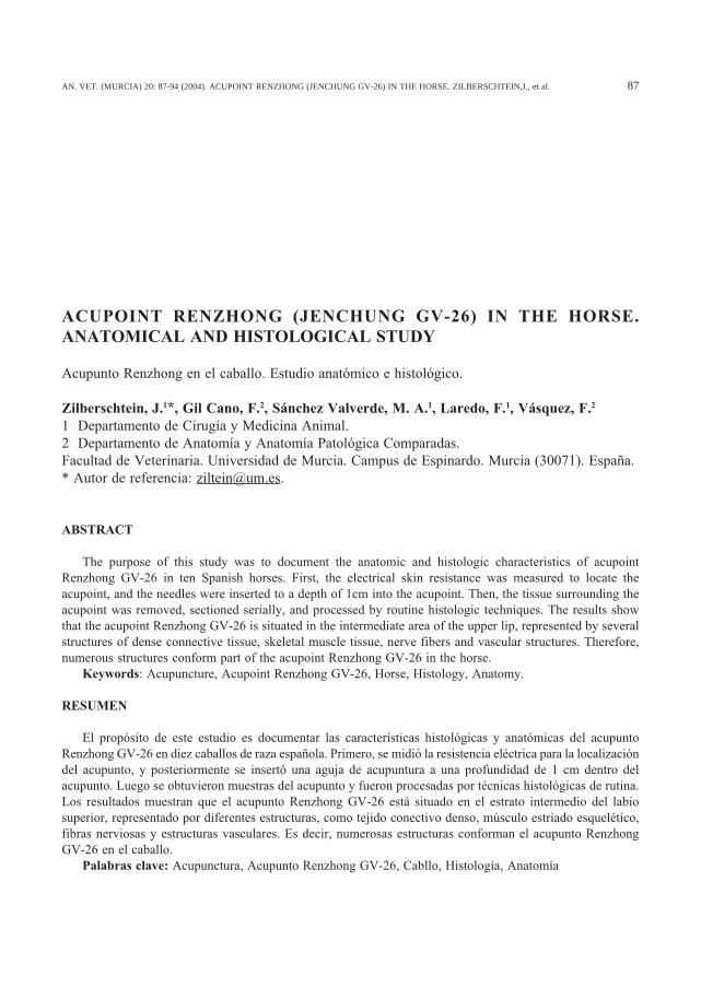
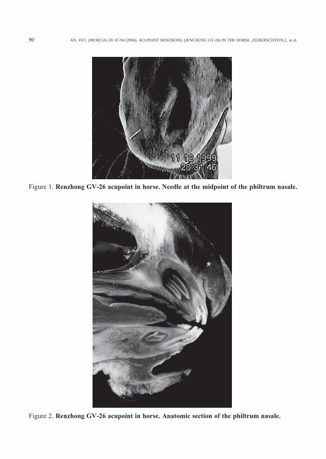
Figure 1. Renzhong GV-26 acupoint in horse. Needle at the midpoint of the philtrum nasale.
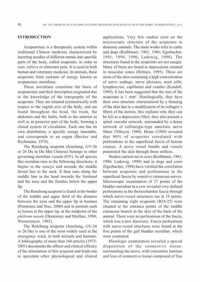
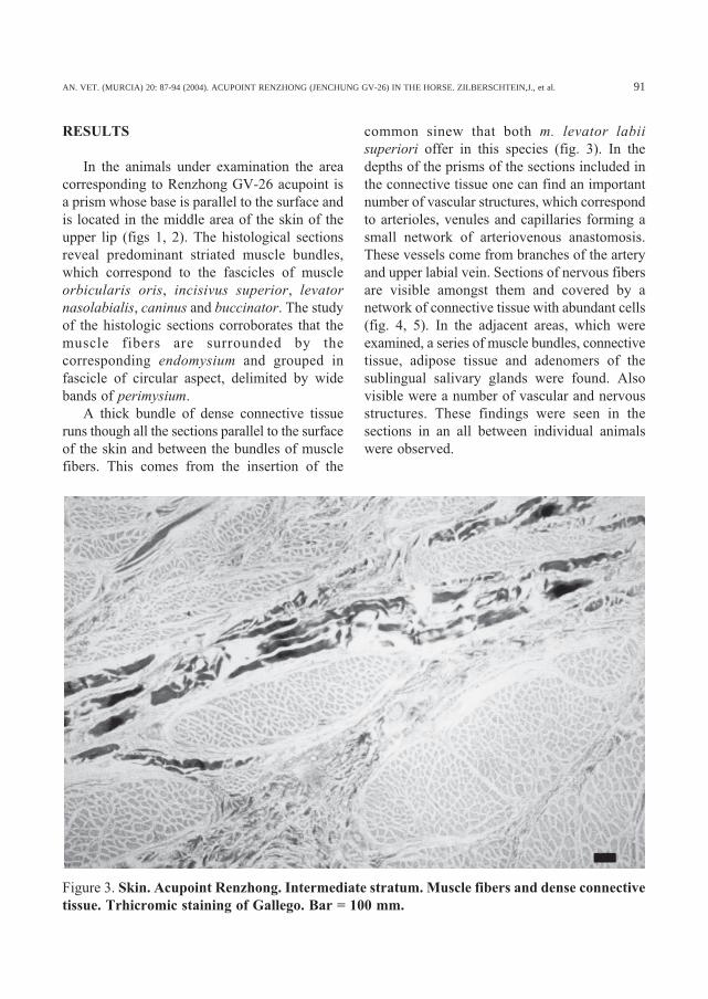
Figure 2. Renzhong GV-26 acupoint in horse. Anatomic section of the philtrum nasale.
AN. VET. (MURCIA) 20: 87-94 (2004). ACUPOINT RENZHONG (JENCHUNG GV-26) IN THE HORSE. ZILBERSCHTEIN,J., et al. 91
RESULTS
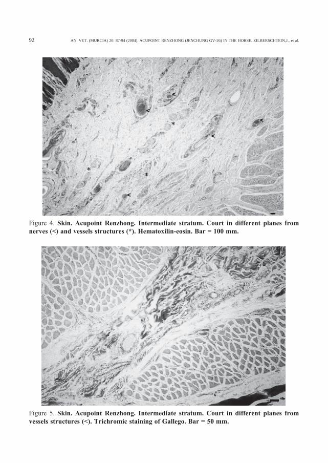
In the animals under examination the areacorresponding to Renzhong GV-26 acupoint isa prism whose base is parallel to the surface andis located in the middle area of the skin of theupper lip (figs 1, 2). The histological sectionsreveal predominant striated muscle bundles,which correspond to the fascicles of muscleorbicularis oris, incisivus superior, levatornasolabialis, caninus and buccinator. The studyof the histologic sections corroborates that themuscle fibers are surrounded by thecorresponding endomysium and grouped infascicle of circular aspect, delimited by widebands of perimysium.
A thick bundle of dense connective tissueruns though all the sections parallel to the surfaceof the skin and between the bundles of musclefibers. This comes from the insertion of the
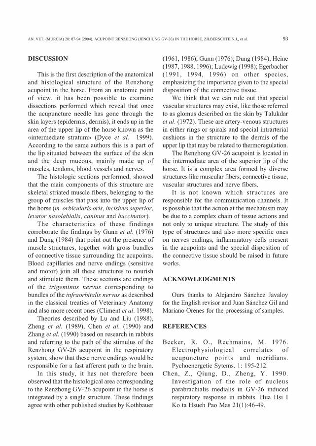
common sinew that both m. levator labiisuperiori offer in this species (fig. 3). In thedepths of the prisms of the sections included inthe connective tissue one can find an importantnumber of vascular structures, which correspondto arterioles, venules and capillaries forming asmall network of arteriovenous anastomosis.These vessels come from branches of the arteryand upper labial vein. Sections of nervous fibersare visible amongst them and covered by anetwork of connective tissue with abundant cells(fig. 4, 5). In the adjacent areas, which wereexamined, a series of muscle bundles, connectivetissue, adipose tissue and adenomers of thesublingual salivary glands were found. Alsovisible were a number of vascular and nervousstructures. These findings were seen in thesections in an all between individual animalswere observed.
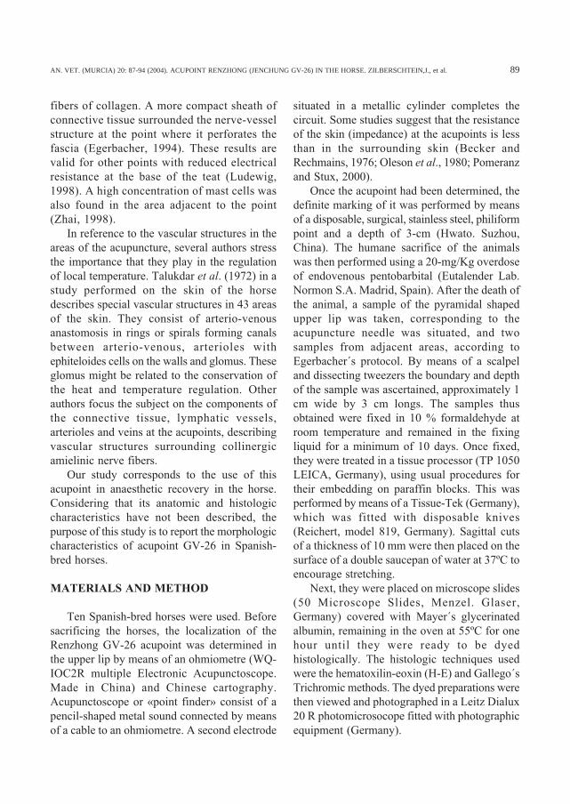
Figure 3. Skin. Acupoint Renzhong. Intermediate stratum. Muscle fibers and dense connectivetissue. Trhicromic staining of Gallego. Bar = 100 mm.
AN. VET. (MURCIA) 20: 87-94 (2004). ACUPOINT RENZHONG (JENCHUNG GV-26) IN THE HORSE. ZILBERSCHTEIN,J., et al.92
Figure 5. Skin. Acupoint Renzhong. Intermediate stratum. Court in different planes fromvessels structures (<). Trichromic staining of Gallego. Bar = 50 mm.
Figure 4. Skin. Acupoint Renzhong. Intermediate stratum. Court in different planes fromnerves (<) and vessels structures (*). Hematoxilin-eosin. Bar = 100 mm.
AN. VET. (MURCIA) 20: 87-94 (2004). ACUPOINT RENZHONG (JENCHUNG GV-26) IN THE HORSE. ZILBERSCHTEIN,J., et al. 93
DISCUSSION
This is the first description of the anatomicaland histological structure of the Renzhongacupoint in the horse. From an anatomic pointof view, it has been possible to examinedissections performed which reveal that oncethe acupuncture needle has gone through theskin layers (epidermis, dermis), it ends up in thearea of the upper lip of the horse known as the«intermediate stratum» (Dyce et al. 1999).According to the same authors this is a part ofthe lip situated between the surface of the skinand the deep mucous, mainly made up ofmuscles, tendons, blood vessels and nerves.
The histologic sections performed, showedthat the main components of this structure areskeletal striated muscle fibers, belonging to thegroup of muscles that pass into the upper lip ofthe horse (m. orbicularis oris, incisivus superior,levator nasolabialis, caninus and buccinator).
The characteristics of these findingscorroborate the findings by Gunn et al. (1976)and Dung (1984) that point out the presence ofmuscle structures, together with gross bundlesof connective tissue surrounding the acupoints.Blood capillaries and nerve endings (sensitiveand motor) join all these structures to nourishand stimulate them. These sections are endingsof the trigeminus nervus corresponding tobundles of the infraorbitalis nervus as describedin the classical treaties of Veterinary Anatomyand also more recent ones (Climent et al. 1998).
Theories described by Lu and Liu (1988),Zheng et al. (1989), Chen et al. (1990) andZhang et al. (1990) based on research in rabbitsand referring to the path of the stimulus of theRenzhong GV-26 acupoint in the respiratorysystem, show that these nerve endings would beresponsible for a fast afferent path to the brain.
In this study, it has not therefore beenobserved that the histological area correspondingto the Renzhong GV-26 acupoint in the horse isintegrated by a single structure. These findingsagree with other published studies by Kothbauer
(1961, 1986); Gunn (1976); Dung (1984); Heine(1987, 1988, 1996); Ludewig (1998); Egerbacher(1991, 1994, 1996) on other species,emphasizing the importance given to the specialdisposition of the connective tissue.
We think that we can rule out that specialvascular structures may exist, like those referredto as glomus described on the skin by Talukdaret al. (1972). These are artery-venous structuresin either rings or spirals and special intrarterialcushions in the structure to the dermis of theupper lip that may be related to thermoregulation.
The Renzhong GV-26 acupoint is located inthe intermediate area of the superior lip of thehorse. It is a complex area formed by diversestructures like muscular fibers, connective tissue,vascular structures and nerve fibers.
It is not known which structures areresponsible for the communication channels. Itis possible that the action at the mechanism maybe due to a complex chain of tissue actions andnot only to unique structure. The study of thistype of structures and also more specific oneson nerves endings, inflammatory cells presentin the acupoints and the special disposition ofthe connective tissue should be raised in futureworks.
ACKNOWLEDGMENTS
Ours thanks to Alejandro Sánchez Javaloyfor the English revisor and Juan Sánchez Gil andMariano Orenes for the processing of samples.
REFERENCES
Becker, R. O., Rechmains, M. 1976.Electrophysiological correlates ofacupuncture points and meridians.Pychoenergetic Sytems. 1: 195-212.
Chen, Z., Qiung, D., Zheng, Y. 1990.Investigation of the role of nucleusparabrachialis medialis in GV-26 inducedrespiratory response in rabbits. Hua Hsi IKo ta Hsueh Pao Mas 21(1):46-49.
AN. VET. (MURCIA) 20: 87-94 (2004). ACUPOINT RENZHONG (JENCHUNG GV-26) IN THE HORSE. ZILBERSCHTEIN,J., et al.94
Climent, S., Sarasa, M., Muniesa, P., Terrado,J. 1998. Manual de Anatomía y Embriologíade los Animales Domésticos: Conceptosbásicos y datos aplicativos. Sistema NerviosoCentral y Órganos de los Sentidos. Ed.Acribia. Zaragoza. España. pp 81-107.
Demontoy, A., Mailhac, J. M. 1980. Le point dereanimation par excellence, RENZHONG.Rec. Med. Vet. 156(3): 241-244.
Dyce, K. M., Sack, W. O., Wensing, C. 1999.Anatomía Veterinaria. 2ª Ed. Mc Graw-HillInteramericana, pp 108-109; 523-524.
Dung, H. C. 1984. Anatomical featurescontributing to the formation of acupuncturepoints. Am. J. Acupunct. 12: 139-143.
Egerbacher, M. 1991. Anatomische undHistologische Untersuchungen zurMorphologie ausgewählter akupunkturpunkteam Rumpf bei Rind und Hund. Diss. Vet.Med. Univ Wien. 27-3.
Egerbacher, M. 1994. Anatomy and Histologyof selected Bovine and canina Acupuncturepoints. Veterinary Acupuncture (ed) 27-31.
Egerbacher, M. 1996. Acupuncture points:macroscopic and microscopic findings inbody- and ear acupuncture points. Wien; 83:359-365.
Gunn, C. C. 1976. Acupuncture loci: a proposalfor their classification according to theirrelationship to known neural structures. Am.J. Chin. Med. 4:183-195.
Heine, H. 1987. Zur Morphologie derAkupunkturpunkte. Dtsch Zschr Akup. 31,26 –30.
Heine, H. 1988. Akupunkturtherapie-Perforatio-nen der oberflächlichen Köperfaszie durchkutane Gefä-Nervenbündel. Therapeutikon.4:238-244.
Heine, H. 1996. Der Akupunkturpunkt – einMeridianorgan. Dtsch Zschr Akup. 39, 75 –80.
Helms, J. M. 1995. Acupuncture energetics: aclinical approach for physicians. MedicalAcupuncture Publishers. Berkeley.
Kendall, D. E. 1989. Parts I and II. A scientificmodel of acupuncture. Am J Acupunct 17:251-268,343-360.
Kothbauer, O. 1961. Über die Druckpunktdiag-nose und Neuraltherapie bei Tieren. WienTierärztl Mschr.; 48, 282-293.
Kothbauer, O. 1986. Studie zur Ohrakupunktur desRindes. Wien Tierärztl Mschr.; 73, 177-180.
Lu, W. Y., Liu, L. 1988. Effects of acupunctureat GV-26 on experimentally-induced disorderof respiratory rhythm. Acup Res. 13(2):124-129.
Ludewig, T. A. 1998. Contribution to themicrocopic anatomy of acupuncture points.Wien Verlag.; 86: 150-154.
Niboyet, J. E. H. 1980. Les points d´acupuncture.Cah. Biothèr. 67, 5-14.
Oleson, T. D., Kroening, R. J., Bresler D. A.1980. An experimental evaluation ofauricular diagnosis: the somatotopic mappingof musculoskeletal pain at acupuncturepoints. Pain. 8: 217-229.
Pomeranz, B., Stux, G. 2000. Fundamentos deAcupuntura. Ed. Springer. Barcelona.
Still, J., Konrad, J. 1985. Verification ofacupuncture resuscitation in some species ofzoo-animals. 26 International Simposium,Brno, Proc Of Meet. 209-214.
Talukdar, A., Colhoun, L., Stinson, W. 1972.Specialized vascular structures in the skin ofthe horse. Am. J. Vet Res. 33(2) 335-338.
Westermayer, E. 1985. The treatments of horseby Acupuncture. Ed. The Company. Essex.
Zhai, N. 1988. Research on the histo-physiological relation of mastocytesandmeridians. Chin. Acup. and Mox. 8: 50-53.
Zhang, H., Zhang, M., Liu, L. 1990. Effect ofpain in the change of respiration induced bystimulation GV-26. East-West Conf Proc.103.
Zheng, Y., Xu, M. L., Yang, S. R. 1989.Mechanisms of effects of electro-acupunctureat GV – 26 on phrenic- nerve discharge inrabbits. Hua Hsi Ko Ta. 20 (4): 384 – 388.