Embed Size (px)
Citation preview
Acute inflammation 5
By Dr. S. Homathy
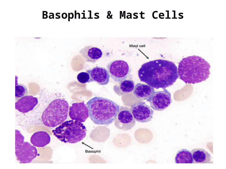
Basophils & Mast Cells
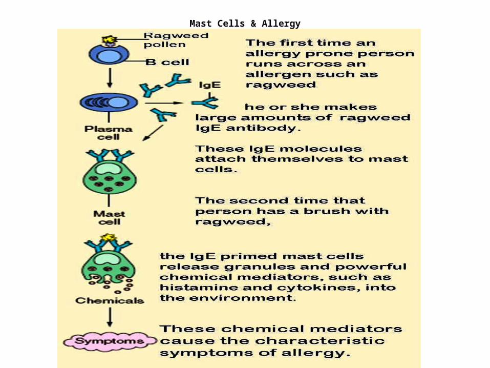
Mast Cells & Allergy
Specific mediators
Serotonin:
• vasodilatory effects similar to those of histamine; • platelet dense-body granules; • release triggered by platelet aggregation
Specific mediators
Plasma proteases– Clotting system– Complement– Kinins
Specific mediators
Arachidonic acid metabolites (eicosanoids)• Prostaglandins and thromboxane: – via cyclooxygenase pathway; – cause vasodilation and prolong edema;– but also protective (gastric mucosa); – COX blocked by aspirin and NSAIDS
Specific mediators
Leukotrienes:• via lipoxygenase pathway; • are • chemotaxins,• vasoconstrictors,
– cause increased vascular permeability, and bronchospasm
Specific mediators
PAF (platelet activating factor)• Derived also from cell membrane phospholipid of– Neutrophils, monocytes, basophils, endothelium and
platelets.• causes – Vasoconstriction and bronchoconstriction– increased vascular permeability, – increases leukocyte adhesion (integrin conformation)– Leukocyte degranulation– Platelet activation– Release of histamine from platelets– chemotaxis
Specific mediators
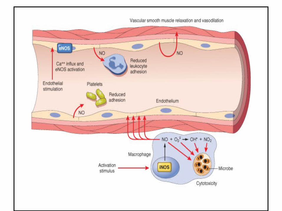
Nitric Oxide• short-acting soluble free-radical gas with many
functions– Produced by endothelial cells, macrophages, specific
neurones in the brain– causes:
• Vascular smooth muscle relaxation and vasodilation• Kills microbes in activated macrophages• Counteracts platelet adhesion, aggregation, and
degranulation• Uncontroled NO production by activated MP in septic
shock can lead to massive peripheral vasodilation and shock
Oxygen derived free radicals• Synthesized via the NADPH oxidase pathway• Released from N and MP after stimulation by – chemotactic agents, – immune cpx, – phagocytic activity
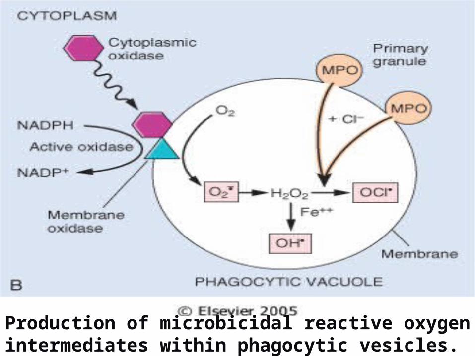
Production of microbicidal reactive oxygen intermediates within phagocytic vesicles.
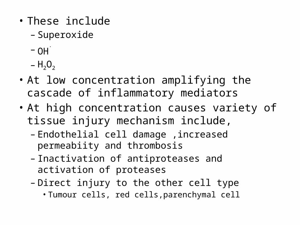
• These include– Superoxide
– OH.
– H2O2
• At low concentration amplifying the cascade of inflammatory mediators
• At high concentration causes variety of tissue injury mechanism include,– Endothelial cell damage ,increased permeabiity and
thrombosis– Inactivation of antiproteases and activation of proteases– Direct injury to the other cell type
• Tumour cells, red cells,parenchymal cell
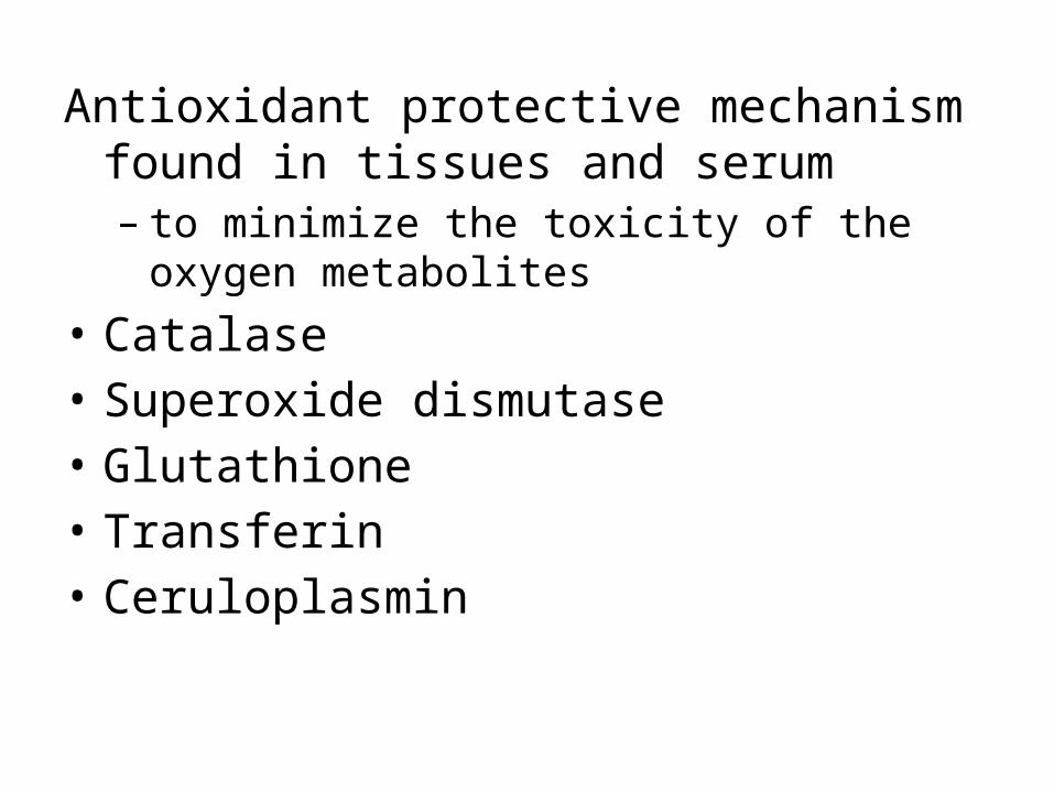
Antioxidant protective mechanism found in tissues and serum – to minimize the toxicity of the oxygen metabolites
• Catalase• Superoxide dismutase• Glutathione• Transferin• Ceruloplasmin
Specific mediators
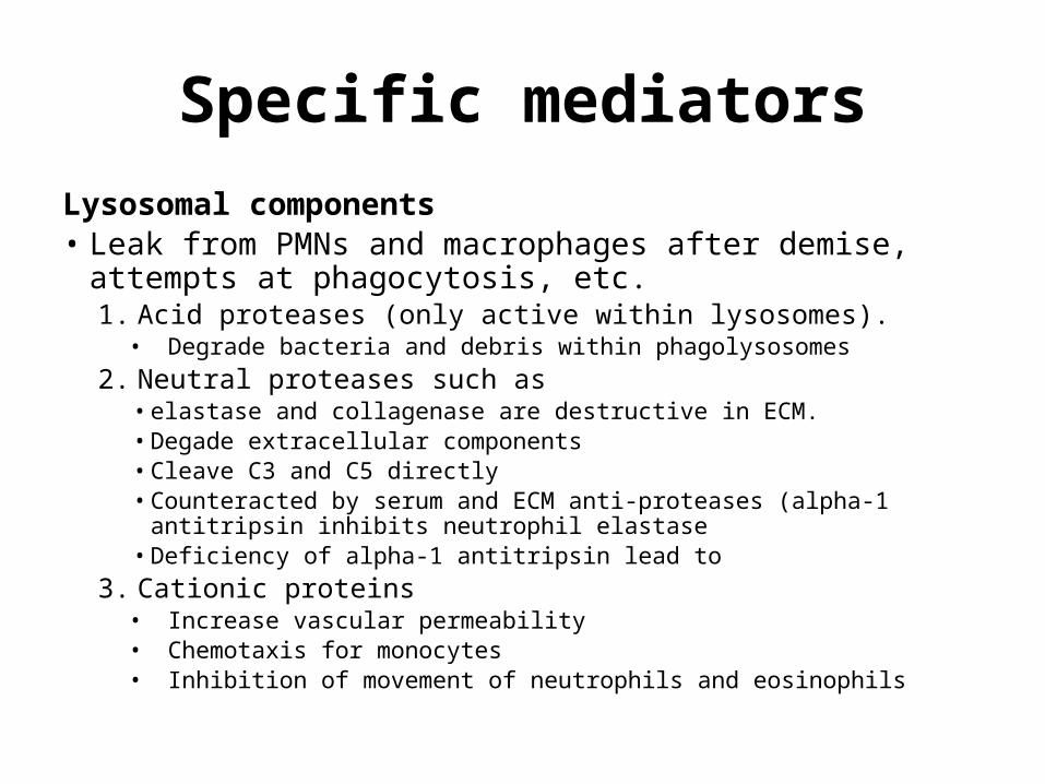
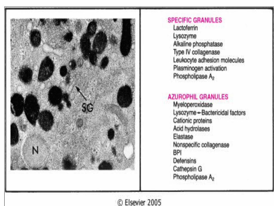
Lysosomal components• Leak from PMNs and macrophages after demise, attempts at
phagocytosis, etc.1. Acid proteases (only active within lysosomes).
• Degrade bacteria and debris within phagolysosomes
2. Neutral proteases such as • elastase and collagenase are destructive in ECM.• Degade extracellular components• Cleave C3 and C5 directly• Counteracted by serum and ECM anti-proteases (alpha-1 antitripsin inhibits
neutrophil elastase• Deficiency of alpha-1 antitripsin lead to
3. Cationic proteins• Increase vascular permeability• Chemotaxis for monocytes• Inhibition of movement of neutrophils and eosinophils
More specific mediators Cytokines• They are polypeptide products of many cell types– Products of activated lymphocytes and macrophages
• They modulate the function of other cell types• Many non lymphoid cells too produce these • The secretion is transient and tightly regulated• Their effects tend to be pleiotrophic – Different cells are affected differently by the same
cytokine
• Produced during immune and inflammatory responses
• Autocrine effect – They can act on the same cell that produces them
• Paracrine effect– Act on other cells in the vicinity
• Endocrine effect– Act systemically
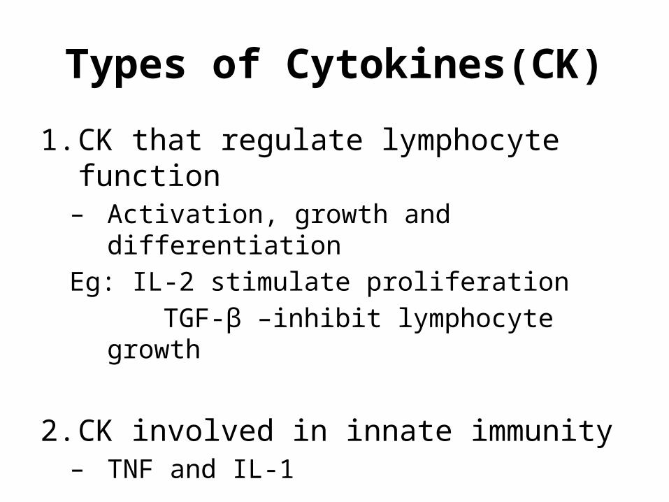
Types of Cytokines(CK)
1. CK that regulate lymphocyte function– Activation, growth and differentiation
Eg: IL-2 stimulate proliferation
TGF-β –inhibit lymphocyte growth
2. CK involved in innate immunity– TNF and IL-1
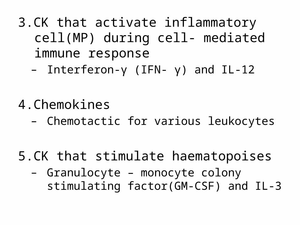
3.CK that activate inflammatory cell(MP) during cell- mediated immune response– Interferon-γ (IFN- γ) and IL-12
4.Chemokines – Chemotactic for various leukocytes
5.CK that stimulate haematopoises– Granulocyte – monocyte colony stimulating
factor(GM-CSF) and IL-3
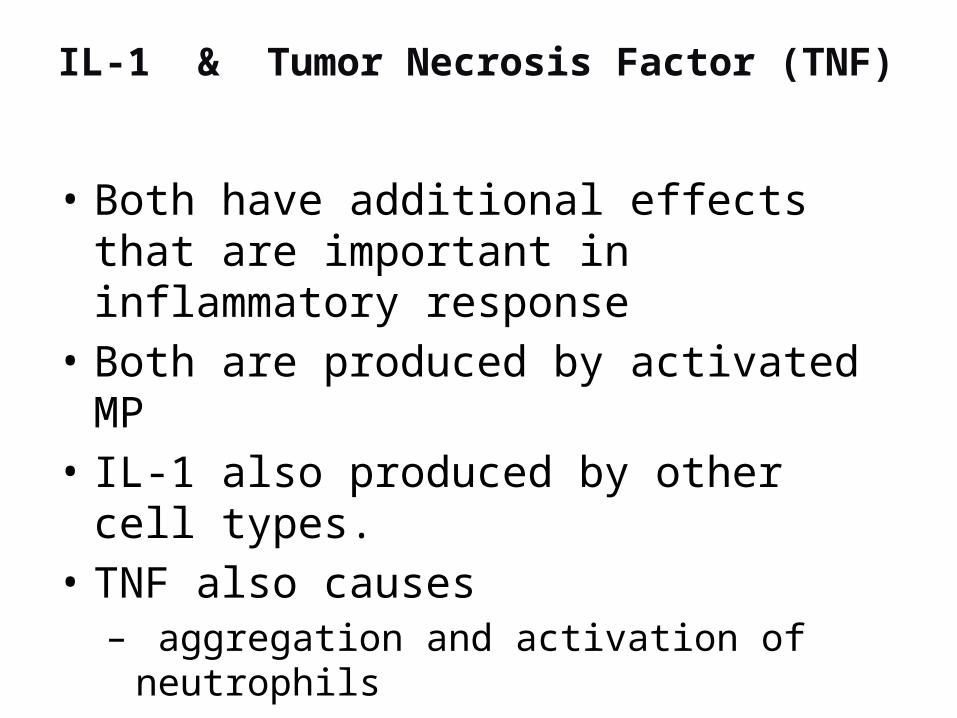
IL-1 & Tumor Necrosis Factor (TNF)
• Both have additional effects that are important in inflammatory response
• Both are produced by activated MP• IL-1 also produced by other cell types.• TNF also causes– aggregation and activation of neutrophils– Release of proteolytic enzymes from mesenchymal
cells- contributing to tissue damage
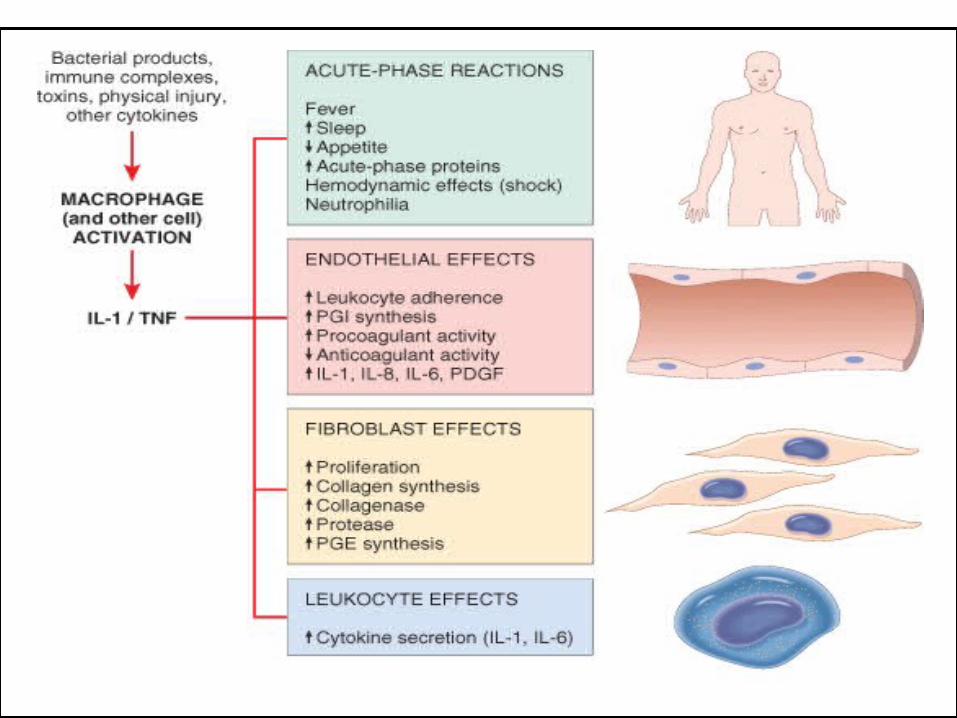
Major effects of interleukin-1 (IL-1) and tumor necrosis factor (TNF) in inflammation
Complement system
• Consist of cascade of plasma proteins (C1-C9)• Play an important role in both immunity and
inflammation• Has 20 component proteins, together with their
cleavage products• Mediate biologic reactions• All of which serve in the defense against
microbial agents
• Their function in immunity primarily by generating membrane attack complex(MAC)– They effectively punches holes in the membranes
of invading microbe
• In the process of producing MAC number of complement fragments are produced.
• Complement components are present in plasma as inactive form
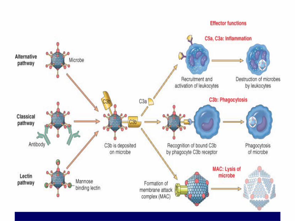
• Most critical step in the elaboration of the biological function of complement is the activation of the 3rd component, C3.
• Activation of complement by different pathways leads to cleavage of C3.
• C3 cleavage occurs via– Classic pathway- triggered by fixation of C1 to
Ag-Ab complex– Alternative pathway- triggered by bacterial
polysaccharide
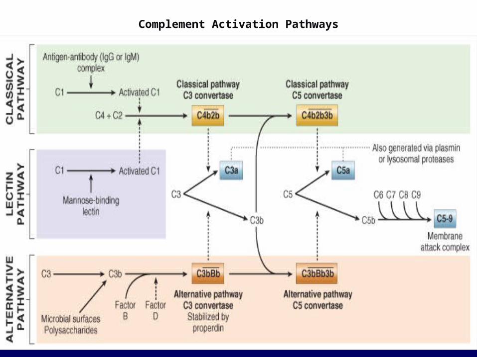
Complement Activation Pathways
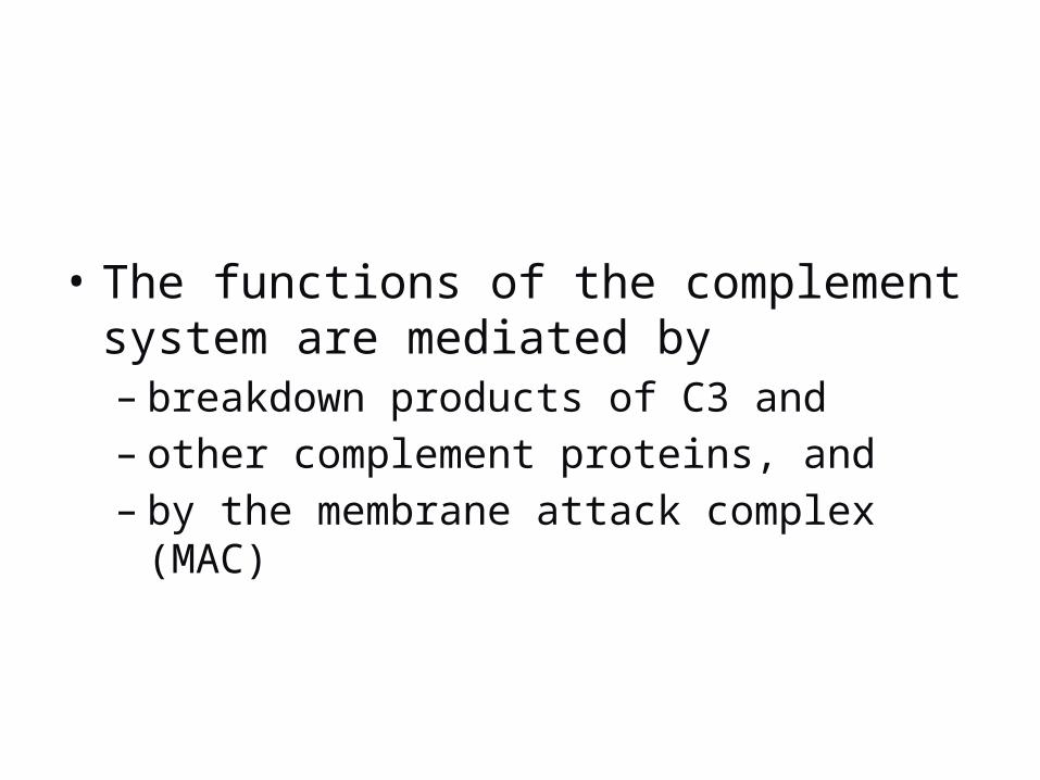
• The functions of the complement system are
mediated by – breakdown products of C3 and – other complement proteins, and – by the membrane attack complex (MAC)
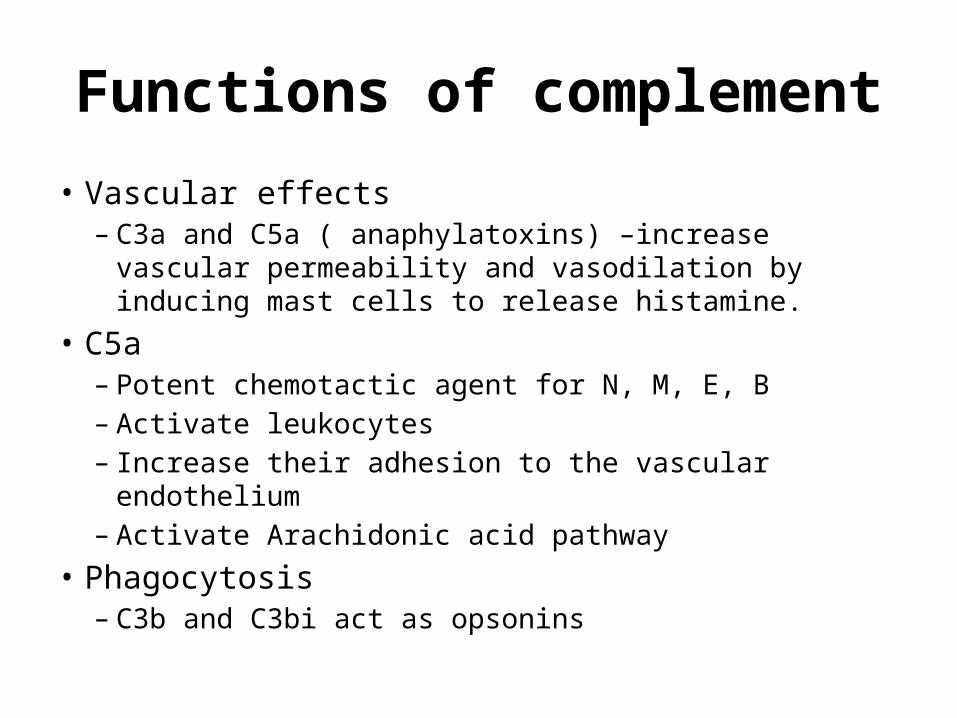
Functions of complement
• Vascular effects – C3a and C5a ( anaphylatoxins) –increase vascular
permeability and vasodilation by inducing mast cells to release histamine.
• C5a – Potent chemotactic agent for N, M, E, B– Activate leukocytes– Increase their adhesion to the vascular endothelium– Activate Arachidonic acid pathway
• Phagocytosis – C3b and C3bi act as opsonins
• C5-9(MAC) –Membrane lysis of cells– Production of oxygen metabolites
• C3 and C5 can be activated by several proteolytic enzymes present in the inflammatory infiltrate– (plasmin, lysosomal enzymes)
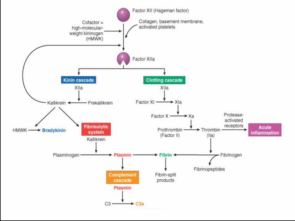
Clotting –fibrinolytic system
• Interrelationships between the four plasma mediator systems triggered by activation of factor XII (Hageman factor).
• Note that thrombin induces inflammation by binding to protease-activated receptors (principally PAR-1) on – platelets, – endothelium, – smooth muscle cells,– other cells.
Clotting cascade
• Cascade of plasma proteases– Hageman factor (factor XII)– Collagen, basement membrane, activated platelets
converts XII to XIIa (active form)– Ultimately converts soluble fibrinogen to insoluble
fibrin clot– Factor XIIa simultaneously activates the “brakes”
through the fibrinolytic system to prevent continuous clot propagation• Counter regulate clotting by cleaving fibrin• thereby solubilizing the the fibrin clot
• Fibrin – insoluble clot• Fibrinopeptide – increased vascular
permeability, chemotaxis• Thrombin – leukocyte adhesion, fibroblast
proliferation
Kinin system
• Leads to formation of bradykinin from cleavage of precursor (HMWK)– Vascular permeability– Arteriolar dilation– Non-vascular smooth muscle contraction (e.g.,
bronchial smooth muscle)– Causes pain– Rapidly inactivated (kininases)
Fibrinolytic system
• Plasminogen is converted into plasmin• Plasmin– lyses fibrin clots– Increase vascular permeability– Activates factor XII- amplify the entire set of
response– Cleaves C3 to produceC3 fragments– Degrades fibrin to fibrin split products
Arachidonic Acid MetabolitesPG,LT3, Lipoxins
• Also called eicosanoids• Derived from the metabolism of AA• Affect variety of biological processes
including– Inflammation and haemostasis
• They are short range hormones– Act locally at the site of generation– Rapidly spontaneously decay / enzymatically
destroyed
• AA is a 20-carbon polyunsaturated fatty acid• Derived from dietary linoleic acid and cell
membrane phospholipids.• AA released from phospholipids via cellular
phospholipases –Which is activated by • mechanical, chemical, physical stimuli • inflammatory mediaters- C5a
–Which is inhibited by steroids.
• AA metabolism proceed along one of the two pathway
• Cyclooxygenase pathway- Synthesizing– prostaglandins and thromboxanes
• Lipoxygenase pathway- Synthesizing– Leukotrienes and lipoxins
• They can mediate virtually every step of inflammation
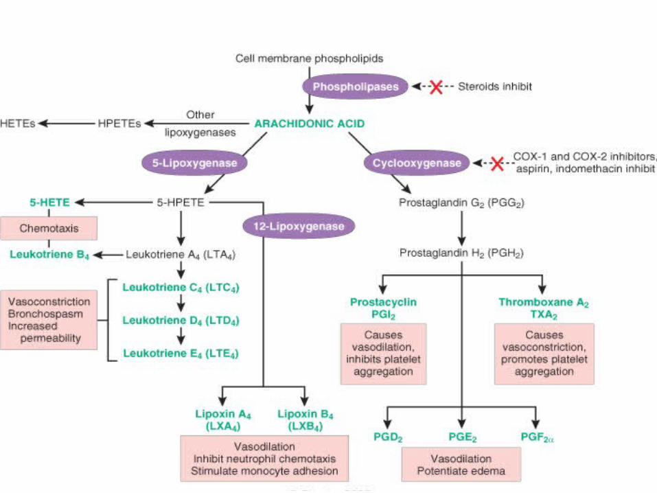
• Generation of arachidonic acid metabolites and their roles in inflammation.
• The molecular targets of some anti-inflammatory drugs are indicated by a red X.
• COX, cyclooxygenase; HETE, hydroxyeicosatetraenoic acid;
• HPETE, hydroperoxyeicosatetraenoic acid.
Prostaglandins
• Major metabolite of the COX pathway in mast cell.
• Regarded as autocoid -Rapidly spontaneously decay / enzymatically destroyed
• Act locally at the site of generation• Also cause pain and fever• Can pass from one cell to another
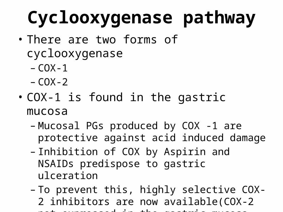
Cyclooxygenase pathway• There are two forms of cyclooxygenase– COX-1– COX-2
• COX-1 is found in the gastric mucosa –Mucosal PGs produced by COX -1 are protective
against acid induced damage– Inhibition of COX by Aspirin and NSAIDs
predispose to gastric ulceration– To prevent this, highly selective COX-2 inhibitors
are now available(COX-2 not expressed in the gastric mucosa
Role of Mediators in Different Reactions of inflammation
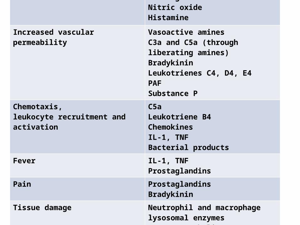
Vasodilatation ProstaglandinsNitric oxideHistamine
Increased vascular permeability Vasoactive aminesC3a and C5a (through liberating amines)BradykininLeukotrienes C4, D4, E4PAFSubstance P
Chemotaxis,leukocyte recruitment and activation
C5aLeukotriene B4ChemokinesIL-1, TNFBacterial products
Fever IL-1, TNFProstaglandins
Pain ProstaglandinsBradykinin
Tissue damage Neutrophil and macrophage lysosomal enzymesOxygen metabolitesNitric oxide
Systemic effects of acute inflammation
Fever• ‘Endogenous pyrogens’ produced:– IL1 and TNFa
• IL-1 act on vascular receptors in the thermoregulatory center of the hypothalamus
• Leads to production of prostaglandins in hypothalamus(hence aspirin etc. reduce fever)
• PGE act on the vasomotor center• Resulting in sympathetic nerve stimulation• Vasoconstriction of skin vessels• Causes reduced heat dissipation and fever.
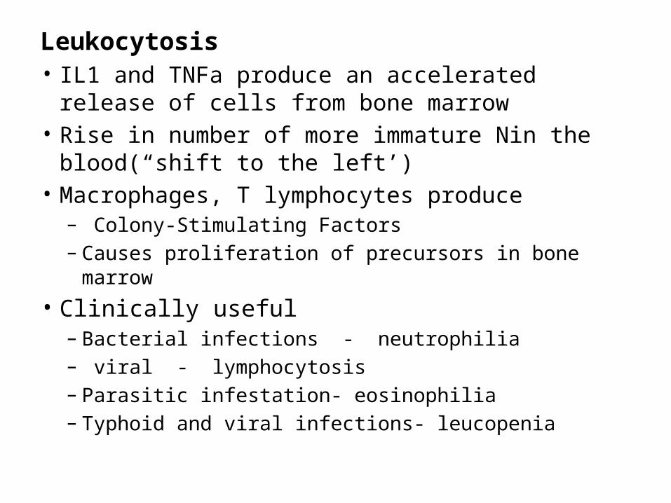
Leukocytosis• IL1 and TNFa produce an accelerated release of
cells from bone marrow• Rise in number of more immature Nin the
blood(“shift to the left’)• Macrophages, T lymphocytes produce– Colony-Stimulating Factors– Causes proliferation of precursors in bone marrow
• Clinically useful– Bacterial infections - neutrophilia– viral - lymphocytosis– Parasitic infestation- eosinophilia– Typhoid and viral infections- leucopenia
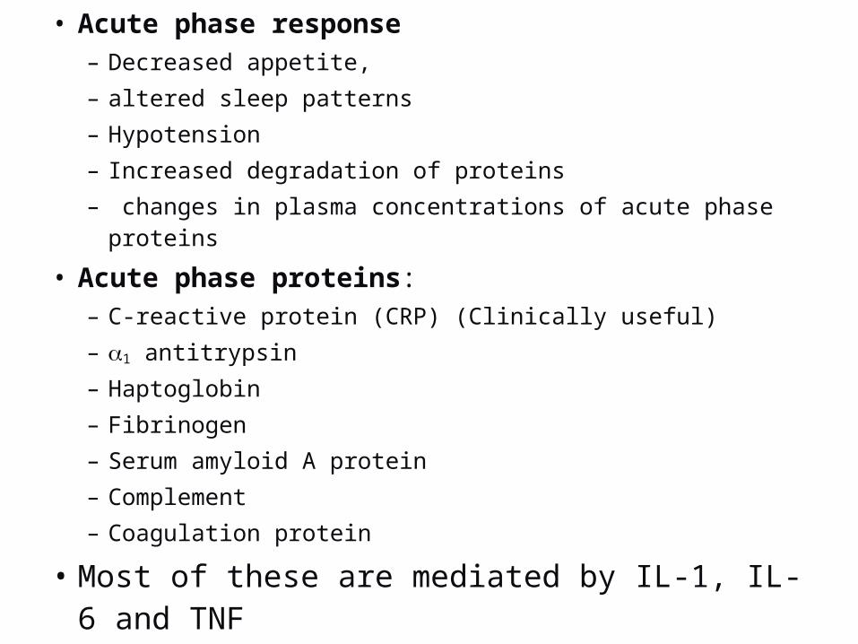
• Acute phase response– Decreased appetite, – altered sleep patterns– Hypotension– Increased degradation of proteins– changes in plasma concentrations of acute phase proteins
• Acute phase proteins:– C-reactive protein (CRP) (Clinically useful)– 1 antitrypsin
– Haptoglobin– Fibrinogen– Serum amyloid A protein– Complement– Coagulation protein
• Most of these are mediated by IL-1, IL-6 and TNF
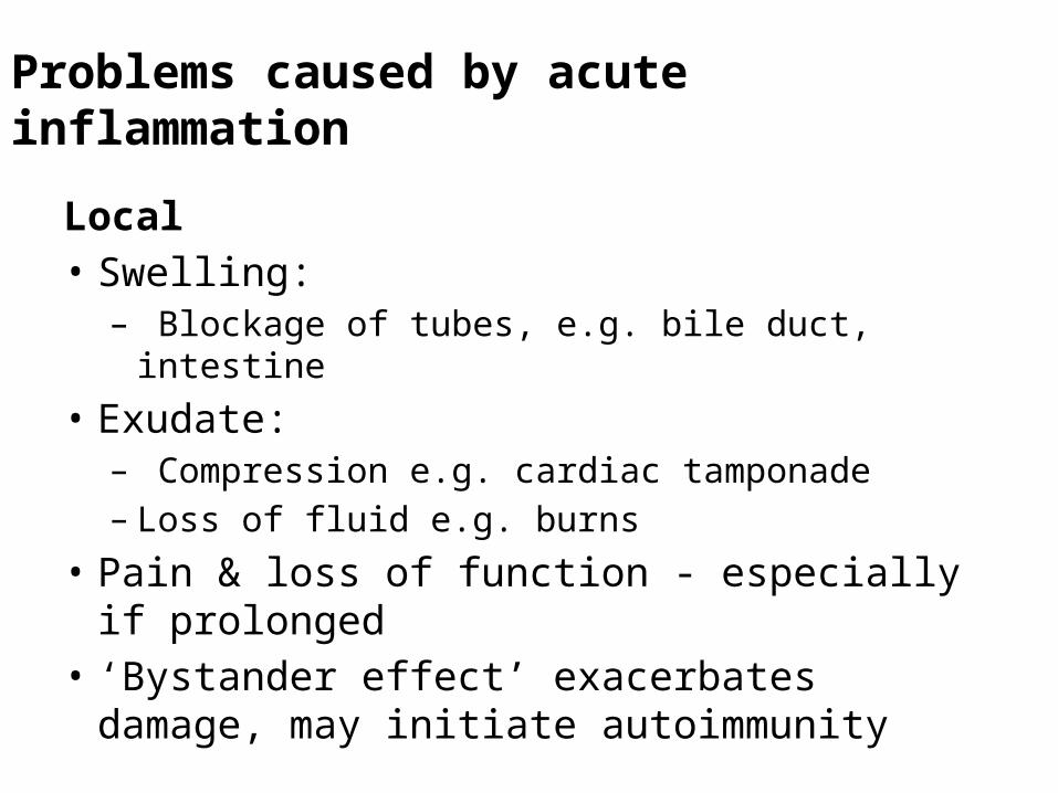
Problems caused by acute inflammation
Local• Swelling:– Blockage of tubes, e.g. bile duct, intestine
• Exudate:– Compression e.g. cardiac tamponade– Loss of fluid e.g. burns
• Pain & loss of function - especially if prolonged• ‘Bystander effect’ exacerbates damage, may
initiate autoimmunity
Systemic• Acute phase response• Spread of micro-organisms and toxins
–SHOCK
Laboratory manifestations
• Leukocytosis
(granulocytosis vs. lymphocytosis)• Elevated serum acute phase proteins
(C-reactive protein, fibrinogen, etc)• Increased ESR
(erythrocyte sedimentation rate)• Hypercoagulability
Diagnosis of acute inflammation
• Cardinal features of inflammation• Fever• Laboratory evidence– ESR– CRP, fibrinogen, haptoglobulin– Increased WBC- neutrophils with toxic granules,
left shift– Examination of inflammatory exudate– Biopsy– Test for specific aetiology• Serum antibody, complement• Culture, grame stained smear
Outcome of acute inflammtion
1. Complete resolution– Little tissue damage– Capable of regeneration
2. Scarring (fibrosis)– In tissues unable to regenerate– Excessive fibrin deposition organized into fibrous
tissue
3. Abscess formation occurs with some bacterial or fungal infections
4. Progression to chronic inflammation (next