Embed Size (px)
Citation preview
Infection & Chemotherapy
Received: August 4, 2014 Revised: August 27, 2014 Accepted: August 28, 2014Corresponding Author : Joon Young Song, MD, PhDDivision of Infectious Diseases, Department of Internal Medicine, Korea University Guro Hospital, Korea University College of Medicine, 148 Gurodong-ro, Guro-gu, Seoul 08308, KoreaTel: +82-2-2626-3052, Fax: +82-2-2626-1105, E-mail: [email protected]
This is an Open Access article distributed under the terms of the Creative Commons Attribution Non-Commercial License (http://creativecommons.org/licenses/by-nc/3.0) which permits unrestricted non-commercial use, distribution, and repro-duction in any medium, provided the original work is properly cited.
Copyrights © 2016 by The Korean Society of Infectious Diseases | Korean Society for Chemotherapy
www.icjournal.org
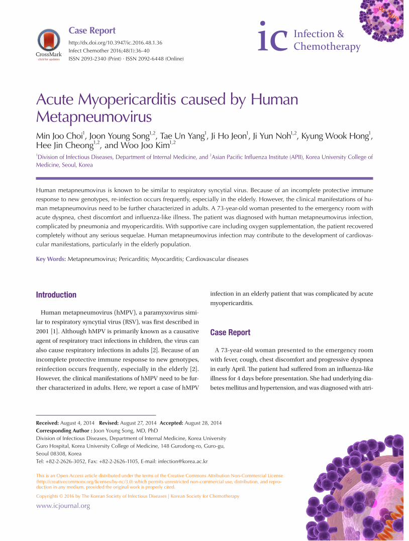
Acute Myopericarditis caused by Human MetapneumovirusMin Joo Choi1, Joon Young Song1,2, Tae Un Yang1, Ji Ho Jeon1, Ji Yun Noh1,2, Kyung Wook Hong1, Hee Jin Cheong1,2, and Woo Joo Kim1,2
1Division of Infectious Diseases, Department of Internal Medicine, and 2Asian Pacific Influenza Institute (APII), Korea University College of Medicine, Seoul, Korea
Human metapneumovirus is known to be similar to respiratory syncytial virus. Because of an incomplete protective immune response to new genotypes, re-infection occurs frequently, especially in the elderly. However, the clinical manifestations of hu-man metapneumovirus need to be further characterized in adults. A 73-year-old woman presented to the emergency room with acute dyspnea, chest discomfort and influenza-like illness. The patient was diagnosed with human metapneumovirus infection, complicated by pneumonia and myopericarditis. With supportive care including oxygen supplementation, the patient recovered completely without any serious sequelae. Human metapneumovirus infection may contribute to the development of cardiovas-cular manifestations, particularly in the elderly population.
Key Words: Metapneumovirus; Pericarditis; Myocarditis; Cardiovascular diseases
Introduction
Human metapneumovirus (hMPV), a paramyxovirus simi-
lar to respiratory syncytial virus (RSV), was first described in
2001 [1]. Although hMPV is primarily known as a causative
agent of respiratory tract infections in children, the virus can
also cause respiratory infections in adults [2]. Because of an
incomplete protective immune response to new genotypes,
reinfection occurs frequently, especially in the elderly [2].
However, the clinical manifestations of hMPV need to be fur-
ther characterized in adults. Here, we report a case of hMPV
infection in an elderly patient that was complicated by acute
myopericarditis.
Case Report
A 73-year-old woman presented to the emergency room
with fever, cough, chest discomfort and progressive dyspnea
in early April. The patient had suffered from an influenza-like
illness for 4 days before presentation. She had underlying dia-
betes mellitus and hypertension, and was diagnosed with atri-
http://dx.doi.org/10.3947/ic.2016.48.1.36
Infect Chemother 2016;48(1):36-40
ISSN 2093-2340 (Print) · ISSN 2092-6448 (Online)
Case Report

http://dx.doi.org/10.3947/ic.2016.48.1.36 • Infect Chemother 2016;48(1):36-40www.icjournal.org 37
al fibrillation 7 days prior to admission.
On physical examination the patient was slightly dyspneic
with a heart rate of 72 beats/min, respiratory rate of 24
breaths/min, blood pressure of 160/77 mmHg and tympanic
body temperature of 38.4°C. Lung auscultation revealed
crackle in both lower lung fields. Initial laboratory tests
showed mild leukopenia, thrombocytopenia, and anemia (Ta-
ble 1). Serum C-reactive protein (97.57 mg/L) and erythrocyte
sedimentation rate (52 mm/h) were elevated, but liver func-
tion tests were within normal limits. Hypoxia (oxygen satura-
tion, 88% in room air) was observed on arterial blood gas
analysis. Levels of creatine kinase-MB (CK-MB, 7.9 ng/mL)
and Pro-B type natriurectic peptide (pro-BNP, 2618 pg/mL)
were elevated. Chest X-ray showed cardiomegaly and diffuse
opacity in both lower lungs (Fig. 1A). Electrocardiogram
showed atrial fibrillation with a normal ventricular response,
and echocardiography showed slightly decreased systolic
function (ejection fraction, 50-55%) with a small amount of
pericardial effusion that was not present 7 days previously at
the time of diagnosis of atrial fibrillation. Chest computed to-
mography (CT) showed ground-glass opacities in bilateral
lungs, right-sided pleural effusion, and pericardial effusion
(Fig. 2). Ultrasound-guided aspiration of pleural effusion re-
vealed a lymphocyte-dominant exudate with pH 7.0, WBC 560
cells/L with relative lymphocytosis (95%), glucose 104 mg/dL,
protein 2.8 g/dL, lactate dehydrogenase (LDH) 356 IU/L and
adenosine deaminase (ADA) 18.3 IU/L. Pericardiocentesis
could not be performed because of safety concerns.
Initially, single-dose intravenous peramivir (300 mg) was
given to the patient assuming influenza viral pneumonia. Be-
Figure 1. Chest X-ray findings show cardiomegaly and diffuse opacity in both lower lungs with the dominance on right lower lobe (A), and marked improvement after a 7-day conservative treatment (B).
BA
Table 1. Laboratory findings at initial presentation
Day 1 Day 3
WBC (count/mm3) 2,500 1,900
Hemoglobin (g/dL) 12.5 11.5
Platelet (count/Ul) 119,000 135,000
ESR (mm/hour) 55 -
CRP (mg/L) 97.6 61.1
AST (IU/L) 62 53
ALT (IU/L) 40 46
Glucose (mg/dL) 156 -
Protein (g/dL) 5.9 5.9
Albumin (g/dL) 3.5 3.5
CPK (IU/L) 443 -
LDH (IU/L) 536 -
BUN (mg/dL) 16.8 14.7
Creatinine (mg/dL) 0.7 0.5
ABGA
PH 7.4 -
SaO2 (%) 88 -
PO2 (mmHg) 54 -
PCO2 (mmHg) 34 -
HCO3- (mmol/L) 22.7 -
CK-MB (ng/mL) 7.9 6.8
Pro-BNP (pg/mL) 2,618 -
WBC, white blood cells; ESR, erythrocyte sedimentation rate; CRP, C-reactive pro-tein; AST, aspartate aminotransferase; ALT, alanine aminotransferase; CPK, creatine phosphokinase; LDH, lactate dehydrogenase; BUN, blood urea nitrogen; ABGA, arterial blood gas analysis; CK-MB, creatine kinase-MB; BNP, B-type natriuretic peptide.

Choi MJ, et al. • Metapneumovirus-induced myopericarditis www.icjournal.org38
cause of concerns about concomitant bacterial pneumonia,
antibiotics were also administered (ceftriaxone 2.0 IV every 24
hours for 7 days and azithromycin 250mg orally bid for 3
days).
A throat swab sample was taken from the patient and poly-
merase chain reaction (PCR) was performed to detect respira-
tory viruses using SeeplexⓇ RV15 ACE Detection kit (Seegene,
Seoul, Korea). PCR analysis was positive only for hMPV on the
fourth day after hospitalization; tests for other respiratory vi-
ruses (influenza virus, RSV, parainfluenza virus, coronavirus,
rhinovirus, enterovirus, adenovirus, and bocavirus) were neg-
ative. Laboratory tests [sputum gram stain and culture, blood
cultures, urinary antigen tests for Streptococcus pneumoniae,
and serologic tests for atypical pathogens (Chlamydophila
pneumoniae, Mycoplasma pneumoniae, and Legionella spe-
cies)] were performed on suspicion of concomitant bacterial
pneumonia on admission, but none gave positive results.
Finally, the patient was diagnosed with hMPV infection
complicated by pneumonia and myopericarditis. With sup-
portive care including oxygen supplementation, the fever sub-
sided after 2 days of hospitalization and shortness of breath
improved progressively. Pneumonia and cardiomegaly were
improved on chest X-ray (Fig. 1B). Systolic function was nor-
malized (ejection fraction, 60-65%), and pericardial effusion
disappeared on follow-up echocardiography (7th day after
hospitalization). The patient recovered without any serious
sequelae.
Discussion
hMPV is a newly discovered respiratory pathogen belonging
to the Metapneumovirus genus within the Paramyxoviridae
family [2]. hMPV is distributed worldwide and shows seasonal
variation, peaking in the late winter and early spring [3]. Par-
ticularly in Korea, as reported during early April in this case,
hPMV co-circulate with influenza B in spring from late March
to early May [4]. hMPV is most prevalent in the pediatric pop-
ulation with almost 100% seroprevalence at the age of five, but
with advances in diagnostic technology the virus is increas-
ingly becoming recognized as a significant pathogen in older
adults [2]. We experienced an interesting case of hMPV infec-
tion complicated by acute myopericarditis that was resolved
after conservative management.
Although hMPV is known to cause various upper and lower
respiratory syndromes including common cold, bronchiolitis,
pneumonia, and asthma exacerbation, the full spectrum of
clinical manifestations remains to be clarified [2]. Similar to
influenza A virus, hMPV infection can cause primary viral
pneumonia, and it be accompanied by concomitant bacterial
pneumonia also [4-6]. In a mouse model, hMPV infection fa-
cilitated severe bacterial infection by obstruction of higher
levels of airways and induction of inflammatory cytokines [7].
Compared with influenza A infection, the impairment of
pneumococcal clearance in the mouse model was shorter for
hMPV infection [8]. In the present study, although a bacterial
pathogen was not isolated, antibiotics were administered be-
cause of concerns regarding concomitant bacterial pneumo-
nia.
Figure 2. Chest computed tomography shows ground-glass opacity in both lower lungs (A) and pericardial effusion with right-sided pleural effusion (B).
BA

http://dx.doi.org/10.3947/ic.2016.48.1.36 • Infect Chemother 2016;48(1):36-40www.icjournal.org 39
Although respiratory tract infections are the most common
manifestations of hMPV infection, cardiovascular problems
may be a feature that distinguishes hMPV infection from other
respiratory viral infections; cardiovascular diseases were more
common in patients with hMPV infections compared to those
with influenza and RSV infections [9, 10]. Previously, a Korean
pediatric study identified hMPV as one of the etiologic agents
of acute myocarditis [11], while a Japanese group suggested
an association between hMPV seroprevalence and hyperten-
sion in elderly subjects [12]. It is not clear whether the virus
has a tropism for the myocardium or patients with underlying
cardiovascular disease are more vulnerable to hMPV. In either
case, myocardial involvement can represent a life-threatening
condition and the association between hMPV and cardiovas-
cular diseases needs to be further clarified with respect to
both host and virus.
This report has several limitations. First, myocardial and
pericardial biopsy was not taken because of safety concerns;
therefore, pathologic confirmation of the diagnosis of myo-
pericarditis was not possible. Second, CK-MB was elevated,
but cardiac troponin levels were not checked. The elevations
of cardiac troponin I or T levels are known to be more com-
mon than CK-MB elevation in patients with biopsy-proven
myocarditis, and reflect acute, early-onset myocarditis [13,
14]. Third, cardiac magnetic resonance (CMR) imaging was
not taken in this case. CMR imaging has emerged as the most
important diagnostic tool of myocarditis, distinguishing be-
tween ischemic and non-ischemic cardiomyopathy [15, 16].
Although CMR image was not available however, coronary
angiography showed normal appearance of coronary arteries
only with mild atherosclerosis. Finally, bronchoscopy was not
taken, so respiratory virus PCR was just carried out with upper
respiratory specimen. Although clinically compatible with
hMPV pneumonia, positive PCR with throat swab specimen
would not mean lower respiratory tract infection.
In conclusion, hMPV infection may contribute to the devel-
opment of cardiovascular manifestations, particularly in the
elderly population. We should consider the possibility of myo-
cardial involvement in adult patients with hMPV infection if
they have suspicious symptoms or signs.
Conflict of InterestNo conflict of interest.
ORCID
Joon Young Song http://orcid.org/0000-0002-0148-7194
References
1. van den Hoogen BG, de Jong JC, Groen J, Kuiken T, de
Groot R, Fouchier RA, Osterhaus AD. A newly discovered
human pneumovirus isolated from young children with
respiratory tract disease. Nat Med 2001;7:719-24.
2. Haas LE, Thijsen SF, van Elden L, Heemstra KA. Human
metapneumovirus in adults. Viruses 2013;5:87-110.
3. Kahn JS. Human metapneumovirus: a newly emerging re-
spiratory pathogen. Curr Opin Infect Dis 2003;16:255-8.
4. Seo YB, Song JY, Choi MJ, Kim IS, Yang TU, Hong KW,
Cheong HJ, Kim WJ. Etiology and clinical outcomes of
acute respiratory virus infection in hospitalized adults. In-
fect Chemother 2014;46:67-76.
5. Yoo JY, Eun JY, Lee EJ, Kim TH, Choo EJ, Jeon MH. A case
of human metapneumovirus pneumonia in an immuno-
competent adult patient mimicking with influenza (A/
H1N1-2009) pandemic. Infect Chemother 2011;43:217-21.
6. Lim HJ, Lee JW, Park YS, Kim NH, Kim M, Yim JJ, Yang SC,
Yoo CG, Kim YW, Han SK, Shim YS, Lee SM. A case of se-
vere human metapneumovirus pneumonia requiring me-
chanical ventilation in an immunocompetent adult. Tu-
berc Respir Dis 2009;67:135-9.
7. Kukavica-Ibrulj I, Hamelin ME, Prince GA, Gagnon C,
Bergeron Y, Bergeron MG, Boivin G. Infection with human
metapneumovirus predisposes mice to severe pneumo-
coccal pneumonia. J Virol 2009;83:1341-9.
8. Ludewick HP, Aerts L, Hamelin ME, Boivin G. Long-term
impairment of Streptococcus pneumoniae lung clearance
is observed after initial infection with influenza A virus
but not human metapneumovirus in mice. J Gen Virol
2011;92:1662-5.
9. Johnstone J, Majumdar SR, Fox JD, Marrie TJ. Viral infec-
tion in adults hospitalized with community-acquired
pneumonia: prevalence, pathogens, and presentation.
Chest 2008;134:1141-8.
10. Widmer K, Zhu Y, Williams JV, Griffin MR, Edwards KM,
Talbot HK. Rates of hospitalizations for respiratory syncy-
tial virus, human metapneumovirus, and influenza virus
in older adults. J Infect Dis 2012;206:56-62.
11. Kim HJ, Yoo GH, Kil HR. Clinical outcome of acute myo-
carditis in children according to treatment modalities. Ko-
rean J Pediatr 2010;53:745-52.
12. Zeng L, Chen R, Ishigami K, Atsumi M, Koizumi Y, Sato K,
Iritani O, Okuro M, Morimoto S. Association between hu-
man metapneumovirus seroprevalence and hypertension
in elderly subjects in a long-term care facility. Hypertens

Choi MJ, et al. • Metapneumovirus-induced myopericarditis www.icjournal.org40
Res 2011;34:474-8.
13. Lauer B, Niederau C, Kühl U, Schannwell M, Pauschinger
M, Strauer BE, Schultheiss HP. Cardiac troponin T in pa-
tients with clinically suspected myocarditis. J Am Coll
Cardiol 1997;30:1354-9.
14. Smith SC, Ladenson JH, Mason JW, Jaffe AS. Elevations of
cardiac troponin I associated with myocarditis. Experi-
mental and clinical correlates. Circulation 1997;95:163-8.
15. Friedrich MG, Marcotte F. Cardiac magnetic resonance
assessment of myocarditis. Circ Cardiovasc Imaging 2013;
6:833-9.
16. Friedrich MG, Sechtem U, Schulz-Menger J, Holmvang G,
Alakija P, Cooper LT, White JA, Abdel-Aty H, Gutberlet M,
Prasad S, Aletras A, Laissy JP, Paterson I, Filipchuk NG,
Kumar A, Pauschinger M, Liu P; International Consensus
Group on Cardiovascular Magnetic Resonance in Myocar-
ditis. Cardiovascular magnetic resonance in myocarditis:
A JACC White Paper. J Am Coll Cardiol 2009;53:1475-87.