Embed Size (px)
Citation preview

551
□ CASE REPORT □
Pneumocystis Pneumonia Concomitant with Ectopic ACTHSyndrome Caused by a Large Cell Neuroendocrine
Carcinoma of the Thymus
Naohiro Oda 1, Nobuaki Miyahara 1,2, Masahiro Tabata 1, Daisuke Minami 3,
Kiichiro Ninomiya 1, Arihiko Kanehiro 1, Motoshi Komatsubara 4, Kenichi Inagaki 4,
Mitsune Tanimoto 1 and Katsuyuki Kiura 1
Abstract
We herein report the case of a 44-year-old man who was diagnosed with pneumocystis pneumonia (PCP)
concomitant with ectopic adrenocorticotropic hormone (ACTH) syndrome, which had been caused by a large
cell neuroendocrine carcinoma of the thymus. Chest computed tomography revealed ground-glass opacities in
the lungs. PCP was diagnosed by a polymerase chain reaction with bronchoalveolar lavage. The levels of cor-
tisol were slowly corrected with an adrenal enzyme inhibitor, and the exacerbation of PCP was successfully
avoided. Our case indicates that in addition to prophylaxis, the early diagnosis of PCP and the slow correc-
tion of hypercortisolemia should be considered in order to prevent an exacerbation due to the reconstitution
of the immune function in patients with ectopic ACTH syndrome.
Key words: ectopic ACTH syndrome, Cushing’s syndrome, thymus, neuroendocrine tumor, large cell
neuroendocrine carcinoma, pneumocystis pneumonia
(Intern Med 56: 551-555, 2017)(DOI: 10.2169/internalmedicine.56.7655)
Introduction
Ectopic adrenocorticotropic hormone (ACTH) syndrome
is an endocrine disorder that is caused by excessive levels of
the endogenous corticosteroid hormone cortisol arising from
an ACTH-producing tumor that was located outside the pi-
tuitary. The most common cause of ectopic ACTH syndrome
is small cell lung cancer (45%), followed by thymic (15%),
bronchial (15%), and pancreatic neuroendocrine tumors
(10%) (1). Among these tumors, large cell neuroendocrine
carcinoma (LCNEC) of the thymus is a very rare cause of
ectopic ACTH syndrome, and only one case has ever been
reported in the literature (2).
Ectopic ACTH syndrome comes with a high risk of op-
portunistic infection, such as pneumocystis pneumonia
(PCP), which has a significant impact on the prognosis of
ectopic ACTH syndrome (3-5). We herein report a case of
PCP concomitant with ectopic ACTH syndrome that had
been caused by LCNEC of the thymus, in which PCP was
successfully treated and the patient’s hypercortisolemia was
corrected.
Case Report
A 44-year-old man had been diagnosed with LCNEC of
the thymus with bone metastasis at a different hospital 3
years previously. The serum levels of ACTH and cortisol
were elevated, but brain magnetic resonance imaging re-
vealed that his pituitary gland was normal. However, immu-
nohistochemical staining of the thymic tumor tissue revealed
partial anti-ACTH antibody positivity. The tumor was there-
1Department of Allergy and Respiratory Medicine, Okayama University Hospital, Japan, 2Department of Medical Technology, Okayama Univer-
sity Graduate School of Health Sciences, Japan, 3Department of Respiratory Medicine, NHO Okayama Medical Center, Japan and 4Endocrine
Center, Okayama University Hospital, Japan
Received for publication April 28, 2016; Accepted for publication June 19, 2016
Correspondence to Dr. Nobuaki Miyahara, [email protected]
Intern Med 56: 551-555, 2017 DOI: 10.2169/internalmedicine.56.7655
552
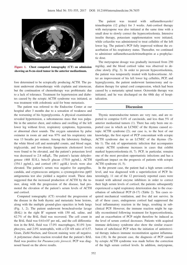
Figure 1. Chest computed tomography (CT) on admission showing an 8-cm-sized tumor in the anterior mediastinum.
fore determined to be ectopically producing ACTH. The pa-
tient underwent chemotherapy with cisplatin and irinotecan,
but the continuation of chemotherapy was problematic due
to a lack of tolerance. Treatment for hypertension and diabe-
tes caused by the ectopic ACTH syndrome was initiated, as
was treatment with zoledronic acid for bone metastasis.
The patient was referred to the Endocrine Center at our
hospital after 3 months due to a sensation of weakness and
the worsening of his hyperglycemia. A physical examination
revealed hypertension, a subcutaneous mass that was palpa-
ble in the anterior chest, and redness and swelling of the left
lower leg without fever, respiratory symptoms, hypoxemia,
or abnormal chest sounds. The oxygen saturation by pulse
oximeter in room air and was 97% and his respiratory rate
was 13 breaths per minute. Among the laboratory findings,
the white blood cell and neutrophil counts, and blood sugar,
triglyceride, and low-density lipoprotein cholesterol levels
were found to be elevated, and the patient’s potassium level
was decreased to 2.8 mEq/L. The patient’s lactate dehydro-
genase (468 IU/L), beta-D glucan (370.8 pg/mL), ACTH
(354.1 pg/mL), and cortisol (49.1 μg/dL) levels were also
elevated. The patient’s serum was negative for aspergillus,
candida, and cryptococcus antigens; a cytomegalovirus pp65
antigenemia test also yielded a negative result. These data
suggested that the increased production of ACTH by the tu-
mor, along with the progression of the disease, had pro-
moted the elevation of the patient’s serum levels of ACTH
and cortisol.
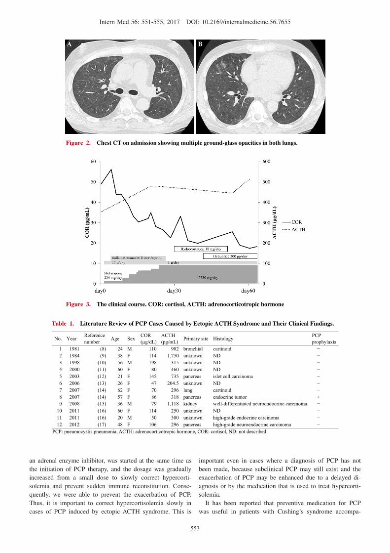
Computed tomography (CT) revealed the progression of
the disease in the both thymic and metastatic bone lesions,
along with the multiple ground-glass opacities in both lungs
(Fig. 1, 2). The patient underwent bronchoalveolar lavage
(BAL) in the right B5 segment with 150 mL saline, and
62.7% of the BAL fluid was recovered. The cell count in
the BAL fluid was 0.61×10⁵ per mL, and the cell differentia-
tion in the BAL fluid was 17.0% macrophages, 81.0% lym-
phocytes, and 2.0% neutrophils, with a CD 4/8 ratio of 0.57.
Gram, Ziehl-Neelsen, and Grocott staining were all negative.
A polymerase chain reaction revealed that the patient’s BAL
fluid was positive for Pneumocystis jirovecii. PCP was diag-
nosed based on the above results.
The patient was treated with sulfamethoxazole /
trimethoprim (12 g/day) for 3 weeks. Anti-cortisol therapy
with metyrapone was also initiated at the same time with a
small dose to slowly correct the hypercortisolemia. Intensive
insulin therapy, potassium supplementation were initiated,
while cefazolin was administered to treat cellulitis of the left
lower leg. The patient’s PCP fully improved without the ex-
acerbation of his respiratory status. Thereafter, we continued
to administer sulfamethoxazole/trimethoprim at a prophylac-
tic dose.
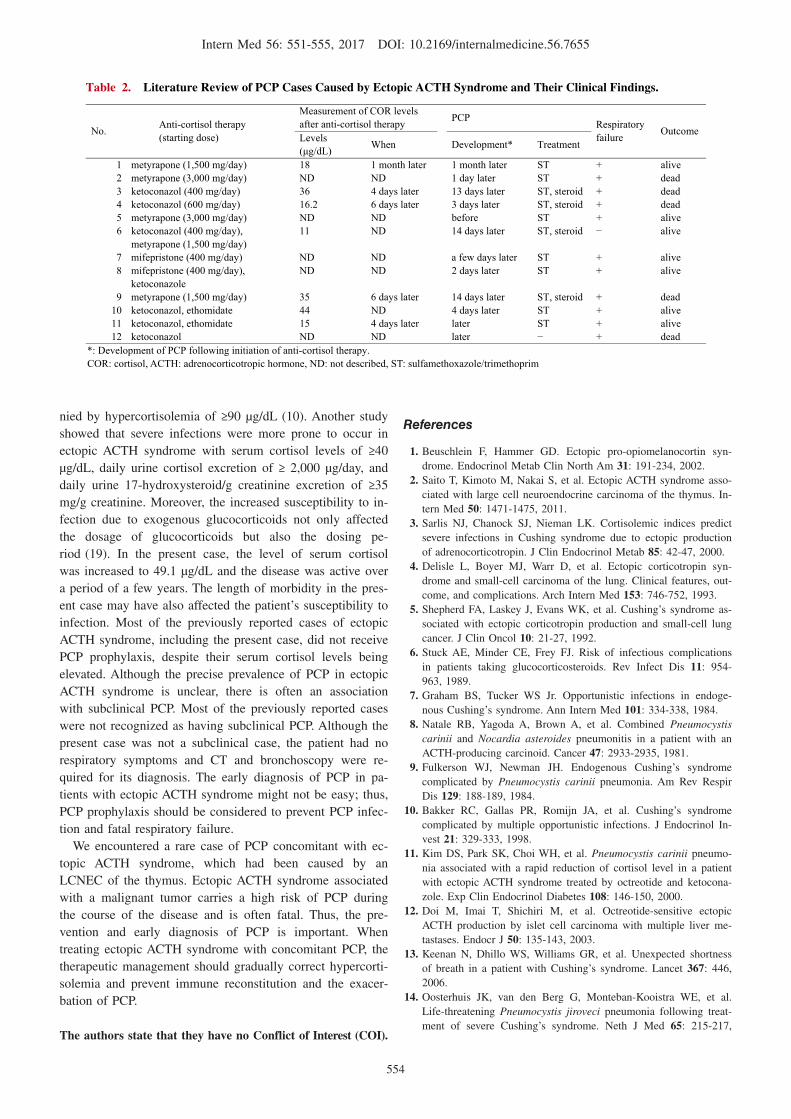
The metyrapone dosage was gradually increased from 250
mg/day, and the blood cortisol value was observed to de-
cline slowly (Fig. 3). In order to prevent hypocortisolemia,
the patient was temporarily treated with hydrocortisone. Af-
ter an improvement of his left lower leg cellulitis, PCP, and
hyperglycemia, the patient underwent laminectomy and ra-
diation therapy for spinal cord compression, which had been
caused by a metastatic spinal tumor. Octreotide therapy was
initiated, and he was discharged on the 68th day of hospi-
talization.
Discussion
Thymic neuroendocrine tumors are very rare, and are re-
ported to comprise 0.4% of carcinoids, and less than 5% of
anterior mediastinal tumors (1). There has only been one re-
ported case in which an LCNEC of the thymus caused ec-
topic ACTH syndrome (2); our case is, to the best of our
knowledge, the first report of PCP concomitant with ectopic
ACTH syndrome due to an LCNEC of the thymus (Ta-
ble 1). The risk of opportunistic infection that accompanies
ectopic ACTH syndrome increases in cases that exhibit
marked hypercortisolemia (6, 7). PCP, in particular, remains
one of the most prevalent opportunistic infections and has a
significant impact on the prognosis of patients with ectopic
ACTH syndrome (4, 5).
In the present case, the patient had a high serum cortisol
level, and was diagnosed with a superinfection of PCP. In-
terestingly, 11 out of the 12 previously reported cases were
treated with adrenal enzyme inhibitors in order to correct
their high serum levels of cortisol; the patients subsequently
experienced a rapid respiratory deterioration due to the exac-
erbation of subclinical PCP (8-17) (Table 2). Ten cases re-
quired mechanical ventilation, and five did not survive. In
all of these cases, endogenous cortisol had suppressed the
local inflammatory reaction in the lungs, resulting in sub-
clinical PCP. However, the immune reaction might be rap-
idly reconstituted following treatment for hypercortisolemia,
and an exacerbation of PCP might therefore be induced as
the level of serum cortisol decreases. Patients with acquired
immune deficiency syndrome also show the similar exacer-
bation of subclinical PCP when the initiation of antiretrovi-
ral therapy induces immune reconstitution against inflamma-
tion (18). In the present case, the diagnosis of PCP caused
by ectopic ACTH syndrome was made before the correction
of the high serum cortisol levels. In addition, metyrapone,
Intern Med 56: 551-555, 2017 DOI: 10.2169/internalmedicine.56.7655
553
Figure 2. Chest CT on admission showing multiple ground-glass opacities in both lungs.
Figure 3. The clinical course. COR: cortisol, ACTH: adrenocorticotropic hormone
Table 1. Literature Review of PCP Cases Caused by Ectopic ACTH Syndrome and Their Clinical Findings.
No. Year Reference number Age Sex COR
(μg/dL)ACTH(pg/mL) Primary site Histology PCP
prophylaxis1 1981 (8) 24 M 110 902 bronchial cartinoid2 1984 (9) 38 F 114 1,750 unknown ND3 1998 (10) 56 M 198 315 unknown ND4 2000 (11) 60 F 80 460 unknown ND5 2003 (12) 21 F 145 735 pancreas islet cell carcinoma 6 2006 (13) 26 F 47 204.5 unknown ND7 2007 (14) 62 F 70 296 lung cartinoid8 2007 (14) 57 F 86 318 pancreas endocrine tumor +9 2008 (15) 36 M 79 1,118 kidney well-differentiated neuroendocrine carcinoma
10 2011 (16) 60 F 114 250 unknown ND11 2011 (16) 20 M 50 300 unknown high-grade endocrine carcinoma12 2012 (17) 48 F 106 296 pancreas high-grade neuroendocrine carcinoma
PCP: pneumocystis pneumonia, ACTH: adrenocorticotropic hormone, COR: cortisol, ND: not described
an adrenal enzyme inhibitor, was started at the same time as
the initiation of PCP therapy, and the dosage was gradually
increased from a small dose to slowly correct hypercorti-
solemia and prevent sudden immune reconstitution. Conse-
quently, we were able to prevent the exacerbation of PCP.
Thus, it is important to correct hypercortisolemia slowly in
cases of PCP induced by ectopic ACTH syndrome. This is
important even in cases where a diagnosis of PCP has not
been made, because subclinical PCP may still exist and the
exacerbation of PCP may be enhanced due to a delayed di-
agnosis or by the medication that is used to treat hypercorti-
solemia.
It has been reported that preventive medication for PCP
was useful in patients with Cushing’s syndrome accompa-
Intern Med 56: 551-555, 2017 DOI: 10.2169/internalmedicine.56.7655
554
Table 2. Literature Review of PCP Cases Caused by Ectopic ACTH Syndrome and Their Clinical Findings.
No. Anti-cortisol therapy(starting dose)
Measurement of COR levels after anti-cortisol therapy PCP Respiratory
failure OutcomeLevels
g/dL) When Development* Treatment
1 metyrapone (1,500 mg/day) 18 1 month later 1 month later ST + alive2 metyrapone (3,000 mg/day) ND ND 1 day later ST + dead3 ketoconazol (400 mg/day) 36 4 days later 13 days later ST, steroid + dead4 ketoconazol (600 mg/day) 16.2 6 days later 3 days later ST, steroid + dead5 metyrapone (3,000 mg/day) ND ND before ST + alive6 ketoconazol (400 mg/day),
metyrapone (1,500 mg/day)11 ND 14 days later ST, steroid alive
7 mifepristone (400 mg/day) ND ND a few days later ST + alive8 mifepristone (400 mg/day),
ketoconazoleND ND 2 days later ST + alive
9 metyrapone (1,500 mg/day) 35 6 days later 14 days later ST, steroid + dead10 ketoconazol, ethomidate 44 ND 4 days later ST + alive11 ketoconazol, ethomidate 15 4 days later later ST + alive12 ketoconazol ND ND later + dead
*: Development of PCP following initiation of anti-cortisol therapy.COR: cortisol, ACTH: adrenocorticotropic hormone, ND: not described, ST: sulfamethoxazole/trimethoprim
nied by hypercortisolemia of �90 μg/dL (10). Another study
showed that severe infections were more prone to occur in
ectopic ACTH syndrome with serum cortisol levels of �40
μg/dL, daily urine cortisol excretion of �2,000 μg/day, and
daily urine 17-hydroxysteroid/g creatinine excretion of �35
mg/g creatinine. Moreover, the increased susceptibility to in-
fection due to exogenous glucocorticoids not only affected
the dosage of glucocorticoids but also the dosing pe-
riod (19). In the present case, the level of serum cortisol
was increased to 49.1 μg/dL and the disease was active over
a period of a few years. The length of morbidity in the pres-
ent case may have also affected the patient’s susceptibility to
infection. Most of the previously reported cases of ectopic
ACTH syndrome, including the present case, did not receive
PCP prophylaxis, despite their serum cortisol levels being
elevated. Although the precise prevalence of PCP in ectopic
ACTH syndrome is unclear, there is often an association
with subclinical PCP. Most of the previously reported cases
were not recognized as having subclinical PCP. Although the
present case was not a subclinical case, the patient had no
respiratory symptoms and CT and bronchoscopy were re-
quired for its diagnosis. The early diagnosis of PCP in pa-
tients with ectopic ACTH syndrome might not be easy; thus,
PCP prophylaxis should be considered to prevent PCP infec-
tion and fatal respiratory failure.
We encountered a rare case of PCP concomitant with ec-
topic ACTH syndrome, which had been caused by an
LCNEC of the thymus. Ectopic ACTH syndrome associated
with a malignant tumor carries a high risk of PCP during
the course of the disease and is often fatal. Thus, the pre-
vention and early diagnosis of PCP is important. When
treating ectopic ACTH syndrome with concomitant PCP, the
therapeutic management should gradually correct hypercorti-
solemia and prevent immune reconstitution and the exacer-
bation of PCP.
The authors state that they have no Conflict of Interest (COI).
References
1. Beuschlein F, Hammer GD. Ectopic pro-opiomelanocortin syn-
drome. Endocrinol Metab Clin North Am 31: 191-234, 2002.
2. Saito T, Kimoto M, Nakai S, et al. Ectopic ACTH syndrome asso-
ciated with large cell neuroendocrine carcinoma of the thymus. In-
tern Med 50: 1471-1475, 2011.
3. Sarlis NJ, Chanock SJ, Nieman LK. Cortisolemic indices predict
severe infections in Cushing syndrome due to ectopic production
of adrenocorticotropin. J Clin Endocrinol Metab 85: 42-47, 2000.
4. Delisle L, Boyer MJ, Warr D, et al. Ectopic corticotropin syn-
drome and small-cell carcinoma of the lung. Clinical features, out-
come, and complications. Arch Intern Med 153: 746-752, 1993.
5. Shepherd FA, Laskey J, Evans WK, et al. Cushing’s syndrome as-
sociated with ectopic corticotropin production and small-cell lung
cancer. J Clin Oncol 10: 21-27, 1992.
6. Stuck AE, Minder CE, Frey FJ. Risk of infectious complications
in patients taking glucocorticosteroids. Rev Infect Dis 11: 954-
963, 1989.
7. Graham BS, Tucker WS Jr. Opportunistic infections in endoge-
nous Cushing’s syndrome. Ann Intern Med 101: 334-338, 1984.
8. Natale RB, Yagoda A, Brown A, et al. Combined Pneumocystiscarinii and Nocardia asteroides pneumonitis in a patient with an
ACTH-producing carcinoid. Cancer 47: 2933-2935, 1981.
9. Fulkerson WJ, Newman JH. Endogenous Cushing’s syndrome
complicated by Pneumocystis carinii pneumonia. Am Rev Respir
Dis 129: 188-189, 1984.
10. Bakker RC, Gallas PR, Romijn JA, et al. Cushing’s syndrome
complicated by multiple opportunistic infections. J Endocrinol In-
vest 21: 329-333, 1998.
11. Kim DS, Park SK, Choi WH, et al. Pneumocystis carinii pneumo-
nia associated with a rapid reduction of cortisol level in a patient
with ectopic ACTH syndrome treated by octreotide and ketocona-
zole. Exp Clin Endocrinol Diabetes 108: 146-150, 2000.
12. Doi M, Imai T, Shichiri M, et al. Octreotide-sensitive ectopic
ACTH production by islet cell carcinoma with multiple liver me-
tastases. Endocr J 50: 135-143, 2003.
13. Keenan N, Dhillo WS, Williams GR, et al. Unexpected shortness
of breath in a patient with Cushing’s syndrome. Lancet 367: 446,
2006.
14. Oosterhuis JK, van den Berg G, Monteban-Kooistra WE, et al.
Life-threatening Pneumocystis jiroveci pneumonia following treat-
ment of severe Cushing’s syndrome. Neth J Med 65: 215-217,

Intern Med 56: 551-555, 2017 DOI: 10.2169/internalmedicine.56.7655
555
2007.
15. Arlt A, Harbeck B, Anlauf M, et al. Fatal pneumocystis jirovecii
pneumonia in a case of ectopic Cushing’s syndrome due to
neuroendocrine carcinoma of the kidney. Exp Clin Endocrinol
Diabetes 116: 515-519, 2008.
16. Gabalec F, Zavrelová A, Havel E, et al. Pneumocystis pneumonia
during medicamentous treatment of Cushing’s syndrome: a de-
scription of two cases. Acta Medica (Hradec Králové) 54: 127-
130, 2011.
17. Chowdry RP, Bhimani C, Delgado MA, et al. Unusual suspects:
pulmonary opportunistic infections masquerading as tumor metas-
tasis in a patient with adrenocorticotropic hormone-producing pan-
creatic neuroendocrine cancer. Ther Adv Med Oncol 4: 295-300,
2012.
18. Miller RF, Huang L, Walzer PD. Pneumocystis pneumonia associ-
ated with human immunodeficiency virus. Clin Chest Med 34:
229-241, 2013.
19. Cutolo M, Seriolo B, Pizzorni C, et al. Use of glucocorticoids and
risk of infections. Autoimmun Rev 8: 153-155, 2008.
The Internal Medicine is an Open Access article distributed under the Creative
Commons Attribution-NonCommercial-NoDerivatives 4.0 International License. To
view the details of this license, please visit (https://creativecommons.org/licenses/
by-nc-nd/4.0/).
Ⓒ 2017 The Japanese Society of Internal Medicine
http://www.naika.or.jp/imonline/index.html