Embed Size (px)
Citation preview
Dr. LEUNG Yu-lung, DexterMBChB, BMedSci(Hons),FRCS (Glas),MRCS(Edin), DRCOphth(London),FCOphthHK, FHKAM(Ophth)
Associate ConsultantClinical Assistant Professor (Honorary)
Deputy Chief, Glaucoma ServiceDepartment of Ophthalmology & Visual Sciences
The Chinese University of Hong KongHong Kong Eye Hospital
3 Dec 2008
Acute Ophthalmology for A&E Practice
Acknowledgment
HK College of Emergency Medicine
Dr. HO Hui-fai
Dr. Tommy Chan
Recognise the NORMAL…Vs. ABNORMAL
One of the key to make it easy is to… …
1. Normality & Special Points in Ophthalmic P/E and Imaging
2. Pattern Recognition– Clinical Slide Show
3. Question and Answer Session
With these in mind, Outline of Talk as follows:
What is Normal?
Note: Seeing Collarettes of Iris in a Clear Cornea
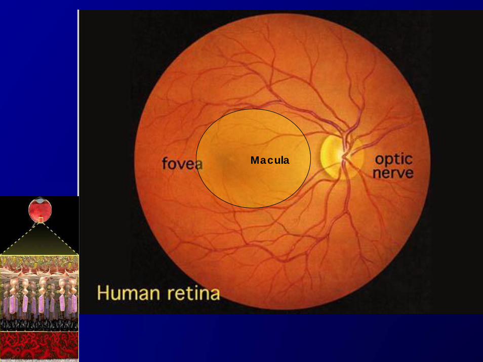
Macula
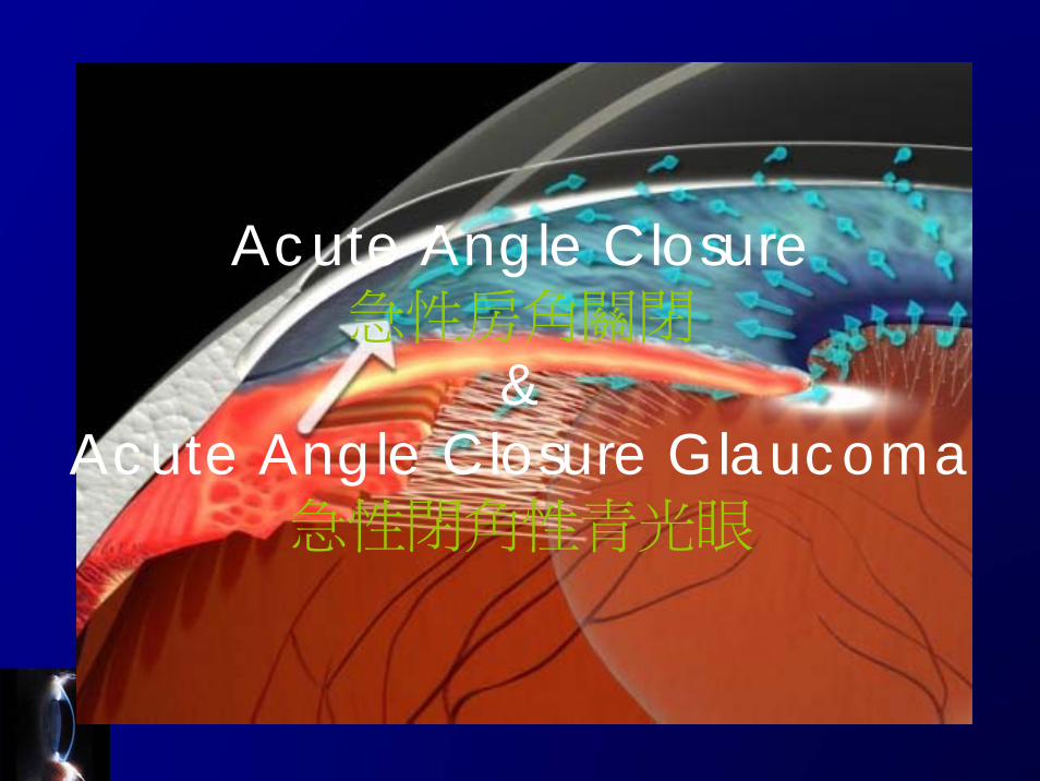
Acute Angle Closure急性房角關閉
&
急性閉角性青光眼Acute Angle Closure Glaucoma
Important: Examine as many normaleyes as you can, with Slit lamp and direct ophthalmoscopy with dilated pupils!
No slit lamp? Magnifying glass + bright torch will do!
Approach to Ocular Abnormality
• Time honored:– Careful History– Thorough Physical Examination– Logical appropriate investigation
• Importance of Pattern Recognition in Ophthalmology (esp. signs)
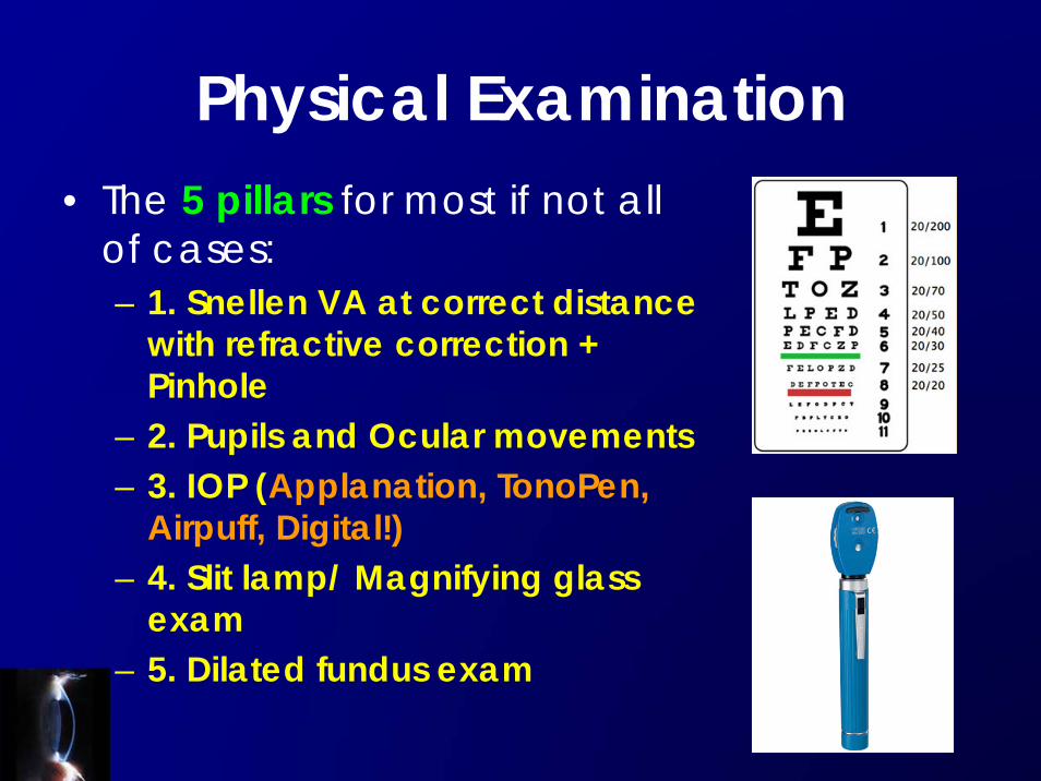
Physical Examination• The 5 pillars for most if not all
of cases:– 1. Snellen VA at correct distance
with refractive correction + Pinhole
– 2. Pupils and Ocular movements– 3. IOP (Applanation, TonoPen,
Airpuff, Digital!)– 4. Slit lamp/ Magnifying glass
exam– 5. Dilated fundus exam
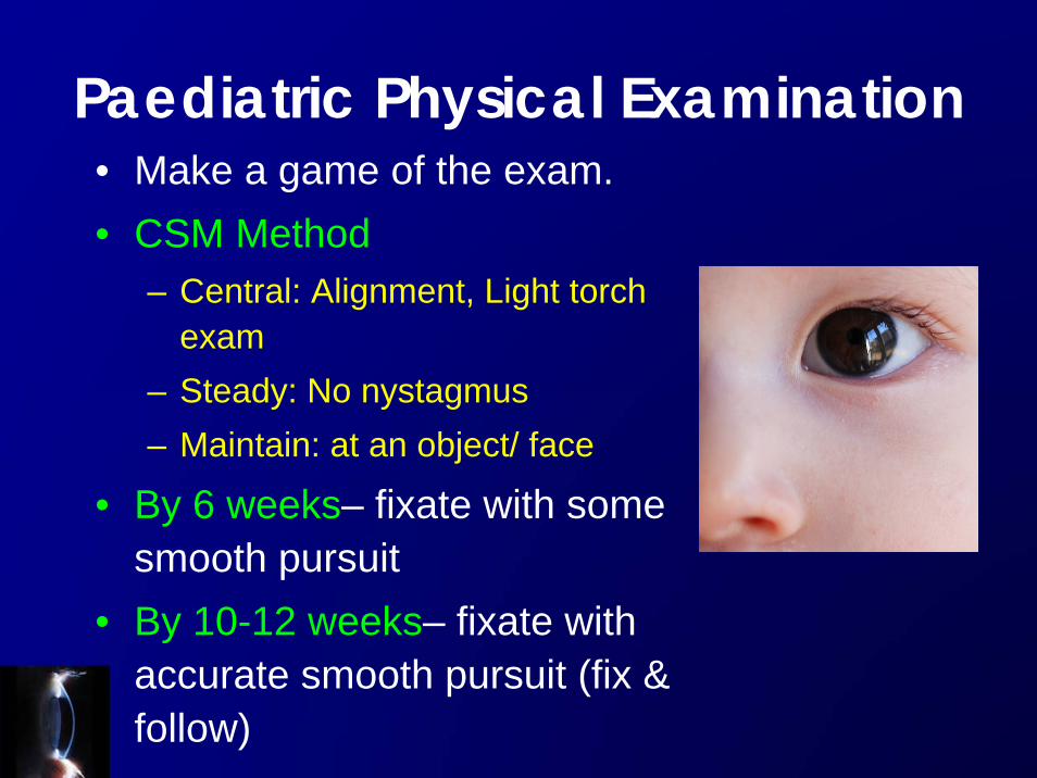
Paediatric Physical Examination• Make a game of the exam.• CSM Method
– Central: Alignment, Light torch exam
– Steady: No nystagmus– Maintain: at an object/ face
• By 6 weeks– fixate with some smooth pursuit
• By 10-12 weeks– fixate with accurate smooth pursuit (fix & follow)
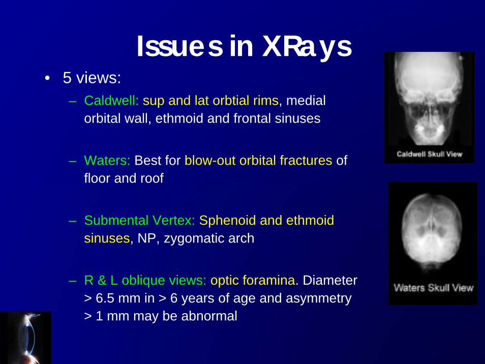
Issues in XRays• 5 views:
– Caldwell: sup and lat orbtial rims, medial orbital wall, ethmoid and frontal sinuses
– Waters: Best for blow-out orbital fractures of floor and roof
– Submental Vertex: Sphenoid and ethmoidsinuses, NP, zygomatic arch
– R & L oblique views: optic foramina. Diameter > 6.5 mm in > 6 years of age and asymmetry > 1 mm may be abnormal
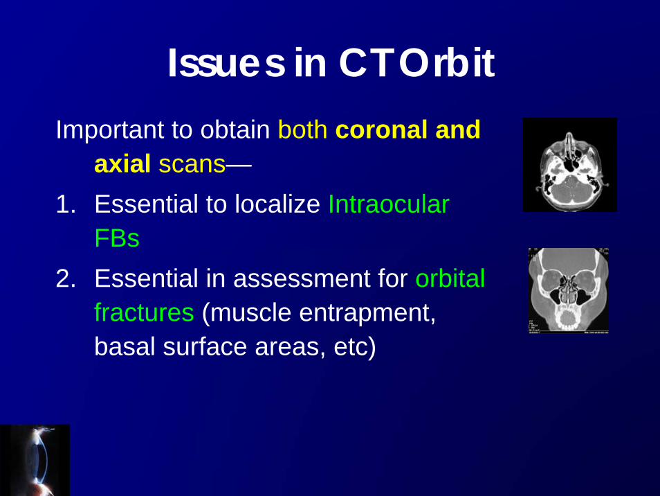
Issues in CT OrbitImportant to obtain both coronal and
axial scans—1. Essential to localize Intraocular
FBs2. Essential in assessment for orbital
fractures (muscle entrapment, basal surface areas, etc)
Clinical Slide Show…
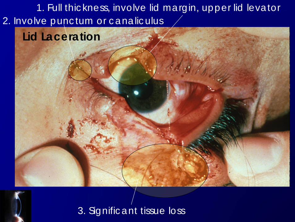
Lid Laceration
1. Full thickness, involve lid margin, upper lid levator2. Involve punctum or canaliculus
3. Significant tissue loss
ALWAYS rule out (1)a ruptured globe(2)retrobulbar hemorrhage with compartmental Sx(3)FBin these cases -- perform a complete ophthalmic exams!!
Management
1. Assessment –NATURE of trauma, extent, associated
injuries
2. Antibiotic prophylaxis: topical +/- systemic--if wound is
not clean/ complicated laceration
3. Tetanus prophylaxis
4. Rabies prophylaxis if indicated
5. Adequate wound irrigation & cleansing with FB removal
6. LA– beware of globe penetration
7. Tarsus– 6-O Vicryl; Skin- 8-O Silk
8. +/- Ice pack first 24-48 hrs
Management
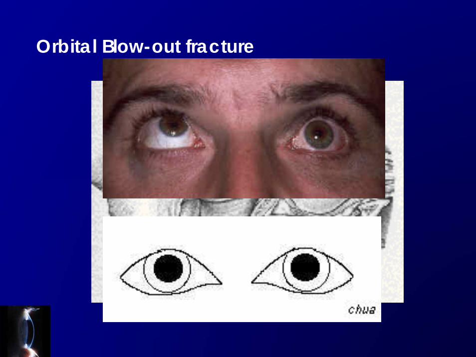
Orbital Blow-out fracture
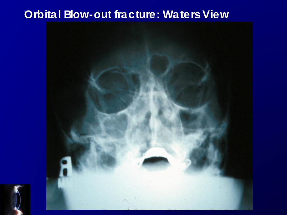
Orbital Blow-out fracture: Waters View
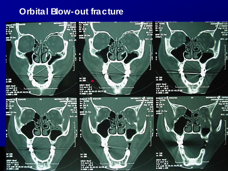
Orbital Blow-out fracture
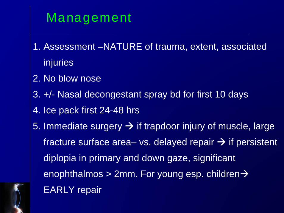
1. Assessment –NATURE of trauma, extent, associated
injuries
2. No blow nose
3. +/- Nasal decongestant spray bd for first 10 days
4. Ice pack first 24-48 hrs
5. Immediate surgery if trapdoor injury of muscle, large
fracture surface area– vs. delayed repair if persistent
diplopia in primary and down gaze, significant
enophthalmos > 2mm. For young esp. children
EARLY repair
Management
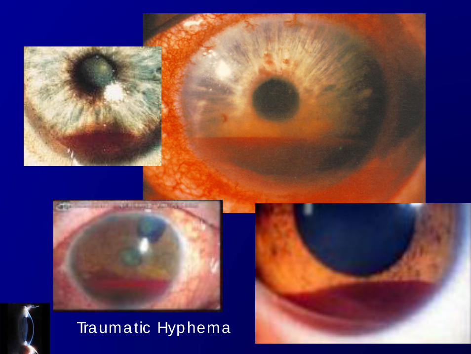
Traumatic Hyphema
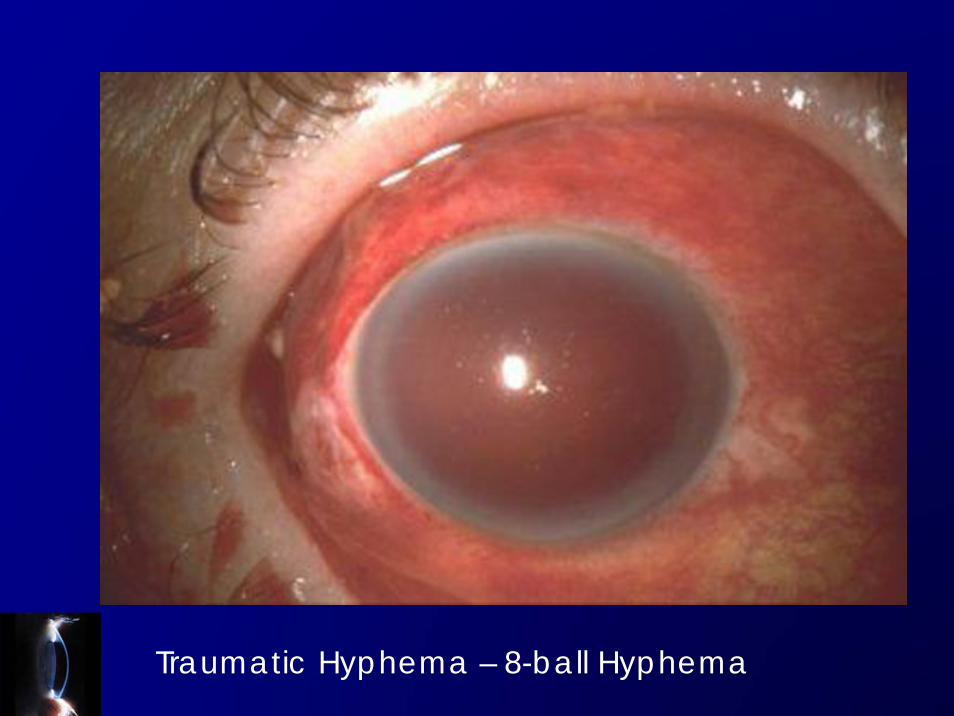
Traumatic Hyphema – 8-ball Hyphema
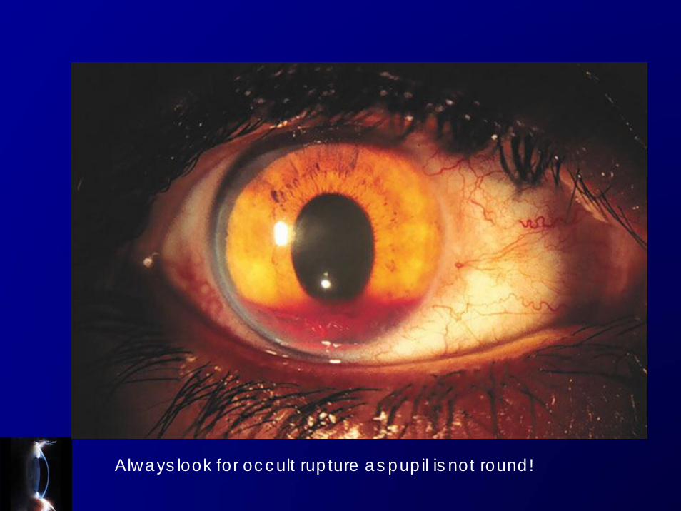
Always look for occult rupture as pupil is not round!
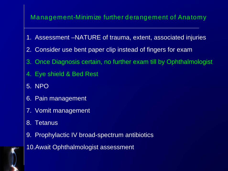
1. Assessment –NATURE of trauma, extent, associated injuries
2. Bed Rest with Head up
3. Eye shield
4. Consider stopping Aspirin
5. Topical steroid (gutt. Pred forte qid to q2h +/- topical antibiotics +/-
cycloplegics (pros & cons– gutt. 4% homatropine bd)
6. +/- topical glaucoma meds
7. +/- Aminocaproic acid (antifibrinolytic) 50mg/kg q4h for 5 days, reduces
incidence of rebleeds in prospective clinical trials.
8. Beware of sickling Cx in African American
9. Surgical: Paracentesis, AC Washout if indicated according to guidelines.
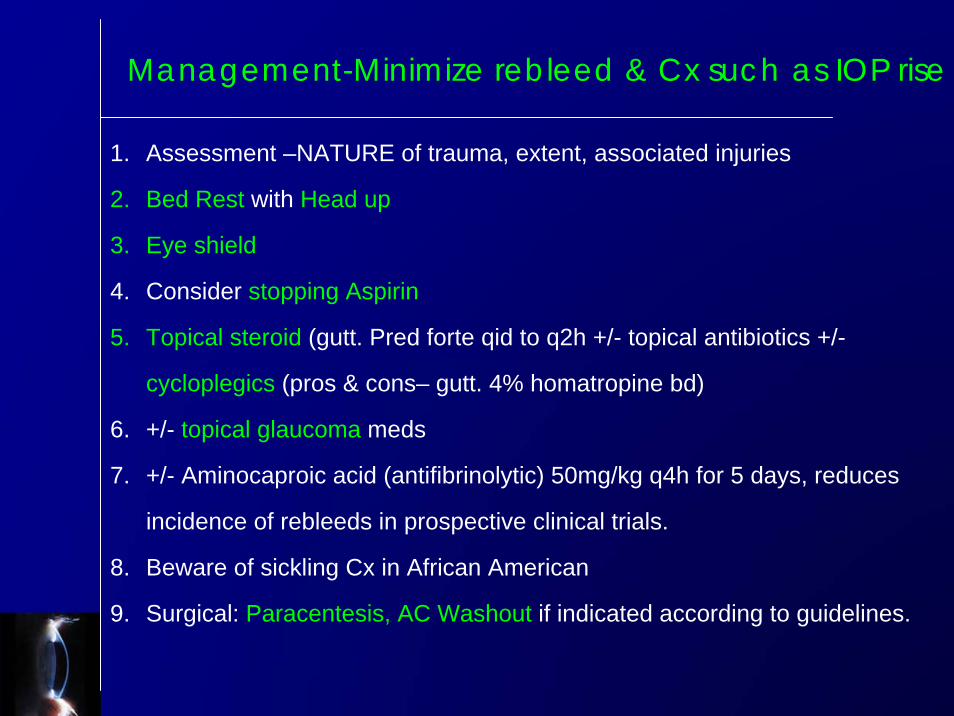
Management-Minimize rebleed & Cx such as IOP rise
1. Assessment –NATURE of trauma, extent, associated injuries
2. Consider use bent paper clip instead of fingers for exam
3. Once Diagnosis certain, no further exam till by Ophthalmologist
4. Eye shield & Bed Rest
5. NPO
6. Pain management
7. Vomit management
8. Tetanus
9. Prophylactic IV broad-spectrum antibiotics
10.Await Ophthalmologist assessment
Management-Minimize further derangement of Anatomy
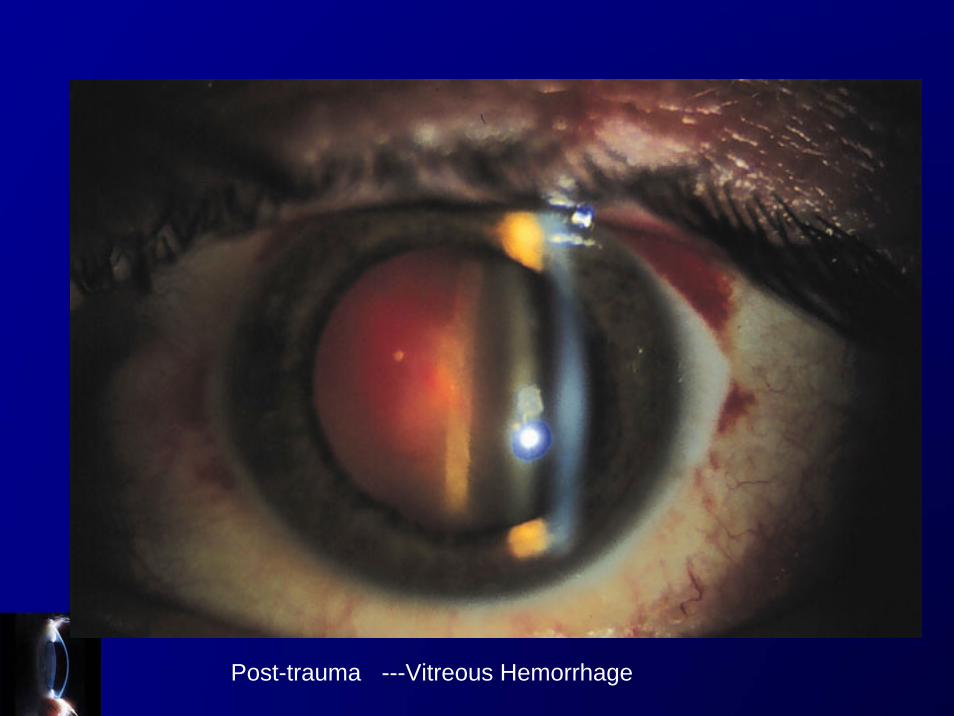
Post-trauma ---Vitreous Hemorrhage
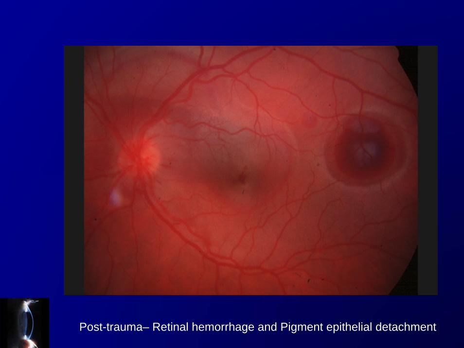
Post-trauma– Retinal hemorrhage and Pigment epithelial detachment

VA= 3/60 VA w gls = 6/6 IOP = 12 mmHg BP = normal
Simple Subconjunctival Hemorrhage
Rx: Nil after BP +/- coagulation assessment; OR +/- topical lubricants
RED EYERED EYE
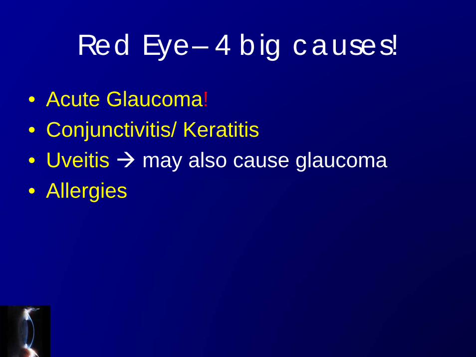
Red Eye– 4 big causes!
• Acute Glaucoma!• Conjunctivitis/ Keratitis• Uveitis may also cause glaucoma• Allergies
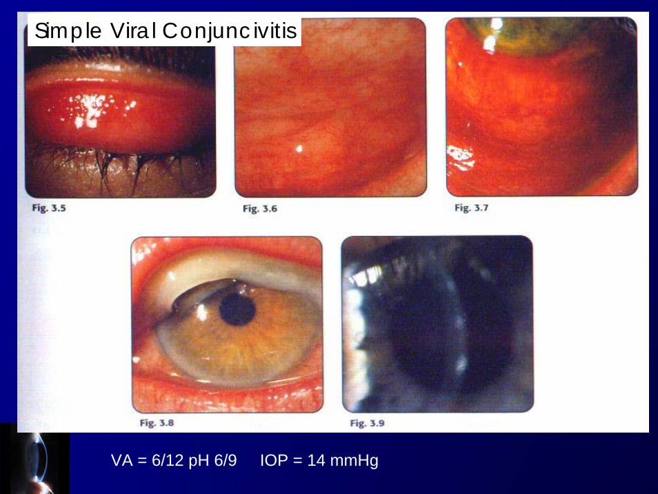
VA = 6/12 pH 6/9 IOP = 14 mmHg
Simple Viral Conjuncivitis
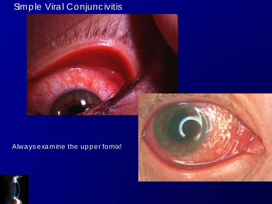
Always examine the upper fornix!
Simple Viral Conjuncivitis
Management
1. Wash your hands and instruments afterwards!!
2. Consider C/ST if looks atypical
3. Supportive Rx: Cool compression, topical
lubricants, Pain management
4. Red Eye education & Hygiene
5. ?Use of topical antibiotics and astringents
6. Treat corneal subepithelial infiltrate with mild
topical steroid (by ophthalmologist)
7. What do you tell your patient?
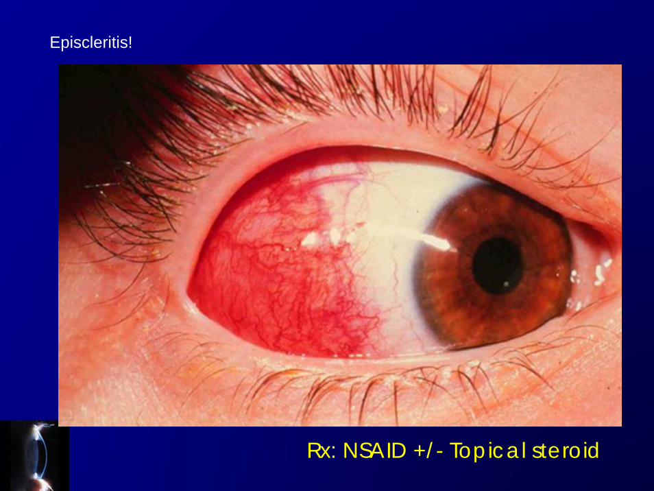
Episcleritis!
Rx: NSAID +/- Topical steroid
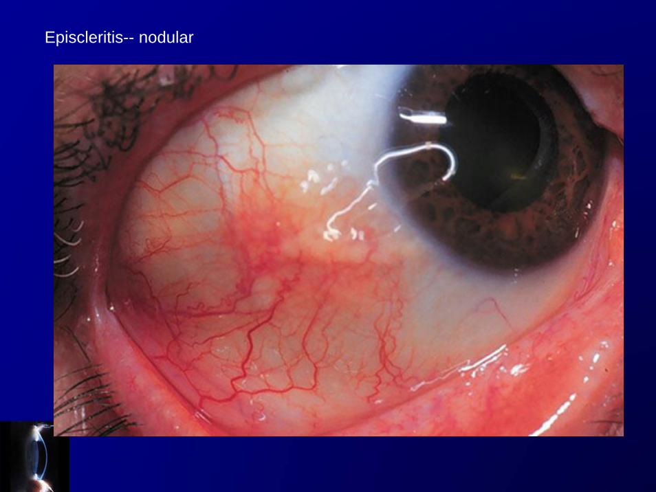
Episcleritis-- nodular