Embed Size (px)
Citation preview
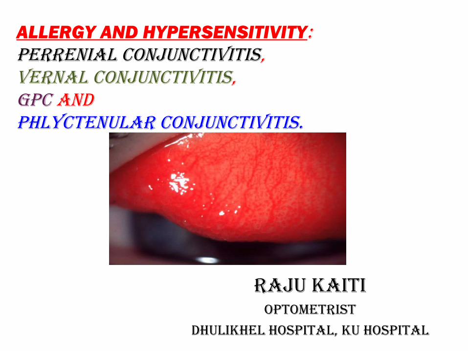
ALLERGY AND HYPERSENSITIVITY:PERRENIAL CONJUNCTIVITIS,VERNAL CONJUNCTIVITIS,GPC AND PHLYCTENULAR CONJUNCTIVITIS.
RAJU KAITIOPTOmETRIST
DHULIKHEL HOSPITAL, KU HOSPITAL
IMMUNITY
• Immune system is an interacting set of specialized cells and proteins designed to identify and destroy foreign invaders or abnormal substances before they damage the body
• Sequence of cellular and molecular events designed to rid the host of an offending stimulus
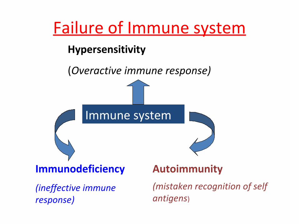
Failure of Immune system
Immune system
Hypersensitivity
(Overactive immune response)
Immunodeficiency
(ineffective immune response)
Autoimmunity
(mistaken recognition of self antigens)
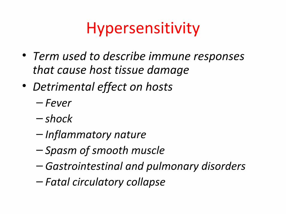
Hypersensitivity
• Term used to describe immune responses that cause host tissue damage
• Detrimental effect on hosts– Fever– shock– Inflammatory nature– Spasm of smooth muscle– Gastrointestinal and pulmonary disorders– Fatal circulatory collapse
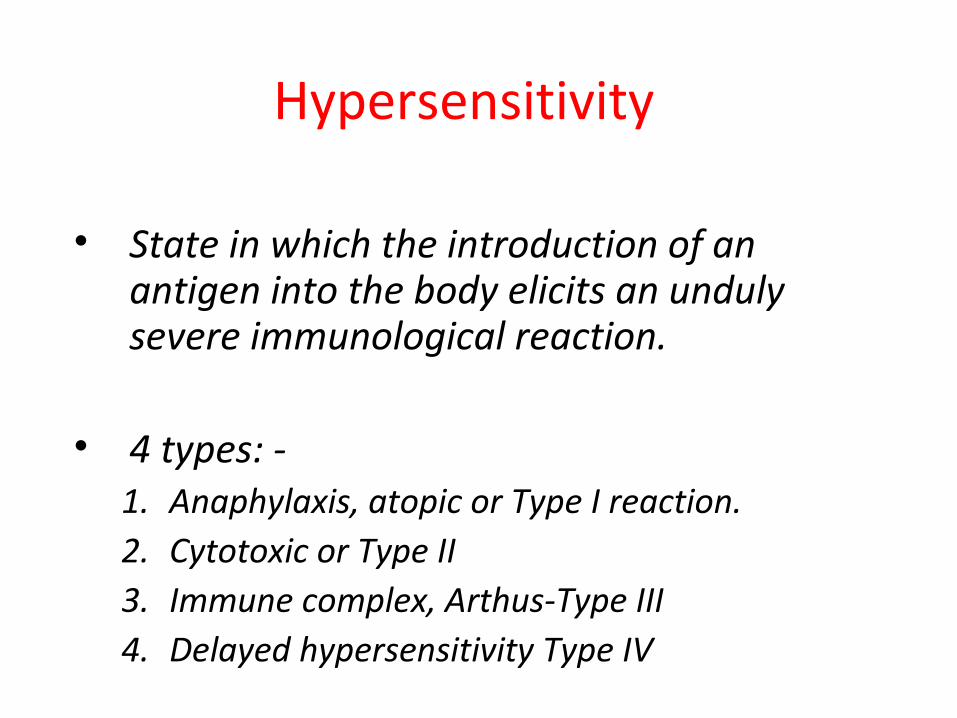
Hypersensitivity
• State in which the introduction of an antigen into the body elicits an unduly severe immunological reaction.
• 4 types: -1. Anaphylaxis, atopic or Type I reaction.2. Cytotoxic or Type II3. Immune complex, Arthus-Type III4. Delayed hypersensitivity Type IV
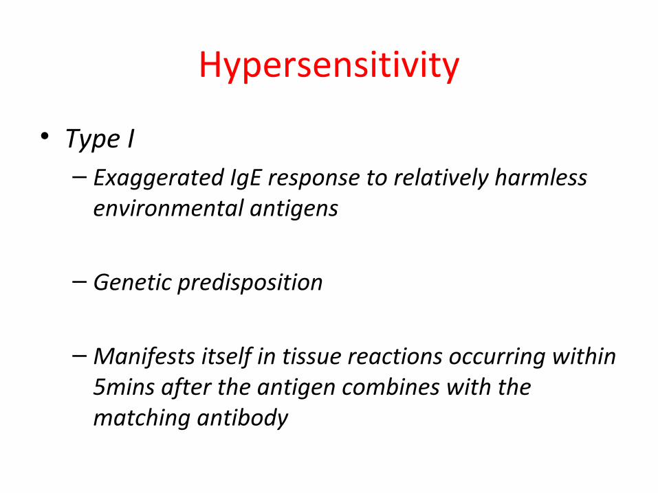
Hypersensitivity
• Type I– Exaggerated IgE response to relatively harmless
environmental antigens
– Genetic predisposition
– Manifests itself in tissue reactions occurring within 5mins after the antigen combines with the matching antibody
• Results in release of several active substances including histamine, slow reacting substance and an eosinophils chemo tactic factor within minutes
• Also may be a second “late phase”
• TYPE 1 HYPERSENSITIVITY MEDIATORS• Histamine• Prostaglandin and thromboxanes• cytokines
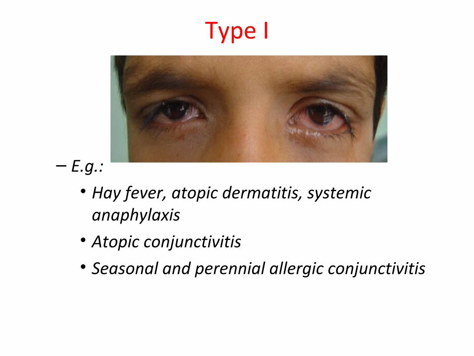
Type I
– E.g.: • Hay fever, atopic dermatitis, systemic
anaphylaxis• Atopic conjunctivitis• Seasonal and perennial allergic conjunctivitis
Type II
– Antibody mediated hypersensitivity against self cells or receptors or membranes
– Mediated by IgG or IgM antibodies against tissue antigens, resulting in organ-specific antibody production
– Antibody binds to cells or tissues and causes local complement activation, influx of leukocytes, and tissue destruction
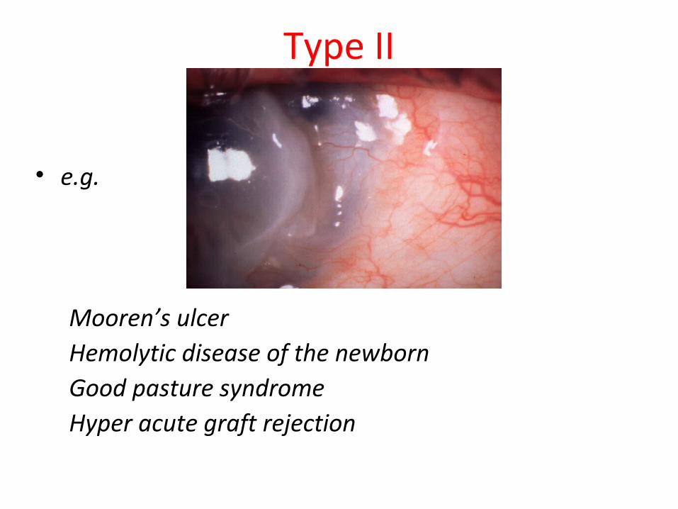
Type II
• e.g.
Mooren’s ulcer Hemolytic disease of the newbornGood pasture syndromeHyper acute graft rejection
Type III
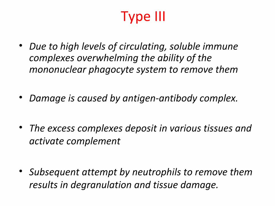
• Due to high levels of circulating, soluble immune complexes overwhelming the ability of the mononuclear phagocyte system to remove them
• Damage is caused by antigen-antibody complex.
• The excess complexes deposit in various tissues and activate complement
• Subsequent attempt by neutrophils to remove them results in degranulation and tissue damage.
Type III
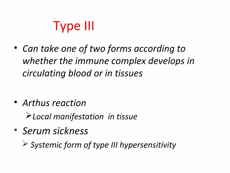
• Can take one of two forms according to whether the immune complex develops in circulating blood or in tissues
• Arthus reactionLocal manifestation in tissue
• Serum sickness Systemic form of type III hypersensitivity
Type III
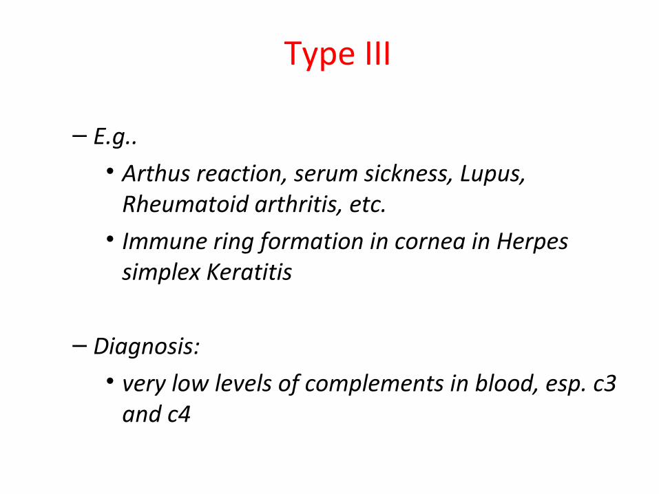
– E.g.. • Arthus reaction, serum sickness, Lupus,
Rheumatoid arthritis, etc.• Immune ring formation in cornea in Herpes
simplex Keratitis
– Diagnosis:• very low levels of complements in blood, esp. c3
and c4

Immune ring formation in herpes simplex Keratitis
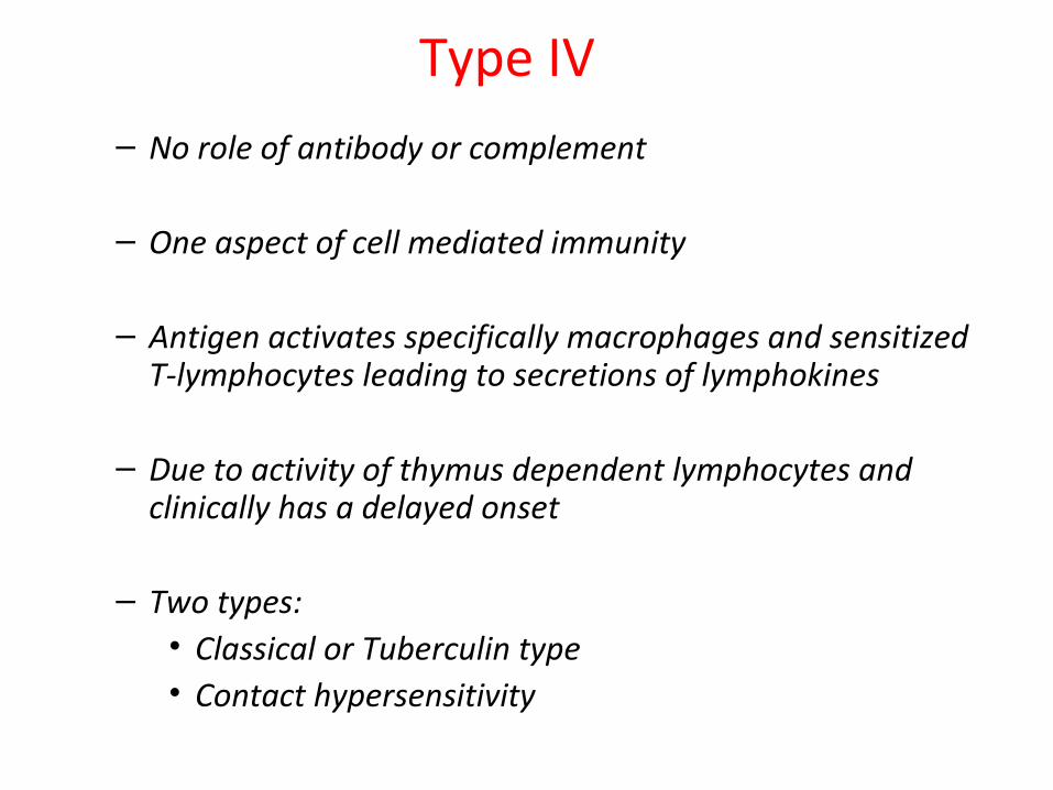
Type IV– No role of antibody or complement
– One aspect of cell mediated immunity
– Antigen activates specifically macrophages and sensitized T-lymphocytes leading to secretions of lymphokines
– Due to activity of thymus dependent lymphocytes and clinically has a delayed onset
– Two types:• Classical or Tuberculin type • Contact hypersensitivity
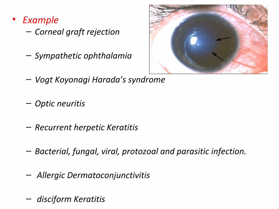
• Example– Corneal graft rejection
– Sympathetic ophthalamia
– Vogt Koyonagi Harada’s syndrome
– Optic neuritis
– Recurrent herpetic Keratitis
– Bacterial, fungal, viral, protozoal and parasitic infection.
– Allergic Dermatoconjunctivitis
– disciform Keratitis


Allergic conjunctivitis:
• Inflammation of conjunctiva due to allergic or hypersensitive reaction which may be immediate (humoral ) or delayed (cellular) to specific antigens
Types
• SIMPLE ALLERGIC• VERNAL KERATOCONJUNCTIVITIS• ATOPIC KERATCONJUNCTIVITIS• GIANT PAPILLARY CONJUNCTIVITIS• PHLYCTENULAR CONJUNCTIVITIS• CONTACT DERMATOCONJUNCTIVITIS
Simple allergic conjunctivitis
• Hay fever conjunctivitis
• Seasonal allergic conjunctivitis
• Perennial allergic conjunctivitis
• Acute allergic conjunctivitis
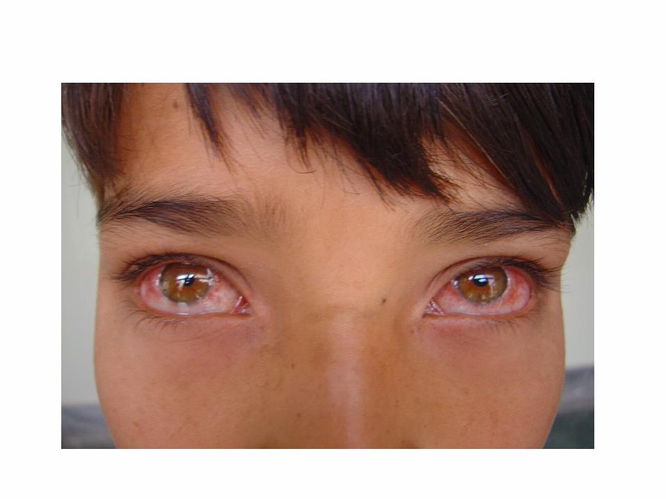
Perennial allergic conjunctivitis(PAC)
• Occurs at any age
• History of personal and/or familial allergies
• Seen all year- round
• Causes can include animal dander, insects and dust mites
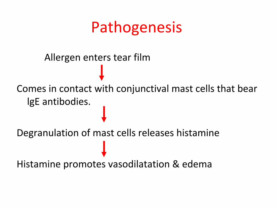
Pathogenesis
Allergen enters tear film
Comes in contact with conjunctival mast cells that bear lgE antibodies.
Degranulation of mast cells releases histamine
Histamine promotes vasodilatation & edema
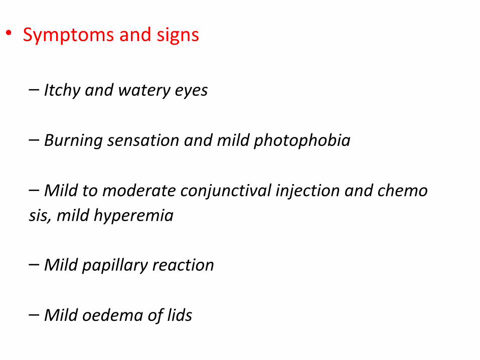
• Symptoms and signs
– Itchy and watery eyes
– Burning sensation and mild photophobia
– Mild to moderate conjunctival injection and chemosis, mild hyperemia
– Mild papillary reaction
– Mild oedema of lids
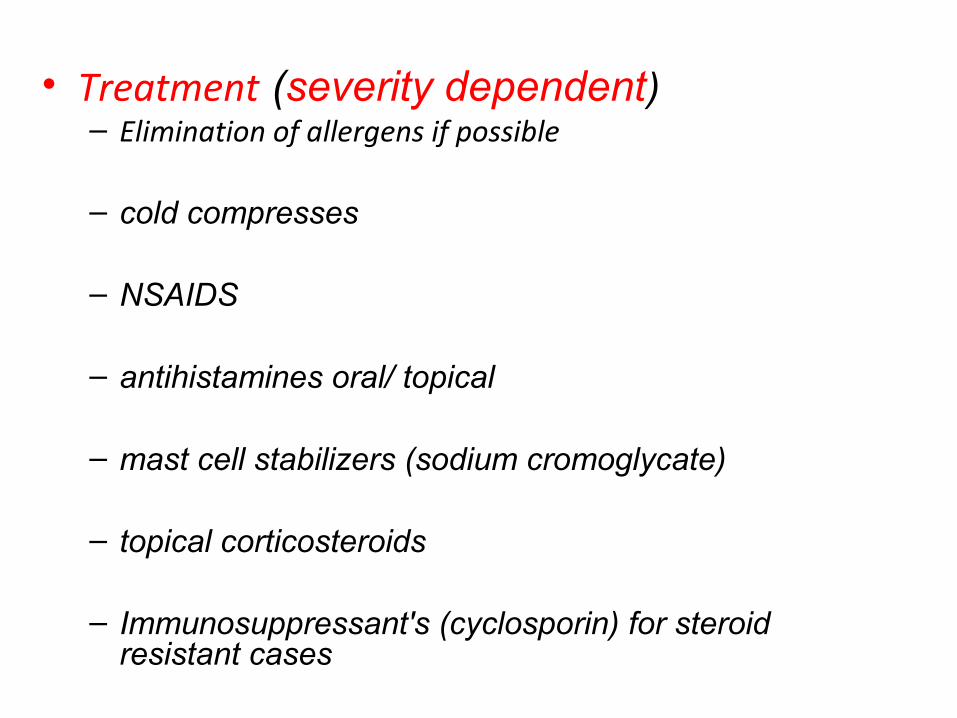
• Treatment (severity dependent)– Elimination of allergens if possible
– cold compresses
– NSAIDS
– antihistamines oral/ topical
– mast cell stabilizers (sodium cromoglycate)
– topical corticosteroids
– Immunosuppressant's (cyclosporin) for steroid resistant cases

Vernal keratoconjunctivitis or spring catarrh
– Recurrent, Bilateral , self limiting allergic inflammation of the conjunctiva affecting children and young adults
– more common in males
– allergic disorder in which IgE and cell mediated immune mechanism play an important role
• Clinical features :
– 98% bilateral, can be asymmetric
– Intense ocular itching, Lacrimation, Photophobia, blepharospasm, blurred vision, FB sensation , burning and difficulty opening eyes in the morning.
– Thick mucous ropy discharge , Psudoptosis due to large papillae.
– Giant papillae on the superior Palpebral conjunctiva are the clinical hallmark.

• Papillae in limbal area have a white, chalky area at apex(Tranta’s dot or Horner's points)
• Peripheral neovascularisation may occur.
• Tarsal papillae in 83-84% of VKC patients.
• Bulbar papillae in 7%. of VKC patients.
• Both forms in 9-17% of VKC patients.
• Cobblestone papillae in 16%(approx).
» Ref: illustrated ophthalmic pathologies-Dr. C. S. Miranda
• Pain if corneal involvement.
• Corneal complications-common; include-Punctate erosions which coalesce to form painful macroerosions.
• Accumulations of inflammatory debris in macroerosions prevents epithelization of cornea.
• Non-healed epithelial defects-shield ulcers
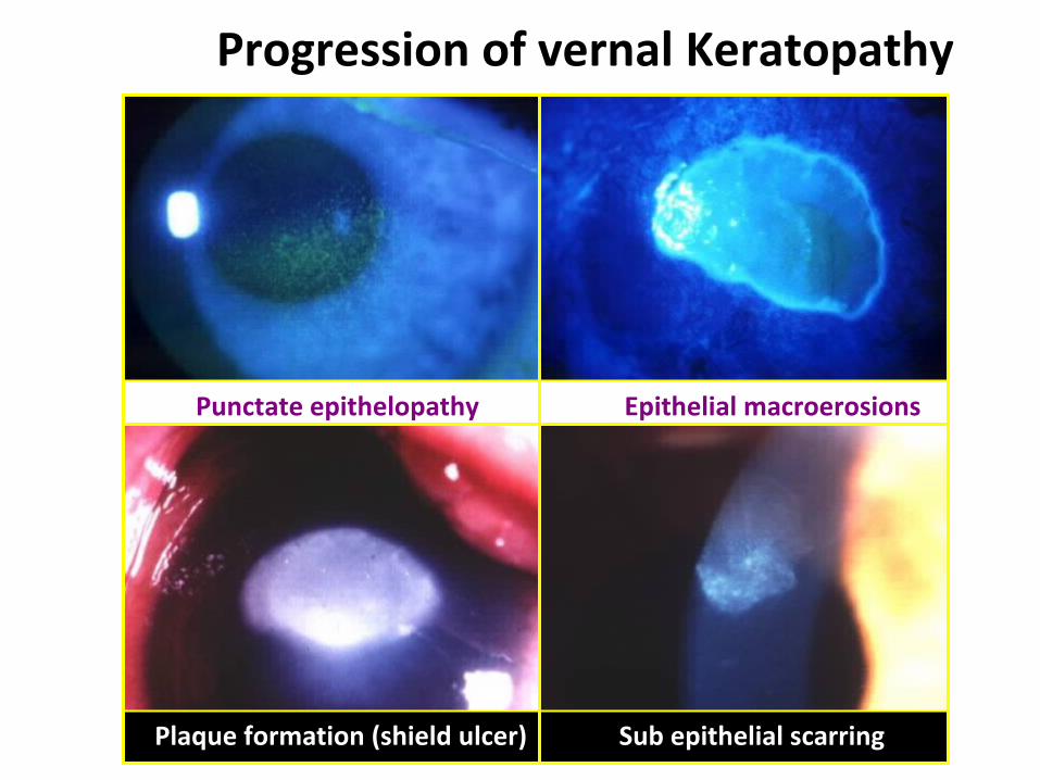
Progression of vernal Keratopathy
Punctate epithelopathy Epithelial macroerosions
Plaque formation (shield ulcer) Sub epithelial scarring
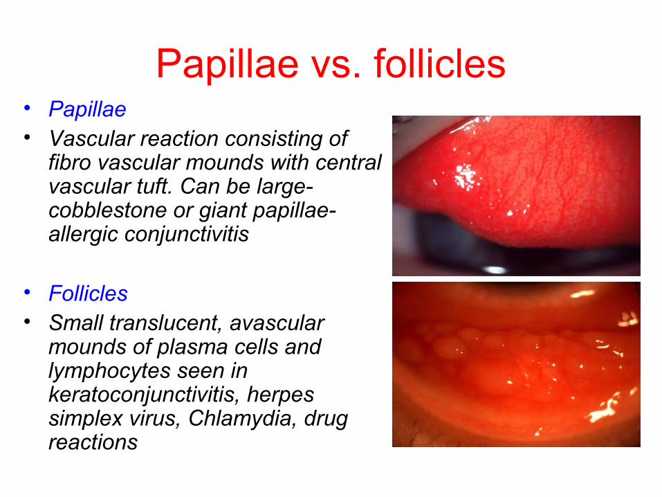
Papillae vs. follicles• Papillae• Vascular reaction consisting of
fibro vascular mounds with central vascular tuft. Can be large- cobblestone or giant papillae- allergic conjunctivitis
• Follicles• Small translucent, avascular
mounds of plasma cells and lymphocytes seen in keratoconjunctivitis, herpes simplex virus, Chlamydia, drug reactions
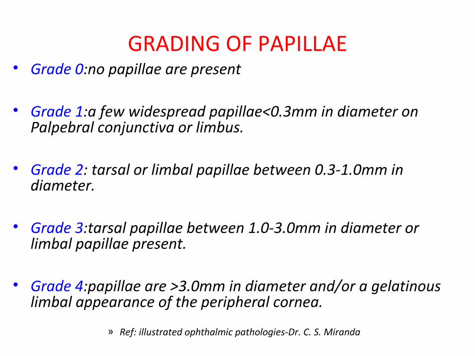
GRADING OF PAPILLAE• Grade 0:no papillae are present
• Grade 1:a few widespread papillae<0.3mm in diameter on Palpebral conjunctiva or limbus.
• Grade 2: tarsal or limbal papillae between 0.3-1.0mm in diameter.
• Grade 3:tarsal papillae between 1.0-3.0mm in diameter or limbal papillae present.
• Grade 4:papillae are >3.0mm in diameter and/or a gelatinous limbal appearance of the peripheral cornea.
» Ref: illustrated ophthalmic pathologies-Dr. C. S. Miranda

• Predisposing factors :• Age -
– Onset usually after 4 years .
– Resolves around puberty .
• Season -– More common in dry and warm climate .
– Peak incidence - April and August.
– But can occur year around .
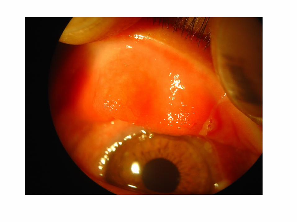
• Clinical types :– Palpebral, Limbal and Mixed .
• Palpebral VKC :– Conjunctival hyperemia followed by diffuse papillary
hypertrophy ( superior tarsus )
– Papillae enlarged and have flat topped polygonal appearance ( cobblestone ).
– Formation of giant septa.
– Active disease - Redness, swelling , tightly packed papillae.
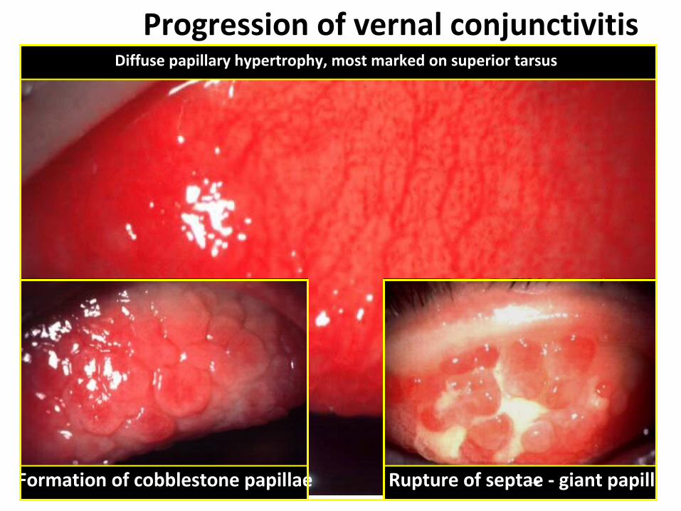
Progression of vernal conjunctivitis Diffuse papillary hypertrophy, most marked on superior tarsus
Formation of cobblestone papillae Rupture of septae - giant papillae
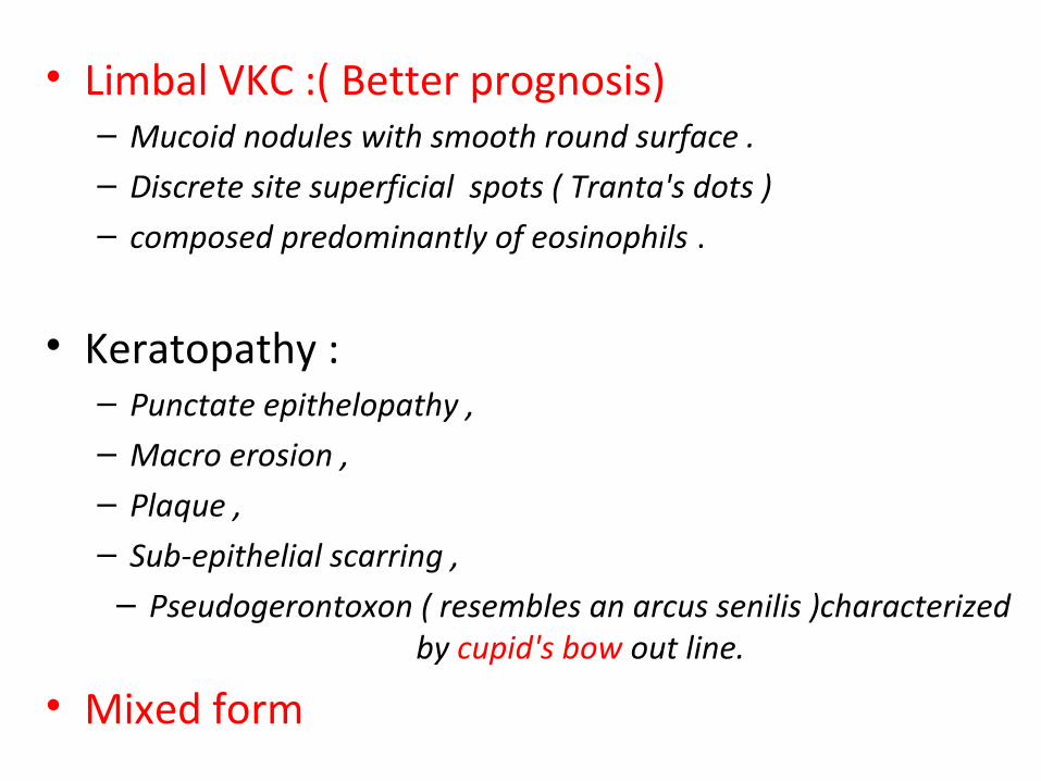
• Limbal VKC :( Better prognosis)– Mucoid nodules with smooth round surface .– Discrete site superficial spots ( Tranta's dots )– composed predominantly of eosinophils .
• Keratopathy :– Punctate epithelopathy ,– Macro erosion , – Plaque ,– Sub-epithelial scarring ,
– Pseudogerontoxon ( resembles an arcus senilis )characterized by cupid's bow out line.
• Mixed form
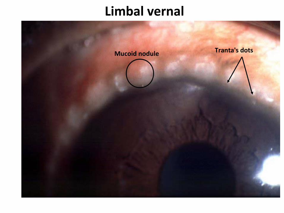
Limbal vernal
Tranta's dotsMucoid nodule

Management
• Therapy should be based on the severity of the symptoms.
• For mild cases – Cold compression– Photo chromic glasses– Topical antihistaminic– NSAIDs such as ketorolac 0.5%

For severe cases
• Topical corticosteroids or topical immunomodulatory agents such as cyclosporine
• Intermittent or pulse therapy of corticosteroid is very effective – Topical steroids used at high frequency & tapered
slowly.
• Less potent soluble steroids are frequently used.
• Patient & family member should be informed of potential dangers of chronic steroid therapy.
• Supratarsal injection of 0.5-1.0 ml of short acting steroid can be given for co-operative patients.
For most severe cases
• Topical cyclosporine 2% twice daily.• Cyclosporine exerts immunomodulatory
effects on both afferent & efferent limbs of the cellular immune response.
• Side effects– Punctate epithelial Keratopathy– Ocular surface irritation.

Phlyctenular conjunctivitis :
• Nodular affection occurring as an allergic response by conjunctiva and corneal epithelium to some endogenous allergens .
• Etiology -– Delayed hypersensitivity ( type I ) response to
endogenous microbial proteins : Tuberculous protein Staphylococcal protein , parasitic protein .
• Predisposing factors -– Age (3 to 15 yrs.) , Sex(girls ), Undernourished one . – Under hygienic and overcrowded living condition .– More common in summer and spring .
• Pathology -– Stage of nodule formation : exudation and infiltration
of lymphocytes – Stage of ulceration : Necrosis of apex of nodule
leading to ulcer formation ,– Stage of granulation – Stage of healing .
• Clinical presentation -– Simple Phlyctenular conjunctivitis – Nercotizing Phlyctenular conjunctivitis – Miliary Phlyctenular conjunctivitis .
• Symptoms -– Lacrimation , discomfort , watering – Mucopurulent conjunctivitis due to secondary
bacterial infection .
• Signs -– Typical pinkish nodule surrounded by hyperemia on
bulbar conjunctiva near the limbus .– Single or multiple phlycten.
• Phlyctenular Keratitis - may occur secondarily.
• Clinical course - – Self limiting disease– phlycten disappear in 8 to 10 days – Recurrence are common.
• Treatment -– steroid eye drops ,– Antibiotic drops ( secondary infection )– specific therapy • Tuberculosis • septic focus should be treated • parasitic infestation - stool examination .
– General measures - improve health of child .

Giant papillary conjunctivitis
• GPC most commonly develops after prolonged conjunctival contact with a foreign substance such as contact lens
• Also reported with exposure to ocular sutures or prosthesis
• Often it is not contact lens itself that causes GPC, but it is deposits or allergens
• Soft contact lens cause GPC more commonly
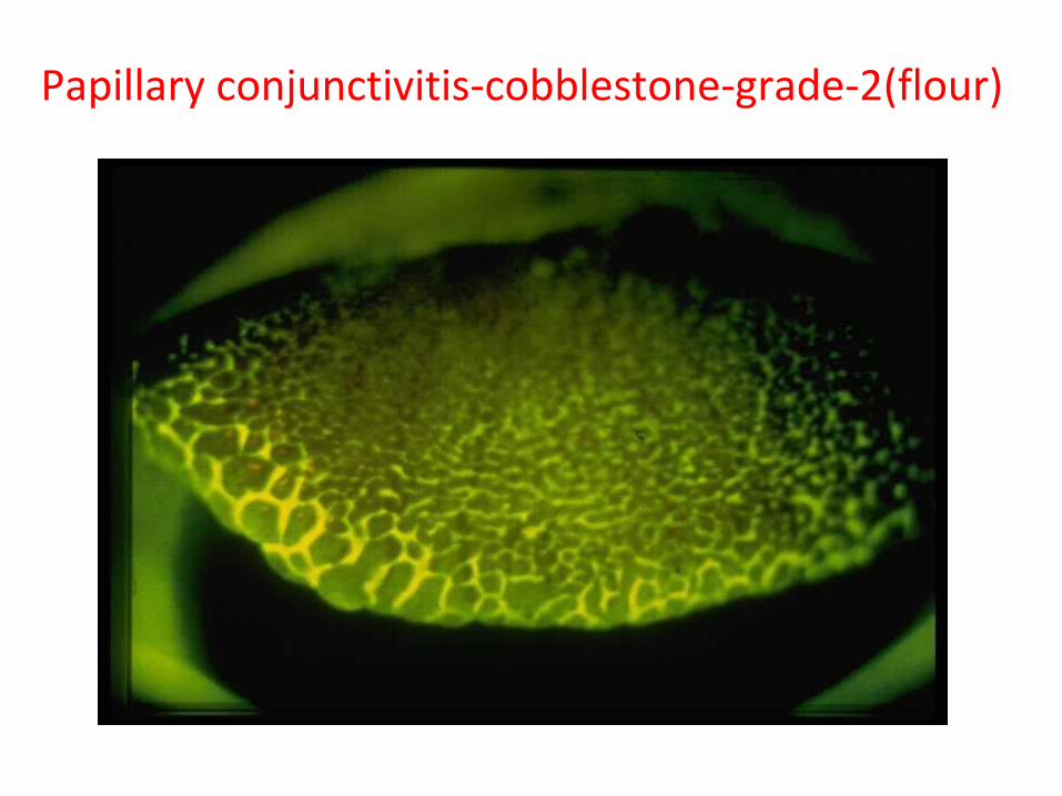
Papillary conjunctivitis-cobblestone-grade-2(flour)

Papillary conjunctivitis-cobblestone-grade 3
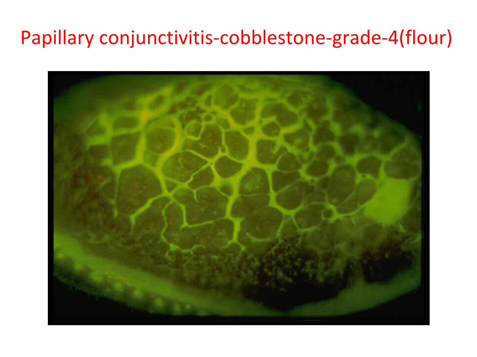
Papillary conjunctivitis-cobblestone-grade-4(flour)
• SYMPTOMS AND SIGNS– Thick mucous discharge, inflamed superior papillae and blurry
vision
– GPC staging• Stage 1:itching and decreased lens tolerance
• Stage 2:blurred vision
• Stage 3:excessive contact lens movement because tarsal papillae don’t allow smooth movement of lid over CL
• Stage 4:similar appearance to mild VKC
» Ref: illustrated ophthalmic pathologies-Dr. C. S. Miranda
• Treatment– Offending causes should be removed
– Disodium cromoglycate-relieve symptoms and enhance the rate of resolution
– Steroids not of much use
REFERENCES• Illustrated ophthalmic pathologies-Dr.C.S.Miranda• Ophthalmology-A.K.Khurana• Clinical ophthalmology-Jack.J.Kanski• Medical microbiology-Geo.F. Brooks,Janet.S.Butel,Stephen A.
Morse• Short text book of medical microbiology, 6th edition, Satish
Gupte• Lippincott’s microbiology• Robbin’s pathology• Ocular Pathology by Myron Yanoff and Ben S. Fine• Internet• Class notes

…THANK YOU !!!