Embed Size (px)
Citation preview
AD_________________ Award Number: W81XWH-04-1-0024 TITLE: Regulation of Normal and Malignant Prostate Growth by the Glucocorticoid Receptor PRINCIPAL INVESTIGATOR: Michael Garabedian, Ph.D. CONTRACTING ORGANIZATION: New York University School of Medicine New York, NY 10016 REPORT DATE: August 2005 TYPE OF REPORT: Final PREPARED FOR: U.S. Army Medical Research and Materiel Command Fort Detrick, Maryland 21702-5012 DISTRIBUTION STATEMENT: Approved for Public Release; Distribution Unlimited The views, opinions and/or findings contained in this report are those of the author(s) and should not be construed as an official Department of the Army position, policy or decision unless so designated by other documentation.

REPORT DOCUMENTATION PAGE Form Approved
OMB No. 0704-0188 Public reporting burden for this collection of information is estimated to average 1 hour per response, including the time for reviewing instructions, searching existing data sources, gathering and maintaining the data needed, and completing and reviewing this collection of information. Send comments regarding this burden estimate or any other aspect of this collection of information, including suggestions for reducing this burden to Department of Defense, Washington Headquarters Services, Directorate for Information Operations and Reports (0704-0188), 1215 Jefferson Davis Highway, Suite 1204, Arlington, VA 22202-4302. Respondents should be aware that notwithstanding any other provision of law, no person shall be subject to any penalty for failing to comply with a collection of information if it does not display a currently valid OMB control number. PLEASE DO NOT RETURN YOUR FORM TO THE ABOVE ADDRESS. 1. REPORT DATE (DD-MM-YYYY)01-11-2005
2. REPORT TYPEFinal
3. DATES COVERED (From - To)1 Nov 2003 – 31 Oct 2005
4. TITLE AND SUBTITLE Regulation of Normal and Malignant Prostate Growth by the Glucocorticoid
5a. CONTRACT NUMBER
Receptor 5b. GRANT NUMBER W81XWH-04-1-0024
5c. PROGRAM ELEMENT NUMBER
6. AUTHOR(S) Michael Garabedian, Ph.D.
5d. PROJECT NUMBER
5e. TASK NUMBER
E-mail: [email protected]
5f. WORK UNIT NUMBER
7. PERFORMING ORGANIZATION NAME(S) AND ADDRESS(ES)
8. PERFORMING ORGANIZATION REPORT NUMBER
New York University School of Medicine New York, NY 10016
9. SPONSORING / MONITORING AGENCY NAME(S) AND ADDRESS(ES) 10. SPONSOR/MONITOR’S ACRONYM(S)U.S. Army Medical Research and Materiel Command Fort Detrick, Maryland 21702-5012 11. SPONSOR/MONITOR’S REPORT NUMBER(S)
12. DISTRIBUTION / AVAILABILITY STATEMENT Approved for Public Release; Distribution Unlimited
13. SUPPLEMENTARY NOTES 14. ABSTRACT The glucocorticoid receptor (GR) is a hormone-dependent transcription factor involved in the regulation of a wide range of metabolic anddevelopmental processes by controlling the expression of target genes in a hormone- and cell-specific manner. However, the expressionand activity of GR in normal and malignant prostate growth is unclear. We have recently developed a GR phosphorylation site specificantibody to serine 211 of human GR (GR-S211-P) and found a strict correlation between phosphorylation of GR at this site and receptortranscriptional activity. Thus, GR phosphorylation at S211 is a surrogate marker for the ligand-bound and transcriptionally active form ofGR in vivo. Using this antibody to survey GR phosphorylation in human tissues by immuohistochemistry, we came across the remarkablefinding that ligand bound and transcriptionally active phospho-GR is present in the stroma and epithelium of normal prostate tissue,including basal and luminal epithelial cells. This was not the case for other tissues examined and suggests that the prostate is becontinually exposed to glucocorticoids, such that GR is actively signaling in the prostate. T he experiments described in this proposal aredesigned to elucidate t he role of GR in prostate cell growth.
15. SUBJECT TERMSProstate cancer, glucocorticoid receptor, conditional knock out mouse 16. SECURITY CLASSIFICATION OF:
17. LIMITATION OF ABSTRACT
18. NUMBER OF PAGES
19a. NAME OF RESPONSIBLE PERSONUSAMRMC
a. REPORT U
b. ABSTRACTU
c. THIS PAGEU
UU
36
19b. TELEPHONE NUMBER (include area code)
Standard Form 298 (Rev. 8-98)Prescribed by ANSI Std. Z39.18

Table of Contents
Cover…………………………………………………………………………………… 1
SF 298……………………………………………………………………………..…… 2
Introduction…………………………………………………………….………….... 4
Body……………………………………………………………………………………. 4
Key Research Accomplishments………………………………………….……… 4
Reportable Outcomes………………………………………………………………. 4
Conclusions………………………………………………………………………….. 4
References…………………………………………………………………………… 5
Appendices…………………………………………………………………………… 5-36
W81XWH-04-1-0024 PI Garabedian, Michael, J
4
IntroductionGlucocorticoids, via the glucocorticoid receptor (GR), regulate the expression of
target genes in a hormone- and cell-specific manner to suppress cell growth. Clinically,glucocorticoids are used as a last resort to treat hormone refractory prostate cancer andhave been shown to have little clinical benefit. Limited data on GR expression in theprostate suggests that GR levels in the epithelium are reduced in cancer relative to normaltissue, suggestive of a role for GR in growth suppression of prostate epithelial cells.However, a direct demonstration that GR regulates prostate cell growth anddifferentiation has not been shown.
BodyBecause GR typically stop cells from growing, we hypothesized that GR is
regulating genes that restrict prostate epithelial cell growth and that its loss wouldpromote prostate cellular proliferation and prostate cancer. To test this hypothesis, wepropose to generate a mouse lacking GR specifically in prostate epithelial cells andexamine changes in proliferation and differentiation.
Since a conventional knockout of the GR gene in the mouse is lethal at birth, weneeded to produce a “conditional” GR knock out mouse where we could remove GRspecifically from prostate epithelial cells and determine the contribution of GR to prostateepithelial cell growth.
Key research accomplishmentsWe have successfully generated a conditional GR knock out mouse where the GR
gene can be selectively removed by the Cre recombinase. Prior to embarking on the GRknock out in prostate epithelial cells, we needed to validate our mouse model in a GRexpressing cell type where the removal of the receptor had been previously shown tohave functional consequences in vivo. Therefore, we choose to authenticate our model inT-cells, where the loss of GR in early thymocyte development has been shown to result insevere gastrointestinal inflammation and mortality upon T-cell activation (1).
We were successful in selectively ablating GR from thymocytes andrecapitulating the above phenotypes, thus establishing our mouse model as a valuablenew tool to analyze GR function in vivo. Now that we have a mouse generated a mousewhere the GR gene can be excised in a tissue specific manner, we will initiate studies onthe consequences of the loss of GR in the prostate using a prostate epithelial cell specificCre recombinase-expressing mouse (2).
Reportable outcomesSee attached manuscript.
ConclusionsWe have generated a “conditional” GR knock out mouse and have validated this
new mouse model in thymocytes, a known GR target cell, as a prelude to the inactivationof the GR gene in prostate epithelial cells. These studies undoubtedly will shed light onthe mechanism of growth regulation by GR in the prostate.

W81XWH-04-1-0024 PI Garabedian, Michael, J
5
BibliographyIsmaili, N., Pineda Torra, I., Shen, Y., Lee M-J. Littman, D.R., Garabedian, M.J.
Stage specific T-cell responses in mice lacking the glucocorticoid receptor (submitted)
Personnel receiving pay from the research effort:Naima Ismaili, Ph.D.Michael J. Garabedian, Ph.D.
References1 Brewer, J. A., B. Khor, S. K. Vogt, L. M. Muglia, H. Fujiwara, K. E. Haegele,
B. P. Sleckman, and L. J. Muglia. 2003. T-cell glucocorticoid receptor isrequired to suppress COX-2-mediated lethal immune activation. Nat Med9:1318-22.
2 Maddison LA, Nahm H, DeMayo F, Greenberg NM. 2000. Prostate specificexpression of Cre recombinase in transgenic mice. Genesis 26:154-6.
Appendices:See attached manuscript

6
Stage specific T-cell responses in mice lacking the glucocorticoid
receptor
Naima Ismaili 1,5
Ines Pineda Torra 1
Yuelei Shen 4
Men-Jean Lee 3, 5
Dan R. Littman 4
Michael J. Garabedian 1,2 *
Departments of Microbiology1, Urology2, Obstetrics and Gynecology3
Skirball Institute of Biomolecular Medicine and
the Howard Hughes Medical Institute 4
NYU School of Medicine
550 First Avenue
New York, NY 10016
Corresponding author:Department of MicrobiologyNYU School of Medicine550 First AvenuePhone 212 263-7662FAX 212 263-8276Email [email protected]
5 Present Address: Department of Obstetrics and Gynecology, Yale School of Medicine
Running title: GR stage-specific effects in thymocytes
7
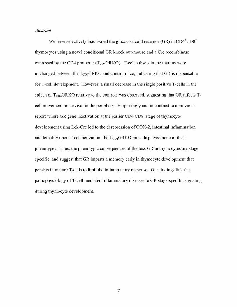
Abstract
We have selectively inactivated the glucocorticoid receptor (GR) in CD4+CD8+
thymocytes using a novel conditional GR knock out-mouse and a Cre recombinase
expressed by the CD4 promoter (TCD4GRKO). T-cell subsets in the thymus were
unchanged between the TCD4GRKO and control mice, indicating that GR is dispensable
for T-cell development. However, a small decrease in the single positive T-cells in the
spleen of TCD4GRKO relative to the controls was observed, suggesting that GR affects T-
cell movement or survival in the periphery. Surprisingly and in contrast to a previous
report where GR gene inactivation at the earlier CD4-CD8- stage of thymocyte
development using Lck-Cre led to the derepression of COX-2, intestinal inflammation
and lethality upon T-cell activation, the TCD4GRKO mice displayed none of these
phenotypes. Thus, the phenotypic consequences of the loss GR in thymocytes are stage
specific, and suggest that GR imparts a memory early in thymocyte development that
persists in mature T-cells to limit the inflammatory response. Our findings link the
pathophysiology of T-cell mediated inflammatory diseases to GR stage-specific signaling
during thymocyte development.

8
Introduction
Glucocorticoids are known to influence the immune function and have long been
used as anti-inflammatory and immunospressive agents (8). Glucocorticoids mediate
their immunoregulatory effect by binding to the intracellular glucocorticoid receptor
(GR) (31). GR is a transcriptional regulatory protein with three functional domains: an
N-terminal transcriptional activation function (AF) 1, a central zinc finger DNA binding
domain (DBD) and a C-terminal hormone-binding region that contains a ligand-
dependent AF2 (32). In the absence of hormone, the Hsp90-based chaperone complex
represses GR regulatory activities (20). Hormone binding relieves this repression and
results in a conformational change in the receptor, which promotes DNA binding as well
as an association with transcriptional regulatory cofactors to enhance the transcription of
target genes. GR also modulates transcription independent of direct DNA recognition via
protein-protein interactions (14). Such a “tethering” mechanism is responsible for the
repressive effect of GR on transcription factors such as AP1 and NFkB, to suppress the
inflammatory response.
It is well established that thymocytes are extremely sensitive to
glucocorticoid–mediated apoptosis (2, 25). Thymocytes are divided into different subsets
based on the expression of cell surface markers. Thymocytes differentiate from the CD4-
CD8- double negative (DN) cells to the intermediate CD4+CD8+ double positive (DP)
stage. The majority of the DP cells present sub-threshold affinity to the major
histocompatibity complex (MHC) and undergo apoptosis or death by neglect. The
remaining DP cells with the ability to recognize self major histocompatibity complex
MHC-II or MHC-I molecules are positively selected and differentiate respectively into

9
either CD4+CD8- or CD4-CD8+ single positive (SP) cells. DP cells with high affinity to
the MHC destined to become self-reactive thymocytes are eliminated.
Steroidogenic enzymes are present in the thymic epithelium, which may produce
glucocorticoids locally to influence thymocyte survival and differentiation (19, 30).
Thus, the thymus may represent a unique microenvironment that could allow directed
delivery of corticosteroids to thymocytes (2).
High doses of glucocorticoids are known to induce thymocyte apoptosis and this
process is inhibited by T-cell receptor (TCR)-mediated activation of the ERK signaling
pathway. Conversely, induction of apoptosis by TCR ligation is repressed by
glucocorticoids (1, 13). In view of this "mutual antagonism", it was suggested that GR
plays a role in thymopoiesis by regulating the threshold of TCR-mediated positive and
negative selection (2).
To assess GR function during T-cell development in vivo, several groups have
manipulated the level of GR in the mouse and analyzed T-cell development with
conflicting results (reviewed in 2, 7, 15). Using two independent transgenic mouse
models expressing an anti-sense GR cDNA (referred to as TKO), two groups reported
contradictory results regarding the involvement of GR in T-cell development: while King
et al reported a 90% reduction of the number of DP thymocytes, Sacedon et al, reported a
significantly increased thymocyte number in a comparable model (17, 24). The disparity
between these studies may reflect differences in the promoters used to drive the antisense
transgenic expression.
Mice with a disrupted GR gene (GR null mutant, GRKO) die at birth due to
defects in lung maturation (4). Total thymocyte number, and single positive subsets were
10
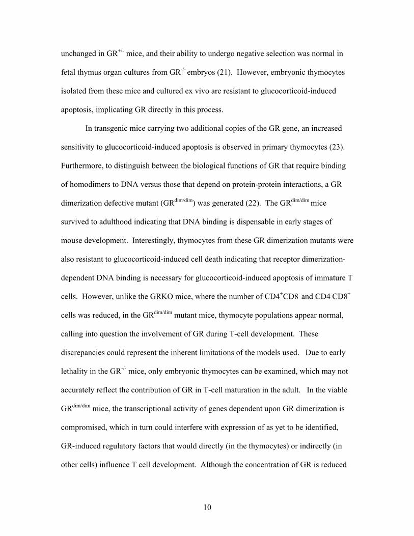
unchanged in GR+/- mice, and their ability to undergo negative selection was normal in
fetal thymus organ cultures from GR-/- embryos (21). However, embryonic thymocytes
isolated from these mice and cultured ex vivo are resistant to glucocorticoid-induced
apoptosis, implicating GR directly in this process.
In transgenic mice carrying two additional copies of the GR gene, an increased
sensitivity to glucocorticoid-induced apoptosis is observed in primary thymocytes (23).
Furthermore, to distinguish between the biological functions of GR that require binding
of homodimers to DNA versus those that depend on protein-protein interactions, a GR
dimerization defective mutant (GRdim/dim) was generated (22). The GRdim/dim mice
survived to adulthood indicating that DNA binding is dispensable in early stages of
mouse development. Interestingly, thymocytes from these GR dimerization mutants were
also resistant to glucocorticoid-induced cell death indicating that receptor dimerization-
dependent DNA binding is necessary for glucocorticoid-induced apoptosis of immature T
cells. However, unlike the GRKO mice, where the number of CD4+CD8- and CD4-CD8+
cells was reduced, in the GRdim/dim mutant mice, thymocyte populations appear normal,
calling into question the involvement of GR during T-cell development. These
discrepancies could represent the inherent limitations of the models used. Due to early
lethality in the GR-/- mice, only embryonic thymocytes can be examined, which may not
accurately reflect the contribution of GR in T-cell maturation in the adult. In the viable
GRdim/dim mice, the transcriptional activity of genes dependent upon GR dimerization is
compromised, which in turn could interfere with expression of as yet to be identified,
GR-induced regulatory factors that would directly (in the thymocytes) or indirectly (in
other cells) influence T cell development. Although the concentration of GR is reduced
11
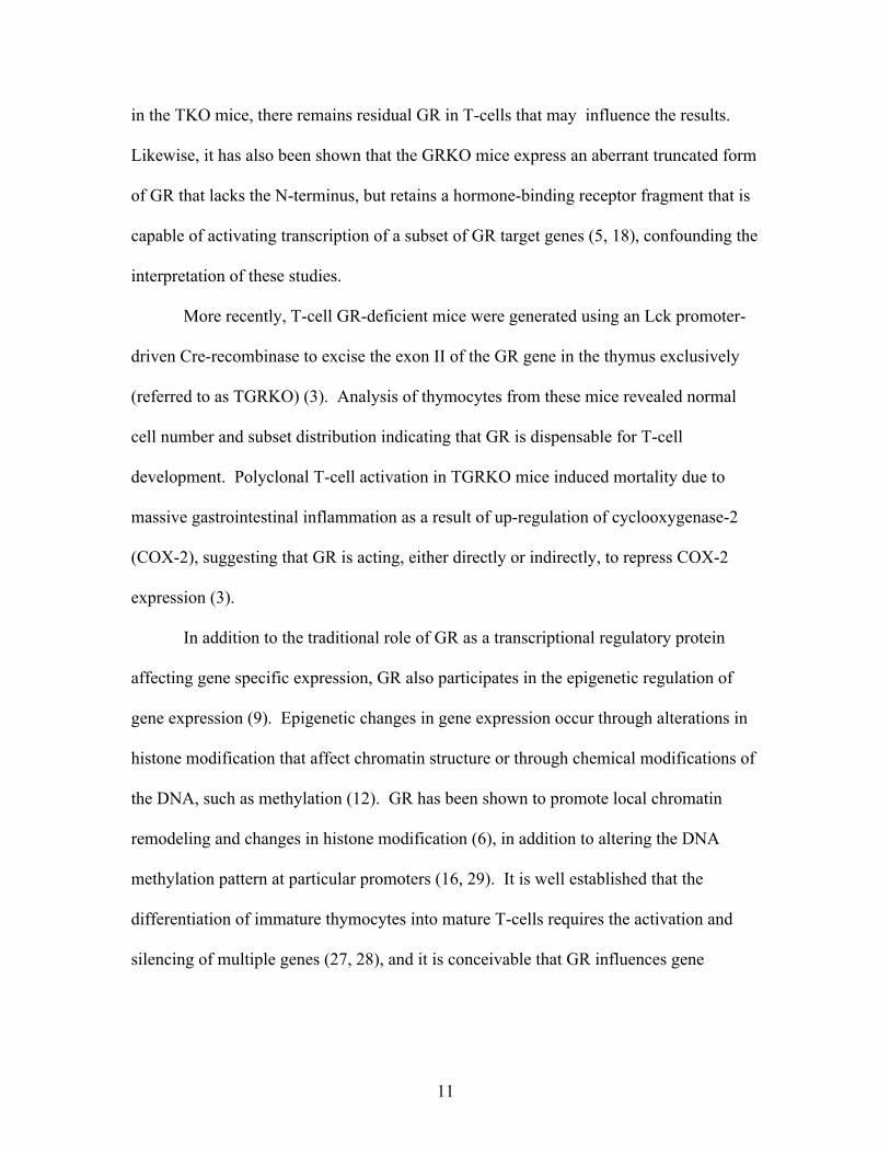
in the TKO mice, there remains residual GR in T-cells that may influence the results.
Likewise, it has also been shown that the GRKO mice express an aberrant truncated form
of GR that lacks the N-terminus, but retains a hormone-binding receptor fragment that is
capable of activating transcription of a subset of GR target genes (5, 18), confounding the
interpretation of these studies.
More recently, T-cell GR-deficient mice were generated using an Lck promoter-
driven Cre-recombinase to excise the exon II of the GR gene in the thymus exclusively
(referred to as TGRKO) (3). Analysis of thymocytes from these mice revealed normal
cell number and subset distribution indicating that GR is dispensable for T-cell
development. Polyclonal T-cell activation in TGRKO mice induced mortality due to
massive gastrointestinal inflammation as a result of up-regulation of cyclooxygenase-2
(COX-2), suggesting that GR is acting, either directly or indirectly, to repress COX-2
expression (3).
In addition to the traditional role of GR as a transcriptional regulatory protein
affecting gene specific expression, GR also participates in the epigenetic regulation of
gene expression (9). Epigenetic changes in gene expression occur through alterations in
histone modification that affect chromatin structure or through chemical modifications of
the DNA, such as methylation (12). GR has been shown to promote local chromatin
remodeling and changes in histone modification (6), in addition to altering the DNA
methylation pattern at particular promoters (16, 29). It is well established that the
differentiation of immature thymocytes into mature T-cells requires the activation and
silencing of multiple genes (27, 28), and it is conceivable that GR influences gene

12
expression in a stage specific manner in T-cell development through both genetic and
epigenetic events.
To address the possibility that the effect of GR on T-cell development is stage
specific, we generated independently a T-cell-specific GR-deficient mouse using Cre-
recombinase driven by CD4-promoter (referred to as TCD4GRKO), which removes GR at
the DP stage of thymocyte development. As with the TGRKO mice, T-cell subsets in the
thymus were unchanged between the TCD4GRKO and control mice, indicating that GR is
dispensable regardless of the time in T-cell development in which GR is ablated.
However, we found a small decrease in the SP T-cells in the spleen of TCD4GRKO
relative to controls. Surprisingly, activation of T-cells by anti-CD3 antibody in the
TCD4GRKO mice did not result in inflammation of the intestine or depression of COX-2
as observed in the TGRKO mice. Our finding indicates that GR is most likely controlling
biological events responsible for polyclonal T-cell activation early in thymocyte
development (DN to DP), which becomes dispensable at later stages (DP to SP).
13
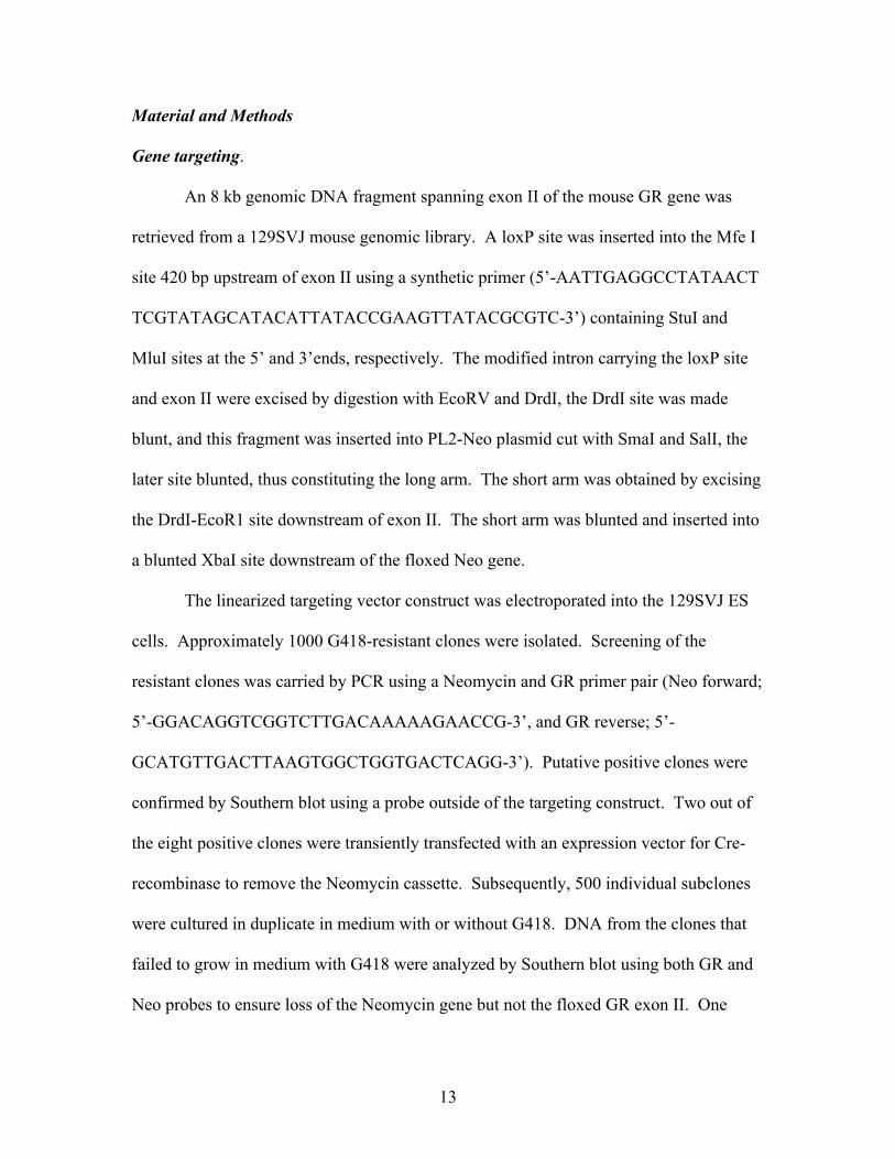
Material and Methods
Gene targeting.
An 8 kb genomic DNA fragment spanning exon II of the mouse GR gene was
retrieved from a 129SVJ mouse genomic library. A loxP site was inserted into the Mfe I
site 420 bp upstream of exon II using a synthetic primer (5’-AATTGAGGCCTATAACT
TCGTATAGCATACATTATACCGAAGTTATACGCGTC-3’) containing StuI and
MluI sites at the 5’ and 3’ends, respectively. The modified intron carrying the loxP site
and exon II were excised by digestion with EcoRV and DrdI, the DrdI site was made
blunt, and this fragment was inserted into PL2-Neo plasmid cut with SmaI and SalI, the
later site blunted, thus constituting the long arm. The short arm was obtained by excising
the DrdI-EcoR1 site downstream of exon II. The short arm was blunted and inserted into
a blunted XbaI site downstream of the floxed Neo gene.
The linearized targeting vector construct was electroporated into the 129SVJ ES
cells. Approximately 1000 G418-resistant clones were isolated. Screening of the
resistant clones was carried by PCR using a Neomycin and GR primer pair (Neo forward;
5’-GGACAGGTCGGTCTTGACAAAAAGAACCG-3’, and GR reverse; 5’-
GCATGTTGACTTAAGTGGCTGGTGACTCAGG-3’). Putative positive clones were
confirmed by Southern blot using a probe outside of the targeting construct. Two out of
the eight positive clones were transiently transfected with an expression vector for Cre-
recombinase to remove the Neomycin cassette. Subsequently, 500 individual subclones
were cultured in duplicate in medium with or without G418. DNA from the clones that
failed to grow in medium with G418 were analyzed by Southern blot using both GR and
Neo probes to ensure loss of the Neomycin gene but not the floxed GR exon II. One

14
positive clone was selected for injection into C57BL/6 blastocytes and resulted in germ
line transmitting chimeras.
Animal breeding and selection
The male chimeras were bred to C57BL/6 females and the tail DNA from the
progeny was genotyped using primer flanking the loxP site upstream of exon II (lox 1
forward; 5’-GGCACAGGTGAAATTGTGA-3’, and lox 2 reverse; 5’-
ACACATTTGGGTAAGCATGGA-3’). The heterozygous mice (GRflox/wt) were
interbred to produce homozygous mice (GRflox/flox). To generate mice with deletion of
GR in the thymus, the GRflox/flox were bred to Lck-Cre or CD4-Cre mice. The resulting
GRflox/wt, Lck-Cre+ or GRflox/wt, CD4-Cre+ were interbred to produce the littermates
GRflox/flox, Cre+ mice or GRwt/wt, Cre+ mice that we used as controls in all our
experiments. The primers for genotyping Lck-Cre mice are: Lck forward; 5’-
CCTCCTGTAACTTGGTGCTTGAG-3’ and Lck reverse; 5’-
TGCATCGACCGGTAATGCAG-3’. The primers for genotyping CD4-Cre mice are:
CD4 forward; 5’-TCTCTGTGGCTGGCAGTTTCTCCA-3’, and CD4 reverse; 5’-
TCAAGGCCAGACTAGGCTGCCTAT-3’.
All mouse protocols were in accordance with National Institute of Health
guidelines and were approved by the Animal Care and Use Committee of NYU School of
Medicine (New York, NY).

15
Flow cytometry
Thymocytes were harvested from 6-week old male mice and 2-10x106 cells were
incubated with anti-CD16/CD32 (2.4G2) antibody for 15 min on ice and then stained
with combination of antibodies against the following cell surface proteins: anti-CD4-
APC (GK1.5), anti-CD4-APC-Cy7 (GK1.5), anti-CD8α-PE-Cy7 (53-6.7), at appropriate
concentrations for 30 min on ice. The cells were then washed with twice with PBS and
analyzed on a BD-LSRII flow cytometer (Becton Dickinson, Mountain View, CA). For
the apoptosis experiments, isolated thymocytes were treated with either 10-6 M
Dexamethasone or an equal volume of the ethanol vehicle in tissue culture media (RPMI-
1640 with 5% FCS, 2mM Glutamine, penicillin/streptomycin 5 Units/ml) for 24h at
37oC, 5% CO2. Annexin-V staining was performed following the protocol provided by
the manufacturer (BD PharMingen, San Diego, CA). After two washes with
cytoperm/cytowash, the cells were re-suspended in FACS buffer and analyzed byflow
cytometry. All data were analyzed using Flowjo flow cytometric data analysis software
(Tree Star, Ashland, OR). Statistical analysis of T-cell populations was performed using
T-tests. Statistical significance was considered for P-values < 0.05.
Protein analysis
Whole cell protein lysates were prepared from whole thymus, isolated thymic
CD4+ or CD8+ T-cells, or splenic T-cells sorted using CD90 (Thy 1.2) microbeads and
the MACS separation columns (Miltenyi biotec). Cells were lysed in 0.1 ml of buffer
containing 50 mM HEPES (pH 7.5), 150 mM NaCl, 1 mM EDTA, 1 mM EGTA, 1 mM
NaF, 1% Triton X-100, 10% glycerol, and additional protease and phosphatase inhibitors

16
[1 mM phenylmethylsulfonyl fluoride, 20 mM β-glycerophosphate, 8 mM sodium
pyrophosphate, 1µg/ml leupeptin, 1 µg/ml pepstatin A, and 1 µg/ml aprotinin. The
lysates were centrifuged at 12,000 rpm for 15 min at 4oC. The soluble supernatants were
normalized for total protein concentration using the Bio-Rad protein assay. Samples
were boiled for 3 min in 2 X SDS sample buffer and stored at -20oC.
For Western blotting, 30 µg of protein extracts were resolved on a 10% SDS-
PAGE, transferred unto PVDF membranes and probed with antibodies to the GR N-
terminal (M20; Santa-Cruz Biotechnology) or C-terminus (P-20; Santa-Cruz
Biotechnology), mouse monoclonal COX-2 antibody (5E10/D10; abcam), mouse
monoclonal antibody against tubulin (TU27; Covance) and a polyclonal antibody against
hsp90 (Santa-Cruz Biotechnology) as a control for loading.
Real time PCR for RNA quantification
Total RNA from purified T-cells cells was extracted with Trizol (Invitrogen) as
described by the manufacturer. cDNA specific for each gene was subsequently
synthesized using the Enhanced Avian Reverse Transcriptase (Sigma) and random primer
hexamers (Pharmacia) following the manufacturer’s instructions. cDNAs were amplified
using the SYBR Green Quantitative PCR Kit (Sigma) on a LightCycler (Roche).
Reactions were carried out in a 20 µl reaction containing a 500 nM concentration of each
primer and the SYBR Green Taq ReadyMix for Quantitative RT-PCR (Sigma) as
recommended by the manufacturer with the following conditions: 95°C for 2 min,
followed by 42 cycles of 5 sec at 95°C, 5 sec at 55 °C and 10 sec at 72°C. COX-2,
mRNA levels were normalized to 28S expression. All RT-PCR products were analyzed

17
in a post-amplification fusion curve to ensure that a single amplicon was obtained.
Primers used were: COX-2-forward-(5’-ATCCCCCCACAGTCAAAGACA-3’); COX-2-
reverse, (5’-CATACATCATCAGACCAGGCACC-3’); GR-forward (5 -
CCTAAGGACGGTCTGAAGAGC-3); GR-reverse, (5 -
GCCAAGTCTTGGCCCTCTAT-3); 28 S rRNA-forward (5'-
AAACTCTGGTGGAGGTCCGT-3’) and 28 S rRNA-reverse (5'-
CTTACCAAAAGTGGCCCACTA-3’).
Histology
Tissues were fixed in 10% neutral buffered formalin overnight, washed in 70%
ethanol and embedded in paraffin. Hematoxylin and eosin staining was performed on 5
µm-wide sections. Micrographs were captured on an Axioplan 2 Zeiss microscope.

18
Results
T-cell glucocorticoid receptor (GR)-deficient mice were generated using CD4
promoter-driven, Cre recombinase-mediated excision of a floxed exon II of the GR gene
(Figure 1). The mice homozygous for the floxed GR gene and harboring the CD4-Cre
transgene (designated TCD4GRKO) were as healthy as their CD4-Cre negative
homozygous floxed GR littermates. Efficient excision of exon II in the thymus was
confirmed by Southern blot (Figure 2D). In addition, we found little glucocorticoid
receptor mRNA (Figure 2E) and protein (Figures 2F and 2H) in the thymus. No
glucocorticoid receptor was detected in purified CD4+ and CD8+ single positive
thymocytes from TCD4GRKO mice, indicating the efficient loss of GR at the specified
stage (Figure 2G). Detection of GR protein was carried using either an antibody to the
GR amino-terminus, recognizing an epitope in exon II, or an antibody to the GR carboxy-
terminus, recognizing an epitope outside the deleted exon. In both cases, intact GR was
not detected and no specific bands were evident in thymocytes from TCD4GRKO versus
wt control mice, excluding the possibility of a remaining C-terminal truncated form of
GR in our mouse model (Figure 2H). GR expression remained intact in the lung (Figure
2G), and other tissues (not shown) where CD4-Cre would not be expressed. Therefore,
the GR is efficiently and specifically deleted in thymocytes in TCD4GRKO mice.
We next analyzed T-cell subsets in the thymus and spleen from TCD4GRKO and
control mice. We noted no significant difference in the cell number or subset distribution
in the thymus between genotypes. In contrast, in the spleen we observed a small but
significant decrease (~40%) in both CD4+CD8- and CD4- CD8+ cell populations. Thus,
GR deficiency at the CD4+CD8+ stage is not required for thymocyte development or

19
negative selection, whereas GR may have a small effect on the trafficking or survival of
mature T-cells in the spleen.
We also monitored T-cell apoptosis in vitro in response to Dexamethasone
treatment by Annexin-V staining. When treated with Dexamethasone, CD4+CD8+ and
CD4+CD8- T-cells from control mice expressing GR underwent apoptosis, whereas
thymocytes from TCD4GRKO mice were largely resistant to glucocorticoid-mediated cell
death (Figure 4). This is in agreement with the role of GR in thymocyte apoptosis.
T-cell receptor activation induces the expression of a host of proinflammatory
mediators, including TNF-α, INF-γ, and COX-2 (10, 11). A previous study in mice
where GR had been removed at the early double negative CD4-CD8- stage of T-cell
development by Lck-Cre (TGRKO), resulted in a profound depression of COX-2 mRNA,
intestinal inflammation and death upon T-cell activation after administering an antibody
to CD3 (3).
To examine whether the proinflammatory effect of GR-deficiency in activated T-
cells is stage specific, we examined COX-2 expression after administering anti-CD3
specific antibody to TCD4GRKO and control mice. We anticipated that if the function of
GR in thymocytes was stage independent, then the removal of GR either early (CD4-
CD8-) or late (CD4+CD8+) stages in T-cell development would result similar phenotypes,
including depression of COX-2 and enhanced proinflammatory response upon T-cell
activation. Alternatively, if the requirement for GR were stage specific and, for example,
was necessary at only the early CD4-8- stage of thymocyte development, then deregulated
COX-2 expression and inflammatory phenotype in GR-deficient T-cells would only be

20
manifest if GR were removed early (TLckGRKO), but not late (TCD4GRKO), in thymocyte
development, which is what is observed.
Although COX-2 mRNA and protein levels were induced in purified T-cells after
treatment with antibody to CD-3 in TCD4GRKO and control mice, there was no difference
in the regulation of COX-2 between TCD4GRKO and controls, despite a lack of GR
mRNA and protein (Figure 5). This finding indicates that deregulation of COX-2 in T-
cells in the absence of GR is thymocyte stage-specific and is required only during early
thymocyte development.
To ensure that the differences in response in GR-deficient T-cells between
TGRKO and TCD4GRKO were stage specific rather than a result of variation in the GR
targeted mouse strain between groups, we generated our own Lck-driven thymus-specific
GR knock out mouse (referred to as TLckGRKO) using the same GR conditional allele as
in the TCD4GRKO. We examined the TLckGRKO and TCD4GRKO by histology for signs
of intestinal inflammation after T-cell activation by anti-CD3 administration. The
TLckGRKO mice displayed intestinal inflammation upon polyclonal T-cell activation
(Figure 6) as previously reported. In contrast, TCD4GRKO mice showed no signs of
gastrointestinal inflammatory in response to T-cell activation, consistent with the lack of
elevated COX-2 expression relative to control. Thus, removal of GR early in thymocyte
development (TLckGRKO) results in gastrointestinal inflammation when mature T-cells
are activated. Surprisingly, this effect is not observed when GR is removed later in
thymocyte development. Thus, GR imparts a memory to thymocytes early in
development that persists in mature T-cell stages.

21
Discussion
Thymocyte differentiation and selection leads to large variety of T-cells, most
notably CD4-T helper cells, CD8-cytotoxic cells and CD4-regulatory or suppressor cells
that reside in various peripheral organs to regulate the adaptive immune response (28).
The role GR plays in thymocyte differentiation as well as the subsequent effects that GR
exerts in adult T-cells lineages has not been fully elucidated (2). An important study
from the Muglia laboratory addressed the role of GR in T-cell activation in adult mice by
generating a conditional GR knockout mouse and ablating GR at the early CD4-CD8-
stage of thymocyte development using Lck-Cre recombinase. T-cell GR deficiency
resulted in a profound up-regulation of COX-2 mRNA, gastrointestinal inflammation and
death upon polyclonal T-cell activation (3). Our results also show that GR-deficient
TLckGRKO mice develop intestinal inflammation as a consequence of T-cell activation.
However, although our TLckGRKO mice appear ill after administration of anti-CD3
antibody they do not die. This lack of lethality may reflect environmental differences in
animal housing between studies as our mice were maintained under pathogen-free
conditions.
While GR-deficiency in early T-cell development (TLckGRKO) leads to
inflammation and pathology, the TCD4GRKO animals display, interestingly, no signs of
gastrointestinal inflammation or COX-2 overexpression relative to control when T-cells
are activated by anti-CD3 antibody. The striking stage specific phenotypic consequences
of the loss GR, suggest that early in T-cell development, GR institutes a genetic program
that persists throughout development and, once established, GR is then dispensable.
This pattern may be established by GR activation in thymocytes through the paracrine

22
action of glucocorticoids produced locally by the thymic epithelium.
Our findings suggest a model whereby early in thymocyte development GR is
activated by glucocorticoids emanating from the thymic epithelium, which induces
epigenetic alterations, such as DNA methylation or histone modifications, resulting in the
decreased transcription of proinflammatory genes (Figure 7). If GR were removed early
in thymic development, then this epigenetic suppression would not occur. However, if
GR were removed at a later point in thymocyte development, then the repression of
promoters by GR would have already been established. According to this model,
subsequent hyperinduction of proinflammatory mediators upon T-cell activation, such as
COX-2, would be observed only if GR were removed early in development and is
consistent with the derepression of COX-2 when GR is ablated at the beginning of
thymocyte maturation.
Indeed, the COX-2 upstream regulatory region harbors CpG islands and
undergoes DNA methylation. Suppression of this methylation by inhibitors, such as 5-
azacytidine, results in the hyperinduction of COX-2 upon stimulation, presumably by
enabling transcription factors to bind and activate the promoter (26). Thus, GR may
induce a particular epigenetic state early in thymocyte development that establishes a
threshold for the activation of proinflammatory genes. This proposed epigenetic GR anti-
inflammatory mechanism is novel and distinct from the traditional GR anti-inflammatory
mechanism by direct repression of gene expression through protein-protein interaction.
Perturbations of GR-regulated events in early in thymocyte development could lead to the
enhanced expression of proinflammatory mediators by activated T-cells, and contribute
to the etiology of T-cell-dependent inflammatory diseases such as lupus and psoriasis.

23
Acknowledgements
We thank Dr. Louis Muglia for helpful discussion and Drs. Susan Logan and
Ellinor Oxelmark for critically reading the manuscript. We also thank Dr. Herman Yee
for help with the histology. Grants from the NIH (DRL, MJG), the DOD (W81XWH-04-
1-0024) (MJG), the NYU School of Medicine CFAR pilot project program (MJG), and
the Reproductive Scientist Development Program/Wyeth Award (M-J.L) supported this
study.

24
References
1. Ashwell, J. D., L. B. King, and M. S. Vacchio. 1996. Cross-talk between the Tcell antigen receptor and the glucocorticoid receptor regulates thymocytedevelopment. Stem Cells 14:490-500.
2. Ashwell, J. D., F. W. Lu, and M. S. Vacchio. 2000. Glucocorticoids in T celldevelopment and function*. Annu Rev Immunol 18:309-45.
3. Brewer, J. A., B. Khor, S. K. Vogt, L. M. Muglia, H. Fujiwara, K. E. Haegele, B.P. Sleckman, and L. J. Muglia. 2003. T-cell glucocorticoid receptor is required tosuppress COX-2-mediated lethal immune activation. Nat Med 9:1318-22.
4. Cole, T. J., J. A. Blendy, A. P. Monaghan, K. Krieglstein, W. Schmid, A. Aguzzi,G. Fantuzzi, E. Hummler, K. Unsicker, and G. Schutz. 1995. Targeted disruptionof the glucocorticoid receptor gene blocks adrenergic chromaffin celldevelopment and severely retards lung maturation. Genes Dev 9:1608-21.
5. Cole, T. J., K. Myles, J. F. Purton, P. S. Brereton, N. M. Solomon, D. I. Godfrey,and J. W. Funder. 2001. GRKO mice express an aberrant dexamethasone-bindingglucocorticoid receptor, but are profoundly glucocorticoid resistant. Mol CellEndocrinol 173:193-202.
6. Flavin, M., L. Cappabianca, C. Kress, H. Thomassin, and T. Grange. 2004. Natureof the accessible chromatin at a glucocorticoid-responsive enhancer. Mol CellBiol 24:7891-901.
7. Godfrey, D. I., J. F. Purton, R. L. Boyd, and T. J. Cole. 2000. Stress-free T-celldevelopment: glucocorticoids are not obligatory. Immunol Today 21:606-11.
8. Goulding, N. J. 2004. The molecular complexity of glucocorticoid actions ininflammation - a four-ring circus. Curr Opin Pharmacol 4:629-36.
9. Grange, T., L. Cappabianca, M. Flavin, H. Sassi, and H. Thomassin. 2001. In vivoanalysis of the model tyrosine aminotransferase gene reveals multiple sequentialsteps in glucocorticoid receptor action. Oncogene 20:3028-38.
10. Hunter, C. A., and S. L. Reiner. 2000. Cytokines and T cells in host defense. CurrOpin Immunol 12:413-8.
11. Iniguez, M. A., C. Punzon, and M. Fresno. 1999. Induction of cyclooxygenase-2on activated T lymphocytes: regulation of T cell activation by cyclooxygenase-2inhibitors. J Immunol 163:111-9.
12. Jaenisch, R., and A. Bird. 2003. Epigenetic regulation of gene expression: howthe genome integrates intrinsic and environmental signals. Nat Genet 33Suppl:245-54.
13. Jamieson, C. A., and K. R. Yamamoto. 2000. Crosstalk pathway for inhibition ofglucocorticoid-induced apoptosis by T cell receptor signaling. Proc Natl Acad SciU S A 97:7319-24.
14. Jenkins, B. D., C. B. Pullen, and B. D. Darimont. 2001. Novel glucocorticoidreceptor coactivator effector mechanisms. Trends Endocrinol Metab 12:122-6.
15. Jondal, M., A. Pazirandeh, and S. Okret. 2004. Different roles for glucocorticoidsin thymocyte homeostasis? Trends Immunol 25:595-600.
16. Kagoshima, M., T. Wilcke, K. Ito, L. Tsaprouni, P. J. Barnes, N. Punchard, and I.M. Adcock. 2001. Glucocorticoid-mediated transrepression is regulated byhistone acetylation and DNA methylation. Eur J Pharmacol 429:327-34.
25
17. King, L. B., M. S. Vacchio, K. Dixon, R. Hunziker, D. H. Margulies, and J. D.Ashwell. 1995. A targeted glucocorticoid receptor antisense transgene increasesthymocyte apoptosis and alters thymocyte development. Immunity 3:647-56.
18. Mittelstadt, P. R., and J. D. Ashwell. 2003. Disruption of glucocorticoid receptorexon 2 yields a ligand-responsive C-terminal fragment that regulates geneexpression. Mol Endocrinol 17:1534-42.
19. Pazirandeh, A., Y. Xue, I. Rafter, J. Sjovall, M. Jondal, and S. Okret. 1999.Paracrine glucocorticoid activity produced by mouse thymic epithelial cells.Faseb J 13:893-901.
20. Pratt, W. B., M. D. Galigniana, Y. Morishima, and P. J. Murphy. 2004. Role ofmolecular chaperones in steroid receptor action. Essays Biochem 40:41-58.
21. Purton, J. F., Y. Zhan, D. R. Liddicoat, C. L. Hardy, A. M. Lew, T. J. Cole, andD. I. Godfrey. 2002. Glucocorticoid receptor deficient thymic and peripheral Tcells develop normally in adult mice. Eur J Immunol 32:3546-55.
22. Reichardt, H. M., K. H. Kaestner, J. Tuckermann, O. Kretz, O. Wessely, R. Bock,P. Gass, W. Schmid, P. Herrlich, P. Angel, and G. Schutz. 1998. DNA binding ofthe glucocorticoid receptor is not essential for survival. Cell 93:531-41.
23. Reichardt, H. M., T. Umland, A. Bauer, O. Kretz, and G. Schutz. 2000. Mice withan increased glucocorticoid receptor gene dosage show enhanced resistance tostress and endotoxic shock. Mol Cell Biol 20:9009-17.
24. Sacedon, R., A. Vicente, A. Varas, M. C. Morale, N. Barden, B. Marchetti, and A.G. Zapata. 1999. Partial blockade of T-cell differentiation during ontogeny andmarked alterations of the thymic microenvironment in transgenic mice withimpaired glucocorticoid receptor function. J Neuroimmunol 98:157-67.
25. Scollay, R., and K. Shortman. 1983. Thymocyte subpopulations: an experimentalreview, including flow cytometric cross-correlations between the major murinethymocyte markers. Thymus 5:245-95.
26. Song, S. H., H. S. Jong, H. H. Choi, H. Inoue, T. Tanabe, N. K. Kim, and Y. J.Bang. 2001. Transcriptional silencing of Cyclooxygenase-2 by hyper-methylationof the 5' CpG island in human gastric carcinoma cells. Cancer Res 61:4628-35.
27. Su, R. C., R. Sridharan, and S. T. Smale. 2005. Assembly of silent chromatinduring thymocyte development. Semin Immunol 17:129-40.
28. Taniuchi, I., W. Ellmeier, and D. R. Littman. 2004. The CD4/CD8 lineage choice:new insights into epigenetic regulation during T cell development. Adv Immunol83:55-89.
29. Thomassin, H., M. Flavin, M. L. Espinas, and T. Grange. 2001. Glucocorticoid-induced DNA demethylation and gene memory during development. Embo J20:1974-83.
30. Vacchio, M. S., V. Papadopoulos, and J. D. Ashwell. 1994. Steroid production inthe thymus: implications for thymocyte selection. J Exp Med 179:1835-46.
31. Yamamoto, K. R. 1995. Multilayered control of intracellular receptor function.Harvey Lect 91:1-19.
32. Zhou, J., and J. A. Cidlowski. 2005. The human glucocorticoid receptor: onegene, multiple proteins and diverse responses. Steroids 70:407-17.

26
Figure Legends
Figure 1 Generation of a floxed conditional allele of GR (GRflox).
(A) Schematic representations of a) the wild-type GR locus flanking exon II, b) targeting
vector, c) recombined locus, and d) the final conditional allele of GR(GR flox) with the
PGK-neo selection cassette removed are shown. B) Confirmation of the GRflox allele by
Southern blot. DNA prepared from ES cell clones was digested Bcl1 and Southern-
blotted for hybridization with the 3' and neo probes indicated in A. C) Presence of the
loxP site in the GRflox allele was confirmed by PCR. DNA from ES cells was amplified
by PCR using F1 and B1 primers. The PCR products, either uncut (-) or digested with
Stu I (+) were resolved on an agarose gel and visualized by ethidium bromide staining.
Figure 2 Deletion of GR in T-cells of GRflox mice.
(A) Schematic diagram of the GRflox conditional allele. B) Southern analysis of GRflox
mice. DNA from wild type (W/W), heterozygous (F/W) or homozygous GRflox (F/F)
mice was digested with StuI and Southern-blotted for hybridization with the 3' end probe
indicated in A. B) Genotyping of GRflox mice by PCR. DNA was amplified by PCR
using Lox1 and Lox2 primers and the resulting DNA fragments were resolved on an
agarose gel and visualized by ethidium bromide staining. D) DNA prepared from the
thymus of mice were digested with Bcl1 and Southern-blotted using a 3’ probe. (E)
mRNA abundance of GR in CD4+ or CD8+ T-cells was assessed by qPCR using 28S
RNA as the normalization control. F) Western blot analysis of protein extracted from the
whole thymus using an antibody against GR and tubulin. G) Western blot analysis of
protein extracted from purified CD4+CD8+ T-cells or lungs from mice of the indicated

27
genotype using an antibody against GR and tubulin. H) GR protein immunoblot of whole
thymus using antibodies to the GR N-terminus (left panel) and C-terminus (right panel).
A similar non-specific pattern of bands is observed in both CD4/GRwt/wt (WT) and
CD4/GR f/f (Flox) samples.
Figure 3 Distribution of T-cell subsets in TCD4GRKO thymus and spleen
Thymus and spleen were removed from 6-7 week old CD4/GRwt/wt (WT) and CD4/GR f/f.
mice. T-cells were isolated and analyzed by flow cytometry as described in the Materials
and Methods. *n=4, p<0.005
Figure 4 TCD4GRKO thymocytes are largely resistant to Dex-induced apoptosis
CD4+CD8+ and CD4+CD8+ SP T-cells were isolated from the thymus of wild type
CD4/GR+/+(black bars) and GRflox CD4/GR f/f (TCD4GRKO) (white bars). Isolated
thymocytes were treated with either 10-6 M dexamethasone or vehicle only in tissue
culture media for 24h at 37oC. Annexin V-FITC staining was performed and cells were
sorted into CD4+CD8+ and CD4+CD8+ by flow cytometry and presented as percent of
Annexin V-staining cells.
Figure 5 Induction of COX-2 by T-cell activation is GR independent in
TCD4GRKO mice
Analysis of COX-2 and GR in purified T cells from control (CD4/GRwt/wt) and
TCD4GRKO (CD4/GR f/f) mice after injection with PBS (-) or antibody to CD3 (+).
mRNA abundance of COX-2 and GR in CD4+ CD8+ T-cells was assessed by qPCR using

28
28S RNA as the normalization control. The CD4/GRwt/wt PBS control samples were
arbitrarily set as 1. Samples were run in triplicate and the error bars represent SD.
Western blot analysis of protein extracted from purified T-cells using an antibody against
COX-2 and tubulin, or GR and hsp90. The experiment was repeated twice with similar
results.
Figure 6 Differential inflammatory responses in T-cell deficient mice.
Control (LCK/GRwt/wt) and TLckGRKO (LCK/GRf/f) mice or control (CD4/GRwt/wt)
TCD4GRKO (CD4/GRf/f) mice treated with PBS or an antibody to CD3 (anti-CD3).
Histological analysis of ceca in mice treated with antibody to CD3. Arrow denotes a
region of inflammation in TLckGRKO mice treated with anti-CD3. Sections are
representative of n = 3 mice.
Figure 7 Model for the stage specific regulation of gene expression by GR in
thymocytes
Schematic representation of a thymic lobule and thymocyte developmental
pathway. Glucocorticoid hormone (black circle) is produced by the thymic epithelium
and activates GR (gray square). The receptor in turn affects the overall program of gene
expression by epigenetic changes (depicted by a ball and stick) in DNA methylation or
histone modification of the regulatory regions of certain proinflammatory target genes.
This change is maintained in the mature T-cells in the periphery. Upon T-cell receptor
activation (wiggly arrow) in WT and TLckGRKO, epigenetic imprinting is already
established, such that only a single transcription factor (parallelogram) binds to the

29
promoter and activates the expression of this pro-inflammatory gene to the appropriate
level. When epigenetic imprinting is lost, as in the TLckGRKO, then additional
transcription factors are capable of binding (stripped oval) and hyperactivating the target
gene upon TCR signaling, leading to overexpression of proinflammatory mediators, such
as COX-2.

A
B
Figure 1
c d c da d c a d c
3’ Neo
C
Exon II PGK-neo
RV StuI StuI R1
1kB
Exon II
RV StuI MfeI ATG R1 R1 ScaI
Exon II
RV StuI StuI
Cre recombinase
Exon II
RV StuI StuI
a
b
c
dF1 B1
PGK-neo
probe
3’ probe
StuI
500750
1000150020002500
MW
3000
Bcl1 Bcl1
Bcl1
Bcl1
R1 R1 ScaI Bcl1
R1 R1 ScaI Bcl1
Bcl1

Figure 2
F/FF/W
Thymus
W/W F/F W/W F/F
Lung
W/W
T cellsF
BF/FF/WW/W F/FF/WW/W
StuI PCR
wtfloxed
floxedwt
floxeddeleted
W/W F/F
Bcl1
3’ probe 3’ probe
α- tubulin
α-GR
tubulin
GR
DC
F/FW/W F/FW/W
C-terminalN-terminalα-GR
ThymusG H
Exon IIlox1 lox2 3’ probe
AExon II
RV StuI StuIBcl1 R1 R1 ScaI Bcl1 StuI
Rela
tive
GR
mRN
A
0
0.2
0.4
0.6
0.8
1.0W/W F/F
E

Figure 3
0 20 40 60 80 100
CD4+ 8-
CD4- 8+
GR +/+
GR f/fCD4- 8-
CD4+ 8+
thymus (% cells)
0 20 40 60 80 100
spleen (% cells)
*
*

Figure 4
0 20 40 60 80 100
CD4+ 8-
GR +/+
GR f/f
CD4+ 8+No Dex
+ Dex
No Dex
+ Dex
% Annexin V positive cells

Anti-CD3
0.51.01.02.02.53.03.54.04.55.0
Rela
tive
COX-
2 m
RNA
0
0.2
0.4
0.6
0.8
1
1.2
CD4/GRwt/wt CD4/GRf/f
GR
CD4/GRwt/wt
PBS
Anti-CD3
CD4/GRf/f
hsp90
Cox-2
tubulin
CD4/GRwt/wt
Anti-CD3
CD4/GRf/f
Figure 5
Rela
tive
GR
mRN
A

Lck/GRwt/wt
Lck/GRf/f
PBS Anti-CD3
CD4/GRwt/wt
CD4/GRf/f
Figure 6
a b
c d
e f
g h

DN
DP
TLCKGRKO TCD4GRKO
Thym
usPe
riphe
ry
Thym
ic ep
ithel
ium
WT
H
GRH
GR
H H H
SP
Figure 7
GRH GRH