Embed Size (px)
Citation preview
00
• ~AD,
BINDING ASSAYS FOR THE QUANTITATIVE DETECTION OF ASPOLYETHER NEUROTOXINS IN BIOLOGICAL SAMPLES AND ANTIBODIES ASTHERAPEUTIC AIDS FOR POLYETHER MARINE INTOXICATION
S~ANNUAL REPORT
DTICDaniel C. Baden S ELECTE£
15 December 19880 1983k
Supported byUS. ARMY MEDICAL RESEARCH AND'DEVELOPMENT COMMA
Fort Detrick, Frederick, Maryland 21701-5012
Contract No. DAND17-87-C-700O
University of MiamiCoral Gables, Florida 33149-1098
Approved for public release;distribution unlimited
The findings in this report Are not to be construed as anofficial Depart3ent of th. Army position unless so designatedby other authorized documents.
89 `2 3 136
RZCIRITY CL.ASS F CA T:0_N Q_0 SrC
REPORT DOCUMENTATION PAGE For O7Op-Qlf
la, REPORT SECURITY CLASS.FCATION I10 RES-.RIC7;Vi MARK.NGS 018N.00 8
unclassified2a. SECURITY CLASSIFICATON1 AUTH.ORITY I 3 3STRf8UTIONIAVAiLAdILIT-Y OF REPORT
_______________________________ A pproved for public release;2b DCL$SFI.TION/DOWNGRAOING SCHEDULE distribution unlimited
4 PERFORMING ORGANIZATION REPORT NUMBER(S) S. MONITORING ORGANtZATiON REPORT NUMBER(S)
6&. NAME OF PERFORMING ORGAN~IZATiON 60 Q~F;CE SYMSOL IS NAME OF MONITORING ORGANIZATION
6c. ADDRESS XCit'f. Stato. and ZIP Cod*) 70 AaORESSkCity. Stat.. adn Zi~pCoot)
Coral Gables, F1 33149-1098
34. NAME Of P1I~NOIG4SPoLSORiNG laoObfICE SYMBOL 9 PROCUREMENT INSTRUMENT O0ENTIFI4CArION'NUMBER4ORGANIZATIOAu .6. Arny Medic IapJcbe I
Res. 'and Develop. Commanf DAMD17-87-C-7001Rc. ADDRESS (City, State. and ZIP CatleA '0 SOURCE OF gjND0.NG NIUMBERS
PROGRAM VRT5 ORK UNITFort Detrick, Frederick, MD 21701-5012 ELiEMEN NO.,0. [CCESSION NO,
62770A 162770A87l AA 402I I TITLE (InCelio S#Cuffty ClasubcatioflJ*Binding Assays for the Quantitative Detection of P. brevis Polyether Neuro-
toxins in Biological Samples and Antibodies as Therapeutic Aids for Poly-12. PERSONAL AUtHOR(S) UM.Sn2Z M-arine Intoxin -
Daniel. G. Baden tionIJTYPE OF REPORT ¶ tLb TIME COVERED 4DATE Of REPORT IYeor, P4onef. Oep) 15. PAGE COUNTAnnual IFROM 2Zj/%7r lO-I/304 8 15 December 1988 29
14, SUPPLEMENTARY NOTAniON
17 COSATI CODES 14I. SUOJECT TERMS (Conmnuo on r~v.., of nocosu.y an4 do."eWfy by b0o*0 nvimbw)$41.0 GROUP- $US-GROUPI K~eywordso marine toxins, antibodies, sodium
Ij t channels, receptor binding, brevetoxinA
19..ABSTRACT (Continge on rwvqnm f noces~iy and rdentpAy by bW00 nuimber)The polyether lipid-soluble toxins isolated from the marine dinoflaqellatePtychodiscus brevis (formerly Gvm~nodinium brfeve) can be detected using twosfýparate types of specific binding reaction. 'Using tritiated PbTx-3 as a specific probe for binding to voltage-dependent sodium channels in rat brain synaptosories or to specific polyclonal antibodies, binding equilibria and dis-pi irf~ment by unlabeled brevetoxins have been compared. Labeled toxin can bpdi,;pIlsced in a competitivp manner by any of the other 5 naturally-occurrinq-.oxinnr; the qu~antitative displacement ability of eact, appears to reflect in-div.idual paotncy in fish bioassay. A comparison of ED50 in Rad'ioimmunoassay,ind FED'0 in .:;yr.aptosome binding assay indicates that the former assay is use-ý,il for dototf-c ion of toxins which posspss the structural backbone of PbTx-3,tho i~ninuni~inq hapton. Thus, the two assays. havt' quantitativp applicability;,i -;odiuim rth.innol with rosrpct to potfency and the antibodioq with respec<t to
20 O)Ti~'N A/WBLT F AWSRACT 'AdS-RACT SECURITY CLASSIF;CAT:ONC týCILAWSF F~nifi MiTFD tj SAME AS APT C)( sfa j unr ic :
00 Farm 1473. JUN 84 prevwous "Ilvofaa aewobolfro. S(IRTY AS!F!(AV N F4I
19 (cont)
component and enzyme-linked toxin hapten have been successful indindicate a general applicability of colorimetric. prototypes. There,is however, considerable manipulation required to decrease non-specific binding of the hydrophobic toxin-enzyme complex to the plates.Preliminary studies aimed at producing monoclonal antibodies have beenexplored using brevetoxins linked to keyhole limpet hemocyanin (KLH).Purification of the specific binding components have been attempted,)using toxin affinity column, molecular size partitioning chromatographyand Protein G-Sepharose columns.
\
Foreword
Citations of commercial organizations and trade names in thisreport do not constitute an official Department of the Army endorsementor approval of the products or services of these organizations.
In conducting the research described in this report, theinvestigator(s) hayed adhered to the "Guide Zor the Care and Use ofLaboratory Animals", prepared by the Committee on Care and Use ofLaboratory Animals of the Institute of LUboratory Animal Resources,National Research Council (DHHS Publication No. (NIH) 86-23, revised1985).
K---
D~t . ... . -.--
..-.. -
,L iI __III _
TABLE OF CONTENTS
Page #
I. List of Appendices, Illustrations, Tables 3
II. Statement of Problem 4
III. Background 4
A. Toxins 4B. Molecular Pharmacology 4C. Immunology 6
IV. Technical Approach 8
A. Synaptosomal Binding Assay 8B. Radioimmunoassay 9C. Microtitre Plate Assays 1i
V. Results and Discussion 15
A. Synaptosomal Binding Assay 15B. Radioimmunoassay 17C. Microtitre Plate Assays 18D. Summary Discussion 23
VI. Conclusions 25
VII. Recommendations 26
VIII. Literature Cited 27
IX. Distribution List 29
I. List of Appendices, Illustrations, TablesPage No.
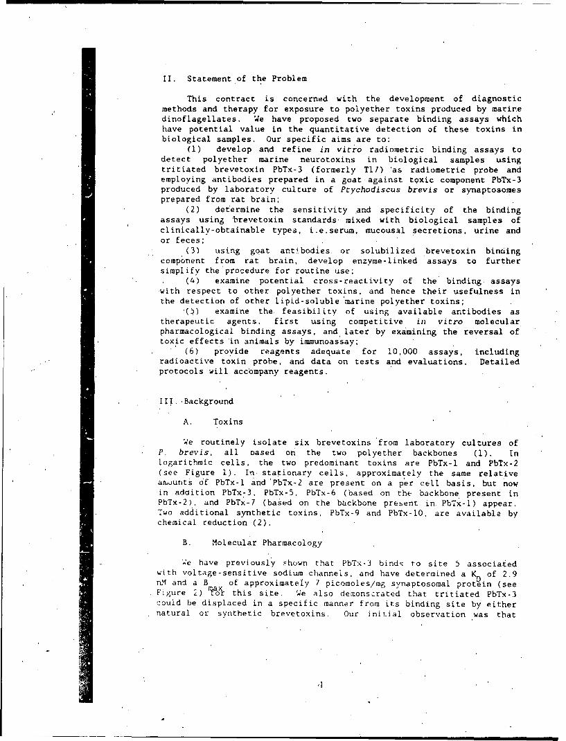
Figure I. Structures of the Brevetoxins 5
Figure 2. Non-Competitive Brevetoxin-Antibody- IProtein A Urease ELISA
Figure 3. Microtiter Plate Assays Utilizing Brevetoxin- 13Urease Conjugates
Figure 4. Enzyme Immunoassays Employing Antibodies 14Linked to Peroxidase.
Figure 5. Protein G-Sepharose Purification of 17Brevetoxin Specific Antibodies
Figure 6. Adsorption and Desorption of Specific 18Brevetoxin Antibodies from a Brevetoxin-Sepharose Affinity Column
Figure 7. Toxin-Urease Assay Conducted as Presented in 20Figure 3 Schematic.
'Table I. Inhibition Constants for Derivative Brevetoxins 6Derived from the Cheng-Prusoff Equation
Table II. Correlation of Potency with Radioimmunoassay 7and Synaptosome Assays
Table Ili. Comparison of Dissociation Constant (Kd) 15and Binding Maximum (B max) in Fish,Turtles, and Rats
Table IV. Comparison of K and B for Four Different 15Brevetoxin Proges in a Brain Synaptosomes
Table V. The Two Brevetoxin Binding Sites 16
"Table VI. Summary of Brevetcxin Microtiter Plate Assays 23
, a
II. Statement of the Problem
This contract is concerned with the development of diagnosticmethods and therapy for exposure to polyether toxins produced by marinedinoflagellates. We have proposed two separate binding assays whichhave potential value in the quantitative detection of these toxins inbiological samples. Our specific aims are to:
(1) develop and refine in vitro radiometric binding assays todetect polyether marine neurotoxins in biological samples usingtritiated brevetoxin PbTx-3 (formerly T17) 'as radiometric probe andemploying antibodies prepared in a goat against toxic component PbTx-3produced by laboratory culture of Ptychodiscus brevis or synaptosomesprepared from rat brain;
(2) det'ermine the sensitivity and specificity of the bindingassays using brevetoxin standards mixed with biological samples ofclinically-obtainable types, i.e.serum, mucousal secretions, urine andor feces;
(3) using goat antibodies or solubilized brevetoxin bindingcomponent from rat brain, develop enzyme-linked assays to furthersimplify the'procedure for routine use;
(4) examine potential cross-reactivity of the binding, assayswith respect to other polyether toxins, and hence their usefulness inthe detection of other lipid-soluble 'marine polyether toxins;
'(ý) examine the. feasibility of using available antibodies astherapeutic agents, first using competitive in vitro molecularpharmacological binding assays, and, later by examining the reversal oftoxic effects in animals by immunoassay;
(6) provide reagents adequate for 10,000 assays, includingradioactive toxin probe, and data on tests and evaluations. Detailed
"* protocols will accompany reagents.
III.-Background
A. Toxins
4e routinely isolate six brevetoxins 'from laboratory cultures ofP. brevis, all oased on the two polyether backbones (1). Inlogarithmic cells, the two predominant toxins are PbTx-i and PbTx-2(see Figure 1). In stationary cells, approximately the same relativeamounts of PbTx-1 and 'PbTx-2 are present on a per cell basis, but nowin addition PbTx-3, PbTx-5, PbTx-6 (based on the backbone present inPbTx-2), and PbTx-7 (based on the backbone present in, PbTx-l) appear.Two additional synthetic toxins, PbTx-9 and PbTx-l0, are available bychemical reduction (2).
B. Molecular Pharmacology
4e have previously shown that PbTx-3 binds to site 5 associatedwith voltage-sensitive sodium channels, and, have determined a K of 2.9n.M and a B of approximately 7 picomoles/mg synaptosomal protein (seeFigure 2) laor this site. We also demonstrated that tritiated PbTx-3could be displaced in a specific manner from its binding site by eithernatural or synthetic brevetoxins. Our initial observation was that
,|
displacement ceficiency was linked in a positive fashion with potencyin animals. Specific displacement curves correlated well with thepotency of each individual parified tc.xirn. Differential lipidsolubility of each of the natural brevetoxins made i': imperative toinclude Emulphor EL-620 in all experimental tubes.
In addition to developing displacement curves for the six toxins(n-2), we had sufficient toxin material for PbTx-l,-2,-3, and -7' tocalculate K s. These are summarized for sever'al species in Table 1.
1
-m3 -- ',
Type-71
Type-2
PbT.-O . • . 8mol:
Figure 1. Structures of the Breveroxins.
i i i l-i- ,- -
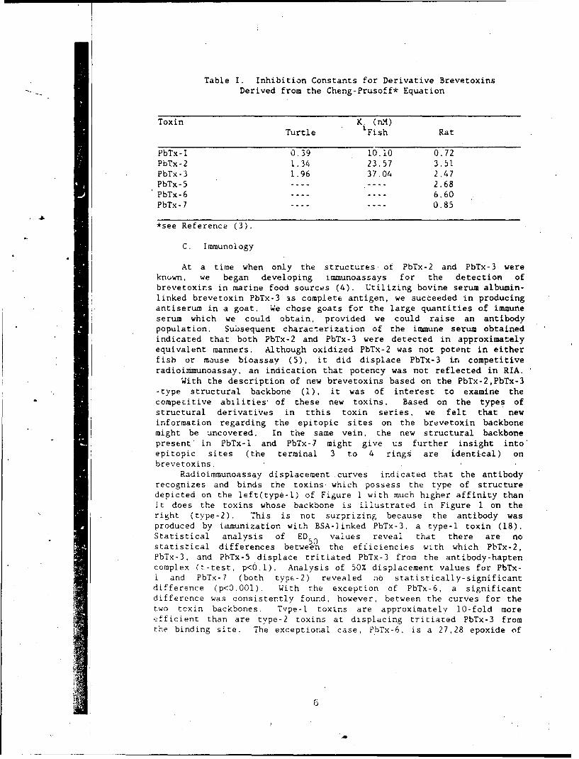
Table I. Inhibition Constants for Derivative BrevetoxinsDerived from the Cheng-Prusoff* Equation
Toxin K. (nM).Turtle 'Fish Rat
PbTx-l 0.39 10.10 0.72PbTx-2 1.34 23.57 3.51PbTx-3 1.96 37.04 2.47PbTx-5 ---- ---- 2.68PbTx-6 ---- ---- 6.60PbTx-7 ---- ---- 0.85
*see Reference (3).
C. Immunology
At a time when only the structures of PbTx-2 and PbTx-3 wereknuwn, we began developing immunoassays for the detection ofbrevetoxins in marine food sources (4). Utilizing bovine serum albumin-linked brevetoxin PbTx-3 as complete antigen, we succeeded in producingantiserum in a goat. We chose goats for the large quantities of immuneserum which we could obtain, provided we could raise an antibodypopulation. Subsequent characterization of the immune serum obtainedindicated that both PbTx-2 and PbTx-3 were detected in approximatelyequivalent manners. Although oxidized PbTx-2 was not potent in eitherfish or mouse bioassay (5), it did displace PbTx-3 in competitiveradioimmunoassay, an indication that potency was not reflected in RIA.
With the description of new brevetoxins based on the PbTx-2,PbTx-3-type structural backbone (1), it was of interest to examine thecompetitive abilities' of these new toxins. Based on the types ofstructural derivatives in tthis toxin series, we felt that newinformation regarding the epitopic sites on the brevetoxin backbonemight be uncovered. In the same vein, the new structural backbonepresent' in PbTx-l and PbTx-7 might give us further insight into'epitopic sites (the terminal 3 to 4 rings are identical) onbrevetoxins.
Radioimmunoassay displacement curves indicated that the antibodyrecognizes and binds the toxinsý which possess the type of structuredepicted on the left(type-l) of Figure 1 with much higher affinity thanit does the toxins whose backbone is illustrated in Figure I on theright (type-2). This is not surprizing because the antibody wasproduced by immunization with BSA-linked PbTx-3, a type-I toxin (18).Statistical analysis of ED values reveal that there are nostatistical differences between the efficiencies with which PbTx-2,PbTx-3, and PbTx-5 displace tritiated PbTx-3 from the antibody-haptencomplex (t-test, p<6.l). Analysis of 50% displacement values for PbTx-i and PbTx-7 (both type-2) revealed aO statistically-significantdifference (p<0.001). With the exception of PbTx-6, a significantdifference was consistently found, however, between the curves for thetwo tcxin backbones. Tvpe-I toxins are approximately 10-fold moreefficient than are type-2 toxins at displacing tritiated PbTx-3 fromthe binding site. The exceptional case, PbTx-6, is a 27,28 epoxide of
6
a type-i toxin. An epitope on the toxin molecule may involve theconfiguration around the 27.28 carbon unsacuration (summarized in Table11) (6).
TABLE II. CORRELATION OF POTENCY WITH RADIO 1XHUNOASSAYAND SYNAPTOSOME ASSAYS
Toxin Synaptosome LDRadioimmunoassayED K. EDD.Qls0 (MM) ED50()
--------------------------------------------------------
PbTx-1 3.5 1.1 4.4 93.0PbTx -7 4.,1 .1 4. 9 92.0?bTx-2 17.0 ib.1 21.8 22.0PbTx-3 12.0 37.0 10.9 20.0PbTx-5 13.0 ---- 42.5 10.1PbTx-6 32.0 -3- 'a 350 122.0
ED are uefined as the to~xin conc at which 50% displacement oftr1?2iated ?bTx-3 froa sodium c . anne13 or antibody occurs.' LD 50are determined by incubation of amb, n~ it toxin in 20
mL seawater for'60 minutes. K. are determined as described i'n thetext.
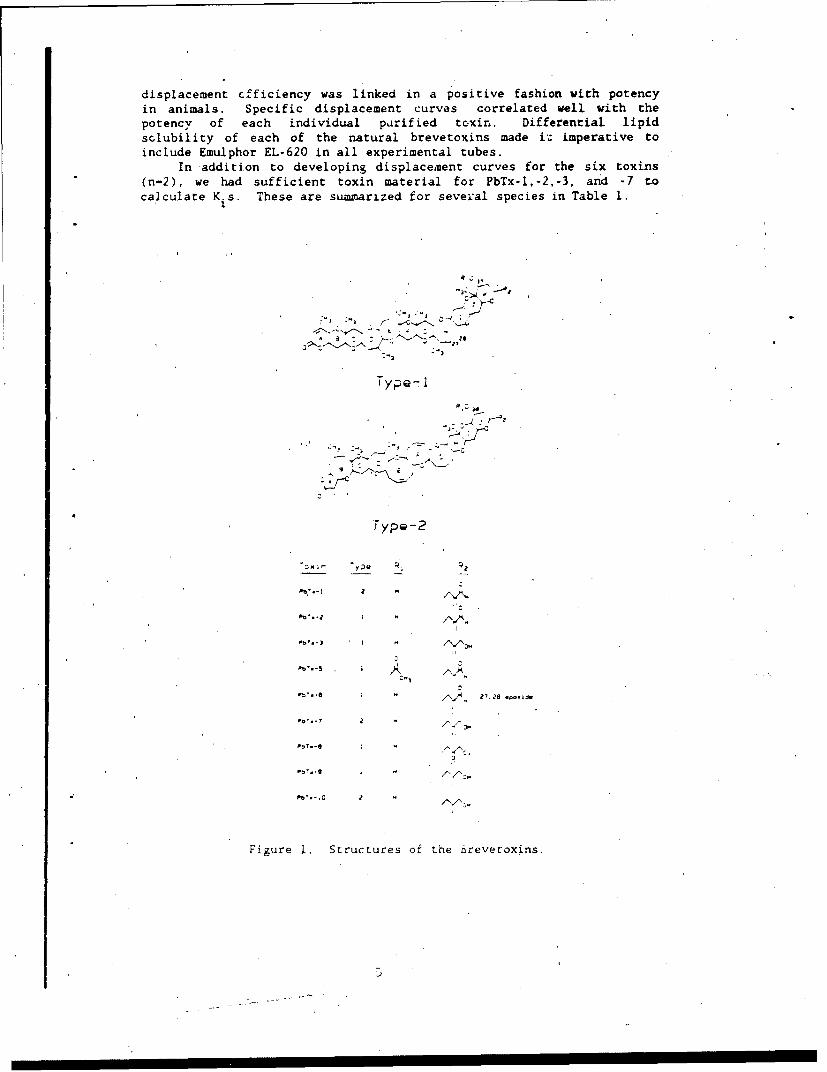
In'additio'n, we began. to explore methods for c'onverting the RIA toan enzyme linked form. o~e sought, to use an enzyme sv,ýtem which wasstable, produced a color re.::tion wh;.ch wouid be visible to the nakedIPve. (even though our evaluation woul.o takze pliae in. a microtitre platereader), would lend itself to coupling enzyme to either toxin oran. ibody, and would possess an enzymatic ac-4vitv that was absent inmammalian systems (to reduce background color reactions).
The basic assay under deve-lopment followed a noncompetitive enzymeimnmunoassa ' sandwich techniqu±e (figure 2). Heterogeneous system assays(7,8) such as these may be performed as' either competitive orrioncompet 'itive types, and may be either enzyme-antibody labeled orenzyme -hapten (antigen) labeled. 71hus, the greatest flexibility isi7.a ined employing such techniques, and many different variations may be,dieveloped to meet defined criteria.
Urease
1 2 34
Fi;gIre 2. Non-Competitiye Breetoxii,,Ant ibody- Protein A Crease ELISA.
7
In order for the proposed assay to work, toxin PbTx-2 'illustratedas triangles) had to be successfully bound to the microtitre platewells. Unlike standard enzyme immunoassay proceduies, where water-soluble IgG 'is adsorbed to the plastic plates, it was necessary toinvestigate the binding kinetics and equilibria of toxin binding. Itis imperative that the solid phase ihould adsorb an adequate amounthapten in a reproducible 'manner. and that variability at this stagewill affect the ultimate precision of the assay.
Binding of PbTx-2 (step I of figure 2) was evaluated in threemedia : ethanol, a solvent in which the toxins are reasonably soluble;phosphate buffered saline, in order to promote partitioning onto thehydrophobic polystyrene surface; and carbonate buffer of pH 9.6, whichis routinely used to bind IgG to plates. Following binding, completeProtein A-urease sandwich assays (steps 2-6) were carried out. Weillustrated that PBS is the most suitable medium for toxin incubation.',e demonstrated the linearity of the assay with respect to reactiontime, illustrating the lack of end-product inhibition of the ureasesystem (9).
The stability of the toxin-antitoxin adsorbed on the microtitreplate (step 3), when stored in a dry atmosphere at room temperature,indicated a probable long shelf-life of the reagents . Stabilitycurves were carried out for 2 months with no loss in activity.Regardless of the blocking agent (shown as a hatched line in steps2,3, and 4) we used, however, substantial amounts of non-specificbinding of Protein A- urease was observed. Thus, backgrounds were manytimes very high. contributing to a. low d&gree of specificsensitivity. In addition, the enzyme urease, while exhibiting a highturnover number and relative insensitivity to temperature duringincubation, was very sensitive to heavy metals and pH (as well as theindicator dye, which is pH sensitive).' Moaifications to the ELISAtechnique were explored this contract year, investigating (i) toxin-enzyme conjugates, (ii)' commercial enzyme-anti IgG conjugates, variousblocking agents for the microtitce plates, a different enzyme andsubstrate for visualization, and monoclonal antibrevetoxin antibodies.
IV. Technical Approach
A. Synaptosome Binding Assay
DJolo~ica1 Preparation. Svnaptosomes were prepared fresh daily
from rat brair, using the tecnniques described by Dodd et al. (10).YZnaptosome integrity was evaluated using electron microscopy, or by
Na influx experiments. To prepare lysed synaptosomal fragments, thesynaptosomal pellet was resuspended in 5 mIM sodium phosphate (pH 7.4)and incubated with occasional stirring for 30 min in an ice bath.Protein was measured on resuspended intact synaptosomes or lysedsynaptosomes just prior to bindir. 6 experiments uEing the techniquedescribed by Bradford (11).
Toxin 2robe preparation. Natural toxins were used as obtained,purifed from cultures. Synthetic tritiated PbTx-3 and unlabeled PbTx-3were prepared by chemical reduction of PbTx-2 using sodium borotritiideor sodium borohydride, respectively. Toxin PbTx-7 was produced byidentical chemical reduction of PbTx-l using borohvdride. Precursor
toxins were mixed with equimaoir reductant, -ach present -'a saturatedsolution. Under utirrni-; conditoon:, the redctants were mixed andallowed to react for 3.5 m~in, ai-Er whicn t-me excess acetcne uas addeu4s sacrificiai substr..e kr•iced to propoi,.ol). The solvent andpropanol was evaporated, a:'d the residue was redissolved ii, minitialacetone Acetone-soluble material was thin-layer chromatographed onsilica gel plates using ethyl acet.Ate/petroleum ether 70/30 as solvent,foliowed bv high pressure liquid c,-romatography using an isocratic.-lution '1.4 mL/tin) soivent of 854 methanol/15Z water and monitoringibsorbancrc at 215 nam.
Tritiatt-d toxin was quantiie.e ýmplcving uv HPLC detector tracingsand sranda id curves wtrfc a :vt, p• c- usirn!; unlabeled toxin PbTx-3.
,iýdioact lvit; wad deterfnlr,.Ld u l.rg . quýr scintillation techniques and,tppr,,priate ,tjenchr trItir.xi 5 rýncxards. nriC-purified radioactive PbTx-3 has a specifir a.ctivitV o- ,)-lb Ci..Taole, or one-fburth the specificactivity of the chenical reductant. Aliquots of tritiated toxin are:;tored undtr iitroien -tiospher. ,,t - n ethyl alcohol solution.'i.belti oxien is szbe for 1-6 ,•;nt•hs, rtpurification by HPLC beingperforuid as necessary.
,)* 'tr to.xins, Other brvt ýxins were used as. purified fromu: I urtv. Pot'encv o:, rndiv~auai brevetokins was measured using
<,,,mbtsia fish bio,,s.%d (4 ..a; __L i• bir nd .. r. tritiataa toxin was measured using a
ra~pid centcritu4atior, .ecnnque (D'. 6inding assays were performed in aKiodirng ,aedr~ con ~:r/g of. , .' HEES (pH 7.4). 130 mM choline,-hiorid=. 5.') -4 ,.ucose. 6.S x.Ž mgnesLu" sulfate, 5,4 mM potassiumchiuride 'i og/mL bovine serum iibumir., and 0.01% Emulphor EL-620 as ane;nuii S ecf tI..- * 'tZror 1oirg necessary to solubilize the highconcentrat on .' I.r e r,;,x- used in measurement of nonspecificbindin?. Birnciing exiýe-r.r nts were also conducted in a depolarizingmedium consisting of 35 ;,&M K,!. :. m.M glucose, 0.8 mM magnesiumsulf ate. I mrgmL bovine se:%-n albumin in 50 M ' HEPES (pH , 7.4).Svnaptosomes f/u-o0 ug rr:al protein), suspended in 0.1 mL. binding,nedium nmnus BSA were acOeti to a reacion mixture containing tritiatedPbTx-3 and , other ef . :. 9 =.. binding medium in 1.5 mLpoiypropvene microcentr.:iZA,4 r.,oD ... Atter mixing and incubating at
C for 1 hour, sawpies were .centcfk.zged at 15000 x g for 2 minutes..uperrarnr .oi lt Ions wert: aspirattec And the pellets were rapidly-.ashed with several .drops cf . wasn meditm (0). The pellet-- were thentransferred to liquid scntin'iation m inIvials containing 3 mL.. ;' nti!V.,nt and the bound waoioactv• v w.'.s estimated using liquid•.:inr:..r:or, :ec.nŽe5. •).n.rpec :c bir.dir.; was m'easured in ther:resence ot a sat'iratinc ror' :tror un.ioeied ?b-x-3 {l0 uM) and
w~s subtract.i •om t.• - ' :.. '. C!d s -ecifc c binding. Freetritiated prcbe was deturc.zii-. :/! coun:nrg; directly an aliquot of thesupernatant solutions prior 'o .. it~t: 1o.
%..'i Z ý,7 t n _L1__L._1 'L (-n 2 " , , :. - •as piirified to HPLC.munO-V is d1tc 'I cd r- :o', I -,i C Vr.- .cv was confirmed .using the
-v 'A •- n was .nlikely to be:-,ue ,t Its c.:'...a .z;anrd thus it was necessary to
riple ý,s hapren 'o a .OiLt3')i- antigeric carrier, in these cases to
bovine serum albumin (BSA) or keyhole limpet hemocvanin (KLH). Weutilized the aliphatic aldehyde functions present in PbTx-2 as thecoupling site, principally because the Aldehyde is located on theterminal portion of each molecule and thus the toxin's spatial exposureduring immunization would be enhanced.
Homogeneous toxin PbTx-2 was added to acetonitrile to yield afinal concentration of 1 mg/sL. To this solution was added (in 3 equalincrements atone minute intervals) sodium boroh,/,dride (as a saturatedsolution in acr.onitrile). The final acetonotrile stoichiometry addedwas on the order of one mole reducing equivaients per mole toxin. Forissessmert of reducing elfticlency.' and as a tracer for later couplingsteps. ine uCi tritium labeled borohvdride was added to the, reactionmixture. Following reaction for 0 uinutes under conditions of constant';t irrinA. excess borohvdride wts degraded by the addition of one mL,tcetonf, (which is reduced to 4 ropanol). The resulting solution wasthin-laver chromatog'raphed on silica gel and ultimately was purifiedto lCmoweeti'i tV using Hpi:.(. For purposes of our later coupling. we",tifht to ut ili- only the Pbs- . reduction product. However. we wishlo po Int otit I at Phbx-*4. wrichi corresionds. to a doubly reducedprotduc't . .l so possesses a primary alcohol and could be used for",'llpl ring .% Wei ,
l'ur i t id PbTx. ( r ePiAA ti't-/) w.u dissolved in a minimal-- J1 mp of ro-distil ld pvr-di,(itn. ad.4 tfnl olA molar excess of succinic.,+t,: vir i(is- In pvri(Ir e was .ad-!(i wkti it iI r ri, The reAction vial was,,.Ied. .trd was hv,, t,, '- <, to s.td it. i r, -d for 2 tiours in an oil
b.th Vol lowing r,,action, PAch soitifon w s dried under a stream ofnitrogen, rre'issolved in mintimal meith.an'l And was chromatographedoniilira ),- in /0/30 othvi acetate/ petrolie• either. Portions of eachplat, wero spr:tvod with bromcretol green Iolution for detection oficcidii. ind I cm port ions (t e ach p'are were scraped and assayed forrad ioacttivi tv by liquid icinti ll ation techniques. Fractions whichpr,.,duced both acid-poittive raction (succinic acid) and radioactivity.ftoxin) were scraped, ,lIted. a4d wetihed,'
The fre, -arboxvl fAjnctiý,.n on each to>.n-%uct:t-.ate derivative wasfv,"alentlv coupied to the ,-amino group of "-;inp restids in ASA or KIJH
bv to-P of 't ilndard I-crmnicpis we h.'vA emplovd previously (4) Thep',(crJure used was . hat. empl ovd !or the c-oviiont mc'd it cat ion of.. r ,r'i hormone whn i-wpl, ed t o :ro;,)! , 'i c.trriers r12 e xcept !hat the
It .itl 'otd*nsat ion it Ie W4I\ N e!, 1t" he ned to 12 houktr% Fol lowing' 0;; 1i I•'. ! t;.. rbI XtoIli€" w'..: ,•i .i ,i 'd .4• aifst: 0t 1 .'i ".,d a U .pho,•phnte
It, tor (;7H /,4) for 2.- iooini . irAi. ,-. ;,ho, ph t ehNuOIerod. I ' ) ,.i' t ti i•, o' it t ion Was .4,1 +i t tI t ox in
' ~ ~ ~ ~ ~ ~ jj va ril 6f • i I''• ) l r 1 simtll~*; i ,*,a t i onIt.
: A .L , , ' t , ' ii.,," , .,.tr ) .I. ;<,'; .I [ . , 't•isto .t);x I -pv) .%-l1ent I of ejA Cf.> .. * .\ Ifit Vt l . in .4 W t'itK .1 int Vt V4 %
If•r ,-,I.ht w,,-ks . the f r.t .. it ,,,n ,' . ~tit , ;,,)I r l, teP o ntlldI,! 'iviIrlt , anrd .1jb%P,?Aient co S, , being ,,oipelid ill i:I riIo' :it V i F t• Anir'd.,, ;',tit There:of ter. .)oo%.t i r,- pi ko( '.-dl ,i .i i lay It"t erv.1l i \; et IS.*1 , ,ioned I•I %' pri r , - t!' , ' l ;r.iVIi.r,4! iw ''111d we-k i md
r V t ' I " . .t" % t l
if) 1 .*'a I1i I.. I.)'V ,1 .l0 ',- 14lI I A * t t. in W V r r
A iI1)'U:i0'1 di iia rt 1 ':4-1 abov.' e" 'i i t w) 4if'1 i1,4 1i ' C titr t-tt I vl he I tng
,naii,tained. the first being boosted with BSA-toxin and the second beingboosted with KLU-toxin..
Preuardt• ;on ,• , AntisLr m was treated withiatmmnium sultate to yield a A 1 1a-d i..,lt ccncentr, ttion of 1.9 M. Themixture was stirred at I, I,) I kiour and was then centrifuged at12,000 x g at 40 C tor 30 minutes. Precipitates ere washed once with1,4 .4 ammonium sulf-:te, dissoived in PBS, And dialyzed against PBS for)4 hours The resuitlng crude antibody solution were adjusted to 25mgirl protein for titre•r evaiuation. Protein concentrations weremeasured iccording to the nethod ,a 6radford ( i).
L.,'ua* ,, it d. ,,L_ L , i !i AF -tbdpe.r to each bleeding, 30 uL.I i quot,, of antibodv preparation (J. V) m•y, pro :( .n) were added to
, CAp V tIIbtei ,ort~itiniUg I) ) mL P6S. and i creasing amounts of
1 i bT r, A ., v f rrom 2 ,, r, 21)ý ,ii . A pir liel null ixperimentp. , r t o-' ctnd tA. I I!,, ore- i iwu:;, t' e. t rl'r t ions. for evaluation of
,ptc it Ac vet not brevetx• an.a rT boo%,--spe,:ii ic b nding. Non-specificrI , N11 All t-.ith • -*, wis dit- , it t he pree~n Ce of 10 u.W4 unlabeled
I •'v.-) ,: ,,i~r W:pn. rmacological binding,XJ,#-r Iat,.I ,,I qt, I L ,I r,:.41 ; 1 I .II , nUd 'Ats dttference between*til at,,1 , . ! lidrg, 1 t , a cA lculated value. TheI Aý rt lic't t, * dr wetfn spe, ,I A, , 1w,1 :,I '." " ýIt.' in pre- and post-
I i i ýI t I en ::,,r.i i "t A i ., i *, , -n..,surtI t specific antibody
7A if i "n t.,l , :I;:,v,,' ,A J e. e r , A hw d exhljtt only non-specificJi.lt , 1t i,t1* , * brv,.t týxin. ,r c i,,,t Ion t i, s and assay protocol
S , ,r,),I'dLre ,A t , i.,vd was that described by,. .. , i:... 1 /•, tr t ro i.i.nt tat it()n o:' !*. rum di Foxigeftn levels in
\i l t') o,,t .i 10t,,t ,,o,1t (,r ,0. !,)-1 t .15 MA .rotain) werei ht,,i , ,•t' ib ; ar'.:,t.s .;r,; Ut ) ml, PBS. 2 ng 1 H)PbTx-3. aid
I W1 w I Ai'., nt I I A .,t sti,,!,,jd !,xi;i rat.,tKing trom 0.6 to 200 nig.A, i c a ti rot i t,,be s wr,',, icl A ir v(d . but wit hour uinlabeled toxin
t r, il ,ii,?; h, tnid by in?. hrdv), or .;n the b.,i {-e of antibody (total. I " ,r d,,-,- Fh.o if, ,a, ),n v)l iimv, were Kept. constant by the., Hi I t i , ' ' 4'.., 1 .,I
. AL ..j -. , .t r It,c,,b, t A ,>n .,it .14 C for one hour, I tl %t ,*'.I' 1I tl t il , r!'V t r p,vr.itr, A ) ul of a suspensl.on
.'.slui / .I t1 ' ' M aI r J4 k , 'N .;A, , <. A "..kn in ,IP S was t-hen,',',,,1 , ,, iri t .,t i<,i, Ats- x,A ..... t tor l o.., , rito.ia,1ng no antibody
fto ht- mI .iý',(l .4:'d Ih.a oi 1t 1 Vi.' I v , n<te, and then
itt, , ,,,., it , , 4 1 1r ,' 2, .IAt. I I s of 0. m til werei,,,,, I tl, ' "I,, J , •, . . , :,, I, :. V .1, . ),",v' ( ,,c with A ml liqu id
' .. 't at ; .. a.. .' ,, , ti,.:. , ,: . " in a 8iýckratn LSC
Si , I n : ,' , . ,).i, 1 W o it ad for
,..e.v., wit,, .at .0 . ,,.I.'i polvitvrenp* : ,+ ;',I ' ''*i . " . -.1"a il + A I I *I. .apdcktY y 4 6.4
'+~ ~~~~~ A,, , 1 ,r " ', + ., A•:. +, .4:,-+ v 1.:t ". e r,lt ed , %te matd"
,, ;'., " . 'it . , * i 'v. y ,ar i .n %o
11 114
"I"I0 M
microtiter plate assays, once we had developed toxin-enzyme conjugateswhic were active materials.
Iimunoassays employinz orotein A-urease. The assay we developedlast year (Figure 2) was a ProteLn-A-urease linked sandwich assay. We.chose this for the following reasons: (i) protein A binds specificallyto the Fc region of IgG and thus will minimize interference with Fab-toxin interactions; (2) protein-A interacts with most IgG Fc's, thuspermitting its use for many antibodies created; (3) protein-A-urease isavailable commercially, thus assuring quality control; (4) urease has'ahigh turn-over substrate rate and is not typically a mammalianenzyme, thus providing low background when examining biological fluids;(5) the assay is conveniently monitored using a dye-coupled (590 nmdetection) reaction in response to released ammonium ion. allowing formicrotitre plate monitoring colorimetricallv.
"That assay utilized initial binding of hydrophobic toxin topolystyrene (virtually quantitative) (step 1). followed by specificantibody binding (step 3). followed by washing. Protein A-urease wasnext added to bind specifically any toxin-ýpecific antibody bound toadsorbed toxin (step 4). After a final wash, urea substrate jolutioncontaining bromcresol purple dye was added and the color reaction isevaluated photometrically.
PbTx-2 was used as hapten bound to the plate because of its higherhvdruphobicltv. Parameters such as optimal toxin concentration perwpll (in 200 uL PBS), optimal buffering solution for toxin adsorption,and optimum time and temperature for binding. Following toxin binding.breveroxin specific antibody was added, and parameters of time andtempe:ature tor binding, antibody concentration, and nonspecificprotein blocking or lack of blocking were evaluated. Both of theseporturbations were evaluated bv classical "chequer board' arrays in themnicrotitre plates. Evaluation of stability of the microtitre plats-toxin-IgG complex in lyophilized state was evaluated for shelf-life.\s indicated already, because of the unstable nature of the urease',rltvmO in Protein A coupled form, we abandonea Protein A-urease as apotential reagent for assaVs,
Lul yme .mmunussays ýmoloyinr brvyetoxin-urease conjugates. We,;oight to examine the. stability, and etficacy of utilizing. brevetoxin,'jvmletv-1i inked to urease enzyme, The toxin-enzcme conjugate is(-,nstrurtepd in an entirelv analogous ;aatner to the construction ofcomplete antigen from brevetoxin ibTx-l and protein. Thls--is describedon page 12. The only deviation from t.his procedure is that we utilizedA• limiting concentration ot toxin- ,ucc-natt- in the coupling procedureto encourage a one-to-one stoichiometrv of toxin to enzyme. This isfffcr ed by adding toxin-succinate con ]u.Ite to protein solution.
ra.ther rh04n the converse of ,td(iru;g en.ývme to toxin- suc-inate solution.Fli coupling stoichiomet ry ,,girin can be convenient v measured using a';in.Ill ualntity of tritlat.ed i'hTx-3 a*dded to solution during coupling.
I'hl I I stolchiometry is or ii ,. based on the. intuitive notion that(I) t iniri:mal number of tox:n ,a•, :i--• per en.:vme moletcule will lessen' he I ik#l ihood of active sitt .iddit ion in the enzvye, and (2) that a,.rat er toxin o . roichiometrv wouli i crea,;,r Ohe hvdrophobic character of• ,. (',, 0! e . thereby inc .ic ;i nr non-specif i c a.dsorpt ion to the
I plaros A A.,chemat ic ot t ih aescrihbed prot ccol is 1 llusttawed
ureast
1 2 3
"urea + H20 = C02 + 2 NH3
Figure 3. Microtiter Plate Assays Utilizing Brevetoxin-UreaseConjugates
Checkerboard experi•ients on 96-well piatcs were carried out usingtoxin-enzyme concentrations ranging in 2-told dilutions from neat to1/512: (2 picograms/wel. to 1.02 nanograms/well) and dilution ofpolyclonal antibodies ranging, from neat -to 1/1024 dilutions (0.75 mg to0.1 Mg/well). The' initial step (step 1) involved antibody binding tothe microtiter plate surfaces in thhe concentrations indicated.Adsorption butter included 0.1 M Phosphate Buffered Saline (PBS, pH7.4), 0.15 .4 Phosphate Buffered Saline (PBS, pH 7.2), and sodiumcarbonate, buffer, pH 9.6 As potential blockers of non-specificbinding sites (step 2), non-fat. dry milk (Blotto), nonflavored gelatinat various concentrations (GelBlock), bovine serum albumin (BSA), andpre-immune serum (PIS) were utilized to minimize non-specific bindingof toxin-urease conjugate in st.p 3. In' step 3, the various dilutionsof toxin urease were evaluated. The various Incubation steps werecarried out at room temperature, 31°0C. and at 4°C. Appropriate blankswere evaluate-i, as well as the stability of the toxin-enzyme conjugatein neat form.
Enzyme imrunoassays er.oo.vn& ant ibrevetoxin IgG-peroxidase(sunu.ateg. Brevetoxin specitic a.ntibodies were prepared as earlierdescritbed. Aliquots were Npecificaily adsorbed to Protein G-Sepharosecolumns (Pharmacia) and washed to remove iexcess protein not of an Igonature. L;G was then desorbed trom the column using high ionicstrength glycine buffer, arid was coricentratc-d by molecular sievec.ntrif.Jg.ation (.4W exclusion i0,5);O). Concentrated IgG was loaded on abrevetoxin 'afLnity ,oiumn pr•.dou by lv hi :..rbodiimide condensationrpaction between PbTx-3' succ i rtLe and Amino hexyl Sepharose(PhIrrmacia). (An identical proc udLrv t) th-.t utilized for toxin",oupling to protein was utilii.d to coupe PbTx-3 succinate to theami no groups of the S,-pharose 1.,ri vat ve). Following specific,iisorpt i on of the brevetox i n .nt , ,,d i es on the column, unadsorbedmaterial wais washed frree bv PSS, . /H I4 ,,c ific brevetoxin antibodywas washed from the colt.rnn with 3 'A %a(_ and was desalted bydialvis. This material was itili•ud :o)r coupllin4 to peroxidase below.
Hor'e radi ih p.eroxid,ls,, ( i i., di s :;o ved in 0 3 ml freshlyprepared ]0(i( rim NaIilW . in a, :.ia t 1 , oditim periodat. (8 mM,, . 3hml) wa.; added with .,irr i .ni .. w:is illowed to r,?- " 2 hr atroom tremperature. Throe ml ot anti - hr,*vtoxin goat IgG (abou ? mgproteitn) was desalted on a !..iali *:ainum .ir1 was placed in 100 m1M sodium
13
carbonate buffer, pH 9.2. The activated peroxidase and the IgG wererapidly mixed and pipetted into dry Sephadex G-25 where they areallowed to react for 3 hr at room temperature.
Following reaction, the conjugate was eluted from the 6ephadexutilizing sodium carbonate buffer (pH 9.2), and was neutralized withfreshly prepared NaBH dissolved in O.1M NaOH. After incubation at 40 Cfor 1 hr, protease in iibitors are added and the whole mix is diluted totwice volume with glycerol and stored at -20 C for later use.
For this particular assay, similar types of parameters wereevaluated to ascertain optimum conditions. The protocol is illustratedin Figure 4 below. Initially, we sought to use unlinked toxin asinitiaL adsorption step', as we did in Figure 1 for urease assays.However, it appears that this step is critical with hydrophobicmaterials such as these toxins and often they yield unpredictableresults. Instead we utilized the keyhole limpet hemocyanin (KLH) toxin
perouidate
1 2 34
Figure 4. Enzyme immunoassays employing antibodies linked toperoxidase. The assays can utilize-either brevetoxin-specific
antibodies linked to peroxidase (Ab in step 3 would be linked), oranti-goat IgG to visualize brevetoxin specific antibodies adsorbed
to toxin-KLH (as indicated in the figure).
conjugate es a primary. adsorbant (Step 1). The material is quitehydrophobic, possesses an estimated 50-100 brevetoxin molecules per KLHmolecule, and possesses 'characteristics exploitable of protein and theplastic plate interaction. In addition, the linker arm between thetoxin and protein allows for a greater freedom of toxin-antibodyinteraction. Again, we evaluated many different KLH-toxinconcentrations (step L), the many different blockers (step 2) alreadyenumerated, and several 'different washing and binding buffers.Commercial antigoat IgG-peroxidase conjugate (step 4) %as utilized toevaluate the binding of brevetoxin specific antibodies to toxin-KLUadsorbed to the plate.
Enzyme immunoassays utilizing binding components derived fromsynaptosomes. The P3 fraction, resulting from the procedures employedfor preparation of synaptosomes from rat brain (page 10), was utilizedas biological preparation in identical dilutions as employed forantibody preparations described above. Standard checkerboard patternswere developed employing dilutions of each specific binding component,and toxin-enzyme conjugate. Dilutions of synaptosomes were incubatedin microtitre plates for one hour, followed by brief washing withstandard binding medium. Gelatin blocks followed for one hour, andwere then rinsed with standard binding medium. Dilutions of toxin-urease conjugate were employed to acertain specific binding.
, II II
V. Results and Discussion
A. Synaptosomal Bindirg Assay
Species similarity of binding. Synaptosowes from rats, turtles,or fish were prepared to examine the binding characteristics of eachwith respect to brevetoxins. Table III outlines the results of thecomparison, and i.ilustrates that any of the three systems examined bindbrevetoxins in a reproducible manner with approximately equal efficacy.
Table Ill. Comparison of Dissociation Constant (K ) and Binding
Maximum (Bm) in Fish. Turtles, and Rais*
K Temp. Optimum Specific BindingSpecies (A) (pMol/mg Protein) (°C) at K
d
Fish 6.1 .-; 23 80XTurtle 1.5 " 2.25 4 80%
Rat 2.6 6.80 4 90%
*mean values tor K and B , n-9,4,6 for fish, turtles, and rats
respcctively, max
pe biL.dI.i or .. -ia.. tIwar2d brevetoxins in' rat brains,;riaotosomes. A preliminary comnparison of specific binding of tritiatedPbTx-3. PbTx-?, PbTx-9, and PbTx-LO indicates an equivalent B and amax.progression of K values wcilch parallel the relative potencies of the
labeled brevetoxins, This ib a further indication to us that binding
affinity is the conservative requirement in the potency of thebrevetoxins (Table IV), and further, that we may be able to utilize thetoxins whtcn are of higher specific activity for more detailed receptorScharacterization (14).
Table IV. Cronparson of K. iar•a 6 for Fouir Different Tritiated6revetoxin erhbes in &a orain Synaptosomes
Toxin K B. (poles/mg protein)
P r'Fx- 2. 3 ) 6 99PbTx- 9 8. /,5 6. 15
PbTx- I 1 .91 6.38PITx-10 1.)6 6.46
Otir vidence indicates thAt. at a K 'oncentration of tritiated'YT:<-3. the t .,r c,- .. a.. , : ites .;.•pLoximate i- 2 minutes. A
" ,-pproxu• Ualon ,-anht [ &,r:vuJ •aLilizing presenz: protocols.There is no -nembrane pot.tntll dependence ot brevetoxin binding to therigh aftinjt'l, low rapacity 1binding sirv known as Site 5. K d- 2.6
I I ! 1 I I I I I I I II I I II I I 1 d
(intact), 2.9 (lysed), 3.3 (depolarized) and B - 6.01 (intact), 5.83(lysed) and 5.75 pmoles/mg protein (depolarizedjMa•5).
Regardless of the organism used for synaptosomal preparations, itis apparent to us that the topographic characteristics of thebrevetoxin binding site on the VSSC are comparable. Using brevetoxinsPbTx-1-3, and PbTx-5-7, K. data for specific displacement of tritiatedPbTx-3 shows comparable data in each case (Table I in Introduction).The more hydrophobic type-2 brevetoxins are most efficacious in theirability to compete for site 5 binding (6).
Classes of brevetoxin binding sites, Two separate brevetoxinbinding sites have been discovered in rat brain synaptosomes. Thebrevetoxins bind with an affinity constant which is consistently in thei-5 nM concentration range, in 'good agreement with affinity-data forother potent marine toxins like saxitoxin (16); In addition, thebinding maximum in synaptosomes is also'in good agreement with data forSite I toxins, which are known to bind to channels with a 1:1stoiciometry. However, the allosteric modulauIon of sodium channelbinding by other natural toxins bj brevetoxins occurs at brevetoxinconcentrations much higher, ca. 20-100 nM (17).. This 'data isinconsistent with high affinity, low capacity binding.
Converse to this allosteric modulation which occurs at higherbrevetoxin conc ' itrations, is the finding that membranedepolarization, -Na influx and competitive displacement of tritiatedbrevetoxin binding by unlabeled competitors, is dose dependent in thesame' concentration ranges observed for the high affinity binding site(5). Thus., the allosteric modulation at other sodium channel bindingsites appears to arise from brevetoxin interaction 'with a loweraffinity, high capacity binding site.
Using classical Rosenthal analysis, we have been able todistinguish two separate specific brevetoxin sites (Table V).
Table V. The Two Brevetoxin Binding Sites
Site Kd B .x AllostericModulator
5 2.6-3.3 5.7-6.8 No
* /9.1-300. 63.7-180 Yes
*not numbered until further work can be accomplished.
The two site hypothesis is supported by brevetoxin inhibitionconstant data and double reciprocal cometition plots, which indicate adeviation from competitive type patterns to non-tompetitive typepatterns at higher competitor brevetoxin concentrations. The non-competitive displacement appears to be specific in nature, and is notlikely due to changes in membrane fluidity. But, certainly moreinvesti&ation is required before concrete conclusions can be offered.
B. Radioimmunoassay
The radioimmunassay was developed last year has been used thisyear almost entirely to give a baseline value to ELISA protocols, whichhave been a major thrust. Two potentially important developments havetaken place this year, both involving a purification of brevetoxinspecific antibodies for use both in RIA and ELISA. No further specificwork has been undertaken with RIA, except that the KLH-Pblx-3 immunogenhas been successful in eliciting antibody production in a goat. Titersare being evaluated weekly, Aith a biweekly immunization schedule.Currently, titers have exceeded BSA-PbTx-3 elicited titers, based onqualitative assessment and Ouchtelony plates employing whole antigenconjugate and specific sera.
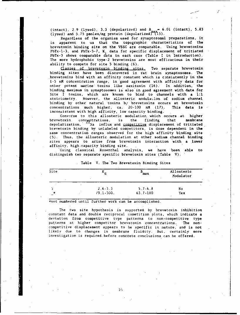
Protein C-affinity columns, Goat serum was purified by loading 4ml of a 10 mg/ml IgG solution onto a column constructed to contain 4 mgof PbTx-3 specifically bound. Single pass adsorpcion of the serum(6828 total units applied) resulted in 5254 Specific Binding Units(SBU) adsorbing and 1896 SBU passing through without adsorption, or a73% specific adsorption of antibrevetoxin IgG to the column. 'Elutionof the adsorbed IgG was accomplished using 3 column volumes of 0.15 Mglycine-HCl buffer, pH 2.8., followed by desalting against PBS pH 7.4ona commercial 10 ml desalting coiumn (BioRad) (figure 5). Standardradioimmunoassays were performed on eluted fractions which were pooledto calculate specific binding units recovered.
2.000
1.500-
0.000 -
0 5 10 15 ao 25 30 35
Protein G Sepharose. Fraction Number
Figure 5. Protein G-Sepharose purification of brevetoxin specificantibodies. 4 ml of antibrevetoxin IgG was loaded on a 3 ml protein G-Sepharcse column pre-equilibrated with PBS, pH 7.4. The sample was,loaded as a single pass, measuring protein concentration in dropwisefractions. Following decreases in protein concentration to backgroundvalues (fractions 15-20), the eluting buffer was changed from PBS to0.15 M glycine (pH *2.8) to elute specifically bound IgG. Proteincorresponding to eluted IgG elutes from fractions 23-40, fractions 25-35 being pooled as purified IgG. These pooled fractions correspond to73% of specific binding units loaded on the column.
17
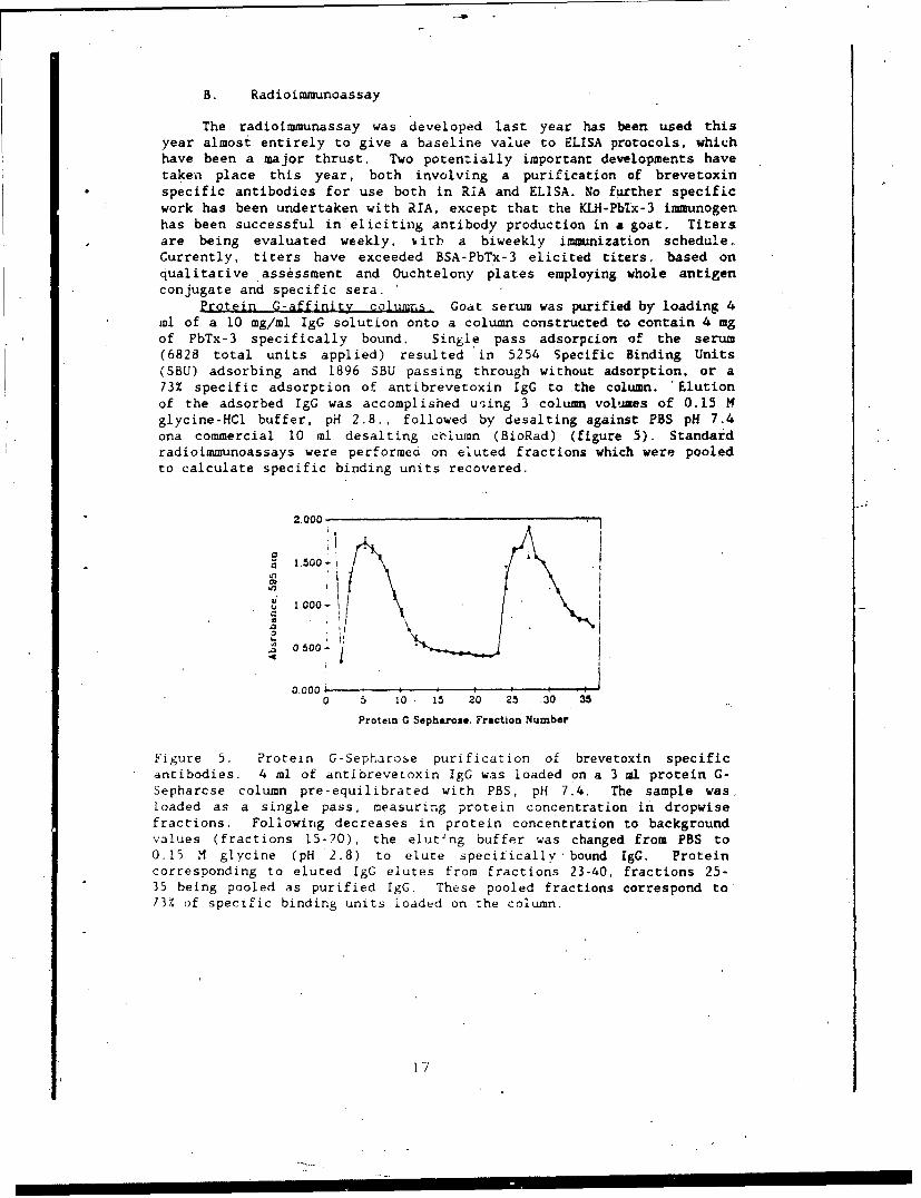
Brevrtoxin affinity column. The brevetoxin affinity column hasbeen utilized for IgG purification following Protein G-Sepharoseseparation. The pooled sample of IgG from the Protein G column (5254SBU) was loaded on the Brevetoxin-Sepharose affinity column in minimalPBS, pH 7.4. Following single pass adsorpti6n and elution with 3 MNaCl. 2210 SBU were recovered (7.2 mg protein), with a specific bindingactivity of 307 Umg protein (figure 6). The peak not adsorbing to thecolumn correpsonds to about 16.8 mg of protein (from a total of 24.4 mgloaded on the column) and possessed no specific binding activity. Wefeel therefore that the column is a useful tool for purification of IgGspecific for brevetoxins.
1.2'1
0.4 -
00''
0.4- ,*am ..
0
FRACTION. NUMBER
Figure 6. Adsorption and desortion of specific brevetoxin antibodies,from a brevetoxin-Sepharose affinity column. A total of 5254 SBU (24.4mg protein) of brevetoxin specific antibodies was loaded on brevetoxin-Sepharose affinity column prepared by carbodiimide condensation of PbTx-3 with aminohexyl Sepharose 4B. Following adsorption, the column waswashed with 3 column volumes of PBS, pH 7.4 and the fractions werepooled. The first peak corresponds to 16.8 mg protein ( and nospecific binding units) which was not adsorbed. At the fractionindicated in the figure by the vertical line, the elution buffer waschanged to 3 M NaCl and the peak which eluted was pooled. The peakcorresponded to 2210 SBU and 7.2 mg total protein. The eluted peak wasutilized for development of the microtiter 'plate assays describedbelow.
C. Microtiter Plate:Assays
Microtiter plate assays have been developed in five differentways, four utilizing antibodies and one utilizing synaptosomes. Eachassay has distinct advantages and , disadvantages. The highhydrophobicity of the toxins and their derivatives has been a principaldifficulty in all our attempts at converting to enzyme-linked assays.
For most ELISAs, hydrophobicity can be exploited to "stick"antigen or antibody to the plate solid support. For brevetoxinmicrotiter plate assays, however, it 'was necessary to minimizenonspecific binding of toxin. Basic among our studies was the
18
opportunity to explore different methodologies for minimizing toxins,or toxin-conjugates, from non-specific adsorption to the plasticplates. Each assay will be outlined, identifying the finalizedprotocol, the problems encountered, the results obtained, and potentialfuture work.
Protein A-urease assays (figure 2).
Protocol:[1] incubate overnight at 4°C with PbTx-2 in PBS pH 7.4.
toxin conc at 1ng/well and 0.1 ml volume;
[2i aspirate and incubate 1 hour at 37 0 C with goat IgG at0.75 mg/well in 0.1 ml volume., Aspirate IgG solution;
[31 aspirate and block I hour at room temp. with 0.5% gelatin,0.3 ml/well. Aspirate gelatin;
[4] add commercial protein A-urease at lOX excess theoretical IgG
concentration in 0.3 ml, volume. Incubate 1 hour at room
temperature;[51 aspirate urease conjugate and wash once with PBS (pH 7,.4) aad
twice with distilled water;
L5] add urease substrate (consisting of per 100 ml in 0.1 mM
sodium hydroxide: 8 mg bromocresol purple, 100 mg urea, 0.2
mM EDTA, all adjusted to pH 4.8) and monitor color
development at 588 rim.
Results:. Micro iter plates could be prepared as depicted in.
figure 2, and could be stored dessicated at either step three or four,i.e at the plate-toxin-IgG step or the plate-toxin-IgG-Protein A-ureasestep. Time course studies indicated no loss of enzyme activity under
such conditions. The problems enumerated below preclude further
development of the assay.
Problems:
11 high non-specific binding even when blockers were used;
(2] low specific antibody binding to adsorbed toxin on plate;
[31 protein A urease unstable in refrigerator and loit activity;'4] protein A binds to only 2 IgG subclasses form goats;[5] competition a.osays using dilutions of brevetoxin were
unsuccessful.
Future work: None. This assay appearsto present too-many
difficulties to proceed further.
Toxin-urease'assays (figure 3).Protocol:
[1] incubate overnight with antibody solution (4 ng/well in PBS,
pH 7.4;
72] aspirate and perform a I hour block:!31 aspirate blocking agent and incubate with toxin-Qrease (range
of 15 mg to 200 ng protein eq./well) for 2 hours at 370 C,
[41 aspirate and add urease reagent (as described above). Monitor
at 590 rim.
19
I I 'I I I I I I ~II I I I I I I
Problems:(1] high nonspecific binding of toxin-urease to plate;[2] blocking agent tried to reduce nonspecific incl ded 0.1-0.5%
gelatin, 1% bovine serum albumin, each in the presence andabsence of 0.01% tween;
[31 urease enzyme-toxin conjugate lost activity with time (overa 6-month period).
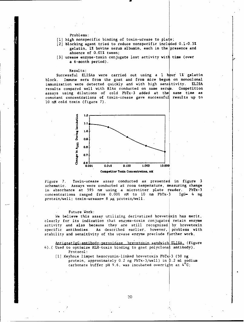
Results:Successful ELISAs were carried out using a I huor 1% gelatin
block. Immune sera from the goat and from mice begu on monoclonalimmunization were detected quickly and with high sens tivity. ELISAresults compared well with RIAs conducted on same serum. Competitionassays using dilutions of cold PbTx-3 added at the same time asconstant concentrations of toxin-urease gave successful results up to10 nM cold toxin (figure 7).
= 1.20
O.a
0.9
A 0.81
* 0.7:
a 0.6 I ,0.001 0.00 0.100 1.000 10.00
Competitor Tozin Concentration. aM
Figure 7. Toxin-urease assay conducted as presented in figure 3schematic. Assays were conducted at room temperature, m asuring changein absorbance at 595 nr using a microtiter plate reader. PbTx-3concentrations ranged from 0.001 nM to 10 run PbTx-3 IgG- 4 ngprotein/well; toxin-urease- 8 Ag protein/well.
Future Work:We believe this assay utilizing derivatized brevet~xin has merit,
clearly for its indication that enzyme-toxin conjugates retain enzymeactivity and also because they are still recognized by brevetoxinspecific antibodies. As described earlier, however, problems withstability and sensitivity of the urease enzyme preclude firther work.
AntigoatlgG-antibody-Deroxidase brevetoxin sandw ELISA. (figure4).( Used to optimize KLH-toxin binding to goat polyclona antibody).
Protocol:(1[ Keyhole limpet hemocyanin-linked brevetoxin PbTx 3 (50 ng
protein, approximately 0.2 ng PbTx-3/well) in D.2 ml sodiumcarbonate buiffer pH 9.6, was incubated overrig ýt at 40C;
20
[2] aspirate and add goat polyclonal antibody (mg to ug proteinconcentration ranges) to optimize Ab-Ag concentrations for Ihour at room temp;
L3] aspirate and add commercial rabbit antigoat-horse radishperoxidase. Incubate 2 hours at room temp.;
[41 aspirate commercial preparation and wash plate with buffer;[5] add commercial ABTS (2,2'-azino-bis-(3-ethylbenzthiazoline-6-
sulfonic acid) reagent and monitor development of colorreaction.
Results: 6:'1 Strong positivfs and low blanks at 4p to 1:10 goat antibody
dilutions (7.5 x 10- mg/well) and 1:10 rabbit antigoat (raggperoxidasge dilutions. 1:50 dilution of rag detects Yp to 1:10dilution goat Ab. 1:500 dilution of rag detects up to 1:10 .;
2; Immulon 4 plates (Dynatech) appeared to give highest amount, ofspecific KLH-toxin binding and lowest nonspecific binding of antibodies
.and antibody-enzyme conjugates;'3.] Preimmune goat serum as blocker gave best results for
minimizing nonspecific binding of goat antobidy to plates, and alsoyielded the highest degree of specific reaction with KLH-toxin;
4* Non fat dry milk was also an effective blocker.
Future'Work:The assay works well 'and is useful for evaluation of serum tIiters.
of antibody from animals being immunized. Our goat bleeds can beevaluated using this assay, employing each bleed serum in step 3 of theassay (figure 3) and employing the commerical rag antibody uniformly.This is the first assay we have developed of an ELISA format whichappears to work reprodzcibly and which has practical applications. Webelieve that this is in part due to the time we have spent developingit. but further that the reagents and substrate (plates) were developedover many years to a totally reproducible state. Thus, the assayspractical application should be developed further to capitalize onthese aspects; i.e. the only variables not are KlM-toxin and goat (or,other species as necessary) antibody, both of which are now routinelyavailable in purified form.
Our future plans with this assay are to utilize the Protein G-sepharose column to pre-purify goat antibodies, and then follow withthe toxin-sephadex affinity columns to further purify specific goatantitoxin antibodies, and to re-evaluate the yields at each step usingThe rag ELISA. We believe this assay will be a primary analytical toolfor the laboratory.
Goat antibrevetoxin antibody-peroxidase assay, (figure 3, whereantibody in step 3 is peroxidase linked).
Protocol:"1 Goat polyclonal antibodies, pre-purified by sequential Protein
G-Sepharose and PbTx-3-Sephadex affinity columns, werelinked to horseradish peroxidase using a standard soCiummeta-periodate method 18);
2; incubate KLH-toxin conjugate in microtiter plates overnight at4°C (dilutions from neat to 1:20000);
: , , o . . .. -- , '
[31 aspirate and block I hour at room temp with nonfat dry milk;[41 aspirate and incubate 2 hours at room temperature with
antibrevetoxin-peroxidase (0.6 Ag to 6.0 pg.protein/well);[5] aspirate and add ABTS substrate and monitor at 405 nm.
Results:Strong signals were observed down to 1:20,000 dilution KLH-toxin
concentrations, indicating that this reaction is both highlyreproducible and optimized. However. tull strength antibrevetoxin-peroxidase conjugate is required for the reaction. Two alternativesexist for this observation: (1) the conjugated peroxidase enzyme lose'ssome acitivity upon brevetoxin covalent binding; or, (2) brevetoxinstoichiometries are not what we calculate.
Future Work:The assay will be further refined to yield consistent results at
high sensitivity. Conjugation, procedure for toxin to peroxidase willbe optimized including proper sto-ichioT-cry and enzyme activityevaluations with increasing toxin coupling. Some initial work,employing the following protocol, has been attempted:
Protocol:'I1 incubate overnight at 40 C with goat antibody in microtiter
plate (mg to ng/well):'2i aspirate and block for I hour at room temperature with 4%
nonfat dry milk in PBS;:3i aspirate and incubate 2 hours at room temperature with unbound
toxin PbTx-3;'4] aspirate and incubate 2 hours at rocm tem)erature with
antibrevetoxin antibody linked to peroxidase;151 aspirate'and add ABTS substrate and monitor change in
absorbance at 405 nm.
Results:Currently, this assay shows high nonspecific binding of antibody-
peroxidase to the plates. Until we resolve the stoichiometry, problem,we are uncertain if this is unaltered peroxidase binding, or antibody-peroxidase contributing to the -higi. enzyme activity in blanks. 'Onereason we are continuing to pursue this assay is that if unknown toxinconcentrations are substituted for known toxin in'step :3i above, theresulting color development in step '5* will quantify toxin in,,,nknowns. Thus the greater color development in, assays will reflectgreater toxin. concentrations. rather than the converse (i.e. lessercolor development for greater toxin concentrations) which ischaracteristic of the other assays.
Synaptosome assays in microtiter plates. (figure 3, substituting.;ynaptosomes for brevetoxin-specific IgG).
Protocol:I' incubate 80 ug-0. 8 ug/well synaptosome preparation in Standard
Binding Medium for 2 hours at room temperature:2' wash three times with 0.3ml PBS pH. 7.4 washing buffer:3 block with 300 ulIXi gelatin and incubate 1 hour at room temp,
'4 rinse 3 times with 0.3ml PBS wash buffer;'5 add toxin-urease diluted in PBS (neat to 1:200 dilution) with
22

1% bovine serum albumin and in..ubate 2 hours at 37 C;[61 rinse 3 times with 0.3 ml distilled water;[7) add urease reagent and bromocreiol purple indicator;[(8 monitor reaction at 595 rm.
Results:As in a'l assays employing the enzyme urease, some inconsistencies
were noted. In general, the checkboard assays indicated that bindingof toxin-urease to synaptosomes was taking place, with detectablechanges occurring with synaptosome concentrations as low as 1.6 Mgprotein and toxin-urease dilutions of 1:20.
Future Work:The assays were performed at a time when only toxin-urease
conjugate was available. We plan to utilize synaptosomes with trxin-peroxidase and in sandwich assays using synAptosomes as primaryadsorbant in the coming year. We believe the results are promising,and have begun to anticipate development of a synaptosome-toxin-antibody-peroxidase assay.
D. Summary Discussion
Table VI. Summary of Brevetoxin Microtiter Plate Assays
Adsorbant SensitivityPrimary Secondary Tertiary (ng/well)
PbTx-2 IgG a PbTx-3'4 Protein A-urease 1.0IgG a PbTx4 3 5 PbTx-3-ureasa ...... 0.001KLH-PbTx-3 ' IgG a PbTx-3 r x g IgG-peroxidase 6 0.2Snaptosome 4' 7 PbTx-3-urease 4 . ------ 0.2IgG a PbTx-3 PbTx-3-peroxidase - 4,10 0.001Synaptosome PbTx-3 igG-peroxidase a PbTx-3 ....
2maximum sensitivity demonstrated3goat antibrevetoxin prepared by our laboratory,commercial preparation
5 brevetoxin-enzyme conjugate prepared ov our laboratory6 kevhole limpet hemocyanin-linked brevetoxinIrabbit antigoat IgG-peroxidase commercial preparation8P3 fraction prepared in our laboratory9brevetoxin-urease conjugate prepared by our laboratory
Larevetoxin-peroxidase conjugate prepared by our laboratorygoit antibrevetoxin peroxidase conjugate produced by our laboratory
During the past year, we have examined several differentmicrotiter plate assays with respect to brevetoxin detection. Theseare summarized above in Table VI. We have experienced mixed results ineach assay explored, for a variety of reasons. We are, none-the-less,Optimistic that full development will occur soon, based on the positiveresults we have obtained with each of the assays we have explored. The
23
first two assays utilize the enzyme urease. and were the first twoapproaches we developed.
Urease was selected for a number of reasons. Principal amongstthe advantages was a general lack of urease occurrence in mammaliancells, thus. theoretically reducing background activity resulting fromenzyme activity in biological fluids. Second, urease is an enzymewhich possesses a high turnover number, thus increasing sensitivity.However, the sensitivity of the indicator dye to pH changes and theextreme sensitivity' of urease to heavy metals (both of which areexpected to fluctuate in biological samples or food sources), precludesfurther development.
Our init~al studies revealed several important factors: (i)brevtoxins alone are not good. adsorbants on microtiter plates.pre., mably because of their proximity to surfaces and limitedaccessibility by antibodies to epitopes on the toxin: (ii) rtgardless,those antibodies which associate with bound toxin can be recognized by%pecific adsorbants like protein A, indicating a general applicabilityof 'sandwich type assays, (iii) brevetoxins can be successfully coupledto atu en..yme without substantiailly r,,ducing either the enzyme activity,"-r the toxin's ability to be recognized by specific antibrevetoxininntibodv. Using antibrevetoxin 1gG as primary adsorbant and brevetoxin-,rease conjugate as enzyme probe, we observed a pg/well sensitivity:Anrd. (iv) svnaptosomes retain specific brevetoxin bindtng affinity inadsorbed form. Specifically. brevetoxin-urease is recognized byadsorbed svnapto:iomes. Thus, it appeared our only shortcoming was the-ptizvme we employed for detection.
Further work employing various peroxidase conjugates of both.ommerciil and our synthetic orig,.n Indicated that our initialindications of toxin-enzymte recognitLon and activity were correct.Sensitlvities of various peroxidase conjugates are currently evaluated'it I .o) ps/well to 200 pg/will and we expect further refinements of bothblo(king agent and stoiciometrv to proceed in a straightforward mannerin the comitn year. '..e have tlso developed a toxin-KU4 derivativewhich hvdrophubicallv binds with high affinity to microtiter plates.ind alrlows the rpitop(4s on the toxin greater access to solutions ofartibodv applied. All issays using peroxidase are currently underfurther ev.iluiation and refinement.
',ýe expecr that prottin G coluans used for purif'csation of IgGclasses from our goat. inriserum wi~l reduce the background levels of
",Irpcrablo protein not of p'peCific 'g(; classes. This purification step,ha II pr*,('fde : .itfinitv ad ourption on coIumns contafln vg i mobili .ed,r ','.t ooxin, whi c'h I;hal I d.'ie brt -- et ox, n sp cif 1. ic G from goatm~t .u'r~um Thus. we oxpect u•r AssavS to 'be rt-tihvd principally by
n f ', our ant body ,nd toxin-conrju;at e e., eents.
VI. Conclusions
li] Antrbrevetoxin antibodies can be produced in goats usingbrevetoxin PbTx-3 covalently linked to either bovine serum albumin orkeyhole limpet hemocyanin;1 [2] Antibodies induced can be purified from serum -using acombination of ammonium sulfate precipitation followed by Protein G
,column cnromatography and affinity column chromatography utilizingimmobilized brevetoxin;
(31 Radioitmunoassays can be used to evaluate serum titers ofantibody. These employ tritiated brevetoxins, produced by reductivetritiation;
4 Enzyme-linked immunoassays for brevetoxins can be developedusing enzyme-linked brevetoxin, enzyme-linked antibrevetoxinantibodies, enzyme-linked antigoat IgG, or enzyme-linked protein A;
[51 Immunoassays employing urease enzyme are unstable andexhibit a high degree of backgrow..d activity due to nonspecificadsorption. Regardless. the assays,e-e capable of detecting brevetoxinin the nano- to pico-gram concentratioi ranges;
:6] Immunoassays employing peroxidase as the probe enzyme appearmore stable arw. reproducible, and also can be utilized in severaldifferent forms a& in 4: above;
'7' Synaptosobea adhere to microtiter plates and bind brovetoxinin a specific fashion. Br.vetoxin-onzyme conjugates are recognized bythe binding site in synAptosomes and thus colrmetric assays based onthis specific binding reaction should be possible;
'81 All micrctiter plate assays for the brevetoxins exhibitvarying degrees of nonspecific color development.
VII. Recommendations
(11 Complete investigation of specific binding of four tritiatedbrevetoxin probes with goat IgG, predominantly to evaluate the specificamount of type 2 toxins bound to antibodies directed against type Itoxins;
[2] continue to boost both goats, using KLH-brevetoxin and BSA-brevetoxin immunogens;
[3[ once titers have plateaued, begin plasmaphoresis, preparingsera for IgG purification in large quantities. Purify using acombination of ammonium sulfate precipitation, Protein 'G affinitychromatography, and brevetoxin-affinity chromatography;
:4" re-evaluate RIAs using purified IgG from goats;f5; continue refinement of microtiter plate assays using
perpxidase enzyme, concentrating on stoichiometry and reproducibilityutilizing (i) brevetoxin-peroxidase probes and each antibodies andsynaptosomes, (ii) antibrevetoxin-peroxidase assays and KLH-brevetoxin;
[61 begin competitive assays and displacment assays usingunbound toxin to develop standard curves;
[7[ explore various biological fluids containing "spikes" ofknown brevetoxin to evaluate interfering materials;
ý8i evaluate purified antibodies for their abilities to displacebound tritiated btevetoxin from synaptosomes.
NC)
VIII. Literature Cited
(1) Shimizu, Y.,. Chou, H.N., Bando, H.. Van Duyne, G., Clardy, J.(1986) Structure of Brevetoxin-A (GB-i Toxin), the Most Potent Toxin inthe Florida Red Tide Organism Gymnodinium breve (Ptychodiscus brevis).JACS 108 , 514.
(2) Baden. D.G., Mende, T.J.. and Trainer. V.L. (1989) Derivatizedbrevetoxins and their use as quantitative tools in detection. In LthIUPAC Symposium on Mycotoxins and Phycotoxins (S. Natori, Ed.) TokyoJapan, in press.
(3) Cheng, Y.C., and Prusoff. W.H. (1973) Relationship between theinhibition constant (Ki) and the concentration of inhibitor whichcauses 50 per cent inhibition (150) of an enzymatic reaction. Bi1*L.Pharmaco.22. 3099.
(4) Baden, D.G., Mende, T.J., Walling, J., Schultz, D. (1984) SpecificAntibodies Directed Against Toxins of Ptvchodiscus brevis (Florida'sRed Tide Dinoflagellate). Toxicon 22,7 8 3 .
(5) Poli, M.A., Mende, T.J., Baden. D.C. (1986) Brevetoxins, UniqueActivators of Voltage-Sensitive'Sodium Channels, Bind to Specific Sitesin Rat Brain Synaptosomes. Mol. Pharmacol. 30 . 129.
(6) Baden. D.C., Mende, T.J., Szmant. A.M., Trainer, V.L., Edwards',R.E., Roszell, L.E. (1988) Brevetoxin Binding: Molecular PharmacologyVersus Immunoassay. Toxic=n 2, 97.(7) Scharpe, S.L., Cooreman, W.M., Blomme, W.J.. Laekaan, G.M.(1976)
Clinical Chem. 22, 733.
(8) Wisdom, G.B.(1976) Clinical Chem. 22, 1243.
(9) Baden, D.G. (1987) Binding assays for the quantitative detectionof P. brevia polyether neurotoxins in biological samples and antibodiesas therapeutic aids for polyether marine intoxication. Annual SummaryReport DAMD17-87-C-7001, U.S. Army Research and Development Command,Fort Detrick, Frederick, Maryland, 31 pp.
(10) Dodd, P.R., Hardy, J.A., Oakley. A.E., £dwardson, J.A., Perry,E.K., Delaunoy, J.P. (1981) A Rapid Method for Preparing Synaptosomes:Comparison with Alternate Procedures. Brain Research 226, 107.
(11) Bradford. M.M.(1976) A% Rapid and Sensitive Method for theQuantitation of Microgram Quantities of Protein. Analyt. Biochem. 72,248.
(12) Abraham. G.E.. and Grover, P K. (1971) Covalent Linkage of SteroidHormones to Protein Carriers for use in Radioimmunoassay. InomDeritive Protein Binding Assays (W.Odell, W. Daughaday, eds.) J.B.
Lippincott, Philadelphia, 140.
27
(13) Bigazz!, ?., Wicker, K., Gorzyski, E., Zeschke, R., Puleo, J.,Andres, G., Gutcho, S. (1973) Procedures Using Labeled Antibodies orAntigens. In Methods in Inmunodiagnosis (N.R. Rose, P.E. Bigazzi, eds.)John Wiley, New York, 107.
(14) Baden, D.G. and Mende, T.J. (1988) Characterization of the L.brevis poiyether neurotoxin binding component in excitable membranes.Final Report DAMD17-85-C-5171, U.S. Army Medical Research andDevelopment Command, Fort Detrick, Frederick Maryland, 16 pp.
(15) Edwards. R.A.(1988) Turtle brain synaptosomes: preparation andbrevetoxin binding. M.S. thesis, University of Miami, 79 pp.
(16.) CatLerall, W.A. and M.A. Risk (1981) Toxin T46 from Ptychodiscus
brevis (formerly Gymnodinium breve) enhances activation of voltage-sensitive sodium channels by veratridine. Mol. Pharmacol. 19, 345.
(17) Catterall, W.A., and Gainer, M. (1985) Interaction of brevetoxin-Awith a new receptor on the sodium channel. Toxicon 23, 497.
(13) Tijssen, P. (1985) Practice and Theory of Enzymatic Immunoassays.Elsevier Science Publishers, Amsterdam, 549.'pp.
28
DISTRIBUTION LIST
5 copies CommanderUS Army Medical Research Institute of
Infectious DiseasesATTN: SGRD-UIZ-EFort Derrick, Frederick, Maryland 21701-5011
1 copy CommanderUS Army Medical Research and Development
CommandATTN: SGRD-R4S-RMI-SFort Detrick, Frederick, Maryland 21701-5012
1 copy DeanSchool of MedicineUniformed Services University of the
Health Sciences4301 Jones Bridge RoadBethesda, MD. 20814-4799
1 copy CommandantAcademy of Health Sciences, US ArmyATTN: AHS-CDMFort Sam Houston, TX 78234-6100
2 copies Defense Technical Information Center (DTIC)ATTN: DTIC-DDACCameron StationAlexandria, Virginia 22304-6145
29