Embed Size (px)
Citation preview
LETTER TO THE EDITOR
Additional imaging features of intramuscular capillary-typehemangioma: the importance of ultrasound
Arnold C. Merrow & Anita Gupta & Denise M. Adams
Received: 14 February 2014 /Accepted: 30 March 2014# Springer-Verlag Berlin Heidelberg 2014
Sir,We would like to congratulate Yilmaz et al. [1] on theirarticle “Intramuscular capillary-type hemangioma:radiologic-pathologic correlation.” Their excellent imag-ing and histological description of this uncommon entityis important for separating out this unique vascularlesion from other vascular anomalies, the terminologyof which has long been misapplied in the medicalliterature despite the seminal work of one of the authorsmore than 30 years ago [2].
We have recently had two cases of pathologicallyconfirmed intramuscular capillary-type hemangioma(ICTH) evaluated in our department and would like tocomment on the value of particular imaging findings inour cases that were best demonstrated by ultrasound.
These findings were not discussed by the authors butappear to be represented in their figures [1].
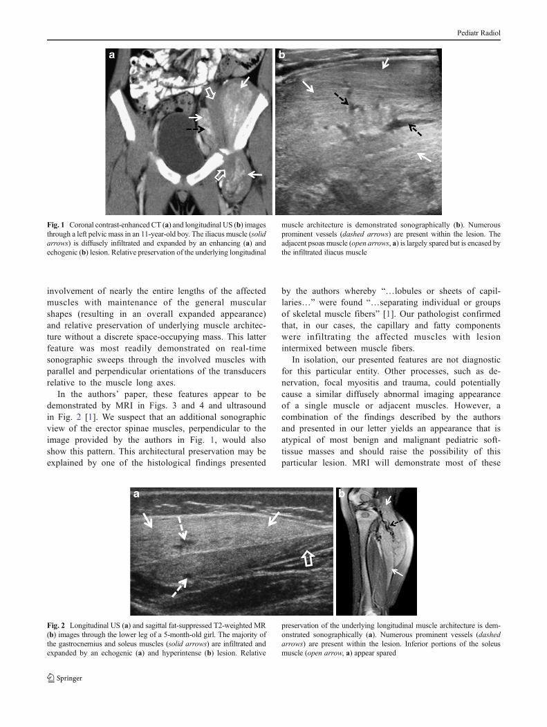
In the first case, an 11-year-old boy with a remotehistory of palatal rhabdomyosarcoma presented to anoutside hospital with a palpable left pelvic mass.Contrast-enhanced CT showed a heterogeneouslyhyperenhancing lesion infiltrating nearly the entirety ofthe left iliacus muscle (Fig. 1) and containing numerousprominent vessels; a fatty rim could be seen alongportions of the lesion. Ultrasound (Fig. 1) similarlyshowed diffuse expansion of almost the entire lengthof the muscle with relative preservation of the musclearchitecture, prominent vessels and lack of a discretespace-occupying mass.
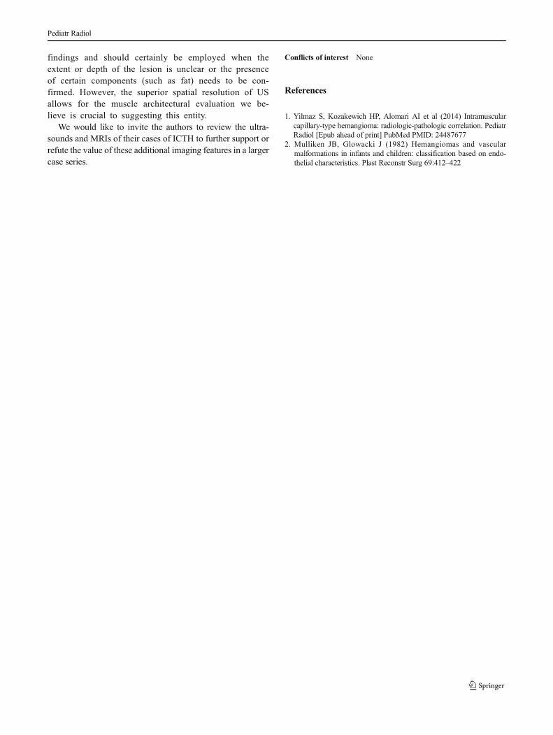
In the second case, a 5-month-old girl presented to anoutside hospital with a palpable bump in the left calf thatwas first noted at 2 weeks of life. The left leg had grownout of proportion to the right leg since that time, but thepatient had no pain or functional problems. Ultrasoundand MRI (Fig. 2) demonstrated an extensive abnormalityof the gastrocnemius and soleus muscles through nearlytheir entire lengths. Both studies implicated a moderateamount of fat throughout the lesion with diffuse muscleexpansion, relatively preserved muscle architecture andincreased vascularity. Follow-up imaging during the next2 years showed continued but proportionate growth of thelesion relative to the patient.
Most of these imaging findings, including localizationwithin a single muscle or two adjacent muscles, intra-and perilesional fatty components and increased vascu-larity, were detailed by the authors [1]. However, addi-tional striking features in our cases were the extent of
A. C. Merrow (*)Department of Radiology, Cincinnati Children’sHospital Medical Center,3333 Burnet Avenue, MLC 5031,Cincinnati, OH 45229, USAe-mail: [email protected]
A. GuptaDivision of Pathology and Laboratory Medicine,Department of Pediatrics,Cincinnati Children’s Hospital Medical Center,Cincinnati, OH, USA
D. M. AdamsHemangioma and Vascular Malformation Center,Cancer and Blood Diseases Institute,Department of Pediatrics,Cincinnati Children’s Hospital Medical Center,Cincinnati, OH, USA
Pediatr RadiolDOI 10.1007/s00247-014-2989-x
involvement of nearly the entire lengths of the affectedmuscles with maintenance of the general muscularshapes (resulting in an overall expanded appearance)and relative preservation of underlying muscle architec-ture without a discrete space-occupying mass. This latterfeature was most readily demonstrated on real-timesonographic sweeps through the involved muscles withparallel and perpendicular orientations of the transducersrelative to the muscle long axes.
In the authors’ paper, these features appear to bedemonstrated by MRI in Figs. 3 and 4 and ultrasoundin Fig. 2 [1]. We suspect that an additional sonographicview of the erector spinae muscles, perpendicular to theimage provided by the authors in Fig. 1, would alsoshow this pattern. This architectural preservation may beexplained by one of the histological findings presented
by the authors whereby “…lobules or sheets of capil-laries…” were found “…separating individual or groupsof skeletal muscle fibers” [1]. Our pathologist confirmedthat, in our cases, the capillary and fatty componentswere infiltrating the affected muscles with lesionintermixed between muscle fibers.
In isolation, our presented features are not diagnosticfor this particular entity. Other processes, such as de-nervation, focal myositis and trauma, could potentiallycause a similar diffusely abnormal imaging appearanceof a single muscle or adjacent muscles. However, acombination of the findings described by the authorsand presented in our letter yields an appearance that isatypical of most benign and malignant pediatric soft-tissue masses and should raise the possibility of thisparticular lesion. MRI will demonstrate most of these
Fig. 1 Coronal contrast-enhanced CT (a) and longitudinal US (b) imagesthrough a left pelvic mass in an 11-year-old boy. The iliacus muscle (solidarrows) is diffusely infiltrated and expanded by an enhancing (a) andechogenic (b) lesion. Relative preservation of the underlying longitudinal
muscle architecture is demonstrated sonographically (b). Numerousprominent vessels (dashed arrows) are present within the lesion. Theadjacent psoas muscle (open arrows, a) is largely spared but is encased bythe infiltrated iliacus muscle
Fig. 2 Longitudinal US (a) and sagittal fat-suppressed T2-weighted MR(b) images through the lower leg of a 5-month-old girl. The majority ofthe gastrocnemius and soleus muscles (solid arrows) are infiltrated andexpanded by an echogenic (a) and hyperintense (b) lesion. Relative
preservation of the underlying longitudinal muscle architecture is dem-onstrated sonographically (a). Numerous prominent vessels (dashedarrows) are present within the lesion. Inferior portions of the soleusmuscle (open arrow, a) appear spared
Pediatr Radiol
findings and should certainly be employed when theextent or depth of the lesion is unclear or the presenceof certain components (such as fat) needs to be con-firmed. However, the superior spatial resolution of USallows for the muscle architectural evaluation we be-lieve is crucial to suggesting this entity.
We would like to invite the authors to review the ultra-sounds and MRIs of their cases of ICTH to further support orrefute the value of these additional imaging features in a largercase series.
Conflicts of interest None
References
1. Yilmaz S, Kozakewich HP, Alomari AI et al (2014) Intramuscularcapillary-type hemangioma: radiologic-pathologic correlation. PediatrRadiol [Epub ahead of print] PubMed PMID: 24487677
2. Mulliken JB, Glowacki J (1982) Hemangiomas and vascularmalformations in infants and children: classification based on endo-thelial characteristics. Plast Reconstr Surg 69:412–422
Pediatr Radiol