Embed Size (px)
Citation preview
AngewandteChemie
Biomimetic SynthesisDOI: 10.1002/anie.200700331
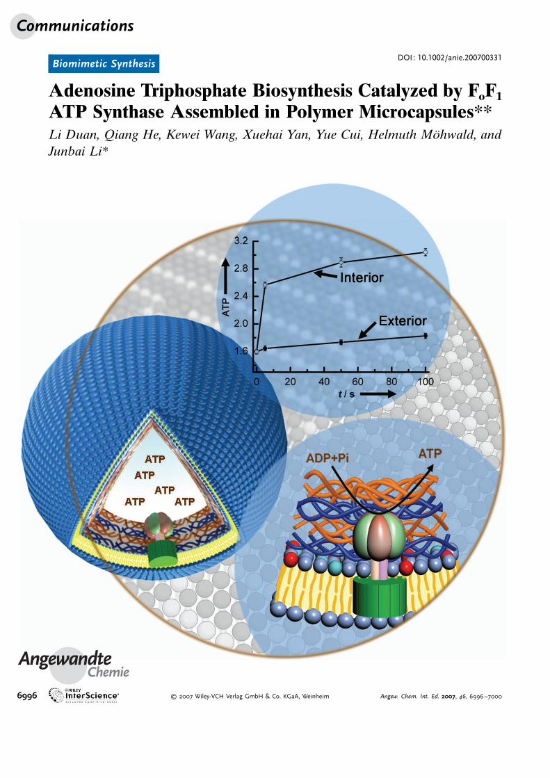
Adenosine Triphosphate Biosynthesis Catalyzed by FoF1
ATP Synthase Assembled in Polymer Microcapsules**Li Duan, Qiang He, Kewei Wang, Xuehai Yan, Yue Cui, Helmuth M�hwald, andJunbai Li*
Communications
6996 � 2007 Wiley-VCH Verlag GmbH & Co. KGaA, Weinheim Angew. Chem. Int. Ed. 2007, 46, 6996 –7000
ATP synthase (ATPase), the smallest molecular motor innature, is composed of two linked multisubunit complexes, amembrane-embedded Fo part and a hydrophilic F1 part, andcan drive the production of adenosine triphosphate (ATP) byutilizing proton gradients. The functionality of ATPase hasattracted great interest in the last decade. Numerous potentialapplications, from the generation of bioenergy to the fabri-cation of nanodevices, have been suggested.[1–3] As a mem-brane-bound protein, ATPase has been successfully recon-stituted in liposomes acting as biomimetic membranes.[4–8]
However, the limitations of the size and stability of theassembled liposome complexes result in difficulty in under-standing and analyzing the system. Herein, we report thereconstitution of ATPase in assembled lipid-coated polymermicrocapsules to imitate the system in living cells governed bymolecular motors.
Layer-by-layer assembly of oppositely charged macro-molecules onto removable colloidal particles has beenutilized to construct ultrathin hollow shells from nano- tomicrosize.[9–14] The assembled capsules have well-controlledsize, shape, and wall thickness. The wall composition can bereadily changed to adjust their physicochemical propertiesand permeability. These hollow capsules are often consideredto have potential applications in the delivery and release ofdrugs, catalysis, biomedicine, and biomaterials. In our pre-vious study,[13] we reported that lipid-coated microcapsulescould be constructed through the conversion of liposomesinto lipid bilayers to cover the capsule surface, in analogy tothe cell membrane.
In the present work, we reconstitute chloroplastic FoF1
(CFoF1) ATPase in the outer shell of assembled polymermicrocapsules containing a lipid bilayer. The concept of alipid-modified capsule with the incorporation of the CFoF1
complex is shown schematically in Figure 1. CFoF1 ATPasewas first reconstituted into liposomes based on the previouslyreported method.[4] Briefly, the liposomes were added into aTriton X-100-solubilized CFoF1-ATPase buffer solution, andthen the Triton X-100 was slowly removed with SM-2 Bio-beads. CFoF1 proteoliposomes were obtained. The micro-capsules were assembled by the alternating adsorption of
negatively charged poly(acrylic acid) (sodium salt) (PAA)and positively charged poly(allylamine hydrochloride) (PAH)onto 3.93-mm positively charged melamine formaldehyde(MF) particles as templates, followed by removal of the MFtemplates by using hydrogen chloride (HCl). Combination ofthe CFoF1 proteoliposomes and the microcapsule solutionallows the proteolipid to adsorb favorably on the outer shellof the microcapsules through the electrostatic interaction ofphosphatidic acid with PAH.[13] Lipid-modified microcapsuleswith incorporated CFoF1 ATPase were thus obtained. Itshould be noted that only in the case of F1 subunits of theCFoF1 complex extending toward the interior of the assem-bled capsules ATP synthesis inside the capsules could becomepossible.
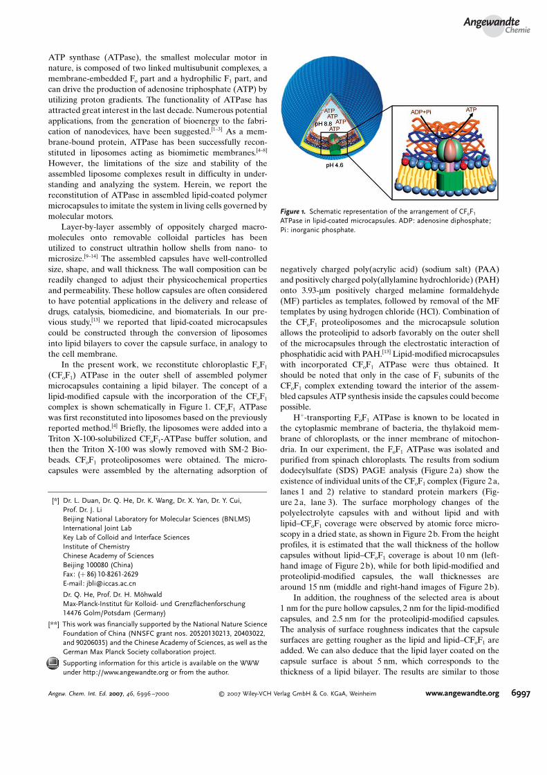
H+-transporting FoF1 ATPase is known to be located inthe cytoplasmic membrane of bacteria, the thylakoid mem-brane of chloroplasts, or the inner membrane of mitochon-dria. In our experiment, the FoF1 ATPase was isolated andpurified from spinach chloroplasts. The results from sodiumdodecylsulfate (SDS) PAGE analysis (Figure 2a) show theexistence of individual units of the CFoF1 complex (Figure 2a,lanes 1 and 2) relative to standard protein markers (Fig-ure 2a, lane 3). The surface morphology changes of thepolyelectrolyte capsules with and without lipid and withlipid–CFoF1 coverage were observed by atomic force micro-scopy in a dried state, as shown in Figure 2b. From the heightprofiles, it is estimated that the wall thickness of the hollowcapsules without lipid–CFoF1 coverage is about 10 nm (left-hand image of Figure 2b), while for both lipid-modified andproteolipid-modified capsules, the wall thicknesses arearound 15 nm (middle and right-hand images of Figure 2b).
In addition, the roughness of the selected area is about1 nm for the pure hollow capsules, 2 nm for the lipid-modifiedcapsules, and 2.5 nm for the proteolipid-modified capsules.The analysis of surface roughness indicates that the capsulesurfaces are getting rougher as the lipid and lipid–CFoF1 areadded. We can also deduce that the lipid layer coated on thecapsule surface is about 5 nm, which corresponds to thethickness of a lipid bilayer. The results are similar to those
Figure 1. Schematic representation of the arrangement of CFoF1
ATPase in lipid-coated microcapsules. ADP: adenosine diphosphate;Pi: inorganic phosphate.
[*] Dr. L. Duan, Dr. Q. He, Dr. K. Wang, Dr. X. Yan, Dr. Y. Cui,Prof. Dr. J. LiBeijing National Laboratory for Molecular Sciences (BNLMS)International Joint LabKey Lab of Colloid and Interface SciencesInstitute of ChemistryChinese Academy of SciencesBeijing 100080 (China)Fax: (+86)10-8261-2629E-mail: [email protected]
Dr. Q. He, Prof. Dr. H. M?hwaldMax-Planck-Institut fBr Kolloid- und GrenzflEchenforschung14476 Golm/Potsdam (Germany)
[**] This work was financially supported by the National Nature ScienceFoundation of China (NNSFC grant nos. 20520130213, 20403022,and 90206035) and the Chinese Academy of Sciences, as well as theGerman Max Planck Society collaboration project.
Supporting information for this article is available on the WWWunder http://www.angewandte.org or from the author.
AngewandteChemie
6997Angew. Chem. Int. Ed. 2007, 46, 6996 –7000 � 2007 Wiley-VCH Verlag GmbH & Co. KGaA, Weinheim www.angewandte.org
obtained by single-particle light-scattering measurements inour previously published work[12] and by freeze–fractureelectron microscopy measurements.[15]
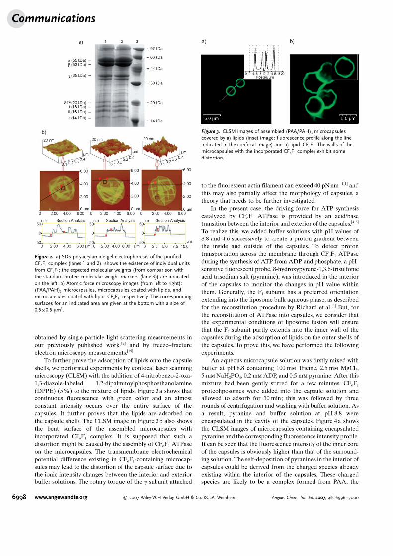
To further prove the adsorption of lipids onto the capsuleshells, we performed experiments by confocal laser scanningmicroscopy (CLSM) with the addition of 4-nitrobenzo-2-oxa-1,3-diazole-labeled 1,2-dipalmitoylphosphoethanolamine(DPPE) (5%) to the mixture of lipids. Figure 3a shows thatcontinuous fluorescence with green color and an almostconstant intensity occurs over the entire surface of thecapsules. It further proves that the lipids are adsorbed onthe capsule shells. The CLSM image in Figure 3b also showsthe bent surface of the assembled microcapsules withincorporated CFoF1 complex. It is supposed that such adistortion might be caused by the assembly of CFoF1 ATPaseon the microcapsules. The transmembrane electrochemicalpotential difference existing in CFoF1-containing microcap-sules may lead to the distortion of the capsule surface due tothe ionic intensity changes between the interior and exteriorbuffer solutions. The rotary torque of the g subunit attached
to the fluorescent actin filament can exceed 40 pNnm�1[1] andthis may also partially affect the morphology of capsules, atheory that needs to be further investigated.
In the present case, the driving force for ATP synthesiscatalyzed by CFoF1 ATPase is provided by an acid/basetransition between the interior and exterior of the capsules.[4,6]
To realize this, we added buffer solutions with pH values of8.8 and 4.6 successively to create a proton gradient betweenthe inside and outside of the capsules. To detect protontransportation across the membrane through CFoF1 ATPaseduring the synthesis of ATP from ADP and phosphate, a pH-sensitive fluorescent probe, 8-hydroxypyrene-1,3,6-trisulfonicacid trisodium salt (pyranine), was introduced in the interiorof the capsules to monitor the changes in pH value withinthem. Generally, the F1 subunit has a preferred orientationextending into the liposome bulk aqueous phase, as describedfor the reconstitution procedure by Richard et al.[4] But, forthe reconstitution of ATPase into capsules, we consider thatthe experimental conditions of liposome fusion will ensurethat the F1 subunit partly extends into the inner wall of thecapsules during the adsorption of lipids on the outer shells ofthe capsules. To prove this, we have performed the followingexperiments.
An aqueous microcapsule solution was firstly mixed withbuffer at pH 8.8 containing 100 mm Tricine, 2.5 mm MgCl2,5 mm NaH2PO4, 0.2 mm ADP, and 0.5 mm pyranine. After thismixture had been gently stirred for a few minutes, CFoF1
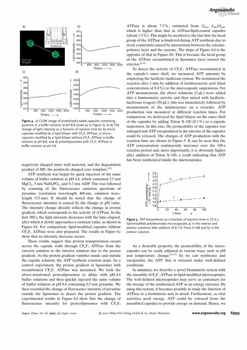
proteoliposomes were added into the capsule solution andallowed to adsorb for 30 min; this was followed by threerounds of centrifugation and washing with buffer solution. Asa result, pyranine and buffer solution at pH 8.8 wereencapsulated in the cavity of the capsules. Figure 4a showsthe CLSM images of microcapsules containing encapsulatedpyranine and the corresponding fluorescence intensity profile.It can be seen that the fluorescence intensity of the inner coreof the capsules is obviously higher than that of the surround-ing solution. The self-deposition of pyranines in the interior ofcapsules could be derived from the charged species alreadyexisting within the interior of the capsules. These chargedspecies are likely to be a complex formed from PAA, the
Figure 2. a) SDS polyacrylamide gel electrophoresis of the purifiedCFoF1 complex (lanes 1 and 2). shows the existence of individual unitsfrom CFoF1; the expected molecular weights (from comparison withthe standard protein molecular-weight markers (lane 3)) are indicatedon the left. b) Atomic force microscopy images (from left to right):(PAA/PAH)3 microcapsules, microcapsules coated with lipids, andmicrocapsules coated with lipid–CFoF1, respectively. The correspondingsurfaces for an indicated area are given at the bottom with a size of0.5 J 0.5 mm2.
Figure 3. CLSM images of assembled (PAA/PAH)3 microcapsulescovered by a) lipids (inset image: fluorescence profile along the lineindicated in the confocal image) and b) lipid–CFoF1. The walls of themicrocapsules with the incorporated CFoF1 complex exhibit somedistortion.
Communications
6998 www.angewandte.org � 2007 Wiley-VCH Verlag GmbH & Co. KGaA, Weinheim Angew. Chem. Int. Ed. 2007, 46, 6996 –7000
negatively charged inner wall material, and the degradationproduct of MF, the positively charged core template.[14]
ATP synthesis was begun by quick injection of the samevolume of buffer solution at pH 4.6, which contained 2.5 mm
MgCl2, 5 mm NaH2PO4, and 0.2 mm ADP. This was followedby scanning of the fluorescence emission spectrum ofpyranine (excitation wavelength 460 nm, emission wave-length 513 nm). It should be noted that the change offluorescence intensity is caused by the change in pH value.The intensity change directly reflects the change in protongradient, which corresponds to the activity of ATPase. In thefirst 300 s, the light intensity decreases with the time elapsed,after which it slowly approaches a constant value, as shown inFigure 4b. For comparison, lipid-modified capsules withoutCFoF1 ATPase were also prepared. The results in Figure 4cshow that no intensity decrease occurs.
These results suggest that proton transportation occursacross the capsule walls through CFoF1 ATPase from theexterior solution to the interior solution due to the protongradient. As the proton gradient vanishes inside and outsidethe capsule solution, the ATP synthesis reaction stops. As acontrol experiment, the proton gradient in liposomes withreconstituted CFoF1 ATPase was measured. We took theabove-mentioned proteoliposomes to dilute with pH 4.6buffer solutions and then quickly injected the same volumeof buffer solution at pH 8.8 containing 0.5 mm pyranine. Wethen recorded the change of florescence intensity of pyranineoutside the liposomes to detect the proton gradient. Theexperimental results in Figure 4d show that the change offluorescence intensity for proteoliposomes with CFoF1
ATPase is about 7.5%, estimated from (Imax�Imin)/Imin,which is higher than that in ATPase/lipid-coated capsules(about 1.5%). This might be ascribed to the fact that the headgroup of the ATPase is hindered during ATP synthesis due tosteric constraints caused by interactions between the cationic-polymer layer and the enzyme. The slope of Figure 4d is theopposite of that in Figure 4b. This is because the head groupof the ATPase reconstituted in liposomes faces toward theexterior.[4–8]
To detect the activity of CFoF1 ATPase reconstituted inthe capsuleCs outer shell, we measured ATP amounts byemploying the luciferin–luciferase system. We terminated thereaction after 1 min by addition of trichloroacetic acid (finalconcentration of 0.4%) to the microcapsule suspensions. ForATP measurement, the above solutions (5 mL) were addedinto a luminometer cuvette and then mixed with luciferin–luciferase reagent (50 mL); this was immediately followed bymeasurement of the luminescence on a recorder. ATPproduction was measured at different reaction times. Forcomparison, we destroyed the lipid bilayer on the outer shellof the capsules by adding Triton X-100 (0.1%) to a capsulesuspension. In this case, the permeability of the capsules wasenlarged and ATP encapsulated in the interior of the capsulescould be released. The changes of ATP production with thereaction time are shown in Figure 5. It can be seen that theATP concentration continuously increases over the 100-sreaction period and, more importantly, it is obviously higherafter addition of Triton X-100, a result indicating that ATPhas been synthesized inside the microcapsules.
As a desirable property, the permeability of the micro-capsules can be easily adjusted in various ways, such as pHand temperature change.[16,17] So we can synthesize andencapsulate the ATP that is released under well-definedconditions.
In summary, we describe a novel biomimetic system withthe assembly of FoF1 ATPase in lipid-modified microcapsules.The well-defined microcapsules may serve as containers forthe storage of the synthesized ATP as an energy currency. Byusing this system, it becomes possible to study the function ofATPase in a biomimetic unit in detail. Furthermore, as vitalactivities need energy, ATP could be released from theassembled capsules to provide energy on demand. Hence, we
Figure 4. a) CLSM image of proteolipid-coated capsules containingpyranine in a buffer solution at pH 8.8 (inset as in Figure 3). b–d) Thechange of light intensity as a function of reaction time for b) micro-capsules modified by a lipid bilayer with CFoF1 ATPase, c) micro-capsules modified by a lipid bilayer without CFoF1 ATPase in buffersolution at pH 8.8, and d) proteoliposomes with CFoF1 ATPase inbuffer solution at pH 4.6.
Figure 5. ATP biosynthesis as a function of reaction time in CFoF1/lipid-modified polyelectrolyte microcapsules a) in the interior andexterior solutions after addition of 0.1% Triton X-100 and b) in theexterior solution.
AngewandteChemie
6999Angew. Chem. Int. Ed. 2007, 46, 6996 –7000 � 2007 Wiley-VCH Verlag GmbH & Co. KGaA, Weinheim www.angewandte.org
have built a micrometer-sized energy-storage device suitableto power biological activity.
Received: January 24, 2007Revised: May 22, 2007Published online: July 6, 2007
.Keywords: lipids · microcapsules · molecular motors ·nucleotides · synthases
[1] H. Noji, R. Yasuda, M. Yoshida, K. Kinosita, Jr., Nature 1997,386, 299 – 302.
[2] R. K. Soong, G. D. Bachand, H. P. Neves, A. G. Olkhovets, H. G.Craighead, C. D. Montemagno, Science 2000, 290, 1555 – 1558.
[3] M. Yoshida, E. Muneyuki, T. Hisabori, Nat. Rev. Mol. Cell Biol.2001, 2, 669 – 677.
[4] P. Richard, J. L. Rigaud, P. GrHber, Eur. J. Biochem. 1990, 193,921 – 925.
[5] G. Steinberg-Yfrach, E. N. Durantini, A. L. Moore, D. Gust,T. A. Moore, Nature 1998, 392, 479 – 482.
[6] P. Turina, D. Smaoray, P. GrHber, EMBO J. 2003, 22, 418 – 426.[7] M. Diez, B. Zimmermann, M. BJrsch, M. KJnig, E. Schwein-
berger, S. Steigmiller, R. Reuter, S. Felekyan, V. Kudryavtsev,C. A. M. Seidel, P. GrHber, Nat. Struct. Mol. Biol. 2004, 11, 135 –141.
[8] T.-J. M. Luo, R. Soong, E. Lan, B. Dunn, C. Montemagno, Nat.Mater. 2005, 4, 220 – 224.
[9] E. Donath, G. B. Sukhorukov, F. Caruso, S. A. Davis, H.MJhwald, Angew. Chem. 1998, 110, 2323 – 2327; Angew. Chem.Int. Ed. 1998, 37, 2201 – 2205.
[10] a) D. T. Haynei, N. Palath, Y. Liu, B. Y. Li, Langmuir 2005, 21,1136 – 1138; b) G. Decher, Science 1997, 277, 1232 – 1237;c) Z. H. An, G. Lu, H. MJhwald, J. B. Li, Chem. Eur. J. 2004,10, 5848 – 5852.
[11] S. P. Zheng, C. Tao, Q. He, H. F. Zhu, J. B. Li,Chem.Mater. 2004,16, 3677 – 3681.
[12] Z. H. An, C. Tao, G. Lu, H. MJhwald, S. P. Zheng, Y. Cui, J. B.Li, Chem. Mater. 2005, 17, 2514 – 2519.
[13] a) L. Q. Ge, H. MJhwald, J. B. Li, Biochem. Biophys. Res.Commun. 2003, 303, 653 – 659; b) L. Q. Ge, H.MJhwald, J. B. Li,Chem. Eur. J. 2003, 9, 2589 – 2594; c) L. Q. Ge, H. MJhwald, J. B.Li, ChemPhysChem 2003, 4, 1351 – 1355.
[14] C. Y. Gao, H. MJhwald, J. C. Shen, Adv. Mater. 2003, 15, 930 –933.
[15] S. Moya, W. Richter, S. Leporatti, H. BHumler, E. Donath,Biomacromolecules 2003, 4, 808 – 814.
[16] Z. H. An, H. MJhwald, J. B. Li, Biomacromolecules 2006, 7,580 – 585.
[17] J. A. Jaber, J. B. Schlenoff, Biomacromolecules 2005, 5, 1089 –1096.
Communications
7000 www.angewandte.org � 2007 Wiley-VCH Verlag GmbH & Co. KGaA, Weinheim Angew. Chem. Int. Ed. 2007, 46, 6996 –7000