Embed Size (px)
Citation preview
V O L . 7, N O . 12 , D E C E M B E R 1 9 6 8
Studies on Adenosine Triphosphate Transphosphorylases. VII. Isolation of the Crystalline Adenosine Triphosphate-Creatine Transphosphorylase from Calf Brain*
Hans J. Keutel, Hans K. Jacobs, Keiichiro Okabe, Robert H. Yue, and Stephen A. Kuby
ABSTRACT : A procedure is described for the isolation of crystalline adenosine triphosphate-creatine transphos- phorylase in good yield, from calf brain extracts. Some observations on the stability properties of the enzyme are presented with respect to pH, temperature, and re- ducing agents, to provide a basis for a systematic physi- cal-zhemical and kinetic characterization,
With the isolation of this enzyme from calf brain, a comparison is now possible with its crystalline isoenzyme
A denosine triphosphate-creatine transphospho- rylase is presumed to play an important role in the metabolism of cerebral tissues (e.g., McIlwain, 1966; Nachmansohn and Wilson, 1951) in a manner analogous to its now accepted physiological and catalytic role in muscle tissues (e.g., Davies el ai., 1967; Mommaerts and Wallner, 1967). Thus, it is presumed that it provides a means for the resynthesis of ATP cia creatine phosphate through ADP, and it should not be forgotten that this concept had its origins in the pioneer work of Meyerhof and associates (e.g., Meyerhof and Lohmann, 1932).
Relatively few enzymes have been isolated and ob- tained in crystalline form from extracts of mammalian cerebral tissues, where the recognized problems of phos- pholipids often present obstacles to the isolation of pro- teins in reasonable yield and purity and also in a repro- ducible manner. (But a preliminary report of one case has recently appeared, eiz., that of the crystalline triose phosphate dehydrogenase from rabbit brain ; Lebherz and Rutter, 1967.) Efforts to purify the ATP-creatine transphosphorylase from calf brain extracts were re- ported early (Kuby and Mahowald, 1958), and the ini- tial steps in the purification scheme to be reported here were retained from the earlier procedure. More re- cently, other attempts to isolate this enzyme from cere- bral tissues have been reported and a highly purified preparation was obtained, e.g., from ox brain (Wood, 1963a,b); and Eppenberger et ai. (1967) have attempted to isolate a brain-type enzyme from chicken heart, which they have stated to be identical with the chicken brain enzyme itself.
* From the Laboratory for the Study of Hereditary and Meta- bolic Disorders and the Departments of Biological Chemistry and Medicine, University of Utah, Salt Lake City, Utah. Re- ceiaed June 20, 1968. This work was supported in part by grants from the National Science Foundation and from the National Institutes of Health. Yue e t al. (1967) is no. VI of this series.
counterpart from calf muscle (Jacobs, H. K. Keutel, H. J., Yue, R. H., Okabe, K., and Kuby, S. A (1968), Federation Proc. 27,640) as well as with crystalline enzymes that were isolated from the brain and skeletal muscle of man (Jacobs, H. K., Keutel, H. J., Yue, R. H., Okabe, K., and Kuby, S. A. (1968), Federation Proc. 27, 640) and of rabbit (Kuby, S. A., Noda, L., and Lardy, H. A. (1954), J . Bid . Chem. 209, 291).
Since studies from this laboratory have dealt with the general problem of ATP transphosphorylation, and, in particular, with three types of ATP transphosphorylases which had been isolated in crystalline form (the ATP- creatine transphosphorylase from rabbit muscle (Kuby et ai., 1954), the ATP-AMP transphosphorylase from rabbit muscle (Noda and Kuby, 1957), and the nucleo- side diphosphokinase from breweis’ yeast (Ratliff et af., 1964)), and since the rabbit muscle ATP-creatine transphosphorylase has been the subject of extensive investigation (e.g., see Kuby and Noltmann, 1962), it was of interest to broaden the physicochemical com- parison to include the isoenzymes of the ATP- creatine transphosphorylase from several mammalian species. It was hoped, thereby, to reveal from such com- parative studies those similarities and differences which may be ultimately correlated with, and applied to a more intimate understanding of its mechanism(s) of catalysis. Futllre reports will deal with studies on the crystalline ATP-creatine transphosphorylases isolated from the muscle and brain of the calf, human, and rab- bit. The enzymes isolated from human tissues will likely be of interest to those concerned with the prsblem of progressive muscular dystrophy where the appearance of the enzymatic activity in the serum seems to reflect the disorder (Tyler, 1966).
This report will deal specifically with the isolation of the crystalline calf brain enzyme and a brief description of its stability properties. Reports will follow which will deal (a) with a homogeneity analysis by several tech- niques; with its molecular weight and subunit struc- ture, as deduced physically (Yue et al., 1968); (b) with an analysis of the reactive SH groups of the native mole- cule and the total SH content, asalso related to the sta- bility and to the molecular structure; and finally, (c) with a kinetic analysis of its catalyzed reaction which may have a bearing on its native molecular structure 4283
C A L F B R A I N A T P - C R E A T I N E T R A N S P H 0 S P H 0 R Y L A S E. C R Y S T A L L I Z A T IO N
B I O C H E M I S T R Y
and mechanism of action. Additional chemical studies on the protein in particular on its amino acid composi- tion and terminal groups, will also be the subject of a later communication. A preliminary report of some of these investigations has been given (Jacobs et al., 1968).
Experimental Procedure
Materials and Methods. Calf brains were removed from the animals immediately after slaughter and transported in ice to the laboratory. The hemispheres were excised, dura and external blood vessels were re- moved, washed (ice water) free of external blood clots, sealed in cellophane bags, and frozen at -15". The frozen tissue (hemispheres), which was stored for pe- riods of 6-50 days at - 15 ", was the source for isolation of the enzyme.
(NH&SOa was obtained from Mann (Ultra Pure) or from Mallinckrodt (A. R., recrystallized from aqueous ethanol), disodium EDTA from Fisher Scientific Co. ; Tris, and the sodium salts of ATP, of creatine phos- phate, and of ADP from Sigma. 0-Mercaptoethanol (Eastman Chemical) was redistilled under vacuum; 95% ethanol was redistilled as described (Ratliff et a/., 1964). The following chemicals were obtained from the sources indicated: creatine (Eastman Chemicals' White Label), glycine (Matheson Coleman and Bell), crystal- line bovine plasma albumin (Armour), and succinic acid (Mallinckrodt, A. R.). All other reagents were of ana- lytical grade. Twice-distilled deionized water was used for preparation of all reagents including the dialysis fluids; in some cases, solutions were prepared in de- gassed (by boiling), distilled water or saturated with nitrogen. Lots of DEAE-cellulose (Bio-Rad Cellex-D anion exchanger, capacity ca. 0.66 mequiv/g) were freed of fines by decantation, cycled through 0.2 N NaOH- HzO-0.2 N HC1-HzO-0.2 N NaOH-H20 as described (Ratliff et al., 1964), and stored as a moist filter cake (averaging 30% dry weight) in sealed containers. Phos- phocellulose (Bio-Rad Cellex-P cation exchanger, ca- pacity 0.88-1.06 mequiv/g) was freed of fines, con- verted into the sodium phase with a 20-fold excess of 0.5 M NaCl in 0.5 M NaOH, washed until neutrality, and transferred into the Hf phase by titration (ice bath) with 1.0 N HCI to pH 2.5; after washing, by filtration, with 0.01 N HCI (in the cold) followed by water until free of C1-, it was also stored as a moist filter cake, whose dry weight was determined. Sephadex G-75 and G-100 (bead form) were products of Pharmacia and were swelled at 3" forseveral days in a solution of 30% saturated (NH4)2S04 (1.17 M) containing 0.01 M EDTA (pH 7.4) prior to preparation of the column.
Measurements of Enzymatic Activity. To permit a comparison of the specific activity of this preparation from calf brain with that of the rabbit muscle, the origi- nal colorimetric procedure for creatine phosphate and reaction mixture conditions of Kuby et al. (1954) were employed during the isolation procedure with only slight modifications in the dilutions of the enzyme. Dilutions, at ice-bath temperature, were made in 1 X 10- M glycine (pH 9.0) ( 5 " ) , in 1 X 10- M cysteine-
M EDTA (pH 7.0) ( 5 " ) , or in 0.1 M P-mer- 4284 1 X
captoethanol-1 X M EDTA (pH 7.4) at the ter- minal stages of isolation. Microaliquots ( 5 or 10 1.11) of properly diluted enzyme were pipetted directly into 10 ml of reaction mixture to minimize inactivation at high dilutions, and bovine serum albumin (1 mg/ml) was added to the glycine or cysteine solutions, if dilutions were greater than 1 X lo6. The definition of a unit of activity under these conditions was as described origi- nally (Kuby et al., 1954) in terms of an apparent second- order velocity constant, k' (1 unit = k' = 1 ml pmole-' min-1).
In addition, the titrimetric pH-Stat method pre- viously described (Mahowald et a/., 1962) was utilized to follow the purification, but with a reaction mixture which consisted of: 0.04 M creatine, 0.004 M ATP, 0.004 M MgS04, and 1 mg/ml of albumin, 30°, pH 8.8; with 0.010 N NaOH as titrant (see also Specifications and Criteria, 1967), and with an 8.0-ml reaction volume under a nitrogen barrier. The definition of a unit of activity for this case was in terms of 1 unit equal to 1 ,uequiv/min, as measured from the initial velocity. To interconvert from one set of conditions to the other, it was found that the pH-Stat-defined unit (1 pequiv/min) was approximately 1.8-2 times that determined under colorimetric conditions (and defined with a k' = 1 ml pmole-1 min-1).
Protein was determined by the colorimetric biuret procedure of Gornall et al. (1949) or in later stages by its extinction coefficient at 280 mp. Appropriate biuret factors and extinction coefficients for some fractions are presented in the text and these values for the isolated enzyme will be presented (Yue et a/., 1968). Specific activity is expressed in terms of units per milligram of protein.
Isolation Procedure (from Calf' Brain). All steps were carried out in a cold room (2-4") or in an ice bath, un- less otherwise specified (e.g., -10" for the solvent steps); pH measurements were made at or near 5 O with a Radiometer TTTla meter, equipped with scale-expander PHA 630Ta, and A. H. Thomas glass-calomel elec- trodes. The required amounts of solvents and (NH& so4, as well as the concentrations of these reagents in the precipitated pellets, were estimated by the formulas summarized by Noltmanm et a/. (1961), and a saturated solution of (NH&S04 was taken to be 3.9 M at 0".
FRACTION I. The frozen brain tissue (ca. 6 kg) is thawed overnight (at 2-5"), passed through a meat grinder, and collected in a tared polyethylene tray. The tissue is then blendorized for 30 sec (gallon-size Waring Blendor) in chilled 0.1 M ammonium acetate (pH 9 3 , with use of 2.0 l./kg of ground tissue, and the homoge- nate was stirred slowly in the cold room for 24 hr.
FRACTION 11. The suspension without centrifugation is transferred to a -10" bath and chilled to 0", with stirring. A volume of 95 x ethanol (- lo") equal to the total homogenate volume is added slowly with good mechanical stirring (the temperature should not rise above 3 "). After the ethanol addition is completed and now presumed to be at 50% (v/v) of 9 5 x ETOH, the temperature of the mixture is allowed to adjust, with stirring, to -8 to -10". The mixture is centrifuged at 13OOg for 45 min at -lo", the supernatant liquid in-
K E U T E L , J A C O B S , O K A B E , Y U E , A N D K U B Y
cluding loosely packed material is decanted, and its volume is determined. The solution (-10') is ad- justed to a final concentration of 0.015 M MgS04 by the slow addition, with stirring, of a calculated volume of 2.0 M MgS04 (pH 8.7) (measured at 2 5 9 , and the ethanol concentration is increased to 70% (v/v of 95 %). The suspension is centrifuged at 1300g for 45 min, at lo", and the tightly packed reddish pellet is suspended rapidly at 0" in a volume of 0.001 M NH40H equal to ' / z o of the calculated supernatant liquid volume of frac- tion I1 and dialyzed with continuous flow (Noltmann et af., 1961) against 50 1. of 0.001 M ",OH overnight. After dialysis, the insoluble material is removed by cen- trifugation at 15,OOOg for 60 min. The protein concen- tration of the supernatant liquid (fraction 11) is deter- mined (after precipitation with 10 trichloroacetic acid, followed by the biuret procedure (Gornall et ai., 1949) with a factor of 32.5 mg/lO-ml volume per unit of absorbance) and the protein concentration is adjusted, if possible, to 8.0 mg/ml with the dialysis fluid; other- wise, to 6 mg/ml.
FRACTION 111. For an initial protein concentration of 8 mg/ml, fractionation between 0.43 and 0.64 (NH& SO4 saturation is employed; but for the case of 6 mg/ml, 0.43-0.67 saturation is used. In an ice bath with stirring, solid (NH4),S04 is slowly added and the pH is simultaneously maintained at 7.0 by the addition of either 1 M ",OH or 1 N H2S04. After the (NH4),S04 concentration has been adjusted to 0.43 saturation, it is allowed to stir slowly for 2 hr, followed by centrifuga- tion at 15,OOOg for 45 min. The ("&SO4 concentra- tion of the supernatant liquid is increased to 0.64 satura- tion (or 0.67) by the addition of solid ("&SO4, at pH 7.0; and after a 2-hr stirring period, the precipitated protein is centrifuged and dissolved in a volume of 0.001 M EDTA (pH 8.0) equal to ' / i o the volume of fraction 11. It is then dialyzed by continuous flow against 50 I . of 0.001 M EDTA (pH 8.0) overnight at which time the dialysis fluid should be free of SO4*-. The protein solu- tion is clarified by centrifugation and is designated frac- tion 111, whose protein concentration and volume are determined.
FRACTION I V . During the dialysis period, the phos- phocellulose is equilibrated as follows. A calculated amount of the moist cake is suspended in 0.001 M EDTA (Na+) (pH 6.8) such that the final concentration (in dry weight) is 30 mg/ml and the total amount will be equal to at least 30 times the protein concentration of fraction 111. The suspension is titrated with stirring to pH 6.8 (with 1 N NaOH) at 5 " , transferred quantitatively to a sintered-glass funnel, washed with 0.001 M EDTA equal to four times the volume initially employed, re- suspended in 0.001 M EDTA, and the pH is readjusted, if necessary. The following day the procedure is re- peated until the pH is stable at 6.8, and a calculated volume of the phosphocellulose containing 30 times the protein weight of fraction 111, is then deaerated by suction, transferred to a 5.0 X 50 cm column, packed
'Determined by centrifuging an aliquot at 100,OOOg for 6 miti.
under a few pounds of NP pressure and finally with a peristaltic pump (Harvard Model 600-1200) at a flow rate of ca. 100 ml/hr, to yield a bed height of ca. 30 cm when tightly packed. Fraction I11 is adjusted to pH 6.8 (with 0.1 N NaOH or 0.01 N HCl) and applied to the phosphocellulose column under Nz pressure (ca. 5-6 Ibs). The single unretarded peak (containing the enzy- matic activity), which is monitored at 280 mp (Van- guard 1056 or GME, and a Texas instrument recorder), is displaced off the column at ca. 100 d / h r with the same solvent (0.001 M EDTA, pH 6.8). A large reddish- brown band (hemoglobin and derivatives) is retained by the column. The protein peak is collected, adjusted to 0.85 saturation (NH4)*S04, at pH 7.0, and after a 2-hr period (with slow stirring) the precipitate is col- lected by centrifugation at 15,OOOg for 75 min. The pel- let is resuspended (in one-fifth volume of fraction 111) in a solution of 0.005 M succinate (0.0115 M Tris)-0.001 M EDTA-0.001 M fl-mercaptoethanol (pH 7.5) and dialyzed by continuous flow us. 50 1. of the same solu- tion overnight. After the S042- concentration of the di- alysis fluid has been reduced to a negligible concentra- tion, the protein solution is clarified by centrifugation (25,OOOg, 60 min) and the volume of fraction IV and its protein concentration is determined.
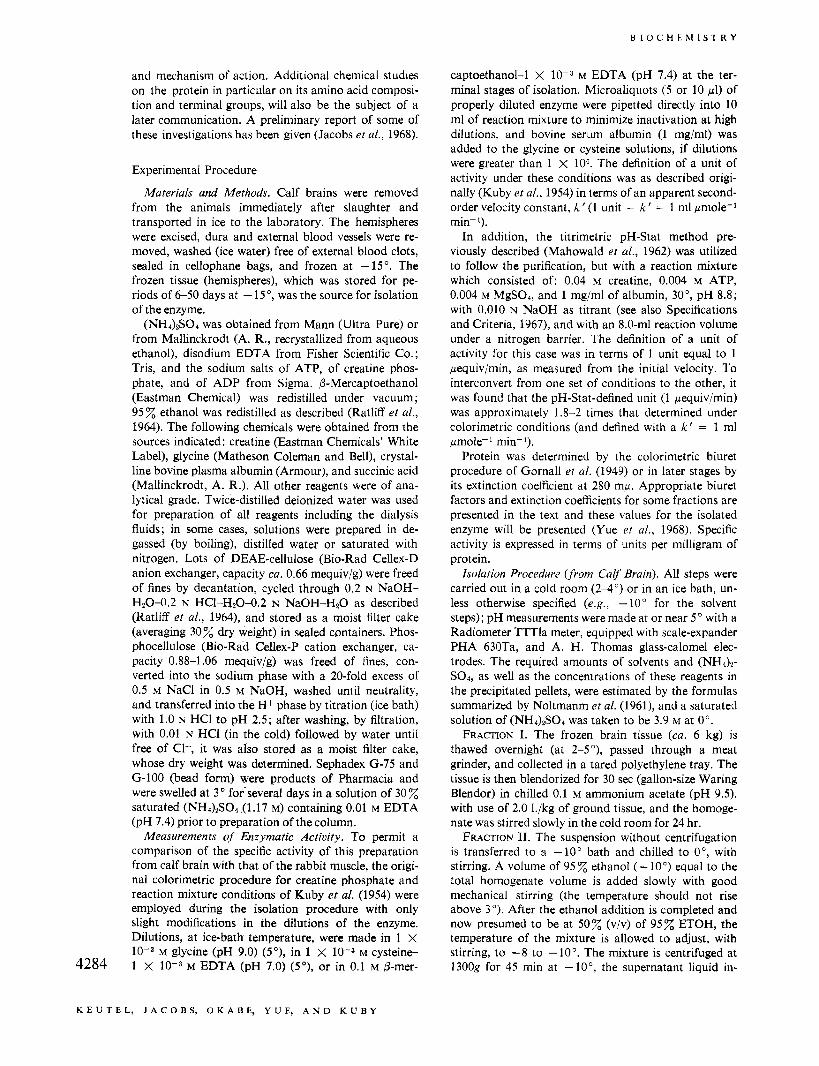
FRACTION V. An amount of DEAE-cellulose equal to at least 20 times the total protein of fraction 111 is suspended in 0.005 M succinate (0.0114 M Tris)-0.001 M EDTA (pH 7.5) at a concentration of ca. 30 mg (dry weight)/nil; its pH is readjusted and allowed to equili- brate overnight. It is then washed (by filtration) with four times the original volume of buffer originally used, resuspended in the same buffer, and an amount equal to 25-35 times the protein of fraction IV is transferred to a 2 x 150 cm column. It is now further equilibrated by pumping two bed volumes (at ca. 100 ml/hr) of the same buffer which now contains 0.001 M P-mercapto- ethanol. Fraction IV is applied to the column and after passage of one bed volume (at 1.7 ml/min), a linear gradient with respect to succinate ion is initiated by the use of two cylinders, each containing 1.5 1. of buffer: the mixing chamber (8.9-cm i.d. X 48.4 cm) containing 0.005 M succinate (0.01 15 M Tris)-0.001 M EDTA-0.001 M P-mercaptoethanol (pH 7.5) and the reservoir cham- ber, 0.09 M succinate (0.186 M Tris)-0.001 M EDTA- 0.001 M P-mercaptoethanol (pH 7.5). The chromato- gram is monitored at 280 mp and analyzed for enzy- matic activity (see Figure 1). A complex chromatogram is evident, and the enzyme appears beneath a skewed protein peak between conductance values of ca. 1.9 and 3.8 mmhos (as measured, e.g., with a Radiometer con- ductivity meter DCM-2d) but with no evidence at this point of multiple enzymatic species. The fractions con- taining enzymatic activity are pooled and its final pro- tein concentration is estimated at 280 mp, assuming that 1 mg/ml has an ODnso of 0.805 at this point. The protein is concentrated as follows, by adsorption on and elution from DEAE-cellulose. The succinate concentration of the protein solution is diluted to a final concentration of 0.01 5 M succinate (checked conductometrically) by the addition of 0.001 M EDTA (pH 7.5). An aliquot of DEAE suspension, equilibrated as above and equivalent 4285
( ' A L P B R A I N A T P - C ' K E A T I N L : T K A N S P H O S P H O R Y L A S E . C R Y S T A L L I Z A T I O N
B I O C H E M I S T R Y
r 3 0 0
-225
FIGURE 1: Chromatography of fraction IV (see text) of ATP-creatine transphos- phorylase from calf brain on DEAE- cellulose 2.0 X 150 cm, 1.7 ml/min. The
- - developing systems and the use of a linear gradient in succinate ion concen- tration are described in the text and
~ - 75 ; pertinent information is presented in the - 4 & 5 above figure. (- - - -) Optical density - 2 p Z recorded at 280 mp; (0-0) enzy-
' . ...._._.., 8 4
I60 lb matic activity in units per milliliter (1 unit = k' = 1; see text); (M) con- ductance in millimhos. GRADIENT STOPPED
C l 5 M S U C C I N A T E I T R I S l p H 7 5
t t 4o 6 O
0005M SUCCINATE GRADIENT STARTED I T R I S l p H 7 5 TO 009M S U C C l N b T E l T R l S l p H 7 5
to 40 times (dry weight) of the protein, is filtered on a coarse, sintered-glass filter; and the filter pad is trans- ferred to the protein solution which is then suspended by stirring, with the pH maintained at 7.5. After 30 min, the DEAE-cellulose is transferred quantitatively to the same funnel by the addition of a small volume of 0.005 M succinate-0.001 M EDTA-O.OO1 M mercaptoethanol (pH 7.5); and the enzyme is eluted batchwise by the addition of 150 ml of 0.2 M succinate (-0.425 M Tris)- 0.001 M EDTA-0.001 M mercaptoethanol (pH 7.5) divided into four volumes. To the combined filtrates, solid (NHJzSO4 is added to 0.90 saturation at pH 7.5; after at least 2 hr, with slow stirring, the precipitate is collected by centrifugation (25,OOOg) and the pellet is dissolved in 0.001 M EDTA (Na+)-0.01 M mercapto- ethanol (pH 7.5) to yield a final protein concentration of ca. 25 mg/ml.
FRACTION VI. The ("&SO4 concentration of frac- tion V is estimated from the volume increase of the dis- solved pellet (i.e., 5''' = AV(S ' ) /V ' ; Noltmann et al., 1961) and increased to 0.40 saturation at pH 7.5. After removal of a small brownish precipitate by centrifuga- tion (35,00Og), the supernatant liquid is brought to 0.58 saturation, at pH 7.5. The precipitate is collected by centrifugation (35,00Og, 60 min) and dissolved in 0.01 M EDTA (Na+)-0.01 M mercaptoethanol (pH 7.5). The ("&SO4 concentration is again estimated, ad- justed to 0.30 saturation, and applied to a 1.5 X 90 cm column (Pharmacia) packed with Sephadex G-75 (later studies employed G-100) equilibrated with 0.30 saturation ("&SO4 (1.17 M) in 0.01 M EDTA-0.01 M mercaptoethanol (pH 7.5). A flow rate of 0.37 ml/min (maintained with either a hydrostatic head, or with a peristaltic pump) is employed to displace the enzyme peak, monitored at 280 mp. A very small minor peak may appear near the excluded volume of Sephadex G-100 followed closely by the major peak containing the enzyme, and finally a trace diffuse peak. Tubes is con- taining the major portion of the enzymatic activity (major component) are combined and the protein is con- centrated by increasing the ("&SO4 concentration from 0.30 to 0.90 saturation (pK 7.4). The precipitated enzyme can stand for 4-6 hr without loss of activity after which it is centrifuged (35,OOOg, 56 min) and the pellet is dissolved in a minitnum volume (1.5-3.0 ml) of 0.01 M EDTA-0.01 mercaptoethanol (pH 7.4), such that the final protein concentration will lie between 75 and 90 mg per ml (fraction VI).
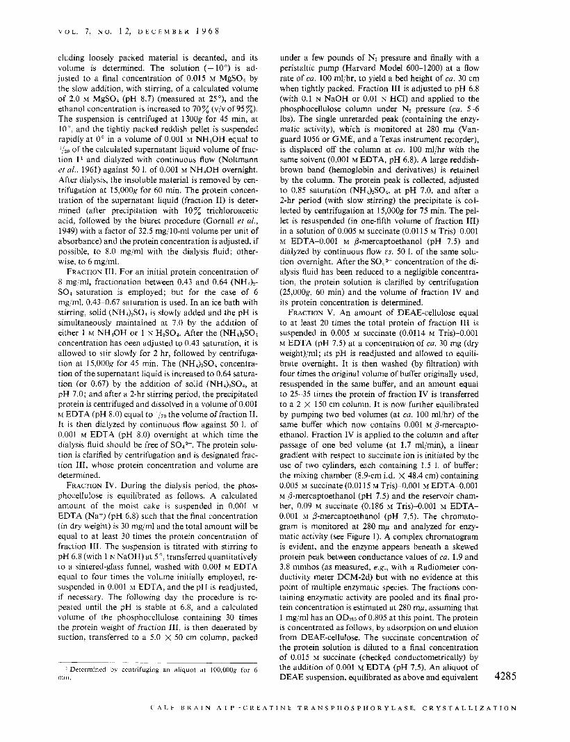
Crystallization. Crystallization is conducted in a 4286
dialysis bag and is induced by a slow adjustment of (NHJ2S04 concentration cia dialysis, in a closed con- tainer filled with liquid (with the gas phase eliminated) and with the dialysis fluid saturated with Nz. The pro- tein solution is dialyzed with magnetic stirring us. a solution of 0.40 saturation ("&SO4 in 0.01 M EDTA- 0.01 M mercaptoethanol (pH 7.4). (The (NH4)S04- EDTA solution is saturated with Nz gas which has been washed with water, before the final addition of 0- mercaptoethanol.) After 24 hr, the (NH4)2S04 concen- tration is increased by 0.01 saturation, and if a small amorphous precipitate appears, it is removed by cen- trifugation, otherwise the dialysis is continued for 12 hr and finally, the saturation is increased to 0.42. At this point, the viscosity of the solution usually shows a noticeable increase followed by incipient crystallization within 12-26 hr. The enzyme crystallizes in the form of relatively large octahedrons (Figure 2) which tend to settle in the dialysis bag. After standing undisturbed (without stirring), qualitatively one observes that the maximum yield of enzyme in crystalline form is usually obtained after a large crop of the crystals had settled and the mother liquor above the settled crystals ap- pears clear but with now a noticeable decrease in vis- cosity. (Sometimes this occurs by increasing the ("&- SO4 saturation by an additional 0.005-0.02.) Recrystal- lization is conducted in the same fashion and is con- tinued (two to three times) until the specific activity of the mother liquor approaches that of the crystalline enzyme.
Results and Discussion
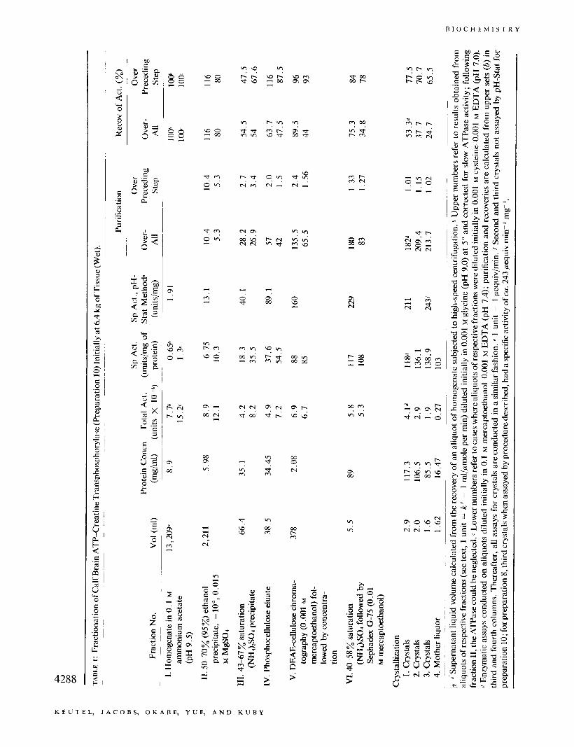
A summary of the data obtained on a typical prepara- tion (preparation 10) on a 6-kg scale is given in Table I. The procedure permits the isolation of the enzyme after some 200-fold purification, and in reasonable yield. It often leads to a final preparation which has approxi- mately a specific activity of ca. 130-140 units/mg, in terms of the apparent second-order velocity constant employed earlier for the crystalline rabbit muscle en- zyme (Kuby et al., 1954) or ca. 240-250 units/mg at 30", when assayed by the pH-Stat procedure, also developed originally for rabbit muscle enzyme.
It is apparent (Table I) that during the initial steps of the preparation, the enzyme is activated by reducing agents (e.g. , cysteine, dithiothreitol, dithioerythritol, or 0-mercaptoethanol has been similarly employed) and which is in agreement with the observations of
K E U T E L , J A C O B S , O K A B E , Y U E , A N D K U B Y
V O L . 7, N O . 1 2 , D E C E M B E R 1 9 6 8
FIGURE 2 : Crystals 01 calf brain ATP-creatine transphosphorylase. The photomicrographs were obtained with a Zeiss phase- contrast microscone at a temnerature of 3”. The maenifications are indicated by the 100- and 20-!2 lines drawn on the respective photographs.
Wood (1963a) on the ox brain enzyme. At the point where 8-mercaptoethanol had been introduced (fraction V), and the enzyme presumably “reduced,” the activa- tion disappears and in fact a slight inhibition is some- times noticeable with cysteine as a diluting reagent, pas- sibly the result of mixed disulfide formation since both thiols and their oxidation products would be present; thereafter, 8-mercaptoethanol is retained for dilution of the enzyme prior to assay.
The conditions used for extraction and the low-tem- perature fractionation step in the presence of Mgz+ are based on earlier observations (Kuby and Mahowald, 1958); the direct addition of aqueous ethanol at low temperatures to the homogenate facilitates further ex- traction of the enzyme, and enzyme assays on the high- speed supernatant liquid of the homogenate (fraction I) are likely to be low. The initial extraction, ethanol steps, and dialysis solve many of the problems (hut not all) concerned with phospholipid removal and permit further purification of the enzyme. The use of phospho- cellulose has a twofold purpose. It separates the hulk of the contaminating hemoglobin-like proteins as well as neutral and basic proteins from the enzyme (at an early stage); and would completely separate any pos- sible traces, should they be present, of muscle-type ATP-creatine transphosphorylase from brain type (as will be made clearer in a later communication on the calf muscle enzyme), as a result of the enormous dif- ferences in charge distribution between the two en- zymes V u e er al., 1968). To avoid the possibility of the brain enzyme, separating into several fractions (e.&!.,
by virtue of different stages of oxidation), mercapto- ethanol is introduced no earlier than at the point of the DEAE-cellulose chromatography. Prior to this point, the purification procedure is designed to isolate in good yield the total enzymatic activity, and should there be more than one brain isoenzyme, this separation would be effected by the terminal stages of purification uia chromatography. Tris-succinate buffers, which Peterson and Chiazze (1962) had successfully employed for separating serum protein by gradient chromatog- raphy on DEAE-cellulose, provide an excellent resolving system for the present case, which is based primarily on ionic strength (i.e., gradients in succinate ion) under con- trolled pH conditions where the enzyme is maximally stable. The relatively low isoelectric point of the en- zyme (Yue et ai., 1968) makes the coupled use of phos- phocellulose, followed by DEAE-chromatography, at suitable pH values, the method of choice. Sephadex gel filtration is primarily employed to remove traces of con- taminating material and trace products which possibly are derived from the enzyme (via oxidation, denatura- tion, or other conformational changes) and which make the crystallization step difficult. The final removal of small protein contaminants is then achieved uia crystal- lization (Figure 2) which proceeds easily and repro- ducibly at this stage of purification, provided the pre- cautions are taken to stabilize the enzyme as outlined above. The relatively large dimensions of the crystals shown, and the fact that they do not appear to have greatly dissimilar dimensions along their three geo- metric axes, may make them amenable to crystallo- 4287
C A L P B R A I N A T P - C R E A T I N E T R A N S P H O S P H O R Y L A S E. C R Y S T A L L 1 2 A T 1 0 N
B I O C H E M I S T R Y
w o 3 w 3
w o - 0 0 3
m P m N . . - -
3 h 3 h
N N
c 0
f
. . 0 3
m I - m . .
00 Q1
m h w h
W
? " w m ~ w o o 0 0
M
Ici 4
E W 3
9 m
N
U
> I . 3 4288
K E U T E L , J A C O B S , O K A B E , Y U E , A N D K U B Y
V O L . 7, N O . 1 2 , D E C E M E E R 1 9 6 8
' ,p ,35'C w i t h
graphic analyses; especially, since with care at the third crystallization stage, much larger crystals than those shown in Figure 2 may be readily grown.
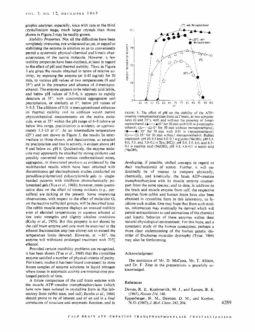
Stability Properties. Not all the difficulties have been completely overcome, nor understood as yet, in regard to stabilizing the enzyme in solution so as to conveniently permit a systematic physical-chemical and kinetic char- acterization of the native molecule. However, a few stability properties have been outlined, at least in regard to the effect of pH and thermal stability. Thus, in Figure 3 are given the results obtained in terms of relative ac- tivity, by exposing the enzyme (at 0.45 mg/ml) for 30 min, to various pH values at two temperatures (0 and 35') and in the presence and absence of P-mercapto- ethanol. The enzyme appears to be relatively acid labile, and below pH values of 5.5-6, it appears to rapidly denature at 35 ', with concomitant aggregation and precipitation, or similarly at O", below pH values of 5-5 .5 . The addition of 0.01 M mercaptoethanol enhances its thermal stability and its addition would permit physicochemical measurements on the native mole- cule, even at 35" within the pH range of 6-8 (above or below this range, precipitation is evident), or approxi- mately 5.5-10 at 0". At an intermediate temperature (25") and not shown in Figure 3, the results lie inter- mediate to those shown, and denaturation, as revealed by precipitation and loss in activity, is evident above pH 9 and below ca. pH 6. Qualitatively, the enzyme mole- cule may apparently be attacked by strong oxidants and possibly converted into various conformational states, aggregates, or dissociated products as evidenced by the multibanded results which have been obtained with discontinuous gel electrophoresis studies conducted on persulfate-polymerized polyacrylamide gels cs. single- banded patterns with riboflavin photochemically po- lymerized gels (Yue et d., 1968); however, more quanti- tative data on the effect of strong oxidants (e.g., per- sulfate) are lacking at the present. Some interesting observations, with respect to the effect of molecular O2 on the reactive sulfhydryl groups, will be described later. The rabbit muscle enzyme displays an unusual stability even at elevated temperatures to aqueous ethanol at low ionic strengths and slightly alkaline conditions (Kuby et al., 1954). But this property is not shared by the calf brain enzyme and care must be exercised in the ethanol fractionation step (see above) not to exceed the temperature limits denoted. However, at -lo", the enzyme will withstand prolonged treatment with 70 % ethanol.
Provided certain instability problems are recognized, it has been shown (Yue et al., 1968) that the crystalline enzyme satisfied a number of physical criteria of purity. For kinetic studies it has been found convenient to store frozen samples of enzyme solutions in liquid nitrogen where losses in enzymatic activity are minimal over pro- longed periods of time.
A future comparison of the calf brain enzyme with the muscle ATP-creatine transphosphorylases (which have now been isolated in crystalline form in this lab- oratory from rabbit man, and calf; Jacobs et al., 1968) should prove to be of interest and of an aid in a final correlation of structure and enzymatic function, and in
,O*C w i th M e r c o p t o e t h o n o l
FIGURE 3: The effect of pH on the stability of the ATP- creatine transphosphorylase from calf brain, at two tempera- tures (0 and 35"), with and without the presence of 2-mer- captoethanol.(A---A)Oc for 30 min with 0.01 M 2-mercapto- ethanol; (A-A) 0" for 30 min without mercaptoethanol; (0-0) 35" for 30 min with 0.01 M mercaptoethanol; ( L O ) 35" for 30 min without mercaptoethanol. Buffers employed: pH 10,9.5 and 9.0-0.1 ~glycine(Na0H); pH 8.5, 8.0,7.5,and7.0-0.1~Tris(HCI);pH5.0,5.5,6.0,and6.5- 0.1 M succinic acid (NaOH); pH 4.5, 4.0-0.1 M acetic acid (NaOH).
developing, if possible, unified concepts in regard to their mechanism(s) of action. Further, it will un- doubtedly be of interest to compare physically, chemically, and kinetically, the brain ATP-creatine transphosphorylase with its muscle enzyme counter- part from the same species; and to date, in addition to the brain and muscle enzyme from calf, the respective enzymes from rabbit and human brain have also been obtained in crystalline form in this laboratory, to fa- cilitate such studies. One may hope that from such stud- ies, information may eventually be derived which will permit extrapolations to and estimations of the chemical and kinetic behavior of these enzymes within their natural physiological environment. For this hope, and a systematic study of the human isoenzymes, perhaps a more clear understanding of the human genetic dis- order of Duchenne muscular dystrophy (Tyler, 1966) may also be forthcoming.
Acknowledgment
The assistance of Mr. D. McCann, Mr. T. Allison, and Dr. F. Ziter in the preparations is gratefully ac- knowledged.
References
Davies, R. E., Kushmerick, M. J., and Larson, R. E.
Eppenberger, H. M., Dawson, D. M . , and Kaplan, (1967), Nature 214, 148.
N. 0. (1967), J . Biol. Chem. 242,204. 4289
C A L F B R A I N A T P - C R E A T I N E T R A N S P H O S P H O R Y L A S E. C R Y S T A L L 1 Z A T I O N
B I O C H E M I S T R Y
Gornall, A. G., Bardawill, C. J., and David, M. M. (1949), J. Biol. Chem. 177,751.
Jacobs, H. K., Keutel, H. J., Yue, R. H., Okabe, K., and Kuby, S. A. (1968), Federation Proc. 27 640.
Kuby, S. A., and Mahowald, T. A. (1958), Federation Proc. 17,258.
Kuby, S. A., Noda, L., and Lardy, H. A. (1954), J. B i d . Chem. 209,191.
Kuby, S. A., and Noltrnann, E. A. (1962), Enzymes 6, 515.
Lebherz, H. G., and Rutter, W. J. (1967), Science 157, 1198.
Mahowald, T. A., Noltmann, E. A., and Kuby, S. A. (1962), J. Biol. Chem. 237, 1535.
McIlwain, H. (1966), Biochemistry of the Central Nervous System, 3rd ed, London, J. and A. Churchill.
Meyerhof, O., and Lohmann, K. (1932), Biochem. 2. 253, 431.
Mommaerts, W. F. H. M., and Wallner, A. (1967), J . Physiol. 193,343.
Nachmansohn, D., and Wilson, E. B. (1951), Adcan. Enzymol. 12,259.
Noda, L., andKuby, S. A. (1957), J. Biol. Chem. 226,541. Noltmann, E. A,, Gubler, C . J., and Kuby, S . A. (1961),
J. Biol. Chem. 236,1225. Peterson, E. A., and Chiazze, E. A. (1962), Arch. Bio-
chem. Biophys. 99,136. Ratliff, R. L., Weaver, R. H., Lardy, H. A,, and Kuby,
S. A. (1964), J . Biol. Chem. 239, 301. Specifications and Criteria for Biochemical Compounds
(1967), 2nd ed, Publication 1344, Washington, D. C . , National Academy of Science., National Research Council, p 231.
Tyler, F. H. (1966), in the Metabolic Basis of Inherited Disease, Stanbury, J. B., Wyngaarden, J. B., and Fredrickson, B. S., 2nd ed, New York, N. Y., Mc- Graw-Hill, p 939.
Wood, T. (1963a), Biochem. J . 87,453. Wood, T. (1963b), Biochem. J. 89,210. Yue, R. H., Jacobs, H. K., Okabe, K., Keutel, H. J.,
and Kuby, S. A. (1968), Biochemistry 7 , 4291 (this issue; following paper).
Yue, R. H., Palrnieri, R. H., Olson, 0. E., and Kuby, S. A. (1967), Biochemistry 6,3204.
4290
K E U T E L , J A C O B S , O K A B E , Y U E , A N D K U B Y


![Increased Rate of Adenosine Triphosphate …...(CANCER RESEARCH 55, 4352-4360, October 1, 1995] Increased Rate of Adenosine Triphosphate-dependent Etoposide (VP-16) Efflux in a Murine](https://img.pdfslide.net/doc/110x75/5e7e8d68c5d0407f2447f2a9/increased-rate-of-adenosine-triphosphate-cancer-research-55-4352-4360-october.jpg)