Embed Size (px)
Citation preview
Mihajlo Lojpur, M.D., Ph.D. - Adult basic life support
1
ADULT BASIC LIFE SUPPORT
_______________________________________________________________________________________________________________________
Mihajlo Lojpur, M.D., Ph.D.
1. VITAL SIGNS
Vital signs include taking the patient’s pulse, respiration, blood pressure, and temperature.
1. Pulse
The ventricles (right and left) have two phases: diastole or the time when the ventricles 'rest' so they can fill with blood, and systole, the time when the
ventricles contract to send blood either to the lungs (from the right side of the heart), or to the rest of the body (from the left side of the heart).
The pulse represents the variation in blood pressure from diastole to systole. During diastole blood pressure falls, but increases after systole as the heart
pumps more blood into the arteries. You feel this difference when taking your pulse.
When taking a patient’s pulse, you should be concerned with two factors: rate and character.
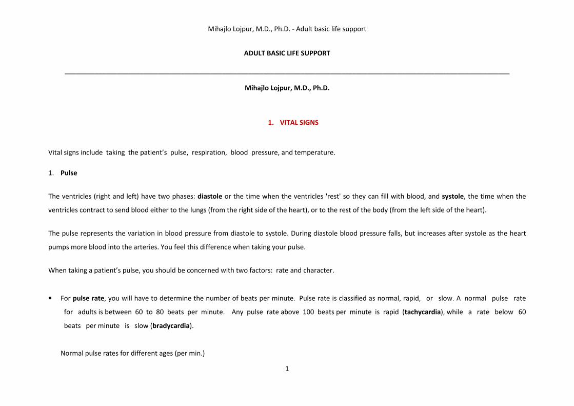
• For pulse rate, you will have to determine the number of beats per minute. Pulse rate is classified as normal, rapid, or slow. A normal pulse rate
for adults is between 60 to 80 beats per minute. Any pulse rate above 100 beats per minute is rapid (tachycardia), while a rate below 60
beats per minute is slow (bradycardia).
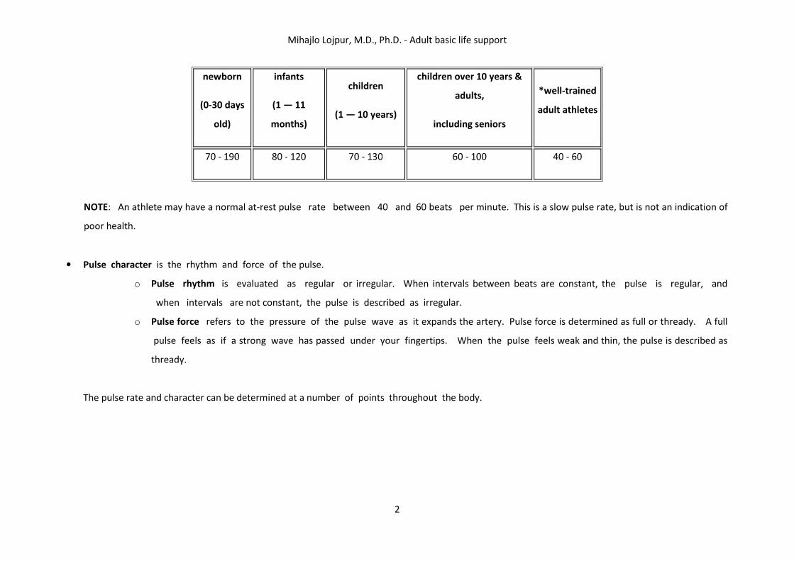
Normal pulse rates for different ages (per min.)
Mihajlo Lojpur, M.D., Ph.D. - Adult basic life support
2
newborn
(0-30 days
old)
infants
(1 — 11
months)
children
(1 — 10 years)
children over 10 years &
adults,
including seniors
*well-trained
adult athletes
70 - 190 80 - 120 70 - 130 60 - 100 40 - 60
NOTE: An athlete may have a normal at-rest pulse rate between 40 and 60 beats per minute. This is a slow pulse rate, but is not an indication of
poor health.
• Pulse character is the rhythm and force of the pulse.
o Pulse rhythm is evaluated as regular or irregular. When intervals between beats are constant, the pulse is regular, and
when intervals are not constant, the pulse is described as irregular.
o Pulse force refers to the pressure of the pulse wave as it expands the artery. Pulse force is determined as full or thready. A full
pulse feels as if a strong wave has passed under your fingertips. When the pulse feels weak and thin, the pulse is described as
thready.
The pulse rate and character can be determined at a number of points throughout the body.
Mihajlo Lojpur, M.D., Ph.D. - Adult basic life support
3
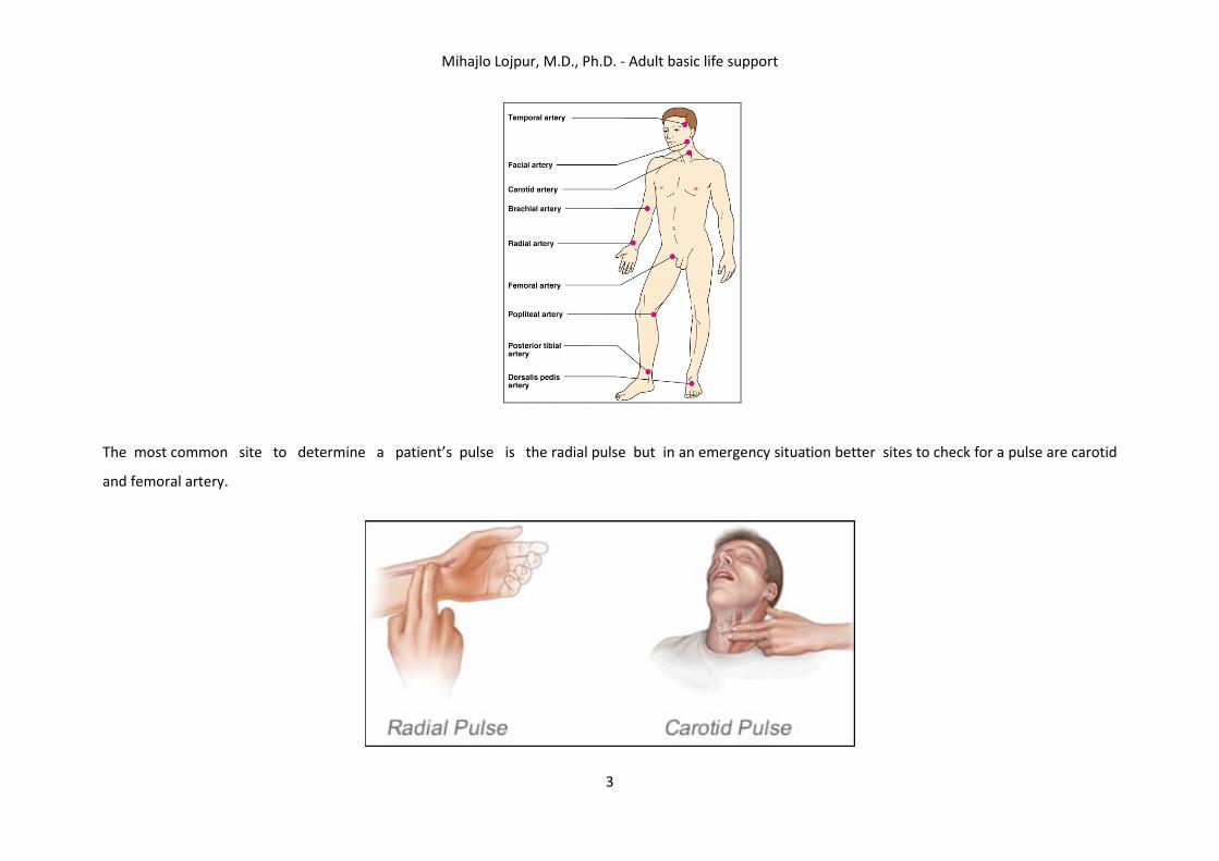
The most common site to determine a patient’s pulse is the radial pulse but in an emergency situation better sites to check for a pulse are carotid
and femoral artery.
Mihajlo Lojpur, M.D., Ph.D. - Adult basic life support
4
How to taking pulse at these sites :
• Radial Pulse: Take two fingers, preferably the 2nd and 3rd finger, and place them in the groove in the wrist that lies beneath the thumb. Move your
fingers back and forth gently until you can feel a slight pusation - this is the pulse of the radial artery which delivers blood to the hand. Don't press too
hard, or else you'll just feel the blood flowing through your fingers!
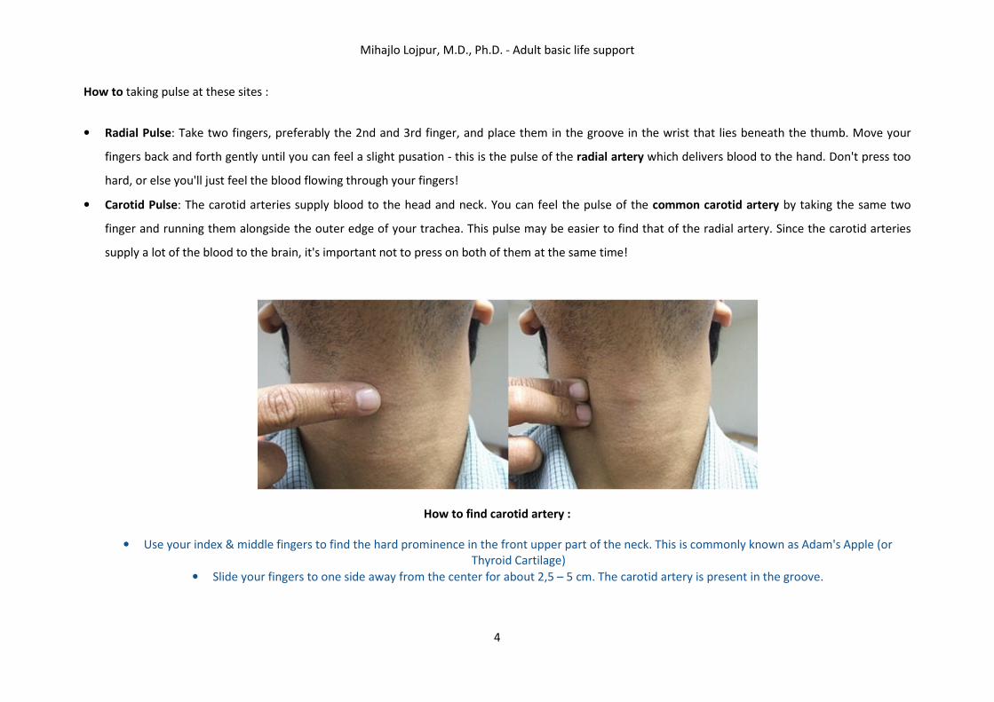
• Carotid Pulse: The carotid arteries supply blood to the head and neck. You can feel the pulse of the common carotid artery by taking the same two
finger and running them alongside the outer edge of your trachea. This pulse may be easier to find that of the radial artery. Since the carotid arteries
supply a lot of the blood to the brain, it's important not to press on both of them at the same time!
How to find carotid artery :
• Use your index & middle fingers to find the hard prominence in the front upper part of the neck. This is commonly known as Adam's Apple (or
Thyroid Cartilage)
• Slide your fingers to one side away from the center for about 2,5 – 5 cm. The carotid artery is present in the groove.
Mihajlo Lojpur, M.D., Ph.D. - Adult basic life support
5
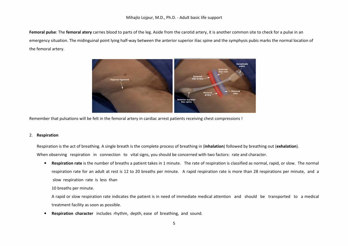
Femoral pulse: The femoral atery carries blood to parts of the leg. Aside from the carotid artery, it is another common site to check for a pulse in an
emergency situation. The midinguinal point lying half-way between the anterior superior iliac spine and the symphysis pubis marks the normal location of
the femoral artery.
Remember that pulsations will be felt in the femoral artery in cardiac arrest patients receiving chest compressions !
2. Respiration
Respiration is the act of breathing. A single breath is the complete process of breathing in (inhalation) followed by breathing out (exhalation).
When observing respiration in connection to vital signs, you should be concerned with two factors: rate and character.
• Respiration rate is the number of breaths a patient takes in 1 minute. The rate of respiration is classified as normal, rapid, or slow. The normal
respiration rate for an adult at rest is 12 to 20 breaths per minute. A rapid respiration rate is more than 28 respirations per minute, and a
slow respiration rate is less than
10 breaths per minute.
A rapid or slow respiration rate indicates the patient is in need of immediate medical attention and should be transported to a medical
treatment facility as soon as possible.
• Respiration character includes rhythm, depth, ease of breathing, and sound.
Mihajlo Lojpur, M.D., Ph.D. - Adult basic life support
6
o Respiration rhythm refers to the manner in which a person breathes. Respiration rhythm is classified as regular or irregular. A
regular rhythm is when the interval between breaths is constant, and an irregular rhythm is when the interval between breaths
varies.
o Respiration depth refers to the amount of air moved between each breath. Respiration depth is classified as normal, deep, or
shallow.
o Ease of breathing can be judged while you are judging depth. Ease of breathing may be judged as labored, difficult, or painful.
o Sound of respiration include snoring , wheezing, crowing (birdlike sounds) and gurgling (sounds like breaths are passing through
water).
You should count respirations as soon as you have determined the pulse rate. Count the number of breaths taken by the patient during 30 seconds
and multiply by 2 to obtain the breaths per minute. While you are counting breaths, note the rhythm, depth, ease of breathing, and sounds
of respiration.
3. Blood Pressure
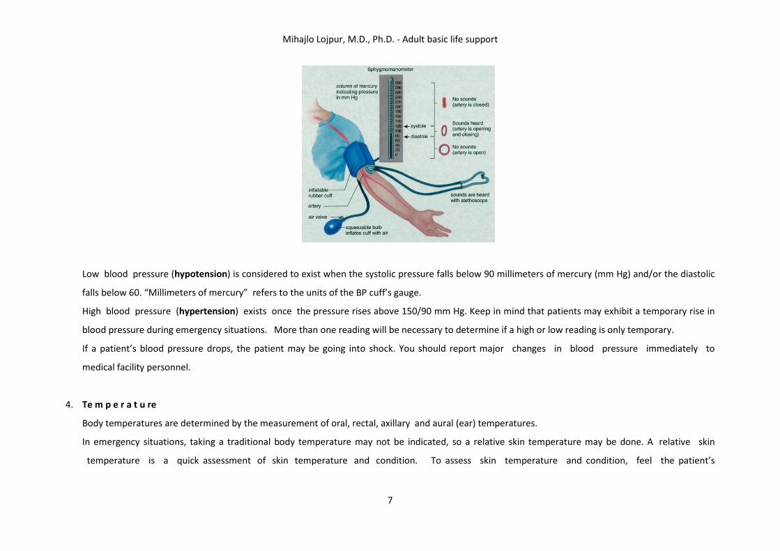
Blood Pressure is the pressure which blood exerts against blood vessel walls, usually arteries.
The pressure created in the arteries when the heart pumps blood out into circulation (heart beat) is called the systolic blood pressure. The pressure
remaining in the arteries when the heart is relaxed (between beats) is called the diastolic blood pressure. The systolic pressure is always
reported first and the diastolic pressure second (e.g., 120 over 80).
Blood pressure varies from one person to another and is measured with a stethoscope and a sphygmomanometer (BP cuff).
Mihajlo Lojpur, M.D., Ph.D. - Adult basic life support
7
Low blood pressure (hypotension) is considered to exist when the systolic pressure falls below 90 millimeters of mercury (mm Hg) and/or the diastolic
falls below 60. “Millimeters of mercury” refers to the units of the BP cuff’s gauge.
High blood pressure (hypertension) exists once the pressure rises above 150/90 mm Hg. Keep in mind that patients may exhibit a temporary rise in
blood pressure during emergency situations. More than one reading will be necessary to determine if a high or low reading is only temporary.
If a patient’s blood pressure drops, the patient may be going into shock. You should report major changes in blood pressure immediately to
medical facility personnel.
4. Te m p e r a t u re
Body temperatures are determined by the measurement of oral, rectal, axillary and aural (ear) temperatures.
In emergency situations, taking a traditional body temperature may not be indicated, so a relative skin temperature may be done. A relative skin
temperature is a quick assessment of skin temperature and condition. To assess skin temperature and condition, feel the patient’s
Mihajlo Lojpur, M.D., Ph.D. - Adult basic life support
8
forehead with the back of your hand. In doing this, note if the patient’s skin feels normal, warm, hot, cool, or cold. At the same time, see if the skin is
dry, moist, or clammy.
2. CARDIAC ARREST
Cardiac arrest, (also known as cardiopulmonary arrest or circulatory arrest) is the cessation of normal circulation of the blood due to failure of the heart to
contract effectively.
Brain injury is likely if cardiac arrest goes untreated for more than five minutes. For the best chance of survival and neurological recovery, immediate and
decisive treatment is imperative. The treatment for cardiac arrest is cardiopulmonary resuscitation (CPR) to provide circulatory support, followed by
defibrillation if a shockable rhythm is present.
Causes
Coronary heart disease is the leading cause of sudden cardiac arrest. Many other cardiac and non-cardiac conditions also increase ones risk
• Approximately 60–70% of cardiac arrest is related to cardiac disease.
o Among adults, ischemic heart disease is the predominant cause of arrest. No less than 30% of them at autopsy showing signs of recent
myocardial infarction.
o A number of other cardiac abnormalities can increase the risk of cardiac arrest including: cardiomyopathy, cardiac rhythm disturbances,
hypertensive heart disease, congestive heart failure...
• Cardiac arrest is unrelated to heart problems in 35% of cases.
Mihajlo Lojpur, M.D., Ph.D. - Adult basic life support
9
o The most common non-cardiac causes: trauma, non-trauma related bleeding (such as gastrointestinal bleeding, aortic rupture, and
intracranial hemorrhage), overdose, drowning and pulmonary embolism.
In infants and children, the most common cause of cardiac arrest is respiratory arrest. Respiratory disorders most often resulting in cardiac arrest include
airway obstruction, smoke inhalation, drowning, infection and sudden infant death syndrome. In adults, the opposite usually occurs - cardiac arrest leads to
respiratory arrest.
Signs and symptoms
Cardiac arrest is an abrupt cessation of pump function in the heart, as evidenced by the absence of a palpable pulse. Arrested blood circulation prevents
delivery of oxygen to the body. Due to inadequate cerebral perfusion, the patient will be unconscious and will have stopped breathing.
Diagnosis
The main diagnostic criterion to diagnose a cardiac arrest is lack of circulation, however there are a number of ways of determining this.
1. A cardiac arrest is usually diagnosed clinically by the absence of a pulse. In many cases lack of carotid pulse is the gold standard for diagnosing cardiac
arrest, but lack of a pulse (particularly in the peripheral pulses) may be a result of other conditions (e.g. shock), or simply an error on the part of the
rescuer. Studies have shown that rescuers often make a mistake when checking the carotid pulse in an emergency, whether they are healthcare
professionals or lay persons.
Mihajlo Lojpur, M.D., Ph.D. - Adult basic life support
10
Owing to the inaccuracy in this method of diagnosis, some bodies such as the European Resuscitation Council (ERC) have de-emphasised its importance.
The Resuscitation Council (UK), in line with the ERC's recommendations and those of the American Heart Association, have suggested that the technique
should be used only by healthcare professionals with specific training and expertise, and even then that it should be viewed in conjunction with other
indicators such as agonal respiration.
2. Various other methods for detecting circulation have been proposed. Guidelines following the 2000 International Liaison Committee on Resuscitation
(ILCOR) recommendations were for rescuers to look for "signs of circulation", but not specifically the pulse. These signs included coughing, gasping,
colour, twitching and movement.
However, in face of evidence that these guidelines were ineffective, the current recommendation of ILCOR is that cardiac arrest should be diagnosed
in all casualties who are unconscious and not breathing normally.
3. ADULT BASIC LIFE SUPPORT
Basic life support includes the maintenance of an airway and the support of breathing and the circulation without using equipment other than a simple
airway device or protective shield. A combination of expired air ventilation (rescue breathing) and chest compression is known as cardiopulmonary
resuscitation (CPR), which forms the basis of modern basic life support.
The term "cardiac arrest" implies a sudden interruption of cardiac output, which may be reversible with appropriate treatment. It is important that those
who may be present at the scene of a cardiac arrest should have learnt the appropriate resuscitation skills and be able to put them into practice.
Mihajlo Lojpur, M.D., Ph.D. - Adult basic life support
11
Simplification of the BLS sequence continues to be a feature of these guidelines, but, in addition, there is now advice on who should be taught what skills,
particularly chest-compression-only or chest compression and ventilation.
All rescuers, trained or not, should provide chest compressions to victims of cardiac arrest :
• If a bystander is not trained in CPR, he or she should provide compression-only CPR for the adult victim who suddenly collapses, with an emphasis
to “push hard and fast” on the center of the chest, or follow the directions of the EMS dispatcher. The rescuer should continue compression-only
CPR until an AED arrives and is ready for use or EMS providers or other responders take over care of the victim.
• All trained lay rescuers should, at a minimum, provide chest compressions for victims of cardiac arrest. In addition, if the trained lay rescuer is able
to perform rescue breaths, compressions and breaths should be provided in a ratio of 30 compressions to 2 breaths. The rescuer should continue
CPR until an AED arrives and is ready for use or EMS providers take over care of the victim.
Continued emphasis has been placed on high-quality CPR (with chest compressions of adequate rate and depth, allowing complete chest recoil after each
compression, minimizing interruptions in compressions, and avoiding excessive ventilation) :
• Compression rate should be at least 100/min (rather than “approximately” 100/min).
• Compression depth for adults has been changed from the range of 4 to 5 cm to at least 5 cm
Mihajlo Lojpur, M.D., Ph.D. - Adult basic life support
12
Adult basic life support
A. Adult basic life support sequence
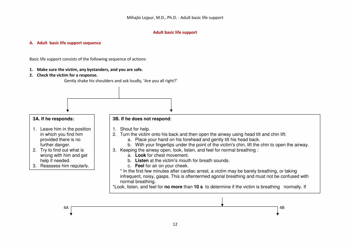
Basic life support consists of the following sequence of actions:
1. Make sure the victim, any bystanders, and you are safe.
2. Check the victim for a response.
Gently shake his shoulders and ask loudly, ‘Are you all right?’
4A 4B
3A. If he responds:
1. Leave him in the position in which you find him provided there is no further danger.
2. Try to find out what is wrong with him and get help if needed.
3. Reassess him regularly.
3B. If he does not respond:
1. Shout for help. 2. Turn the victim onto his back and then open the airway using head tilt and chin lift:
a. Place your hand on his forehead and gently tilt his head back. b. With your fingertips under the point of the victim's chin, lift the chin to open the airway.
3. Keeping the airway open, look, listen, and feel for normal breathing : a. Look for chest movement. b. Listen at the victim's mouth for breath sounds. c. Feel for air on your cheek.
* In the first few minutes after cardiac arrest, a victim may be barely breathing, or taking infrequent, noisy, gasps. This is oftentermed agonal breathing and must not be confused with normal breathing.
*Look, listen, and feel for no more than 10 s to determine if the victim is breathing normally. If
Mihajlo Lojpur, M.D., Ph.D. - Adult basic life support
13
5A 6
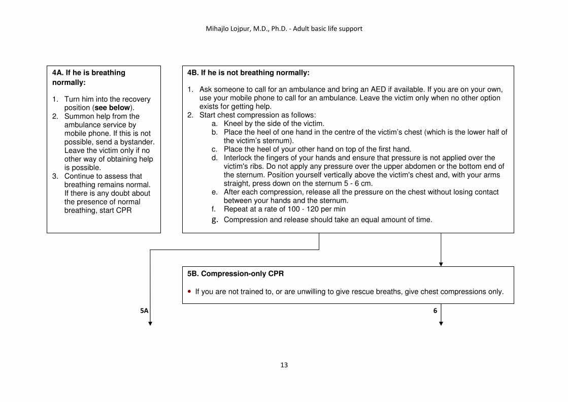
4A. If he is breathing
normally:
1. Turn him into the recovery position (see below).
2. Summon help from the ambulance service by mobile phone. If this is not possible, send a bystander. Leave the victim only if no other way of obtaining help is possible.
3. Continue to assess that breathing remains normal. If there is any doubt about the presence of normal breathing, start CPR
4B. If he is not breathing normally:
1. Ask someone to call for an ambulance and bring an AED if available. If you are on your own, use your mobile phone to call for an ambulance. Leave the victim only when no other option exists for getting help.
2. Start chest compression as follows: a. Kneel by the side of the victim. b. Place the heel of one hand in the centre of the victim’s chest (which is the lower half of
the victim’s sternum). c. Place the heel of your other hand on top of the first hand. d. Interlock the fingers of your hands and ensure that pressure is not applied over the
victim's ribs. Do not apply any pressure over the upper abdomen or the bottom end of the sternum. Position yourself vertically above the victim's chest and, with your arms straight, press down on the sternum 5 - 6 cm.
e. After each compression, release all the pressure on the chest without losing contact between your hands and the sternum.
f. Repeat at a rate of 100 - 120 per min
g. Compression and release should take an equal amount of time.
5B. Compression-only CPR
• If you are not trained to, or are unwilling to give rescue breaths, give chest compressions only.
Mihajlo Lojpur, M.D., Ph.D. - Adult basic life support
14
5A 6
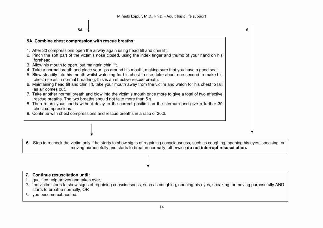
5A. Combine chest compression with rescue breaths:
1. After 30 compressions open the airway again using head tilt and chin lift. 2. Pinch the soft part of the victim’s nose closed, using the index finger and thumb of your hand on his
forehead. 3. Allow his mouth to open, but maintain chin lift. 4. Take a normal breath and place your lips around his mouth, making sure that you have a good seal. 5. Blow steadily into his mouth whilst watching for his chest to rise; take about one second to make his
chest rise as in normal breathing; this is an effective rescue breath. 6. Maintaining head tilt and chin lift, take your mouth away from the victim and watch for his chest to fall
as air comes out. 7. Take another normal breath and blow into the victim’s mouth once more to give a total of two effective
rescue breaths. The two breaths should not take more than 5 s. 8. Then return your hands without delay to the correct position on the sternum and give a further 30
chest compressions. 9. Continue with chest compressions and rescue breaths in a ratio of 30:2.
6. Stop to recheck the victim only if he starts to show signs of regaining consciousness, such as coughing, opening his eyes, speaking, or moving purposefully and starts to breathe normally; otherwise do not interrupt resuscitation.
7. Continue resuscitation until: 1. qualified help arrives and takes over, 2. the victim starts to show signs of regaining consciousness, such as coughing, opening his eyes, speaking, or moving purposefully AND
starts to breathe normally, OR 3. you become exhausted.
Mihajlo Lojpur, M.D., Ph.D. - Adult basic life support
15
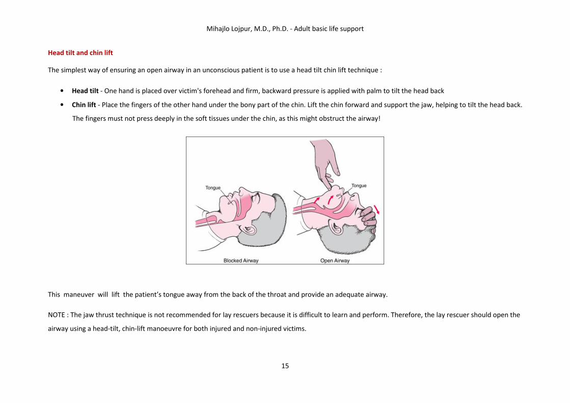
Head tilt and chin lift
The simplest way of ensuring an open airway in an unconscious patient is to use a head tilt chin lift technique :
• Head tilt - One hand is placed over victim's forehead and firm, backward pressure is applied with palm to tilt the head back
• Chin lift - Place the fingers of the other hand under the bony part of the chin. Lift the chin forward and support the jaw, helping to tilt the head back.
The fingers must not press deeply in the soft tissues under the chin, as this might obstruct the airway!
This maneuver will lift the patient’s tongue away from the back of the throat and provide an adequate airway.
NOTE : The jaw thrust technique is not recommended for lay rescuers because it is difficult to learn and perform. Therefore, the lay rescuer should open the
airway using a head-tilt, chin-lift manoeuvre for both injured and non-injured victims.
Mihajlo Lojpur, M.D., Ph.D. - Adult basic life support
16
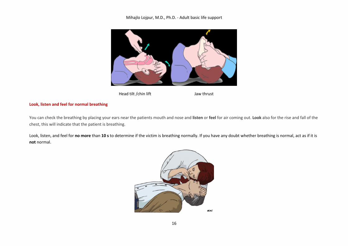
Head tilt /chin lift Jaw thrust
Look, listen and feel for normal breathing
You can check the breathing by placing your ears near the patients mouth and nose and listen or feel for air coming out. Look also for the rise and fall of the
chest, this will indicate that the patient is breathing.
Look, listen, and feel for no more than 10 s to determine if the victim is breathing normally. If you have any doubt whether breathing is normal, act as if it is
not normal.
Mihajlo Lojpur, M.D., Ph.D. - Adult basic life support
17
NOTE : Rescuers are often warned against mistaking agonal breathing, which is a series of noisy gasps occurring in around 40% of cardiac arrest victims, for
normal breathing.
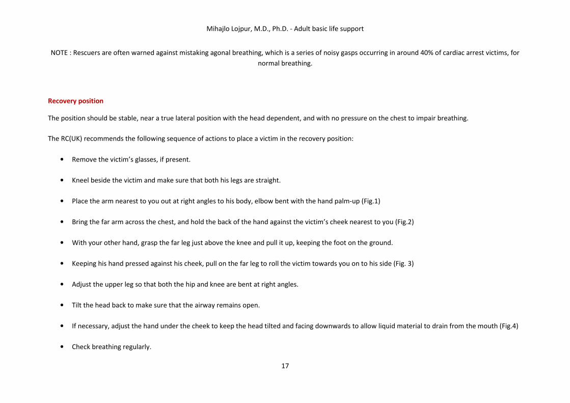
Recovery position
The position should be stable, near a true lateral position with the head dependent, and with no pressure on the chest to impair breathing.
The RC(UK) recommends the following sequence of actions to place a victim in the recovery position:
• Remove the victim’s glasses, if present.
• Kneel beside the victim and make sure that both his legs are straight.
• Place the arm nearest to you out at right angles to his body, elbow bent with the hand palm-up (Fig.1)
• Bring the far arm across the chest, and hold the back of the hand against the victim’s cheek nearest to you (Fig.2)
• With your other hand, grasp the far leg just above the knee and pull it up, keeping the foot on the ground.
• Keeping his hand pressed against his cheek, pull on the far leg to roll the victim towards you on to his side (Fig. 3)
• Adjust the upper leg so that both the hip and knee are bent at right angles.
• Tilt the head back to make sure that the airway remains open.
• If necessary, adjust the hand under the cheek to keep the head tilted and facing downwards to allow liquid material to drain from the mouth (Fig.4)
• Check breathing regularly.
Mihajlo Lojpur, M.D., Ph.D. - Adult basic life support
18
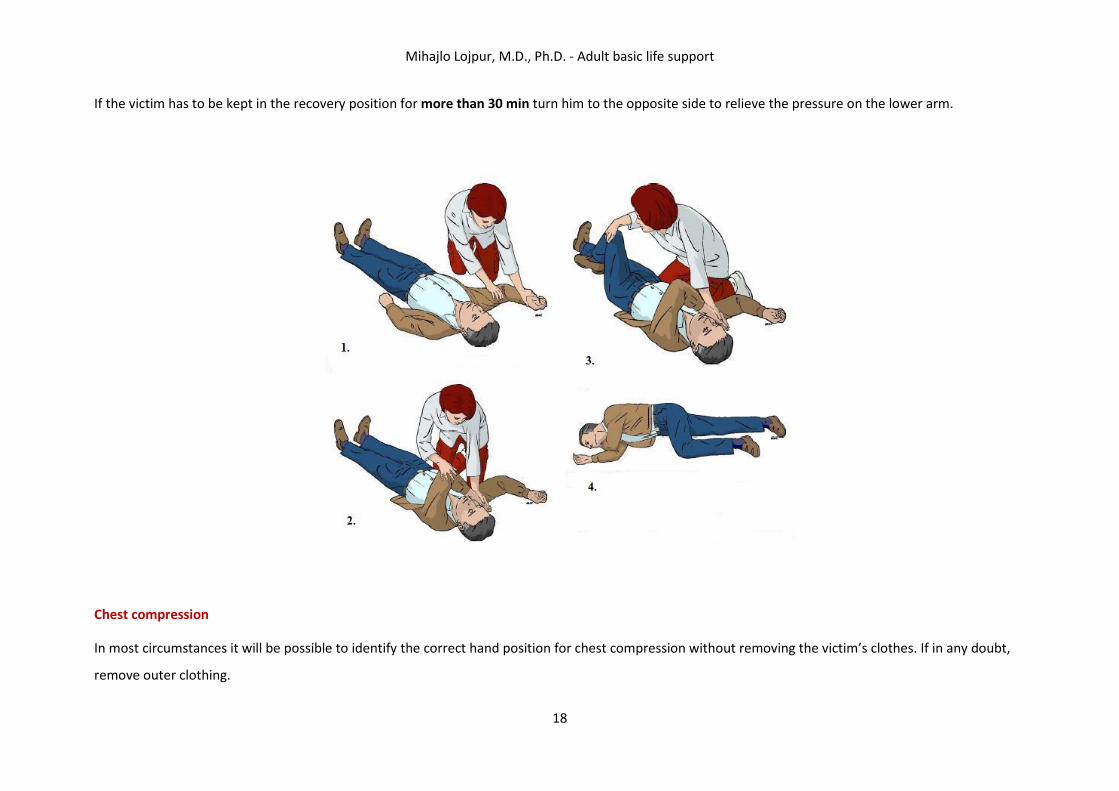
If the victim has to be kept in the recovery position for more than 30 min turn him to the opposite side to relieve the pressure on the lower arm.
Chest compression
In most circumstances it will be possible to identify the correct hand position for chest compression without removing the victim’s clothes. If in any doubt,
remove outer clothing.
Mihajlo Lojpur, M.D., Ph.D. - Adult basic life support
19
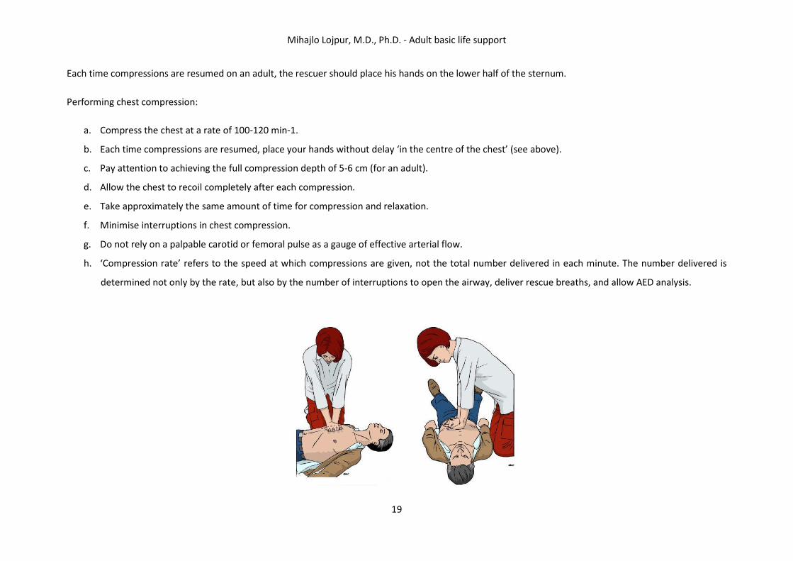
Each time compressions are resumed on an adult, the rescuer should place his hands on the lower half of the sternum.
Performing chest compression:
a. Compress the chest at a rate of 100-120 min-1.
b. Each time compressions are resumed, place your hands without delay ‘in the centre of the chest’ (see above).
c. Pay attention to achieving the full compression depth of 5-6 cm (for an adult).
d. Allow the chest to recoil completely after each compression.
e. Take approximately the same amount of time for compression and relaxation.
f. Minimise interruptions in chest compression.
g. Do not rely on a palpable carotid or femoral pulse as a gauge of effective arterial flow.
h. ‘Compression rate’ refers to the speed at which compressions are given, not the total number delivered in each minute. The number delivered is
determined not only by the rate, but also by the number of interruptions to open the airway, deliver rescue breaths, and allow AED analysis.
Mihajlo Lojpur, M.D., Ph.D. - Adult basic life support
20
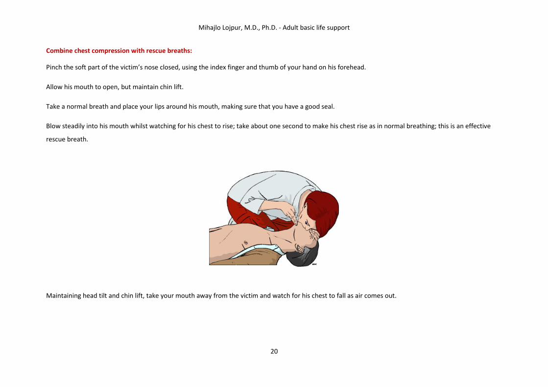
Combine chest compression with rescue breaths:
Pinch the soft part of the victim’s nose closed, using the index finger and thumb of your hand on his forehead.
Allow his mouth to open, but maintain chin lift.
Take a normal breath and place your lips around his mouth, making sure that you have a good seal.
Blow steadily into his mouth whilst watching for his chest to rise; take about one second to make his chest rise as in normal breathing; this is an effective
rescue breath.
Maintaining head tilt and chin lift, take your mouth away from the victim and watch for his chest to fall as air comes out.
Mihajlo Lojpur, M.D., Ph.D. - Adult basic life support
21
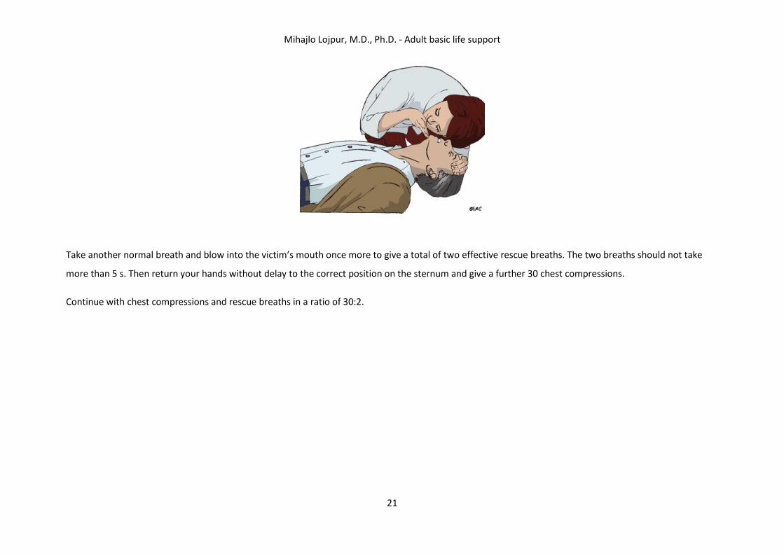
Take another normal breath and blow into the victim’s mouth once more to give a total of two effective rescue breaths. The two breaths should not take
more than 5 s. Then return your hands without delay to the correct position on the sternum and give a further 30 chest compressions.
Continue with chest compressions and rescue breaths in a ratio of 30:2.
Mihajlo Lojpur, M.D., Ph.D. - Adult basic life support
22
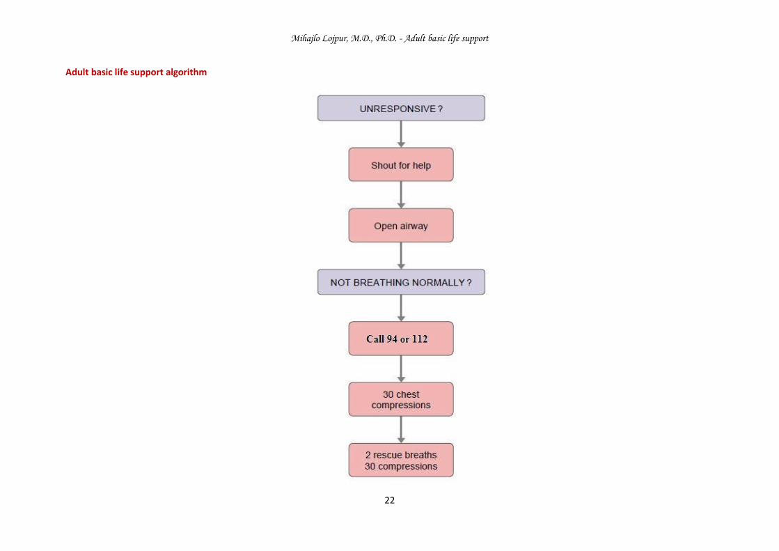
Adult basic life support algorithm
Mihajlo Lojpur, M.D., Ph.D. - Adult basic life support
23
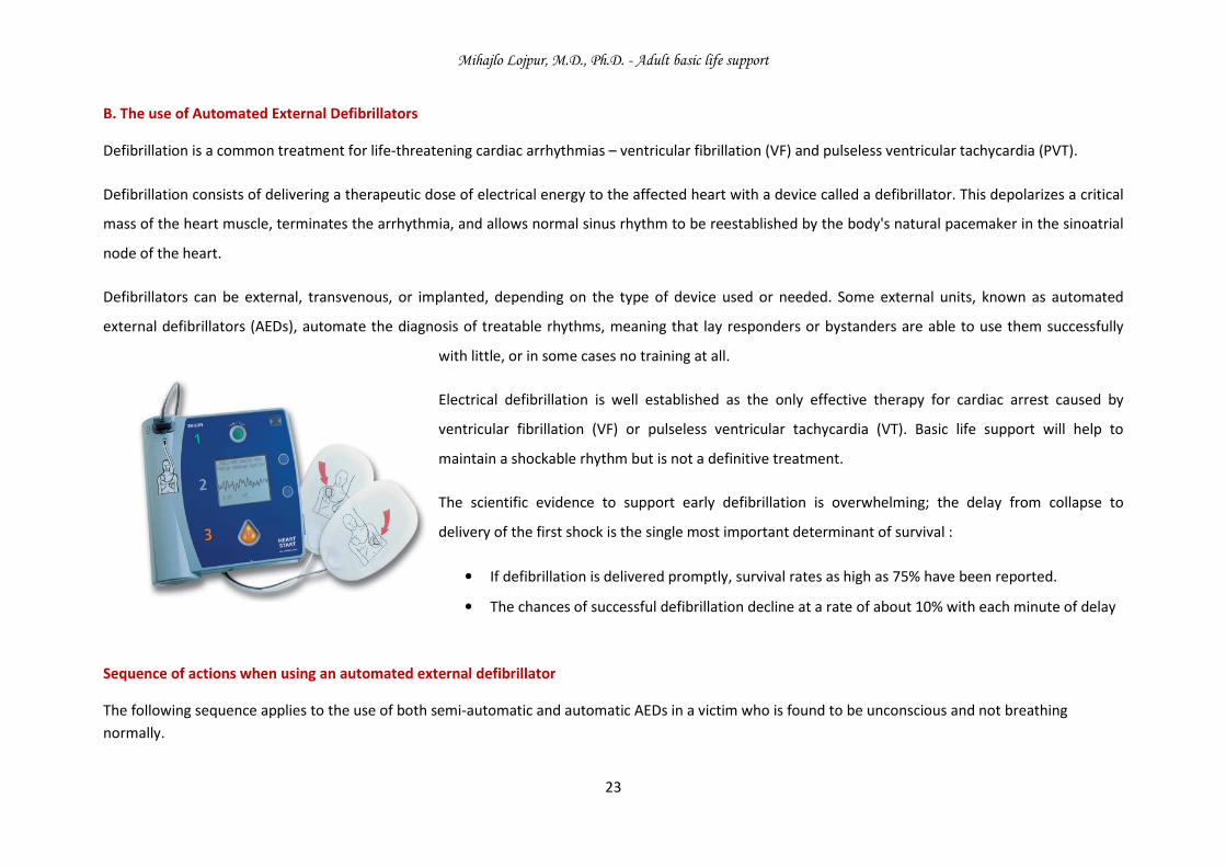
B. The use of Automated External Defibrillators
Defibrillation is a common treatment for life-threatening cardiac arrhythmias – ventricular fibrillation (VF) and pulseless ventricular tachycardia (PVT).
Defibrillation consists of delivering a therapeutic dose of electrical energy to the affected heart with a device called a defibrillator. This depolarizes a critical
mass of the heart muscle, terminates the arrhythmia, and allows normal sinus rhythm to be reestablished by the body's natural pacemaker in the sinoatrial
node of the heart.
Defibrillators can be external, transvenous, or implanted, depending on the type of device used or needed. Some external units, known as automated
external defibrillators (AEDs), automate the diagnosis of treatable rhythms, meaning that lay responders or bystanders are able to use them successfully
with little, or in some cases no training at all.
Electrical defibrillation is well established as the only effective therapy for cardiac arrest caused by
ventricular fibrillation (VF) or pulseless ventricular tachycardia (VT). Basic life support will help to
maintain a shockable rhythm but is not a definitive treatment.
The scientific evidence to support early defibrillation is overwhelming; the delay from collapse to
delivery of the first shock is the single most important determinant of survival :
• If defibrillation is delivered promptly, survival rates as high as 75% have been reported.
• The chances of successful defibrillation decline at a rate of about 10% with each minute of delay
Sequence of actions when using an automated external defibrillator
The following sequence applies to the use of both semi-automatic and automatic AEDs in a victim who is found to be unconscious and not breathing
normally.
Mihajlo Lojpur, M.D., Ph.D. - Adult basic life support
24
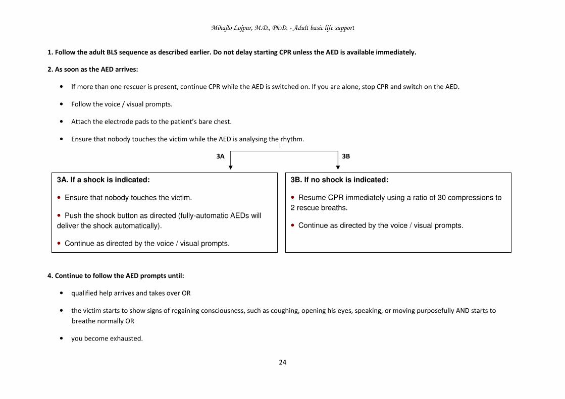
1. Follow the adult BLS sequence as described earlier. Do not delay starting CPR unless the AED is available immediately.
2. As soon as the AED arrives:
• If more than one rescuer is present, continue CPR while the AED is switched on. If you are alone, stop CPR and switch on the AED.
• Follow the voice / visual prompts.
• Attach the electrode pads to the patient’s bare chest.
• Ensure that nobody touches the victim while the AED is analysing the rhythm.
3A 3B
4. Continue to follow the AED prompts until:
• qualified help arrives and takes over OR
• the victim starts to show signs of regaining consciousness, such as coughing, opening his eyes, speaking, or moving purposefully AND starts to
breathe normally OR
• you become exhausted.
3A. If a shock is indicated:
• Ensure that nobody touches the victim.
• Push the shock button as directed (fully-automatic AEDs will
deliver the shock automatically).
• Continue as directed by the voice / visual prompts.
3B. If no shock is indicated:
• Resume CPR immediately using a ratio of 30 compressions to
2 rescue breaths.
• Continue as directed by the voice / visual prompts.
Mihajlo Lojpur, M.D., Ph.D. - Adult basic life support
25
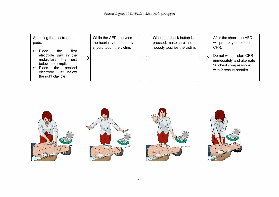
Attaching the electrode
pads.
• Place the first electrode pad in the midaxillary line just below the armpit.
• Place the second electrode just below the right clavicle
While the AED analyses
the heart rhythm, nobody
should touch the victim.
When the shock button is
pressed, make sure that
nobody touches the victim.
After the shock the AED
will prompt you to start
CPR.
Do not wait — start CPR
immediately and alternate
30 chest compressions
with 2 rescue breaths
Mihajlo Lojpur, M.D., Ph.D. - Adult basic life support
26
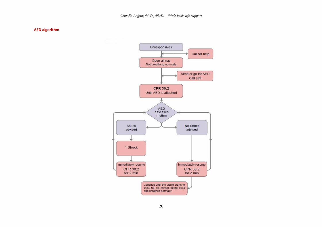
AED algorithm
Mihajlo Lojpur, M.D., Ph.D. - Adult basic life support
27
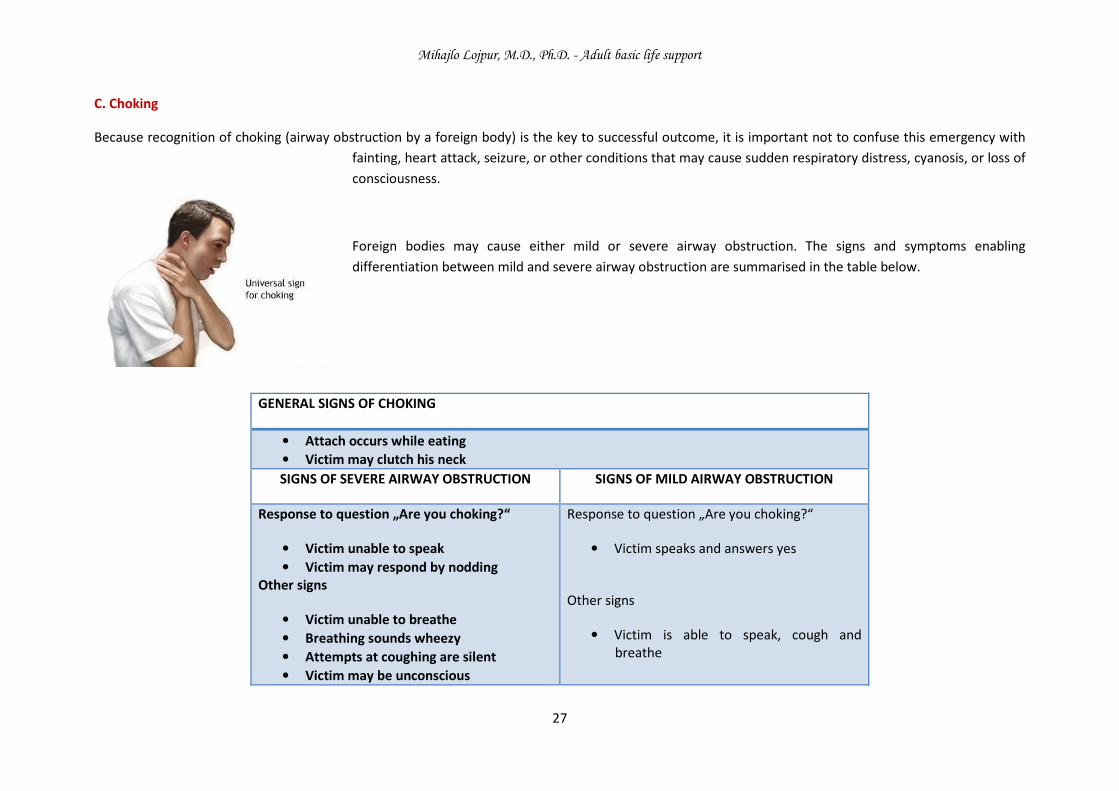
C. Choking
Because recognition of choking (airway obstruction by a foreign body) is the key to successful outcome, it is important not to confuse this emergency with
fainting, heart attack, seizure, or other conditions that may cause sudden respiratory distress, cyanosis, or loss of
consciousness.
Foreign bodies may cause either mild or severe airway obstruction. The signs and symptoms enabling
differentiation between mild and severe airway obstruction are summarised in the table below.
GENERAL SIGNS OF CHOKING
• Attach occurs while eating
• Victim may clutch his neck
SIGNS OF SEVERE AIRWAY OBSTRUCTION SIGNS OF MILD AIRWAY OBSTRUCTION
Response to question „Are you choking?“
• Victim unable to speak
• Victim may respond by nodding
Other signs
• Victim unable to breathe
• Breathing sounds wheezy
• Attempts at coughing are silent
• Victim may be unconscious
Response to question „Are you choking?“
• Victim speaks and answers yes
Other signs
• Victim is able to speak, cough and
breathe
Mihajlo Lojpur, M.D., Ph.D. - Adult basic life support
28
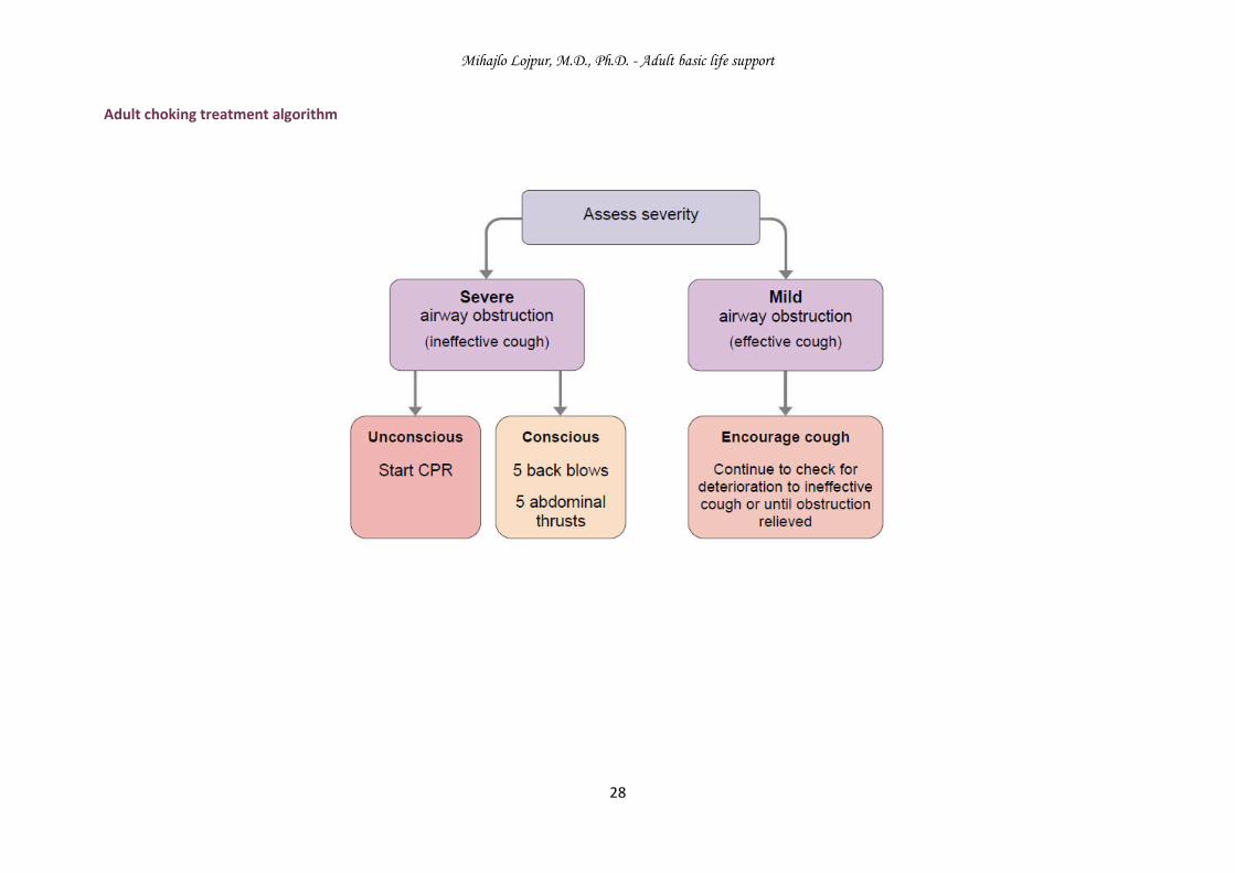
Adult choking treatment algorithm
Mihajlo Lojpur, M.D., Ph.D. - Adult basic life support
29
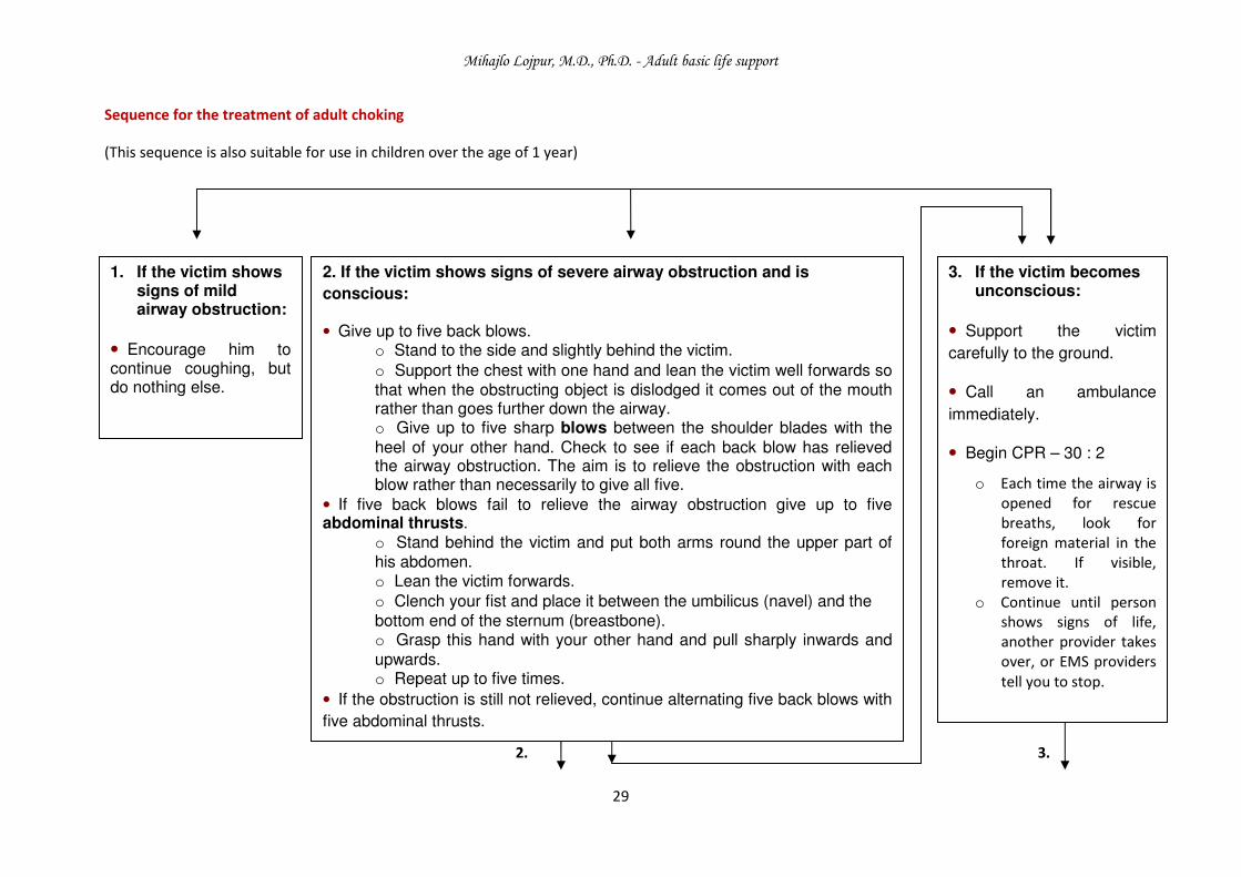
Sequence for the treatment of adult choking
(This sequence is also suitable for use in children over the age of 1 year)
2. 3.
1. If the victim shows signs of mild airway obstruction:
• Encourage him to continue coughing, but do nothing else.
2. If the victim shows signs of severe airway obstruction and is
conscious:
• Give up to five back blows. o Stand to the side and slightly behind the victim.
o Support the chest with one hand and lean the victim well forwards so that when the obstructing object is dislodged it comes out of the mouth rather than goes further down the airway. o Give up to five sharp blows between the shoulder blades with the
heel of your other hand. Check to see if each back blow has relieved the airway obstruction. The aim is to relieve the obstruction with each blow rather than necessarily to give all five.
• If five back blows fail to relieve the airway obstruction give up to five abdominal thrusts.
o Stand behind the victim and put both arms round the upper part of his abdomen. o Lean the victim forwards.
o Clench your fist and place it between the umbilicus (navel) and the
bottom end of the sternum (breastbone). o Grasp this hand with your other hand and pull sharply inwards and
upwards. o Repeat up to five times.
• If the obstruction is still not relieved, continue alternating five back blows with
five abdominal thrusts.
3. If the victim becomes unconscious:
• Support the victim
carefully to the ground.
• Call an ambulance
immediately.
• Begin CPR – 30 : 2
o Each time the airway is
opened for rescue
breaths, look for
foreign material in the
throat. If visible,
remove it.
o Continue until person
shows signs of life, another provider takes
over, or EMS providers
tell you to stop.
Mihajlo Lojpur, M.D., Ph.D. - Adult basic life support
30
2. 3.
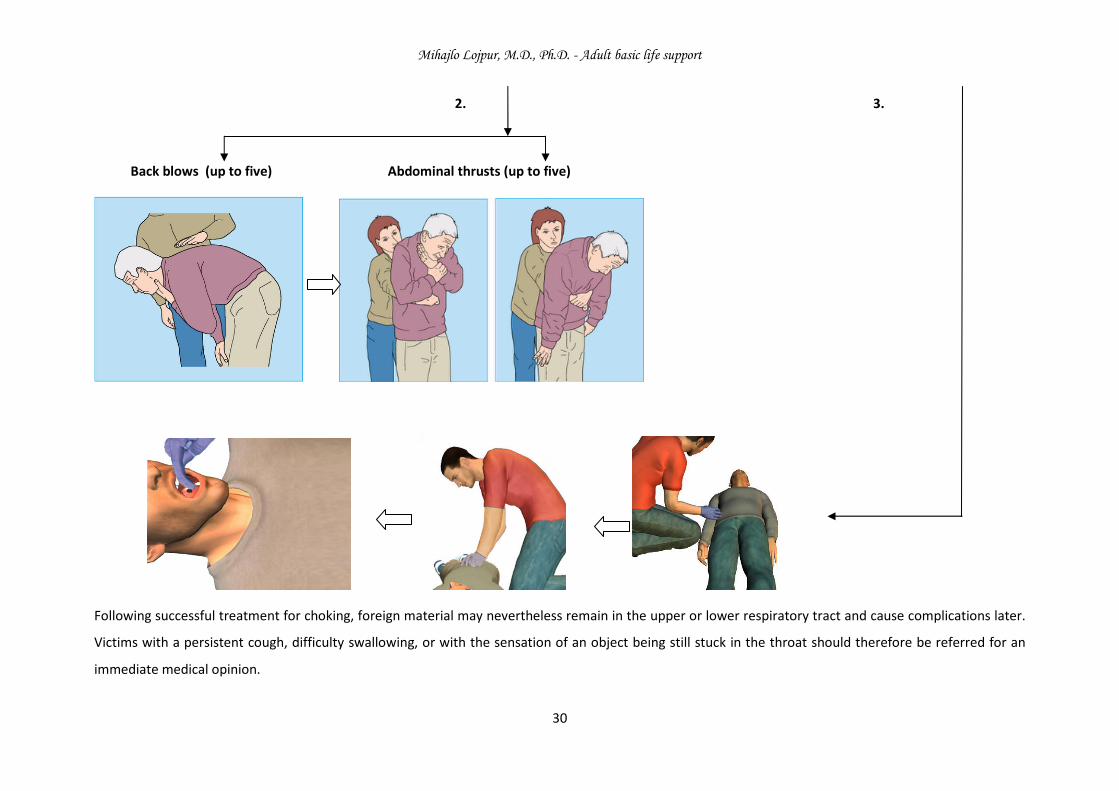
Back blows (up to five) Abdominal thrusts (up to five)
Following successful treatment for choking, foreign material may nevertheless remain in the upper or lower respiratory tract and cause complications later.
Victims with a persistent cough, difficulty swallowing, or with the sensation of an object being still stuck in the throat should therefore be referred for an
immediate medical opinion.
Mihajlo Lojpur, M.D., Ph.D. - Adult basic life support
31
D. Further points related to basic life support
1. Use of oxygen during basic life support
There is no evidence that oxygen administration is of benefit during basic life support in the majority of cases of cardiac arrest before healthcare
professionals are available with equipment to secure the airway.
Its use may lead to interruption in chest compressions, and is not recommended, except in cases of drowning (see below).
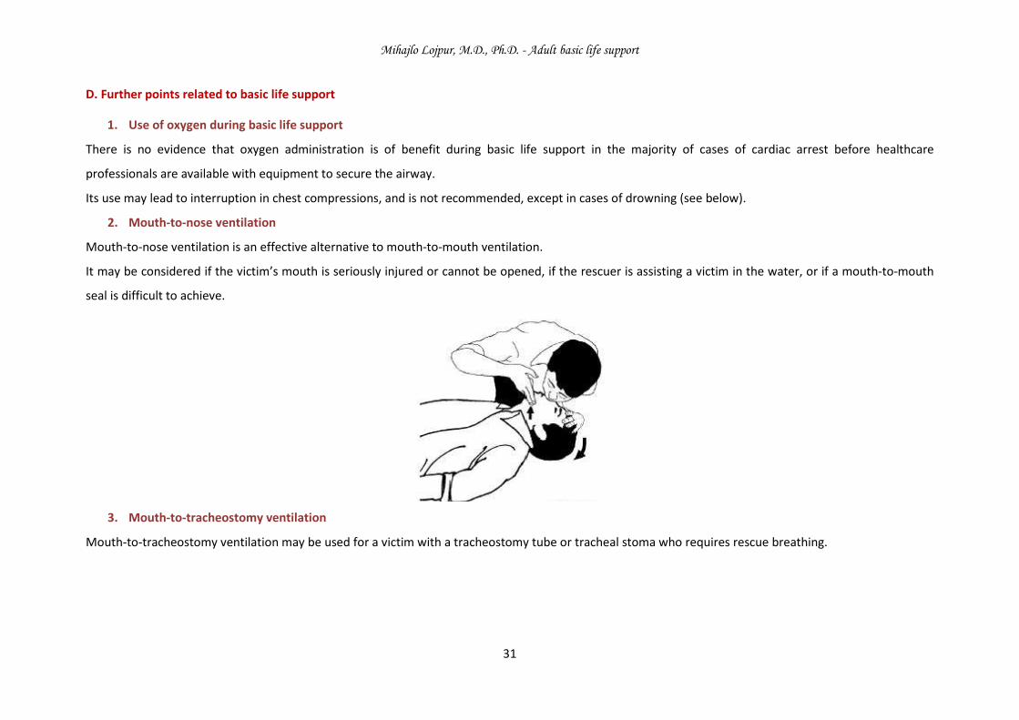
2. Mouth-to-nose ventilation
Mouth-to-nose ventilation is an effective alternative to mouth-to-mouth ventilation.
It may be considered if the victim’s mouth is seriously injured or cannot be opened, if the rescuer is assisting a victim in the water, or if a mouth-to-mouth
seal is difficult to achieve.
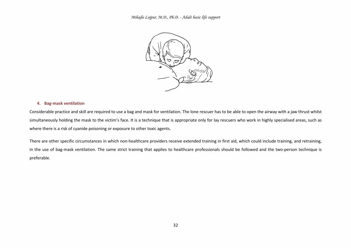
3. Mouth-to-tracheostomy ventilation
Mouth-to-tracheostomy ventilation may be used for a victim with a tracheostomy tube or tracheal stoma who requires rescue breathing.
Mihajlo Lojpur, M.D., Ph.D. - Adult basic life support
32
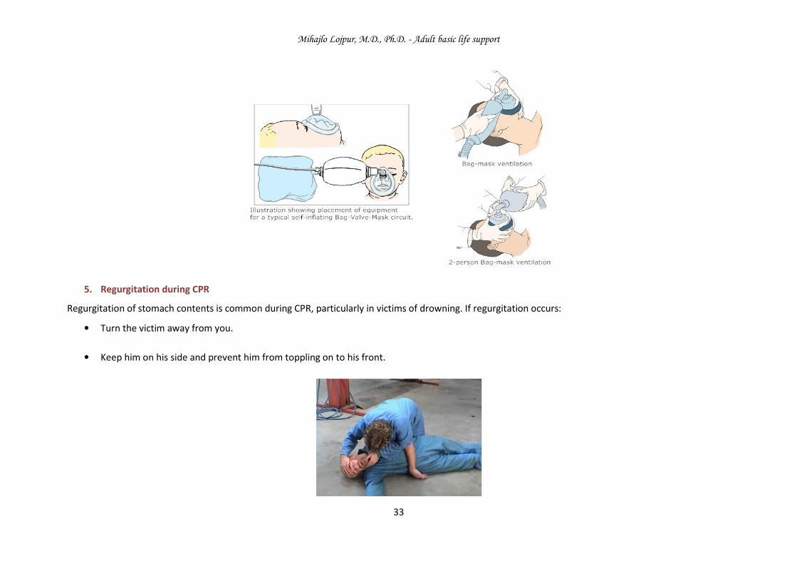
4. Bag-mask ventilation
Considerable practice and skill are required to use a bag and mask for ventilation. The lone rescuer has to be able to open the airway with a jaw thrust whilst
simultaneously holding the mask to the victim’s face. It is a technique that is appropriate only for lay rescuers who work in highly specialised areas, such as
where there is a risk of cyanide poisoning or exposure to other toxic agents.
There are other specific circumstances in which non-healthcare providers receive extended training in first aid, which could include training, and retraining,
in the use of bag-mask ventilation. The same strict training that applies to healthcare professionals should be followed and the two-person technique is
preferable.
Mihajlo Lojpur, M.D., Ph.D. - Adult basic life support
33
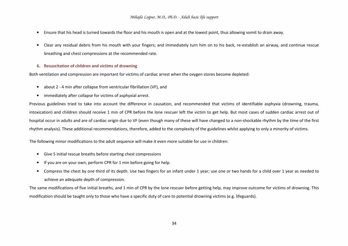
5. Regurgitation during CPR
Regurgitation of stomach contents is common during CPR, particularly in victims of drowning. If regurgitation occurs:
• Turn the victim away from you.
• Keep him on his side and prevent him from toppling on to his front.
Mihajlo Lojpur, M.D., Ph.D. - Adult basic life support
34
• Ensure that his head is turned towards the floor and his mouth is open and at the lowest point, thus allowing vomit to drain away.
• Clear any residual debris from his mouth with your fingers; and immediately turn him on to his back, re-establish an airway, and continue rescue
breathing and chest compressions at the recommended rate.
6. Resuscitation of children and victims of drowning
Both ventilation and compression are important for victims of cardiac arrest when the oxygen stores become depleted:
• about 2 - 4 min after collapse from ventricular fibrillation (VF), and
• immediately after collapse for victims of asphyxial arrest.
Previous guidelines tried to take into account the difference in causation, and recommended that victims of identifiable asphyxia (drowning, trauma,
intoxication) and children should receive 1 min of CPR before the lone rescuer left the victim to get help. But most cases of sudden cardiac arrest out of
hospital occur in adults and are of cardiac origin due to VF (even though many of these will have changed to a non-shockable rhythm by the time of the first
rhythm analysis). These additional recommendations, therefore, added to the complexity of the guidelines whilst applying to only a minority of victims.
The following minor modifications to the adult sequence will make it even more suitable for use in children:
• Give 5 initial rescue breaths before starting chest compressions
• If you are on your own, perform CPR for 1 min before going for help.
• Compress the chest by one third of its depth. Use two fingers for an infant under 1 year; use one or two hands for a child over 1 year as needed to
achieve an adequate depth of compression.
The same modifications of five initial breaths, and 1 min of CPR by the lone rescuer before getting help, may improve outcome for victims of drowning. This
modification should be taught only to those who have a specific duty of care to potential drowning victims (e.g. lifeguards).
Mihajlo Lojpur, M.D., Ph.D. - Adult basic life support
35
If supplemental oxygen is available, and can be brought to the victim and used without interruption in CPR (e.g., by attaching to a resuscitation face mask), it
may be of benefit.
Drowning is easily identified. It can be difficult, on the other hand, for a layperson to recognise when trauma or intoxication has caused cardiorespiratory
arrest. If either cause is suspected the victim should be managed according to the standard BLS protocol.
* Many children do not receive resuscitation because potential rescuers fear causing harm. This fear is unfounded; it is far better to use the adult BLS
sequence for resuscitation of a child than to do nothing. For ease of teaching and retention, laypeople should be taught to use the adult sequence for
children who are not responsive and not breathing normally, with the single modification that the chest should be compressed by one third of its depth.
7. Defibrillation if the victim is wet
As long as there is no direct contact between the user and the victim when the shock is delivered, there is no direct pathway that the electricity can take
that would cause the user to experience a shock.
Dry the victim’s chest so that the adhesive AED pads will stick and take particular care to ensure that no one is touching the victim when a shock is delivered.
8. Defibrillation in the presence of supplemental oxygen
There are no reports of fires caused by sparking where defibrillation was delivered using adhesive pads.
If supplemental oxygen is being delivered by a face mask, remove the face mask and place it at least one metre away before delivering a shock. Do not allow
this to delay shock delivery.
9. Risks to the rescuer and victim
The safety of both the rescuer and victim are paramount during a resuscitation attempt. There have been few incidents of rescuers suffering adverse effects
from undertaking CPR, with only isolated reports of infections such as tuberculosis (TB) and severe acute respiratory distress syndrome (SARS). Transmission
of HIV during CPR has never been reported.
Mihajlo Lojpur, M.D., Ph.D. - Adult basic life support
36
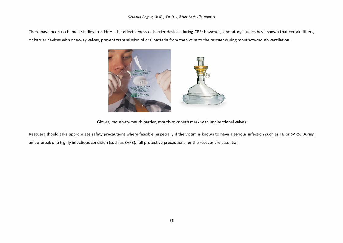
There have been no human studies to address the effectiveness of barrier devices during CPR; however, laboratory studies have shown that certain filters,
or barrier devices with one-way valves, prevent transmission of oral bacteria from the victim to the rescuer during mouth-to-mouth ventilation.
Gloves, mouth-to-mouth barrier, mouth-to-mouth mask with undirectional valves
Rescuers should take appropriate safety precautions where feasible, especially if the victim is known to have a serious infection such as TB or SARS. During
an outbreak of a highly infectious condition (such as SARS), full protective precautions for the rescuer are essential.