Embed Size (px)
Citation preview
ADVANCES IN DIAGNOSES OF INFECTIOUS DISEASE IN DOGS IN CLINICAL PRACTICE THROUGH Molecular biology POC
DVM VITOR MÁRCIO RIBEIRO
THE CHALLENGE OF MEDICAL PRACTICE
Knowledge Diagnosis Management – Infection stage identification - Treatment Control Prevention

KNOWLEDGE X EXPERIENCE
The only source of
knowledge is
experience
ESSENTIAL ITEMS
Knowledge Diagnosis Management - Infection stage identification - Treatment Control Prevention
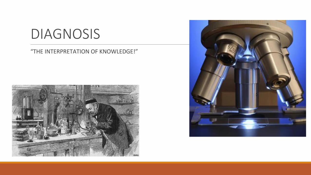
DIAGNOSIS
“THE INTERPRETATION OF KNOWLEDGE!”
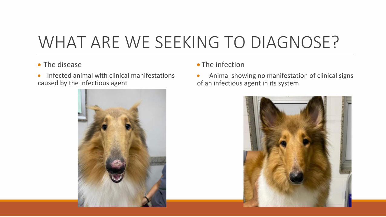
WHAT ARE WE SEEKING TO DIAGNOSE?
The disease
Infected animal with clinical manifestations caused by the infectious agent
The infection Animal showing no manifestation of clinical signs of an infectious agent in its system
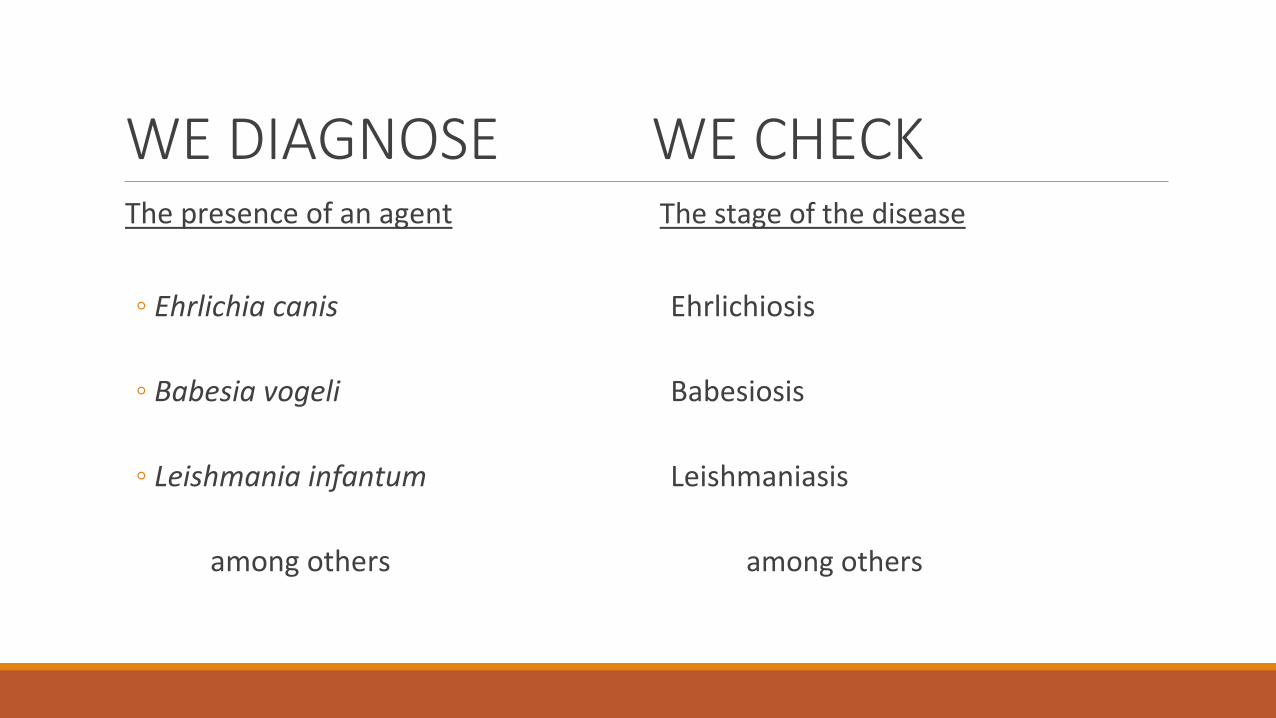
WE DIAGNOSE WE CHECK
The presence of an agent The stage of the disease
◦ Ehrlichia canis Ehrlichiosis
◦ Babesia vogeli Babesiosis
◦ Leishmania infantum Leishmaniasis
among others among others
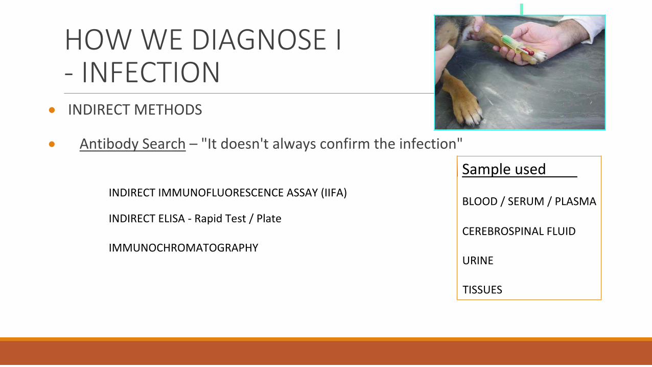
HOW WE DIAGNOSE I - INFECTION
INDIRECT METHODS Antibody Search – "It doesn't always confirm the infection"
INDIRECT IMMUNOFLUORESCENCE ASSAY (IIFA)
Sample used BLOOD / SERUM / PLASMA
INDIRECT ELISA - Rapid Test / Plate CEREBROSPINAL FLUID
IMMUNOCHROMATOGRAPHY URINE
TISSUES
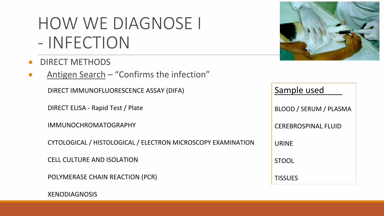
HOW WE DIAGNOSE I - INFECTION
DIRECT METHODS Antigen Search – “Confirms the infection”
DIRECT IMMUNOFLUORESCENCE ASSAY (DIFA)
DIRECT ELISA - Rapid Test / Plate
IMMUNOCHROMATOGRAPHY
CYTOLOGICAL / HISTOLOGICAL / ELECTRON MICROSCOPY EXAMINATION
CELL CULTURE AND ISOLATION
POLYMERASE CHAIN REACTION (PCR)
XENODIAGNOSIS
Sample used
BLOOD / SERUM / PLASMA
CEREBROSPINAL FLUID
URINE
STOOL
TISSUES
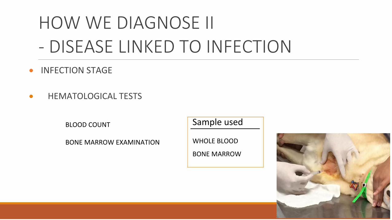
HOW WE DIAGNOSE II - DISEASE LINKED TO INFECTION
INFECTION STAGE HEMATOLOGICAL TESTS
BLOOD COUNT
BONE MARROW EXAMINATION
Sample used
WHOLE BLOOD
BONE MARROW
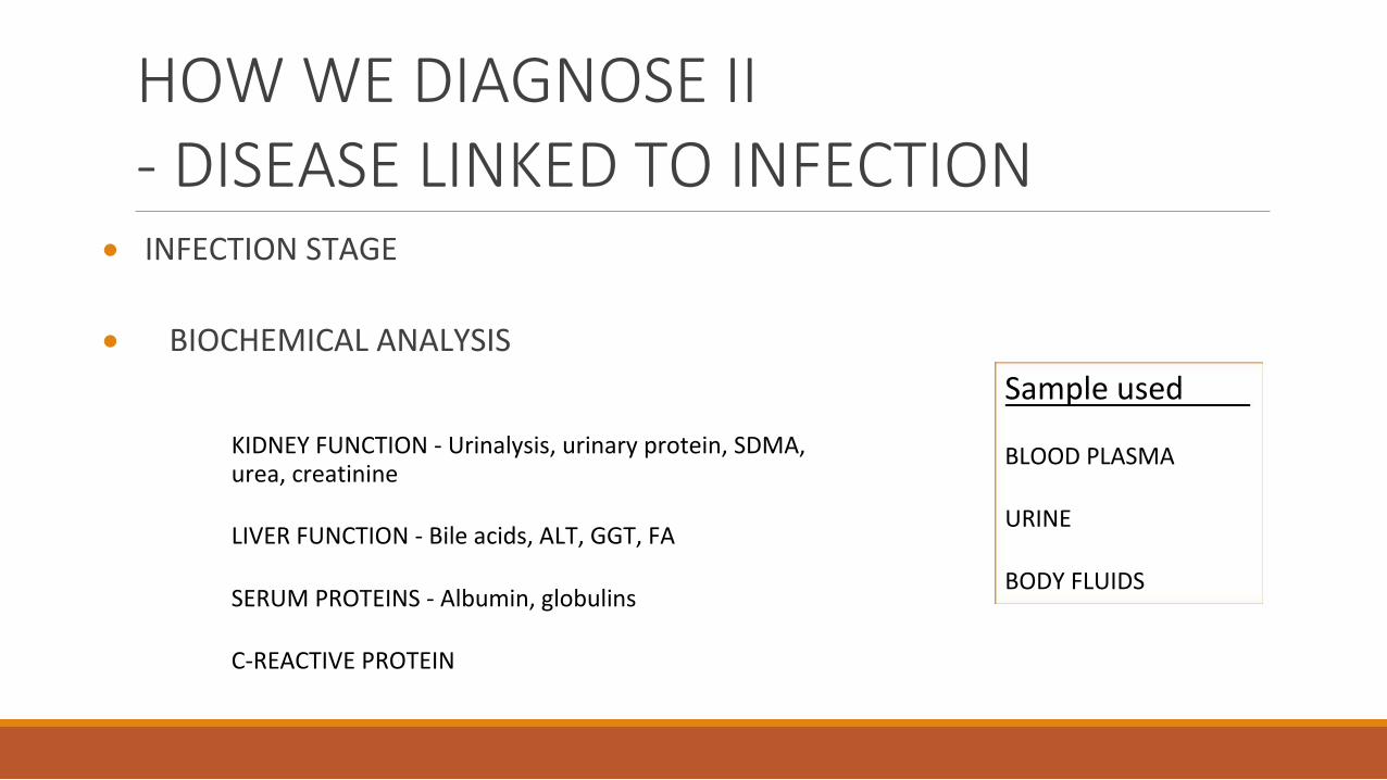
HOW WE DIAGNOSE II - DISEASE LINKED TO INFECTION
INFECTION STAGE BIOCHEMICAL ANALYSIS
KIDNEY FUNCTION - Urinalysis, urinary protein, SDMA, urea, creatinine
LIVER FUNCTION - Bile acids, ALT, GGT, FA
SERUM PROTEINS - Albumin, globulins
C-REACTIVE PROTEIN
Sample used
BLOOD PLASMA
URINE
BODY FLUIDS
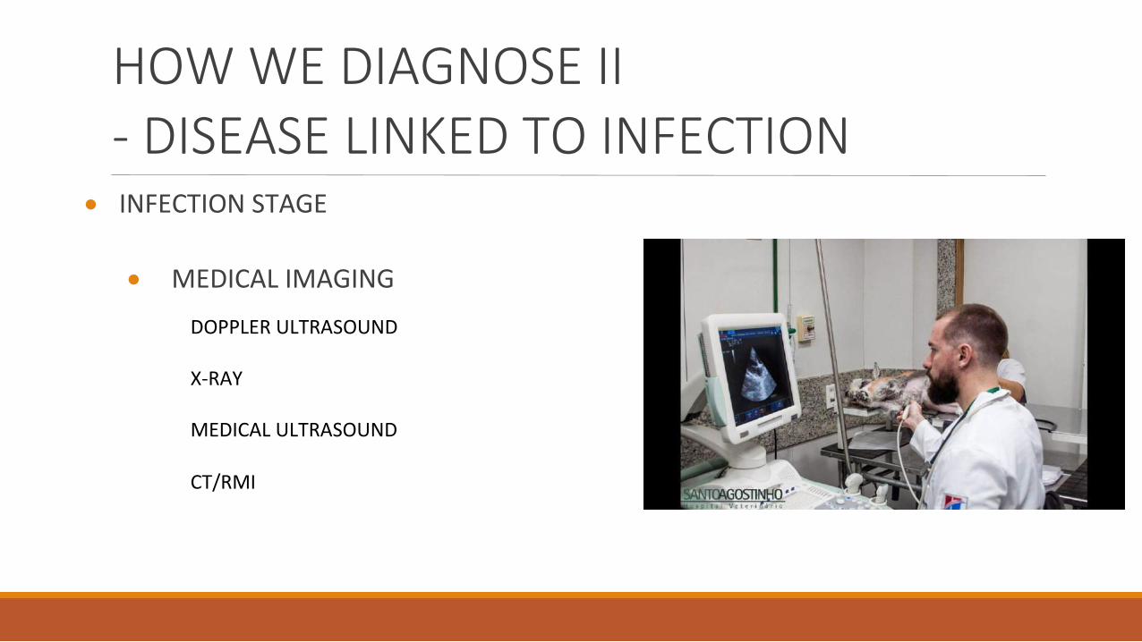
HOW WE DIAGNOSE II - DISEASE LINKED TO INFECTION
INFECTION STAGE
MEDICAL IMAGING
DOPPLER ULTRASOUND
X-RAY
MEDICAL ULTRASOUND CT/RMI

NEEDS AND ADVANCES
MORE SENSITIVE ANALYSES
SPECIFIC ANALYSES FASTER METHODS AVAILABLE TECHNIQUES SUITABLE COST
MOLECULAR DIAGNOSIS
PCR
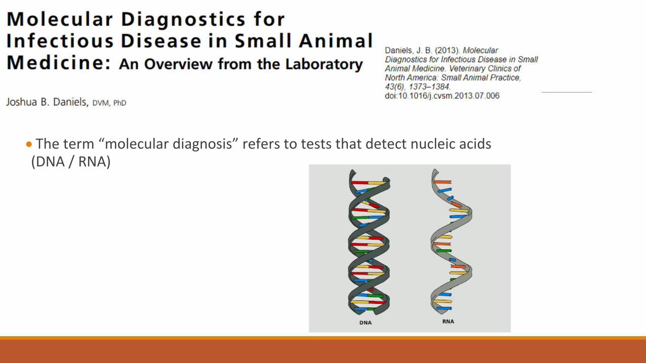
The term “molecular diagnosis” refers to tests that detect nucleic acids (DNA / RNA)
The methods most commonly used by diagnostic laboratories for detection and characterization of nucleic acids are:
◦ 1. Polymerase Chain Reaction (PCR) ◦ 2. Quantitative Real-Time PCR (qPCR) ◦ 3. Reverse Transcription PCR (RT-PCR) ◦ 4. Duplex and Multiplex Real-Time PCR ◦ 5. DNA Sequencing
IS MOLECULAR DIAGNOSTICS A SOLUTION?
Molecular diagnostic tests offer some advantages depending on the infection and the course of the disease, but it is essential to understand their limitations Clinicians must properly order molecular diagnostic tests according to the course of the disease Other diagnostic tests should not be abandoned because they can provide valuable incidental information
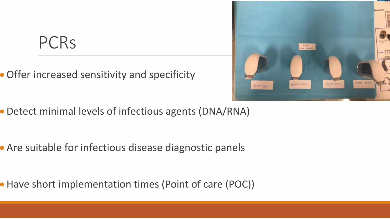
PCRs
Offer increased sensitivity and specificity Detect minimal levels of infectious agents (DNA/RNA) Are suitable for infectious disease diagnostic panels
Have short implementation times (Point of care (POC))
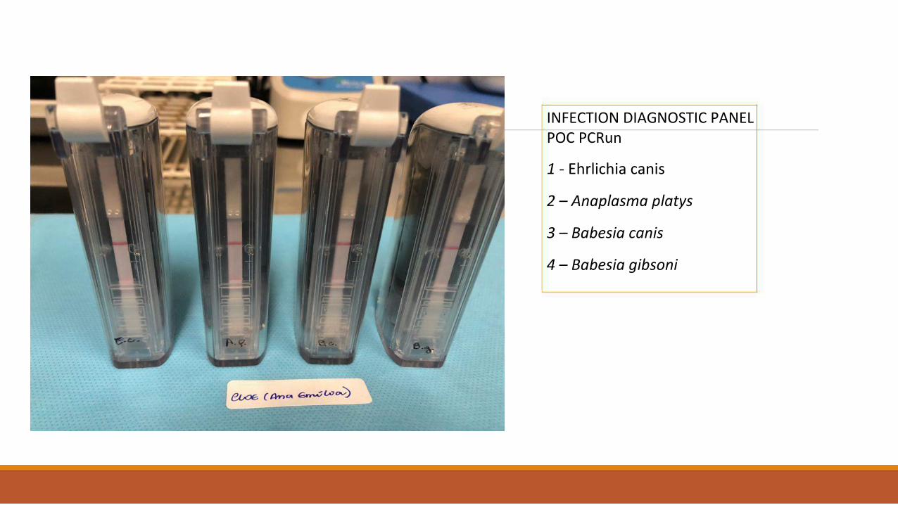
INFECTION DIAGNOSTIC PANEL POC PCRun
1 - Ehrlichia canis
2 – Anaplasma platys
3 – Babesia canis
4 – Babesia gibsoni
Are better able to diagnose subclinical infections There is a need to improve techniques that allow Lower costs
Smaller laboratory structure
Large-scale utilization
WHAT HAPPENS IN AN INFECTION
VIRAL INFECTION
The viral messenger RNA enters the ribosome causing the cell to begin producing the proteins of
viral reproduction Viruses are obligate intracellular parasites: the lack of hyaloplasm and ribosomes prevents them
from having metabolism In their life cycle, viruses need a cellular environment containing ribosomes and other necessary
substances to synthesize viral proteins, allowing multiplication of their genetic material
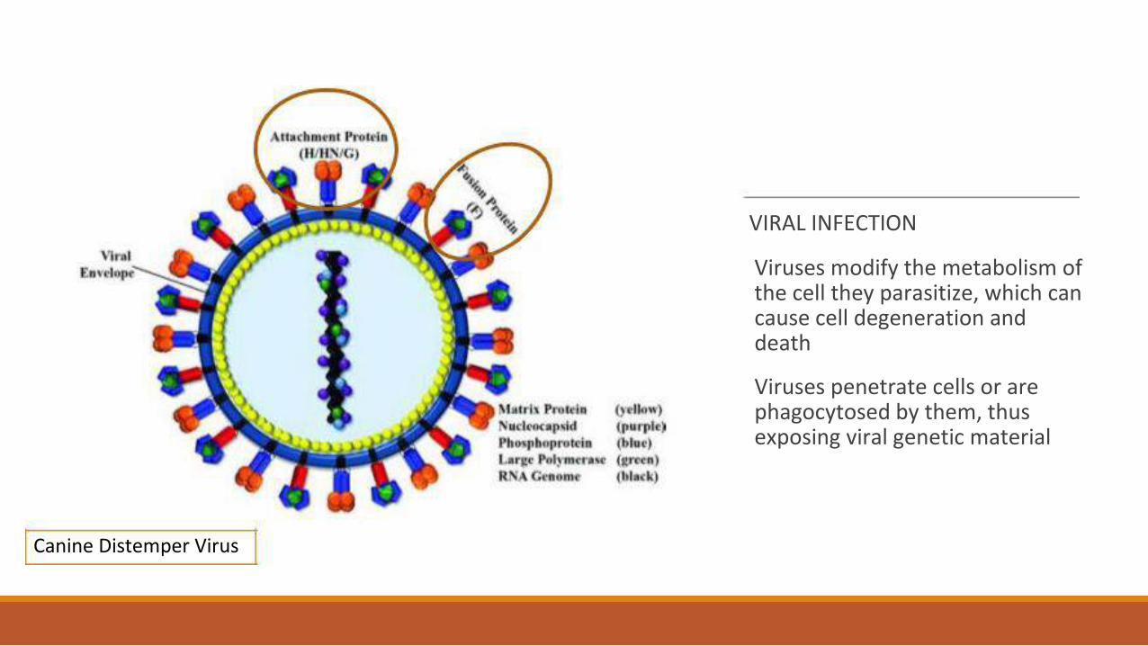
VIRAL INFECTION
Viruses modify the metabolism of the cell they parasitize, which can cause cell degeneration and death Viruses penetrate cells or are phagocytosed by them, thus exposing viral genetic material
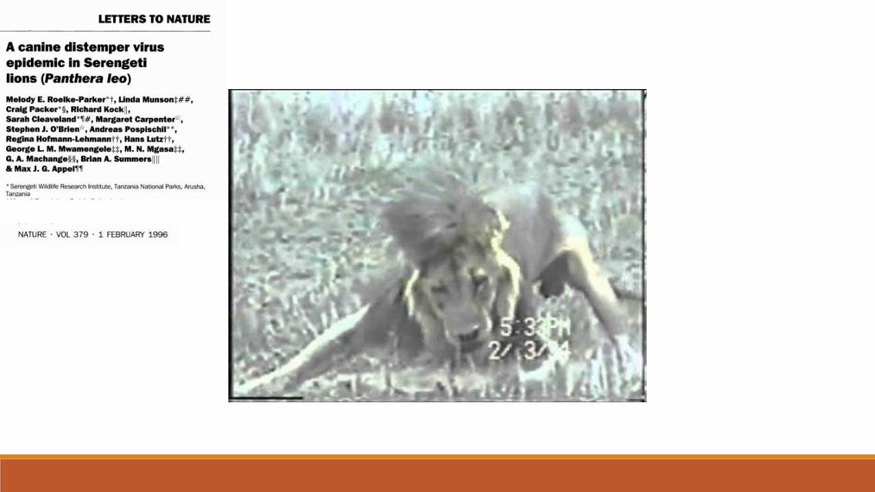
Canine Distemper Virus

CLINICAL CASE


available at:
https://www.researchgate.net/publication/14641773_A_canin
e_distemper_virus_epidemic_in_Serengeti_lions_Panthera_leo
[accessed December 8, 2020].
Extreme climatic conditions can change historical relationships between host and pathogen, and synchronize temporal and spatial convergence of multiple infectious agents, triggering more deadly epidemics than those due to single pathogens.
In 1994, A CDV epidemic affected Serengeti lions (Panthera leo), killing 1/3 of the population In 2001, a second high-mortality CDV epidemic hit the lion population of Ngorongoro Crater
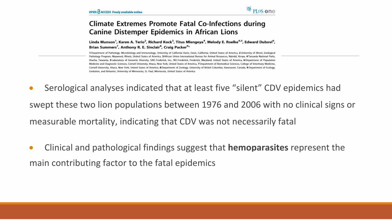
Serological analyses indicated that at least five “silent” CDV epidemics had
swept these two lion populations between 1976 and 2006 with no clinical signs or
measurable mortality, indicating that CDV was not necessarily fatal Clinical and pathological findings suggest that hemoparasites represent the
main contributing factor to the fatal epidemics

Using quantitative real-time PCR, we measured the magnitude of infections with hemoparasites in these populations over 22 years Significantly high levels of Babesia were observed during the 1994 and 2001 epidemics when, in addition to CDV exposure within the groups, conditions of extreme drought along with high mortality of herbivores and high outbreaks of ticks were also observed Lions were infected by an unusual number of Babesia parasites, which were more pathogenic due to the immunosuppressive effect of CDV co-infection, leading to high mortality These mass mortality events may become increasingly common if extreme weather changes disrupt the stable historical relationship between coexisting pathogens and their susceptible hosts
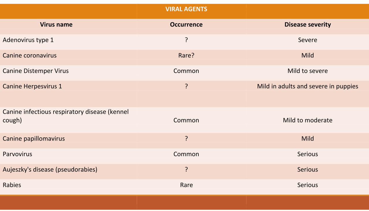
VIRAL AGENTS
Virus name Occurrence Disease severity
Adenovirus type 1 ? Severe
Canine coronavirus Rare? Mild
Canine Distemper Virus Common Mild to severe
Canine Herpesvirus 1 ? Mild in adults and severe in puppies
Canine infectious respiratory disease (kennel cough) Common Mild to moderate
Canine papillomavirus ? Mild
Parvovirus Common Serious
Aujeszky's disease (pseudorabies) ? Serious
Rabies Rare Serious
WHATS HAPPENS IN AN INFECTION
BACTERIAL INFECTION
INTRACELLULAR BACTERIA
Chlamydia spp., Anaplasma spp., Ehrlichia spp., Rickettsia spp., Orientia spp. and Coxiella spp. replicate exclusively within eukaryotic host cells
EXTRACELLULAR BACTERIA
Extracellular bacteria are free-living organisms
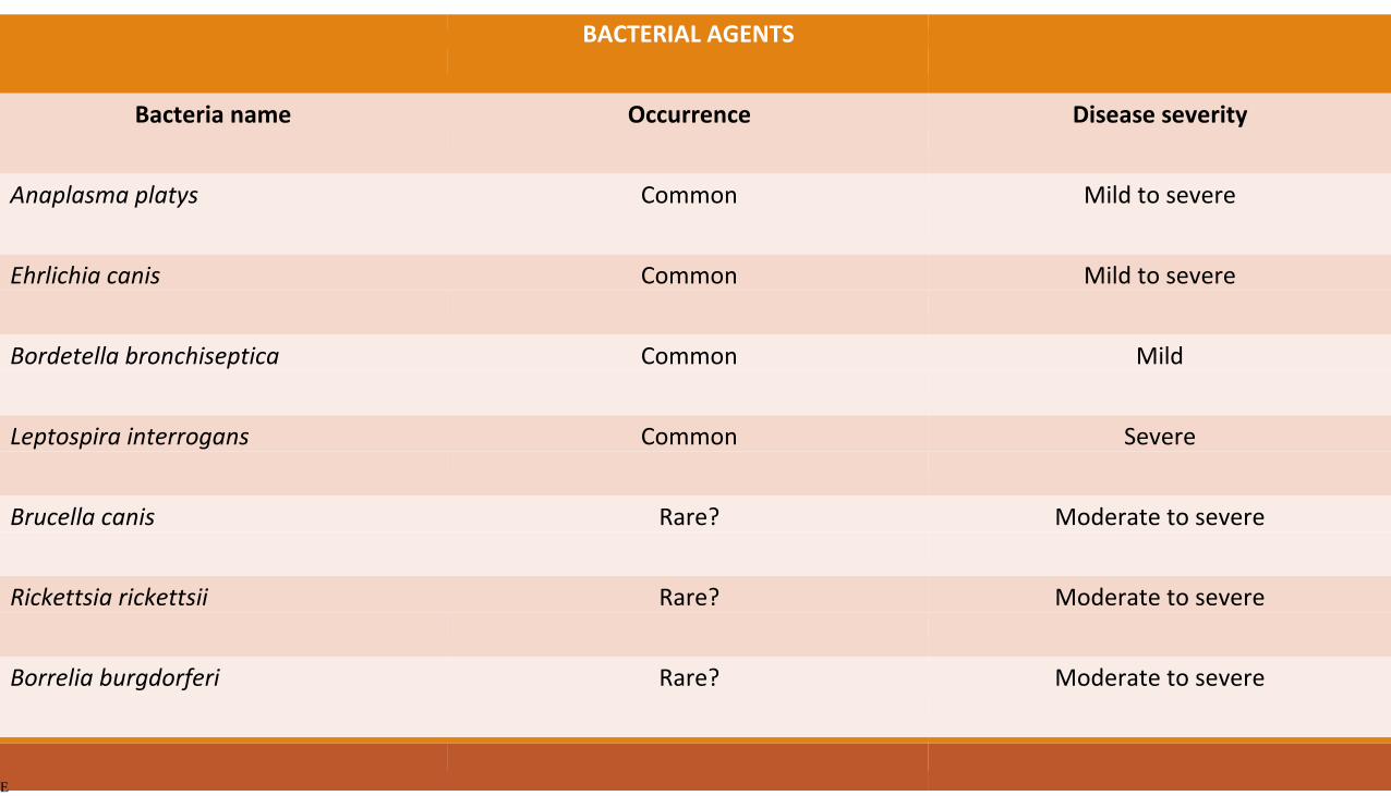
BACTERIAL AGENTS
Bacteria name Occurrence Disease severity
Anaplasma platys Common Mild to severe
Ehrlichia canis Common Mild to severe
Bordetella bronchiseptica Common Mild
Leptospira interrogans Common Severe
Brucella canis Rare? Moderate to severe
Rickettsia rickettsii Rare? Moderate to severe
Borrelia burgdorferi Rare? Moderate to severe
E

Ehrlichia sp
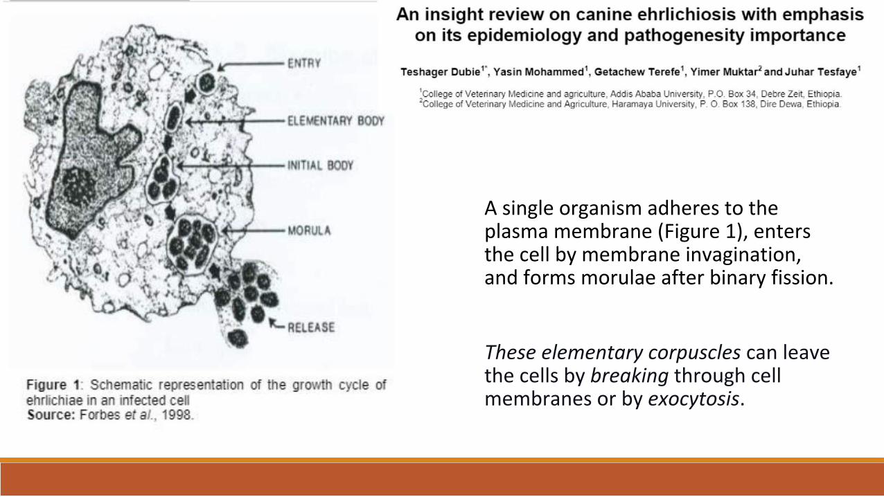
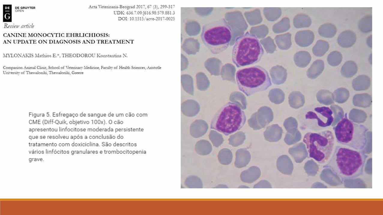
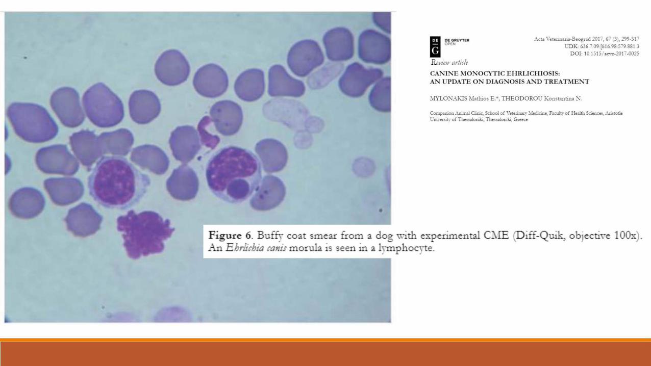
A single organism adheres to the plasma membrane (Figure 1), enters the cell by membrane invagination, and forms morulae after binary fission.
These elementary corpuscles can leave the cells by breaking through cell membranes or by exocytosis.
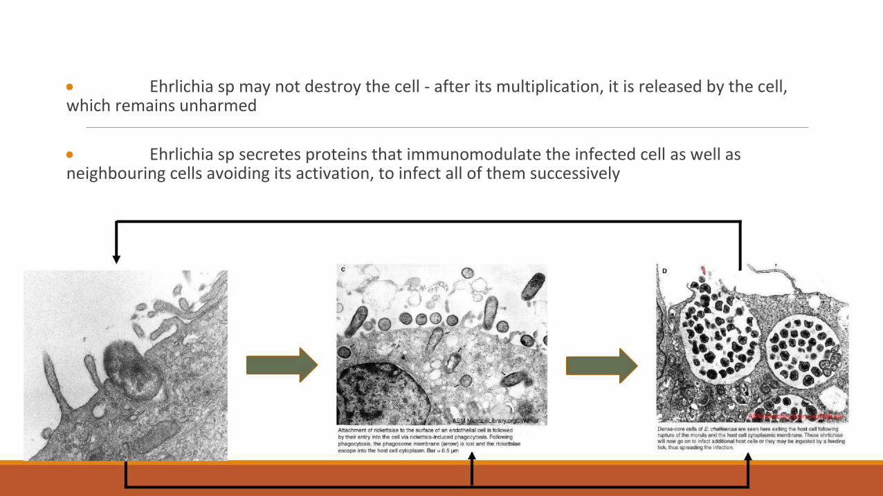
Ehrlichia sp may not destroy the cell - after its multiplication, it is released by the cell, which remains unharmed Ehrlichia sp secretes proteins that immunomodulate the infected cell as well as neighbouring cells avoiding its activation, to infect all of them successively
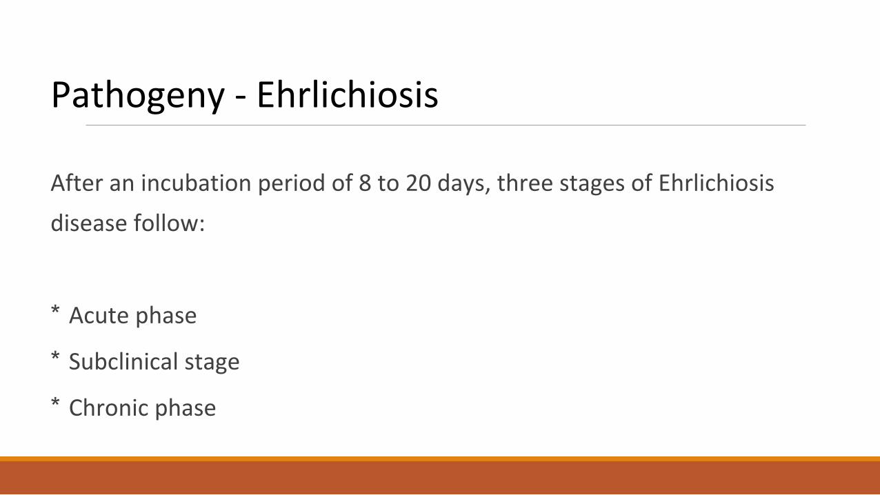
Pathogeny - Ehrlichiosis
After an incubation period of 8 to 20 days, three stages of Ehrlichiosis
disease follow:
* Acute phase * Subclinical stage * Chronic phase
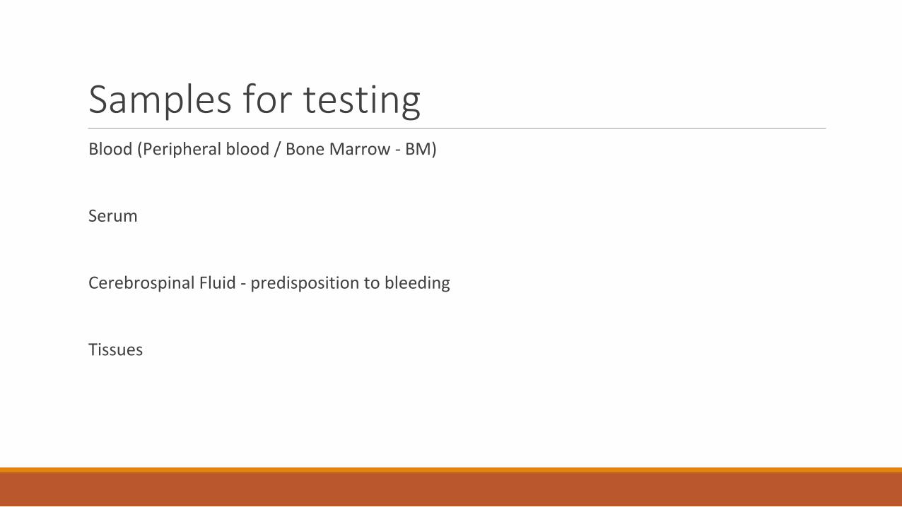
Samples for testing
Blood (Peripheral blood / Bone Marrow - BM)
Serum
Cerebrospinal Fluid - predisposition to bleeding
Tissues
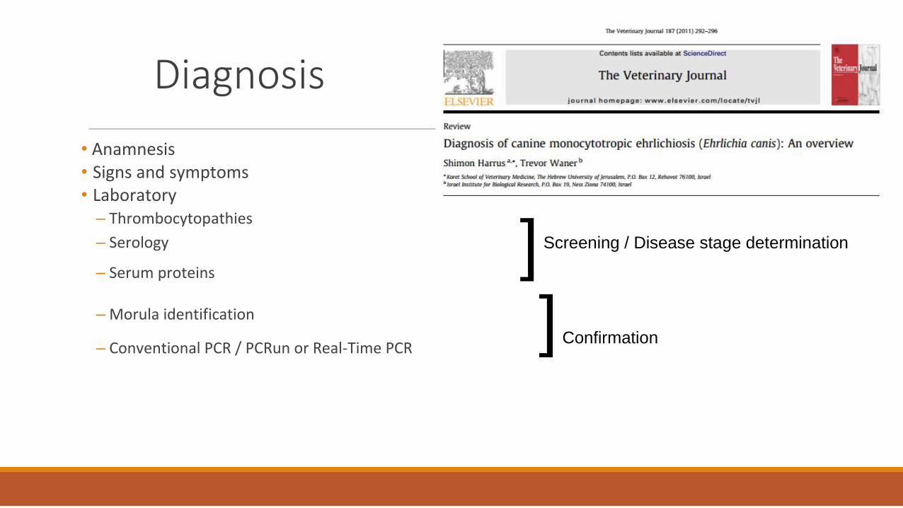
Diagnosis
• Anamnesis
• Signs and symptoms
• Laboratory
– Thrombocytopathies
]
– Serology Screening / Disease stage determination
– Serum proteins
] Confirmation
– Morula identification
– Conventional PCR / PCRun or Real-Time PCR
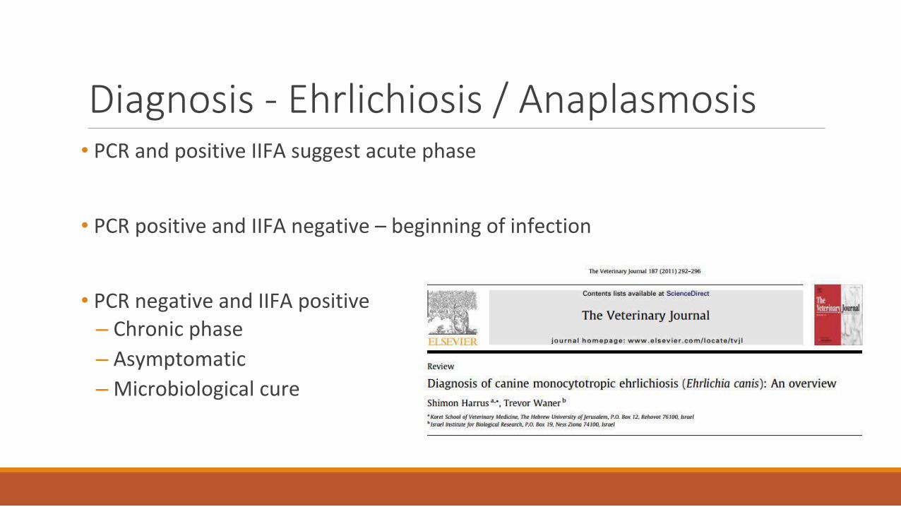
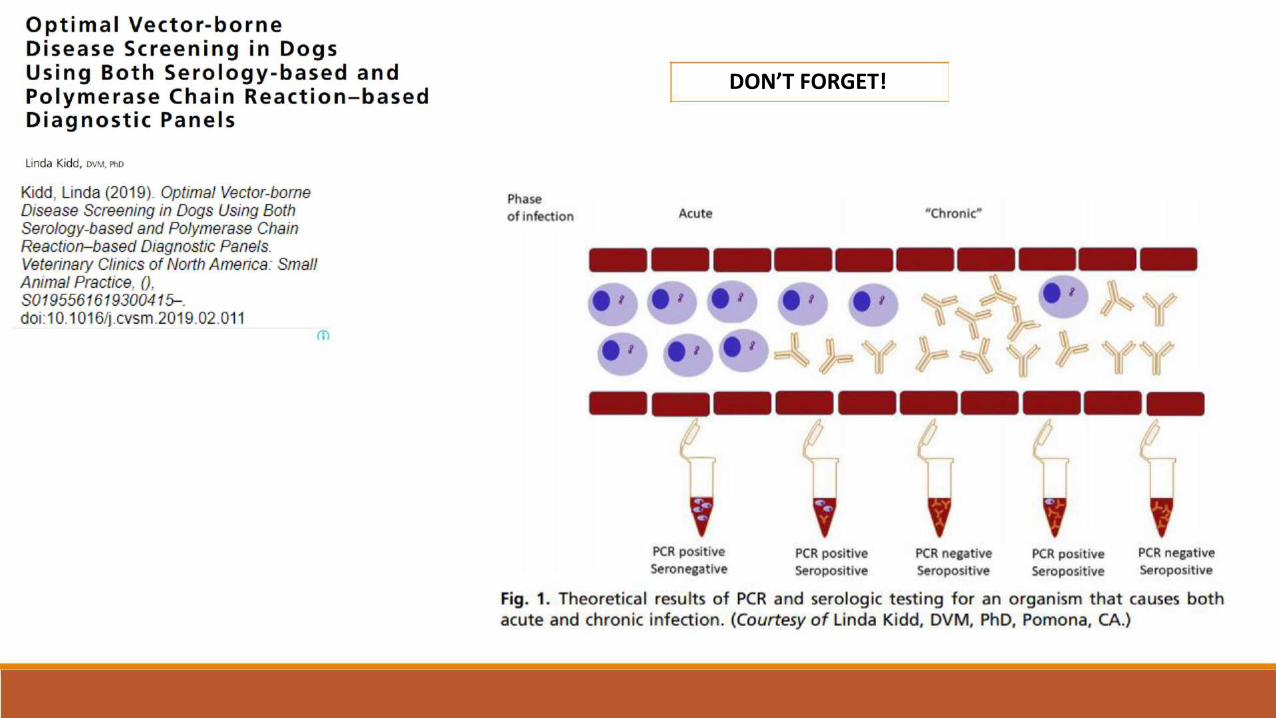
Diagnosis - Ehrlichiosis / Anaplasmosis
• PCR and positive IIFA suggest acute phase
• PCR positive and IIFA negative – beginning of infection
• PCR negative and IIFA positive
– Chronic phase
– Asymptomatic
– Microbiological cure
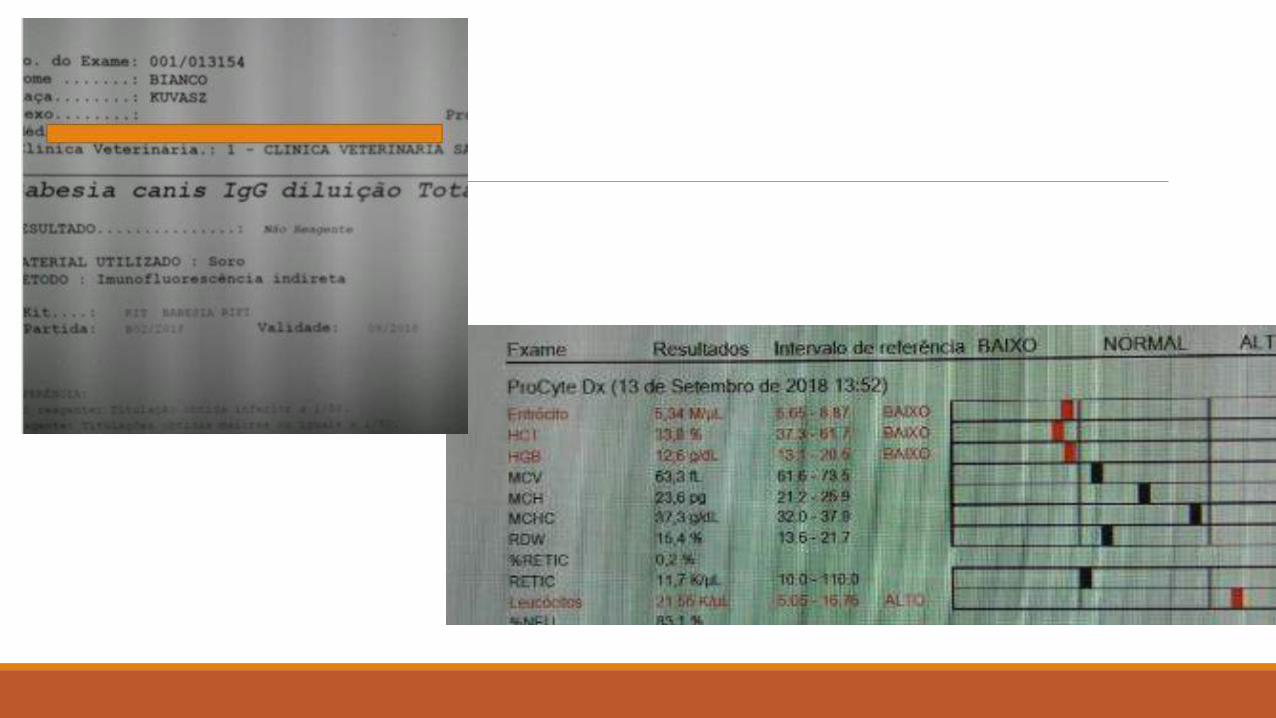
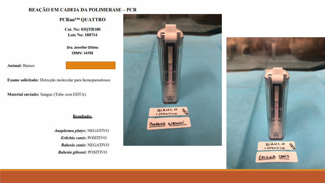
CLINICAL CASE
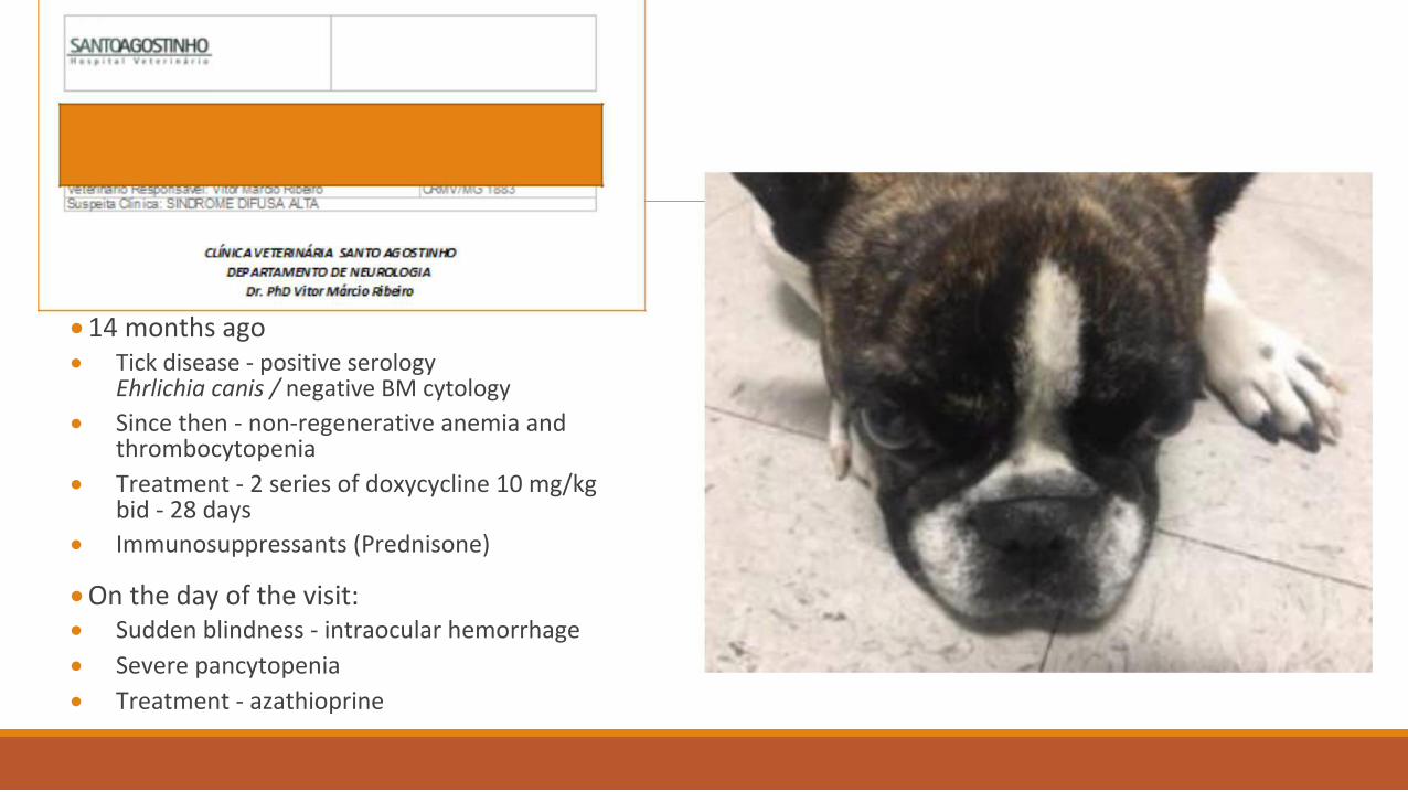
14 months ago
Tick disease - positive serology Ehrlichia canis / negative BM cytology Since then - non-regenerative anemia and thrombocytopenia Treatment - 2 series of doxycycline 10 mg/kg bid - 28 days Immunosuppressants (Prednisone)
On the day of the visit: Sudden blindness - intraocular hemorrhage Severe pancytopenia Treatment - azathioprine
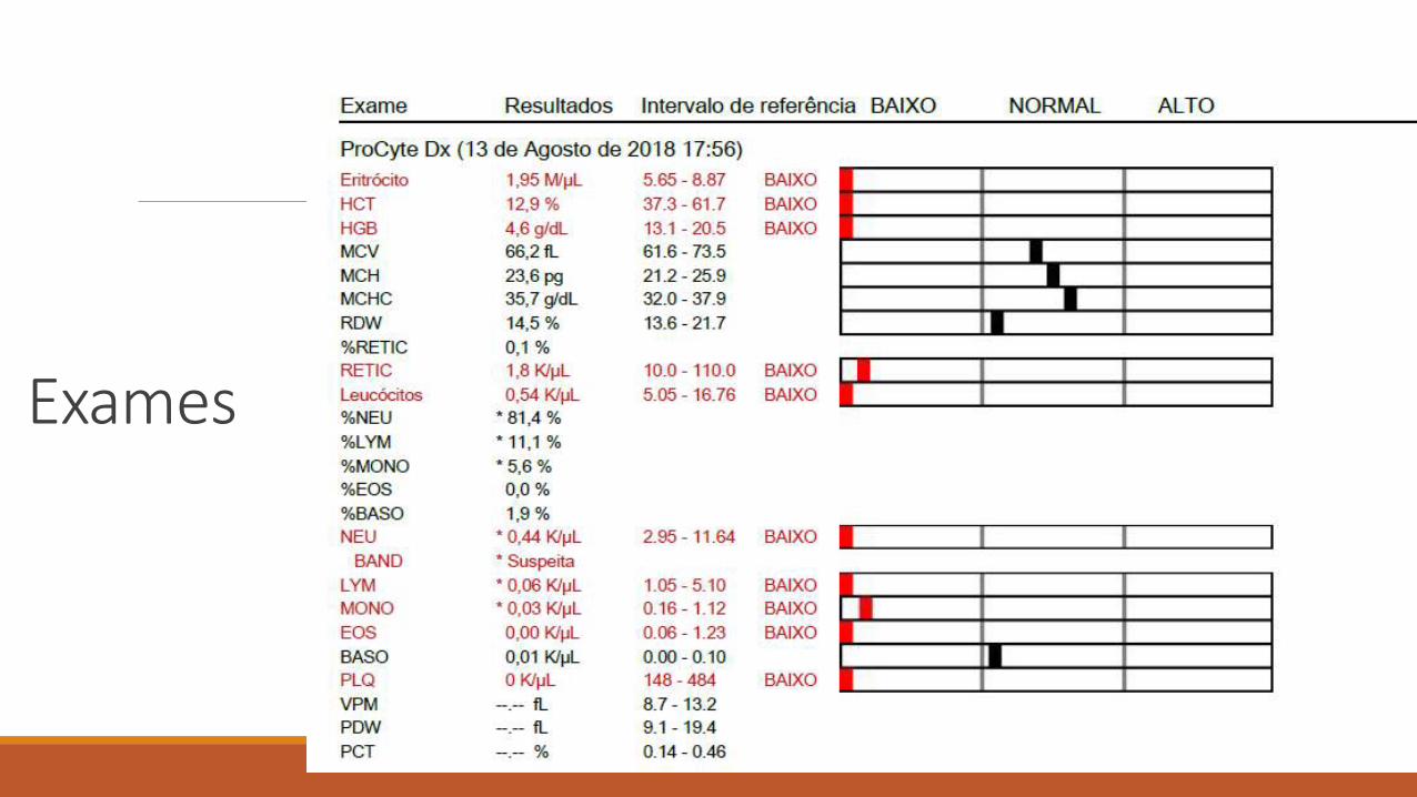
Exames
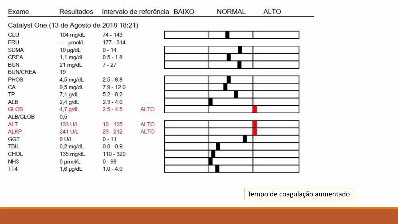
Tempo de coagulação aumentado
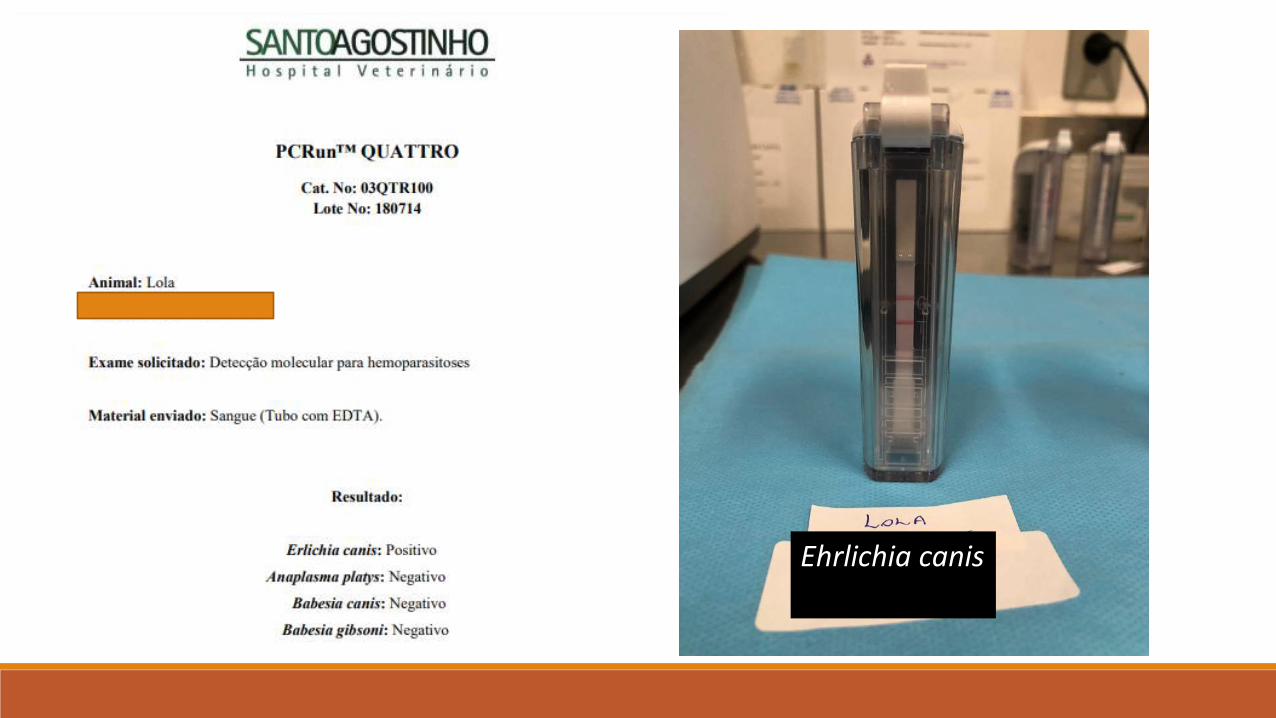
Ehrlichia canis
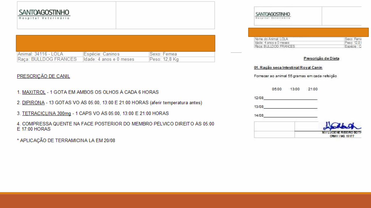
Prescrição
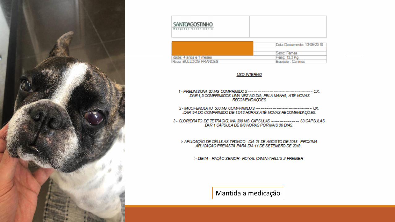
Mantida a medicação
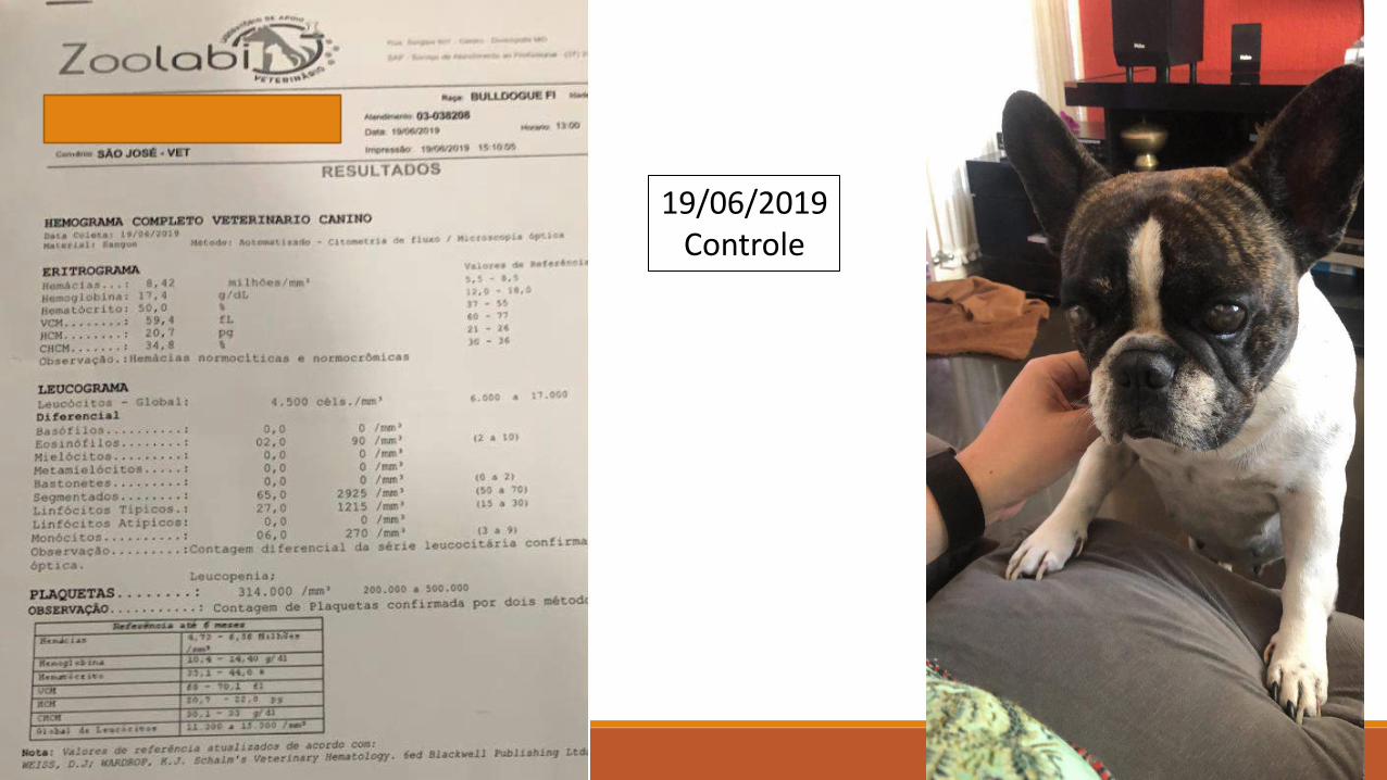
19/06/2019Controle

09/07/2020
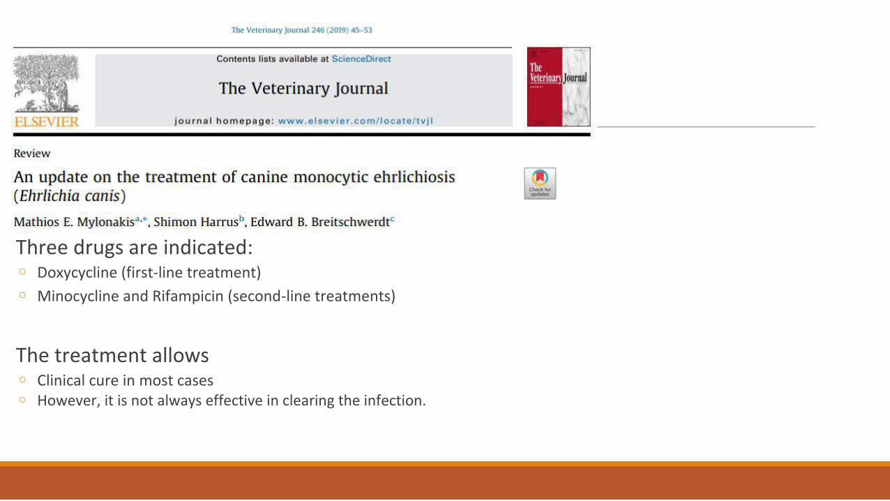
Three drugs are indicated:
◦ Doxycycline (first-line treatment) ◦ Minocycline and Rifampicin (second-line treatments)
The treatment allows
◦ Clinical cure in most cases ◦ However, it is not always effective in clearing the infection.
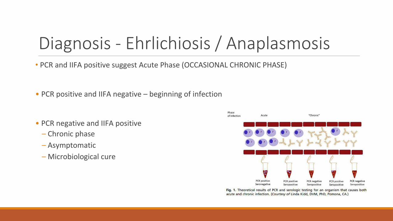
Diagnosis - Ehrlichiosis / Anaplasmosis
• PCR and IIFA positive suggest Acute Phase (OCCASIONAL CHRONIC PHASE)
• PCR positive and IIFA negative – beginning of infection
• PCR negative and IIFA positive – Chronic phase – Asymptomatic – Microbiological cure
Reviews – disease stage determination
Review relapses
Possibility of maintenance of the infection - RECIDIVISM
Follow-up by quantitative serological analyses can be a guide
Blood PCR to control parasitemia

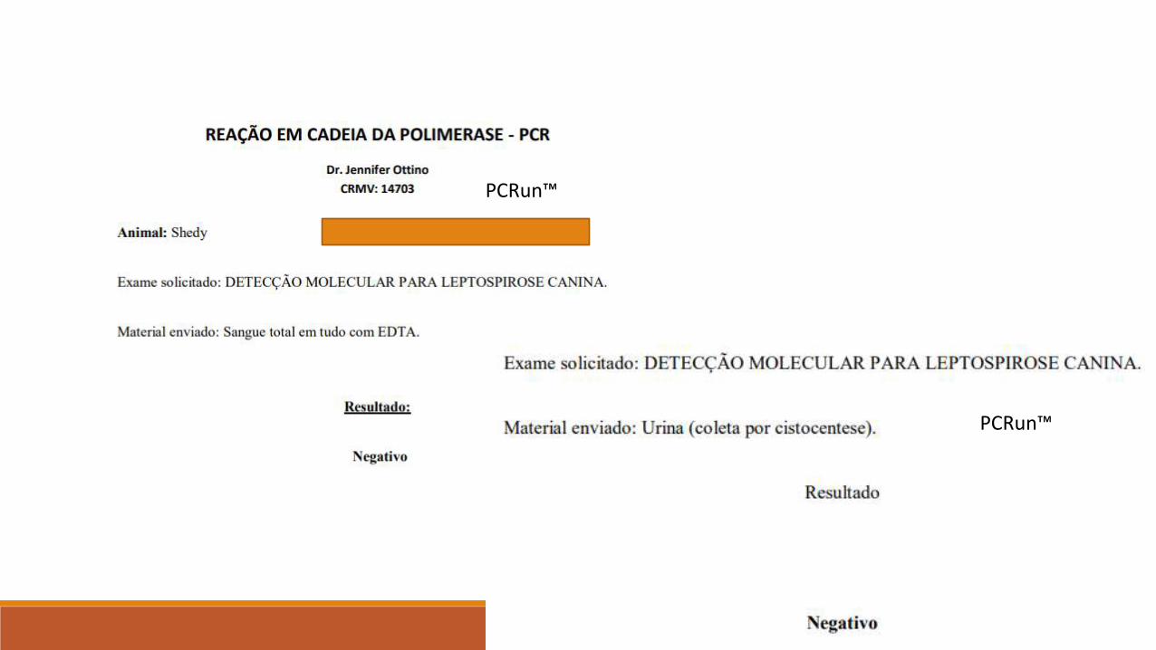
Leptospira interrogans
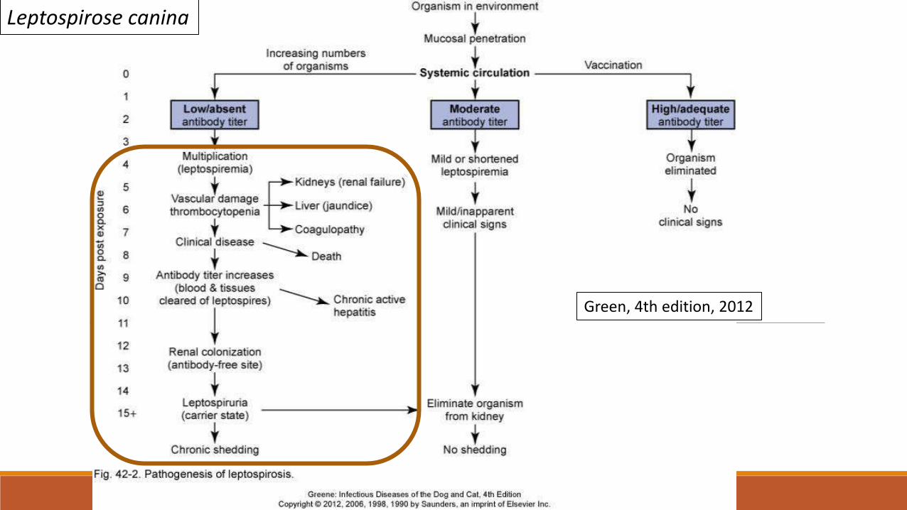
Leptospirose canina
Green, 4th edition, 2012

CLINICAL CASE

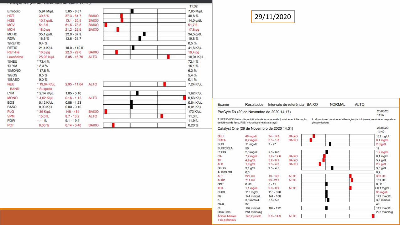
29/11/2020
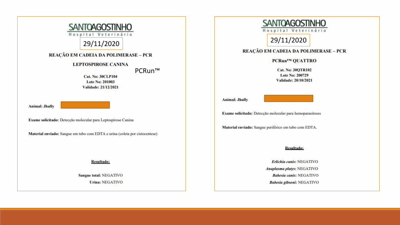
PCRun™
29/11/2020 29/11/2020

PROTOZOAN AGENTS
Bacteria name Occurrence Disease severity
Babesia vogeli Common Mild to severe
Babesia gibsoni Common Mild to severe
Leishmania infantum Common Mild
Rangelia vittali Common Severe
Trypanosoma sp Rare? Moderate to severe
Giardia lamblia (assemblies) Rare? Moderate to severe
Hepatozoon canis Rare? Moderate to severe
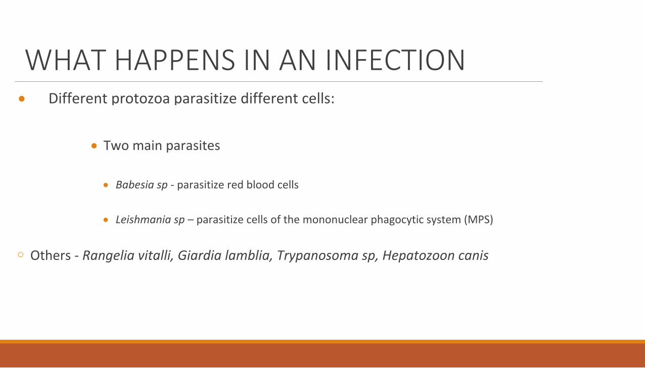
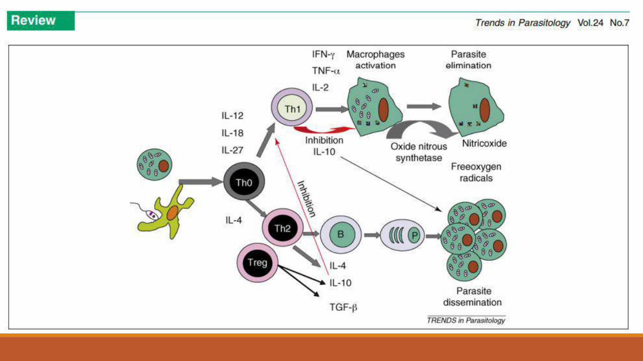
WHAT HAPPENS IN AN INFECTION
Different protozoa parasitize different cells:
Two main parasites
Babesia sp - parasitize red blood cells
Leishmania sp – parasitize cells of the mononuclear phagocytic system (MPS)
◦ Others - Rangelia vitalli, Giardia lamblia, Trypanosoma sp, Hepatozoon canis

Babesia sp
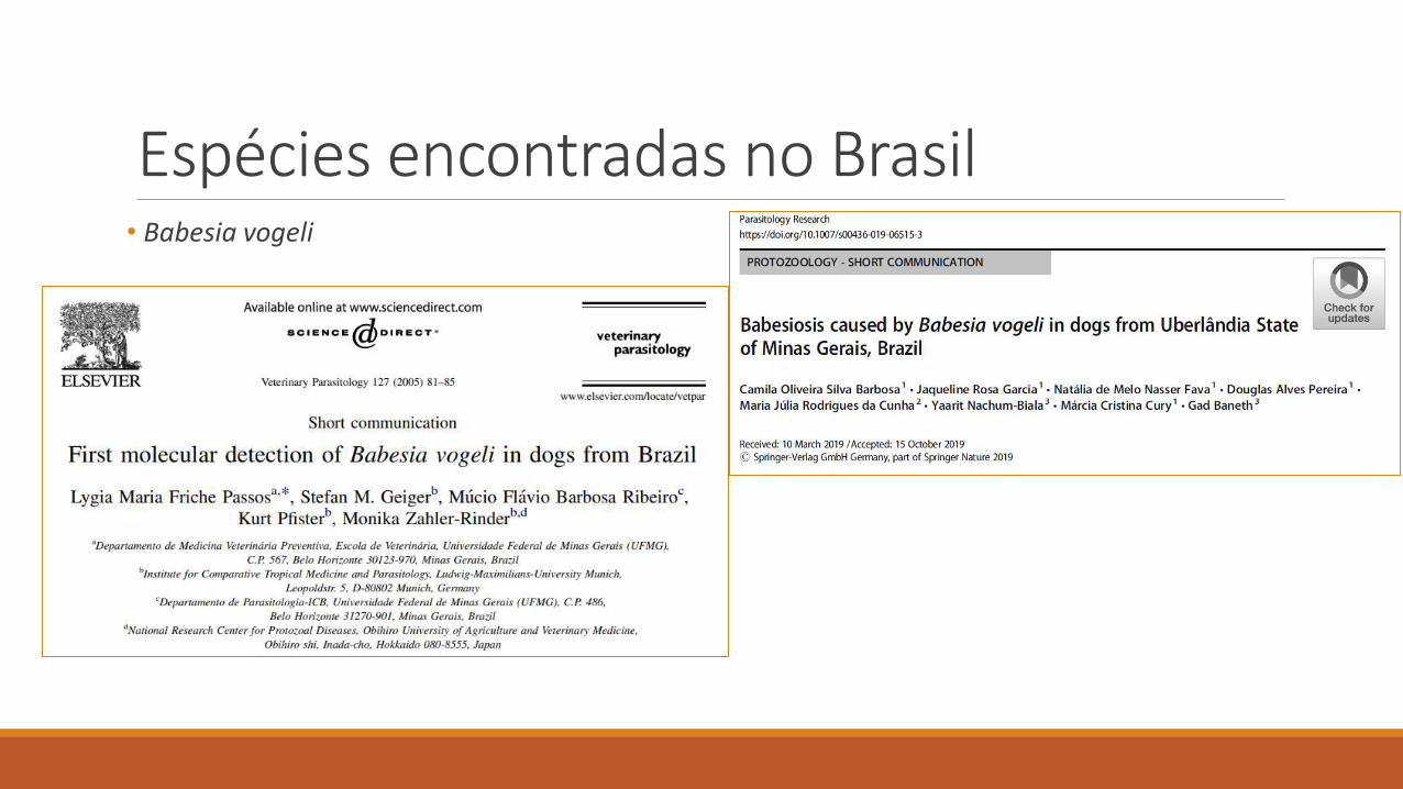
Espécies encontradas no Brasil• Babesia vogeli
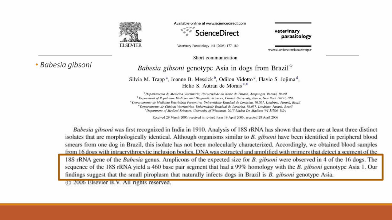
• Babesia gibsoni
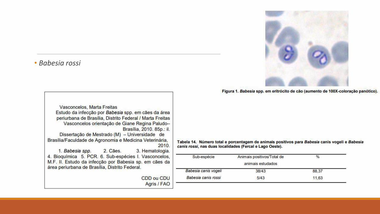
• Babesia rossi
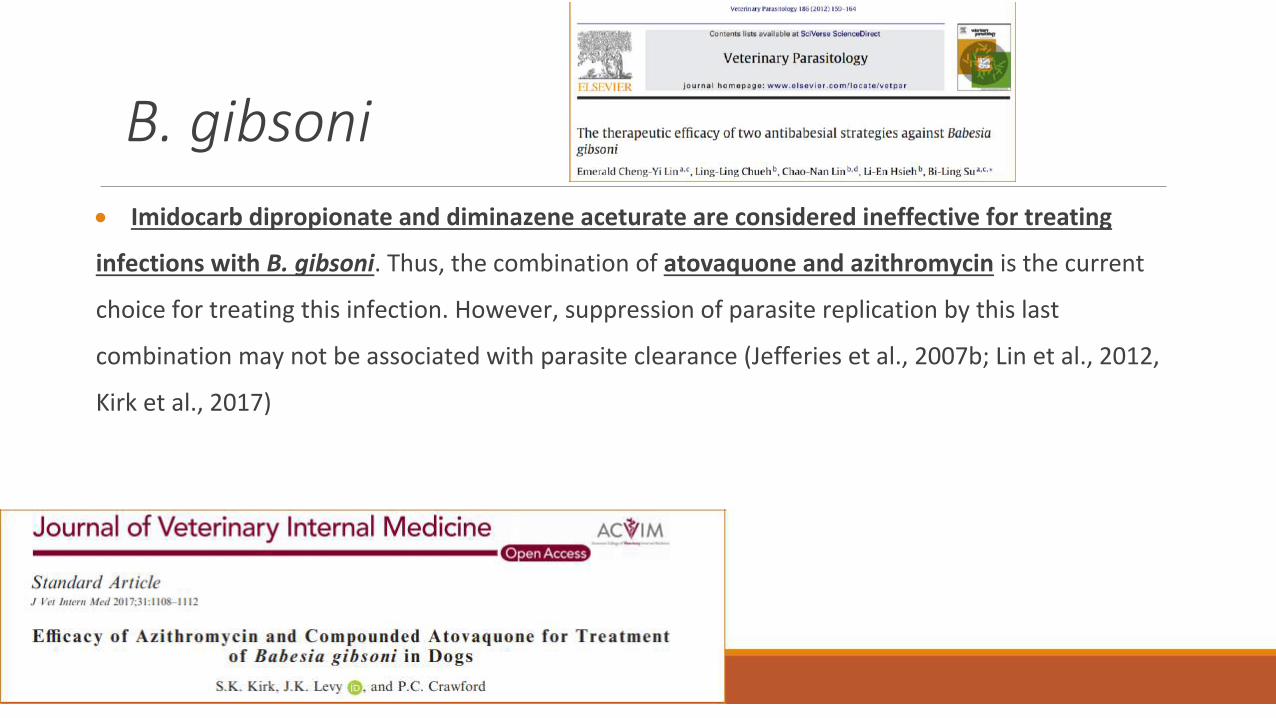
B. gibsoni
Imidocarb dipropionate and diminazene aceturate are considered ineffective for treating
infections with B. gibsoni. Thus, the combination of atovaquone and azithromycin is the current
choice for treating this infection. However, suppression of parasite replication by this last
combination may not be associated with parasite clearance (Jefferies et al., 2007b; Lin et al., 2012,
Kirk et al., 2017)
Conclusions
Treated dogs must be followed up and monitored by blood tests and PCR to
determine persistence of infection and parasitemia. In addition, new and
supplementary drugs and the synergistic effects of current drug combinations
against infection with Babesia should be studied to find effective treatments for
canine babesiosis.
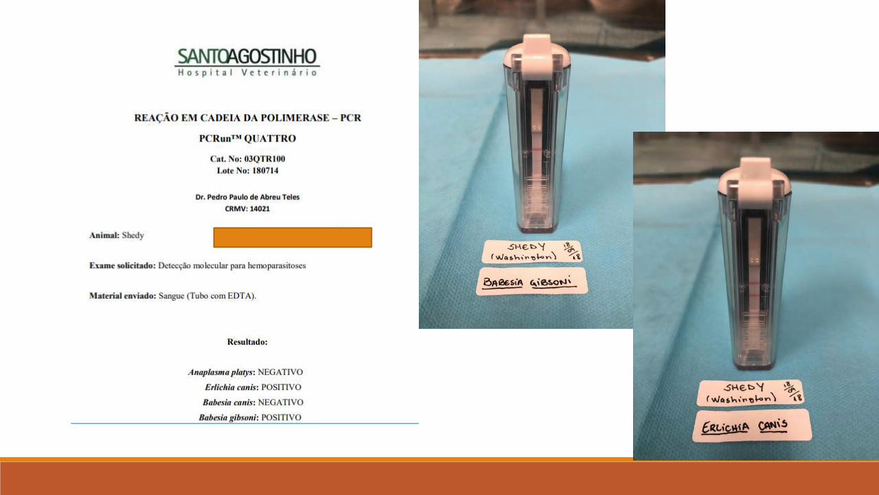
CASO CLÍNICO
CLINICAL CASE
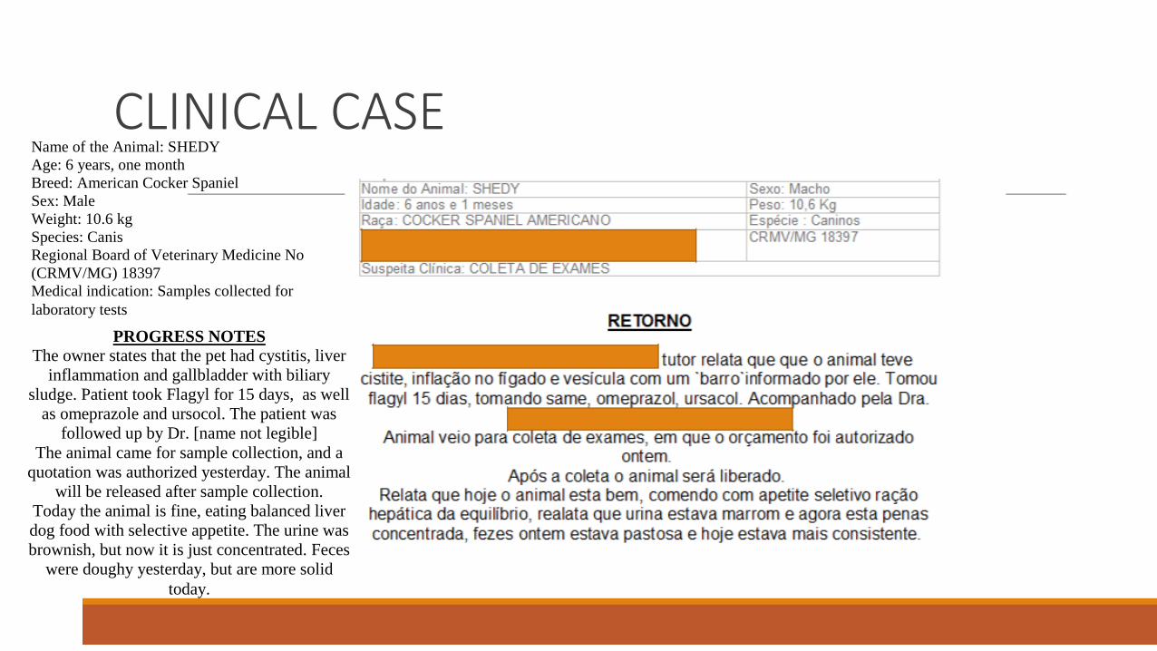
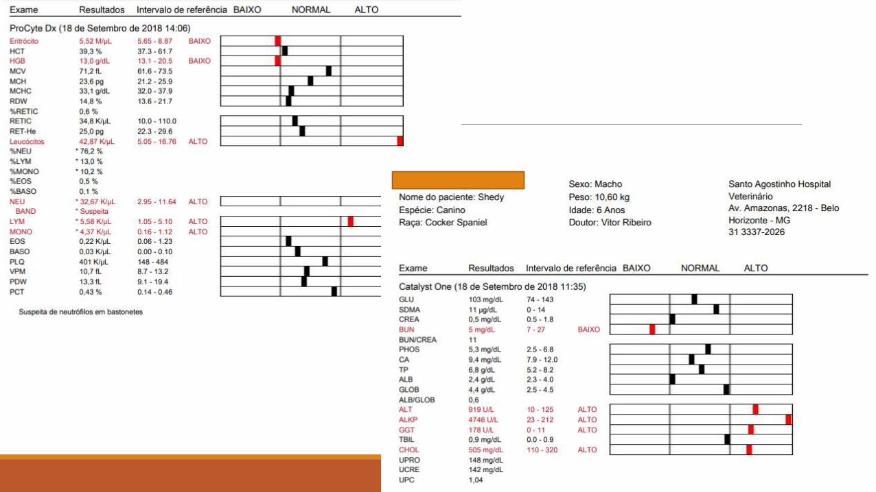
Name of the Animal: SHEDY
Age: 6 years, one month
Breed: American Cocker Spaniel
Sex: Male
Weight: 10.6 kg
Species: Canis
Regional Board of Veterinary Medicine No
(CRMV/MG) 18397
Medical indication: Samples collected for
laboratory tests
PROGRESS NOTES
The owner states that the pet had cystitis, liver
inflammation and gallbladder with biliary
sludge. Patient took Flagyl for 15 days, as well
as omeprazole and ursocol. The patient was
followed up by Dr. [name not legible]
The animal came for sample collection, and a
quotation was authorized yesterday. The animal
will be released after sample collection.
Today the animal is fine, eating balanced liver
dog food with selective appetite. The urine was
brownish, but now it is just concentrated. Feces
were doughy yesterday, but are more solid
today.
PCRun™
PCRun™

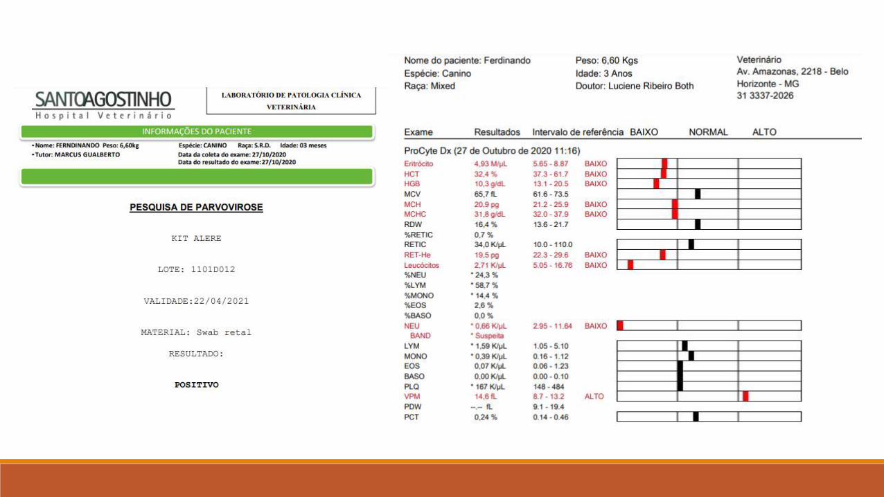
CLINICAL CASE
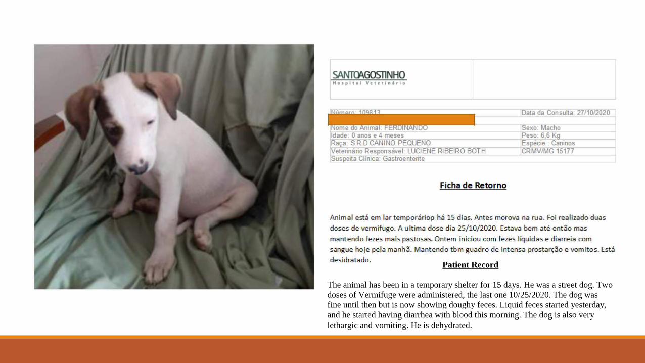
Patient Record
The animal has been in a temporary shelter for 15 days. He was a street dog. Two
doses of Vermifuge were administered, the last one 10/25/2020. The dog was
fine until then but is now showing doughy feces. Liquid feces started yesterday,
and he started having diarrhea with blood this morning. The dog is also very
lethargic and vomiting. He is dehydrated.

Babesia vogeli // Babesia gibsoni

Leishmania infantum
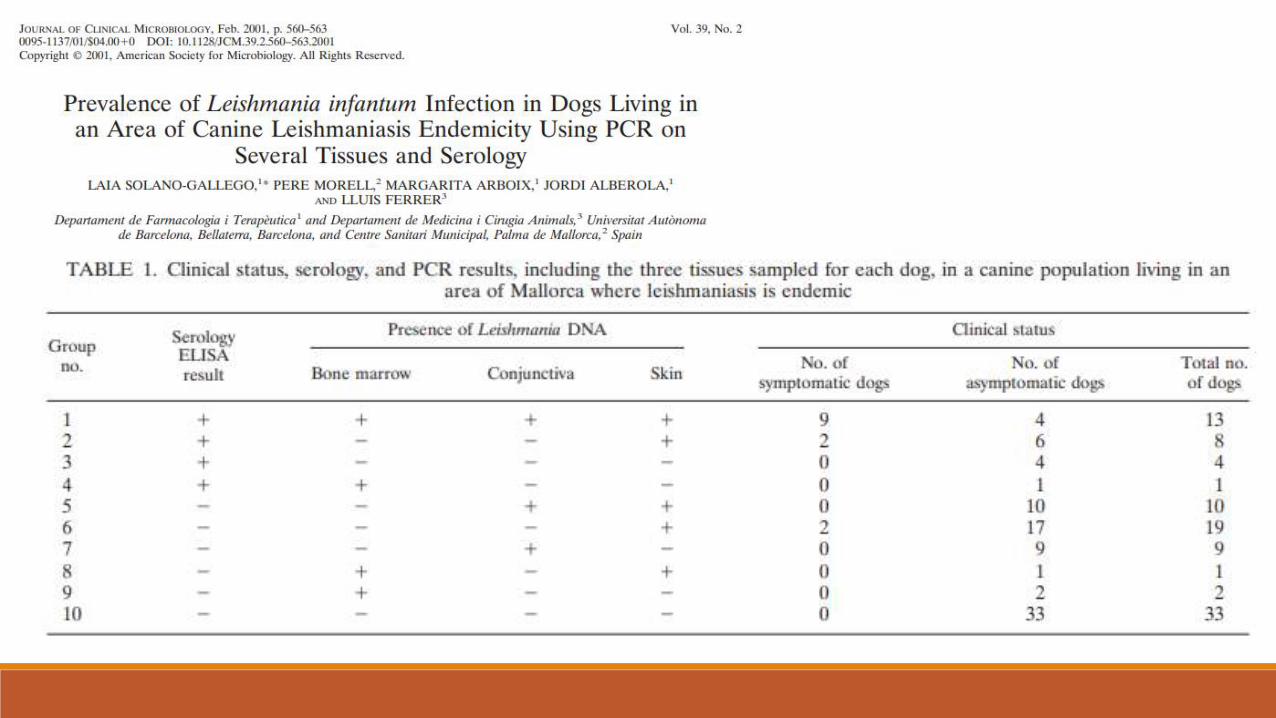

• Clinical disease – 13%
• Seropositives – 26%
• PCR positive – 63%
• 67% - sum of positives per serology or by PCR
IMPORTANT!
The study shows that The prevalence of infection by Leishmania in an enzootic area is greater than assumed The main tissue’s parasite reserve was the dog's skin This result demonstrates the importance of sensitive tests that detect both etiological agents and if animals have immunological protection
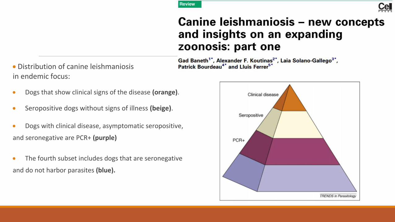
Distribution of canine leishmaniosis in endemic focus:
Dogs that show clinical signs of the disease (orange). Seropositive dogs without signs of illness (beige). Dogs with clinical disease, asymptomatic seropositive,
and seronegative are PCR+ (purple) The fourth subset includes dogs that are seronegative
and do not harbor parasites (blue).
INFECTION WITH L. infantum
• Half of the serological information in a study using eight different serological tests in a clinical cohort with suspicion of Leishmania canis infection proved inconclusive, offering an admixture of seropositive and seronegative data
Rapid Tests (RTs) have greater sensitivity in serum samples with higher antibody titers than the IIFA
TRs exhibited false negatives in samples with low antibody titers
The use of RTs for diagnosis of Visceral Leishmaniasis (VL) in dogs with low antibody titers, such as asymptomatic dogs, should be carefully evaluated - vaccination criteria
IIFA with the highest number of false positives
OBJECTIVE:
◦ This study aimed to assess the accuracy of ◦ TR-DPP (Biomanguinhos®) ◦ EIE Canine-Visceral-Leishmaniasis-Biomanguinhos (EIE-CVL) (Biomanguinhos®) ◦ Enzyme immunoassay (ELISA) rK39 (in-house) ◦ Direct agglutination test (DAT-Canis)
◦ Reference standard comprising parasitological and molecular techniques.
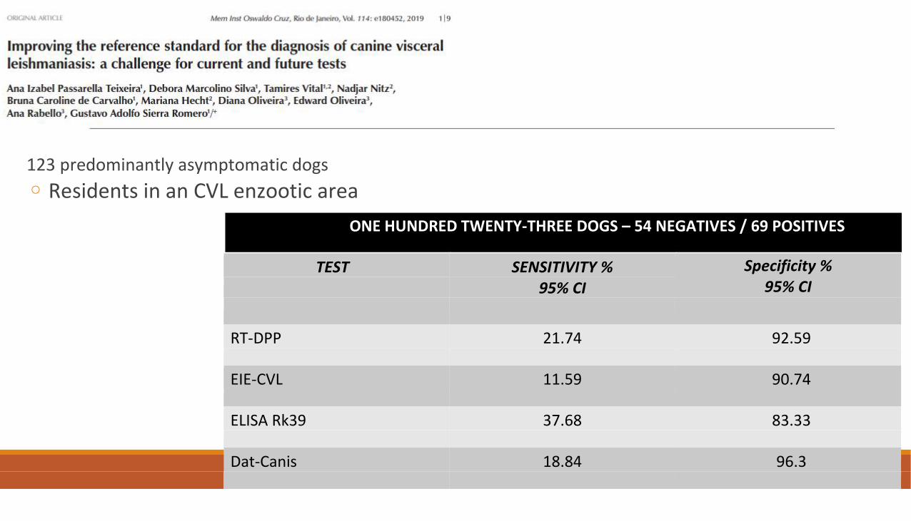
123 predominantly asymptomatic dogs
◦ Residents in an CVL enzootic area
ONE HUNDRED TWENTY-THREE DOGS – 54 NEGATIVES / 69 POSITIVES
TEST SENSITIVITY % Specificity %
95% CI
95% CI
RT-DPP 21.74 92.59
EIE-CVL 11.59 90.74
ELISA Rk39 37.68 83.33
Dat-Canis 18.84 96.3
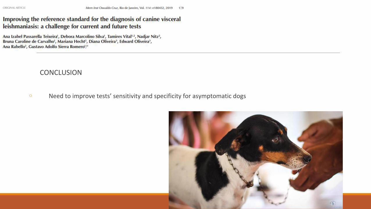
CONCLUSION
◦ Need to improve tests’ sensitivity and specificity for asymptomatic dogs
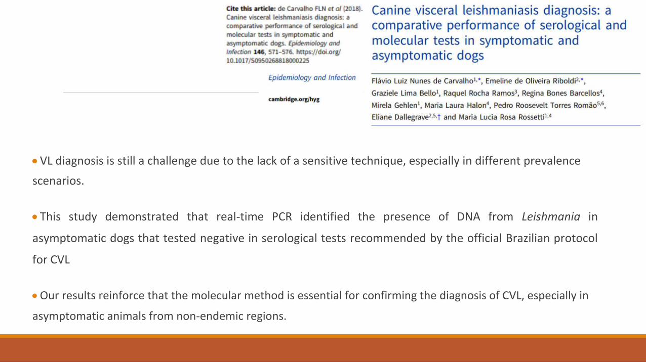
VL diagnosis is still a challenge due to the lack of a sensitive technique, especially in different prevalence
scenarios. This study demonstrated that real-time PCR identified the presence of DNA from Leishmania in
asymptomatic dogs that tested negative in serological tests recommended by the official Brazilian protocol
for CVL Our results reinforce that the molecular method is essential for confirming the diagnosis of CVL, especially in
asymptomatic animals from non-endemic regions.

CLINICAL CASE
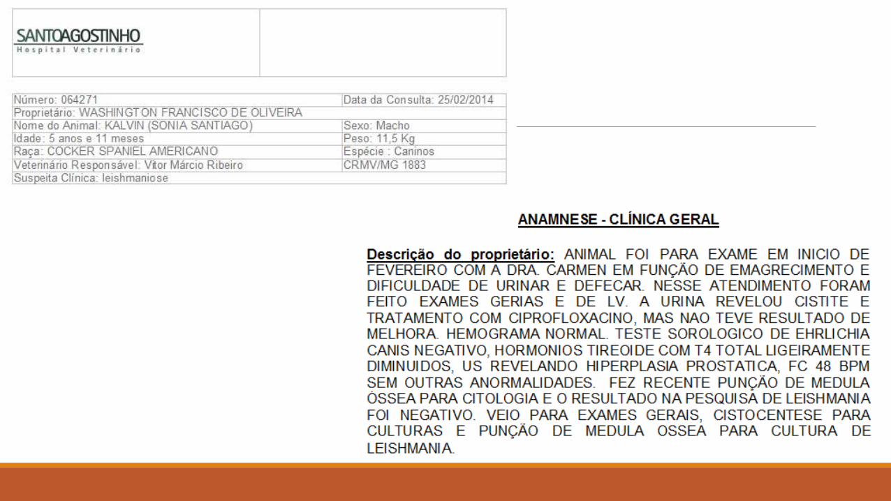
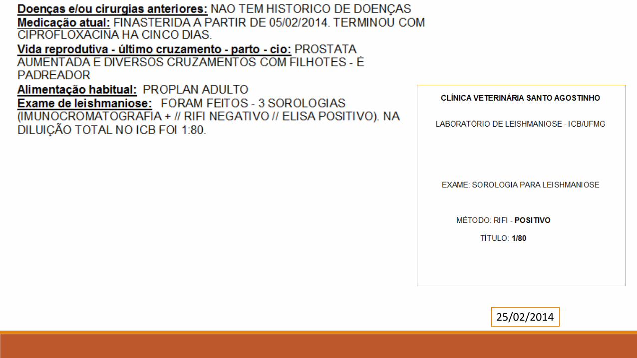
25/02/2014
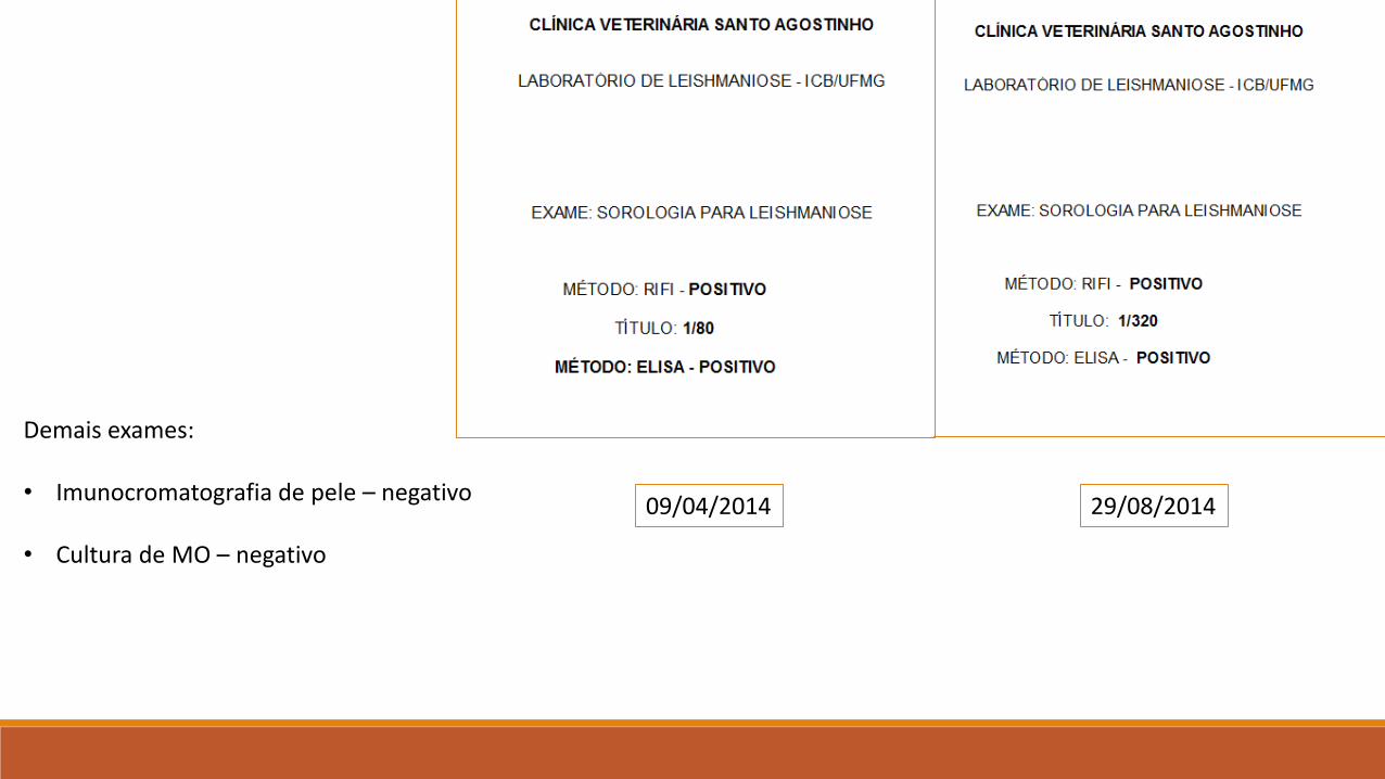
09/04/2014 29/08/2014
Demais exames:
• Imunocromatografia de pele – negativo
• Cultura de MO – negativo
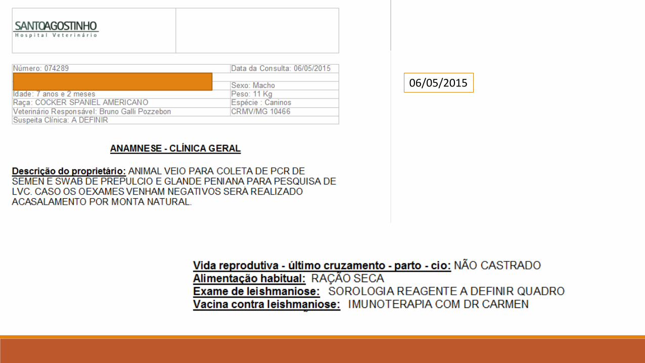
06/05/2015

11/05/2015
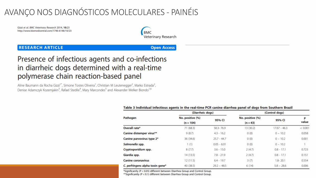
AVANÇO NOS DIAGNÓSTICOS MOLECULARES - PAINÉIS

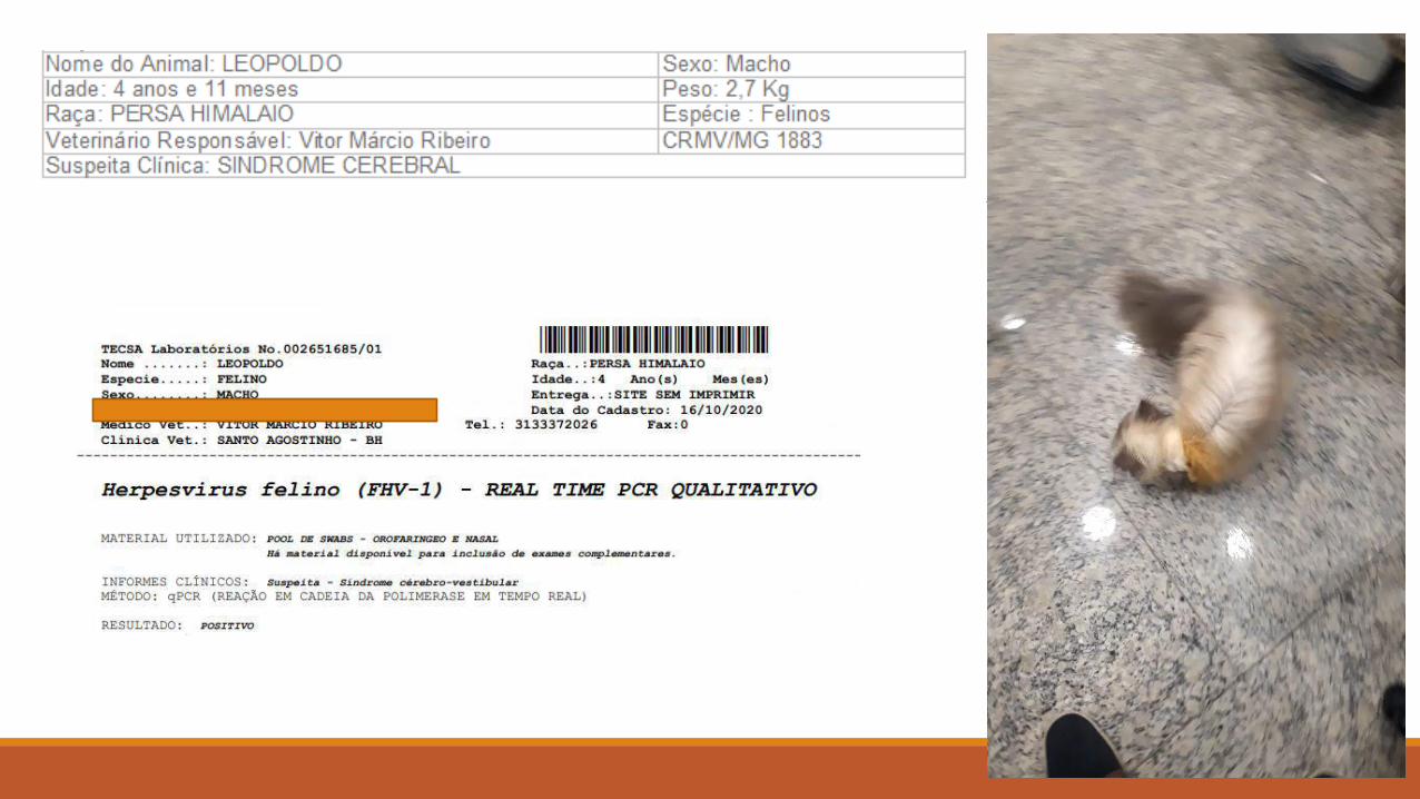
CLINICAL CASE
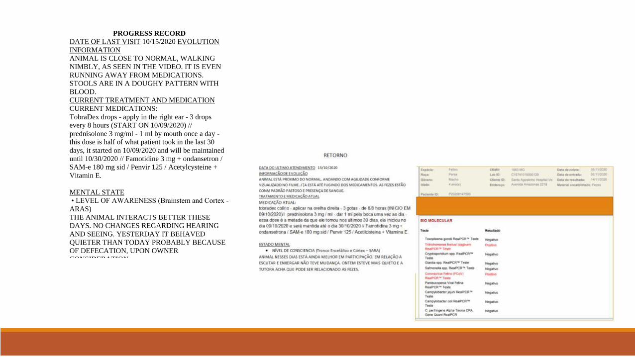
PROGRESS RECORD
DATE OF LAST VISIT 10/15/2020 EVOLUTION
INFORMATION
ANIMAL IS CLOSE TO NORMAL, WALKING
NIMBLY, AS SEEN IN THE VIDEO. IT IS EVEN
RUNNING AWAY FROM MEDICATIONS.
STOOLS ARE IN A DOUGHY PATTERN WITH
BLOOD.
CURRENT TREATMENT AND MEDICATION
CURRENT MEDICATIONS:
TobraDex drops - apply in the right ear - 3 drops
every 8 hours (START ON 10/09/2020) //
prednisolone 3 mg/ml - 1 ml by mouth once a day -
this dose is half of what patient took in the last 30
days, it started on 10/09/2020 and will be maintained
until 10/30/2020 // Famotidine 3 mg + ondansetron /
SAM-e 180 mg sid / Penvir 125 / Acetylcysteine +
Vitamin E.
MENTAL STATE
• LEVEL OF AWARENESS (Brainstem and Cortex -
ARAS)
THE ANIMAL INTERACTS BETTER THESE
DAYS. NO CHANGES REGARDING HEARING
AND SEEING. YESTERDAY IT BEHAVED
QUIETER THAN TODAY PROBABLY BECAUSE
OF DEFECATION, UPON OWNER
CONSIDERATION.
The most used molecular tests for detection of nucleic acids are:
◦ 1. Polymerase Chain Reaction (PCR) ◦ 2. Quantitative Real-Time PCR (qPCR) ◦ 3. Reverse Transcription PCR (RT-PCR) ◦ 4. Duplex and Multiplex Real-Time PCR ◦ 5. DNA sequencing

New times
Molecular microbiology has revolutionized the field of microbiology by reducing test response time from weeks to HOURS Provided elevated SENSITIVITY - SPECIFICITY -
QUANTIFICATION Molecular assays provide improved DIAGNOSIS in suspected bacterial, viral, and protozoan infectious events
Molecular microbiology approaches such as POLYMERASE CHAIN REACTION (PCR) seek to: Detect targeted portions of genetic material (DNA or RNA) from biological samples The material is amplified - producing large numbers of copies and detecting even very small amounts of microbial genetic material
In general, these molecular methods are considered Moderately or very complex
• Require extensive training
]
This makes them unfeasible in many labs.
• Sterile technique
• Post-analytical assessments
• Take hours to execute
INNOVATION
More recent sample-to-response tests are simplified and involve minimal processing and hands-
on time. With these simplified molecular assays, more technologists and laboratories perform these tests
outside clinical microbiology laboratories, increasing their use and the number of people involved in
the process.
HENCE
The POINT OF CARE molecular diagnosis currently allows: Research on the main day-to-day etiological agents Results available in less than two hours Carried out at the clinic itself After screening with rapid tests, the possibility of confirmation by PCR POC in a short time
The prognostic implication of an adequate treatment guided by quick molecular diagnosis and
monitoring of the “cure” (QUALITATIVE / QUANTITATIVE ASSESSMENT)
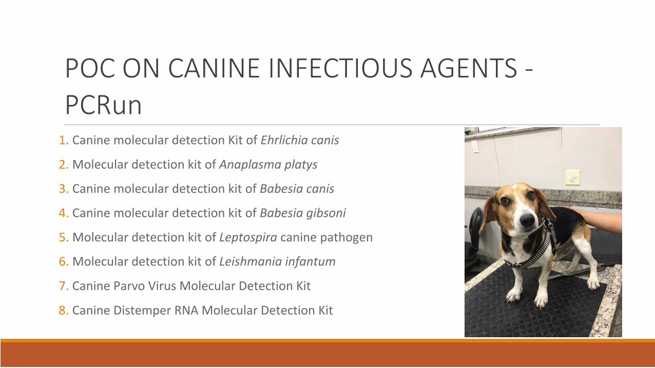
POC ON CANINE INFECTIOUS AGENTS - PCRun
1. Canine molecular detection Kit of Ehrlichia canis
2. Molecular detection kit of Anaplasma platys
3. Canine molecular detection kit of Babesia canis
4. Canine molecular detection kit of Babesia gibsoni
5. Molecular detection kit of Leptospira canine pathogen
6. Molecular detection kit of Leishmania infantum
7. Canine Parvo Virus Molecular Detection Kit
8. Canine Distemper RNA Molecular Detection Kit
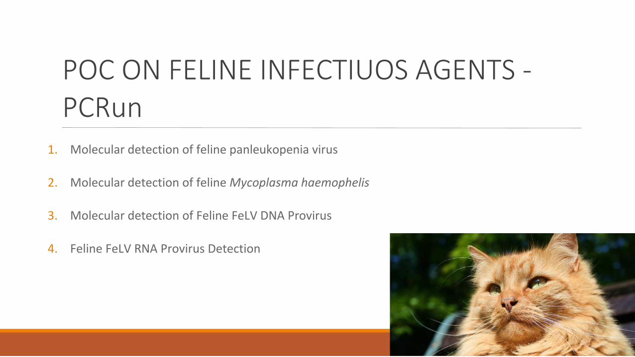
POC ON FELINE INFECTIUOS AGENTS - PCRun
1. Molecular detection of feline panleukopenia virus 2. Molecular detection of feline Mycoplasma haemophelis 3. Molecular detection of Feline FeLV DNA Provirus 4. Feline FeLV RNA Provirus Detection
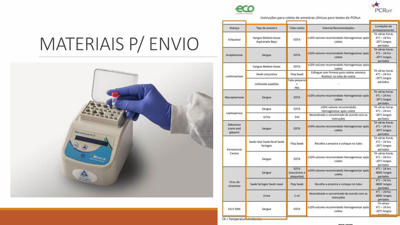
MATERIAIS P/ ENVIO

INCLUSIVIDADE / EXCLUSIVIDADE
DON’T FORGET!
CONCLUSIONS
ADVANCES HAVE PROVIDED GREATER POSSIBILITIES FOR DIAGNOSIS TRY TO ASSOCIATE METHODS, AND ALWAYS SEEK TO INTERPRET THEM FOR DECISION MAKING CLINICS AND HOSPITALS CAN USE ADVANCED AND SIMPLE IMPLEMENTATION TECHNOLOGIES THESE METHODOLOGIES HAVE INCREASED THE SPEED AT WHICH RESULTS ARE PROVIDED - BEFORE THEM WE HAD GREAT DIFFICULTY AGILELY PROVIDING RESULTS
MOLECULAR EXAMS MUST BE INCLUDED IN THE STAGE DETERMINATION OF ALL DISEASES


![Pedi [Type the document subtitle] atric Infectious ......were discharged with final diagnoses of Tetralogy of Fallot not in failure, Cavitary TB and Pneumonia, Septic Shock and 2 did](https://img.pdfslide.net/doc/110x75/5e8b2b316759b34fd1569592/pedi-type-the-document-subtitle-atric-infectious-were-discharged-with.jpg)